Efficacy of a novel sphingosine kinase inhibitor in experimental Crohn’s disease
13
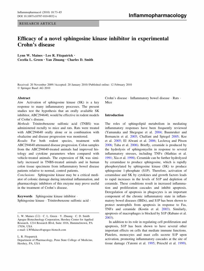
RESEARCH ARTICLE Efficacy of a novel sphingosine kinase inhibitor in experimental Crohn’s disease Lynn W. Maines • Leo R. Fitzpatrick • Cecelia L. Green • Yan Zhuang • Charles D. Smith Received: 20 November 2009 / Accepted: 20 January 2010 / Published online: 12 February 2010 Ó Springer Basel AG 2010 Abstract Aim Activation of sphingosine kinase (SK) is a key response to many inflammatory processes. The present studies test the hypothesis that an orally available SK inhibitor, ABC294640, would be effective in rodent models of Crohn’s disease. Methods Trinitrobenzene sulfonic acid (TNBS) was administered rectally to mice and rats. Rats were treated with ABC294640 orally alone or in combination with olsalazine and disease progression was monitored. Results For both rodent species, treatment with ABC294640 attenuated disease progression. Colon samples from the ABC294640-treated animals had improved his- tology and cytokine parameters when compared with vehicle-treated animals. The expression of SK was simi- larly increased in TNBS-treated animals and in human colon tissue specimens from inflammatory bowel disease patients relative to normal, control patients. Conclusions Sphingosine kinase may be a critical medi- ator of colonic damage during intestinal inflammation, and pharmacologic inhibitors of this enzyme may prove useful in the treatment of Crohn’s disease. Keywords Sphingosine kinase inhibitor Á Sphingosine kinase Á Trinitrobenzene sulfonic acid Á Crohn’s disease Á Inflammatory bowel disease Á Rats Á Mice Introduction The roles of sphingolipid metabolism in mediating inflammatory responses have been frequently reviewed (Yamanaka and Shegogue et al. 2004; Baumruker and Bornancin et al. 2005; Chalfant and Spiegel 2005; Kee et al. 2005; El Alwani et al. 2006; Leclercq and Pitson 2006; Taha et al. 2006). Briefly, ceramide is produced by the hydrolysis of sphingomyelin in response to several inflammatory stresses, including TNFa (Mathias et al. 1991; Xia et al. 1998). Ceramide can be further hydrolyzed by ceramidase to produce sphingosine, which is rapidly phosphorylated by sphingosine kinase (SK) to produce sphingosine 1-phosphate (S1P). Therefore, activation of ceramidase and SK by cytokines and growth factors leads to rapid increases in the levels of S1P and depletion of ceramide. These conditions result in increased inflamma- tion and proliferation cascades and inhibit apoptosis. Deregulation of apoptosis in phagocytes is an important component of the chronic inflammatory state in inflam- matory bowel diseases (IBDs), and S1P has been shown to protect neutrophils from apoptosis in response to Fas, TNFa and ceramide (Kostin et al. 2003). Similarly, apoptosis of macrophages is blocked by S1P (Rabano et al. 2003). In addition to its role in regulating cell proliferation and apoptosis, S1P has been shown to have several other important effects on cells that mediate immune functions. Platelets, monocytes and mast cells secrete S1P upon activation, promoting inflammatory cascades at the site of tissue damage (Yatomi et al. 1995; Prieschl et al. 1999). L. W. Maines (&) Á C. L. Green Á Y. Zhuang Á C. D. Smith Apogee Biotechnology Corporation, Hershey Center for Applied Research, 1214 Research Blvd, Suite 1016, Hummelstown, PA 17036, USA e-mail: [email protected] L. R. Fitzpatrick Department of Pharmacology, Penn State College of Medicine, Hershey, PA, USA Inflammopharmacol (2010) 18:73–85 DOI 10.1007/s10787-010-0032-x Inflammopharmacology
-
Upload
independent -
Category
Documents
-
view
4 -
download
0
Transcript of Efficacy of a novel sphingosine kinase inhibitor in experimental Crohn’s disease
RESEARCH ARTICLE
Efficacy of a novel sphingosine kinase inhibitor in experimentalCrohn’s disease
Lynn W. Maines • Leo R. Fitzpatrick •
Cecelia L. Green • Yan Zhuang • Charles D. Smith
Received: 20 November 2009 / Accepted: 20 January 2010 / Published online: 12 February 2010
� Springer Basel AG 2010
Abstract
Aim Activation of sphingosine kinase (SK) is a key
response to many inflammatory processes. The present
studies test the hypothesis that an orally available SK
inhibitor, ABC294640, would be effective in rodent models
of Crohn’s disease.
Methods Trinitrobenzene sulfonic acid (TNBS) was
administered rectally to mice and rats. Rats were treated
with ABC294640 orally alone or in combination with
olsalazine and disease progression was monitored.
Results For both rodent species, treatment with
ABC294640 attenuated disease progression. Colon samples
from the ABC294640-treated animals had improved his-
tology and cytokine parameters when compared with
vehicle-treated animals. The expression of SK was simi-
larly increased in TNBS-treated animals and in human
colon tissue specimens from inflammatory bowel disease
patients relative to normal, control patients.
Conclusions Sphingosine kinase may be a critical medi-
ator of colonic damage during intestinal inflammation, and
pharmacologic inhibitors of this enzyme may prove useful
in the treatment of Crohn’s disease.
Keywords Sphingosine kinase inhibitor �Sphingosine kinase � Trinitrobenzene sulfonic acid �
Crohn’s disease � Inflammatory bowel disease � Rats �Mice
Introduction
The roles of sphingolipid metabolism in mediating
inflammatory responses have been frequently reviewed
(Yamanaka and Shegogue et al. 2004; Baumruker and
Bornancin et al. 2005; Chalfant and Spiegel 2005; Kee
et al. 2005; El Alwani et al. 2006; Leclercq and Pitson
2006; Taha et al. 2006). Briefly, ceramide is produced by
the hydrolysis of sphingomyelin in response to several
inflammatory stresses, including TNFa (Mathias et al.
1991; Xia et al. 1998). Ceramide can be further hydrolyzed
by ceramidase to produce sphingosine, which is rapidly
phosphorylated by sphingosine kinase (SK) to produce
sphingosine 1-phosphate (S1P). Therefore, activation of
ceramidase and SK by cytokines and growth factors leads
to rapid increases in the levels of S1P and depletion of
ceramide. These conditions result in increased inflamma-
tion and proliferation cascades and inhibit apoptosis.
Deregulation of apoptosis in phagocytes is an important
component of the chronic inflammatory state in inflam-
matory bowel diseases (IBDs), and S1P has been shown to
protect neutrophils from apoptosis in response to Fas,
TNFa and ceramide (Kostin et al. 2003). Similarly,
apoptosis of macrophages is blocked by S1P (Rabano et al.
2003).
In addition to its role in regulating cell proliferation and
apoptosis, S1P has been shown to have several other
important effects on cells that mediate immune functions.
Platelets, monocytes and mast cells secrete S1P upon
activation, promoting inflammatory cascades at the site of
tissue damage (Yatomi et al. 1995; Prieschl et al. 1999).
L. W. Maines (&) � C. L. Green � Y. Zhuang � C. D. Smith
Apogee Biotechnology Corporation, Hershey Center for Applied
Research, 1214 Research Blvd, Suite 1016, Hummelstown, PA
17036, USA
e-mail: [email protected]
L. R. Fitzpatrick
Department of Pharmacology, Penn State College of Medicine,
Hershey, PA, USA
Inflammopharmacol (2010) 18:73–85
DOI 10.1007/s10787-010-0032-x Inflammopharmacology

The activation of SK is essential for the signaling responses
to TNFa, since its ability to induce adhesion molecule
expression via activation of NFjB is mimicked by S1P,
and is blocked by the SK inhibitor dimethylsphingosine
(Xia et al. 1998). S1P is also a mediator of Ca2? influx
during neutrophil activation by TNFa and other stimuli,
leading to the production of superoxide and other toxic
radicals (MacKinnon et al. 2002; Itagaki and Hauser 2003).
When considering the accumulating evidence for a
pivotal role of SK in the regulation of inflammatory pro-
cesses, pharmacological inhibition of SK is a new potential
means of preventing and/or treating IBDs. However, ther-
apeutically useful SK inhibitors have been described only
recently (French et al. 2003a, b, Maines et al. 2006, 2008).
These compounds inhibit S1P formation in intact cells, and
demonstrate a high degree of selectivity for SK versus
other lipid and protein kinases. Our lead SK inhibitor,
ABC294640, has excellent oral bioavailability and in vivo
SK inhibitory activity in rodents and can be administered
chronically to rats and mice in efficacious doses without
limiting systemic toxicities (Smith and French 2008).
ABC294640 has been shown in a contracted 20-kinase
panel to be specific towards SK and our recent data indi-
cates it is specific to SK-2 with a Ki of approximately
30 lM (unpublished data). Importantly, ABC294640 rep-
resents the first pharmacologic probe to evaluate the
biological roles of this SK isozyme.
In our previous work (Maines et al. 2008), we demon-
strated that ABC294640 reduces the inflammatory disease
parameters in the acute and chronic dextran sulfate sodium
(DSS) models of ulcerative colitis (UC) in mice. The
pathogenesis of the DSS model is different from the tri-
nitrobenzene sulfonic acid (TNBS) model. Specifically,
TNBS-induced colitis is thought to involve Th-1 and Th-
17-directed immune responses. This immunological-based
colitis is more responsive to typical IBD-drugs like corti-
costeroids than is DSS-induced colitis. Therefore, we have
now evaluated ABC294640 against TNBS-induced colitis
in rats and mice, to gain efficacy data in two species for a
subsequent Investigational New Drug Application for
ABC294640 and to gain a better understanding of the
global therapeutic potential of this compound for IBD.
Materials and methods
Reagents
Trinitrobenzene sulfonic acid and prednisolone were
purchased from Sigma–Aldrich (St. Louis, MO).
ABC294640�HCl [3-(4-chlorophenyl)adamantane-1-car-
boxylic acid (pyridin-4-ylmethyl)amide, hydrochloride
salt] was synthesized as described by Maines et al. (2008),
and is hereafter referred to as ABC294640. Mice were
dosed with drug in solution and for studies with rats,
10.5 ± 0.5 mg quantities of ABC294640 were filled into
size 9 porcine hard gelatin capsules (Torpac; Fairfield, NJ).
Rabbit polyclonal antibodies were raised against a 19
amino acid peptide unique to SK1 (Genbank Accession
Number: AF200328) by Biosynthesis Inc. (Lewisville,
TX), and have been previously demonstrated to detect
human SK by immunohistochemistry (Maines et al. 2008).
Mouse TNBS model
On experimental days 0 and 7, age matched (46–63 days)
male C57/BL/6 mice were anaesthetized with intraperito-
neal ketamine/xylazine (136 mg/kg and 9.6 mg/kg,
respectively) and a stainless steel catheter was carefully
inserted into the colon (4 cm proximal to the anus). A
solution of TNBS in 50% ethanol (0.1 ml volume of
40 mg/kg TNBS) was slowly administered with the ani-
mals being kept in a head-down position until they
regained consciousness. Two control groups comprised
mice administered either 50% ethanol or PBS in an iden-
tical fashion. Starting on day 6 and proceeding through day
9, animals were treated twice daily by oral gavage with
either PBS containing 0.375% Tween-80 (vehicle) or
50 mg of ABC294640/kg) in vehicle. ABC294640 was
administered by oral gavage at 50 mg/kg twice daily,
which has been determined to be an effective dose in other
inflammation models (French et al. 2006; Maines et al.
2008) with no related toxicities (unpublished data). On day
10, animals were killed by CO2 asphyxiation and cervical
dislocation per institutional IACUC requirements and the
colons were removed. The colons were measured, weighed
and scored for macroscopic inflammation as indicated
below. The distal 3 cm was then transected for histology
and biochemical analyses, as this was where disease
manifestations were primarily observed.
Rat TNBS model
On day 0, female Sprague–Dawley rats (approximately
210 g) were anaesthetized (i.p.; 79.92 mg/kg ketamine plus
3.96 mg/kg xylazine). A stainless steel catheter was care-
fully inserted into the colon (8 cm proximal to the anus).
Then, 1.0 ml total volume of TNBS solution (30 mg TNBS
in 20% ethanol in PBS) was slowly delivered with animals
being kept for 15-min intervals in a head-down position,
level and head-up position. Treatment groups received oral
doses of vehicle (0.375% Tween in PBS), prednisolone
(5 mg/kg) in vehicle, osalazine in gelcaps, or ABC294640 in
gelcaps. Animals were killed on day 6 by CO2 asphyxiation
and cervical dislocation per institutional IACUC require-
ments and colons removed. The colons were measured,
74 L. W. Maines et al.

weighed and scored for macroscopic inflammation as indi-
cated below. The distal 6 cm were then transected for
histology and biochemical analyses. Colonic TNFa and IL-
1b levels were determined as described previously by our
laboratory (Fitzpatrick et al. 2000; Maines et al. 2008), using
commercially available kits from Pierce Endogen (Rock-
ford, IL).
Macroscopic score
Macroscopic inflammation within the distal 3 cm of each
colon was scored using the scoring system as described by
other investigators (Morris et al. 1989; McCafferty et al.
1999).
Histology score
The colon tissues were fixed with formalin overnight,
followed by embedding in paraffin, sectioning and staining
with haematoxylin–eosin. The sections were microscopi-
cally examined for histopathologic changes using the
following scoring system. The histology score was deter-
mined by multiplying the percent involvement for each of
the three following histological features by the percent
area of involvement (Williams et al. 2001; Maines et al.
2008). Therefore, the minimal score is 0, and the maximal
score is 40.
Myeloperoxidase assay
Mucosal myeloperoxidase (MPO) activity (Fitzpatrick
et al. 2000) was determined as a measure of granulocyte
infiltration into the colon.
Human tissue samples
Colon specimens were harvested from patients undergoing
surgery at the Hershey Medical Center for a diagnosis of
intestinal disease. Informed consent was obtained before
surgery, as stipulated in the IRB-approved protocol #
19084. Two groups of patients were included in this study
as described previously (Fitzpatrick et al. 2007). After
bowel resection by a surgeon from the Department of
Colon and Rectal Surgery, the intestinal tissue was sent to
the Department of Anatomic Pathology Laboratory for
Confirmation of the Diagnosis, and was banked within
30 min. For our studies, paraffin sections were obtained
from macroscopically normal areas of the intestine from
the non-IBD patients, and from areas of the intestine with
obvious disease from the IBD patients, and stained for SK
expression as described below.
Immunohistochemical analyses of SK expression
The expression of SK in colon specimens from the human
patients, as well as from the rodent TNBS models was
examined by immunohistochemistry. Briefly, sections were
de-paraffinized in xylene and rehydrated in a series of
ethanol dilutions. Sections were permeabilized with 0.5%
Triton X-100 in PBS, and boiled for 10 min in 10 mM
sodium citrate buffer for antigen retrieval. Slides were then
incubated 10 min in 3% hydrogen peroxide to quench
endogenous peroxidase. After washing in PBS, sections
were blocked for 1 h with reagents from the VECTA-
STAIN ABC Kit (Vector Laboratories Inc.), and then
incubated for 30 min at room temperature with the primary
SK antibody. The samples were then washed with PBS, and
incubated with biotinylated anti-rabbit IgG antibody for
30 min, followed by 30 min of incubation with DAB
reagent for color development. Sections were lightly
counterstained with hematoxylin, rehydrated, and mounted
for analysis by bright-field microscopy. The percent area of
SK expression was measured with a 25-mm ocular grid
attached to an Olympus CH light microscope at a 4009
magnification. Six areas of colonic tissue were evaluated
per slide.
Statistics
Statistical analyses were performed using InStat (Version
3.01, GraphPad Software, Inc.). A one-way analysis of
variance was performed with Tukey–Kramer multiple
comparisons tests for evaluation of differences between
groups.
Ethical considerations
All animal experimentation was conducted with procedures
approved and monitored by the Penn State College of
Medicine Institutional Animal Care and Use Committee
(Office of Laboratory Animal Welfare Assurance Number
A3045-01).
Results
Mouse TNBS model
To extend our previous findings that the SK inhibitor
ABC294640 attenuates the development of DSS-induced
UC in mice (Maines et al. 2008), we have studied this
compound in the TNBS model of IBD, which models CD
(Murthy and Flanigan 1999).
Change in body weight was the most global parameter
of the IBD phenotype measured. As expected, non-TNBS-
Sphingosine kinase inhibitor, Rodent Crohn’s 75

treated mice gained weight while those receiving TNBS
lost weight (Fig. 1a). Administration of ABC294640 nearly
normalized the growth of the animals and this was asso-
ciated with a general improvement in the appearance of the
animals. Notably, the positive control drug prednisolone
increased the loss of body weight (Fig. 1a), consistent with
its known toxicity at therapeutic doses. The macroscopic
scores for animals that received TNBS were consistently
elevated in comparison with the ethanol alone controls, and
this was largely normalized in the group that also received
ABC294640. The treatment of the mice with prednisolone
significantly attenuated macroscopic damage to a similar
level found in PBS-treated mice.
As an objective measurement of inflammation-mediated
oedema, hypertrophy and fibrosis, the weight of the distal
3 cm of each colon was determined (Fig. 1c). As with the
macroscopic score, there was a substantial increase in
colon weight of mice treated with ethanol alone compared
with PBS. The addition of TNBS to the treatment protocol
produced a further significant increase in colon weight, and
this increase was significantly blocked by treatment of the
mice with ABC294640. A comparable reduction was seen
with prednisolone.
Granulocyte infiltration is frequently associated with
IBD, and is markedly increased in rodent models of IBD
(McCafferty et al. 1999; Williams et al. 2001; Maines et al.
2008). Therefore, MPO activity was assayed in the colon
samples from the mice. As indicated in Fig. 1d, colonic
MPO activity was substantially elevated in the TNBS-
treated animals, when compared with control animals. The
TNBS-induced increase in MPO activity was reduced by
approximately 60% in mice receiving daily doses of
ABC294640. As anticipated from the literature (Videla
et al. 2006), prednisolone also suppressed neutrophil
accumulation in the colon. Variability did not allow any
changes to reach statistical significance.
Colons from mice with histology scores that correlated
closely to the mean of their group were sectioned and
stained and are shown in Fig. 2. Histological results of
colon sections from the various treatment groups were
consistent with the macroscopic scores and colon weights,
revealing inflammation and damage in the TNBS-vehicle
group (Fig. 2b, c) that was reduced or negated in the
ABC294640-treated animals (Fig. 2d, e). It was also
apparent that inflammatory cell influx was more substantial
in TNBS-treated animals not receiving ABC294640 com-
pared with those receiving the compound. The histology
evaluations also suggested that the size of the muscularis
layer, as well as oedema of the submucosa, was lessened in
ABC294640-treated animals as compared to vehicle-trea-
ted animals (Fig. 2b, e). These observations are likely an
explanation for the significant reductions in colon weight
that were observed in the ABC294640-treated mice.
As a quantifiable measure of histological damage
(Fig. 1e), animals receiving TNBS and oral vehicle typi-
cally had higher histology scores of 15–20 (representing
moderate colitis) than non-TNBS controls (mean score
\10). As with the other assays, the histology scores of
TNBS mice given oral ABC294640 were consistently
lower than the TNBS-vehicle animals, although some
animals were only partially protected.
We have previously demonstrated that SK mRNA levels
are elevated in human tumors (French et al. 2003a, b), and
SK protein expression is increased in the colons of mice in
the DSS model of UC (Maines et al. 2008). A similar
increase in SK expression was observed in the colons from
TNBS-induced colitis mice, when compared with PBS
control animals (Fig. 3). SK staining was prominent in the
mucosal epithelial cells, but was also seen in infiltrating
leukocytes of TNBS-treated mice.
Rat TNBS model–dose response study
The rat TNBS model was used to consistently produce
moderately severe colonic pathology. As shown in Fig. 4a,
a discrete area of ulceration was typically produced in the
rat distal colon on day 6 after TNBS administration. As
shown in picture form (Fig. 4b) and by semi-quantitative
analysis (Fig. 5b), colonic ulceration was less severe in
animals treated with ABC294640. The mean body weight
change was measured from days 0 to 6. As shown in Fig. 5,
rats treated with either vehicle or ABC294640 had modest
weight gains and animals that received prednisolone lost
significant weight. On day 6, colons were harvested and
evaluated for macroscopic damage. The vehicle group
exhibited a macroscopic damage score relating to moderate
colitis (5.57). All treatment groups had reduced scores
(Fig. 5b).
As an objective measurement of inflammation-mediated
edema, hypertrophy and fibrosis, the weight of the distal
6 cm of each colon was determined (Fig. 5c). As with the
macroscopic score, the vehicle group had the largest
colonic weight. Improvements were seen in all drug
treatment groups.
Histological examination of colon sections (Fig. 5d)
yielded histology scores that were generally consistent with
the macroscopic scores and colon weights. Inflammation
and damage were evident in the vehicle group that was
reduced in the drug-treated animals. Previous studies have
shown that the Ki of SK2 for ABC294640 is 10 lM and
circulating levels of ABC294640 in plasma reach levels in
this range at a dose of 25 mg/kg (Fitzpatrick et al. 2010)
and putatively explains the comparable results in the 25
and 50 mg/kg treatment groups. Representative colon
sections from the vehicle-, prednisolone- and ABC294640
(25 mg/kg) treatment groups are shown in Fig. 6. Marked
76 L. W. Maines et al.

inflammation and damage in the TNBS-vehicle group
(Fig. 6a) was significantly reduced in both the prednisolone
and ABC294640-treated animals (Figs. 6b, c, respec-
tively). As in the mouse model, it was also apparent that
leukocytes were present in much higher levels in vehicle-
treated animals not receiving prednisolone or ABC294640.
These histology studies also revealed reduced edema of the
submucosa in prednisolone and ABC294640-treated ani-
mals as compared to vehicle animals (Fig. 6), as well as
some evidence of attenuated colonic hypertrophy of the
-15
0
15
A
Induction: PBS EtOH TNBS TNBS TNBSTreatment: Vehicle Vehicle Vehicle ABC294640 Prednisolone
##,++Bo
dy
wei
gh
t ch
ang
e(%
)
*
##,++
0
10
20E
His
tolo
gy S
core
Induction: PBS EtOH TNBS TNBS TNBSTreatment: Vehicle Vehicle Vehicle ABC294640 Prednisolone
0
1
2
3
4
5
6B
Induction: PBS EtOH TNBS TNBS TNBSTreatment: Vehicle Vehicle Vehicle ABC294640 Prednisolone
Mac
rosc
opic
Sco
re
##
**
0.0
0.1
0.2
0.3D
MP
O A
ctiv
ity(u
nits
/ m
g of
tiss
ue)
Induction: PBS EtOH TNBS TNBS TNBSTreatment: Vehicle Vehicle Vehicle ABC294640 Prednisolone
#
0.000
0.025
0.050
0.075
0.100
0.125
0.150C
Induction: PBS EtOH TNBS TNBS TNBSTreatment: Vehicle Vehicle Vehicle ABC294640 Prednisolone
Col
on W
eigh
t (g) #
##,+
**
Fig. 1 Effects of ABC294640 on disease parameters in the mouse
TNBS-colitis model. C57BL/6 mice were treated as follows: rectal
PBS and oral vehicle (black bars); rectal 50% ethanol and oral vehicle
(diagonal hatched bars); rectal TNBS and oral vehicle (open bars);
rectal TNBS and oral ABC294640 (gray bars 50 mg/kg b.i.d.); or
rectal TNBS and oral prednisolone (horizontal hatched bars 5 mg/
kg b.i.d). At the time of killing (day 10), the following parameters
were measured as described in ‘‘Materials and methods’’. a Body
weight change, b macroscopic score, c colon weight, d myeloperox-
idase activity and e histology score. Values represent the
mean ± SEM for 4–5 mice per group. *P \ 0.05 or **P \ 0.01
versus vehicle/TNBS treatment, #P \ 0.05 or ##P \ 0.01 or###P \ 0.001 versus PBS/vehicle group, ?P\0.05 or ??P \ 0.01
versus EtOH/vehicle group
Sphingosine kinase inhibitor, Rodent Crohn’s 77

muscularis propria layer. These observations again are a
likely explanation for the significant reductions in colon
weight that were observed in the prednisolone- and
ABC294640-treated mice.
Rat TNBS model—combination therapy study
The previous studies demonstrate that ABC294640 pro-
tects against colonic damage similar to the steroid
prednisolone. Because olsalazine (Dipentum), a 5-amino-
salicylic acid prodrug, is frequently used for maintenance
therapy for UC, the ability of ABC294640 to be com-
bined with this drug was examined in the rat TNBS
model.
Disease parameters were measured in rats treated with
ABC294640 or olsalazine alone or in combination. As
shown in Fig. 7a, rats treated with vehicle or olsalazine lost
weight between days 0 and 6; whereas, animals treated
Fig. 2 Effects of ABC294640 on colon histology in the mouse
TNBS-colitis model. Sections of colons from the animals described in
Fig. 1 were stained with H&E and examined for pathologic changes.
a Rectal PBS, oral vehicle-treated control animal representative of a
colon with no disease morphology. b Rectal TNBS, oral vehicle-
treated animal; top arrow shows moderate crypt disruption, middlearrow indicates leukocyte infiltration and bottom arrow points out
severe thickening of the muscularis propria layer, c as in b, rectal
TNBS and oral vehicle; top arrow shows an area of severe crypt
damage and bottom arrow indicates substantial mucosal leukocyte
infiltration. d A representative rectal TNBS, oral ABC294640-treated
mouse with average disease intensity; left arrow shows that leuko-
cytes are present at the bottom of the crypts, while the right arrowshows infiltrating leukocytes in the submucosa. e A rectal TNBS, oral
ABC294640-treated complete responder showing normal crypt mor-
phology similar to that of the rectal PBS-treated animals. f A rectal
TNBS, oral prednisolone-treated mouse with some leukocyte infil-
tration in the lamina propria (top arrow) as well as at the base of the
crypts near the muscularis mucosa (bottom arrow). The magnification
was 1509 for all panels
78 L. W. Maines et al.

with ABC294640 alone or in combination with olsalazine
gained weight.
Colons were harvested on day 6 and evaluated for
macroscopic damage. As indicated in Fig. 7b, the vehicle
group exhibited a macroscopic damage score of 7.1, indi-
cating more severe damage than in the previous dose–
response study. All treatment groups showed reduced
scores with the combination therapy having the lowest
score.
The weight of the distal 6 cm of each colon was mea-
sured (Fig. 7c). The vehicle group had the largest colonic
weight, indicative of the most damage, with improvements
being seen in all treatment groups. In addition, the best
result was seen in the combination therapy group.
To assess the extent of neutrophil infiltration, MPO
activity was assayed in colon samples. As indicated in
Fig. 7d, colonic MPO activity was significantly decreased
in the combination therapy group when compared with the
vehicle group.
Histological examination of colon sections was per-
formed (Fig. 7e), and scores were consistent with the
macroscopic scores and colon weights. Specifically, the
results revealed reduced colonic damage and inflammation
in all drug treatment groups, although statistical signifi-
cance was not reached.
We also measured the levels of the proinflammatory
cytokines TNFa and IL-1b. As expected from the relevant
literature (Stasi et al. 2004; Zhang et al. 2006), colonic
levels of both cytokines were markedly increased in the
vehicle/TNBS group. The treatment of rats ABC294640
alone or in combination with olsalazine significantly
reduced (P \ 0.05) the colonic cytokine levels to control
levels (Fig. 8). In contrast, olsalazine alone was ineffective
at reducing TNFa and IL-1b levels.
Figure 9 shows the SK immunohistochemistry results
from the rat colon. Generally, the colonic SK staining
pattern was similar to that found with murine TNBS-
induced colitis. Increased SK staining was found in areas
Fig. 3 SK expression in the mouse TNBS-colitis model. a A
representative example of the mild SK expression pattern in the
colon of a control, i.e. non-TNBS-treated, mouse. The black arrowshows staining of a surface epithelial cell. b A representative section
of a TNBS-treated animal and reveals a general increase in SK
staining with the most pronounced increase in the remnant surface
colonic epithelial cells (black arrow), as well as in patches of
infiltrating lamina propria leukocytes (blue arrows). c A representa-
tive section from another TNBS-treated animal, and elevated SK
expression is evident in crypt colonic epithelial cells (black arrow), as
well as in infiltrating leukocytes within the lamina propria and
submucosa (blue arrows)
Fig. 4 Effect of ABC294640 on macroscopic damage to the rat colon
in the TNBS-colitis model. Colons were harvested from TNBS-
exposed rats treated with vehicle (a) or ABC249640 at 50 mg/kg,
b.i.d. (b). The double arrows demarcate a large ulcer within the distal
4 cm segment of the rat colon. Colonic thickening is clearly evident
in the adjacent ulcer area. There is a much smaller ulcer (arrow) at the
4.5-cm mark of the distal colon of the ABC294640-treated animal
Sphingosine kinase inhibitor, Rodent Crohn’s 79
-15
-10
-5
0
5
10
15
Vehicle Prednisolone ABC294640 ABC294640 ABC294640 (2x50) (2x25) (2x10)
A
*
Bod
y w
eigh
t ch
ang
e(%
)
Vehicl
e
Predn
isolon
e
ABC2946
402X
50
ABC2946
40 2
x25
ABC2946
40 2
X100.0
2.5
5.0
7.5B
*
Mac
rosc
op
ic S
core
Vehicl
e
Predn
isolon
e
ABC2946
40 2
X50
ABC2946
402x
25
ABC2946
40 2
X100
10
20
30D
***
His
tolo
gy
Sco
re
Vehicl
e
Predn
isolon
e
ABC2946
40 2
X50
ABC2946
40 2
x25
ABC2946
40 2
X100.0
0.5
1.0
1.5C
Co
lon
Wei
gh
t (g
)
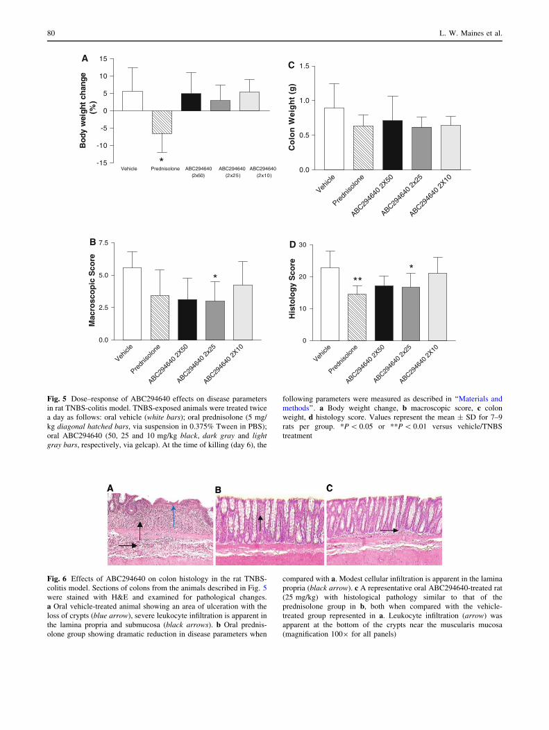
Fig. 5 Dose–response of ABC294640 effects on disease parameters
in rat TNBS-colitis model. TNBS-exposed animals were treated twice
a day as follows: oral vehicle (white bars); oral prednisolone (5 mg/
kg diagonal hatched bars, via suspension in 0.375% Tween in PBS);
oral ABC294640 (50, 25 and 10 mg/kg black, dark gray and lightgray bars, respectively, via gelcap). At the time of killing (day 6), the
following parameters were measured as described in ‘‘Materials and
methods’’. a Body weight change, b macroscopic score, c colon
weight, d histology score. Values represent the mean ± SD for 7–9
rats per group. *P \ 0.05 or **P \ 0.01 versus vehicle/TNBS
treatment
Fig. 6 Effects of ABC294640 on colon histology in the rat TNBS-
colitis model. Sections of colons from the animals described in Fig. 5
were stained with H&E and examined for pathological changes.
a Oral vehicle-treated animal showing an area of ulceration with the
loss of crypts (blue arrow), severe leukocyte infiltration is apparent in
the lamina propria and submucosa (black arrows). b Oral prednis-
olone group showing dramatic reduction in disease parameters when
compared with a. Modest cellular infiltration is apparent in the lamina
propria (black arrow). c A representative oral ABC294640-treated rat
(25 mg/kg) with histological pathology similar to that of the
prednisolone group in b, both when compared with the vehicle-
treated group represented in a. Leukocyte infiltration (arrow) was
apparent at the bottom of the crypts near the muscularis mucosa
(magnification 1009 for all panels)
80 L. W. Maines et al.

of TNBS-induced mucosal damage (black arrows in
Fig. 9b). Moreover, SK staining was apparent in both
colonic epithelial cells and infiltrating leukocytes, fol-
lowing the intracolonic administration of TNBS to rats
(Fig. 9c).
Human IBD
Although the rodent models have proven to be very useful
in drug evaluation, it is critical to confirm that molecular
alterations in these models mimic changes in human IBD.
-5
0
5
10
Vehicle Olsalazine ABC294640 Olsalazine+ABC294640
A
Bo
dy
wei
gh
t ch
ang
e(%
)
Vehicl
e
Olsalaz
ine
ABC2946
40
Olsalaz
ine +
ABC29
40.0
0.1
0.2
0.3
0.4
0.5D
*
MP
O A
ctiv
ity
(un
its
/ mg
of
tiss
ue)
Vehicl
e
Olsalaz
ine
ABC2946
40
Olsalaz
ine +
ABC294
0.0
2.5
5.0
7.5
10.0
Mac
rosc
op
ic S
core
B
Vehicl
e
Olsalaz
ine
ABC2946
40
Olsalaz
ine +
ABC29
40
10
20
30
40E
His
tolo
gy
Sco
re
Vehicl
e
Olsalaz
ine
ABC2946
40
Olsalaz
ine +
ABC29
40
1
2
3
4C
Co
lon
Wei
gh
t (g
)
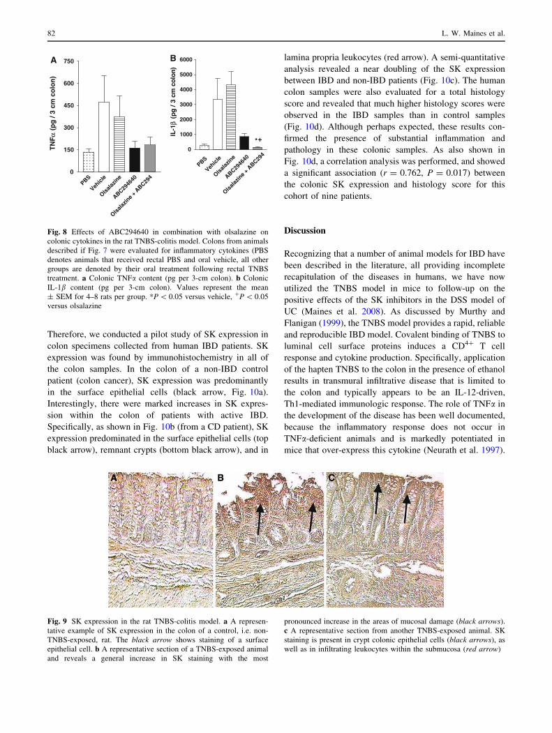
Fig. 7 Effects of ABC294640 in combination with olsalazine on
disease parameters in the rat TNBS-colitis model. Animals were
treated twice a day as follows: oral sham gavage (white bars); oral
olsalazine (50 mg/kg horizontal hatched bars, via gel cap);
ABC294640 (25 mg/kg black bars, via gelcap); olsalazine and
ABC294640 (50 and 25 mg/kg, respectively, gray bars, via gel
cap). At the time of killing (day 6), the following parameters were
measured as described in ‘‘Materials and methods’’. a Body weight
change, b macroscopic score, c colon weight, d myeloperoxidase
activity, e histology score. Values represent the mean ± SD for 5–8
rats per group
Sphingosine kinase inhibitor, Rodent Crohn’s 81
Therefore, we conducted a pilot study of SK expression in
colon specimens collected from human IBD patients. SK
expression was found by immunohistochemistry in all of
the colon samples. In the colon of a non-IBD control
patient (colon cancer), SK expression was predominantly
in the surface epithelial cells (black arrow, Fig. 10a).
Interestingly, there were marked increases in SK expres-
sion within the colon of patients with active IBD.
Specifically, as shown in Fig. 10b (from a CD patient), SK
expression predominated in the surface epithelial cells (top
black arrow), remnant crypts (bottom black arrow), and in
lamina propria leukocytes (red arrow). A semi-quantitative
analysis revealed a near doubling of the SK expression
between IBD and non-IBD patients (Fig. 10c). The human
colon samples were also evaluated for a total histology
score and revealed that much higher histology scores were
observed in the IBD samples than in control samples
(Fig. 10d). Although perhaps expected, these results con-
firmed the presence of substantial inflammation and
pathology in these colonic samples. As also shown in
Fig. 10d, a correlation analysis was performed, and showed
a significant association (r = 0.762, P = 0.017) between
the colonic SK expression and histology score for this
cohort of nine patients.
Discussion
Recognizing that a number of animal models for IBD have
been described in the literature, all providing incomplete
recapitulation of the diseases in humans, we have now
utilized the TNBS model in mice to follow-up on the
positive effects of the SK inhibitors in the DSS model of
UC (Maines et al. 2008). As discussed by Murthy and
Flanigan (1999), the TNBS model provides a rapid, reliable
and reproducible IBD model. Covalent binding of TNBS to
luminal cell surface proteins induces a CD4? T cell
response and cytokine production. Specifically, application
of the hapten TNBS to the colon in the presence of ethanol
results in transmural infiltrative disease that is limited to
the colon and typically appears to be an IL-12-driven,
Th1-mediated immunologic response. The role of TNFa in
the development of the disease has been well documented,
because the inflammatory response does not occur in
TNFa-deficient animals and is markedly potentiated in
mice that over-express this cytokine (Neurath et al. 1997).
PBS
Vehicl
e
Olsala
zine
ABC2946
40
Olsalaz
ine +
ABC29
40
150
300
450
600
750AT
NF α
(p
g /
3 cm
co
lon
)
PBS
Vehicl
e
Olsalaz
ine
ABC2946
40
Olsalaz
ine +
ABC29
40
1000
2000
3000
4000
5000
6000
*+
B
IL-1
β (
pg /
3 cm
co
lon)
Fig. 8 Effects of ABC294640 in combination with olsalazine on
colonic cytokines in the rat TNBS-colitis model. Colons from animals
described if Fig. 7 were evaluated for inflammatory cytokines (PBS
denotes animals that received rectal PBS and oral vehicle, all other
groups are denoted by their oral treatment following rectal TNBS
treatment. a Colonic TNFa content (pg per 3-cm colon). b Colonic
IL-1b content (pg per 3-cm colon). Values represent the mean
± SEM for 4–8 rats per group. *P \ 0.05 versus vehicle, ?P \ 0.05
versus olsalazine
Fig. 9 SK expression in the rat TNBS-colitis model. a A represen-
tative example of SK expression in the colon of a control, i.e. non-
TNBS-exposed, rat. The black arrow shows staining of a surface
epithelial cell. b A representative section of a TNBS-exposed animal
and reveals a general increase in SK staining with the most
pronounced increase in the areas of mucosal damage (black arrows).
c A representative section from another TNBS-exposed animal. SK
staining is present in crypt colonic epithelial cells (black arrows), as
well as in infiltrating leukocytes within the submucosa (red arrow)
82 L. W. Maines et al.

In addition, antibodies against TNFa and pentoxifylline
strongly reduced colonic and systemic inflammation in the
TNBS model (Armstrong et al. 2001). The end result of the
inflammatory cascade is the production of colonic ulcera-
tion and transmural inflammation (Murthy and Flanigan
1999). Indeed, in this study, we confirmed that TNFa (as
well as IL-1b) was increased in the rat colon following
TNBS exposure.
Previously, we showed that both colonic SK expression
and SIP levels were increased following the administration
of DSS to C57 BL/6 mice (Maines et al. 2008). Similarly,
the current data suggest increased SK expression in the
colons of mice and rats with TNBS-induced colitis, as well
as in the colons of IBD patients. This target validation in
the human is important evidence that the SK inhibitors may
be effective in the clinical setting. As a whole, our results
indicate that the TNBS model replicates the increased SK
expression with intestinal inflammation, as observed in
human IBD. Consequently, this data validate the use of the
TNBS-colitis model for studying the effects of SK inhibi-
tors in IBD.
Corticosteroids, such as dexamethasone can inhibit the
inflammatory process in the TNBS model, suggesting this
colitis is sensitive to the pharmacological action of these
drugs (Nakase et al. 2001). With the goal of advancing a
lead SK inhibitor into the clinic, we chose prednisolone as
a comparator drug in our IBD studies. Conventional cor-
ticosteroids have demonstrated efficacy in active CD, but
are used as sparingly due to deleterious side effects
including immune suppression and osteoporosis (Sandborn
et al. 2007). In pediatric IBD, steroids have even been
shown to reduce bone density and formation (Canalis et al.
2002). In our studies, prednisolone-treated rats exhibited
significant weight loss. Current medication strategies seek
to eliminate a reliance on prednisone as corticosteroids are
not an ideal first-line therapy for CD (Sandborn et al.
2007).
The search for a drug with similar efficacy to steroids
without the side effects is thus of the utmost importance for
the clinical management of IBD. As one option, 5-ASA
drugs are considered marginally effective in the manage-
ment of CD (Sandborn et al. 2007). Our combined data
suggest that the novel SK-2 inhibitor ABC294640 performs
comparably to prednisolone over multiple parameters,
without the undesired weight loss. In addition, the combi-
nation of ABC294640 with olsalazine was tested as a
possible scenario in which the SK inhibitor would be used
as an add-on therapy to 5-ASA. The results from these
studies are encouraging, with an apparent further
improvement with the combination as was anticipated due
to the different mechanisms of action of the two com-
pounds. Interestingly, combination treatment with
olsalazine and ABC294640 resulted in near normalization
of the colonic TNFa and IL-1b contents, while olsalazine
was devoid of anti-cytokine activity. In this regard, acti-
vation of SK is essential for the signaling responses to
Fig. 10 Immunohistochemical
staining of SK in the colons of
human patients. a A
representative result of SK
expression (arrow) in a control
patient sample, i.e. non-IBD.
b A marked increase in SK
staining in the epithelial cells
(black arrows) as well as in the
lamina propria (red arrow) from
an IBD patient tissue sample.
c A quantitative analysis of the
percent SK expression results
from all human samples tested,
and d a correlation between SK
expression levels and disease
intensity in human control (c)
and IBD tissue samples
Sphingosine kinase inhibitor, Rodent Crohn’s 83

TNFa because its ability to induce adhesion molecule
expression via activation of NFjB is mimicked by S1P,
and is blocked by the SK inhibitor dimethylsphingosine
(Xia et al. 1998). We have previously shown that
ABC294640 blocks TNFa-induced VCAM expression
(Maines et al. 2008). The clear role of SK in the mecha-
nism of action of TNFa (Xia et al. 1999; Niwa et al. 2000;
Osawa et al. 2001) suggests that SK inhibitors could have
additive or synergistic effects with other therapeutic
modalities, such as remicade.
The present work extends our previous results in the
DSS colitis models (Maines et al. 2008) and demonstrates
that a novel inhibitor of SK (ABC294640) provides pro-
tection against IBD in the TNBS-colitis model in an
overall similar manner to prednisolone. Moreover, the
side-effect profile of ABC294640 was superior to that of
prednisolone. ABC294640 may find utility as a single
therapeutic agent, or as part of a targeted combination
therapy approach for the treatment of intestinal inflam-
mation. Taken together, these animal and human data lend
further evidence to warrant investigation of a first in class
small molecule SK-2 inhibitor in a clinical setting for the
treatment of IBDs.
Acknowledgments We thank Dr. Walter Koltun (Department of
Surgery) of the Penn State College of Medicine for providing the
human tissue samples.
References
Armstrong AM, Foulkes R et al (2001) Tumour necrosis factor
inhibitors reduce the acute-phase response in hapten-induced
colitis. Br J Surg 88(2):235–240
Baumruker T, Bornancin F et al (2005) The role of sphingosine and
ceramide kinases in inflammatory responses. Immunol Lett
96(2):175–185
Canalis E, Pereira RC et al (2002) Effects of glucocorticoids on the
skeleton. J Pediatr Endocrinol Metab 15(5):1341–1345
Chalfant CE, Spiegel S (2005) Sphingosine 1-phosphate and
ceramide 1-phosphate: expanding roles in cell signaling. J Cell
Sci 118(Pt 20):4605–4612
El Alwani M, Wu BX et al (2006) Bioactive sphingolipids in the
modulation of the inflammatory response. Pharmacol Ther
112(1):171–183
Fitzpatrick LR, Wang J et al (2000) In vitro and in vivo effects of
gliotoxin, a fungal metabolite: efficacy against dextran sodium
sulfate-induced colitis in rats. Dig Dis Sci 45(12):2327–2336
Fitzpatrick LR, Small JS et al (2007) Enhanced intestinal expression
of the proteasome subunit low molecular mass polypeptide 2 in
patients with inflammatory bowel disease. Dis Colon Rectum
50(3):337–348
French K, Zhuang Y et al. (2010) Pharmacology and Antitumor
Activity of ABC294640, a selective inhibitor of sphingosine
kinase-2. JPET. doi:10.1124/JPET.109.163444 (in press)
French KJ, Schrecengost RS et al (2003a) Discovery and evaluation
of inhibitors of human sphingosine kinase. Cancer Res
63(18):5962–5969
French KJ, Schrecengost RS et al (2003b) Discovery and evaluation
of inhibitors of human sphingosine kinase. Cancer Res
63(18):5962–5969
French KJ, Upson JJ et al (2006) Antitumor activity of sphingosine
kinase inhibitors. J Pharm Exp Ther 318(2):596–603
Itagaki K, Hauser CJ (2003) Sphingosine 1-phosphate, a diffusible
calcium influx factor mediating store-operated calcium entry. J
Biol Chem 278(30):27540–27547
Kee TH, Vit P et al (2005) Sphingosine kinase signalling in immune
cells. Clin Exp Pharmacol Physiol 32(3):153–161
Kostin S, Pool L et al (2003) Myocytes die by multiple mechanisms in
failing human hearts. Circ Res 92(7):715–724
Leclercq TM, Pitson SM (2006) Cellular signalling by sphingosine
kinase and sphingosine 1-phosphate. IUBMB Life 58(8):467–472
MacKinnon AC, Buckley A et al (2002) Sphingosine kinase: a point
of convergence in the action of diverse neutrophil priming
agents. J Immunol 169(11):6394–6400
Maines LW, French KJ et al (2006) Pharmacologic manipulation of
sphingosine kinase in retinal endothelial cells: implications for
angiogenic ocular diseases. Invest Ophthalmol Vis Sci
47(11):5022–5031
Maines LW, Fitzpatrick LR et al (2008) Suppression of ulcerative
colitis in mice by orally available inhibitors of sphingosine
kinase. Dig Dis Sci 53(4):997–1012
Mathias S, Dressler KA et al (1991) Characterization of a ceramide-
activated protein kinase: stimulation by tumor necrosis factor
alpha. Proc Natl Acad Sci USA 88(22):10009–10013
McCafferty DM, Miampamba M et al (1999) Role of inducible nitric
oxide synthase in trinitrobenzene sulphonic acid induced colitis
in mice. Gut 45(6):864–873
Morris GP, Beck PL et al (1989) Hapten-induced model of chronic
inflammation and ulceration in the rat colon. Gastroenterology
96(3):795–803
Murthy S, Flanigan A (1999) Animal models of inflammatory bowel
disease. Birkhauser Verlag, Basel
Nakase H, Okazaki K et al (2001) An oral drug delivery system
targeting immune-regulating cells ameliorates mucosal injury in
trinitrobenzene sulfonic acid-induced colitis. J Pharmacol Exp
Ther 297(3):1122–1128
Neurath MF, Fuss I et al (1997) Predominant pathogenic role of tumor
necrosis factor in experimental colitis in mice. Eur J Immunol
27(7):1743–1750
Niwa M, Kozawa O et al (2000) Tumor necrosis factor-alpha-
mediated signal transduction in human neutrophils: involvement
of sphingomyelin metabolites in the priming effect of TNF-alpha
on the fMLP-stimulated superoxide production. Life Sci
66(3):245–256
Osawa Y, Banno Y et al (2001) TNF-alpha-induced sphingosine
1-phosphate inhibits apoptosis through a phosphatidylinositol
3-kinase/Akt pathway in human hepatocytes. J Immunol
167(1):173–180
Prieschl EE, Csonga R et al (1999) The balance between sphingosine
and sphingosine-1-phosphate is decisive for mast cell activation
after Fc epsilon receptor I triggering. J Exp Med 190(1):1–8
Rabano M, Pena A et al (2003) Sphingosine-1-phosphate stimulates
cortisol secretion. FEBS Lett 535(1–3):101–105
Sandborn WJ, Feagan BG et al (2007) Medical management of mild
to moderate Crohn’s disease: evidence-based treatment algo-
rithms for induction and maintenance of remission. Aliment
Pharmacol Ther 26(7):987–1003
Smith CD, French KJ et al. (2008) Sphingosine kinase inhibitors. US,
Apogee Biotechnology Corporation, US 7,338,961 B2
Stasi MA, Ruggiero V et al (2004) Ameliorating effects of
the immunomodulator 3-(2-ethylphenyl)-5-(3-methoxyphenyl)-
1H–1, 2, 4-triazole in an experimental model of colitis in the rat.
Eur J Pharmacol 494(2–3):263–272
84 L. W. Maines et al.

Taha TA, Hannun YA et al (2006) Sphingosine kinase: biochemical
and cellular regulation and role in disease. J Biochem Mol Biol
39(2):113–131
Videla S, Vilaseca J et al (2006) Selective inhibition of phosphodi-
esterase-4 ameliorates chronic colitis and prevents intestinal
fibrosis. J Pharmacol Exp Ther 316(2):940–945
Williams KL, Fuller CR et al (2001) Enhanced survival and mucosal
repair after dextran sodium sulfate-induced colitis in transgenic
mice that overexpress growth hormone. Gastroenterology
120(4):925–937
Xia P, Gamble JR et al (1998) Tumor necrosis factor-alpha induces
adhesion molecule expression through the sphingosine kinase
pathway. Proc Natl Acad Sci USA 95(24):14196–14201
Xia P, Wang L et al (1999) Activation of sphingosine kinase by tumor
necrosis factor-alpha inhibits apoptosis in human endothelial
cells. J Biol Chem 274(48):34499–34505
Yamanaka M, Shegogue D et al (2004) Sphingosine kinase 1 (SPHK1)
is induced by transforming growth factor-beta and mediates
TIMP-1 up-regulation. J Biol Chem 279(52):53994–54001
Yatomi Y, Ruan F et al (1995) Sphingosine-1-phosphate: a platelet-
activating sphingolipid released from agonist-stimulated human
platelets. Blood 86(1):193–202
Zhang M, Deng C et al (2006) Curcumin inhibits trinitrobenzene
sulphonic acid-induced colitis in rats by activation of peroxisome
proliferator-activated receptor gamma. Int Immunopharmacol
6(8):1233–1242
Sphingosine kinase inhibitor, Rodent Crohn’s 85