Resting SPECT-neuropsychology correlation in very mild Alzheimer's disease
Brain (1997),120,1229–1244
Spatial- and object-based attentional deficits inAlzheimer’s diseaseRelationship to HMPAO-SPECT measures of parietalperfusion
Brian H. Buck,1,2 Sandra E. Black,1,2 Marlene Behrmann,5 Curtis Caldwell3 and Michael J. Bronskill4
1Research Program in Aging (Sunnybrook Health Science Correspondence to: Dr Sandra Black, Sunnybrook HealthCentre),2Institute of Medical Science, Departments of Science Centre A421, 2075 Bayview Avenue, North York,3Medical Imaging and4Medical Biophysics, University of ON, Canada M4N 3M5Toronto, Toronto, Canada and5Department of Psychology,Carnegie Mellon University, Pittsburgh, USA
SummaryThe purpose of this study was to investigate the ability of compared with age-matched normal controls (n 5 17) using
a cued reaction time task based on Eglyet al.(1994). Regionspatients with Alzheimer’s disease to shift attention betweenspatial locations and between objects, and to examine the of interest were defined semi-automatically on SPECT, and
were anatomically localized with the aid of co-registered MRI.brain regions involved in these cognitive operations usingsingle photon emission computed tomography (SPECT) As hypothesized, in Alzheimer’s disease patients, reaction time
costs of invalid targets eliciting shifts of attention betweenimaging. A recent study of patients with focal lesionsprovided evidence that the right and left parietal lobes are spatial locations were selectively correlated with SPECT
hypoperfusion in the right superior parietal lobe; whiledifferentially involved in shifting selective attention frominvalidly cued spatial locations and objects, respectively (Egly reaction time costs of between-object shifts of attention were
correlated with hypoperfusion in the left inferior parietalet al.J Exp Psychol Gen 1994; 123: 161–77). Accordingly, inAlzheimer’s disease patients, we hypothesized that right lobe. These results provide evidence for the specialized
roles of the right and left parietal regions in the spatialparietal hypoperfusion on SPECT would be associated withdeficits on the spatial-based component of a cued reaction and object components of attentional shifting respectively, and
suggest that the cognitive profile associated with Alzheimer’stime task, and left parietal hypoperfusion would be associatedwith the deficits on the object-based component. Attentional disease includes both spatial- and object-based attentional
impairments.performance of Alzheimer’s disease patients (n 5 29) was
Keywords: visual attention; Alzheimer’s disease; SPECT; neuropsychology; parietal lobe
Abbreviations: AC/PC 5 anterior commissure/posterior commissure; ANOVA5 analysis of variance; AP5 anterior–posterior; DRS5 (Mattis) Dementia Rating Scale; HMPAO5 hexamethylpropylene amine oxime; MMS5 Mini-MentalState Examination; OM5 orbitomeatal; SPECT5 single photon emission computed tomography; srCBF5 standardizedregional cerebral blood flow
IntroductionAlzheimer’s disease is a neurodegenerative disorder of (Terryet al., 1994) and the presence of neurofibrillary tangles
(Arrigada et al., 1992). Of interest, with respect to visualunknown aetiology, and a major cause of dementia in elderlyadults. In Alzheimer’s disease, deficits in cognitive processes attention, is the pathology of the posterior parietal and
prefrontal regions. These cortical areas have beensuch as memory, language and visual attention are theresult of regionally selective neuropathological changes. The hypothesized to form part of a cortical system involved in
the focusing, shifting and maintenance of attention over timeneuropathological events that best correlate with thedeterioration of cognitive function are the loss of synapses (Mesulam, 1990; Morecraftet al., 1993; Posner and Dehaene,
© Oxford University Press 1997
1230 B. H. Bucket al.
1994). Given the topographical distribution of pathology in 1986). The effects of cue validity on response times arerobust, and cueing has also been shown to modulate scalpAlzheimer’s disease, it would be predicted that the visual
attention system would be damaged early in the disease electrical activity in humans (Mangun and Hillyard, 1987)and the excitability of neurons in non-human primatesprocess.
Recent developments in imaging methodologies, most (Mountcastleet al., 1987).When subjects with brain damage following cerebro-notably the development of anatomical and functional
neuroimaging techniques, provide an opportunity to quantify vascular injury are tested on the spatial cueing task, differentdeficits are found depending on the loci of the lesions (Posnerbrain dysfunction in Alzheimer’s disease. Both PET and
single photon emission computed tomography (SPECT) haveet al., 1984; Rafal and Posner, 1987). Subjects with damageto the parietal lobe are impaired in their detection of targetsbeen used to index regional tissue dysfunction in the brains
of Alzheimer’s disease subjects. Based on previous studies, in the contra-lesional visual field, but only when they arefirst miscued (by an invalid cue) to the ipsilesional visualit is known that Alzheimer’s disease is associated with the
following features on PET and SPECT: (i) reduced perfusion field (Posneret al., 1984). These same subjects show almostnormal reaction times to contra-lesional targets that arein the parietotemporal association cortices, even early in the
disease process (Kumaret al., 1991); (ii) the reduction of validly cued, which indicates that they are able to shiftattention to the contra-lesional field. Based on the deficitperfusion is bilateral, although asymmetry in the degree of
hypoperfusion is often observed (Haxbyet al., 1985); (iii) seen in parietally-damaged stroke patients on the spatialcueing task, it has been postulated that the parietal lobe isin more advanced cases, perfusion in the frontal association
cortex is also reduced (Waldemaret al., 1994); (iv) the selectively involved in shifting or ‘disengaging’ attentionfrom previously attended locations in the contra-lesional field.primary sensory and motor cortical regions are relatively
spared (Jagustet al., 1993; Kumaret al., 1991). These PET Further studies have shown that the ‘disengage deficit’ ismore common after damage to the right compared with theand SPECT findings are consistent with the topographical
distribution of pathological markers and suggest that PET left parietal lobe, and is often related to a more generalattentional deficit in which the patient fails to respond orand SPECT can provide an index of the regional distribution
of pathology in Alzheimer’s disease. If imaging measures of orient to stimuli presented contralateral to the brain lesion(i.e. unilateral neglect) (Morrow and Ratcliff, 1988). Thesebrain dysfunction are combined with measures of cognitive
performance, then Alzheimer’s disease potentially provides clinical observations along with more recent PET studies(Corbettaet al., 1993, 1995) provide converging evidencean opportunity to probe the relationship between brain
function and cognitive processes (Parasuramanet al., 1992; that the right posterior parietal lobe in humans is specializedfor directing attention to spatial locations and may also bePennielloet al., 1995). This approach was utilized in this
study to examine whether the deficits in orienting selective specifically involved in disengaging selective attention.A study by Egly et al. (1994) helped to delineate theattention in Alzheimer’s disease patients are significantly
correlated with indices of regional cerebral blood flow on respective attentional functions of the right and left parietalregions further. They developed an elegant paradigm in whichhexamethylpropylene amine oxime (HMPAO) SPECT
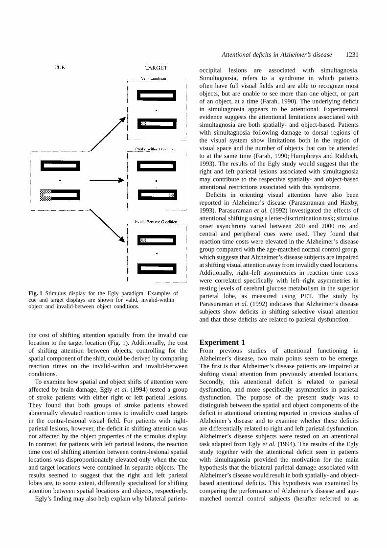
imaging. shifting of attention could occur either between spatiallocations enclosed within a single object or between spatialTo study attentional orienting without accompanying eye
movements, a spatial cueing paradigm can been used (Posner, locations located in separate objects. The objects consistedof two rectangles that appeared either above and below or1980; Posneret al., 1984). With this paradigm, subjects are
asked to maintain fixation on a central point and to respond to the left and right of a central fixation point. The tworectangles were identical in size and were positioned so thatby pressing a key to the appearance of a target at a peripheral
location on a computer display. The target is preceded by a the internal distance between ends of a single rectangle wasequal to the perpendicular distance between the ends ofcue that summons attention either to the target location (i.e.
a valid cue) or to the wrong location (i.e. an invalid cue). the two rectangles (Fig. 1). Cueing was accomplished bybrightening (i.e. changing from grey to white) the end of theCues can be either an abrupt visual onset or a centrally
presented arrow-shaped stimulus. Responses to targets on rectangle. The subject’s task was to press a key whenever atarget (a square) was detected at one of the four rectangle ends.valid and invalid trials require the same perceptual and
motor processing but differ in their attentional requirements. This experiment had two conditions: (i) valid trials, inwhich the target appeared at the cued end of the rectangleSpatial cueing tasks have been used repeatedly and
successfully in normal subjects to show that targets presented and (ii) invalid trials, in which the target appeared at anuncued end of one of the rectangles. On invalid trials theat validly cued locations are responded to more rapidly than
targets appearing at invalidly cued locations (Posner, 1980). target appeared either at the end of the rectangle opposite tothe cue (i.e. invalid-within) or at the end of the uncuedThere is a consensus that the response-time difference
between trials with valid and invalid cues results from the rectangle (i.e. invalid-between) (Fig. 1). The two invalid cueconditions allowed for the separate calculation of spatial andadditional time required for the subject to reorient attention
from the incorrectly cued location to the correct target object reaction time costs. By comparing reaction time onvalid and invalid-within trials it was possible to determinelocation on invalid trials (Posner, 1980; Eriksen and St James,
Attentional deficits in Alzheimer’s disease 1231
occipital lesions are associated with simultagnosia.Simultagnosia, refers to a syndrome in which patientsoften have full visual fields and are able to recognize mostobjects, but are unable to see more than one object, or partof an object, at a time (Farah, 1990). The underlying deficitin simultagnosia appears to be attentional. Experimentalevidence suggests the attentional limitations associated withsimultagnosia are both spatially- and object-based. Patientswith simultagnosia following damage to dorsal regions ofthe visual system show limitations both in the region ofvisual space and the number of objects that can be attendedto at the same time (Farah, 1990; Humphreys and Riddoch,1993). The results of the Egly study would suggest that theright and left parietal lesions associated with simultagnosiamay contribute to the respective spatially- and object-basedattentional restrictions associated with this syndrome.
Deficits in orienting visual attention have also beenreported in Alzheimer’s disease (Parasuraman and Haxby,1993). Parasuramanet al. (1992) investigated the effects ofattentional shifting using a letter-discrimination task; stimulusonset asynchrony varied between 200 and 2000 ms andcentral and peripheral cues were used. They found thatreaction time costs were elevated in the Alzheimer’s diseasegroup compared with the age-matched normal control group,which suggests that Alzheimer’s disease subjects are impairedat shifting visual attention away from invalidly cued locations.Additionally, right–left asymmetries in reaction time costswere correlated specifically with left–right asymmetries inresting levels of cerebral glucose metabolism in the superior
Fig. 1 Stimulus display for the Egly paradigm. Examples of parietal lobe, as measured using PET. The study bycue and target displays are shown for valid, invalid-within
Parasuramanet al. (1992) indicates that Alzheimer’s diseaseobject and invalid-between object conditions.subjects show deficits in shifting selective visual attentionand that these deficits are related to parietal dysfunction.
the cost of shifting attention spatially from the invalid cuelocation to the target location (Fig. 1). Additionally, the cost Experiment 1
From previous studies of attentional functioning inof shifting attention between objects, controlling for thespatial component of the shift, could be derived by comparing Alzheimer’s disease, two main points seem to be emerge.
The first is that Alzheimer’s disease patients are impaired atreaction times on the invalid-within and invalid-betweenconditions. shifting visual attention from previously attended locations.
Secondly, this attentional deficit is related to parietalTo examine how spatial and object shifts of attention wereaffected by brain damage, Eglyet al. (1994) tested a group dysfunction, and more specifically asymmetries in parietal
dysfunction. The purpose of the present study was toof stroke patients with either right or left parietal lesions.They found that both groups of stroke patients showed distinguish between the spatial and object components of the
deficit in attentional orienting reported in previous studies ofabnormally elevated reaction times to invalidly cued targetsin the contra-lesional visual field. For patients with right- Alzheimer’s disease and to examine whether these deficits
are differentially related to right and left parietal dysfunction.parietal lesions, however, the deficit in shifting attention wasnot affected by the object properties of the stimulus display. Alzheimer’s disease subjects were tested on an attentional
task adapted from Eglyet al. (1994). The results of the EglyIn contrast, for patients with left parietal lesions, the reactiontime cost of shifting attention between contra-lesional spatial study together with the attentional deficit seen in patients
with simultagnosia provided the motivation for the mainlocations was disproportionately elevated only when the cueand target locations were contained in separate objects. The hypothesis that the bilateral parietal damage associated with
Alzheimer’s disease would result in both spatially- and object-results seemed to suggest that the right and left parietallobes are, to some extent, differently specialized for shifting based attentional deficits. This hypothesis was examined by
comparing the performance of Alzheimer’s disease and age-attention between spatial locations and objects, respectively.Egly’s finding may also help explain why bilateral parieto- matched normal control subjects (herafter referred to as
1232 B. H. Bucket al.
control subjects) on a version of the Egly task. It was rectangles were 3.8° from the centre of fixation. Therectangles were oriented parallel to each other, so that thepredicted that the predominantly bilateral parietal damage
associated with Alzheimer’s disease would result in impaired distance between the ends of the two rectangles was 7.6°.Cueing was accomplished by superimposing three 1.9° whiteshifting of attention both between- and within-objects.lines over one end of a rectangle, which had the effect ofbrightening the rectangle end. The target was a solid whitesquare measuring 1.8°31.9°. All stimuli were presented onMethodsa black background.
SubjectsTwo groups of subjects participated in this experiment, agroup with mild to moderate Alzheimer’s disease (15 males,Apparatus14 females) and a group of healthy age-, sex- and education-Stimulus presentation and recording of response time werematched control subjects (eight males, nine females). Allcontrolled using SuperLab software on a Macintosh IIci withsubjects had normal or corrected vision of at least 20/40.a 14-in colour monitor (70-Hz refresh rate). Subjects madeAlzheimer’s disease subjects had standard neurological andresponses to the target using their dominant hand to press abiochemical tests in addition to MRI to rule out secondarykey interfaced to the computer.causes of dementia. Subjects in the Alzheimer’s diseasegroup met the NINCDS–ADRDA (National Institute ofNeurological and Communicative Disorders and Stroke-ProcedureAlzheimer’s Disease and Related Disorders Association WorkSubjects were tested in a quiet room, with subdued lighting.Group) diagnostic criteria for ‘probable’ Alzheimer’s diseaseTrials began with the presentation of a fixation display for(McKhann et al., 1984). To reduce the possibility of 1000 ms. Following the fixation display, one of the ends ofconcomitant vascular disease, all Alzheimer’s disease subjectsthe two rectangles was cued by brightening the rectangle.had a modified Hachinski score ofø4 (Hachinski et al., The cue lasted for 200 ms, after which time the cued end1975). Except for two Alzheimer’s disease subjects (onereturned to its original colour (grey). Then, the fixationmale, one female), all subjects were right-handed. Thedisplay was immediately presented for another 200 ms, afterdemographic characteristics of the Alzheimer’s disease andwhich time the target square appeared (i.e. the inter-stimuluscontrol subjects are shown on Table 1. In addition to mentalinterval was 200 ms). The target square remained visiblestatus testing, Alzheimer’s disease subjects also received auntil the subject responded, or for 2000 ms if there was nomore detailed battery of neuropsychological tests, the resultsresponse. Once the subject responded, or 2000 ms lapsed,of which are summarized in Table 1. the screen was blank (black) for 500 ms and then the
For the control group, standard neurological andnext trial began. On ‘catch’ trials, the fixation display waspsychological exclusion criteria were applied in selectingpresented for 2000 ms following the cue, and no target wassubjects for participation in the study. There were nopresented.significant differences in the age [t(42) 5 1.53,P 5 0.1327] Both key-response accuracy and reaction time wereand education [t(38) 5 1.70,P 5 0.0955] of the two groups; measured. Subjects were told to maintain fixation on thehowever, as expected, the Alzheimer’s disease group scoredfixation point throughout each trial, and that, although reactionlower on the Mini-Mental State Examination (MMS) (Folstein time was being recorded, it was equally important to respondet al., 1975) [t(37) 5 9.52, P 5 0.0000] (Table 1). Of the only when the target was detected. Trials on which the subjects29 Alzheimer’s disease subjects, 12 subjects had mild deficitswere distracted or required re-instruction were marked by[MMS 20–28, Mattis Dementia Rating Scale (DRS) (Mattis, the experimenter and excluded from analysis. All subjects1976), 115–132], and the remaining 17 subjects wereincluded in this analysis had at least 15 trials in each conditionmoderately demented (MMS 10–19, DRS 74–114). Allanalysed. Additionally, response times of,250 ms wereprocedures were approved by the institutional ethics reviewconsidered as anticipation and were not analysed.board at Sunnybrook Health Science Centre and informed Before starting the experiment, subjects were given aconsent was obtained from all subjects and/or their guardianspractice session consisting of 20 trials. The task was explainedfollowing appropriate ethical procedures. during the practice trials. If subjects made more than five
errors (ù25%) on the practice trials, the practice trials wererepeated until the error rate dropped below 25%. Of the 31Alzheimer’s disease subjects who attempted the experiment,Stimuli
The fixation display consisted of two coloured rectangles two were unable to achieve this level of performance andwere excluded from the study, leaving 29 subjects in the(one blue and one green) positioned either above and below,
or to the left and right of, a fixation point measuring 0.5°30.5° Alzheimer’s disease group. All control subjects were able tocomplete the experiment.(when viewed at a distance of 60 cm) (Fig. 1). Each rectangle
measured 9.5°31.9° and was formed from lines that were During test sessions, eye movements were monitoredvisually by the experimenter. Trials on which eye movementssix pixels thick and 50% grey in colour. The centres of the
Attentional deficits in Alzheimer’s disease 1233
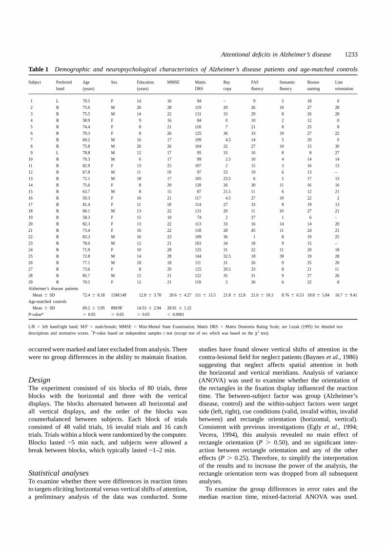
Table 1 Demographic and neuropsychological characteristics of Alzheimer’s disease patients and age-matched controls
Subject Preferred Age Sex Education MMSE Mattis Rey FAS Semantic Boston Line
hand (years) (years) DRS copy fluency fluency naming orientation
1 L 70.5 F 14 16 94 – 9 5 18 0
2 R 75.6 M 20 28 119 29 26 10 27 28
3 R 75.5 M 14 22 131 33 29 8 26 28
4 R 58.9 F 9 16 84 0 10 2 12 0
5 R 74.4 F 8 21 118 7 21 8 25 8
6 R 70.3 F 8 26 125 36 33 10 27 22
7 R 69.2 M 16 17 109 4.5 14 5 20 0
8 R 75.8 M 20 26 104 32 27 10 15 30
9 L 78.8 M 12 17 95 33 10 8 8 27
10 R 70.3 M 6 17 99 2.5 10 4 14 14
11 R 82.9 F 13 25 107 2 15 3 16 13
12 R 67.8 M 11 19 97 23 19 6 13 –
13 R 72.1 M 18 17 105 23.5 6 5 17 13
14 R 75.6 F 8 20 120 26 30 11 16 16
15 R 63.7 M 8 15 87 21.5 11 6 12 21
16 R 50.3 F 16 21 117 4.5 27 10 22 2
17 R 81.4 F 11 18 114 27 33 8 19 13
18 R 60.1 M 13 22 131 20 11 10 27 21
19 R 58.3 F 15 10 74 2 27 1 6 –
20 R 82.3 F 13 22 113 33 16 14 14 20
21 R 73.4 F 16 22 118 28 45 11 24 21
22 R 83.3 M 16 23 109 36 1 8 19 25
23 R 78.0 M 12 21 103 34 18 9 15 –
24 R 71.9 F 10 28 125 31 22 11 20 18
25 R 72.8 M 14 28 144 32.5 18 39 19 28
26 R 77.1 M 18 18 111 31 26 9 25 20
27 R 73.6 F 8 20 125 20.5 33 8 21 11
28 R 85.7 M 12 21 122 35 31 9 27 26
29 R 70.5 F 12 21 119 3 30 6 22 8
Alzheimer’s disease patients
Mean 6 SD 72.4 6 8.18 15M/14F 12.86 3.78 20.66 4.27 1116 15.5 21.86 12.8 21.06 10.3 8.766 6.53 18.86 5.84 16.76 9.41
Age-matched controls
Mean 6 SD 69.2 6 5.95 8M/9F 14.536 2.94 28.916 1.22
P-value* . 0.05 . 0.05 . 0.05 , 0.0001
L/R 5 left hand/right hand; M/F5 male/female; MMSE5 Mini-Mental State Examination; Mattis DRS5 Mattis Dementia Rating Scale;seeLezak (1995) for detailed test
descriptions and normative scores.*P-value based on independent samplest test (except test of sex which was based on theχ2 test).
occurred were marked and later excluded from analysis. There studies have found slower vertical shifts of attention in thecontra-lesional field for neglect patients (Bayneset al., 1986)were no group differences in the ability to maintain fixation.suggesting that neglect affects spatial attention in boththe horizontal and vertical meridians. Analysis of variance(ANOVA) was used to examine whether the orientation ofDesign
The experiment consisted of six blocks of 80 trials, three the rectangles in the fixation display influenced the reactiontime. The between-subject factor was group (Alzheimer’sblocks with the horizontal and three with the vertical
displays. The blocks alternated between all horizontal and disease, control) and the within-subject factors were targetside (left, right), cue conditions (valid, invalid within, invalidall vertical displays, and the order of the blocks was
counterbalanced between subjects. Each block of trials between) and rectangle orientation (horizontal, vertical).Consistent with previous investigations (Eglyet al., 1994;consisted of 48 valid trials, 16 invalid trials and 16 catch
trials. Trials within a block were randomized by the computer. Vecera, 1994), this analysis revealed no main effect ofrectangle orientation (P . 0.50), and no significant inter-Blocks lasted ~5 min each, and subjects were allowed a
break between blocks, which typically lasted ~1–2 min. action between rectangle orientation and any of the othereffects (P . 0.25). Therefore, to simplify the interpretationof the results and to increase the power of the analysis, therectangle orientation term was dropped from all subsequentStatistical analyses
To examine whether there were differences in reaction times analyses.To examine the group differences in error rates and theto targets eliciting horizontal versus vertical shifts of attention,
a preliminary analysis of the data was conducted. Some median reaction time, mixed-factorial ANOVA was used.
1234 B. H. Bucket al.
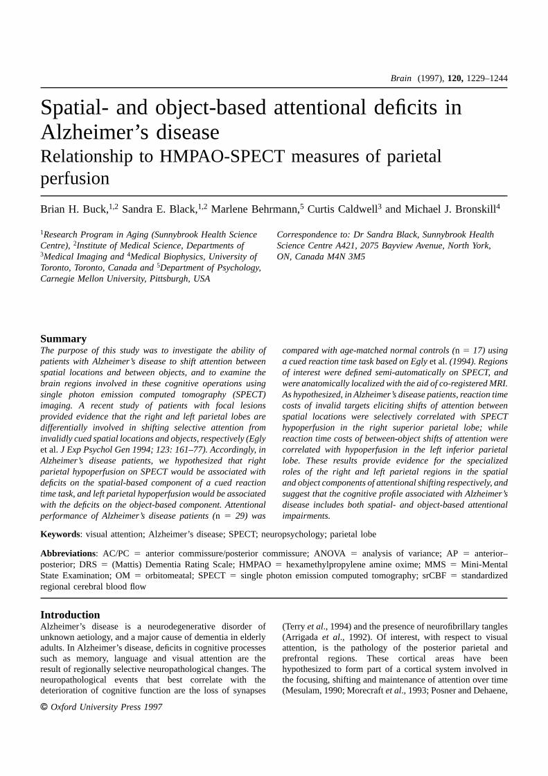
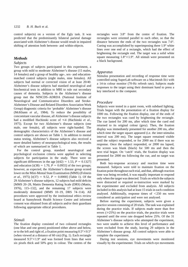
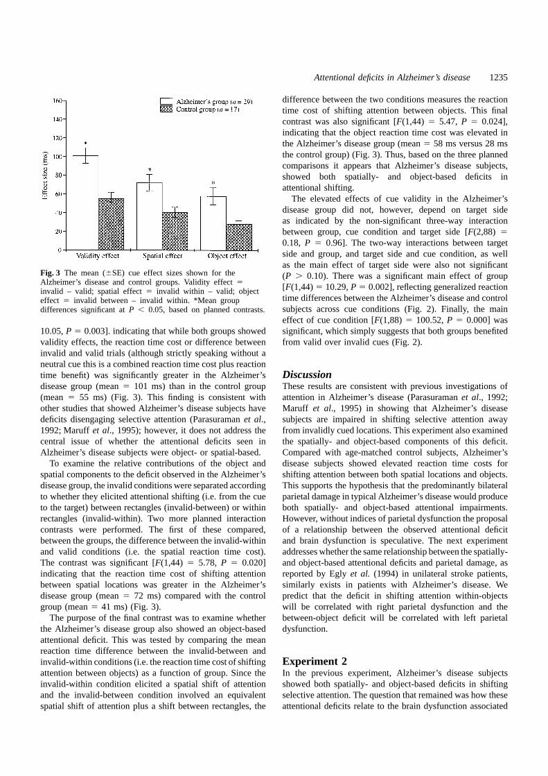
Fig. 2 Mean (6SE) of the median reaction time (RT) as a function of cue condition, target sideand group. Percentage errors for each condition are shown in parenthesis.
The degrees of freedom for within-subjectF-ratios were shows that Alzheimer’s disease subjects made more errorsacross all experimental conditions.corrected using the Greenhouse–Geisser Epsilon (Stevens,
1986). Cue effects were compared between groups usingplanned single degree of freedom interaction-contrasts(Keppel and Zedeck, 1989). All tests were evaluated usingGroup differences in reaction timetwo-tailed probabilities, with the significance level set to 0.05.The mean of the median reaction time for the correct
responses was analysed using a mixed three-factor ANOVA.For the ANOVA, the between-subject factor was group
Results (Alzheimer’s disease, control), and the within-subjectfactors were target side (left, right) and cue condition (valid,Accuracy
The mean hit-rate (6SD) was 93.5 6 5.2% for the invalid within, invalid between). The mean of the medianreaction time as a function of group, target side and cueAlzheimer’s disease group and 97.76 2.7% for the control
group. On catch trials, the Alzheimer’s disease subjects made condition is shown in Fig. 2. ANOVA showed a significanttwo-way interaction between group and cue conditiona significantly greater number of responses than the control
subjects [t(44) 5 –3.64,P 5 0.001]. The mean (6SD) false- [F(1,88) 5 9.48, P 5 0.001]. To identify the source of theinteraction, the 23 3 interaction was decomposed into threealarm rates for the Alzheimer’s disease and control group
was 7.016 6.39% and 1.346 1.95%, respectively. Figure separate planned comparisons, each comparison consistingof a 2 3 2 (group by cue condition) interaction contrast2 shows the percentage errors and the mean of the median
reaction time for the two groups, as a function of cue (Keppel and Zedeck, 1989).The purpose of the first contrast was to compare thecondition and target side. Three-way repeated measures
ANOVA was performed on the error rate for target present difference between the valid and combined invalid conditions(i.e. the effect of cue validity) between the groups. Since thetrials with group (Alzheimer’s disease, control) as the
between-subject factor, and target side (left, right) and cue perceptual–motor processing demands of the invalid andvalid conditions were matched, it was assumed thatcondition (valid, invalid within, invalid between) as within-
subject factors. The only significant source of variance was generalized slowing would equally affect responses on validand invalid trials. The contrast was significant [F(1,88) 5the main effect of group [F(1,44)5 8.56,P 5 0.005], which
Attentional deficits in Alzheimer’s disease 1235
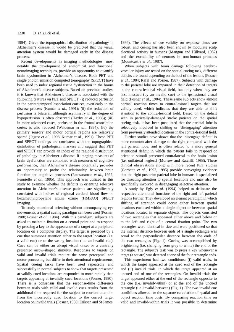
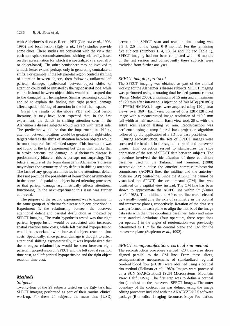
difference between the two conditions measures the reactiontime cost of shifting attention between objects. This finalcontrast was also significant [F(1,44) 5 5.47, P 5 0.024],indicating that the object reaction time cost was elevated inthe Alzheimer’s disease group (mean5 58 ms versus 28 msthe control group) (Fig. 3). Thus, based on the three plannedcomparisons it appears that Alzheimer’s disease subjects,showed both spatially- and object-based deficits inattentional shifting.
The elevated effects of cue validity in the Alzheimer’sdisease group did not, however, depend on target sideas indicated by the non-significant three-way interactionbetween group, cue condition and target side [F(2,88) 50.18, P 5 0.96]. The two-way interactions between targetside and group, and target side and cue condition, as wellas the main effect of target side were also not significant
Fig. 3 The mean (6SE) cue effect sizes shown for the (P . 0.10). There was a significant main effect of groupAlzheimer’s disease and control groups. Validity effect5
[F(1,44)5 10.29,P 5 0.002], reflecting generalized reactioninvalid – valid; spatial effect5 invalid within – valid; objecttime differences between the Alzheimer’s disease and controleffect 5 invalid between – invalid within. *Mean groupsubjects across cue conditions (Fig. 2). Finally, the maindifferences significant atP , 0.05, based on planned contrasts.effect of cue condition [F(1,88) 5 100.52,P 5 0.000] wassignificant, which simply suggests that both groups benefited10.05,P 5 0.003]. indicating that while both groups showed
validity effects, the reaction time cost or difference between from valid over invalid cues (Fig. 2).invalid and valid trials (although strictly speaking without aneutral cue this is a combined reaction time cost plus reactiontime benefit) was significantly greater in the Alzheimer’s Discussion
These results are consistent with previous investigations ofdisease group (mean5 101 ms) than in the control group(mean 5 55 ms) (Fig. 3). This finding is consistent with attention in Alzheimer’s disease (Parasuramanet al., 1992;
Maruff et al., 1995) in showing that Alzheimer’s diseaseother studies that showed Alzheimer’s disease subjects havedeficits disengaging selective attention (Parasuramanet al., subjects are impaired in shifting selective attention away
from invalidly cued locations. This experiment also examined1992; Maruffet al., 1995); however, it does not address thecentral issue of whether the attentional deficits seen in the spatially- and object-based components of this deficit.
Compared with age-matched control subjects, Alzheimer’sAlzheimer’s disease subjects were object- or spatial-based.To examine the relative contributions of the object and disease subjects showed elevated reaction time costs for
shifting attention between both spatial locations and objects.spatial components to the deficit observed in the Alzheimer’sdisease group, the invalid conditions were separated according This supports the hypothesis that the predominantly bilateral
parietal damage in typical Alzheimer’s disease would produceto whether they elicited attentional shifting (i.e. from the cueto the target) between rectangles (invalid-between) or within both spatially- and object-based attentional impairments.
However, without indices of parietal dysfunction the proposalrectangles (invalid-within). Two more planned interactioncontrasts were performed. The first of these compared, of a relationship between the observed attentional deficit
and brain dysfunction is speculative. The next experimentbetween the groups, the difference between the invalid-withinand valid conditions (i.e. the spatial reaction time cost). addresses whether the same relationship between the spatially-
and object-based attentional deficits and parietal damage, asThe contrast was significant [F(1,44) 5 5.78, P 5 0.020]indicating that the reaction time cost of shifting attention reported by Eglyet al. (1994) in unilateral stroke patients,
similarly exists in patients with Alzheimer’s disease. Webetween spatial locations was greater in the Alzheimer’sdisease group (mean5 72 ms) compared with the control predict that the deficit in shifting attention within-objects
will be correlated with right parietal dysfunction and thegroup (mean5 41 ms) (Fig. 3).The purpose of the final contrast was to examine whether between-object deficit will be correlated with left parietal
dysfunction.the Alzheimer’s disease group also showed an object-basedattentional deficit. This was tested by comparing the meanreaction time difference between the invalid-between andinvalid-within conditions (i.e. the reaction time cost of shifting Experiment 2
In the previous experiment, Alzheimer’s disease subjectsattention between objects) as a function of group. Since theinvalid-within condition elicited a spatial shift of attention showed both spatially- and object-based deficits in shifting
selective attention. The question that remained was how theseand the invalid-between condition involved an equivalentspatial shift of attention plus a shift between rectangles, the attentional deficits relate to the brain dysfunction associated
1236 B. H. Bucket al.
with Alzheimer’s disease. Recent PET (Corbettaet al., 1993, between the SPECT scan and reaction time testing was3.3 6 2.6 months (range 0–9 months). For the remaining1995) and focal lesion (Eglyet al., 1994) studies provide
some clues. These studies are consistent with the view that five subjects (numbers 1, 4, 13, 24 and 25;seeTable 1),SPECT imaging had not been completed within 9 monthseach hemisphere controls attentional shifting bilaterally, based
on the representation for which it is specialized (i.e. spatially- of the test session and consequently these subjects wereexcluded from further analyses.or object-based). The other hemisphere may be involved to
a much lesser extent, perhaps only in generating contralateralshifts. For example, if the left parietal region controls shiftingof attention between objects, then following unilateral leftSPECT imaging protocol
The SPECT imaging was obtained as part of the clinicalparietal damage, ipsilesional between-object shifts ofattention could still be initiated by the right parietal lobe, while workup for the Alzheimer’s disease subjects. SPECT imaging
was performed using a rotating dual-headed gamma cameracontra-lesional between-object shifts would be disrupted dueto the damaged left hemisphere. Similar reasoning could be (Picker Model 2000), a minimum of 15 min and a maximum
of 120 min after intravenous injection of 740 MBq (20 mCi)applied to explain the finding that right parietal damageaffects spatial shifting of attention in the left hemispace. of [99mTc]-HMPAO. Images were acquired using 120 planar
views, over 360°. Each view consisted of a 1283128 pixelGiven the results of the above PET and focal lesionliterature, it may have been expected that, in the first image with a reconstructed image resolution of ~10.5 mm
full width at half maximum. Each view took 20 s, with theexperiment, the deficit in shifting attention seen in theAlzheimer’s disease subjects would interact with target side. entire scan session lasting 20 min. Reconstruction was
performed using a ramp-filtered back-projection algorithmThe prediction would be that the impairment in shiftingattention between locations would be greatest for right-sided followed by the application of a 3D low pass post-filter.
During reconstruction, the sets of SPECT images weretargets whereas the shifts of attention between objects wouldbe most impaired for left-sided targets. This interaction was corrected for head-tilt in the sagittal, coronal and transverse
planes. This correction served to standardize the slicenot found in the first experiment but given that, unlike thatin stroke patients, the damage in Alzheimer’s disease is orientation of the sets of SPECT data between subjects. The
procedure involved the identification of three coordinatepredominantly bilateral, this is perhaps not surprising. Thebilateral nature of the brain damage in Alzheimer’s disease baselines used in the Tailarach and Tournoux (1988)
stereotaxic brain atlas: the anterior commissure/posteriormay reduce the asymmetry of any deficits in shifting attention.The lack of any group asymmetries in the attentional deficit commissure (AC/PC) line, the midline and the anterior–
posterior (AP) centre-line. Since the AC/PC line cannot bedoes not preclude the possibility of hemispheric asymmetriesin the control of spatial and object-based orienting processes, visualized on SPECT, the orbitomeatal (OM) line was
identified on a sagittal view instead. The OM line has beenor that parietal damage asymmetrically affects attentionalfunctioning. In the next experiment this issue was further shown to approximate the AC/PC line within 5° (Vanier
et al., 1985). The midline and AP centre-line were selectedaddressed.The purpose of the second experiment was to examine, in by visually identifying the axis of symmetry in the coronal
and transverse planes, respectively. Rotation of the data setsthe same group of Alzheimer’s disease subjects described inExperiment 1, the relationship between the observed was performed in each plane to align the matrix of the image
data sets with the three coordinate baselines. Inter- and intra-attentional deficit and parietal dysfunction as indexed bySPECT imaging. The main hypothesis tested was that right rater standard deviations (four operators, three repetitions
per operator) in the angles of reorientation was previouslyparietal hypoperfusion would be associated with increasedspatial reaction time costs, while left parietal hypoperfusion determined as 1.5° for the coronal plane and 1.6° for the
transverse plane (Stapletonet al., 1992).would be associated with increased object reaction timecosts. Specifically, since parietal damage is thought to affectattentional shifting asymmetrically, it was hypothesized thatthe strongest relationships would be seen between rightSPECT semiquantification: cortical rim method
The reconstruction procedure yielded ~20 transverse slicesparietal hypoperfusion on SPECT and the left spatial reactiontime cost, and left parietal hypoperfusion and the right object aligned parallel to the OM line. From these slices,
semiquantitative measurements of standardized regionalreaction time cost.cerebral blood flow (srCBF) were obtained using a corticalrim method (Hellmanet al., 1989). Images were processedon a SUN SPARCstation2 (SUN Microsystems, Mountain
Methods View, Calif., USA). The first step was to define a corticalrim (annulus) on the transverse SPECT images. The outerSubjects
Twenty-four of the 29 subjects tested on the Egly task had boundary of the cortical rim was defined using the imageediting procedure included with the ANALYZE© 7.5 softwareSPECT imaging performed as part of their routine clinical
work-up. For these 24 subjects, the mean time (6SD) package (Biomedical Imaging Resource, Mayo Foundation,
Attentional deficits in Alzheimer’s disease 1237
USA) (Robb, 1994). This boundary was delimited semi- blind to subject identity) based on visual identification ofanatomical landmarks. Raters were given hardcopies of eachautomatically using a threshold value of 40% of the mean
cerebellar counts. The procedure for determining mean subject’s SPECT scan and were instructed to select the 10transverse slices that most closely matched (i.e. containedcerebellar perfusion is described below. Placement of the
outer boundary was inspected on all slices to ensure that it the same brain structures) the 10 template slices. There wasagreement between raters on 296 out of 380 (78%) of thewas correctly positioned on the surface of the cortex, at the
interface between grey matter and cerebrospinal fluid. The total slices. For the slices on which the two raters disagreed(which never differed by more than one slice), the best sliceinner boundary was defined by moving seven pixels radially
inwards from the outer boundary, using a morphological filter was selected based on a consensus of the two raters.Once the SPECT slices were registered across subjects(i.e. an ‘erosion’ operation). Then the eroded images were
subtracted from the original image, resulting in the ‘cortical into a standardized stereotaxic space, the cortical rimsegments could be localized with reference to the Talairachrim’ image alone.
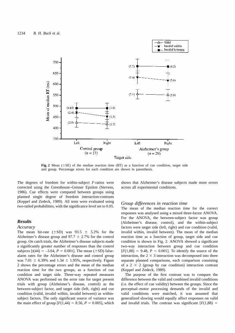
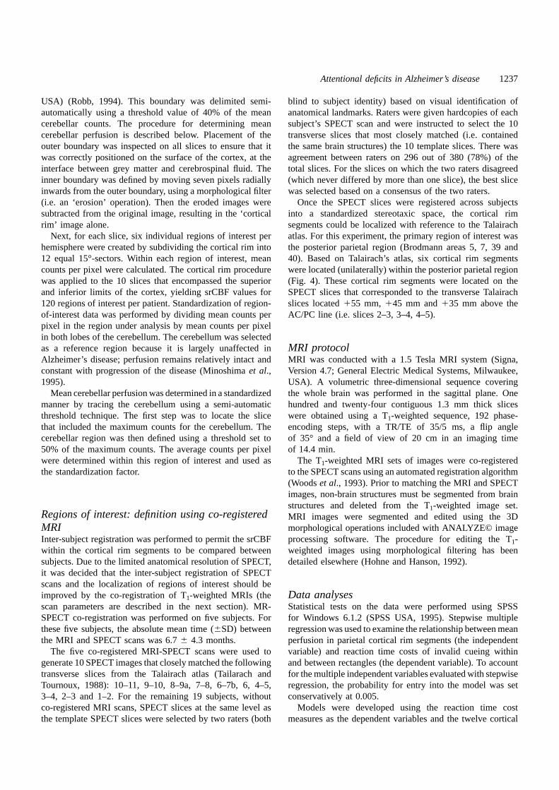
Next, for each slice, six individual regions of interest per atlas. For this experiment, the primary region of interest wasthe posterior parietal region (Brodmann areas 5, 7, 39 andhemisphere were created by subdividing the cortical rim into
12 equal 15°-sectors. Within each region of interest, mean 40). Based on Talairach’s atlas, six cortical rim segmentswere located (unilaterally) within the posterior parietal regioncounts per pixel were calculated. The cortical rim procedure
was applied to the 10 slices that encompassed the superior (Fig. 4). These cortical rim segments were located on theSPECT slices that corresponded to the transverse Talairachand inferior limits of the cortex, yielding srCBF values for
120 regions of interest per patient. Standardization of region- slices located155 mm, 145 mm and135 mm above theAC/PC line (i.e. slices 2–3, 3–4, 4–5).of-interest data was performed by dividing mean counts per
pixel in the region under analysis by mean counts per pixelin both lobes of the cerebellum. The cerebellum was selectedas a reference region because it is largely unaffected inMRI protocol
MRI was conducted with a 1.5 Tesla MRI system (Signa,Alzheimer’s disease; perfusion remains relatively intact andconstant with progression of the disease (Minoshimaet al., Version 4.7; General Electric Medical Systems, Milwaukee,
USA). A volumetric three-dimensional sequence covering1995).Mean cerebellar perfusion was determined in a standardized the whole brain was performed in the sagittal plane. One
hundred and twenty-four contiguous 1.3 mm thick slicesmanner by tracing the cerebellum using a semi-automaticthreshold technique. The first step was to locate the slice were obtained using a T1-weighted sequence, 192 phase-
encoding steps, with a TR/TE of 35/5 ms, a flip anglethat included the maximum counts for the cerebellum. Thecerebellar region was then defined using a threshold set to of 35° and a field of view of 20 cm in an imaging time
of 14.4 min.50% of the maximum counts. The average counts per pixelwere determined within this region of interest and used as The T1-weighted MRI sets of images were co-registered
to the SPECT scans using an automated registration algorithmthe standardization factor.(Woodset al., 1993). Prior to matching the MRI and SPECTimages, non-brain structures must be segmented from brainstructures and deleted from the T1-weighted image set.
Regions of interest: definition using co-registered MRI images were segmented and edited using the 3Dmorphological operations included with ANALYZE© imageMRI
Inter-subject registration was performed to permit the srCBF processing software. The procedure for editing the T1-weighted images using morphological filtering has beenwithin the cortical rim segments to be compared between
subjects. Due to the limited anatomical resolution of SPECT, detailed elsewhere (Hohne and Hanson, 1992).it was decided that the inter-subject registration of SPECTscans and the localization of regions of interest should beimproved by the co-registration of T1-weighted MRIs (the Data analyses
Statistical tests on the data were performed using SPSSscan parameters are described in the next section). MR-SPECT co-registration was performed on five subjects. For for Windows 6.1.2 (SPSS USA, 1995). Stepwise multiple
regression was used to examine the relationship between meanthese five subjects, the absolute mean time (6SD) betweenthe MRI and SPECT scans was 6.76 4.3 months. perfusion in parietal cortical rim segments (the independent
variable) and reaction time costs of invalid cueing withinThe five co-registered MRI-SPECT scans were used togenerate 10 SPECT images that closely matched the following and between rectangles (the dependent variable). To account
for the multiple independent variables evaluated with stepwisetransverse slices from the Talairach atlas (Tailarach andTournoux, 1988): 10–11, 9–10, 8–9a, 7–8, 6–7b, 6, 4–5, regression, the probability for entry into the model was set
conservatively at 0.005.3–4, 2–3 and 1–2. For the remaining 19 subjects, withoutco-registered MRI scans, SPECT slices at the same level as Models were developed using the reaction time cost
measures as the dependent variables and the twelve corticalthe template SPECT slices were selected by two raters (both
1238 B. H. Bucket al.
Fig. 4 The cortical rims extracted from a SPECT image are superimposed on co-registered MRIs oriented parallel to the AC/PCline as described in the text. The distance above the AC/PC line for each slice is indicated in the lower right-hand corner. Forpurpose of illustration, in the left hemisphere, the six superior and inferior parietal segments that were entered into the multiplestepwise regression are shown in black.
rim segments corresponding to left and right, posterior parietal experiments. Specifically, it was important to establish thatthe Alzheimer’s disease subjects selected for the SPECTregions as independent variables. All cortical rim segments
were standardized by dividing the mean counts in each analysis showed, as a group, an impaired ability to shiftattention between both spatial locations and objects.segment by the mean cerebellar counts. Although there were
a priori hypotheses regarding hemispheric specialization for The structure of the ANOVA was identical to theanalysis described in Experiment 1. ANOVA was performedspatial and object attentional shifts, both left and right
hemisphere cortical rim segments were included in each on the mean of the median reaction time for the correctresponses made by the 24 Alzheimer’s disease subjectsmodel, in order to assess the specificity of any detected
relationships between srCBF and task performance. with SPECT imaging, and the same group of controlsubjects described in Experiment 1. Most important, theThe relatively low spatial resolution inherent to SPECT
imaging means that the cortical rim segments are not fully ANOVA revealed a significant group-by-cue conditioninteraction [F(1,78) 5 8.18, P 5 0.003], replicating theindependent. Conventional statistical methods such as
multiple regression may not be ideally suited for examining finding that the cueing effects in the Alzheimer’s diseasesubjects were elevated relative to the control subjects.the relationship between behavioural performance and the
activity of multiple correlated brain regions. Therefore, Furthermore, the reaction time cost of shifting attentionbetween both spatial locations and objects was elevated inthis second experiment was limited to examining the
specific hypothesis that attentional performance would be the Alzheimer’s disease group compared with the controlgroup (P , 0.05) (Table 2).related to parietal hypoperfusion and did not aim to address
the broader issue of which brain systems are involved inperforming this task. More sophisticated data-analysis toolssuch as Partial Least Squares (McIntoshet al., 1996) have Relationship between reaction time costs andbeen proposed to extract this latter type of informationsrCBFfrom imaging data, but these techniques are not yet widelyThe spatial and object reaction time cost measures wereavailable. calculated separately for left- and right-sided targets (as
described in Experiment 1), and were used as dependentvariables in multiple stepwise regression models. For each
Results model, 12 cortical rim segments from the left andright posterior parietal region were included as putativeAnalysis of reaction time data
Figure 5 shows the mean of the median reaction time data independent variables. Segments were included in a modelwhen they accounted for a significant portion of theand percentage errors as a function of cue validity and
target side for the subset of Alzheimer’s disease subjects variance (i.e. ifP , 0.005) in the dependent variable.Results of these analyses are shown in Table 3. As(n 5 24) with SPECT imaging that were used in this
experiment. Prior to examining the relationship between hypothesized, for the right object reaction time cost,a segment located in the left inferior parietal regionmeasures of srCBF and attentional performance, ANOVA
was performed on the reaction time data. The purpose of (Brodmann area 39, 40) entered into the model [F(1,22) 510.00, P 5 0.0045]. The negative slope (β) indicated thatthis analysis was to ensure that the subset of subjects
selected for inclusion in this experiment were representative the relationship was in the expected direction, with reducedsrCBF being associated with increased reaction time costsof the larger group of 29 subjects described in the previous
Attentional deficits in Alzheimer’s disease 1239
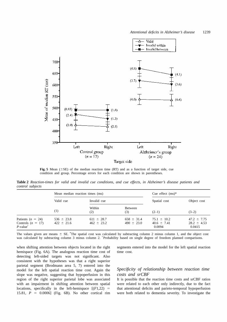
Fig. 5 Mean (6SE) of the median reaction time (RT) and as a function of target side, cuecondition and group. Percentage errors for each condition are shown in parentheses.
Table 2 Reaction-times for valid and invalid cue conditions, and cue effects, in Alzheimer’s disease patients andcontrol subjects
Mean median reaction times (ms) Cue effect (ms)*
Valid cue Invalid cue Spatial cost Object cost
Within Between(1) (2) (3) (2–1) (3–2)
Patients (n 5 24) 536 6 23.8 6116 28.7 6586 31.4 75.16 10.2 47.26 7.75Controls (n 5 17) 422 6 21.6 4626 23.2 4906 23.0 40.66 7.41 28.26 4.53P-value† 0.0094 0.0415
The values given are means6 SE. *The spatial cost was calculated by subtracting column 2 minus column 1, and the object costwas calculated by subtracting column 3 minus column 2.†Probability based on single degree of freedom planned comparisons.
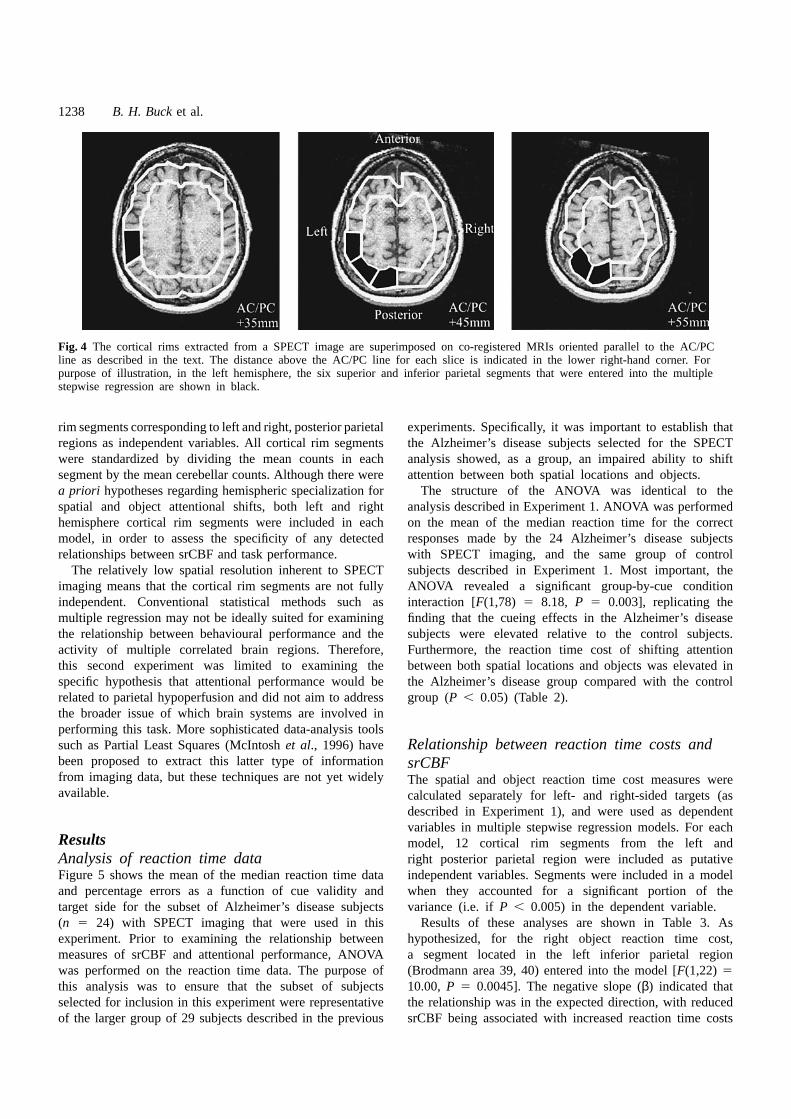
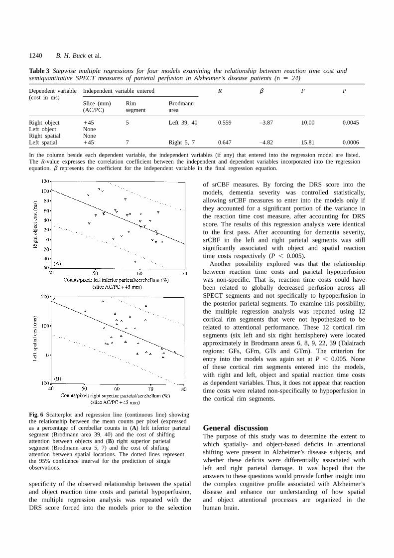
when shifting attention between objects located in the right segments entered into the model for the left spatial reactiontime cost.hemispace (Fig. 6A). The analogous reaction time cost of
detecting left-sided targets was not significant. Alsoconsistent with the hypotheses was that a right superiorparietal segment (Brodmann area 5, 7) entered into the
Specificity of relationship between reaction timemodel for the left spatial reaction time cost. Again theslope was negative, suggesting that hypoperfusion in thiscosts and srCBF
It is possible that the reaction time costs and srCBF ratiosregion of the right superior parietal lobe was associatedwith an impairment in shifting attention between spatial were related to each other only indirectly, due to the fact
that attentional deficits and parieto-temporal hypoperfusionlocations, specifically in the left-hemispace [(F1,22) 515.81, P 5 0.0006] (Fig. 6B). No other cortical rim were both related to dementia severity. To investigate the
1240 B. H. Bucket al.
Table 3 Stepwise multiple regressions for four models examining the relationship between reaction time cost andsemiquantitative SPECT measures of parietal perfusion in Alzheimer’s disease patients (n 5 24)
Dependent variable Independent variable entered R β F P(cost in ms)
Slice (mm) Rim Brodmann(AC/PC) segment area
Right object 145 5 Left 39, 40 0.559 –3.87 10.00 0.0045Left object NoneRight spatial NoneLeft spatial 145 7 Right 5, 7 0.647 –4.82 15.81 0.0006
In the column beside each dependent variable, the independent variables (if any) that entered into the regression model are listed.The R-value expresses the correlation coefficient between the independent and dependent variables incorporated into the regressionequation.β represents the coefficient for the independent variable in the final regression equation.
of srCBF measures. By forcing the DRS score into themodels, dementia severity was controlled statistically,allowing srCBF measures to enter into the models only ifthey accounted for a significant portion of the variance inthe reaction time cost measure, after accounting for DRSscore. The results of this regression analysis were identicalto the first pass. After accounting for dementia severity,srCBF in the left and right parietal segments was stillsignificantly associated with object and spatial reactiontime costs respectively (P , 0.005).
Another possibility explored was that the relationshipbetween reaction time costs and parietal hypoperfusionwas non-specific. That is, reaction time costs could havebeen related to globally decreased perfusion across allSPECT segments and not specifically to hypoperfusion inthe posterior parietal segments. To examine this possibility,the multiple regression analysis was repeated using 12cortical rim segments that were not hypothesized to berelated to attentional performance. These 12 cortical rimsegments (six left and six right hemisphere) were locatedapproximately in Brodmann areas 6, 8, 9, 22, 39 (Talairachregions: GFs, GFm, GTs and GTm). The criterion forentry into the models was again set atP , 0.005. Noneof these cortical rim segments entered into the models,with right and left, object and spatial reaction time costsas dependent variables. Thus, it does not appear that reactiontime costs were related non-specifically to hypoperfusion inthe cortical rim segments.
Fig. 6 Scatterplot and regression line (continuous line) showingthe relationship between the mean counts per pixel (expressedas a percentage of cerebellar counts in (A) left inferior parietal General discussionsegment (Brodmann area 39, 40) and the cost of shifting The purpose of this study was to determine the extent toattention between objects and (B) right superior parietal which spatially- and object-based deficits in attentionalsegment (Brodmann area 5, 7) and the cost of shifting
shifting were present in Alzheimer’s disease subjects, andattention between spatial locations. The dotted lines representwhether these deficits were differentially associated withthe 95% confidence interval for the prediction of single
observations. left and right parietal damage. It was hoped that theanswers to these questions would provide further insight intothe complex cognitive profile associated with Alzheimer’sspecificity of the observed relationship between the spatial
and object reaction time costs and parietal hypoperfusion, disease and enhance our understanding of how spatialand object attentional processes are organized in thethe multiple regression analysis was repeated with the
DRS score forced into the models prior to the selection human brain.
Attentional deficits in Alzheimer’s disease 1241
To examine these issues, covert attentional orienting was The SPECT findings are also consistent with twoprevious studies that examined attentional shifting usingstudied in Alzheimer’s disease patients and control subjects,
using an experimental task, involving the cueing of visual PET (Corbettaet al., 1993; Corbettaet al., 1995). Corbettaet al. (1993) found that Brodmann area 7 was activatedattention prior to the presentation of a target stimulus.
Unlike previous experimental investigations of attentional when attention was shifted between spatial locations. Theright superior parietal lobe showed two distinct foci ofshifting in Alzheimer’s disease, the paradigm used in this
study allowed for the separate examination of the spatially- activation that were differentially related to attentionalshifting in the left and right visual fields, while the leftand object-based components of attentional shifting. It
was hypothesized that the bilateral parietal dysfunction superior parietal lobe showed only a single focus ofactivation related to shifting of attention in the right visualassociated with Alzheimer’s disease would result in
both spatially- and object-based attentional deficits. This field. On this basis, Corbettaet al. (1993) concluded thatwhile both parietal lobes are involved in shifting attentionhypothesis was based on two converging streams of
evidence. First, Eglyet al. (1994) found that unilateral in the contralateral visual field, the right superior parietalregion may be specialized for shifting attention acrossright parietal damage was associated with deficits in shifting
attention to contra-lesional spatial locations contained within spatial locations in both visual fields. In this study, it wasshown that ‘deactivation’ or hypoperfusion in the rightan object, while left parietal damage was associated with
an impairment in shifting attention between objects (Egly superior parietal lobe is correlated with performance deficitsin attentional shifting between spatial locations. In thiset al., 1994). Also, simultagnosia, a complex disorder of
visual perception that includes both spatially- and object- respect, the SPECT imaging study reported in this studycan be viewed as a negative activation paradigm, andbased attentional limitations, is typically found in patients
with bilateral parietal lesions. Consistent with the hypothesis, provides converging evidence to support the view that theright superior parietal lobe is specialized for the spatialin the first experiment, Alzheimer’s disease subjects showed
elevated reaction time costs for invalid cues eliciting shifts orienting of attention. Together with the PET studies ofattention, the present results demonstrate how functionalof attention both between- and within-objects, which
suggests combined spatially- and object-based attentional imaging studies using similar paradigms in normal andbrain-damaged subjects can provide converging evidencedeficits.
In the second experiment, the SPECT scans of 24 for the models relating human brain and cognitive function.To date, there have been no functional imaging studiesAlzheimer’s disease subjects who completed the Egly task
were analysed to derive semi-quantitative indices of parietal showing hemispheric specialization for object-based atten-tional processes. The findings of a recent study of workingdysfunction. In particular, perfusion in superior (Brodmann
areas 5, 7) and inferior (Brodmann area 39, 40) parietal memory for spatial locations and objects, however, providefurther support for the hemispheric specialization hypothesisregions was the focus of this study because previous
human and non-human primate studies (Posneret al., advanced by Eglyet al. (1994). Smith et al. (1995)investigated whether there were separate memory systems1984; Corbettaet al., 1993; Corbettaet al., 1995; Robinson
et al., 1995) have shown these regions to be involved in for spatial and object information. They found a doubledissociation in the regions activated by the spatial andthe shifting of visual attention. If the right and left parietal
regions were differentially specialized for spatially- and object working memory tasks. The spatial task activatedonly right hemisphere regions (occipital, inferior parietalobject-based attentional orienting then, in Alzheimer’s
disease subjects, it was predicted that right parietal and prefrontal areas), whereas the object task activatedprimarily left hemisphere regions (inferotemporal andhypoperfusion would be significantly correlated with
reaction time costs for left-sided targets in the spatial inferior parietal areas). Our results suggest that thisprinciple of left and right hemisphere specialization forcondition, and left parietal hypoperfusion would be
correlated with reaction time costs for right-sided targets spatial and object working-memory buffers may also applyto the networks of brain areas involved in selectivein the object condition. The results were consistent with
these predictions. Multiple regression analyses revealed attention.significant relationships between left spatial reaction timecosts and right superior parietal hypoperfusion (area 5, 7)and right object reaction time costs and left inferior parietal
Relationship of findings to current models ofhypoperfusion (area 39, 40). Both relationships werespecific to targets contralateral to the damaged hemisphereselective attention
Humphreys and Riddoch (1993) have proposed thatand to the parietal regions, and persisted even whendementia severity was controlled. Together, the findings attentional selection results from the interaction of orienting
and maintenance mechanisms that are respectively spatially-support the view that regions of the right and left posteriorparietal lobes are specialized for shifting attention away and object-based. Orienting is a spatial operation that
directs attention to locations of potential significance, whilefrom previously selected spatial locations and objects,respectively. the maintenance mechanism is object-based and selects
1242 B. H. Bucket al.
whole objects rather than regions of space for enhanced proposal of two types of spatially structured representationsand further suggest that these representations are coded inprocessing. Humphreys and Riddoch (1993) further suggest
that deficits in detecting invalidly cued targets result from parallel by the left and right parietal region. Thus, inAlzheimer’s disease patients, deficits in shifting attentiondisruptions in the balance between the orienting and
maintenance systems. Problems in disengaging attention within a rectangle may have correlated with right parietaldamage because this brain region is involved in codingcould stem from two types of deficits: (i) damage to the
orienting system could reduce the ability of targets within-object representations. Furthermore, deficits inshifting attention between rectangles may have correlatedappearing at uncued locations to attract attention normally
resulting in a spatial-based deficit, or (ii) the orienting with left parietal dysfunction due to deficient coding ofthe between-object representations.system could be abnormally inhibited by the maintenance
system resulting in the hypermaintenance of attention oncued objects.
The results reported in this study support the view ofConclusionsThese results suggest that performance on computerizedHumphreys and Riddoch (1993) that separate spatially-
and object-based deficits may underlie impairments in tests of attentional orienting can be related to the locationand severity of brain dysfunction in Alzheimer’s disease.disengaging attention. Right parietal damage in Alzheimer’s
disease subjects was significantly correlated with the Locasioet al. (1995) recently studied cognitive deficits inAlzheimer’s disease to determine which cognitive testsimpairment in detecting invalidly cued targets in both the
cued and uncued rectangle. In terms of the Humphreys were best for detecting Alzheimer’s disease, stagingAlzheimer’s disease and tracking disease progression. Theyand Riddoch model, damage to the right parietal lobe
could have disrupted the spatial-based orienting mechanism found that tests of explicit memory were best suitedfor detecting Alzheimer’s disease due to the earlyso that invalidly cued targets were unable to elicit
attentional orienting to new (uncued) locations, regardless mediotemporal lobe pathology in the majority of cases.All cognitive tests were, however, much less effective atof the object properties associated with the target location.
In contrast, left parietal damage was found to be correlated staging illness. The lack of effectiveness of cognitive testsfor staging dementia reflects the individual variability inwith the impaired detection of invalidly cued targets,
specifically those appearing in the uncued rectangle. Unlike the anatomical distribution of pathology. The spread oflesions to the parietal and frontal lobes is variable andthe right parietal deficit, the deficit associated with left
parietal damage was sensitive to the object properties of often asymmetric between subjects. Consequently, Locasioet al. (1995) recommended that the progression of diseasethe visual display. With respect to the Humphreys and
Riddoch model, this object-based attentional deficit could be monitored with a range of cognitive tests that reflectthe underlying spread of neuronal dysfunction associatedarise from damage to the maintenance mechanism, leading
to the hypermaintenance of attention on cued objects. The with Alzheimer’s disease. Accordingly, tests of visualattention, like the experimental tasks used in this study,results of the present study, when interpreted in the
Humphreys and Riddoch framework, suggest that the right could be combined with other tests of visuospatial andlanguage functions to chart the course of the disease, andsuperior parietal lobe may be involved in the spatial
orienting of attention while the left inferior parietal lobe may also be useful for monitoring the outcome oftherapeutic trials that claim to slow the progression ofmay be involved in the maintenance of attentional selection
on objects of current interest. the disease.Another implication of this study is that Alzheimer’sFinally, in the present study the terms spatially- and
object-based have been applied to the right and left parietal disease combined with functional imaging methodologiescan provide cognitive neuroscience with a useful modelattentional mechanisms and the corresponding deficits.
Recent studies suggest that this terminology may be for exploring brain–behaviour relationships. The braindamage, especially in early Alzheimer’s disease, issomewhat inaccurate, as the representation accessed by
both attentional mechanisms may be spatially structured topographically selective, but still shows tremendousindividual variability. This topographical variability may(Vecera, 1994; Humphreys and Riddoch, 1995). For
example, in a single-case study of a patient with be indexed with brain imaging techniques such as SPECT.The variability in pathology will result in selective cognitivebilateral parietal lesions, Humphreys and Riddoch (1995)
demonstrated neglect on the left or right side depending deficits. By combining brain-imaging techniques with testsof cognitive performance it is possible to relate deficits inon whether the visual stimuli were encoded as parts of a
single perceptual object or as separate perceptual objects. cognitive performance to regional brain dysfunction. Theresults of this and other recent studies (Pennielloet al.,This finding led the authors to suggest that right and left
parietal lobes construct separate parallel representations of 1995) suggest that imaging studies of Alzheimer’s diseasecan complement both neuropsychological investigations andthe relations between parts of single objects (within-object)
and between separate objects (between-object). The results PET-activation studies of healthy individuals, in theinvestigation of brain–behaviour relationships.of this study support Humphreys and Riddoch’s (1995)
Attentional deficits in Alzheimer’s disease 1243
Kornblum S, editors. Attention and performance XIV. CambridgeAcknowledgements(MA): MIT Press, 1993: 143–62.We wish to thank Drs R. McIntosh, A. Sekuler, M.-L.
Smith and J. Shedden for their comments on the M.Sc.Humphreys GW, Riddoch MJ. Separate coding of space withinthesis upon which this work was based. We also wish toand between perceptual objects: evidence from unilateral visual
neglect. Cogn Neuropsychol 1995; 12: 283–311.thank Ms J. Lawrence, Ms J. Pawsey-Corson and Ms KiraBarbour for their assistance with subject recruitment, andJagust WJ, Reed BR, Ellis WG, Eberling JL, Budinger TF.Mr F. Leibovitch for performing the SPECT reconstructions.Single-photon emission computed tomographic perfusion imagingThis research was supported by grants from the Medicalin autopsy-diagnosed dementia. J Neuroimaging 1993; 3: 93–9.Research Council of Canada to S.E.B., C.C., M.J.B.; from
Keppel G, Zedeck S. Data analysis for research designs. Newthe Ontario Mental Health Foundation to S.E.B. and M.B.;York: W.H. Freeman, 1989.and an Ontario Graduate Scholarship to B.H.B.Kumar A, Schapiro MB, Grady C, Haxby JV, Wagner E, SalernoJA, et al. High-resolution PET studies in Alzheimer’s disease.Neuropsychopharmacology 1991; 4: 35–46.References
Arriagada PV, Growdon JH, Hedley-Whyte ET, Hyman BT. Lezak MD. Neuropsychological assessment. 3rd ed. New York:Neurofibrillary tangles but not senile plaques parallel durationOxford University Press, 1995.and severity of Alzheimer’s disease. Neurology 1992; 42: 631–9.
Locascio JJ, Growdon JH, Corkin S. Cognitive test performanceBaynes K, Holtzman JD, Volpe BT. Components of visual in detecting, staging, and tracking Alzheimer’s disease. Archattention. Alterations in response pattern to visual stimuli followingNeurol 1995; 52: 1087–99.parietal lobe infarction. Brain 1986; 109: 99–114.
Mangun GR, Hillyard SA. The spatial allocation of visualCorbetta M, Miezin FM, Shulman GL, Petersen SE. A PET attention as indexed by event-related brain potentials. Humstudy of visuospatial attention. J Neurosci 1993; 13: 1202–26. Factors 1987; 29: 195–211.
Corbetta M, Shulman GL, Miezin FM, Petersen SE. SuperiorMaruff P, Malone V, Currie J. Asymmetries in the covert orientingparietal cortex activation during spatial attention shifts and visualof visual spatial attention to spatial and non-spatial cues infeature conjunction. Science 1995; 270: 802–5. Alzheimer’s disease. Brain 1995; 118: 1421–35.
Egly R, Driver J, Rafal RD. Shifting visual attention between Mattis S. Mental status examination for organic mental syndromeobjects and locations: evidence from normal and parietal lesionin the elderly patient. In: Bellak L, Karasu TB, editors. Geriatricsubjects. J Exp Psychol Gen 1994; 123: 161–77. psychiatry: a handbook for psychiatrists and primary care
physicians. New York: Grune and Stratton, 1976: 77–121.Eriksen CW, St James JD. Visual attention within and aroundthe field of focal attention: a zoom lens model. Percept PsychophysMcIntosh AR, Bookstein FL, Haxby JV, Grady CL. Spatial1986; 40: 225–40. pattern analysis of functional brain images using partial least
squares. Neuroimage 1996; 3: 143–57.Farah MJ. Visual agnosia: disorders of object recognition andwhat they tell us about normal vision. Cambridge (MA): MIT McKhann G, Drachman D, Folstein M, Katzman R, Price D,Press, 1990. Stadlan EM. Clinical diagnosis of Alzheimer’s disease: report of
the NINCDS-ADRDA Work Group under the auspices ofFolstein MF, Folstein SE, McHugh PR. ‘Mini-mental state’. ADepartment of Health and Human Services Task Force onpractical method for grading the cognitive state of patients forAlzheimer’s Disease. Neurology 1984; 34: 939–44.the clinician. J Psychiatr Res 1975; 12: 189–98.
Mesulam MM. Large-scale neurocognitive networks and distributedHachinski VC, Iliff LD, Zilkha E, du Boulay GH, McAllisterprocessing for attention, language, and memory. [Review]. AnnVL, Marshall J, et al. Cerebral blood flow in dementia. ArchNeurol 1990; 28: 597–613.Neurol 1975; 32: 632–7.
Minoshima S, Frey KA, Foster NL, Kuhl DE. Preserved pontineHaxby JV, Duara R, Grady CL, Cutler NR, Rapoport SI. Relationsglucose metabolism in Alzheimer disease: a reference region forbetween neuropsychological and cerebral metabolic asymmetriesfunctional brain image (PET) analysis. J Comput Assist Tomogrin early Alzheimer’s disease. J Cereb Blood Flow Metab 1985;1995; 19: 541–7.5: 193–200.
Morecraft RJ, Geula C, Mesulam MM. Architecture of connectivityHellman RS, Tikofsky RS, Collier BD, Hoffmann RG, Palmerwithin a cingulo-fronto-parietal neurocognitive network for directedDW, Glatt SL, et al. Alzheimer disease: quantitative analysis ofattention. Arch Neurol 1993; 50: 279–84.[123I]iodoamphetamine SPECT brain imaging. Radiology 1989;
172: 183–8.Morrow LA, Ratcliff G. The disengagement of covert attentionand the neglect syndrome. Psychobiology 1988; 16: 261–9.Hohne KH, Hanson WA. Interactive 3D segmentation of MRI
and CT volumes using morphological operations. J Comput AssistMountcastle VB, Motter BC, Steinmetz MA, Sestokas AK.Tomogr 1992; 16: 285–94.Common and differential effects of attentive fixation on theexcitability of parietal and prestriate (V4) cortical visual neuronsHumphreys GW, Riddoch MJ. Interactions between object and
space systems revealed through neuropsychology. In: Meyer DE, in the macaque monkey. J Neurosci 1987; 7: 2239–55.
1244 B. H. Bucket al.
Parasuraman R, Greenwood PM, Haxby JV, Grady CL. Visuospatial Minoshima S. Spatial versus object working memory: PETinvestigations. J Cogn Neurosci 1995; 7: 337–56.attention in dementia of the Alzheimer type. Brain 1992; 115:
711–33. Stapleton SJ, Caldwell CB, Ehrlich LE, Leonhardt C, Black SE,Yaffe MJ. A quantitative method of analyzing99mTc-HMPAOParasuraman R, Haxby JV. Attention and brain function in[abstract]. Med Phys 1992; 19: 780.Alzheimer’s disease: a review. Neuropsychology 1993; 7: 242–72.Stevens J. Applied multivariate statistics for the social sciences.
Penniello MJ, Lambert J, Eustache F, Petit-Taboue MC, BarreHillsdale (NJ): Lawrence Erlbaum, 1986.L, Viader F, et al. A PET study of the functional neuroanatomy
Talairach J, Tournoux P. Co-planar stereotaxic atlas of the humanof writing impairment in Alzheimer’s disease: the role of thebrain: 3-dimensional proportional system: an approach to cerebralleft supramarginal and left angular gyri. Brain 1995; 118: 697–706.imaging. Stuttgart: Thieme, 1988.
Posner MI. Orienting of attention. Q J Exp Psychol 1980; 32: Terry RD, Masliah E, Hansen LA. Structural basis of the2–25. cognitive alterations in Alzheimer disease. In: Terry RD, Katzman
R, Bick KL, editors. Alzheimer disease. New York: Raven Press,Posner MI, Dehaene S. Attentional networks. [Review]. Trends1994: 179–96.Neurosci 1994; 17: 75–9.Vanier M, Lecours AR, Ethier R, Habib M, Poncet M, Milette
Posner MI, Walker JA, Friedrich FJ, Rafal RD. Effects of parietal PC, et al. Proportional localization system for anatomicalinjury on covert orienting of attention. J Neurosci 1984; 4: interpretation of cerebral computed tomograms. J Comput Assist1863–74. Tomogr 1985; 9: 715–24.
Vecera SP. Grouped locations and object-based attention: commentRafal RD, Posner MI. Deficits in human visual spatial attentionon Egly, Driver, & Rafal (1994). J Exp Psychol Gen 1994; 123:following thalamic lesions. Proc Natl Acad Sci USA 1987; 84:316–20.7349–53.
Waldemar G, Bruhn P, Kristensen M, Johnsen A, Paulson OB,Robb RA. Visualization methods for analysis of multimodalityLassen NA. Heterogeneity of neocortical cerebral blood flowimages. In: Thatcher RW, Hallett M, Zeffiro T, Roy John E,deficits in dementia of the Alzheimer type: a [99mTc]-d,l-HMPAOHuerta M, editors. Functional neuroimaging. Technical foundations.SPECT study. J Neurol Neurosurg Psychiatry 1994; 57: 285–95.San Diego: Academic Press, 1994: 181–90.Woods RP, Mazziotta JC, Cherry SR. MRI-PET registration with
Robinson DL, Bowman EM, Kertzman C. Covert orienting of automated algorithm. J Comput Assist Tomogr 1993; 17: 536–46.attention in macaques. II. Contributions of parietal cortex. JNeurophysiol 1995; 74: 698–712.
Received September 4, 1996. Revised January 4, 1997.Accepted February 2, 1997Smith ES, Jonides J, Koeppe RA, Awh E, Schumacher EH,
Copyright © 2022 FDOKUMEN