The Neuropsychology of Male Adults With High-Functioning Autism or Asperger Syndrome

Resting SPECT-neuropsychology correlation in very mild
Alzheimer’s disease
Flavio Nobilia,*, Andrea Brugnoloa, Piero Calvinic, Francesco Copellod, Caterina De Leoa,Nicola Girtlera, Silvia Morbellib, Arnoldo Piccardob, Paolo Vitalia, Guido Rodrigueza
aSection of Clinical Neurophysiology (DISEM), Department of Endocrinological and Metabolic Sciences,
University of Genoa, Viale Benedetto XV, 6, I-16132 Genoa, ItalybSection of Nuclear Medicine, Department of Internal Medicine, University of Genoa, Genoa, Italy
cINFM, Department of Physics, University of Genoa, Genoa, ItalydSection of Preventive Medicine, S. Martino Hospital, Genoa, Italy
Accepted 2 September 2004
Available online 7 October 2004
Abstract
Objective: To investigate the relationships between brain function and some of the most frequently impaired cognitive domains in the first
stages of Alzheimer’s disease (AD), we searched for correlation between the scores on 3 neuropsychological tests and brain perfusion,
assessed by single photon emission computed tomography (SPECT) in patients with very mild AD.
Methods: Twenty-nine consecutive outpatients (mean age 78.2G5.5) affected by probable AD in the very mild phase (i.e. with a score
R20 on the mini-mental state examination, MMSE) underwent brain SPECT with 99mTc-ethylcisteinate dimer. For correlative purposes,
word list learning (by the selective reminding test, SRT), constructional praxis test (CPT) and visual search test (VST) were chosen a priori
out of an extended battery employed to diagnose AD at first patient evaluation. Voxel-based correlation analysis was achieved by statistical
parametric mapping (SPM99) with a height threshold of PZ0.005. Age, years of education and the MMSE score were inserted in the
correlative analysis as confounding variables.
Results: The SRT score showed correlation with brain perfusion in 3 clusters of the left hemisphere, including the post-central gyrus, the
parietal precuneus, the inferior parietal lobule and the middle temporal gyrus, and in one cluster in the right hemisphere including the middle
temporal gyrus and the middle occipital gyrus. The CPT score was significantly correlated with brain perfusion in the parietal precuneus and
the posterior cingulate gyrus in the left hemisphere, whereas the VST score gave a significant correlation with brain perfusion in a left cluster
including the parietal precuneus and the superior temporal gyrus.
Conclusions: Cognitive impairment in very mild AD is reflected by brain dysfunction in posterior associative areas, with peculiar
topographical differences proper of each domain. The parietal precuneus was a common site of correlation of all 3 neuropsychological tests.
This region, together with the posterior cingulate and the superficial posterior temporal–parietal cortex, is thought to be affected by
disconnection from the mesial temporal lobe, besides being directly affected by increased oxidative stress and by atrophy as well. The
impairment of these areas is thought to contribute to cognitive decline in verbal memory, constructional praxis and visual sustained attention
which are indeed among the earliest signs of cognitive impairment in AD.
Significance: Assessing the relationships between neuropsychology and brain functional imaging is a key approach to clarify the
pathophysiology of cognitive failure in AD; the specificity of these findings in AD remains to be proven through comparison with correlation
achieved in matched controls.
q 2004 International Federation of Clinical Neurophysiology. Published by Elsevier Ireland Ltd. All rights reserved.
Keywords: Alzheimer’s disease; Brain SPECT; Neuropsychology; SPM; Verbal memory; Constructional apraxia; Sustained attention
1388-2457/$30.00 q 2004 International Federation of Clinical Neurophysiology.
doi:10.1016/j.clinph.2004.09.001
* Corresponding author. Tel.: C39 010 3537568; fax: C39 010 5556893.
E-mail address: [email protected]
(F. Nobili).
1. Introduction
Both brain perfusion single photon emission computed
tomography (SPECT) and neuropsychological assessment
Clinical Neurophysiology 116 (2005) 364–375
www.elsevier.com/locate/clinph
Published by Elsevier Ireland Ltd. All rights reserved.

F. Nobili et al. / Clinical Neurophysiology 116 (2005) 364–375 365
have been proven to be sensitive tools to assess the
functional deficit in the early stages of Alzheimer’s disease
(AD) (Arnaiz and Almkvist, 2003; Devous, 2002). Positron
Emission Tomography (PET) is also increasingly used,
yielding measurement of glucose utilization with a better
spatial resolution but at a consistently higher cost than
SPECT. SPECT and PET provide a topographic picture of
brain function and allow speculations on functional
connectivity among distant areas. On the other hand,
neuropsychology detects impairment in cognitive domains
by specific tasks and gives the experimental material to
design models of cognitive derangement in AD.
Two main approaches are used to investigate the brain
perfusion/metabolic substrates of cognitive performances.
One is mainly aimed to find out those brain areas that are
involved during the performance of a cognitive task in AD
patients in comparison with controls. From these ‘acti-
activation’ studies, that are especially carried out by PET
and by functional magnetic resonance imaging (fMRI),
information is drawn on putative circuitry that is activated in
AD patients while performing the task (Prvulovic et al.,
2002). The great advantage of activation studies is in
imaging the working brain and thus gaining information on
possibly alternative activity/inactivity of brain regions in
AD. The most serious drawbacks are the sophisticated
methodology and statistics required (making these studies
prone to artefacts), the high compliance required in subjects
(often hard to reach in AD patients) and the need of an a
priori cognitive model to interpret images (Shallice, 2003).
Very recently, the current way of interpretation of
‘activated’ areas in this provocative approach has been
seriously questioned (Sidtis et al., 2003).
The other investigation method is to search for
relationships between the scores on neuropsychological
tests applied to the patients either ad-hoc or while
performing the diagnosis and perfusion/metabolic scans
obtained at rest. This ‘resting’ method leads to understand
how much and where two sound tools to study brain
functional failure are correlated, allowing speculations on
the early involved sites, on how their impairment is reflected
by cognitive failure and possibly on the task that better
represents the brain impairment. The most obvious
advantages are that data is already available in a modern
memory clinic (thus not requiring extra costs to design ad-
hoc investigations) and that patient compliance is generally
good, especially when cognition is still mildly impaired.
The main limitation is that the two examinations are not
simultaneous and thus explore different moments of what
can well be a fluctuating cognitive state.
The two approaches yield very different information that
cannot be joined or directly compared, at least until a
complete theory of cognition is applied (Shallice, 2003). On
the other hand, both approaches are very useful to improve
our understanding of the disease’s pathophysiology from its
beginning.
A number of studies have investigated the relationships
between neuropsychological test scores and brain perfu-
sion/metabolism at rest (Bartenstein et al., 1997; Burns
et al., 1989; Desgranges et al., 1998; Elgh et al., 2002;
Goldenberg et al., 1989; Hirono et al., 2001; Montaldi et al.,
1990; O’Brien et al., 1992). However, most of them suffer
from several limitations. First, AD patients are examined
together, especially in early studies, irrespectively of the
severity of cognitive impairment and in some cases the
severity of the disease is not reported at all (Burns et al.,
1989; Goldenberg et al., 1989). In fact, it is now known that
both the cognitive profile and the perfusion/metabolic
deficits change substantially with the increasing severity
of the disease (Matsuda et al., 2002). Second, several studies
(Bartenstein et al., 1997; Burns et al., 1989; O’Brien et al.,
1992) have employed a correlative analysis between the
subscores of a general battery, such as those of the CAM-
COG, the ADAS-COG, or the MMSE scales, which are
often represented by too few points within each subscore to
allow meaningful linear correlations. Third, methods of
analysis are almost always based on Regions (ROIs) or
Volumes of Interest (VOIs) in SPECT/PET images, thus
inevitably missing a number of brain regions, or considering
in the same ROI/VOI a peculiar anatomical rather than a
functional region. It is especially noteworthy that mesial
brain regions, such as those of the temporal and parietal
lobes which are now known to be so relevantly impaired at
the beginning of the disease, were often not included in the
choice of ROIs or VOIs (O’Brien et al., 1992).
The aim of the present investigation is to assess the
correlation between the scores on specific neuropsycholo-
gical tests and brain perfusion SPECT on a voxel-based
basis in a consecutive series of patients with very mild AD,
taking into account the severity of general cognitive
impairment. The scores on word-list learning, construc-
tional praxis and visual sustained attention were chosen
among a wide standardized battery administered to patients
at the time of their first evaluation. In fact, impairment in
verbal learning is a well known typical hallmark of early
AD, constructional apraxia has been increasingly found in
the mild stages (Fisher et al., 1999), while sustained
attention is a ‘background’ function underlying all the
other tasks (Tales et al., 2002, 2004).
2. Methods
2.1. Patients
During a 1 year period, all the consecutive outpatients with
probable AD (according to the definition of the NINCDS-
ADRDA Work Group) (McKhann et al., 1984) in the early or
very mild stage of the disease (i.e. scoring 20 or higher on the
mini-mental state examination (MMSE)) (Folstein et al.,
1975) who came to our Centre for a first diagnostic evaluation
were considered eligible for this study.

F. Nobili et al. / Clinical Neurophysiology 116 (2005) 364–375366
All the patients underwent a complete diagnostic work-
up according to current standards, which include general
and neurological examinations, a standardized neuropsy-
chological assessment, basal computed tomography or
magnetic resonance imaging and other routine blood and
urine screening investigations to rule out secondary
dementias.
The presence of previous or present major psychiatric
disorders, serious neurological diseases, severe and uncon-
trolled arterial hypertension, diabetes mellitus, renal,
hepatic or respiratory failure, anaemia (Hb level!10 mg/dl) or malignancies were exclusion criteria.
In conformity with the diagnosis of AD, all patients
scored lower than 4 on the Hachinski ischaemic scale.
Lewy-body dementia, frontotemporal dementia and vascu-
lar dementia were excluded in all patients on the basis of
current clinical criteria (McKeith et al., 1999; Roman et al.,
1993; The Lund and Manchester Criteria, 1994).
All patients underwent brain perfusion SPECT examin-
ation by 99mTc-N,N 00-1,2-ethylene diylbis-L-cysteine diethyl
ester dihydrochloride (ECD) as part of the routine
diagnostic procedure in our Centre. The original group
was composed of 31 patients, but two left-handed patients
(Annett’s Questionnaire) were excluded. The study group
thus included 29 patients (23 women and 6 men), aged
66–86 years (mean 78.2G5.5), with years of full education
ranging from 3 to 17 (mean 7.2G4.2).
All the patients were at their first diagnostic evaluation
for cognitive complaints, thus none of them were under
treatment with Acetylcholinesterase inhibitors, nor with
other neuropsychoactive drugs. The patients (or their
relatives) were informed about the finalities of the study
and gave their informed consent.
2.2. Brain SPECT
Brain SPECT was performed employing a single-head
gamma-camera (Apex SPX-4, Elscint, Israel) equipped by a
low-energy, high-resolution collimator. All SPECT acqui-
sitions started between 30 and 120 min after the i.v.
injection of about 1000 MBq of 99mTc-ECD (Neurolite,
Dupont Pharma, USA), according to the guidelines of the
European Society of Nuclear Medicine (Tatsch et al., 2002).
A step-and-shoot acquisition protocol acquired images by
120 projections, with a radius of rotation !15 cm. Total
counts were between 7 and 10 million. SPECT images were
3-dimensionally (3D) reconstructed by the conjugate
gradient (CG) algorithm, a technique initially proposed in
a 2-D version (Formiconi et al., 1989). A 3D version has
been developed by means of the 2DC1 algorithm (Boccacci
et al., 1999), according to the procedure described else-
where (Nobili et al., 2001). The number of iterations was
between 13 and 15, depending on the number of counts
collected in the data. The 2DC1 version of the CG method
used in this study yielded a reconstruction consisting of
stacks of 64 transaxial slices, where the compensation for
attenuation had already been performed at the reconstruc-
tion stage by the iterative algorithm itself. In this case, the
attenuation contour was directly given by the skull contour
and the related anatomical shapes were carefully taken into
account. The CG reconstruction method has already been
employed in clinical studies on dementia, allowing better
spatial resolution and correlative results than the conven-
tional filtered back-projection method (Nobili et al., 2001;
Rodriguez et al., 2000).
2.3. Neuropsychological variables
Patients were carefully screened with the synoptic
batteries of the MMSE and of the CAM-COG battery
(short Italian version) (Neri et al., 1994). The specific
domains of sustained visual attention, verbal learning and
recall, language, abstract reasoning and constructional
praxis were assessed by a standardized battery in use at
our Center. The presence of behavioural and psychological
disturbances was assessed by a structured interview to the
principal caregiver by means of the neuropsychiatric
inventory.
The following neuropsychological tests were chosen a
priori out of the whole battery because they explore
cognitive domains that are typically and sometimes
alternatively impaired in the first stages of AD (verbal
memory and constructional praxis), or concern aspects of
cognition that are a common background for the execution
of several other tasks, such as visual sustained attention.
(i)
The Buschke–Fuld selective reminding test (SRT), inits short version (Masur et al., 1989), is a word-list
learning of 12 words and 6 repetition trials. For the
purpose of this study, only the total number of words
recalled was considered, since delayed recall showed a
clear ground effect, leading to a consistent number of 0
scores. Abnormally low scores were identified accord-
ing to the normative values reported by Masur et al.
(1989);
(ii)
the constructional praxis test (Carlesimo et al., 1996;Gainotti et al., 1977); only the subtest with planning
elements was considered for further analysis, since the
subtest of freehand copying yields a narrow score range
not suitable for linear correlative purposes. This subtest
requires the completion of 2 bi-dimensional and 8 tri-
dimensional figures, of which some parts have been
omitted. The items range from 2 to 10 for each figure
(maximum score 70) (Fig. 1), and abnormal values
were computed according to the reference literature
(Carlesimo et al., 1996);
(iii)
the visual search test (VST), which assesses sustainedattention, in the Italian validated version (Spinnler and
Tognoni, 1987). Three matrices of 110 (10!11)
numbers each, are presented in sequence to the patient
who must identify and mark the number 5 in the first
matrix (10 items), the numbers 2 and 6 in the second

Fig. 1. Two (a cube and a house, both with a tri-dimensional view) of the 10
figures that must be completed during the constructional praxis test.
F. Nobili et al. / Clinical Neurophysiology 116 (2005) 364–375 367
one (20 items) and the numbers 1, 4 and 9 in third one
(30 items). The score is formed by the sum of correct
items identified in 45 s of time for each of the 3
matrices (maximum score 60). The Italian normative
database (Spinnler and Tognoni, 1987) was used to
recognize pathological values.
Verbal learning is recognized as one of the first specific
memory complaints in AD, already detectable in prede-
mentia AD, such as mild cognitive impairment (Petersen et
al., 1999), and the SRT is widely employed in this context.
Constructional apraxia seems to be a somewhat later event
in a number of patients, but it is often the first cognitive
domain to be impaired in AD, besides memory (Fisher et al.,
1999). Finally, sustained visual attention is increasingly
recognized as one of the early deficits in AD (Baddeley et
al., 2001; Foster, 2001; Perry et al., 2000; Tales et al., 2002).
Category fluency verbal (CFVT) was tested for colours,
animals, fruits and towns in four 2 min periods. Letter
fluency verbal (LFVT) required three 1 min items, including
the letters F, L and P, according to the Italian normative
(Capitani et al., 1998). Abstract reasoning was evaluated by
Weigl’s sorting test (WST) which assesses the ability to
categorize 12 coloured items of different shape, colour,
thickness, dimension each engraved with card suits.
As for the 3 tests chosen for correlative analysis, scores
for CFVT, LFVT and WST were evaluated on the basis of
the Italian normative values, which require correction for
age, education and comparison with normal subjects.
Reference normal values are reported by Spinnler and
Tognoni (1987) for both the WST and the CFVT, and by
Capitani et al. (1998) for LFVT. Range values, individual
values, means and SDs for all the neuropsychological
tests performed in the present investigation are reported in
Table 1.
The correlation study between functional imaging and
other neuropsychological tests or scales would be of great
relevance to understand the pathophysiology of cognitive
failure in AD. However, performing more than 3 correlation
analyses seemed unjustified because of the dimension of the
present patient sample. Neuropsychological tests and
SPECT were performed within an interval of 3 months
(mean interval time 38 daysG16).
2.4. Statistical analysis
Correlation between the SPECT data and the neurop-
sychological scores were computed by statistical para-
metric mapping (SPM) (Friston et al., 1991), 1999 version
(SPM99), following the rules of the Consensus of the
European Alzheimer’s Disease Consortium (EADC)
(Frisoni et al., 2003). In a preprocessing step, data
sets were spatially normalized using a 12 point linear
affined transformation into Talairach space (Talairach and
Tournoux, 1988) and smoothed with an isotropic Gaussian
kernel of FWHM 12 mm. SPM99 co-registers the
individual SPECT with the 152 brains average of the
Montreal Neurological Institute. Since this template does
not completely match the Talairach brain, a correction of
the SPM{t} coordinates is needed. This was achieved
using the subroutine implemented by Matthew Brett
(http://www.mrc-cbu.cam.ac.uk/Imaging) which gives the
correspondence between SPM and the Talairach atlas
coordinates. The grey matter threshold was set at 0.8.
Normalization of global CBF to 50 was performed with
proportional scaling. The resulting set of values for
comparison constituted a statistical parametric map of
the statistic SPM{t}. Then, the SPM{t} maps were
transformed to the unit of normal distribution (SPM{z})
and reached a threshold at PZ0.005, which is an accepted
procedure in a clinical setting (Desgranges et al., 1998).
Because of the lack of any topographic a priori
hypothesis, the significance of identified regions was
assessed using P values corrected for multiple compari-
sons (P!0.05 was the first statistical significance to be
accepted at the cluster level).

Table 1
Neuropsychological test data in 29 outpatients with very mild Alzheimer’s disease
Patient no. Sex Age
yrs
Education
yrs
Short
CAM-COG
MMSE WST VST CFVT LFVT SRT CPs CPpe
Range 66–86 3–17 0–62 0–30 0–15 0–60 0–72 0–12 0–70
1 F 80 8 39 24a 10 50 10.67 14 29a 6a 68
2 F 83 5 40 29 3a 57 7.67 14.5 39 5a 60
3 F 86 5 27a 20a 8 46 4.67a 8.5 27a 5a 52a
4 F 81 4 21a 22a 2a 38 5.67a 9 22a 8 70
5 F 79 6 24a 23a 4 39 7.33 10.5 25a 5a 52a
6 F 81 5 26a 21a 6 44 3.0a 9.25 20a 5a 36a
7 F 72 5 26a 24a 4 32 3.0a 7 21a 8 70
8 F 79 5 14a 21a 1a 39 3.33a 7 27a 4a 56a
9 F 74 5 41 27 9 30a 15.33 13.25 29a 10 70
10 F 76 5 22a 20a 3a 55 6.67 11.25 22a 5a 55a
11 M 81 5 30a 27 5 45 8.0 6.5 25a 6a 60
12 M 78 17 33 24a 12 51 7.33 9a 26a 9 68
13 F 66 5 33 26 6 40 4.67a 13.25 21a 3a 24a
14 F 66 5 21a 20a 1a 26a 2.67a 8.25 19a 6a 52a
15 M 85 17 25a 20a 5a 36 8.67 12 18a 9 68
16 F 86 4 34 26 10 47 4.33a 13.5 29a 9 57a
17 F 79 6 37 26 4a 31 4.33a 10.5 24a 6a 58a
18 M 78 8 30 23a 6 34 2.33a 7.75 19a 2a 28a
19 F 73 5 39 29 10 45 11.33 14 25a 3a 60a
20 F 82 10 30 22a 5a 51 13.67 19.75 29a 7 68
21 F 81 6 21a 21a 6 48 7.0 9.5 25a 3a 55a
22 F 73 13 22a 20a 3a 36 7.67 12.5 26a 5a 49a
23 F 82 5 24a 22a 7 26a 5.0a 7.25 25a 5a 65
24 M 78 17 39 28 9 38 6.67 8.5a 10a 4a 67
25 F 82 5 36 25 7 51 4.67a 11 26a 6a 58a
26 M 72 13 47 28 13 30a 16.33 18.5 37 7a 69
27 F 86 3 22a 20a 3 51 4.0a 9.5 26a 5a 46a
28 F 70 3 32 26 10 48 9.0 16 22a 7 52a
29 F 80 8 45 27 10 47 11.0 14 29a 6 60a
Mean 23 F 78.24 7.17 30.34 23.83 6.28 41.76 7.1 11.22 24.9 5.83 57.0
SD 6 M 5.52 4.17 8.29 3.06 3.29 8.77 3.73 3.42 5.55 2.0 11.96
WST, Weigl’s sorting test; VST, visual search test; CFVT, category fluency verbal test; LFVT, letter fluency verbal test; SRT, selective reminding test; CPs,
constructional praxis test, simple (freehand copying of drawings); CPpe, constructional praxis test, with planning elements (copying drawings with landmarks).a Abnormally low value in comparison with normative database of the literature (see text in the Methods section for further details).
F. Nobili et al. / Clinical Neurophysiology 116 (2005) 364–375368
The correlative analysis was applied to SPECT examin-
ations by means of the ‘multi-subjects: co-variate only’
option. The co-variates were the scores on the selective
reminding test, constructional praxis with planning
elements and Visual search test. Age and years of education
were inserted as ‘nuisance’ variables, to account for their
confounding effect on both perfusion data (Salmon et al.,
2000) and neuropsychological scores. The MMSE score was
inserted as the third ‘nuisance’ variable to take into account
the degree of global cognitive impairment even in this
selected series of patients with very mild AD. The SPM
contrastC1 was defined in each correlation to search for
direct (positive) correlation.
3. Results
In this group of 29 patients with very mild AD, the
clinical dementia rating was 0.5 in 11 patients and 1 in 18
patients, whereas the MMSE score ranged from 20 to 29
(mean 23.8G3.1).
Table 1 reports the details of results of neuropsycholo-
gical tests. Briefly, all the patients showed an impairment in
at least two tests. The SRT was impaired in 37 patients
(94.9%), the CPT (freehand copy) in 20 (69%), CPT with
planning elements in 17 (58.6%), and VST in 4 (13.8%).
Among the other tests, the CFVT was altered in 13 patients
(44.8%), the WST in 8 patients (27.6%), and LFVT in 2
(6.9%) patients.
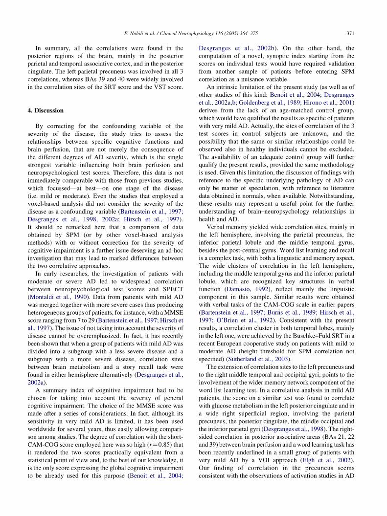
Table 2 reports in detail the sites of significant SPM
correlation between neuropsychological scores and brain
perfusion SPECT. The SRT score gave 3 clusters of
significant correlation in the left hemisphere and one cluster
in the right hemisphere. The 3 left-side clusters included the
post-central gyrus (Brodmann area, BA 3), the parietal
precuneus (BA 39), the inferior parietal lobule (BA 40) and
the middle temporal gyrus (BAs 39 and 21). The right
cluster gave less significant correlation at voxel level and
involved the middle temporal (BA 39) and occipital (BA 19)
gyri (Table 2; Fig. 2).
The constructional praxis score results of correlation
were significant in one cluster involving the left parietal

Table 2
Numerical results of SPM direct correlations (height threshold PZ0.005)
Correlation Cluster level Voxel level
Cluster
extent
Corrected
P value
Cortical region Z score of
maximum
Talairach
coordinates
Cortical region BA
Selective reminding test 861 0.05 L parietal 4.26 (K42, K22, 60) Post-central gy 3
1301 0.013 L parietal and
temporal
4.04 (K38, K62, 33) Precuneus 39
(K44, K39, 39) Inf. parietal lobule 40
(K51, K61, 29) Middle temporal gy 39
887 0.05 L temporal 3.74 (K63, K29, K5) Middle temporal gy 21
924 0.048 R temporal and
occipital
3.24 (36, K70, 29) Middle temporal gy 39
(32, K85, 19) Middle occipital gy 19
(36, K77, 17) Middle occipital gy 19
Constructional praxis test 442 0.05 L parietal 4.46 (K6, K53, 38) Precuneus 7
L posperior
cingulated
(K4, K35, 40) Posterior cingulate 31
Visual search test 1528 0.008 L temporal and
parietal
4.36 (K38, K51, 25) Superior temporal
gy
39
(K40, K48, 17) Superior temporal
gy
13
(K24, K45, 41) Precuneus 5
A value of P!0.05, corrected for multiple comparison at cluster level, was accepted as statistically significant. In the ‘cluster level’ section on left, the number
of voxels, the corrected P value of significance and the cortical region where the voxel is found, are all reported for each significant cluster of correlation. In the
‘voxel level’ section, all the coordinates of the correlation sites (with the Z score of the maximum correlation point), the corresponding cortical region and BA
are reported for each significant cluster. L, left; R, right; BA, Brodmann’s area. In the case that the maximum correlation is achieved in the white matter, the
nearest grey matter is indicated with the corresponding BA.
Fig. 2. z map of SPM correlation (height threshold PZ0.005) between brain perfusion and score on the Buschke–Fuld selective reminding test in a group of 29
patients with very mild AD. Significant areas (shown as glass brain) are found in the post-central gyrus, parietal precuneus, inferior parietal lobule and middle
temporal gyrus in the left hemisphere, and in the middle temporal and middle occipital gyri in the right hemisphere. See Table 2 for details of Talairach
coordinates.
F. Nobili et al. / Clinical Neurophysiology 116 (2005) 364–375 369

Fig. 3. z map of SPM correlation (height threshold PZ0.005) between brain perfusion and constructional apraxia score in a group of 29 patients with very mild
AD. Significant areas (shown as glass brain) are found in the left posterior cingulate and precuneus. See Table 2 for details of Talairach coordinates.
F. Nobili et al. / Clinical Neurophysiology 116 (2005) 364–375370
precuneus (BA 7) and the left posterior cingulate
gyrus (BA 31). This small region was very close to the
median line, though significance was reached in the left
hemisphere (Table 2; Fig. 3). The VST score was
Fig. 4. z map of SPM correlation (height threshold PZ0.005) between brain perfu
AD. Significant areas (shown as glass brain) are found in the left parietal prec
coordinates.
significantly correlated with brain perfusion in a left
cluster involving the parietal precuneus (BA 5) and the
superior temporal gyrus (BAs 13 and 39) (Table 2;
Fig. 4).
sion and score on visual search test in a group of 29 patients with very mild
uneus and superior temporal gyrus. See Table 2 for details of Talairach
F. Nobili et al. / Clinical Neurophysiology 116 (2005) 364–375 371
In summary, all the correlations were found in the
posterior regions of the brain, mainly in the posterior
parietal and temporal associative cortex, and in the posterior
cingulate. The left parietal precuneus was involved in all 3
correlations, whereas BAs 39 and 40 were widely involved
in the correlation sites of the SRT score and the VST score.
4. Discussion
By correcting for the confounding variable of the
severity of the disease, the study tries to assess the
relationships between specific cognitive functions and
brain perfusion, that are not merely the consequence of
the different degrees of AD severity, which is the single
strongest variable influencing both brain perfusion and
neuropsychological test scores. Therefore, this data is not
immediately comparable with those from previous studies,
which focussed—at best—on one stage of the disease
(i.e. mild or moderate). Even the studies that employed a
voxel-based analysis did not consider the severity of the
disease as a confounding variable (Bartenstein et al., 1997;
Desgranges et al., 1998, 2002a; Hirsch et al., 1997).
It should be remarked here that a comparison of data
obtained by SPM (or by other voxel-based analysis
methods) with or without correction for the severity of
cognitive impairment is a further issue deserving an ad-hoc
investigation that may lead to marked differences between
the two correlative approaches.
In early researches, the investigation of patients with
moderate or severe AD led to widespread correlation
between neuropsychological test scores and SPECT
(Montaldi et al., 1990). Data from patients with mild AD
was merged together with more severe cases thus producing
heterogeneous groups of patients, for instance, with a MMSE
score ranging from 7 to 29 (Bartenstein et al., 1997; Hirsch et
al., 1997). The issue of not taking into account the severity of
disease cannot be overemphasized. In fact, it has recently
been shown that when a group of patients with mild AD was
divided into a subgroup with a less severe disease and a
subgroup with a more severe disease, correlation sites
between brain metabolism and a story recall task were
found in either hemisphere alternatively (Desgranges et al.,
2002a).
A summary index of cognitive impairment had to be
chosen for taking into account the severity of general
cognitive impairment. The choice of the MMSE score was
made after a series of considerations. In fact, although its
sensitivity in very mild AD is limited, it has been used
worldwide for several years, thus easily allowing compari-
son among studies. The degree of correlation with the short-
CAM-COG score employed here was so high (rZ0.85) that
it rendered the two scores practically equivalent from a
statistical point of view and, to the best of our knowledge, it
is the only score expressing the global cognitive impairment
to be already used for this purpose (Benoit et al., 2004;
Desgranges et al., 2002b). On the other hand, the
computation of a novel, synoptic index starting from the
scores on individual tests would have required validation
from another sample of patients before entering SPM
correlation as a nuisance variable.
An intrinsic limitation of the present study (as well as of
other studies of this kind: Benoit et al., 2004; Desgranges
et al., 2002a,b; Goldenberg et al., 1989; Hirono et al., 2001)
derives from the lack of an age-matched control group,
which would have qualified the results as specific of patients
with very mild AD. Actually, the sites of correlation of the 3
test scores in control subjects are unknown, and the
possibility that the same or similar relationships could be
observed also in healthy individuals cannot be excluded.
The availability of an adequate control group will further
qualify the present results, provided the same methodology
is used. Given this limitation, the discussion of findings with
reference to the specific underlying pathology of AD can
only be matter of speculation, with reference to literature
data obtained in normals, when available. Notwithstanding,
these results may represent a useful point for the further
understanding of brain–neuropsychology relationships in
health and AD.
Verbal memory yielded wide correlation sites, mainly in
the left hemisphere, involving the parietal precuneus, the
inferior parietal lobule and the middle temporal gyrus,
besides the post-central gyrus. Word list learning and recall
is a complex task, with both a linguistic and memory aspect.
The wide clusters of correlation in the left hemisphere,
including the middle temporal gyrus and the inferior parietal
lobule, which are recognized key structures in verbal
function (Damasio, 1992), reflect mainly the linguistic
component in this sample. Similar results were obtained
with verbal tasks of the CAM-COG scale in earlier papers
(Bartenstein et al., 1997; Burns et al., 1989; Hirsch et al.,
1997; O’Brien et al., 1992). Consistent with the present
results, a correlation cluster in both temporal lobes, mainly
in the left one, were achieved by the Buschke–Fuld SRT in a
recent European cooperative study on patients with mild to
moderate AD (height threshold for SPM correlation not
specified) (Sutherland et al., 2003).
The extension of correlation sites to the left precuneus and
to the right middle temporal and occipital gyri, points to the
involvement of the wider memory network component of the
word list learning test. In a correlative analysis in mild AD
patients, the score on a similar test was found to correlate
with glucose metabolism in the left posterior cingulate and in
a wide right superficial region, involving the parietal
precuneus, the posterior cingulate, the middle occipital and
the inferior parietal gyri (Desgranges et al., 1998). The right-
sided correlation in posterior associative areas (BAs 21, 22
and 39) between brain perfusion and a word learning task has
been recently underlined in a small group of patients with
very mild AD by a VOI approach (Elgh et al., 2002).
Our finding of correlation in the precuneus seems
consistent with the observations of activation studies in AD

F. Nobili et al. / Clinical Neurophysiology 116 (2005) 364–375372
(Backman et al., 1999; Becker et al., 1996) as well as in
normals during the recall of episodic information (Cabeza
et al., 2003; Petrides et al., 1995; Shallice et al., 1994) or
during mental imagery processes (Mellet et al., 1995; Platel
et al., 1997), thus suggesting a role of the precuneus in the
memorization of imageable words (Fletcher et al., 1995).
Some activation studies in AD patients are consistent with
this interpretation. In fact, in a PET study employing a verbal
(episodic) memory task in mild AD (Desgranges et al.,
2002a), several areas of brain metabolic activation were
found in both hemispheres, including the bilateral precuneus
and posterior cingulate gyrus, besides mesial and superior
temporal cortex and the cerebellum. Even more interestingly,
a recent fMRI study has demonstrated that the mesial
parietal-posterior cingulated area is quickly deactivated in
young normal subjects during a semantic classification task
whereas AD patients maintained activation throughout the
task block (Lustig et al., 2003), showing an overlap between
the regions with the most pronounced metabolic deficit at rest
and those which are mostly activated during a semantic task.
Finally, functional activation in the mesial posterior regions
has been found to be correlated with the degree of
hippocampal atrophy during a verbal memory task (Garrido
et al., 2002).
The constructional praxis test yielded significant corre-
lation in the parietal precuneus and posterior cingulate gyrus
in the left hemisphere (very close to the median line, so that
the lack of correlation in the right hemisphere could merely
be the result of chance). Since we only evaluated
significance values corrected for multiple comparisons at
cluster level to account for type II error, the possibility that a
somewhat weaker correlation in homologous structures of
the right hemisphere did not survive correction should be
considered. The test employed in the present study is based
on the cued reproduction of a number of both geometric and
structured figures, the majority with a tri-dimensional view,
and was specifically addressed to investigate constructional
apraxia. Therefore, it is more extended and specific than the
constructional apraxia subtest of the CAM-COG scale
which has been the most widely used in correlation studies,
mainly reporting correlation in the right (Bartenstein et al.,
1997; Hirsch et al., 1997; O’Brien et al., 1992) or in both
(Burns et al., 1989) parietal lobes. The involvement of both
parietal lobes in constructional praxis function has been
recently demonstrated by fMRI in young volunteers as well
(Makuuchi et al., 2003). Giannakopoulos et al. (1998), by
using a constructional praxis test very similar to that used in
the present study, found a significant relationship between
the test score and neurofibrillary tangle density in the
superior parietal, posterior cingulate and occipital cortex of
both hemispheres, suggesting that constructional apraxia is
related to the disruption of cortical pathways mediating
visuospatial cognition in AD. It is therefore thought that AD
patients develop constructional apraxia early in the course
of disease as a likely consequence of impairment of both
parietal lobes (Spinnler, 1996).
The correlation achieved with sustained visual attention
mainly involved the left parietal precuneus and the posterior
part of the left temporal lobe. The majority of available data
in the literature has been obtained by activation PET and
fMRI studies. In healthy subjects employing an attention-
demanding search for a target shape, loci of activation were
found in the left parietal operculum, superior temporal
gyrus, parietal–occipital fissure and precuneus by PET
(Patel and Sathian, 2000), thus very similar to the site of
correlation we found in very mild AD patients. Both the
precuneus and the region of superior and inferior parietal
lobule, which is very close to the superior temporal gyrus
have repeatedly been shown to be involved in visual
sustained attention tasks in normal individuals (Astafiev
et al., 2003; Patel and Sathian, 2000) as well as in AD
patients (Astafiev et al., 2003; Buck et al., 1997; Fujimori
et al., 2000; Johannsen et al., 1999). By a picture description
task, it has been recently demonstrated that the posterior
part of the parietal lobes has a major role in sustained visuo-
spatial attention in AD. In fact, perfusion in a parietal lobe
has been shown to be correlated with visual attention toward
figures in the contralateral space (Meguro et al., 2001).
Finally, a recent fMRI study in healthy humans has
established that during visual search the regions in the
bilateral parietal sulcus are the most involved (Muller et al.,
2003).
The interpretation of these results as specific of the
disease process in AD is limited, however, by the lack of
findings of correlation of the same tests in normal
individuals, as already stated. Notwithstanding, some
speculation can be advanced on the basis of the present
results and of the literature data in AD patients. In fact, the
current model for the interpretation of brain hypometabo-
lism/hypoperfusion findings in early AD is based on the
theory of functional disconnection of posterior temporal–
parietal–occipital associative areas from the mesial tem-
poral cortex, where pathological changes are found early
(Braak and Braak, 1995). The precuneus and the posterior
cingulate have been shown to be among the first regions to
suffer from such a disconnection, both in AD (Matsuda
et al., 2002; Minoshima et al., 1997) and in mild cognitive
impairment (MCI) (Chetelat et al., 2003), and hypometa-
bolism has been confirmed even after correction for atrophy
(Ibanez et al., 1998). Neurochemical evidence suggests the
presence of an active oxidative stress in the posterior
temporal–parietal associative cortex, especially in the
inferior parietal lobule (Aksenov et al., 1999; Hensley
et al., 1995; Lovell et al., 1998). To further stress the
relevance of these regions, the right precuneus has been
reported to be the site of maximum perfusion decrease in a
group of patients with mild to moderate AD who did
not respond to continuous donepezil administration after
15 months (Nobili et al., 2002), and the left precuneus was
the site of maximum correlation between changes in
quantitative electroencephalography and brain perfusion in
a group of AD patients treated with donepezil after 1 year

F. Nobili et al. / Clinical Neurophysiology 116 (2005) 364–375 373
(Rodriguez et al., 2004). The converging mechanisms of
oxidative stress, disconnection and atrophy in these
posterior regions leading to hypoperfusion/hypometabolism
appear as a main substrate of cognitive decline in early AD,
as shown by correlation with the performances on
constructional praxis, visual attention and verbal learning.
In conclusion, impairment in 3 of the cognitive domains
which can be involved early in AD has been shown to
correlate with brain perfusion level in several areas in the
posterior parietal–temporal associative cortex, mainly
including the parietal precuneus, the posterior cingulate
gyrus, the inferior parietal lobule, besides other regions in
the temporal and occipital cortex, more linked to the
specificity of each task. These regions are the core of what is
today thought to suffer from the effects of disconnection
from the mesial temporal lobe, besides being directly
affected by increased oxidative stress and atrophy. The
dysfunction in these areas is thought to give a main
contribution to cognitive decline in verbal memory,
constructional praxis and visual sustained attention which
are early cognitive signs in AD. The future availability of
correlation results in healthy subjects could further qualify
these results obtained in early AD patients.
Acknowledgements
This study has been supported by the grant number
2002013422_004 of the Italian Ministry of University and
Research (MIUR). We are indebted to Mr Bruno Errani for
English editing.
References
Aksenov MY, Tucker HM, Nair P, Aksenova MV, Butterfield DA, Estus S,
Markesbery WR. The expression of several mitochondrial and nuclear
genes encoding the subunits of electron transport chain enzyme
complexes, cytochrome c oxidase and NADH dehydrogenase, in
different brain regions in Alzheimer’s disease. Neurochem Res 1999;
24:767–74.
Arnaiz E, Almkvist O. Neuropsychological features of mild cognitive
impairment and preclinical Alzheimer’s disease. Acta Neurol Scand
Suppl 2003;179:34–41.
Astafiev SV, Shulman GL, Stanley CM, Snyder AZ, Van Essen DC,
Corbetta M. Functional organization of human intraparietal and frontal
cortex for attending, looking, and pointing. J Neurosci 2003;23:
4689–99.
Backman L, Andersson JL, Nyberg L, Winblad B, Nordberg A,
Almkvist O. Brain regions associated with episodic retrieval in normal
aging and Alzheimer’s disease. Neurology 1999;52:1861–70.
Baddeley AD, Baddeley HA, Bucks RS, Wilcock GK. Attentional control
in Alzheimer’s disease. Brain 2001;124:1492–508.
Bartenstein P, Minoshima S, Hirsch C, Buch K, Willoch F, Mosch D,
Schad D, Schwaiger M, Kurz A. Quantitative assessment of cerebral
blood flow in patients with Alzheimer’s disease by SPECT. J Nucl Med
1997;38:1095–101.
Becker JT, Mintun MA, Aleva K, Wiseman MB, Nichols T, DeKosky ST.
Compensatory reallocation of brain resources supporting verbal
episodic memory in Alzheimer’s disease. Neurology 1996;46:692–700.
Benoit M, Clairet S, Koulibaly PM, Darcourt J, Robert PH. Brain perfusion
correlates of the apathy inventory dimensions in Alzheimer disease. Int
J Geriatr Psychiatry 2004;19:864–9.
Boccacci P, Bonetto P, Calvini P, Formiconi AR. A simple model for the
efficient correction of collimator blur in 3D SPECT imaging. Inverse
Probl 1999;15:907–30.
Braak H, Braak E. Staging of Alzheimer’s disease-related neurofibrillary
changes. Neurobiol Aging 1995;16:271–8.
Buck BH, Black SE, Behrmann M, Caldwell C, Bronskill MJ. Spatial- and
object-based attentional deficits in Alzheimer’s disease relationship to
HMPAO-SPECT measures of parietal perfusion. Brain 1997;120:
1229–44.
Burns A, Philpot MP, Costa DC, Ell PJ, Levy R. The investigation of
Alzheimer’s disease with single photon emission tomography. J Neurol
Neurosurg Psychiatry 1989;52:248–53.
Cabeza R, Dolcos F, Prince SE, Rice HJ, Weissman DH, Nyberg L.
Attention-related activity during episodic memory retrieval: a cross-
function fMRI study. Neuropsychologia 2003;41:390–9.
Capitani E, Laiacona M, Basso A. Phonetically cued word-fluency, gender
differences and aging: a reappraisal. Cortex 1998;34:779–83.
Carlesimo GA, Caltagirone C, Gainotti G. The Mental Deterioration
Battery: normative data, diagnostic reliability and qualitative analysis
of cognitive impairment. The group for the standardization of the
Mental Deterioration Battery. Eur Neurol 1996;36:378–84.
Chetelat G, Desgranges B, de la Sayette V, Viader F, Eustache F, Baron J-
C. Mild cognitive impairment. Can FDG-PET predict who is to rapidly
convert to Alzheimer’s disease? Neurology 2003;60:1374–7.
Damasio AR. Aphasia. New Engl J Med 1992;326:531–9.
Desgranges B, Baron J-C, de la Sayette V, Petit-Taboue M-C, Benali K,
Landeau B, Lechevalier B, Eustache F. The neural substrates of
memory systems impairment in Alzheimer’s disease. A PET study of
resting brain glucose utilisation. Brain 1998;121:611–31.
Desgranges B, Baron J-C, Lalavee C, Giffard B, Viader F, de la Sayette V,
Eustache F. The neural substrates of episodic memory impairment in
Alzheimer’s disease as revealed by FDG-PET: relationship to degree of
deterioration. Brain 2002a;125:1116–24.
Desgranges B, Baron J-C, Giffard B, Chetelat G, Lalevee C, Viader F, de la
Sayette V, Eustache F. The neural basis of intrusions in free recall and
cued recall: a PET study in Alzheimer’s disease. NeuroImage 2002b;17:
1658–64.
Devous Sr MD. Functional brain imaging in the dementias: role in early
detection, differential diagnosis, and longitudinal studies. Eur J Nucl
Med 2002;29:1685–7.
Elgh E, Sundstrom T, Nasman B, Ahlstrom KR, Nyberg L. Memory
functions and rCBF 99mTc-HMPAO SPET: developing diagnostics in
Alzheimer’s disease. Eur J Nucl Med 2002;29:1140–8.
Fisher NJ, Rourke BP, Bieliauskas LA. Neuropsychological subgroups of
patients with Alzheimer’s disease: an examination of the first 10 years
of CERAD data. J Clin Exp Neuropsychol 1999;21:488–518.
Fletcher PC, Frith CD, Baker SC, Shallice T, Frackowiak RS, Dolan RJ.
The mind’s eye-precuneus activation in memory-related imagery.
NeuroImage 1995;2:195–200.
Folstein MF, Folstein SE, McHugh PR. ‘Mini Mental State’: a practical
method for grading the cognitive state of patients for clinician.
J Psychiatr Res 1975;12:189–98.
Formiconi A, Passeri A, Pupi A. Compensation of spatial system response
in SPECT with conjugate gradient reconstruction technique. Phys Med
Biol 1989;34:69–84.
Foster JK. Selective attention in Alzheimer’s disease. Front Biosci 2001;6:
D135–D153.
Frisoni GB, Scheltens Ph, Galluzzi S, Nobili FM, Fox NC, Robert PH,
Soininen H, Wahlund LO, Waldemar G, Salmon E. Neuroimaging tools
to rate regional atrophy, subcortical cerebrovascular disease, and
regional cerebral blood flow and metabolism: consensus paper of the
EADC. J Neurol Neurosurg Psychiatry 2003;74:1371–81.

F. Nobili et al. / Clinical Neurophysiology 116 (2005) 364–375374
Friston KJ, Frith CD, Liddle PF. Comparing functional (PET) images: the
assessment of significant changes. J Cereb Blood Flow Metab 1991;11:
690–9.
Fujimori M, Imamura T, Hirono N, Ishii K, Sasaki M, Mori E. Disturbances
of spatial vision and object vision correlate differently with regional
cerebral glucose metabolism in Alzheimer’s disease. Neuropsychologia
2000;38:1356–61.
Gainotti G, Miceli G, Caltagirone C. Constructional apraxia in left brain
damaged patients: a planning disorder? Cortex 1977;13:109–18.
Garrido GE, Furuie SS, Buchpiguel CA, Bottino CM, Almeida OP, Cid CG,
Camargo CH, Castro CC, Glabus MF, Busatto GF. Relation between
medial temporal atrophy and functional brain activity during memory
processing in Alzheimer’s disease: a combined MRI and SPECT study.
J Neurol Neurosurg Psychiatry 2002;73:508–16.
Giannakopoulos P, Duc M, Gold G, Hof PR, Michel JP, Bouras C.
Pathologic correlates of apraxia in Alzheimer disease. Arch Neurol
1998;55:689–95.
Goldenberg G, Podreka I, Suess E, Deecke L. The cerebral localization of
neuropsychological impairment in Alzheimer’s disease: a SPECT
study. J Neurol 1989;236:131–8.
Hensley K, Hall N, Subramaniam R, Cole P, Harris M, Aksenov M,
Aksenova M, Gabbita SP, Wu JF, Carney JM, Lovell M,
Markesbery WR, Butterfield DA. Brain regional correspondence
between Alzheimer’s disease histopathology and biomarkers of protein
oxidation. J Neurochem 1995;65:2146–56.
Hirono N, Mori E, Ishii K, Imamura T, Tanimukai S, Kazui H,
Hashimoto M, Takatsuki Y, Kitagaki H, Sasaki M. Neuronal substrates
for semantic memory: a positron emission tomography study in
Alzheimer’s disease. Dement Geriatr Cogn Disord 2001;12:15–21.
Hirsch C, Bartenstein P, Minoshima S, Mosch D, Willoch F, Buch K,
Schad D, Schwaiger M, Kurz A. Reduction of regional cerebral blood
flow and cognitive impairment with Alzheimer’s disease: evaluation of
an observer-independent analystic approach. Dement Geriatr Cogn
Disord 1997;8:98–104.
Ibanez V, Pietrini P, Alexander GE, Furey ML, Teichberg D, Rajapakse JC,
Rapoport SI, Schapiro MB, Horwitz B. Regional glucose metabolic
abnormalities are not the result of atrophy in Alzheimer’s disease.
Neurology 1998;50:1585–93.
Johannsen P, Jakobsen J, Bruhn P, Gjedde A. Cortical responses to
sustained and divided attention in Alzheimer’s disease. NeuroImage
1999;10:269–81.
Lovell MA, Xie C, Markesbery WR. Decreased glutathione transferase
activity in brain and ventricular fluid in Alzheimer’s disease. Neurology
1998;51:1562–6.
Lustig C, Snyder AZ, Bhakta M, O’Brien KC, McAvoy M, Raichle ME,
Morris JC, Buckner RL. Functional deactivations: change with age and
dementia of the Alzheimer type. Proc Natl Acad Sci USA 2003;100:
14504–9.
Makuuchi M, Kaminaga T, Sugishita M. Both parietal lobes are involved in
drawing: a functional MRI study and implications for constructional
apraxia. Cogn Brain Res 2003;16:338–47.
Masur DM, Fuld PA, Blau AD, Thal LJ, Levin HS, Aronson MK.
Distinguishing normal and demented elderly with selective reminding
test. J Clin Exp Neuropsychol 1989;11:615–30.
Matsuda H, Kitayama N, Ohnishi T, Asada T, Nakano S, Sakamoto S,
Imabayashi E, Katoh A. Longitudinal evaluation of both morphologic
and functional changes in the same individuals with Alzheimer’s
disease. J Nucl Med 2002;43:304–11.
McKeith IG, Perry EK, Perry RH. Report of the second dementia with
Lewy body international workshop: diagnosis and treatment. Con-
sortium on Dementia with Lewy Bodies. Neurology 1999;53:902–5.
McKhann G, Drachman D, Folstein M, Katzman R, Price D, Stadlan E.
Clinical diagnosis of Alzheimer’s disease: report of the NINCDS
ADRDA work group under the auspices of the Department of Health
and Human Services Task Force on Alzheimer’s Disease. Neurology
1984;34:939–44.
Meguro K, Shimada M, Someya K, Horikawa A, Yamadori A. Hemispatial
visual-searching impairment correlated with decreased contralateral
parietal blood flow in Alzheimer disease. Neuropsychiatry Neuropsy-
chol Behav Neurol 2001;14:213–8.
Mellet E, Tzourio N, Denis M, Mazoyer B. A positron emission
tomography study of visual and mental spatial exploration. J Cogn
Neurosci 1995;7:433–45.
Minoshima S, Giordani B, Berent S, Frey K, Foster NL, Kuhl DE.
Metabolic reduction in the posterior cingulate cortex in very early
Alzheimer’s disease. Ann Neurol 1997;42:85–94.
Montaldi D, Brooks DN, McColl JH, Whyper D, Patterson J, Barron E,
McCulloch J. Measurements of regional cerebral blood flow and
cognitive performance in Alzheimer’s disease. J Neurol Neurosurg
Psychiatry 1990;53:33–8.
Muller NG, Donner TH, Bartelt OA, Brandt SA, Villringer A,
Kleinschmidta A. The functional neuroanatomy of visual conjunction
search: a parametric fMRI study. NeuroImage 2003;20:1578–90.
Neri M, Roth M, Mountjoy CQ, Andermarcher E, Rubichi S, Spano A,
Salvioli G, Cipolli C. Validation of the full and short forms of the
CAMDEX interview for diagnosing dementia. Cambridge Examination
for Mental Disorders of the Elderly. Dementia 1994;5:257–65.
Nobili F, Vitali P, Calvini P, Bollati F, Girtler N, Delmonte M, Mariani G,
Rodriguez G. Clinical correlative evaluation of an iterative method for
reconstruction of brain SPECT images. Nucl Med Biol 2001;28:
627–32.
Nobili F, Koulibaly M, Vitali P, Migneco O, Mariani G, Ebmeier K, Pupi A,
Robert PH, Rodriguez G, Darcourt J. Brain perfusion follow-up in
Alzheimer’s patients during treatment with acetylcholinesterase
inhibitors. J Nucl Med 2002;43:983–90.
O’Brien JT, Eagger S, Syed GMS, Sahakian BJ, Levy R. A study of
regional cerebral blood flow and cognitive performances in Alzheimer’s
disease. J Neurol Neurosurg Psychiatry 1992;55:1182–7.
Patel GA, Sathian K. Visual search: bottom-up or top-down? Front Biosci
2000;5:D169–D193.
Perry RJ, Watson P, Hodges JR. The nature and staging of attention
dysfunction in early (minimal and mild) Alzheimer’s disease:
relationship to episodic and semantic memory impairment. Neuropsy-
chologia 2000;38:252–71.
Petersen RC, Smith GE, Waring SC, Ivnik RJ, Tangalos EG, Kokmen E.
Mild cognitive impairment: clinical characterization and outcome. Arch
Neurol 1999;56:303–8.
Petrides M, Alivisatos B, Evans AC. Functional activation of the human
ventrolateral frontal cortex during mnemonic retrieval of verbal
information. Proc Natl Acad Sci USA 1995;92:5803–7.
Platel H, Proce C, Baron JC, Wise R, Lambert J, Frackowiak RS,
Lechevalier B, Eustache F. The structural components of music
perception: a functional anatomical study. Brain 1997;120:229–43.
Prvulovic D, Hubl D, Sack AT, Melillo L, Maurer K, Frolich L,
Lanfermann H, Zanella FE, Goebel R, Linden DE, Dierks T. Functional
imaging of visuospatial processing in Alzheimer’s disease. NeuroImage
2002;17:1403–14.
Rodriguez G, Vitali P, Calvini P, Bordoni C, Girtler N, Taddei G, Mariani
G, Nobili F. Hippocampal perfusion in mild Alzheimer’s disease.
Psychiatry Res: Neuroimaging 2000;100:65–74.
Rodriguez G, Vitali P, Canfora M, Calvini P, Girtler N, De Leo C,
Piccardo A, Nobili F. Quantative EEG and perfusional single photon
emission computed tomography correlation during long-term donepezil
therapy in Alzheimer’s disease. Clin Neurophysiol 2004;115:39–49.
Roman GC, Tatemichi TK, Erkinjuntti T, Cummings JL, Masdeu JC,
Garcia JH, Amaducci L, Orgogozo JM, Brun A, Hofman A. Vascular
dementia: diagnostic criteria for research studies. Report of the NINDS-
AIREN International Workshop. Neurology 1993;43:250–60.
Salmon E, Collette F, Degueldre C, Lemaire C, Franck G. Voxel-based
analysis of confounding effects of age and dementia severity on cerebral
metabolism in Alzheimer’s disease. Hum Brain Mapp 2000;10:39–48.
Shallice T. Functional imaging and neuropsychology findings: how can
they be linked? NeuroImage 2003;20:S146–S154.

F. Nobili et al. / Clinical Neurophysiology 116 (2005) 364–375 375
Shallice T, Fletcher P, Frith CD, Grasby P, Frackowiak RS, Dolan RJ. Brain
regions associated with acquisition and retrieval of verbal episodic
memory. Nature 1994;368:633–5.
Sidtis JJ, Strother SC, Rottenberg DA. Predicting performance from
functional imaging data: methods matter. NeuroImage 2003;20:615–24.
Spinnler H. La malattia di Alzheimer. In: Denes G, Pizzamiglio L, editors.
Manuale di Neuropsicologia. 2nd ed. Bologna: Zanichelli; 1996. p.
912–74.
Spinnler H, Tognoni G. Standardizzazione e taratura italiana di test
neuropsicologici. Ital J Neurol Sci 1987;6(Suppl 8):1–120.
Sutherland JK, Dougall N, Ebmeier K. Brain behaviour relationships in
dementia (neuropsychology). In: Ebmeier KP, editor. SPECT in
dementia. Advances in biological psychiatry, vol. 22. Basel: Karger;
2003. p. 38–50.
Talairach J, Tournoux P. Co-planar stereotaxic atlas of the human brain.
New York:: Thieme Medical; 1988.
Tales A, Butler SR, Fossey J, Gilchrist ID, Jones RW, Troscianko T. Visual
search in Alzheimer’s disease: a deficiency in processing conjunctions
of features. Neuropsychologia 2002;40:1849–57.
Tales A, Muir J, Jones R, Bayer A, Snowden RJ. The effects of saliency and
task difficulty on visual search performance in ageing and Alzheimer’s
disease. Neuropsychologia 2004;42:335–45.
Tatsch K, Asenbaum S, Bartenstein P, Catafau A, Halldin C, Pilowsky LS,
Pupi A. European Association of Nuclear Medicine procedure
guidelines for brain perfusion SPET using 99mTc-labelled radio-
pharmaceuticals. Eur J Nucl Med 2002;29:BP36–BP42.
The Lund and Manchester Groups. Clinical and neuropathological criteria for
frontotemporal dementia. J Neurol Neurosurg Psychiatry 1994;57:416–8.
Copyright © 2022 FDOKUMEN