Accuracy of quantitative reconstructions in SPECT/CT imaging
Upload
independentCategory
view
0download
0
This content has been downloaded from IOPscience. Please scroll down to see the full text.
Download details:
IP Address: 178.248.43.155
This content was downloaded on 01/11/2013 at 05:31
Please note that terms and conditions apply.
Development of an MR-compatible SPECT system (MRSPECT) for simultaneous data
acquisition
View the table of contents for this issue, or go to the journal homepage for more
2010 Phys. Med. Biol. 55 1563
(http://iopscience.iop.org/0031-9155/55/6/002)
Home Search Collections Journals About Contact us My IOPscience
IOP PUBLISHING PHYSICS IN MEDICINE AND BIOLOGY
Phys. Med. Biol. 55 (2010) 1563–1575 doi:10.1088/0031-9155/55/6/002
Development of an MR-compatible SPECT system(MRSPECT) for simultaneous data acquisition
Mark J Hamamura1, Seunghoon Ha1, Werner W Roeck1,L Tugan Muftuler1, Douglas J Wagenaar2, Dirk Meier2, Bradley E Patt2
and Orhan Nalcioglu1
1 Tu & Yuen Center for Functional Onco-Imaging, University of California, Irvine, CA, USA2 Gamma Medica-Ideas, Inc., Northridge, CA, USA
E-mail: [email protected]
Received 1 October 2009, in final form 11 January 2010Published 17 February 2010Online at stacks.iop.org/PMB/55/1563
AbstractIn medical imaging, single-photon emission computed tomography (SPECT)can provide specific functional information while magnetic resonanceimaging (MRI) can provide high spatial resolution anatomical informationas well as complementary functional information. In this study, wedeveloped a miniaturized dual-modality SPECT/MRI (MRSPECT) systemand demonstrated the feasibility of simultaneous SPECT and MRI dataacquisition, with the possibility of whole-body MRSPECT systems throughsuitable scaling of components. For our MRSPECT system, a cadmium-zinc-telluride (CZT) nuclear radiation detector was interfaced with a specializedradiofrequency (RF) coil and placed within a whole-body 4 T MRI system.Various phantom experiments characterized the interaction between the SPECTand MRI hardware components. The metallic components of the SPECThardware altered the B0 field and generated a non-uniform reduction in thesignal-to-noise ratio (SNR) of the MR images. The presence of a magneticfield generated a position shift and resolution loss in the nuclear projectiondata. Various techniques were proposed to compensate for these adverseeffects. Overall, our results demonstrate that accurate, simultaneous SPECTand MRI data acquisition is feasible, justifying the further development ofMRSPECT for either small-animal imaging or whole-body human systems byusing appropriate components.
(Some figures in this article are in colour only in the electronic version)
1. Introduction
Single-photon emission computed tomography (SPECT), like positron emission tomography(PET), is a powerful molecular imaging tool with demonstrated applications in neurology,
0031-9155/10/061563+13$30.00 © 2010 Institute of Physics and Engineering in Medicine Printed in the UK 1563
1564 M J Hamamura et al
cardiology, oncology and, more recently, stem cell research. Through the use of highly specificradiolabeled molecular probes, SPECT can provide insight into a wide range of biologicalprocesses. However, its relatively poor spatial resolution can make unambiguous localizationof the probes extremely difficult, especially when the images lack significant anatomicaldetail for reference. Limited spatial resolution can also hamper quantification of the probeconcentration, especially when localized in small volumes. A unique advantage of SPECTover PET is that simultaneous multiple isotope imaging is also possible if the detector hashigh energy resolution. ‘Multi-dimensional’ molecular imaging to simultaneously investigatedifferent biological processes is thus possible with SPECT by labeling different moleculeswith different radioisotopes.
In contrast to SPECT, magnetic resonance imaging (MRI) can provide exceptionallyhigh spatial resolution anatomical information as well as localized chemical and physicalinformation such as metabolite concentrations and water diffusion characteristics. However,recently developed molecular probes using MR contrast agents have much lower sensitivity(about 10 000-fold) on a ‘per-molecule’ basis than radionuclide techniques. In addition,the nonlinear relationship between probe concentration and the MR signal intensity makesabsolute quantification difficult.
SPECT and MRI each have their respective advantages and limitations. Merging thesetwo modalities in a synergistic manner would allow researchers to exploit the strengths of bothtechniques. Accurate co-registration of anatomical MRI and SPECT data provides informationthat can be used to improve the accuracy of the reconstructed SPECT images. For example,the MRI data can be incorporated into the SPECT reconstruction as a priori informationand/or through a maximization of mutual information to improve the spatial resolution. Suchimproved resolution should reduce partial volume effects, where inaccuracies in quantificationof the radiotracer concentrations result in structures with dimensions less than half the full-width at half-maximum (FWHM) SPECT spatial resolution. This effect becomes significant,for example, in the interpretation of SPECT images of tumors, where a measured decreasein the uptake of a radiotracer following treatment could indicate tumor shrinkage, a changein biological function or both. Segmentation of the MR images can be used to facilitateattenuation correction of the SPECT data, also improving the accuracy of the reconstruction.The MR anatomical images can also provide a reference for the SPECT images, allowing forimproved localization. A comprehensive rationale for combining SPECT with MRI is givenby Wagenaar et al (2006a).
The ability to acquire SPECT and MRI data simultaneously would create new researchopportunities in dynamic imaging using both SPECT radionuclides and MRI contrast agentsat the same time with optimum spatial and temporal co-registration. This would providemotivation for the development of appropriate bi-functional imaging probes. The sensitivityof radiotracer techniques could also be combined with the chemical resolution of magneticresonance spectroscopy (MRS). Temporal correlation of the SPECT and MRS signals wouldbe invaluable in the study of drug pharmacokinetics and metabolism as well as therapeuticmanipulations. Additional advantages of simultaneous SPECT and MRI measurements overseparate or sequential acquisitions include the reduction of co-registration errors, decrease inoverall scan time and the possibility of using the MR images to correct for motion artifacts inthe SPECT data.
While the integration of SPECT and MRI for simultaneous data acquisition offersnumerous advantages and new opportunities, it also presents many technological challenges.The SPECT detectors must function within an operating MRI scanner. Likewise, the SPECThardware must not significantly perturb the MR images. Due to these challenges, thedevelopment of a combined SPECT and MRI system (henceforth called MRSPECT) is in its
Development of an MRSPECT system 1565
infancy, and a very limited amount of research has been reported to date. Breton et al (2007)and Goetz et al (2008) used a strategy similar to PET-CT systems in which a small animalSPECT system was brought in close proximity to a separate low field (0.1 T) MRI system forco-registered imaging. While they demonstrated excellent results, use of a low magnetic fieldis sub-optimal given presently available high field MRI systems. Furthermore, their techniquelacks the characteristics of a truly integrated multimodality system with simultaneous dataacquisition, as they performed sequential SPECT and MR imaging that is susceptible to co-registration errors and not suitable for simultaneous dynamic studies. Meng et al (2007)presented the design of an MR-compatible SPECT system for mouse brain imaging based oncadmium-zinc-telluride (CZT) nuclear radiation detectors. While they investigated the effectsof the SPECT and MRI components on each other, they confined their study to the use of a57Co point source for SPECT imaging and were not able to acquire simultaneous SPECT andMRI experimental data.
Given the infancy of this field, additional work on the development and analysis of theMRSPECT system is required before proceeding to in-depth studies into various potentialapplications. In this study, a miniaturized MRSPECT system was designed and constructedusing a CZT nuclear radiation detector system placed within a 4 T MRI system in order toinvestigate the feasibility of simultaneous SPECT and MR imaging. The effects of the SPECTand MRI components on each other were investigated through various phantom experiments.Once the technical challenges of such dual imaging are addressed, such a system could eitherbe modified with pinhole collimators for small animal imaging (micro-MRSPECT) or scaledup with appropriate components for human imaging.
2. Materials and methods
For SPECT imaging, we utilized a CZT detector system manufactured by Gamma Medica-Ideas, Inc. (Northridge, USA). The CZT element measures 2.54 × 2.54 cm and consists of16 × 16 detector elements. It has a thickness of 5 mm, making it suitable for use with isotopessuch as 99mTc which emit low-energy gamma rays (absorption efficiency: ∼90% at 140 keV).The CZT element is integrated with low-power application-specific integrated circuit (ASIC)readout electronics, and this unit was connected to a carrier board and mounted in a plastic box.The module housing and cables connecting the carrier board to the interface electronics werewrapped with a fine copper mesh for radiofrequency (RF) shielding. Remaining componentssuch as the power supplies, interface electronics and computer were located in a control roomoutside an RF-shielded MRI room.
Wagenaar et al (2006b) previously reported that this CZT detector yields high qualitypulse height spectra (PHS) of a 57Co source (121 keV) in up to a 7 T magnetic field. Theydetermined that the energy resolution was ∼5%, considerably better than that for scintillation-based detectors even in the absence of a magnetic field. The high energy resolution of the CZTdetector allows for much narrower windowing around the photopeak without losing detectionsensitivity, resulting in an improved signal-to-noise ratio (SNR). Such energy discriminationalso makes it possible to perform multi-isotope studies when using combinations of low energygamma ray emitters, such as 99mTc and 123I, 99mTc and 201Tl, and 99mTc and 111In.
A dual-modality MRSPECT system requires the integration of both MRI and SPECThardware components. To this end, we utilized a custom-built RF birdcage coil, in which theseparation between two rungs was increased to allow for the insertion of a 2.54 × 2.54 cmlead parallel-hole collimator (Nalcioglu et al 2008). We selected a parallel-hole collimatorto simplify the setup and maximize sensitivity in this initial feasibility study. The collimatorconsisted of square holes each 1.2 mm2 in area and 25.4 mm deep, with a septal thickness of
1566 M J Hamamura et al
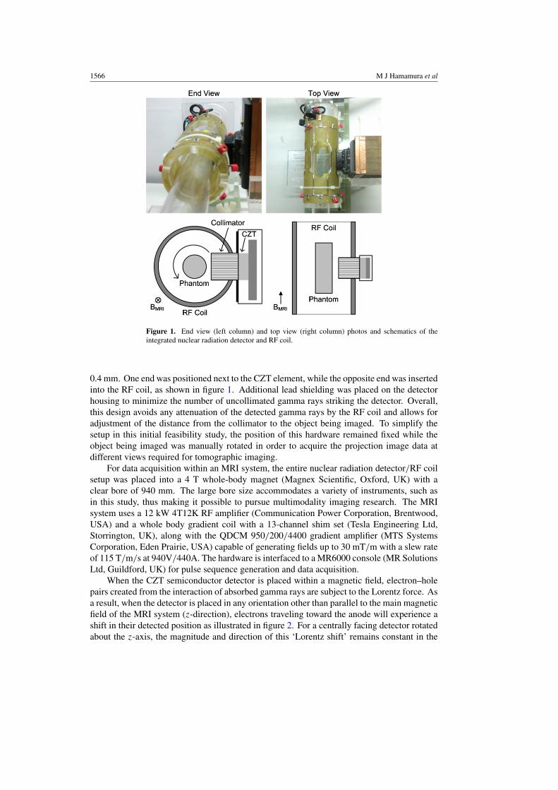
Figure 1. End view (left column) and top view (right column) photos and schematics of theintegrated nuclear radiation detector and RF coil.
0.4 mm. One end was positioned next to the CZT element, while the opposite end was insertedinto the RF coil, as shown in figure 1. Additional lead shielding was placed on the detectorhousing to minimize the number of uncollimated gamma rays striking the detector. Overall,this design avoids any attenuation of the detected gamma rays by the RF coil and allows foradjustment of the distance from the collimator to the object being imaged. To simplify thesetup in this initial feasibility study, the position of this hardware remained fixed while theobject being imaged was manually rotated in order to acquire the projection image data atdifferent views required for tomographic imaging.
For data acquisition within an MRI system, the entire nuclear radiation detector/RF coilsetup was placed into a 4 T whole-body magnet (Magnex Scientific, Oxford, UK) with aclear bore of 940 mm. The large bore size accommodates a variety of instruments, such asin this study, thus making it possible to pursue multimodality imaging research. The MRIsystem uses a 12 kW 4T12K RF amplifier (Communication Power Corporation, Brentwood,USA) and a whole body gradient coil with a 13-channel shim set (Tesla Engineering Ltd,Storrington, UK), along with the QDCM 950/200/4400 gradient amplifier (MTS SystemsCorporation, Eden Prairie, USA) capable of generating fields up to 30 mT/m with a slew rateof 115 T/m/s at 940V/440A. The hardware is interfaced to a MR6000 console (MR SolutionsLtd, Guildford, UK) for pulse sequence generation and data acquisition.
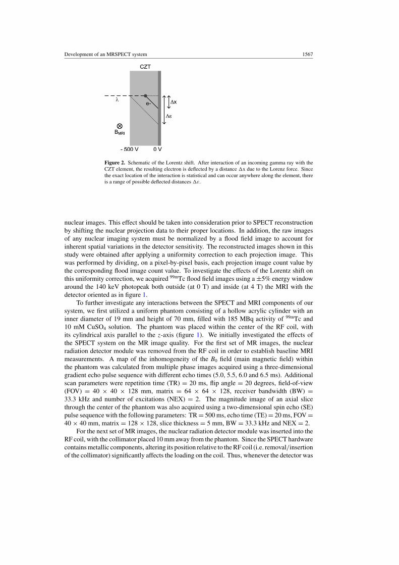
When the CZT semiconductor detector is placed within a magnetic field, electron–holepairs created from the interaction of absorbed gamma rays are subject to the Lorentz force. Asa result, when the detector is placed in any orientation other than parallel to the main magneticfield of the MRI system (z-direction), electrons traveling toward the anode will experience ashift in their detected position as illustrated in figure 2. For a centrally facing detector rotatedabout the z-axis, the magnitude and direction of this ‘Lorentz shift’ remains constant in the
Development of an MRSPECT system 1567
Figure 2. Schematic of the Lorentz shift. After interaction of an incoming gamma ray with theCZT element, the resulting electron is deflected by a distance �x due to the Lorenz force. Sincethe exact location of the interaction is statistical and can occur anywhere along the element, thereis a range of possible deflected distances �ε.
nuclear images. This effect should be taken into consideration prior to SPECT reconstructionby shifting the nuclear projection data to their proper locations. In addition, the raw imagesof any nuclear imaging system must be normalized by a flood field image to account forinherent spatial variations in the detector sensitivity. The reconstructed images shown in thisstudy were obtained after applying a uniformity correction to each projection image. Thiswas performed by dividing, on a pixel-by-pixel basis, each projection image count value bythe corresponding flood image count value. To investigate the effects of the Lorentz shift onthis uniformity correction, we acquired 99mTc flood field images using a ±5% energy windowaround the 140 keV photopeak both outside (at 0 T) and inside (at 4 T) the MRI with thedetector oriented as in figure 1.
To further investigate any interactions between the SPECT and MRI components of oursystem, we first utilized a uniform phantom consisting of a hollow acrylic cylinder with aninner diameter of 19 mm and height of 70 mm, filled with 185 MBq activity of 99mTc and10 mM CuSO4 solution. The phantom was placed within the center of the RF coil, withits cylindrical axis parallel to the z-axis (figure 1). We initially investigated the effects ofthe SPECT system on the MR image quality. For the first set of MR images, the nuclearradiation detector module was removed from the RF coil in order to establish baseline MRImeasurements. A map of the inhomogeneity of the B0 field (main magnetic field) withinthe phantom was calculated from multiple phase images acquired using a three-dimensionalgradient echo pulse sequence with different echo times (5.0, 5.5, 6.0 and 6.5 ms). Additionalscan parameters were repetition time (TR) = 20 ms, flip angle = 20 degrees, field-of-view(FOV) = 40 × 40 × 128 mm, matrix = 64 × 64 × 128, receiver bandwidth (BW) =33.3 kHz and number of excitations (NEX) = 2. The magnitude image of an axial slicethrough the center of the phantom was also acquired using a two-dimensional spin echo (SE)pulse sequence with the following parameters: TR = 500 ms, echo time (TE) = 20 ms, FOV =40 × 40 mm, matrix = 128 × 128, slice thickness = 5 mm, BW = 33.3 kHz and NEX = 2.
For the next set of MR images, the nuclear radiation detector module was inserted into theRF coil, with the collimator placed 10 mm away from the phantom. Since the SPECT hardwarecontains metallic components, altering its position relative to the RF coil (i.e. removal/insertionof the collimator) significantly affects the loading on the coil. Thus, whenever the detector was
1568 M J Hamamura et al
moved, trim capacitors were readjusted to maintain optimal tuning, matching and isolation ofthe two ports of the (quadrature) RF coil.
Initially, the detector was not powered on in order to observe the effects of the SPECTmaterials independent of any electronic interference generated by the SPECT hardware. B0
field data were acquired using the previous parameters, including the same shim values andcenter frequency as used without the detector. Maintaining these settings enabled us tocalculate the change in the B0 field caused by the presence of the SPECT materials. To testthe ability of our shim set to compensate for these changes, the optimum shim channel valueswere then calculated and applied using a constrained least-squares-fit algorithm (Wen andJaffer 1995) and an additional B0 map was acquired for comparison. A magnitude image wasalso acquired using the previous SE parameters.
For the final set of MR images of the uniform phantom, the nuclear radiation detector waspowered up and set to acquire data, as would be the case for simultaneous MRI and SPECTdata acquisition. This enabled us to observe the effects of the SPECT electronics on the MRimages. A B0 map and magnitude image were acquired using the previous parameters.
In addition to the MRI data, nuclear radiation data were acquired to investigate the effectsof the MRI system on the SPECT image quality. First, data were acquired with the nuclearradiation detector/RF coil unit located away from the MRI system (at 0 T) in order to establishbaseline nuclear radiation measurements. Next, data were acquired with the nuclear radiationdetector/RF coil unit located within the MRI system (at 4 T), but with the RF and gradientamplifiers turned off. This enabled us to observe the effects of the magnetic field independentof any interference caused by RF pulses, gradient waveforms or MRI electronics. Finally,to observe the effects of RF pulses, gradient waveforms and the MRI electronics, data wereacquired with the MRI system cycling through the previously detailed SE pulse sequence.
Pulse height spectra (PHS) were measured for each of the nuclear radiation data sets.Profiles for the average of the three CZT detector element rows (4.8 mm) corresponding to thepreviously acquired MR magnitude images were generated in order to determine the distanceof the Lorentz shift and to further investigate the effects of the MRI system. Data acquiredoutside the MRI system were normalized using the flood field image also acquired outsidethe MRI system. For comparison, uniformity correction of the data acquired inside the MRIsystem was performed using the flood field images acquired both outside the magnet andwithin the magnet.
For the next set of experiments, sections of 2.3 mm and 3.0 mm diameter plastic rodswere inserted axially into the previous uniform phantom to generate a resolution phantom (asimaged in figure 6). MRI and SPECT data were collected for 30 equally spaced views around360 degrees about the z-axis for ‘separate’, ‘sequential’and ‘simultaneous’ acquisition modes.For the separate mode, the nuclear radiation detector/RF coil unit was located away fromthe MRI system, and independent SPECT and MRI data were acquired. For the sequentialmode, the nuclear radiation detector/RF coil unit was placed inside the MRI system. Nuclearradiation data were first acquired with the RF and gradient amplifiers turned off. MR imageswere then acquired with the SPECT system turned off. For the simultaneous mode, both theMRI and SPECT data were acquired at the same time using the fully integrated MRSPECTsystem. For each view, data were acquired while the phantom position was fixed and notduring the rotation of the phantom to/from a different view in order to avoid any motionartifacts.
For MR imaging, two axial slices, each corresponding to a different section of the phantomwere acquired at each view using the SE pulse sequence. Nuclear projection data wereacquired using a ±5% energy window around the 140 keV photopeak. For each view, thenuclear projection data acquisition time was set to the length of the MRI scan (∼2 min).
Development of an MRSPECT system 1569
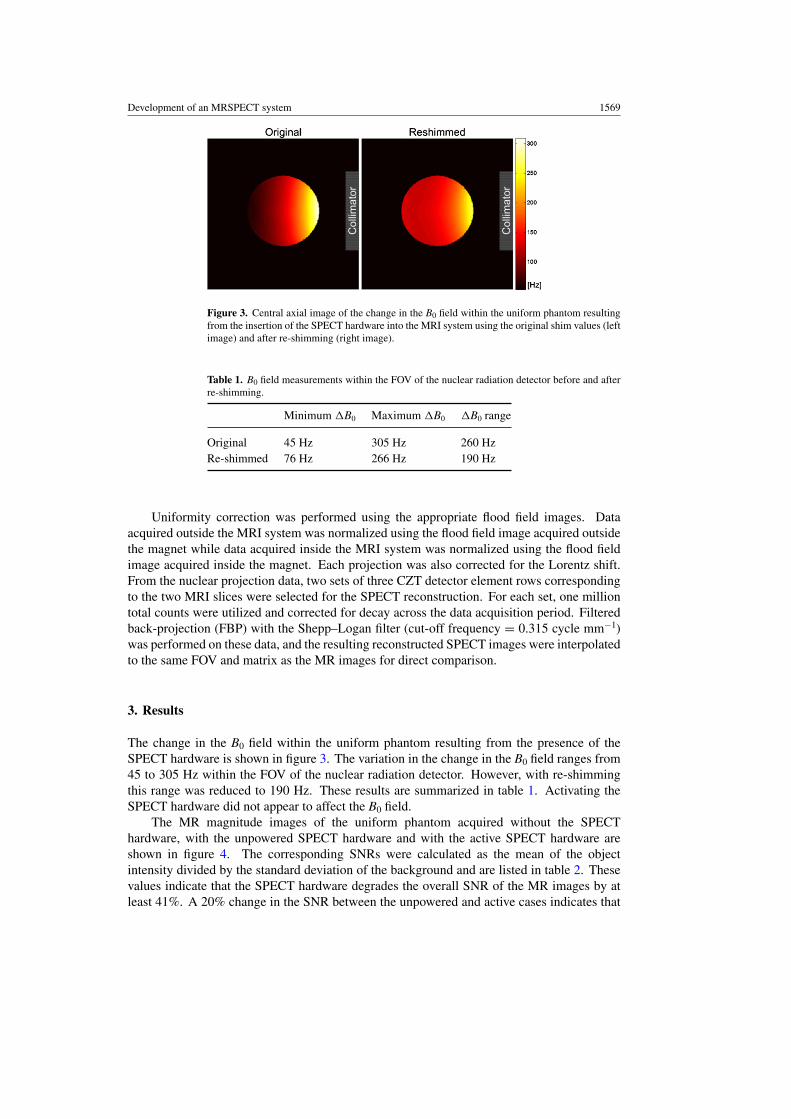
Figure 3. Central axial image of the change in the B0 field within the uniform phantom resultingfrom the insertion of the SPECT hardware into the MRI system using the original shim values (leftimage) and after re-shimming (right image).
Table 1. B0 field measurements within the FOV of the nuclear radiation detector before and afterre-shimming.
Minimum �B0 Maximum �B0 �B0 range
Original 45 Hz 305 Hz 260 HzRe-shimmed 76 Hz 266 Hz 190 Hz
Uniformity correction was performed using the appropriate flood field images. Dataacquired outside the MRI system was normalized using the flood field image acquired outsidethe magnet while data acquired inside the MRI system was normalized using the flood fieldimage acquired inside the magnet. Each projection was also corrected for the Lorentz shift.From the nuclear projection data, two sets of three CZT detector element rows correspondingto the two MRI slices were selected for the SPECT reconstruction. For each set, one milliontotal counts were utilized and corrected for decay across the data acquisition period. Filteredback-projection (FBP) with the Shepp–Logan filter (cut-off frequency = 0.315 cycle mm−1)was performed on these data, and the resulting reconstructed SPECT images were interpolatedto the same FOV and matrix as the MR images for direct comparison.
3. Results
The change in the B0 field within the uniform phantom resulting from the presence of theSPECT hardware is shown in figure 3. The variation in the change in the B0 field ranges from45 to 305 Hz within the FOV of the nuclear radiation detector. However, with re-shimmingthis range was reduced to 190 Hz. These results are summarized in table 1. Activating theSPECT hardware did not appear to affect the B0 field.
The MR magnitude images of the uniform phantom acquired without the SPECThardware, with the unpowered SPECT hardware and with the active SPECT hardware areshown in figure 4. The corresponding SNRs were calculated as the mean of the objectintensity divided by the standard deviation of the background and are listed in table 2. Thesevalues indicate that the SPECT hardware degrades the overall SNR of the MR images by atleast 41%. A 20% change in the SNR between the unpowered and active cases indicates that

1570 M J Hamamura et al
Figure 4. Central axial MR magnitude image of the uniform phantom without the SPECThardware (1st column), with the unpowered SPECT hardware (2nd column), with the activeSPECT hardware (3rd column) and with the active SPECT hardware averaged over 30 viewsabout 360◦ (4th column). The top row is normalized to maximum intensity while the bottom rowis windowed (18% brightness, 90% contrast) to visualize the non-uniformity resulting from thepresence of the SPECT hardware.
Table 2. MRI measurements of the uniform phantom for the various SPECT system configurations.
No SPECT Inactive SPECT Active SPECT Averaged
SNR 599 352 280 1201Non-uniformity 0.068 0.184 0.182 0.059
the SPECT electronics also interfere with the MR image quality, although to a smaller extentthan the SPECT materials.
Proper windowing of the MR images reveals that the SNR degradation increases closerto the SPECT detector that was placed on the right side. This non-uniformity was calculatedas the difference between the minimum and maximum values divided by the mean intensityand is also listed in table 2. The signal drop across the phantom can be attributed to the RFpower absorption and change in the B0 field due to the presence of the metallic components(i.e. lead collimator and shielding).
Our data acquisition procedure suggests a method to compensate for the non-uniformSNR loss. As the detector is rotated relative to the object being imaged, the region of lowestSNR (closest to the collimator) also rotates around the object. Adding together the MRimages acquired at each angular position (and registered to the same object orientation) willboth significantly improve the overall image SNR through signal averaging and smooth outthe non-uniformity. For the uniform phantom, averaging over 30 views resulted in a greaterthan fourfold improvement in the SNR and a 68% reduction in the non-uniformity, as alsoshown in figure 4 and table 2.
Previous studies have shown that the nuclear radiation detector can operate within highmagnetic fields (Wagenaar et al 2006a). To verify this finding and also test whether it can
Development of an MRSPECT system 1571
(a) (b)
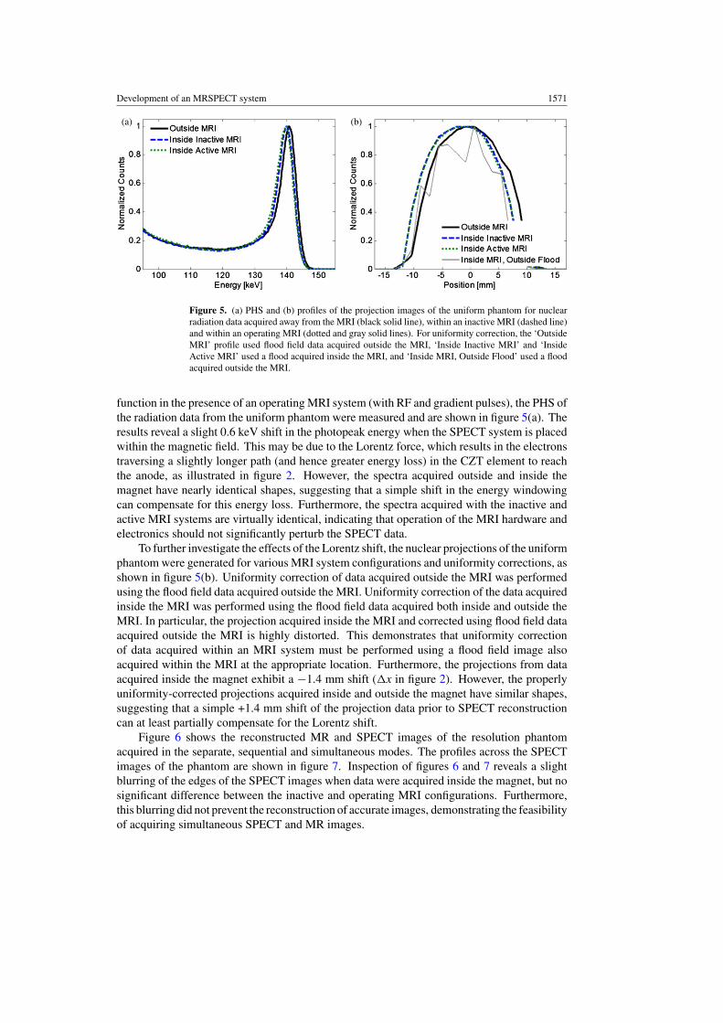
Figure 5. (a) PHS and (b) profiles of the projection images of the uniform phantom for nuclearradiation data acquired away from the MRI (black solid line), within an inactive MRI (dashed line)and within an operating MRI (dotted and gray solid lines). For uniformity correction, the ‘OutsideMRI’ profile used flood field data acquired outside the MRI, ‘Inside Inactive MRI’ and ‘InsideActive MRI’ used a flood acquired inside the MRI, and ‘Inside MRI, Outside Flood’ used a floodacquired outside the MRI.
function in the presence of an operating MRI system (with RF and gradient pulses), the PHS ofthe radiation data from the uniform phantom were measured and are shown in figure 5(a). Theresults reveal a slight 0.6 keV shift in the photopeak energy when the SPECT system is placedwithin the magnetic field. This may be due to the Lorentz force, which results in the electronstraversing a slightly longer path (and hence greater energy loss) in the CZT element to reachthe anode, as illustrated in figure 2. However, the spectra acquired outside and inside themagnet have nearly identical shapes, suggesting that a simple shift in the energy windowingcan compensate for this energy loss. Furthermore, the spectra acquired with the inactive andactive MRI systems are virtually identical, indicating that operation of the MRI hardware andelectronics should not significantly perturb the SPECT data.
To further investigate the effects of the Lorentz shift, the nuclear projections of the uniformphantom were generated for various MRI system configurations and uniformity corrections, asshown in figure 5(b). Uniformity correction of data acquired outside the MRI was performedusing the flood field data acquired outside the MRI. Uniformity correction of the data acquiredinside the MRI was performed using the flood field data acquired both inside and outside theMRI. In particular, the projection acquired inside the MRI and corrected using flood field dataacquired outside the MRI is highly distorted. This demonstrates that uniformity correctionof data acquired within an MRI system must be performed using a flood field image alsoacquired within the MRI at the appropriate location. Furthermore, the projections from dataacquired inside the magnet exhibit a −1.4 mm shift (�x in figure 2). However, the properlyuniformity-corrected projections acquired inside and outside the magnet have similar shapes,suggesting that a simple +1.4 mm shift of the projection data prior to SPECT reconstructioncan at least partially compensate for the Lorentz shift.
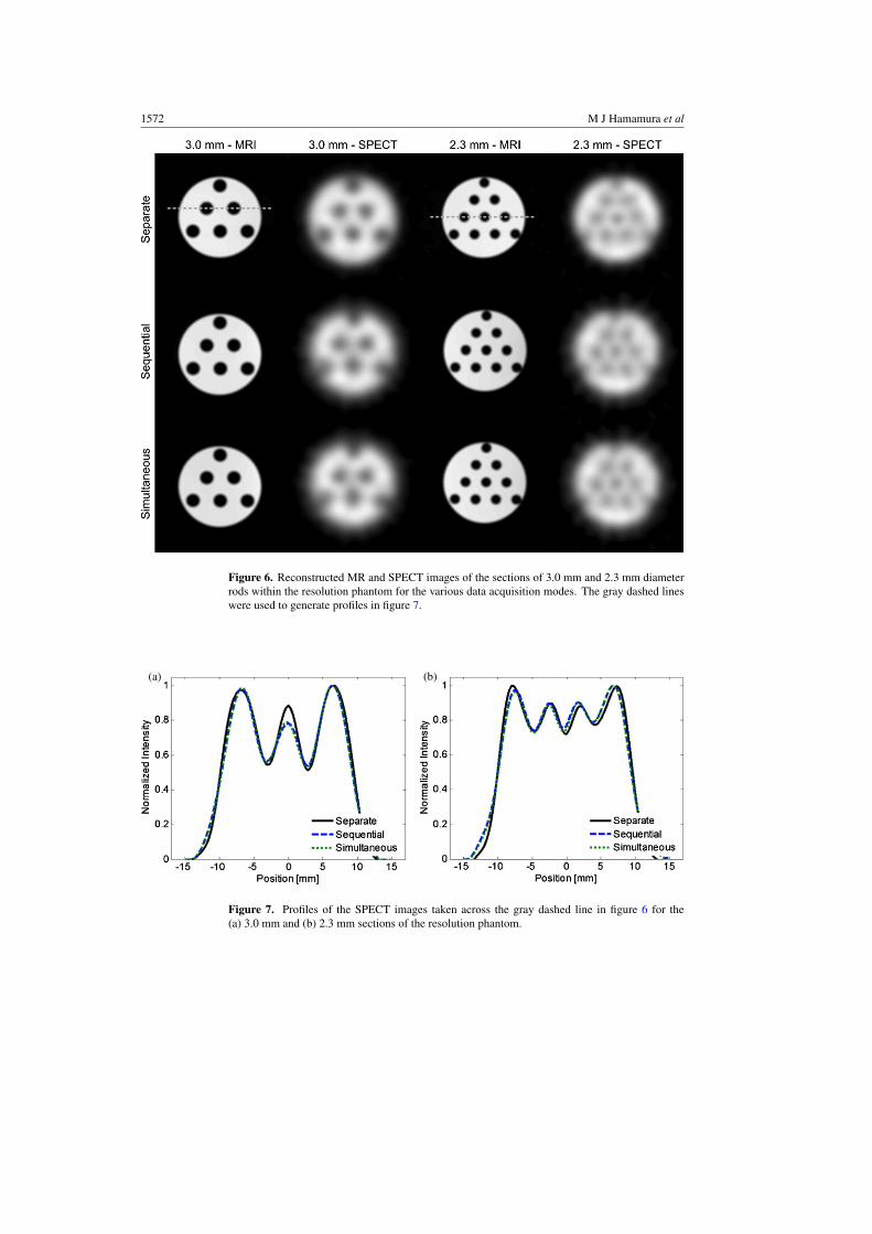
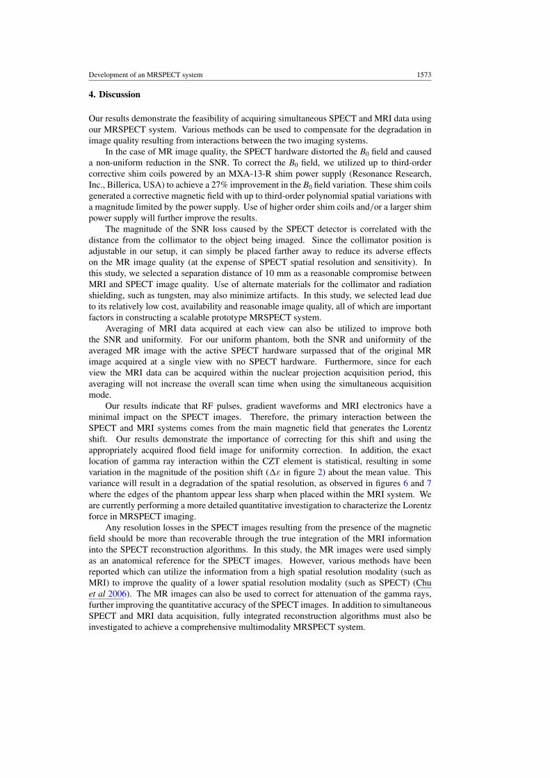
Figure 6 shows the reconstructed MR and SPECT images of the resolution phantomacquired in the separate, sequential and simultaneous modes. The profiles across the SPECTimages of the phantom are shown in figure 7. Inspection of figures 6 and 7 reveals a slightblurring of the edges of the SPECT images when data were acquired inside the magnet, but nosignificant difference between the inactive and operating MRI configurations. Furthermore,this blurring did not prevent the reconstruction of accurate images, demonstrating the feasibilityof acquiring simultaneous SPECT and MR images.
1572 M J Hamamura et al
Figure 6. Reconstructed MR and SPECT images of the sections of 3.0 mm and 2.3 mm diameterrods within the resolution phantom for the various data acquisition modes. The gray dashed lineswere used to generate profiles in figure 7.
(a) (b)
Figure 7. Profiles of the SPECT images taken across the gray dashed line in figure 6 for the(a) 3.0 mm and (b) 2.3 mm sections of the resolution phantom.
Development of an MRSPECT system 1573
4. Discussion
Our results demonstrate the feasibility of acquiring simultaneous SPECT and MRI data usingour MRSPECT system. Various methods can be used to compensate for the degradation inimage quality resulting from interactions between the two imaging systems.
In the case of MR image quality, the SPECT hardware distorted the B0 field and causeda non-uniform reduction in the SNR. To correct the B0 field, we utilized up to third-ordercorrective shim coils powered by an MXA-13-R shim power supply (Resonance Research,Inc., Billerica, USA) to achieve a 27% improvement in the B0 field variation. These shim coilsgenerated a corrective magnetic field with up to third-order polynomial spatial variations witha magnitude limited by the power supply. Use of higher order shim coils and/or a larger shimpower supply will further improve the results.
The magnitude of the SNR loss caused by the SPECT detector is correlated with thedistance from the collimator to the object being imaged. Since the collimator position isadjustable in our setup, it can simply be placed farther away to reduce its adverse effectson the MR image quality (at the expense of SPECT spatial resolution and sensitivity). Inthis study, we selected a separation distance of 10 mm as a reasonable compromise betweenMRI and SPECT image quality. Use of alternate materials for the collimator and radiationshielding, such as tungsten, may also minimize artifacts. In this study, we selected lead dueto its relatively low cost, availability and reasonable image quality, all of which are importantfactors in constructing a scalable prototype MRSPECT system.
Averaging of MRI data acquired at each view can also be utilized to improve boththe SNR and uniformity. For our uniform phantom, both the SNR and uniformity of theaveraged MR image with the active SPECT hardware surpassed that of the original MRimage acquired at a single view with no SPECT hardware. Furthermore, since for eachview the MRI data can be acquired within the nuclear projection acquisition period, thisaveraging will not increase the overall scan time when using the simultaneous acquisitionmode.
Our results indicate that RF pulses, gradient waveforms and MRI electronics have aminimal impact on the SPECT images. Therefore, the primary interaction between theSPECT and MRI systems comes from the main magnetic field that generates the Lorentzshift. Our results demonstrate the importance of correcting for this shift and using theappropriately acquired flood field image for uniformity correction. In addition, the exactlocation of gamma ray interaction within the CZT element is statistical, resulting in somevariation in the magnitude of the position shift (�ε in figure 2) about the mean value. Thisvariance will result in a degradation of the spatial resolution, as observed in figures 6 and 7where the edges of the phantom appear less sharp when placed within the MRI system. Weare currently performing a more detailed quantitative investigation to characterize the Lorentzforce in MRSPECT imaging.
Any resolution losses in the SPECT images resulting from the presence of the magneticfield should be more than recoverable through the true integration of the MRI informationinto the SPECT reconstruction algorithms. In this study, the MR images were used simplyas an anatomical reference for the SPECT images. However, various methods have beenreported which can utilize the information from a high spatial resolution modality (such asMRI) to improve the quality of a lower spatial resolution modality (such as SPECT) (Chuet al 2006). The MR images can also be used to correct for attenuation of the gamma rays,further improving the quantitative accuracy of the SPECT images. In addition to simultaneousSPECT and MRI data acquisition, fully integrated reconstruction algorithms must also beinvestigated to achieve a comprehensive multimodality MRSPECT system.
1574 M J Hamamura et al
Our MRSPECT hardware may be expanded for future studies. In this initial study, thenuclear radiation detector remained fixed while the phantom was manually rotated to obtaindata from multiple views. Construction of a larger rotating gantry would allow for rotation ofthe detector about a (fixed) object. Furthermore, rotation of either the object or the detectorcould be automated through the use of an MR-compatible motor (Roeck et al 2009).
Rotation of the detector (and its lead components) will result in a significant alterationof the B0 field. In this case, it will become necessary to re-shim at each view. We anticipatethat such shimming need not increase the overall time of an MRSPECT experiment, since foreach view the relatively longer nuclear radiation data acquisition period may still exceed anyrequired MR imaging operations. For example, using the parameters of this study, the 2 minanatomical MR image could be preceded by reported fast shimming techniques of about 1 min(Klassen and Menon 2004), totaling only 3 min per view.
For applications using pinhole collimators, a birdcage RF coil may be appropriatelymodified by cutting out regions for the pinholes. While pinhole collimators are necessaryfor micro-SPECT applications, we selected a parallel-hole collimator to simplify the setupand maximize sensitivity in this initial feasibility study. More significantly, our MRSPECTsystem will serve as a scalable prototype for future human studies, where the use of parallel-hole collimators is more common. For a scaled-up collimator larger than the space betweenthe rungs of a birdcage RF coil, the collimator may simply be placed outside the coil andattenuation of the gamma rays by the coil materials taken into consideration during dataprocessing. Alternatively, an array of receiver coil loops may be utilized instead of a birdcagecoil, where a much larger space between the coil elements is permissible for placement ofa collimator. Use of an RF receiver array with integrated nuclear radiation detectors can beimplemented for both small-animal and human-sized scales with improved MR image quality(Ha et al 2009).
5. Conclusion
This study demonstrates the feasibility of co-registered, simultaneous SPECT and MR imagingbased on CZT detector technology. Thus, our miniaturized MRSPECT system could bemodified with pinhole collimators for small animal imaging (micro-MRSPECT) or scaled upusing appropriate components for human imaging. Since both SPECT and MRI modalitiesare applicable to both small animals and humans, we anticipate that MRSPECT will play asignificant role in molecular imaging for various human diseases in the near future.
Acknowledgments
This research was supported in part by CIRM grant RT1-01120, CIRM training grant T1-00008 (for S Ha) and NIH NIBIB grant R44EB006712. The authors thank Keumsil Lee forher assistance with the data acquisition.
References
Breton E, Choquet P, Goetz C, Kintz J, Erbs P, Rooke R and Constantinesco A 2007 Dual SPECT/MR imaging insmall animal Nucl. Instrum. Methods Phys. Res. A 571 446–8
Chu Y, Su MY, Mandelkern M and Nalcioglu O 2006 Resolution improvement in positron emission tomographyusing anatomical magnetic resonance imaging Technol. Cancer Res. Treat. 5 311–7
Goetz C, Breton E, Choquet P, Israel-Jost V and Constantinesco A 2008 MRI system for small-animal imagingJ. Nucl. Med. 49 88–93
Development of an MRSPECT system 1575
Ha S, Hamamura M J, Roeck W W, Muftuler L T and Nalcioglu O 2009 Development of a new RF coil and γ -rayradiation shielding assembly for a SPECT/MRI system Phys. Med. Biol. submitted
Klassen L M and Menon R S 2004 Robust automated shimming technique using arbitrary mapping acquisitionparameters (RASTAMAP) Magn. Reson. Med. 51 881–7
Meng L J, Tan J W and Geng F 2007 Design study of an MRI compatible ultra-high resolution SPECT for in vivomice brain imaging IEEE Nucl. Sci. Sym. (Honolulu) pp 2956–60
Nalcioglu O, Roeck W W and Ha S-H 2008 Multi-modality RF coil (US patent pending)Roeck W W, Ha S-H, Farmaka S and Nalcioglu O 2009 A variable torque motor compatible with magnetic resonance
imaging Rev. Sci. Instrum. 80 046108Wagenaar D J, Kapusta M, Li J and Patt B E 2006a Rationale for the combination of nuclear medicine with magnetic
resonance for pre-clinical imaging Technol. Cancer Res. Treat. 5 343–50Wagenaar D, Nalcioglu O, Muftuler L, Szawlowski M, Kapusta M, Pawlov N, Meier D, Maehlum G and Patt B
2006b Development of MRI-compatible nuclear medicine imaging detectors IEEE Nucl. Sci. Sym. (San Diego)pp 1825–8
Wen H and Jaffer F A 1995 An in-vivo automated shimming method taking into account shim current constraintsMagn. Reson. Med. 34 898–904
Copyright © 2022 FDOKUMEN