Axonal Dynamics of Excitatory and Inhibitory Neurons in Somatosensory Cortex
Sodium and calcium mechanisms of rhythmic bursting inexcitatory neural networks of the pre-Botzinger complex:a computational modelling study
Patrick E. Jasinski,1,* Yaroslav I. Molkov,1,2,* Natalia A. Shevtsova,1 Jeffrey C. Smith3 and Ilya A. Rybak11Department of Neurobiology and Anatomy, Drexel University College of Medicine, Philadelphia, PA, USA2Department of Mathematical Sciences, Indiana University – Purdue University, Indianapolis, IN, USA3Cellular and Systems Neurobiology Section, National Institute of Neurological Disorders and Stroke, National Institutes ofHealth, Bethesda, MD, USA
Keywords: Ca2+-activated nonspecific cation current, Na+/K+ pump, neural oscillations, persistent Na+ current, respiration
Abstract
The neural mechanisms generating rhythmic bursting activity in the mammalian brainstem, particularly in the pre-Botzinger complex(pre-BotC), which is involved in respiratory rhythm generation, and in the spinal cord (e.g. locomotor rhythmic activity) that persistafter blockade of synaptic inhibition remain poorly understood. Experimental studies in rodent medullary slices containing the pre-BotC identified two mechanisms that could potentially contribute to the generation of rhythmic bursting: one based on the persistentNa+ current (INaP), and the other involving the voltage-gated Ca2+ current (ICa) and the Ca2+-activated nonspecific cation current(ICAN), activated by intracellular Ca2+ accumulated from extracellular and intracellular sources. However, the involvement and rela-tive roles of these mechanisms in rhythmic bursting are still under debate. In this theoretical/modelling study, we investigated Na+-dependent and Ca2+-dependent bursting generated in single cells and heterogeneous populations of synaptically interconnectedexcitatory neurons with INaP and ICa randomly distributed within populations. We analysed the possible roles of network connections,ionotropic and metabotropic synaptic mechanisms, intracellular Ca2+ release, and the Na+/K+ pump in rhythmic bursting generatedunder different conditions. We show that a heterogeneous population of excitatory neurons can operate in different oscillatoryregimes with bursting dependent on INaP and/or ICAN, or independent of both. We demonstrate that the operating bursting mecha-nism may depend on neuronal excitation, synaptic interactions within the network, and the relative expression of particular ionic cur-rents. The existence of multiple oscillatory regimes and their state dependence demonstrated in our models may explain differentrhythmic activities observed in the pre-BotC and other brainstem/spinal cord circuits under different experimental conditions.
Introduction
Synchronised neural oscillations are generated in the brain by centralpattern generators, which are centrally located neural networks thatcontrol rhythmic movements, such as walking, swimming, breathing,and chewing (Cohen et al., 1988; Stain et al., 1999; Gossard et al.,2010). Although synaptic inhibition plays a considerable role inthese behaviours, the basic rhythmic activity in the correspondingstructures usually persists after pharmacological blockade of inhibi-tion. This was demonstrated in isolated rodent spinal cord prepara-tions (Cowley & Schmidt, 1995) and medullary slices containingthe pre-Bötzinger complex (pre-BötC, Feldman & Smith, 1989; Ra-mirez et al., 1996), which is the medullary structure involved inrespiratory rhythm generation (Smith et al., 1991; Rekling & Feld-man, 1998; Gray et al., 2001). However, the neural mechanisms
underlying the generation of these important rhythms remain largelyunknown.Butera et al. (1999a,b) suggested that the population bursting
observed in the pre-BötC in vitro arises from the persistent (slowlyinactivating) Na+ current (INaP) in pre-BötC neurons and excitatorysynaptic interactions within populations of these neurons. The pres-ence of INaP in the pre-BötC has been confirmed (Del Negro et al.,2002a; Rybak et al., 2003a; Koizumi & Smith, 2008), and the pre-BötC rhythmic activity in medullary slices from neonatal rats couldbe abolished by the INaP blocker riluzole (Rybak et al., 2003b;Koizumi & Smith, 2008). Alternatively, Thoby-Brisson & Ramirez(2001), using medullary slices from mice at postnatal days 6–12(P6-P12), found two distinct types of intrinsically bursting cells(bursters) in the pre-BötC whose bursting was, respectively, sensi-tive and insensitive to the Ca2+ current blocker Cd2+. Later, Peñaet al. (2004) found that the Cd2+-sensitive bursters were riluzole-insensitive, whereas most of the Cd2+-insensitive ones were riluzole-sensitive. Furthermore, rhythmic activity in the Cd2+-sensitive burst-ers could be blocked by flufenamic acid (FFA), a pharmacologicalblocker of the Ca2+-activated nonspecific cation current (ICAN, Del
Correspondence: Ilya A. Rybak, PhD, as above.E-mail: [email protected]
*P.E.J. and Y.I.M. contributed equally to this work.
Received 11 June 2012, revised 13 August 2012, accepted 26 September 2012
© 2012 Federation of European Neuroscience Societies and Blackwell Publishing Ltd
European Journal of Neuroscience, pp. 1–19, 2012 doi:10.1111/ejn.12042
European Journal of Neuroscience
Negro et al., 2005), suggesting that both INaP and ICAN are involvedin bursting generated in the pre-BötC. The role of ICAN in this burst-ing was further investigated by Del Negro and collaborators (Paceet al., 2007; Pace & Del Negro, 2008; Rubin et al., 2009), who sug-gested a metabotropic mechanism for synaptic activation of ICAN.However, the involvement and specific roles of these mechanisms
in experimentally observed bursting activities remain controversial,and require special investigations. In this modelling study, we con-sider the intrinsic Na+-dependent and Ca2+-dependent bursting gen-erated in single cells and heterogeneous populations of excitatoryneurons with INaP and the voltage-gated Ca2+ current (ICa) randomlydistributed across neurons in the populations. We study the possibleroles of synaptic interactions, ionotropic and metabotropic synapticmechanisms, intracellular Ca2+ release and the Na+/K+ pump in thecellular and network rhythmic bursting. We show that heterogeneouspopulations of excitatory neurons can generate rhythmic burstingdependent on INaP and/or ICAN, or independent of both, and that theinvolvement of each mechanism may depend on the neuronal excita-tion, strength of synaptic interactions, and expression of particularionic channels. We therefore suggest that the rhythmic burstingactivity discovered in the pre-BötC in vitro is state-dependent, andhence, depending on the state, the pre-BötC can operate in multipleoscillatory regimes involving different INaP-dependent and/or ICAN-dependent mechanisms. We also support the previous suggestionthat the electrogenic Na+/K+ pump can play an important role in thegeneration of this rhythmic bursting by performing the burst-termi-nating function in multiple regimes of oscillations. The results ofthis theoretical/modelling study provide important insights into vari-ous rhythmic activities observed in the pre-BötC and other brain-stem and spinal cord circuits.
Methods
Model description
The conductance-based single-compartment model of a single neuronwas developed in the Hodgkin–Huxley style. The neuronal mem-brane potential (V) is defined by a set of membrane ionic currents:
C � dVdt
¼ �INa � INaP � IK � ICa � ICAN � IPump � IL � ISynE; ð1Þ
where C is neuronal membrane capacitance and t is time. The ioniccurrents in the model include fast Na+ (INa with the maximal con-ductance �gNa), persistent (or slowly inactivating) Na+ (INaP with themaximal conductance �gNaP), delayed rectifier K+ (IK with the maxi-mal conductance �gK), high-voltage-activated Ca2+ (ICa with the max-imal conductance �gCa), Ca
2+-activated nonspecific cation (ICAN withthe maximal conductance �gCAN), Na+/K+ pump (IPump), leakage(IL with the constant conductance gL) and excitatory synaptic (ISynE)currents. These currents, except for IPump, are described as follows:
INa ¼ �gNa � m3Na � hNa � ðV � ENaÞ;
INaP ¼ �gNaP � mNaP � hNaP � ðV � ENaÞ;IK ¼ �gK � m4
K � ðV � EKÞ;ICa ¼ �gCa � mCa � hCa � ðV � ECaÞ;ICAN ¼ �gCAN � mCAN � ðV � ECANÞ;IL ¼ gL � ðV � ELÞ;ISynE ¼ gSynE � ðV � ESynEÞ;
ð2Þ
where ENa, EK, ECa, ECAN, EL and ESynE are reversal potentials ofthe corresponding channels:
ENa ¼ ðR � T=FÞ � ln ðNao=NaiÞ;EK ¼ ðR � T=FÞ � ln ðKo=KiÞ;ECa ¼ ðR � T=2FÞ � ln ðCao=CaiÞ;ECAN ¼ 0; EL ¼ �68mV; ESynE ¼ �10 mV:
ð3Þ
In the above equations: R = 8.314 J/(mol K) is the universal gasconstant; T = 308 K is the temperature; and F = 96.485 kC/mol isthe Faraday constant. This makes R � T/F = 26.54 mV. Na, K andCa represent the concentrations of Na+, K+, and Ca2+, respectively.The subscripts ‘o’ (or ‘out’) and ‘i’ (or ‘in’) indicate the concentra-tions of these ions outside and inside the cell, respectively. In ourmodel: Nao = 120 mM; Cao = 4 mM; Ko = 4 mM; Ki = 140 mM;and Nai and Cai were considered to be dynamical variables. There-fore, the K+ reversal potential was constant, EK = –94 mV, whereasENa and ECa varied according to Eqn 3: ENa = 26.54 � ln(120/Nai) mV and ECa = 13.27 � ln(4/Cai) mV. The membrane capaci-tance value was set to C = 36 pF (Rybak et al., 2007; Smith et al.,2007).Activation (m) and inactivation (h) variables for most ionic chan-
nels are described as:
syðVÞ � dydt ¼ ðy1ðVÞ � yÞ; y ¼ m; hf g: ð4Þ
For INa and INaP, steady-state activation (m∞) and inactivation(h∞) and their time constants are described as:
y1ðVÞ ¼ 1=ð1þ expð�ðV � Vy1=2Þ=kyÞÞ;syðVÞ ¼ symax= coshð�ðV � Vsy1=2Þ=ksyÞ:
ð5Þ
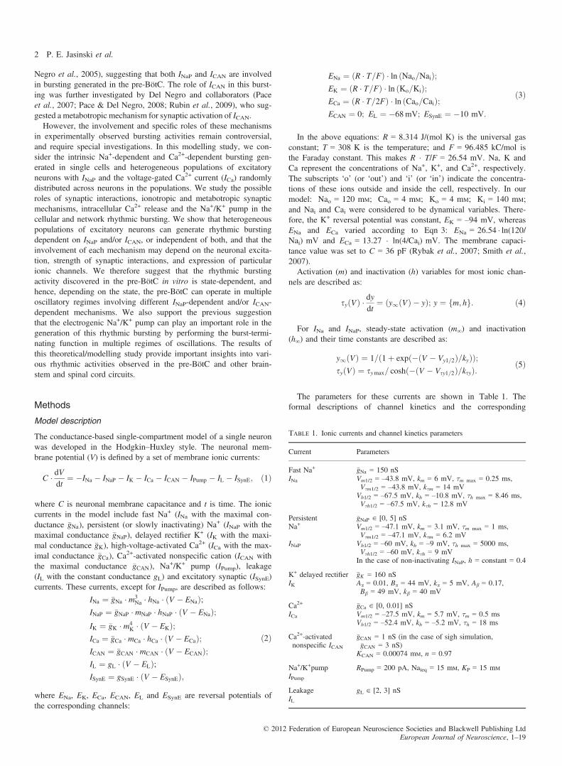
The parameters for these currents are shown in Table 1. Theformal descriptions of channel kinetics and the corresponding
Table 1. Ionic currents and channel kinetics parameters
Current Parameters
Fast Na+
INa�gNa = 150 nSVm1/2 = –43.8 mV, km = 6 mV, τm max = 0.25 ms,Vτm1/2 = –43.8 mV, kτm = 14 mV
Vh1/2 = –67.5 mV, kh = –10.8 mV, τh max = 8.46 ms,Vτh1/2 = –67.5 mV, kτh = 12.8 mV
Persistent �gNaP ∈ [0, 5] nSNa+ Vm1/2 = –47.1 mV, km = 3.1 mV, τm max = 1 ms,
Vτm1/2 = –47.1 mV, kτm = 6.2 mVINaP Vh1/2 = –60 mV, kh = –9 mV, τh max = 5000 ms,
Vτh1/2 = –60 mV, kτh = 9 mVIn the case of non-inactivating INaP, h = constant = 0.4
K+ delayed rectifierIK
�gK = 160 nSAa = 0.01, Ba = 44 mV, ka = 5 mV, Ab = 0.17,Bb = 49 mV, kb = 40 mV
Ca2+
ICa�gCa ∈ [0, 0.01] nSVm1/2 = –27.5 mV, km = 5.7 mV, τm = 0.5 msVh1/2 = –52.4 mV, kh = –5.2 mV, τh = 18 ms
Ca2+-activatednonspecific ICAN
�gCAN = 1 nS (in the case of sigh simulation,�gCAN = 3 nS)
KCAN = 0.00074 mM, n = 0.97
Na+/K+pumpIPump
RPump = 200 pA, Naieq = 15 mM, KP = 15 mM
LeakageIL
gL ∈ [2, 3] nS
© 2012 Federation of European Neuroscience Societies and Blackwell Publishing LtdEuropean Journal of Neuroscience, 1–19
2 P. E. Jasinski et al.

parameters were derived from previous experimental (Rybak et al.,2003a) and modelling (Rybak et al., 2007; Smith et al., 2007)studies.For the non-inactivating K+ delayed rectifier current (IK):
m1 ¼ a1=ða1 þ b1Þ;s1 ¼ 1=ða1 þ b1Þ; ð6Þ
where
a1 ¼ Aa � ðV þ BaÞ=ð1� expð�ðV þ BaÞ=kaÞ;b1 ¼ Ab � expð�ðV þ BbÞ=kbÞ:
ð7Þ
These channel kinetics and the corresponding parameter values,shown in Table 1, were derived from previous models (Huguenard& McCormick, 2004; Rybak et al., 2007; Smith et al., 2007).The parameters for steady-state activation (m∞) and inactivation
(h∞) of ICa are shown in Table 1. This channel description andparameters were accepted from previous models (Rybak et al.,2007; Smith et al., 2007), and had been initially derived from exper-imental studies of Elsen & Ramirez (1998).ICAN has instantaneous activation, which depends on Cai and is
voltage-independent:
mCAN ¼ 1=ð1þ ðKCAN=CaiÞnÞ; ð8Þ
where KCAN represents the half-activation concentration of Cai, andn is the Hill coefficient. These parameters in Table 1 are taken fromToporikova & Butera (2011).The description of the Na+/K+ pump is taken from Li et al.
(1996), with parameters as in Rubin et al. (2009):
IPump ¼ RPump � ðuðNaiÞ � uðNaieqÞÞ; ð9Þ
where, uðxÞ ¼ x3�ðx3 þ K3
PÞ, RPump is the maximal pump current,Naieq is the equilibrium intracellular Na+ concentration, and KP isthe pump parameter (Table 1).The dynamics of the total intracellular Ca2+ concentration within
the cell (Catot) are described as follows:
dCatotdt
¼ �aCa � ICa � Cai=sCa: ð10Þ
In the right part of this equation, the first term represents Ca2+
influx from the extracellular space through voltage-gated Ca2+ chan-nels, and the second term represents the membrane Ca2+ pump,which extrudes free intracellular Ca2+ (Cai) from the cytoplasm (Li& Rinzel, 1994).The dynamics of the intracellular concentration of free Ca2+
([Ca2+]in, Cai) are described by the following differential equation(Li & Rinzel, 1994; Toporikova & Butera, 2011):
dCaidt
¼ �aCa � ICa � Cai=sCa þ fCa � ðJERin � JERoutÞ: ð11Þ
In Eqns 10 and 11: aCa is the conversion factor between currentand rate of concentration change), and τCa is the time constant forthe Ca2+ pump (aCa = 2.5 9 10–5 mM/fC and τCa = 500 ms). InEqn 11, JERin represents the flux of Ca2+ per unit volume from theendoplasmic reticulum (ER) into the cytoplasm, JERout represents theflux of Ca2+ per unit volume from the cytoplasm into the ER,and fCa is the ratio of free to bound Ca2+ in the cytosol(fCa = 2.5 9 10�5).
The flux per unit volume of Ca2+ from the ER to the cytoplasmis hypothesised to be mediated by the inositol triphosphate (IP3)receptor (IP3R), which is modelled as:
JERin ¼�LIP3R þ PIP3R �
�Cai
Cai þ Ka� IP3IP3þ KI
� l3��
� ðCaER � CaiÞ;
ð12Þ
where IP3 represents the intracellular concentration of IP3, which, inour simulations, is considered to be constant (IP3 = 0.5 9 10–3 mM
unless otherwise indicated). Also in this equation: LIP3R is the leakconstant of IP3R (LIP3R = 0.37/ms); PIP3R characterises the perme-ability of the IP3 channels (PIP3R = 31 000/ms); and Kα and KI arethe dissociation constants (Ka = 0.4 9 10–3 mM; KI = 10–3 mM).The values of these parameters have been drawn from Toporikova &Butera (2011). The Ca2+-dependent IP3 gating variable (l) and theCa2+ concentration in the ER (CaER) are described as follows:
dldt
¼ A � ðKd � l � ðCai þ KdÞÞ; ð13Þ
CaER ¼ ðCatot � CaiÞ=rCa;
where A is a conversion factor, Kd is the dissociation constant forIP3 inactivation, and rCa is the ratio of cytosolic to ER volume. Thefollowing parameter values have been used: A = 5 mM/ms;Kd = 0.4 9 10–3 mM; rCa = 0.185 (Toporikova & Butera, 2011).The flux per unit volume of Ca2+ into the ER from the cytoplasm
is hypothesised to be mediated by an ER membrane pump represent-ing the sarcoplasmic/endoplasmic reticulum Ca2+-ATPase (SERCA):
JERout ¼ VSERCA � Ca2i�ðK2
SERCA þ Ca2i Þ ð14Þ
where VSERCA = 0.4 mM/ms is the maximal flux through theSERCA pumps, and KSERCA = 0.2 9 10–3 mM (Toporikova & Bu-tera, 2011).Nai is governed by the following differential equation:
dNaidt
¼ �aNa � ðINa þ INaP þ ICAN þ 3 � IPumpÞ; ð15Þ
where aCa = 5 9 10–5 mM/fC is the conversion factor.The total synaptic conductance of each neuron i (gSynEi) includes
two components: one, gtonic, that characterises an external tonicexcitatory drive to the neuron, and another characterising excitatorysynaptic inputs from all N neurons of the population (includingitself):
gSynEiðtÞ ¼ gtonic þXNj¼1
wji �Xtkj\t
expð�ðt � tkjÞ=sSynEÞ ð16Þ
According to this equation, each spike of neuron j triggering apostsynaptic current in neuron i at time tkj increases the excitatorysynaptic conductance in neuron i by wji, which represents the weightof the synaptic connection from neuron j to neuron i. τSynE = 5 msis the decay time constant. In our simulations, each neuron in thepopulation received the same external drive gtonic ∈ [0, 1.2] nS, andthe weights of its synaptic inputs were normally distributed by useof the assigned average weight �w (�w ∈ [0, 0.2]) nS and the variancerw ¼ 0:2 � �w.A population of N = 50 neurons with all-to-all connections was
simulated. The heterogeneity of neurons within the population wasprovided by the uniformly distributed maximal conductance of leak-age, persistent Na+ and Ca2+ channels. The leakage conductance
© 2012 Federation of European Neuroscience Societies and Blackwell Publishing LtdEuropean Journal of Neuroscience, 1–19
Na+ and Ca2+ mechanisms of rhythmic bursting 3
was uniformly distributed within a range gL ∈ [2, 3] nS, which wasselected on the basis of the experimental measurements of inputresistance in neurons of the rostral ventrolateral medulla and thepre-BötC (500 MΩ in Mazza et al., 2000 and 430 MΩ in DelNegro et al., 2002a; see also Koizumi et al., 2008). The persistentNa+ conductance was distributed within a range �gNaP ∈ [0, 5] nS,which was based on experimental measurements of this conductancein rostral ventrolateral medulla and pre-BötC neurons (Rybak et al.,2003a). The Ca2+ conductance was distributed within a range�gCa ∈ [0, 0.01] nS, unless a different range is stated in the text. Toapply these distributions, a random number generator from the stan-dard library of the Intel C++ compiler was used. To initialise a par-ticular conductance value, a random four-byte integer number (i.e.from 0 to 232) was generated and divided by 232. This provided uswith a random number x, uniformly distributed in the [0, 1] interval.Then, if a randomised parameter g (e.g. one of the above maximalconductances) should be uniformly distributed within a rangeg ∈ [a, b], we calculated g = a + x � (b � a), which served as aparticular g value.The weights of synaptic interactions were also distributed (by use
of a standard normal distribution; see above). The total synapticinput to each neuron from other neurons in the population can becharacterised by N � �w. The initial conditions for membrane poten-tials, intracellular Ca2+ and Na+ concentrations and channel conduc-tances were chosen by use of a uniform distribution within thephysiologically realistic ranges of values for each variable, and asettling period of 10–20 s was allowed in the performed or testingsimulations to ensure that the results are independent of initial con-ditions. Most simulations were repeated 10–20 times, and they dem-onstrated qualitatively similar behaviour for all values of distributedparameters and initial conditions.
Data analysis and representation
Spike timing in individual neurons was detected when the neuronmembrane potential crossed a threshold (�35 mV) from below. Theactivity of the population was defined as the average number ofspikes generated by one neuron of the population per time unit. Todetermine this value, the total number of spikes generated by allneurons of the population during the selected time bin (usually20 ms) was calculated and then normalised by the number of neu-rons and bin size.At the single-neuron level, the record of interspike intervals was
analysed to identify bursting. Owing to the complex nature of thebursting found in our model, which includes such things as mixed-mode oscillations and tonic-like activity preceding bursts, we foundthat a simple threshold based on a fixed interval was not enough todistinguish interburst intervals from interspike intervals. We there-fore used an algorithm that defined an interburst interval as an in-terspike interval that is at least twice as long as the subsequentinterval, and greater than the preceding interval. This interval wassummed together with all preceding intervals that did not meet thiscondition to define bursting period. We found this to be an effec-tive algorithm that is relatively free from artefacts, but if any doubtexisted about whether a burst defined by the algorithm was genu-ine, e.g. near the transition between bursting and tonic spiking, therecord of the interspike intervals was first examined, and if thiswas not enough to make a determination, a simulation was run, themembrane potential was recorded, and the type of neuronal activity(bursting or tonic) and the bursting period (in case of bursting)were determined by visual inspection of the membrane potentialtrace.
At the network activity level, a time series of the integrated activ-ity was generated, and the average activity was calculated. To recog-nise bursts, we introduced a threshold that was defined as a fractionof the average activity. It turned out that the boundaries of theareas of bursting were insensitive to changes of the threshold in the10–50% range, creating a basis for reliable detection of bursting.A level of 20% of the average activity was finally accepted for thethreshold. Time moments when the activity of the populationcrossed the threshold from below were considered as burst onsettimes. The time interval between two consecutive onsets was definedas a burst period.We constructed scatter plots that can be interpreted as maps of
the bursting regions in a two-dimensional (2D) space of parameters(Figs 2A–C, and 4A and B, 8A–D, and 11) in the following way.A simulation was run for a fixed value of the parameter on the y-axis (�gNaP, �gCAN, or N � �w ), in which the parameter on the x-axis(gtonic) was slowly varied over some fixed interval known to containthe bursting region, and interspike intervals (single neuron) or inte-grated activity (network) were recorded. The burst recognition algo-rithms described above were applied to the records, giving theperiod of each burst cycle, and the average value of gtonic overwhich the burst occurred. By running multiple simulations for incre-mentally different values of the y-parameter, and analysing the dataas described, we generated a 2D scatter plot of dots correspondingto bursts in which the colour of each dot corresponded to the periodof the burst cycle containing that burst. We chose the rate of changeof gtonic to be slow enough so that any halving of this rate wouldhave no noticeable effect on the boundaries and the colour of theplot. In practice, we found that a total time of 2000 s for a 1 nSchange in gtonic was sufficient. To further confirm the accuracy ofthis technique, we compared our results with results obtained byrunning a number of simulations at different fixed values of gtonic inwhich the initial transient phase is eliminated by discarding the first10 bursts, and the period is calculated by taking the average of thenext 10 bursts. We found no noticeable differences between the val-ues of period or the boundaries of the transitions between differentoscillatory regimes obtained with this method and with our method.Some graphs (e.g. Figs 2A1–C1 and D, and 4A1 and B1 and C)were explicitly built by use of the average values of the periodobtained with the latter method.All simulations were performed with custom-written C++ software
for a Linux-based operating system, which ran locally on a six-coreworkstation in the laboratory or remotely on the high-performanceparallel cluster Biowulf at the National Institutes of Health, Beth-esda, MD, USA (http://biowulf.nih.gov).
Results
The intrinsic Na+-dependent and Ca2+-dependent mechanismsfor single-neuron bursting
Bursting mechanisms involving INaP
The presence of a persistent (i.e. non-inactivating) Na+ current isnot enough for a neuron to generate intrinsic bursting; there needsto be an additional burst-terminating mechanism. In the well-knownmodel of an INaP-dependent bursting neuron by Butera et al.(1999a; Model 1), the burst-terminating mechanism was based onthe slow inactivation of the persistent Na+ channels themselves(as represented by the hNaP variable in the equation for INaP inEqn 2). The other proposed burst-terminating mechanisms werebased on the slowly activating, voltage-dependent (e.g. Butera et al.,
© 2012 Federation of European Neuroscience Societies and Blackwell Publishing LtdEuropean Journal of Neuroscience, 1–19
4 P. E. Jasinski et al.
1999a; Model 2) or [Ca2+]in-dependent (e.g. El Manira et al., 1994;Ryczko et al., 2010) K+ currents. In the present study, we proposedand investigated another potential burst-terminating mechanism,based on the activity-dependent accumulation of Na+ within the cell,and subsequent activation of the electrogenic Na+/K+ pump (IPump;see Eqn 9) removing the intracellularly accumulated Na+.In our single-neuron simulations of Na+-dependent bursting, all
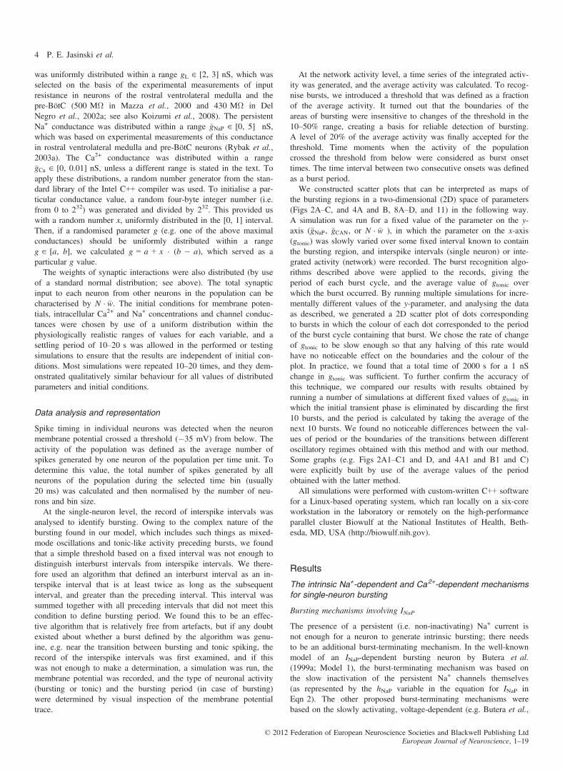
possible effects of Ca2+-dependent mechanisms were eliminated bysetting �gCa ¼ �gCAN ¼ 0. Three single-neuron models exhibiting INaP-dependent bursting were studied, which differed by the burst-termi-nating mechanisms operating in the models (Figs 1 and 2). The firstmodel (Figs 1A and A1, and 2A and A1) was qualitatively similar tothe classical Butera model (Butera et al., 1999a; Model 1), but withparameter values drawn from previous experimental measurements(Rybak et al., 2003a). Burst termination in this model was basedon the slow inactivation of INaP. In this case in Eqn 1,ICa = ICAN = IPump = 0, and hNaP changed in accordance withEqns 4 and 5. In the second model (Figs 1B and B1, and 2B andB1), in addition to the slowly inactivating INaP (as in the first model),we incorporated the burst-terminating mechanism based on the intra-cellular accumulation of Na+ (Eqn 15 at ICAN = 0) followed byintracellular Na+ concentration ([Na+]in)-dependent activation of theNa+/K+ pump (IPump, Eqn 15]. In the third neuron model (Figs 1Cand C1, and 2C and C1), INaP was considered to be non-inactivating(by setting hNaP = constant = 0.4; see Table 1), and burst terminationwas based entirely on the Na+/K+ pump. Not only did this Na+/K+
pump-based mechanism cause burst termination, but its relaxationwas also involved in the recovery of the membrane potential duringthe interburst interval, leading to the onset of the next burst.These models were comparatively investigated with respect to
their response to tonic excitatory drive (gtonic), which was elevatedslowly from 0 to 1 nS to sweep a range of baseline membrane
potentials (Figs 1A–C and A1–C1). In each model, the neuronstarted rhythmic bursting when the drive exceeded some model-spe-cific threshold (Figs 1A–C, and 2A–C, and A1–C1), and the fre-quency of this bursting increased (bursting period decreased) withincreasing gtonic. Then, when the drive exceeded another model-spe-cific threshold, the neuron switched from rhythmic bursting to tonicspiking (Figs 1A and B, and 2A–C and A1–C1).Figure 2A–C, shows respectively the bursting regions for each
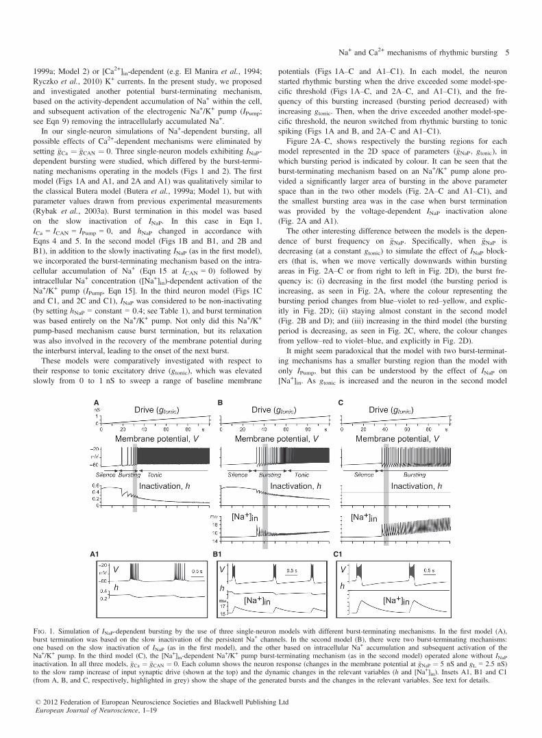
model represented in the 2D space of parameters (�gNaP; gtonic), inwhich bursting period is indicated by colour. It can be seen that theburst-terminating mechanism based on an Na+/K+ pump alone pro-vided a significantly larger area of bursting in the above parameterspace than in the two other models (Fig. 2A–C and A1–C1), andthe smallest bursting area was in the case when burst terminationwas provided by the voltage-dependent INaP inactivation alone(Fig. 2A and A1).The other interesting difference between the models is the depen-
dence of burst frequency on �gNaP. Specifically, when �gNaP isdecreasing (at a constant gtonic) to simulate the effect of INaP block-ers (that is, when we move vertically downwards within burstingareas in Fig. 2A–C or from right to left in Fig. 2D), the burst fre-quency is: (i) decreasing in the first model (the bursting period isincreasing, as seen in Fig. 2A, where the colour representing thebursting period changes from blue–violet to red–yellow, and explic-itly in Fig. 2D); (ii) staying almost constant in the second model(Fig. 2B and D); and (iii) increasing in the third model (the burstingperiod is decreasing, as seen in Fig. 2C, where, the colour changesfrom yellow–red to violet–blue, and explicitly in Fig. 2D).It might seem paradoxical that the model with two burst-terminat-
ing mechanisms has a smaller bursting region than the model withonly IPump, but this can be understood by the effect of INaP on[Na+]in. As gtonic is increased and the neuron in the second model
A B C
A1 B1 C1
Fig. 1. Simulation of INaP-dependent bursting by the use of three single-neuron models with different burst-terminating mechanisms. In the first model (A),burst termination was based on the slow inactivation of the persistent Na+ channels. In the second model (B), there were two burst-terminating mechanisms:one based on the slow inactivation of INaP (as in the first model), and the other based on intracellular Na+ accumulation and subsequent activation of theNa+/K+ pump. In the third model (C), the [Na+]in-dependent Na
+/K+ pump burst-terminating mechanism (as in the second model) operated alone without INaPinactivation. In all three models, �gCa ¼ �gCAN ¼ 0. Each column shows the neuron response (changes in the membrane potential at �gNaP ¼ 5 nS and gL = 2.5 nS)to the slow ramp increase of input synaptic drive (shown at the top) and the dynamic changes in the relevant variables (h and [Na+]in). Insets A1, B1 and C1(from A, B, and C, respectively, highlighted in grey) show the shape of the generated bursts and the changes in the relevant variables. See text for details.
© 2012 Federation of European Neuroscience Societies and Blackwell Publishing LtdEuropean Journal of Neuroscience, 1–19
Na+ and Ca2+ mechanisms of rhythmic bursting 5
becomes increasingly depolarised, INaP is progressively inactivated,so that its contribution to intracellular Na+ accumulation during theactive phase decreases (compare [Na+]in in Fig. 1B1 to that in C1),and burst-terminating IPump becomes progressively weaker.There is also an interesting issue concerning the different effects
of the INaP suppression used to simulate the possible effects of riluz-ole (i.e. a decrease in �gNaP at a constant excitatory drive, whichwould correspond to a vertical downwards shift from burstingregions in each 2D plot in Fig. 2A–C and a right-to-left shift inFig. 2D). It can be seen that, in the first model (Fig. 2A and D, bluecurve), this �gNaP reduction can only cause a switch from bursting tosilence, whereas in the second and third models containing theNa+/K+ pump (Fig. 2B and C), the result depends on the drive. At lowdrive, this INaP suppression also produces a switch from bursting tosilence, but at higher drive, it causes a switch from bursting to tonicspiking in the majority of the bursting region in each of these models(see examples in Fig. 2D, red and green curves, respectively).
Bursting involving ICa and ICAN
This component of the study was motivated by the experimental andmodelling studies of Del Negro and his collaborators (Crowderet al., 2007; Pace et al., 2007; Pace & Del Negro, 2008; Kreyet al., 2010; Dunmyre et al., 2011), as well as Toporikova & Butera(2011), and focused on simulation of an ICAN-dependent burstingmechanism. To exclude the effect of INaP, in all models consideredhere we set �gNaP ¼ 0. In these models, ICAN was activated by intra-cellular Ca2+, whose accumulation was provided by the IP3-depen-dent Ca2+ release from intracellular stores (Eqns 11–14). This
process in our models critically depended on Ca2+ influx throughvoltage-gated Ca2+ channels (i.e. by ICa), which provided an initial[Ca+2]in inducing a nonlinear positive feedback mechanism knownas Ca2+-induced Ca2+ release (CICR). Both the input synaptic acti-vation (provided by synaptic drive) and the [Ca2+]in-dependent acti-vation of ICAN contributed to the initial membrane depolarisation(burst onset), and their contributions depended on the input synapticactivation. Two burst-terminating mechanisms were considered. Onemechanism involved Ca2+-dependent inactivation of IP3R (regulatedby the gating variable l; see Eqn 13), leading to the reduction of[Ca2+]in and deactivation of ICAN (see Toporikova & Butera, 2011).The second mechanism was based on the activity-dependent accu-mulation of intracellular Na+ followed by the [Na+]in-activated IPump,i.e. the same Na+/K+-based mechanism as in the second and thirdINaP-based models described above. However, in contrast to theINaP-based models, the accumulation of intracellular Na+ in thesemodels was mainly provided by ICAN (and partly by INa), but not byINaP, as �gNaP ¼ 0 (see Eqn 15).In order to investigate the effect of these burst-terminating mecha-
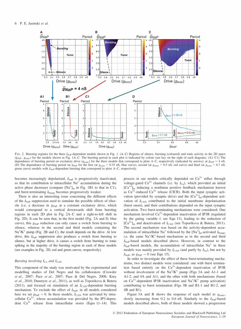
nisms, two distinct models were considered: one with burst termina-tion based entirely on the Ca2+-dependent inactivation of IP3Rwithout involvement of the Na+/K+ pump (Figs 3A and A1-1 andA1-2, and 4A and A1), and the other with both mechanisms (basedon Ca2+-dependent IP3R inactivation and Na+/K+ pump activation)contributing to burst termination (Figs 3B and B1-1 and B1-2, and4B and B1).Figure 3A and B shows the response of each model to gtonic,
slowly increasing from 0.2 to 0.6 nS. Similarly to the INaP-basedmodels described above, both of these models showed a progression
A B C
A1 B1 C1 D
Fig. 2. Bursting regimes for the three INaP-dependent models shown in Fig. 1. (A–C) Regions of silence, bursting (coloured) and tonic activity in the 2D space(�gNaP; gtonic) for the models shown in Fig. 1A–C. The bursting period in each plot is indicated by colour (see key on the right of each diagram). (A1–C1) Thedependence of bursting period on excitatory drive (gtonic) for the three models that correspond to plots A–C, respectively (indicated by arrows), at �gNaP ¼ 4 nS.(D) The dependence of bursting period on �gNaP for the first (at gtonic = 0.35 nS, blue curve), second (at gtonic = 0.5 nS, red curve) and third (at gtonic = 0.7 nS,green curve) models with INaP-dependent bursting that correspond to plots A–C, respectively.
© 2012 Federation of European Neuroscience Societies and Blackwell Publishing LtdEuropean Journal of Neuroscience, 1–19
6 P. E. Jasinski et al.
over the three regimes of silence, bursting and tonic spiking as drivewas increased (Figs 3A and B, and 4A and B), and the burst fre-quency increased in the bursting regime with increasing gtonic(Figs 3A and B, and 4A, B, A1 and B1). The insets in Fig. 3A1-1and B1-1 show burst details in each model at relatively low valuesof drive, whereas the insets in Fig. 3A1-2 and B1-2 show burstdetails in these models at higher values of drive. At low drives(Fig. 3A1-1 and B1-1), the initial increase in [Ca2+]in through ICaand the CICR mechanism precedes membrane depolarisation andburst onset (indicated by the vertical dashed line). This accumulatedintracellular Ca2+ activates ICAN, which, in turn, produces the mem-brane depolarisation and burst onset. In contrast, at higher drives(Fig. 3A1-2 and B1-2), the initial membrane depolarisation is pro-vided by the dynamics of synaptic input (i.e. drive) and IPump
(which decreases during the preceding interburst interval), and hencethe onset of the burst (indicated by the dashed line) precedes the[Ca2+]in-dependent ICAN activation. This can also explain a clear dif-ference in the intraburst spike pattern between the corresponding
bursts (a decreasing spike frequency at lower drives vs. an initialramp increase in spike frequency within the burst at higher drives,Fig. 3B1-1 vs. B1-2). Although ICAN does not play a leading role inburst initiation at higher drives, it contributes to the above patterningof intraburst spikes (the initial ramp in spike frequency) in bothmodels. In addition, ICAN is critical for burst termination in bothmodels, as described below.The above insets demonstrate the differences between the two
models in burst termination. In the first model (Fig. 3A1-1 andA1-2), burst termination (controlled by IP3R inactivation defined byl) occurs when [Ca2+]in drops below the threshold for ICAN activa-tion (indicated by the dot-dashed line). In the second model(Fig. 3B1-1 and B1-2), burst termination is mostly provided by theICAN-dependent intracellular accumulation of Na+ followed by[Na+]in-dependent activation of the Na+/K+ pump (IPump, see the ver-tical dot-dashed line, showing that bursts are terminated not when[Ca2+]in drops below the half-activation concentration for ICAN, as inFig. 3A1-1 and A1-2, but when [Na+]in reaches its maximum).
A
A1-1 A1-2 B1-1 B1-2
B
Fig. 3. Simulation of ICa-dependent and ICAN-dependent bursting by the use of two single-neuron models with different burst-terminating mechanisms. In thefirst model (A), burst termination was based on Ca2+-dependent IP3R inactivation (see the traces for IP3R gating variable l and [Ca2+]in). In the second model(B), there were two burst-terminating mechanisms: one based on Ca2+-dependent IP3R inactivation (as in the first model), and the other based on intracellularNa+ accumulation (see [Na+]in traces) and subsequent activation of the Na+/K+ pump. In both models, �gNaP ¼ 0. Each column shows the neuron response(changes in the membrane potential at �gCa ¼ 0:01 nS, �gCAN ¼ 1 nS, and gL = 2.5 nS) to the slow ramp increase of input synaptic drive (shown at the top).Insets A1-1, A1-2, B1-1 and B1-2 (from A and B traces, respectively, highlighted in grey) show the shapes of generated bursts and the changes in relevant vari-ables. See text for details.
© 2012 Federation of European Neuroscience Societies and Blackwell Publishing LtdEuropean Journal of Neuroscience, 1–19
Na+ and Ca2+ mechanisms of rhythmic bursting 7
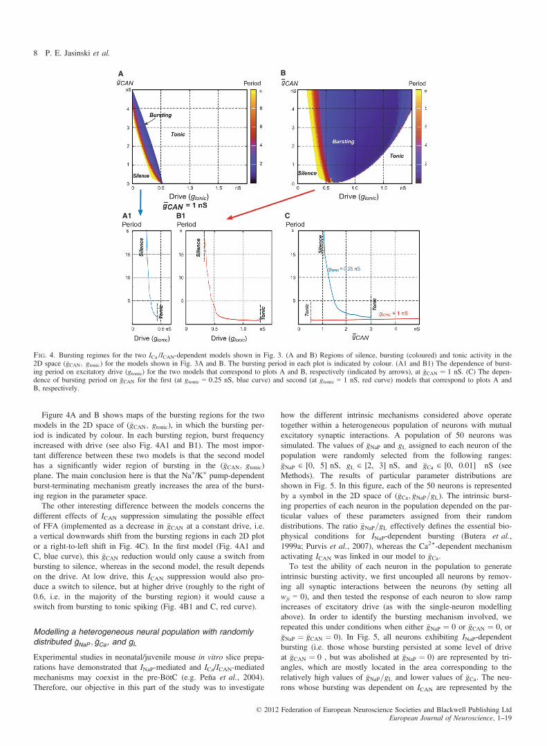
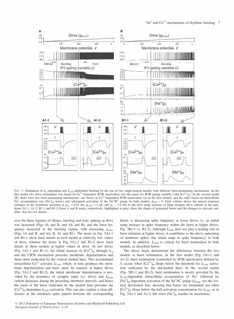
Figure 4A and B shows maps of the bursting regions for the twomodels in the 2D space of (�gCAN; gtonic), in which the bursting per-iod is indicated by colour. In each bursting region, burst frequencyincreased with drive (see also Fig. 4A1 and B1). The most impor-tant difference between these two models is that the second modelhas a significantly wider region of bursting in the (�gCAN; gtonic)plane. The main conclusion here is that the Na+/K+ pump-dependentburst-terminating mechanism greatly increases the area of the burst-ing region in the parameter space.The other interesting difference between the models concerns the
different effects of ICAN suppression simulating the possible effectof FFA (implemented as a decrease in �gCAN at a constant drive, i.e.a vertical downwards shift from the bursting regions in each 2D plotor a right-to-left shift in Fig. 4C). In the first model (Fig. 4A1 andC, blue curve), this �gCAN reduction would only cause a switch frombursting to silence, whereas in the second model, the result dependson the drive. At low drive, this ICAN suppression would also pro-duce a switch to silence, but at higher drive (roughly to the right of0.6, i.e. in the majority of the bursting region) it would cause aswitch from bursting to tonic spiking (Fig. 4B1 and C, red curve).
Modelling a heterogeneous neural population with randomlydistributed �gNaP ; �gCa, and gL
Experimental studies in neonatal/juvenile mouse in vitro slice prepa-rations have demonstrated that INaP-mediated and ICa/ICAN-mediatedmechanisms may coexist in the pre-BötC (e.g. Peña et al., 2004).Therefore, our objective in this part of the study was to investigate
how the different intrinsic mechanisms considered above operatetogether within a heterogeneous population of neurons with mutualexcitatory synaptic interactions. A population of 50 neurons wassimulated. The values of �gNaP and gL assigned to each neuron of thepopulation were randomly selected from the following ranges:�gNaP ∈ [0, 5] nS, gL ∈ [2, 3] nS, and �gCa ∈ [0, 0.01] nS (seeMethods). The results of particular parameter distributions areshown in Fig. 5. In this figure, each of the 50 neurons is representedby a symbol in the 2D space of (�gCa; �gNaP=gL). The intrinsic burst-ing properties of each neuron in the population depended on the par-ticular values of these parameters assigned from their randomdistributions. The ratio �gNaP=gL effectively defines the essential bio-physical conditions for INaP-dependent bursting (Butera et al.,1999a; Purvis et al., 2007), whereas the Ca2+-dependent mechanismactivating ICAN was linked in our model to �gCa.To test the ability of each neuron in the population to generate
intrinsic bursting activity, we first uncoupled all neurons by remov-ing all synaptic interactions between the neurons (by setting allwji = 0), and then tested the response of each neuron to slow rampincreases of excitatory drive (as with the single-neuron modellingabove). In order to identify the bursting mechanism involved, werepeated this under conditions when either �gNaP ¼ 0 or �gCAN ¼ 0, or�gNaP ¼ �gCAN ¼ 0). In Fig. 5, all neurons exhibiting INaP-dependentbursting (i.e. those whose bursting persisted at some level of driveat �gCAN ¼ 0 , but was abolished at �gNaP ¼ 0) are represented by tri-angles, which are mostly located in the area corresponding to therelatively high values of �gNaP=gL and lower values of �gCa. The neu-rons whose bursting was dependent on ICAN are represented by the
A
A1 B1 C
B
Fig. 4. Bursting regimes for the two ICa /ICAN-dependent models shown in Fig. 3. (A and B) Regions of silence, bursting (coloured) and tonic activity in the2D space (�gCAN; gtonic) for the models shown in Fig. 3A and B. The bursting period in each plot is indicated by colour. (A1 and B1) The dependence of burst-ing period on excitatory drive (gtonic) for the two models that correspond to plots A and B, respectively (indicated by arrows), at �gCAN ¼ 1 nS. (C) The depen-dence of bursting period on �gCAN for the first (at gtonic = 0.25 nS, blue curve) and second (at gtonic = 1 nS, red curve) models that correspond to plots A andB, respectively.
© 2012 Federation of European Neuroscience Societies and Blackwell Publishing LtdEuropean Journal of Neuroscience, 1–19
8 P. E. Jasinski et al.
filled circles. The bursting in these neurons persisted at �gNaP ¼ 0,but could be abolished at �gCAN ¼ 0; these neurons are located in thearea corresponding to the higher values of �gCa and relatively lowervalues of �gNaP=gL. The neurons represented by crosses could expressbursting based on any of the above mechanisms; that is, their burst-ing could be abolished only if �gNaP ¼ �gCAN ¼ 0. It is not surprisingthat these neurons are located in the area corresponding to thehigher values of both �gCa and �gNaP=gL. Finally, the neurons unableto express bursting under any condition are represented by squares,and are located in the area corresponding to the lower values ofboth �gCa and �gNaP=gL.Figure 5 shows that, at the ranges assigned for random distribution
of �gNaP; gL; and �gCa, the 50-neuron population of uncoupled neuronscontained 43 potential bursters (86%; this includes all cells exceptthose represented by rectangles). For comparison, Koshiya & Smith(1999) reported that 70% of inspiratory cells in the pre-BötC main-tained bursting (most likely INaP-dependent) in neonatal rat slicesafter neuron uncoupling by 6-cyano-7-nitroquinoxaline-2,3-dioneapplication, which is close to the percentage of bursters in our simu-lation. The ranges used for �gNaP and gL distributions (justified inMethods) resulted in 19 (38%) neurons that could intrinsically gener-ate bursting after blockade of ICa (i.e. at �gCa ¼ 0); these are the neu-rons shown in Fig. 5 by triangles and crosses together. Thepercentage of these bursters in the model looks plausible, given theaccount of the percentage of INaP-dependent bursters estimated inslices from neonatal rats (60%, Koizumi & Smith, 2008) and the per-centage of Cd2+-insensitive bursters found in slices from P11–15mice (29%, Peña et al., 2004). This provided additional support forthe distribution ranges of �gNaP and gL used in our model. Unfortu-nately, no experimental data were found to justify an advancedassignment of the range for �gCa distribution. The use of�gCa � 0.001 nS led to the absence of ICa/ICAN-dependent busters inthe population. Setting a range of �gCa ∈ [0, 0.01] nS (as in the simu-
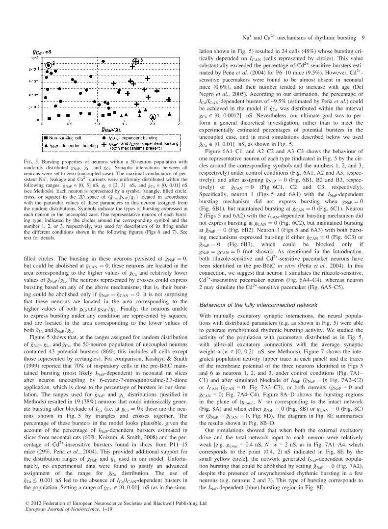
lation shown in Fig. 5) resulted in 24 cells (48%) whose bursting cri-tically depended on ICAN (cells represented by circles). This valuesubstantially exceeded the percentage of Cd2+-sensitive bursters esti-mated by Peña et al. (2004) for P6–10 mice (9.5%). However, Cd2+-sensitive pacemakers were found to be almost absent in neonatalmice (0.6%), and their number tended to increase with age (DelNegro et al., 2005). According to our estimation, the percentage ofICa/ICAN-dependent busters of ~9.5% (estimated by Peña et al.) couldbe achieved in the model if �gCa was distributed within the interval�gCa ∈ [0, 0.0012] nS. Nevertheless, our ultimate goal was to per-form a general theoretical investigation, rather than to meet theexperimentally estimated percentages of potential bursters in theuncoupled case, and in most simulations described below we used�gCa ∈ [0, 0.01] nS, as shown in Fig. 5.Figure 6A1–C1, and A2–C2 and A3–C3 shows the behaviour of
one representative neuron of each type (indicated in Fig. 5 by the cir-cles around the corresponding symbols and the numbers 1, 2, and 3,respectively) under control conditions (Fig. 6A1, A2 and A3, respec-tively), and after assigning �gNaP ¼ 0 (Fig. 6B1, B2 and B3, respec-tively) or �gCAN ¼ 0 (Fig. 6C1, C2 and C3, respectively).Specifically, neuron 1 (Figs 5 and 6A1) with the INaP-dependentbursting mechanism did not express bursting when �gNaP ¼ 0(Fig. 6B1), but maintained bursting at �gCAN ¼ 0 (Fig. 6C1). Neuron2 (Figs 5 and 6A2) with the ICAN-dependent bursting mechanism didnot express bursting at �gCAN ¼ 0 (Fig. 6C2), but maintained burstingat �gNaP ¼ 0 (Fig. 6B2). Neuron 3 (Figs 5 and 6A3) with both burst-ing mechanisms expressed bursting if either �gCAN ¼ 0 (Fig. 6C3) or�gNaP ¼ 0 (Fig. 6B3), which could be blocked only if�gNaP ¼ �gCAN ¼ 0 (not shown). As mentioned in the Introduction,both riluzole-sensitive and Cd2+-sensitive pacemaker neurons havebeen identified in the pre-BötC in vitro (Peña et al., 2004). In thisconnection, we suggest that neuron 1 simulates the riluzole-sensitive,Cd2+-insensitive pacemaker neuron (Fig. 6A4–C4), whereas neuron2 may simulate the Cd2+-sensitive pacemaker (Fig. 6A5–C5).
Behaviour of the fully interconnected network
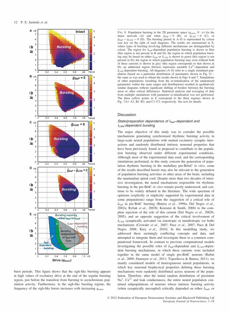
With mutually excitatory synaptic interactions, the neural popula-tions with distributed parameters (e.g. as shown in Fig. 5) were ableto generate synchronised rhythmic bursting activity. We studied theactivity of the population with parameters distributed as in Fig. 5,with all-to-all excitatory connections with the average synapticweight �w ð�w ∈ [0, 0.2] nS, see Methods). Figure 7 shows the inte-grated population activity (upper trace in each panel) and the tracesof the membrane potential of the three neurons identified in Figs 5and 6 as neurons 1, 2, and 3, under control conditions (Fig. 7A1–C1) and after simulated blockade of INaP (�gNaP ¼ 0; Fig. 7A2–C2)or ICAN (�gCAN = 0; Fig. 7A3–C3), or both currents (�gNaP = 0 and�gCAN = 0; Fig. 7A4–C4). Figure 8A–D shows the bursting regionsin the plane of (gtonic, N � �w) corresponding to the intact network(Fig. 8A) and when either �gNaP = 0 (Fig. 8B) or �gCAN = 0 (Fig. 8C)or (�gNaP ¼ �gCAN ¼ 0, Fig. 8D). The diagram in Fig. 8E summarisesthe results shown in Fig. 8B–D.Our simulations showed that when both the external excitatory
drive and the total network input to each neuron were relativelyweak [e.g. gtonic = 0.4 nS, N � �w = 2 nS, as in Fig. 7A1–A4, whichcorresponds to the point (0.4, 2) nS indicated in Fig. 8E by thesmall yellow circle], the network generated INaP-dependent popula-tion bursting that could be abolished by setting �gNaP ¼ 0 (Fig. 7A2),despite the presence of unsynchronised rhythmic bursting in a fewneurons (e.g. neurons 2 and 3). This type of bursting corresponds tothe INaP-dependent (blue) bursting region in Fig. 8E.
Fig. 5. Bursting properties of neurons within a 50-neuron population withrandomly distributed �gNaP, gL, and �gCa. Synaptic interactions between allneurons were set to zero (uncoupled case). The maximal conductance of per-sistent Na+, leakage and Ca2+ currents were uniformly distributed within thefollowing ranges: �gNaP ∈ [0, 5] nS, gL ∈ [2, 3] nS, and �gCa ∈ [0, 0.01] nS(see Methods). Each neuron is represented by a symbol (triangle, filled circle,cross, or square) in the 2D space of (�gCa; �gNaP=gL) located in accordancewith the particular values of these parameters in this neuron assigned fromthe random distributions. Symbols indicate the types of bursting expressed ineach neuron in the uncoupled case. One representative neuron of each burst-ing type, indicated by the circles around the corresponding symbol and thenumber 1, 2, or 3, respectively, was used for description of its firing underthe different conditions shown in the following figures (Figs 6 and 7). Seetext for details.
© 2012 Federation of European Neuroscience Societies and Blackwell Publishing LtdEuropean Journal of Neuroscience, 1–19
Na+ and Ca2+ mechanisms of rhythmic bursting 9

An increase in the total network synaptic input to each neuron atthe same gtonic (e.g. from N � �w ¼ 2 to N � �w ¼ 5 nS, simulating anincrease in the number of neurons in the population; Fig. 7B1–B4),which corresponds to the point (0.4, 5) nS (indicated in Fig. 8E byanother small yellow circle), allowed the population to maintain(ICAN-dependent) population bursting at �gNaP ¼ 0 (Fig. 7B2): thisbursting could be abolished only if both �gNaP ¼ 0 and �gCAN ¼ 0(Fig. 7B4). The same type of bursting could be obtained by increas-ing drive (e.g. gtonic = 0.5 nS) while keeping a low level of excit-atory synaptic interactions within the network (N � �w ¼ 2 nS, notshown). This type of bursting, requiring the presence of either INaPor ICAN, corresponds to the green region in Fig. 8E.A further increase of excitatory drive, e.g. by setting
gtonic = 0.5 nS at N � �w ¼ 5 nS as in Fig. 7C1–C4 (corresponding tothe point (0.5, 5) nS indicated in Fig. 8E by the third small yellowcircle), allowed for population bursting independent of both INaPand ICAN (see Figs 7C4 and 8D, and the grey region in Fig. 8E). Inthis case, relatively strong excitatory synaptic interactions within thepopulation provided burst initiation and then its termination via
intracellular Na+ accumulation and Na+/K+ pump activation). Notethat setting �gCAN = 0 to simulate the suppression of only ICAN couldnot stop population bursting in the above three cases (see Fig. 7A3–C3). Unstable, irregular ICAN-dependent bursting could only exist atvery high drive values with a moderate level of N � �w (see Fig. 8Band the brown region in Fig. 8E), and the biological plausibility ofthis bursting is questionable.
Synchronised Ca2+ oscillations and sigh-like rhythmic activity
Under certain conditions, the model was capable of generating anadditional slow sigh-like bursting activity with augmented amplitudearising from low-frequency Ca2+ oscillations synchronised over thepopulation. In the simulations presented here, we narrowed theallowed ranges of parameters in the random distributions of themaximal Ca2+ and leak conductances (�gCa ∈ [0.0006, 0.0007] nSand gL ∈ [2.25, 2.75] nS). In addition, we increased both the IP3concentration (IP3 = 1.5 9 10–3 mM) and the maximal conductancefor ICAN channels (�gCAN ¼ 3 nS). The results of simulations at
A1 B1 C1
A2 B2 C2
A3 B3 C3
A4 B4 C4
A5 B5 C5
Fig. 6. Firing behaviour of the uncoupled representative neurons with different values of �gNaP; �gCa, and gL. A1–C1, A2–C2 and A3–C3 show the behaviour ofone representative neuron of each type (indicated in Fig. 5 by the circles around the corresponding symbol and the number 1, 2, and 3, respectively) under con-trol conditions (A1, A2, and A3, respectively), and after blocking INaP (�gNaP ¼ 0 in B1, B2, and B3, respectively) or ICAN (�gCAN ¼ 0 in C1, C2, and C3, respec-tively). Specifically, neuron 1 (A1–C1; see also Fig. 5) with the INaP-dependent bursting mechanism did not express bursting when �gNaP ¼ 0 (B1, exhibiting aswitch to tonic spiking with an increase in excitatory drive), but maintained bursting activity at �gCAN ¼ 0 (C1). Neuron 2 (A2–C2; see also Fig. 5) with theICAN-dependent bursting mechanism did not express bursting when �gCAN ¼ 0 (C2), but maintained bursting activity at �gNaP ¼ 0 (B2). Neuron 3 (A3–C3; seealso Fig. 5) with both bursting mechanisms; this neuron expressed bursting when either �gCAN ¼ 0 (C3) or �gNaP ¼ 0 (B3), which could be blocked only if�gNaP ¼ �gCAN ¼ 0 (not shown). A4–C4 show an isolated riluzole-sensitive, Cd2+-insensitive intrinsically bursting ‘pacemaker’ neuron recorded in the pre-BötCin vitro after pharmacological blockade of excitatory and inhibitory synaptic transmission (cocktail, A4), and after application of riluzole (B4) and Cd2+ (C4,modified from Peña et al., 2004, fig. 3, with permission). A5–C5 show an isolated riluzole-insensitive, Cd2+-sensitive intrinsic burster before (cocktail, A4) andafter application of riluzole (B5) and Cd2+ (C5, also from Peña et al., 2004; Fig. 3). See text for details.
© 2012 Federation of European Neuroscience Societies and Blackwell Publishing LtdEuropean Journal of Neuroscience, 1–19
10 P. E. Jasinski et al.

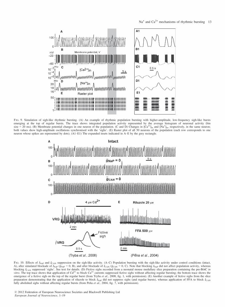
gtonic = 0.7 nS and N � �w ¼ 2:5 nS are shown in Fig. 9. These simu-lations demonstrate the emergence of higher-amplitude, low-fre-quency bursts that appeared periodically at the top of the regularbursts (Fig. 9A and A1). Figure 9C shows the low-amplitude[Ca+2]in transients occurring in each neuron during regular bursts,which were subthreshold for the CICR mechanism. The slow back-ground accumulation of intracellular Ca2+ in each neuron finallyresulted in synchronised CICR producing low-frequency, high-amplitude [Ca+2]in transients, which, through synchronous activationof ICAN, produced higher-amplitude bursts emerging at the top ofthe regular bursts (Fig. 9A and B), forming distinctive biphasicburst shapes (Fig. 9A1) and delaying the onset of the next regularbursts (Fig. 9A, B, E, A1, B1 and E1). These bursts were qualita-
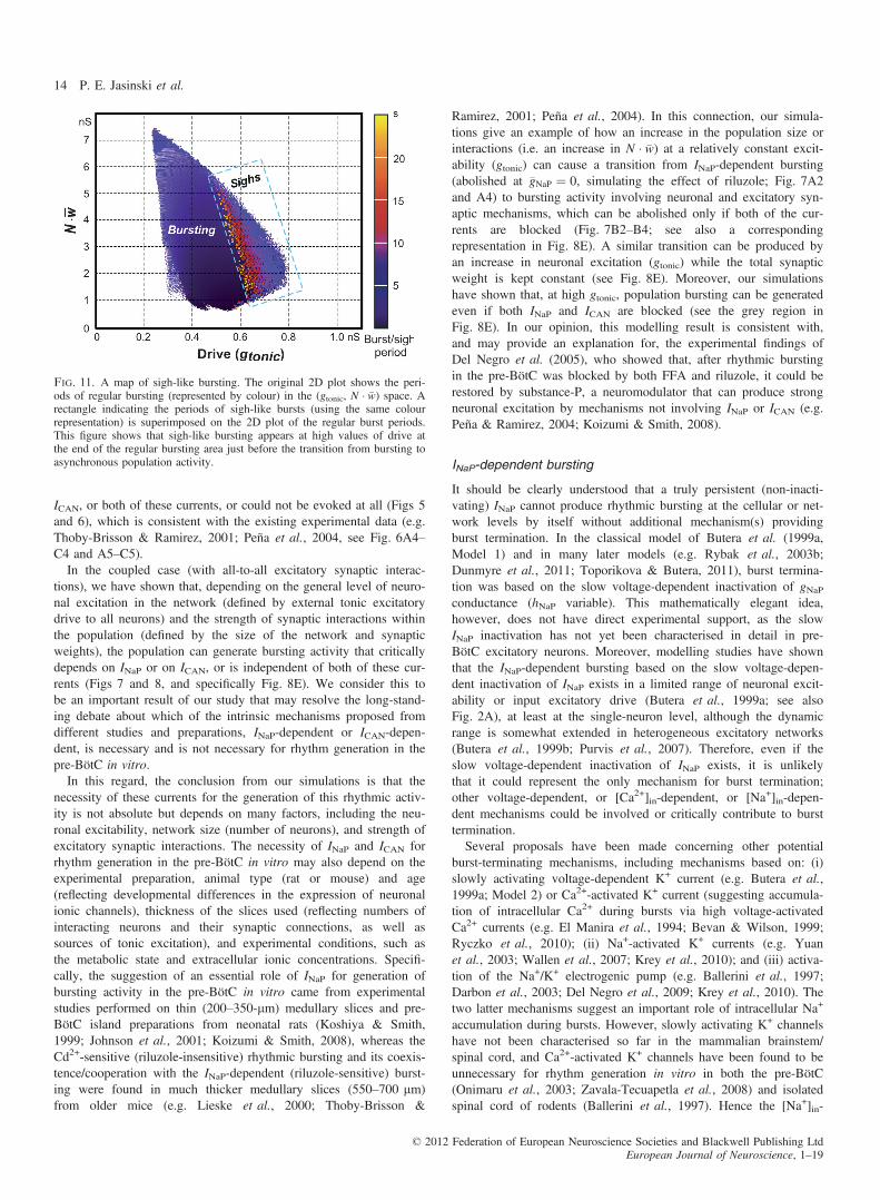
tively similar to the ‘fictive sighs’ that have been previouslydescribed by Ramirez and his collaborators (Lieske et al., 2000;Peña et al., 2004; Tryba et al., 2008, e.g. see Fig. 10D and E). Asrelatively high drives were used, the regular bursting was indepen-dent of both INaP and ICAN (see the previous section), and hencewas not abolished at �gNaP ¼ 0 or �gCAN ¼ 0 (Fig. 10B and C). How-ever, assigning �gCAN ¼ 0 (or �gCa ¼ 0, not shown) fully removed theslow sigh-like oscillations (Fig. 10C), confirming that the generationof sigh-like bursts in our model is ICa/ICAN-dependent.Figure 11 presents a colour-coded 2D plot showing the period of
regular bursting (represented by colour) in the (gtonic, N � �w) plane.A rectangle indicating the period of sigh-like bursts (using the samecolour representation) is superimposed on the 2D plot of the regular
A1 A2 A3 A4
B1 B2 B3 B4
C1 C2 C3 C4
Fig. 7. Rhythmic bursting activity generated by the 50-neuron population with mutual excitatory synaptic interconnections. Each panel shows the integratedpopulation activity represented by the average histogram of neuronal activities [upper trace, y-axis represents an average histogram of population activity inspikes/(neuron�s), bin size = 20 ms] and the membrane potential traces of three neurons, indicated in Figs 5 and 6 as neurons 1, 2, and 3, respectively. The toprow (A1–A4) shows INaP-dependent bursting that occurs in the population at relatively low levels of both neuronal interaction (N � �w =2 nS) and drive(gtonic = 0.4 nS). The middle row (B1–B4) shows population bursting requiring either INaP or ICAN that occurs with increased neuronal interactions(N � �w = 5 nS) at the same level of drive (gtonic = 0.4 nS). The bottom row (C1–C4) shows population bursting independent of INaP and ICAN that occurs athigher levels of interaction (N � �w = 5 nS) and drive (gtonic = 0.5 nS). The first column (A1–C1) shows the population activity and activities of the identifiedneurons 1, 2 and 3 under control conditions; the second column (A2–C2) shows the simulated effects of INaP blockade (�gNaP = 0) on the population and single-neuron bursting in the three cases of drive and neuronal interaction described above; the third column (A3–C3) shows the simulated effects of ICAN blockade(�gCAN = 0) on the population and single-neuron bursting for the same three cases; the fourth column (A4–C4) shows the simulated effects of blockade of bothINaP and ICAN (�gNaP = 0 and �gCAN = 0). See text for details.
© 2012 Federation of European Neuroscience Societies and Blackwell Publishing LtdEuropean Journal of Neuroscience, 1–19
Na+ and Ca2+ mechanisms of rhythmic bursting 11
burst periods. This figure shows that the sigh-like bursting appearsat high values of excitatory drive at the end of the regular burstingregion, just before the transition from bursting to asynchronous pop-ulation activity. Furthermore, in the sigh-like bursting regime, thefrequency of the sigh-like bursts increases with increasing gtonic.
Discussion
State/preparation dependence of INaP-dependent andICAN-dependent bursting
The major objective of this study was to consider the possiblemechanisms generating synchronised rhythmic bursting activity inlarge-scale neural populations with mutual excitatory synaptic inter-actions and randomly distributed intrinsic neuronal properties thathave been previously found or proposed to contribute to the popula-tion bursting observed under different experimental conditions.Although most of the experimental data used, and the correspondingsimulations performed, in this study concern the generation of popu-lation rhythmic bursting in the medullary pre-BötzC in vitro, someof the results described herein may also be relevant to the generationof population bursting activities in other areas of the brain, includingthe mammalian spinal cord. Despite more than two decades of inten-sive investigation, the neural mechanisms responsible for rhythmicbursting in the pre-BötC in vitro remain poorly understood, and con-tinue to be widely debated in the literature. The wide spectrum ofopinions (explicitly or implicitly supported by experimental data insome preparations) range from the suggestion of a critical role ofINaP in pre-BötC bursting (Butera et al., 1999a; Del Negro et al.,2002a; Rybak et al., 2003b; Koizumi & Smith, 2008) to the com-plete rejection of the role of this current (Del Negro et al., 2002b,2005), and an opposite suggestion of the critical involvement ofICAN synaptically activated via ionotropic or metabotropic (or both)mechanisms (Crowder et al., 2007; Pace et al., 2007; Pace & DelNegro, 2008; Krey et al., 2010). In this modelling study, weaddressed these seemingly conflicting concepts and data, andattempted to integrate them and investigate them in a common com-putational framework. In contrast to previous computational modelsinvestigating the possible roles of INaP-dependent and ICAN-depen-dent bursting mechanisms, in which these currents were includedtogether in the same model of single pre-BötC neurons (Rubinet al., 2009; Dunmyre et al., 2011; Toporikova & Butera, 2011), wemainly considered models of heterogeneous neural populations, inwhich key neuronal biophysical properties defining these burstingmechanisms were randomly distributed across neurons of the popu-lation. Therefore, after the initial random distribution of persistentNa+, Ca2+ and leak conductances, the entire neural population con-tained subpopulations of neurons whose intrinsic bursting activity(when synaptically uncoupled) critically depended on either INaP, or
A
B
C
D
E
Fig. 8. Population bursting in the 2D parameter space (gtonic, N � �w) for theintact network (A) and when �gNaP = 0 (B), or �gCAN = 0 (C), or�gNaP ¼ �gCAN ¼ 0 (D). The bursting period in A–D is represented by colour(see key on the right of each diagram). The results are summarised in E,where types of bursting involving different mechanisms are distinguished bycolour. The region for INaP-dependent population bursting is shown in blue(this region is not present in B and D); the region in which population burst-ing may be based on either INaP or ICAN is shown in green (this region is notpresent in D); the region in which population bursting may exist without bothof these currents is shown in grey (this region corresponds to that shown inD); an additional region (brown) represents unstable Ca2+-dependent andICAN-dependent bursting. All diagrams (A–E) refer to a single simulated pop-ulation (based on a particular distribution of parameters shown in Fig. 5) –the same as was used to obtain the results shown in Figs 6 and 7. Simulationsof other populations (resulting from the re-initialisation of the randomisedparameters within the same ranges and distributions) resulted in qualitativelysimilar diagrams without significant shifting of borders between the burstingareas or other critical differences. Statistical analysis and averaging of datafrom multiple simulations with parameter re-initialisation was not performed.The three yellow points in E correspond to the three regimes shown inFig. 7A1–A3, B1–B3, and C1–C3, respectively. See text for details.
© 2012 Federation of European Neuroscience Societies and Blackwell Publishing LtdEuropean Journal of Neuroscience, 1–19
12 P. E. Jasinski et al.
A A1
B1
C1
D1
E1
B
C
D
E
Fig. 9. Simulation of sigh-like rhythmic bursting. (A) An example of rhythmic population bursting with higher-amplitude, low-frequency sigh-like burstsemerging at the top of regular bursts. The trace shows integrated population activity represented by the average histogram of neuronal activity (binsize = 20 ms). (B) Membrane potential changes in one neuron of the population. (C and D) Changes in [Ca2+]in and [Na+]in, respectively, in the same neuron;both values show high-amplitude oscillations synchronised with the ‘sighs’. (E) Raster plot of all 50 neurons of the population (each row corresponds to oneneuron whose spikes are represented by dots). (A1–E1) The expanded insets indicated in A–E by the grey rectangle.
A
B
C
D E
Fig. 10. Effects of INaP and ICAN suppression on the sigh-like activity. (A–C) Population bursting with the sigh-like activity under control conditions (intact,A), after simulated blockade of INaP (�gNaP = 0, B), and after blockade of ICAN (�gCAN = 0, C). Note that blocking INaP did not affect population activity, whereasblocking ICAN suppressed ‘sighs’. See text for details. (D) Fictive sighs recorded from a neonatal mouse medullary slice preparation containing the pre-BötC invitro. The top trace shows that application of Cd2+ to block Ca2+ currents suppressed fictive sighs without affecting regular bursting; the bottom trace shows theemergence of a fictive sigh on the top of the regular burst (from Tryba et al., 2008, fig. 1, with permission). (E) Another example of fictive sighs from the slicepreparation demonstrating that the application of riluzole to block INaP did not suppress sighs (and regular bursts), whereas application of FFA to block ICANfully abolished sighs without affecting regular bursts (from Peña et al., 2004, fig. 7, with permission).
© 2012 Federation of European Neuroscience Societies and Blackwell Publishing LtdEuropean Journal of Neuroscience, 1–19
Na+ and Ca2+ mechanisms of rhythmic bursting 13
ICAN, or both of these currents, or could not be evoked at all (Figs 5and 6), which is consistent with the existing experimental data (e.g.Thoby-Brisson & Ramirez, 2001; Peña et al., 2004, see Fig. 6A4–C4 and A5–C5).In the coupled case (with all-to-all excitatory synaptic interac-
tions), we have shown that, depending on the general level of neuro-nal excitation in the network (defined by external tonic excitatorydrive to all neurons) and the strength of synaptic interactions withinthe population (defined by the size of the network and synapticweights), the population can generate bursting activity that criticallydepends on INaP or on ICAN, or is independent of both of these cur-rents (Figs 7 and 8, and specifically Fig. 8E). We consider this tobe an important result of our study that may resolve the long-stand-ing debate about which of the intrinsic mechanisms proposed fromdifferent studies and preparations, INaP-dependent or ICAN-depen-dent, is necessary and is not necessary for rhythm generation in thepre-BötC in vitro.In this regard, the conclusion from our simulations is that the
necessity of these currents for the generation of this rhythmic activ-ity is not absolute but depends on many factors, including the neu-ronal excitability, network size (number of neurons), and strength ofexcitatory synaptic interactions. The necessity of INaP and ICAN forrhythm generation in the pre-BötC in vitro may also depend on theexperimental preparation, animal type (rat or mouse) and age(reflecting developmental differences in the expression of neuronalionic channels), thickness of the slices used (reflecting numbers ofinteracting neurons and their synaptic connections, as well assources of tonic excitation), and experimental conditions, such asthe metabolic state and extracellular ionic concentrations. Specifi-cally, the suggestion of an essential role of INaP for generation ofbursting activity in the pre-BötC in vitro came from experimentalstudies performed on thin (200–350-lm) medullary slices and pre-BötC island preparations from neonatal rats (Koshiya & Smith,1999; Johnson et al., 2001; Koizumi & Smith, 2008), whereas theCd2+-sensitive (riluzole-insensitive) rhythmic bursting and its coexis-tence/cooperation with the INaP-dependent (riluzole-sensitive) burst-ing were found in much thicker medullary slices (550–700 lm)from older mice (e.g. Lieske et al., 2000; Thoby-Brisson &
Ramirez, 2001; Peña et al., 2004). In this connection, our simula-tions give an example of how an increase in the population size orinteractions (i.e. an increase in N � �w) at a relatively constant excit-ability (gtonic) can cause a transition from INaP-dependent bursting(abolished at �gNaP ¼ 0, simulating the effect of riluzole; Fig. 7A2and A4) to bursting activity involving neuronal and excitatory syn-aptic mechanisms, which can be abolished only if both of the cur-rents are blocked (Fig. 7B2–B4; see also a correspondingrepresentation in Fig. 8E). A similar transition can be produced byan increase in neuronal excitation (gtonic) while the total synapticweight is kept constant (see Fig. 8E). Moreover, our simulationshave shown that, at high gtonic, population bursting can be generatedeven if both INaP and ICAN are blocked (see the grey region inFig. 8E). In our opinion, this modelling result is consistent with,and may provide an explanation for, the experimental findings ofDel Negro et al. (2005), who showed that, after rhythmic burstingin the pre-BötC was blocked by both FFA and riluzole, it could berestored by substance-P, a neuromodulator that can produce strongneuronal excitation by mechanisms not involving INaP or ICAN (e.g.Peña & Ramirez, 2004; Koizumi & Smith, 2008).
INaP-dependent bursting
It should be clearly understood that a truly persistent (non-inacti-vating) INaP cannot produce rhythmic bursting at the cellular or net-work levels by itself without additional mechanism(s) providingburst termination. In the classical model of Butera et al. (1999a,Model 1) and in many later models (e.g. Rybak et al., 2003b;Dunmyre et al., 2011; Toporikova & Butera, 2011), burst termina-tion was based on the slow voltage-dependent inactivation of gNaPconductance (hNaP variable). This mathematically elegant idea,however, does not have direct experimental support, as the slowINaP inactivation has not yet been characterised in detail in pre-BötC excitatory neurons. Moreover, modelling studies have shownthat the INaP-dependent bursting based on the slow voltage-depen-dent inactivation of INaP exists in a limited range of neuronal excit-ability or input excitatory drive (Butera et al., 1999a; see alsoFig. 2A), at least at the single-neuron level, although the dynamicrange is somewhat extended in heterogeneous excitatory networks(Butera et al., 1999b; Purvis et al., 2007). Therefore, even if theslow voltage-dependent inactivation of INaP exists, it is unlikelythat it could represent the only mechanism for burst termination;other voltage-dependent, or [Ca2+]in-dependent, or [Na+]in-depen-dent mechanisms could be involved or critically contribute to bursttermination.Several proposals have been made concerning other potential
burst-terminating mechanisms, including mechanisms based on: (i)slowly activating voltage-dependent K+ current (e.g. Butera et al.,1999a; Model 2) or Ca2+-activated K+ current (suggesting accumula-tion of intracellular Ca2+ during bursts via high voltage-activatedCa2+ currents (e.g. El Manira et al., 1994; Bevan & Wilson, 1999;Ryczko et al., 2010); (ii) Na+-activated K+ currents (e.g. Yuanet al., 2003; Wallen et al., 2007; Krey et al., 2010); and (iii) activa-tion of the Na+/K+ electrogenic pump (e.g. Ballerini et al., 1997;Darbon et al., 2003; Del Negro et al., 2009; Krey et al., 2010). Thetwo latter mechanisms suggest an important role of intracellular Na+
accumulation during bursts. However, slowly activating K+ channelshave not been characterised so far in the mammalian brainstem/spinal cord, and Ca2+-activated K+ channels have been found to beunnecessary for rhythm generation in vitro in both the pre-BötC(Onimaru et al., 2003; Zavala-Tecuapetla et al., 2008) and isolatedspinal cord of rodents (Ballerini et al., 1997). Hence the [Na+]in-
Fig. 11. A map of sigh-like bursting. The original 2D plot shows the peri-ods of regular bursting (represented by colour) in the (gtonic, N � �w) space. Arectangle indicating the periods of sigh-like bursts (using the same colourrepresentation) is superimposed on the 2D plot of the regular burst periods.This figure shows that sigh-like bursting appears at high values of drive atthe end of the regular bursting area just before the transition from bursting toasynchronous population activity.
© 2012 Federation of European Neuroscience Societies and Blackwell Publishing LtdEuropean Journal of Neuroscience, 1–19
14 P. E. Jasinski et al.
dependent burst-terminating mechanisms, such as those involvingactivation of the Na+/K+ pump, currently look most plausible, andthis is supported by experimental studies on both the pre-BötC(Krey et al., 2010) and isolated spinal cord (Ballerini et al., 1997).In this study, we compared three single-neuron models capable of
generating INaP-dependent bursting that differed by the operatingburst-terminating mechanisms. In the first model, burst terminationwas based on the slow inactivation of INaP (Fig. 1A and A1), as inthe original Butera et al. (1999a) model (Model 1). In the secondmodel, burst termination was provided by both the slow inactivationof INaP (as in the first model) and the Na+/K+ pump activated byintracellular Na+ accumulating during bursts (Fig. 1B and B1). Inthe third model, the Na+/K+ pump-based burst-terminating mecha-nism operated alone without INaP inactivation; that is, INaP was con-sidered to be non-inactivating (Fig. 1C and C1). We found that thecontribution of the Na+/K+ pump to the INaP-based bursting as a partof the burst-terminating mechanism significantly increases the rangeof neuronal excitability (external drive) in which INaP-based burstingexists (Fig. 2A–C and A1–C1).An additional point of difference between the first model of Bu-
tera et al. (1999a), and the second and third models incorporatingthe Na+/K+ pump, is that each of the latter models has a wide areaof bursting in which a reduction in �gNaP at constant gtonic producesa switch from bursting to tonic spiking (see Fig. 2B–D), which canexplain how riluzole-sensitive intrinsic bursters can become toni-cally spiking after application of riluzole (see Fig. 6B4 and Peñaet al., 2004). This feature is incompatible with the first model, inwhich burst termination is based only on INaP inactivation (Fig. 2Aand D).The other interesting difference between the three models con-
cerns the dependence of bursting frequency in these models on �gNaP.With decreasing �gNaP at a constant gtonic (to simulate the effects ofpharmacological blockade of INaP, e.g. by riluzole), the burst fre-quency decreased in the first model (Fig. 2A and D), was relativelyconstant in the second model (Fig. 2B and D), and increased in thethird model (Fig. 2C and D). Thus, our model predicts that such dif-ferences can be used experimentally to test for the presence of theseburst-terminating mechanisms and to potentially distinguish them.These features also suggest that the effect of blocking INaP by riluz-ole on burst frequency may depend not on INaP activation as such,but rather on the operating burst-terminating and recovery mecha-nisms, and hence the change in burst frequency with INaP suppres-sion cannot be considered as an argument for or against the criticalrole of INaP in the generation of bursting, as proposed by someauthors (Del Negro et al., 2002b, 2005).
ICAN-dependent bursting
The essential role of ICAN in rhythm generation in the pre-BötC invitro has been suggested as a plausible alternative to INaP-dependentmechanisms. Specifically, a metabotropic mechanism has beenhypothesised, suggesting that synaptically activated metabotropicglutamate receptors (mGluRs) explicitly trigger IP3-mediated intra-cellular Ca2+ release, which in turn activates ICAN (Pace et al.,2007; Pace & Del Negro, 2008).An important issue in this hypothesised mechanism concerns the
onset of bursting, i.e. the initial membrane depolarisation that initi-ates spiking. Do synaptically activated mGluRs directly evoke IP3-dependent intracellular Ca2+ release leading to intracellular Ca2+
accumulation, which in turn activates ICAN, providing membranedepolarisation initiating the burst (metabotropic burst-initiatingmechanism), or does the synaptic activation by ionotropic mecha-
nisms explicitly cause a necessary membrane depolarisation, whichevokes IP3-dependent intracellular Ca2+ release that activates ICAN?In the former case (metabotropic burst-initiating mechanism), we
should expect a critical role of mGluR activation, which is not sup-ported by recent studies showing that bursting in the pre-BötC net-work persists following the blockade of group I mGluRs (Ben-Mabrouk et al., 2012). In this case, we should also expect dynamic(phasic) changes of IP3 production within a time scale compatiblewith the temporal characteristics of bursting. Although it has beenshown that transient stimulation of glutamate receptors can induceCa2+-activated currents (e.g. Berridge, 1998; Anwyl, 1999), thiseffect occurs on a time scale of hundreds of milliseconds or slower,which is not compatible with the temporal characteristics of burstinitiation considered here. Therefore, the dynamic IP3 changes assuch probably do not play a major/critical role here, and may beignored. The IP3 changes, however, may be important in slowerprocesses; for example, they can mediate the effects of neuromodu-lators.In the latter case (ionotropic burst-initiating mechanism), the syn-
aptically evoked membrane depolarisation by glutamate receptoractivation can activate voltage-gated Ca2+ currents, providing an ini-tial accumulation of intracellular Ca2+. The latter can initiate theIP3-dependent CICR mechanism (Berridge, 1998). For this reason,the latter mechanism does not necessarily require changes in the IP3concentration. In this connection, Pace & Del Negro (2008) haveconfirmed a critical involvement of both the influx of Ca2+ throughvoltage-gated Ca2+ channels and the CICR-based mechanism in theactivation of ICAN in pre-BötC bursting at the single-neuron level.Here, it is worth recalling that the idea of an important role of ICANin the pre-BötC bursting was earlier proposed by Peña et al. (2004),who demonstrated that rhythmic activity in the Cd2+-sensitive intrin-sic bursters could be blocked by the ICAN blocker FFA, and thatcombined application of riluzole and FFA abolished rhythmic burst-ing in the pre-BötC. Later, Pace et al. (2007) reported that evenapplication of FFA alone (without riluzole) could stop the rhythm inthe slice, hence suggesting that ICAN alone (i.e. without INaP) couldbe sufficient for bursting in the pre-BötC. However, the possiblenonspecific effects of FFA at the concentration used (300 lM) donot warrant this conclusion, especially as FFA at such a high con-centration has also been shown to affect Na+ channels, by reducingNa+ current availability and slowing down Na+ channel inactivation(Yau et al., 2010).However, the important finding of Peña et al. (2004) was that
Ca2+ influx through voltage-gated Ca2+ channels was actually neces-sary for ICAN-dependent bursting, as the latter could be abolished bythe Ca2+ current blocker Cd2+. This allows the suggestion that the[Ca2+]in necessary for ICAN activation is actually provided by volt-age-gated Ca2+ currents, either directly (through Ca2+ influx viavoltage-gated Ca2+ channels) or indirectly via IP3-dependent CICRmechanisms, which, in any case, contradicts a pure metabotropic(based on mGluR activation) concept. This suggestion has beenexplicitly implemented in our model, in which ICAN-dependentbursting can be abolished in both cases, when either �gCAN ¼ 0 or�gCa ¼ 0, which is consistent with the results of both Peña et al.(2004) and Pace & Del Negro (2008).
Modelling low-frequency, high-amplitude sigh-like bursting
Increases in both IP3 concentration and ICAN (both were tripled)accompanied by a lowering of ICa conductance and a narrowing ofits distribution allowed our model to generate higher-amplitude,low-frequency bursts emerging periodically on the top of regular
© 2012 Federation of European Neuroscience Societies and Blackwell Publishing LtdEuropean Journal of Neuroscience, 1–19
Na+ and Ca2+ mechanisms of rhythmic bursting 15
network bursts (Figs 9 and 10). These slow oscillations appearedvery similar to the ‘fictive sighs’ described previously by Ramirezand his collaborators (Lieske et al., 2000; Peña et al., 2004; Trybaet al., 2008). In our model, with the above augmentation of IP3/ICAN, the sigh-like activity emerged at a high level of external drive(or neuronal excitability, see Fig. 11), when the regular burstingbecame independent of both INaP and ICAN. Therefore, the regularbursting persisted when �gNaP ¼ 0 and/or �gCAN ¼ 0 (Fig. 10B andC), whereas the sigh-like bursts were eliminated during simulatedsuppression of ICAN (Fig. 10C) or ICa, confirming that the sigh-likebursting in our model critically depends on both ICAN and ICa. Onthe other hand, the sigh-like bursting in the model represents a net-work phenomenon, because no sigh-like bursts were seen in theactivity of any neuron (at any drive) if the neurons were uncoupledby setting all wij = 0 (not shown). These modelling results lookconsistent with the previous experimental data showing that bothCd2+ (ICa blocker, Lieske et al., 2000; Tryba et al., 2008, seeFig. 10D) and FFA (ICAN blocker, Peña et al., 2004, see Fig. 10E)could remove sighs generated in the medullary slices (or even sliceislands) containing the pre-BötC without suppressing regular (high-frequency, low-amplitude) bursting activity in these slices. It is pos-sible that a low concentration of Cd2+ (20 lM) used in the experi-ments of Lieske et al. (2000) and Tryba et al. (2008) removedsighs via a reduction of synaptic interactions in the network, assuggested by Lieske & Ramirez (2006). However, the results ofour simulations allow a different explanation for sigh suppressionby low Cd2+ concentrations: such low concentration of Cd2+ couldbe enough to block relatively weak ICa in pre-BötC neurons, which(when not blocked) is sufficient to initiate the CICR mechanism,leading to slow accumulation of intracellular Ca2+ to activate ICANinvolved in the generation of sighs (see Fig. 9C). Pharmacologicalblockade of ICAN by FFA was experimentally demonstrated byPeña et al. (2004) (see Fig. 10E). Although they used a relativelyhigh concentration of FFA (500 lM), which might not be selectiveenough for ICAN to allow conclusions to be drawn about the neces-sity for ICAN, the authors stated that the effect of FFA could bemimicked by 100 lM lanthanum, another blocker of ICAN (Peñaet al., 2004).Besides sharing a similarity in appearance and pharmacological
suppression, the sigh-like bursting in our model has many of thecharacteristic features of experimentally recorded ‘fictive sighs’,such as their biphasic profile as they are triggered by regularbursts, their duration of ~1 s, an increased delay in the onset ofthe next bursts after sighs (Lieske et al., 2000; Peña et al., 2004;Tryba et al., 2008; see Fig. 10 A and D), and the frequency ofsigh-like bursts as reported in some recent studies (Doi & Ramirez,2010). However, we were unable to obtain regular sigh-like oscilla-tions in our model with periods larger than 25–30 s (with theparameters chosen) that were observed in other experimental stud-ies (e.g. Lieske et al., 2000; Peña et al., 2004; Tryba et al., 2008);further reduction of gtonic in all neurons allowed the model to pro-duce intersigh intervals of up to 1–2 min, but these intervals (andthe sigh-like bursting activities) were very irregular. Therefore,additional coordinated experimental, modelling and analytical stud-ies are required to improve the model, find a correct set of parame-ters, and carefully verify whether this modelling phenomenon (andits mechanisms) relate to, and may provide a plausible explanationfor, the ‘sighs’ observed in the pre-BötC in vitro. In particular, ourmodel predicts that CICR may occur in pre-BötC neurons primarilyduring the sigh bursts, suggesting an interaction between purelynetwork and intrinsic cellular processes in producing the sighingrhythm.
Burst-terminating mechanisms and the ‘group-pacemaker’hypothesis
The burst-terminating mechanism that is critical for bursting in thelargest part of the bursting regions determined in the present model(Fig. 8) is based on strong accumulation of intracellular Na+ fol-lowed by activation of the Na+/K+ pump. Another [Na+]in-dependentburst-terminating mechanism could be based on an Na+-activated K+
current (Yuan et al., 2003; Wallen et al., 2007; Krey et al., 2010),which was not considered in the present model. The plausibility of[Na+]in-dependent burst termination in pre-BötC bursting in vitro,and specifically the plausibility of the Na+/K+ pump-based mecha-nism, has been indirectly supported by recent experimental studies(Krey et al., 2010), and requires further investigation. According toour model, the intracellular Na+ accumulation can be provided byseveral cellular mechanisms, such as those involving INaP and ICAN,or network interactions, which, if they are strong enough (definedby N � �w), could allow intracellular Na+ accumulation via neuronaldepolarisation and fast Na+ currents (INa, see Eqn 15), sufficiently toactivate the Na+/K+ pump-dependent burst-terminating mechanism.Correspondingly, our simulations have demonstrated a large regionin the (gtonic, N � �w) parameter space where bursting can occur evenif INaP = ICAN = 0 (Fig. 8D or grey area in Fig. 8E). Interestingly,this emergent, network interaction-dependent bursting (which cannotbe generated by single neurons) may reflect a so-called group-pace-maker mechanism (Rekling & Feldman, 1998). In this case, such agroup-pacemaker bursting mechanism would provide burst initiationby recurrent excitation via excitatory synaptic network interactionsand burst termination by an Na+/K+ pump-dependent burst-terminat-ing mechanism in each neuron. Moreover, generation of such group-pacemaker bursting does not need ICAN, nor does it require anyadditional neuronal/network properties, such as the depolarisationblock and associated transient depression of network synaptic inter-actions suggested by Rubin et al. (2009).
Comparison with other related models
Three computational models have been previously developed to sim-ulate the ICAN-dependent bursting and the possible coexistence of,and interactions between, ICAN-dependent and INaP-dependent oscil-lations in the pre-BötC (Rubin et al., 2009; Dunmyre et al., 2011;Toporikova & Butera, 2011). These models have provided importantinsights into previously hypothesised bursting mechanisms, andsome formal descriptions from these models were explicitly used inour model. Nevertheless, there are several critical points concerningconceptual and technical differences between our model and thesemodels that need to be considered and discussed.In the model of Rubin et al. (2009), the recurrent synaptic excita-
tion evokes IP3-dependent intracellular Ca2+ accumulation fromintracellular stores. Although the authors use a parameter that explic-itly connects intracellular Ca2+ accumulation with synaptic activa-tion, the metabotropic mechanism behind this process is notexplicitly modelled. The accumulated intracellular Ca2+ activatesICAN, which then evokes membrane depolarisation, initiates the neu-ronal burst, and finally produces the population burst via the recur-rent excitatory interactions. This current also initiates accumulationof intracellular Na+. The ICAN-produced membrane depolarisationcauses voltage-dependent spike inactivation, which diminishes recur-rent excitation, causing a short-term depression of excitatory trans-mission (depolarisation block), hence suppressing intracellular Ca2+
accumulation from intracellular stores. The kinetics of IP3 receptoractivation/inactivation, which could also provide burst termination
© 2012 Federation of European Neuroscience Societies and Blackwell Publishing LtdEuropean Journal of Neuroscience, 1–19
16 P. E. Jasinski et al.
(see Toporikova & Butera, 2011, and our model herein), were notconsidered. Finally, the [Na+]in-activated Na+/K+ pump was includedin the model to contribute to burst termination and support the tran-sient quiescent state in the network. Importantly, as burst initiationhere critically depends on recurrent excitation within the network,this model was considered as the first model of a group-pacemakerbursting mechanism operating in the pre-BötC. This model, how-ever, has several important limitations. First, it proposes pure metab-otropic activation of ICAN and ignores a possible role of voltage-gated Ca2+ currents and CICR, which is not supported experimen-tally (Pace & Del Negro, 2008; Ben-Mabrouk et al., 2012). There-fore it cannot reproduce and explain the Ca2+-dependent, Cd2+-sensitive bursting demonstrated experimentally by Ramirez and col-laborators (Thoby-Brisson & Ramirez, 2001; Peña et al., 2004).Second, this model ignores the possible generation of INaP-depen-dent bursting (Rybak et al., 2003b; Peña et al., 2004; Koizumi &Smith, 2008). Therefore, full blockade of ICAN (setting ICAN = 0)should completely stop bursting, which contradicts the experimentaldata (Peña et al., 2004). For some reason, only the ICAN contribu-tion to intracellular Na+ accumulation was considered in this model,whereas the possible contribution of Na+ currents (both fast INa andpersistent INaP) was not taken into account.An important problem with this model involves the issue of exter-
nal control of population bursting. The current view is that the fre-quency and/or amplitude of network oscillations are usuallycontrolled by drives from other neural sources providing tonic syn-aptic input. In this connection, this model has no mechanisms con-trolling intracellular Ca2+ accumulation induced by synaptic drivesfrom external sources. Depolarisation block does not help here,because this mechanism only applies to synaptic interactions withinthe network. Therefore, any tonic excitatory synaptic input from anexternal source would result in constantly increasing intracellularCa2+ accumulation, leading to cellular hyperactivation, which hasnot been investigated.An additional problem in the Rubin et al. model relates to the
possible overestimation of the voltage-dependent spike inactivation(depolarisation block) mechanism, which, in this model, is necessaryto suppress recurrent excitation and initiate burst termination. Thereis often reactivation of the spiking (after the transient spike inactiva-tion) in the latter half of the burst, which must be overcome bysome other burst-terminating mechanism than that incorporated inthe model. In our model, some neurons in the network also exhib-ited transient partial spike inactivation during the burst under someconditions with strong phasic synaptic input (e.g. see Fig. 7B1, B3,C1 and C3), as observed experimentally in some neuronal record-ings, but this phenomenon did not contribute significantly to popula-tion burst termination in our network model.Dunmyre et al. (2011) extended the model described above by
including INaP and considering the behaviour of a self-coupled sin-gle neuron by varying �gCAN and �gNaP, the conductances of ICAN andINaP, respectively. By varying these conductances, the authorsobtained a spectrum of activity patterns, including quiescence, tonicspiking, and two types of bursting, which were classified on thebasis of whether depolarisation block was present or not. Theauthors elucidated the mechanisms underlying each behaviour, aswell as the transitions between the corresponding regimes with theoccurrence of bistability. The obvious advantage of this model isthat it allows for INaP-dependent (ICAN-independent) bursting insome region of the parameter space. As in the Rubin et al. (2009)model, only ICAN could provide intracellular Na+ accumulation, andINa and INaP did not contribute to this process. Setting ICAN = 0reduces this model to the Butera et al. (1999a) model with a narrow
area of bursting in the parameter space. Also as in the Rubin et al.(2009) model, this model suggests metabotropic activation of ICAN,and possible roles of voltage-gated Ca2+ currents and CICR havenot been considered. Hence, it also could not reproduce and explainCa2+-dependent, Cd2+-sensitive bursting (Thoby-Brisson & Ramirez,2001; Peña et al., 2004). The problem with the possible neuronbehaviour under conditions of external (not recurrent) synaptic input(see above) also remained unsolved. Furthermore, just a single self-exciting neuron was considered in this model, and not the behaviourof a population of such neurons. All of the possible issues concern-ing synchronisation of neuronal activity within populations of suchneurons with randomised parameters were not addressed in the Dun-myre et al. model.Toporikova & Butera (2011) have developed a two-compartment
model of a single pre-BötC neuron with two independent burstingmechanisms: an INaP-dependent mechanism with burst terminationbased on INaP slow inactivation, operating in the soma, and an IP3-dependent and ICAN-dependent mechanism operating in the dendriticcompartment. In contrast to the models described above, the authorsof this model incorporated and simulated more realistic and detailedkinetics of the [Ca2+]in-dependent and IP3-dependent intracellularCa2+ release, which can produce both CICR and endogenous[Ca2+]in oscillations (which we explicitly incorporated in our model;see Methods). However, their modelling focused on investigatingneuronal behaviour in response to current injection in the soma, andthe possible effects of neuromodulators that can control cellularactivity via IP3. Hence, synaptic input to the neuron was not consid-ered. Thus, this model did not consider possible interactionsbetween synaptic activation and IP3/ICAN dynamics, or a possiblerole of voltage-gated Ca2+ currents, the dynamics of [Na+]in, and apossible role of the Na+/K+ pump. The burst-terminating mechanismin this model relies on Ca2+-dependent inactivation of intracellularCa2+ release operating in a very narrow parameter space supportingbursting. Moreover, although the model has, importantly, demon-strated that a single neuron can generate Ca2+-dependent (and INaP)-dependent bursting, the possible behaviour of a population of suchcells with mutual excitatory synaptic interconnections and distrib-uted parameters was not simulated and investigated in this study.The ability of such an excitatory network to produce synchronouspopulation bursting is questionable, because endogenous Ca2+ oscil-lations generated in different neurons would not synchronise, as thesingle-neuron model proposed does not have a mechanism by whichsynaptic inputs could affect (and hence synchronise) Ca2+ dynamicsin each neuron.The model of a single neuron built in our study incorporated sev-
eral key features of the above models, including some mathematicalformulations and particular parameters (see Methods). Specifically,the formal description of the Na+/K+ pump was adapted from Rubinet al. (2009) and Dunmyre et al. (2011) (originally from Li et al.,1996), and the description of IP3-dependent Ca2+ dynamics andICAN were taken from Toporikova & Butera (2011). We additionallyincorporated in the single-neuron model a voltage-gated ICa provid-ing initial accumulation of intracellular Ca2+ from the extracellularspace and initiating the CICR process. This made the ionotropicmechanism of synaptic activation more important than the metabo-tropic mechanism (simulated by Rubin et al. and Dunmyre et al.),and allowed a normal neuron response to external synaptic inputs.This also allowed us to simulate the Ca2+-dependent bursters andreproduce the effect of Cd2+ on bursting activity in these neurons(Thoby-Brisson & Ramirez, 2001; Peña et al., 2004). Also, in con-trast to the Rubin et al. and Dunmyre et al. models, the accumula-tion of intracellular Na+ in our model takes into account both fast
© 2012 Federation of European Neuroscience Societies and Blackwell Publishing LtdEuropean Journal of Neuroscience, 1–19
Na+ and Ca2+ mechanisms of rhythmic bursting 17
INa and persistent INaP. Hence, the major [Na+]in-dependent burst-terminating mechanism (based on the Na+/K+ pump) can operateindependently of burst initiation, whether the latter is provided byICAN, or INaP, or synaptic interactions in the network driving activa-tion of the fast INa.The other important feature of our model, distinguishing it from
other models, is that it principally considers a strong population het-erogeneity resulting from distributed intrinsic neuronal properties,which allowed us to investigate the behaviour of a population ofcells with distinct properties and to reproduce many experimentaldata showing the effects of specific pharmacological blockers on theactivity of pre-BötC neurons in the intact (coupled) and uncoupled(after blockade of synaptic transmission) cases, as described inResults.
Model limitations and modelling predictions
Like any other model, our model has a number of limitations thatcould affect our conclusions. Specifically, we have only considerednetworks with all-to-all excitatory interactions, and have not simu-lated physiologically more realistic networks with sparser connec-tions between the neurons. We have only considered single-compartment models of single neurons, although a consideration oftwo-compartment and multi-compartment neuronal models withionic channels and other cellular properties distributed over neuronalcompartments could potentially provide other single-neuron and net-work behaviours that are not available with the single-compartmentrepresentation.One of the predictions of our model is that the rhythm-generating
mechanism operating in the pre-BötC excitatory network (INaP-dependent, or ICa/ICAN-dependent, or independent of both) is state-dependent. It specifically depends on the general level of neuronalexcitation in the network (provided by external excitatory drive) andthe strength of synaptic interactions within the network. Hence, thecritical involvement of a particular cellular mechanism in the popu-lation bursting may depend on the particular conditions, e.g. thosedefining the average neuronal excitation in the network. Specifically,blocking INaP could effectively abolish the population bursting gen-erated at a lower level of neuronal excitation in the network (e.g. ata lower concentration of extracellular K+ used to produce therhythm), whereas at a higher level of neuronal excitation the cessa-tion of the population rhythm would require suppression of bothINaP and ICAN, and, even after that, according to our model predic-tion, the population bursting can be recovered by a subsequent fur-ther increase in neuronal excitation in the network (see Fig. 8E).We also note that many of our results are based on the assump-
tion that free intracellular sodium ions are accumulated during cellu-lar activity (i.e. during bursts) and activate an [Na+]in-dependentburst-terminating mechanism operating in each cell independently ofthe source of intracellular Na+ accumulation. We also suggest thatthis [Na+]in-dependent burst-terminating mechanism is connectedwith the activation of the Na+/K+ pump. Moreover, at a high levelof excitatory synaptic interactions in the network, bursting can occurwithout INaP and ICAN (see Fig 8D and grey area in Fig. 8E). How-ever, such bursting, which is based entirely on excitatory neuralinteractions and the Na+/K+ pump-dependent burst termination, canoccur only at a sufficiently high strength of synaptic interactions (asdefined by N ��w; see Fig 8D and E), and thus may not occur in net-works with sparse or weak interconnections (low N � �w). Note that asimilar model and concept can, in principle, be developed withanother [Na+]in-activated burst-terminating mechanism based on an[Na+]in-activated K+ current, which could perform a function similar
to that of the Na+/K+ pump in the current model. However, despitesome indirect support (Krey et al., 2010), these [Na+]in-activatedmechanisms remain highly hypothetical, and are considered to bemodel predictions requiring more thorough experimental investiga-tions and testing.In summary, we suggest that our analysis represents an important
step forward in understanding the integration at the network level ofa number of currently proposed cellular and network properties forthe generation of Na+-dependent and Ca2+-dependent rhythms in thepre-BötC, and possibly in other brainstem or spinal networks.
Acknowledgements
This study was supported by National Institute of Neurological Disordersand Stroke (NINDS), NIH grants R01 NS057815 and R01 NS069220, and inpart by the Intramural Research Program of the NIH, NINDS (J. C. Smith).
Abbreviations
[Ca2+]in, intracellular Ca2+ concentration; CICR, Ca2+-induced Ca2+ release;ER, endoplasmic reticulum; FFA, flufenamic acid; IP3, inositol triphosphate;IP3R, inositol triphosphate receptor; mGluR, metabotropic glutamate receptor;[Na+]in, intracellular Na
+ concentration; P, postnatal day; pre-BötC, pre-Bötzin-ger complex; SERCA, sarcoplasmic/endoplasmic reticulum Ca2+-ATPase; 2D,two-dimensional.
References
Anwyl, R. (1999) Metabotropic glutamate receptors: electrophysiologicalproperties and role in plasticity. Brain Res. Rev., 29, 83–120.
Ballerini, L., Bracci, E. & Nistri, A. (1997) Pharmacological block of theelectrogenic sodium pump disrupts rhythmic bursting induced by strych-nine and bicuculline in the neonatal rat spinal cord. J. Neurophysiol., 77,17–23.
Ben-Mabrouk, F., Amos, L.B. & Tryba, A.K. (2012) Metabotropic glutamatereceptors (mGluR5) activate TRPC channels to improve the regularity ofthe respiratory rhythm generated by the pre-Bötzinger complex in mice.Eur. J. Neurosci., 35, 1725–1737.
Berridge, M.J. (1998) Neuronal calcium signaling. Neuron, 21, 13–26.Bevan, M.D. & Wilson, C.J. (1999) Mechanisms underlying spontaneousoscillation and rhythmic firing in rat subthalamic neurons. J. Neurosci.,19, 7617–7628.
Butera, R.J., Rinzel, J. & Smith, J.C. (1999a) Models of respiratory rhythmgeneration in the pre-Bötzinger complex. I. Bursting pacemaker neurons.J. Neurophysiol., 82, 382–397.
Butera, R.J., Rinzel, J.R. & Smith, J.C. (1999b) Models of respiratoryrhythm generation in the pre-Bötzinger complex: II. Populations of cou-pled pacemaker neurons. J. Neurophysiol., 82, 398–415.
Cohen, A., Rossignol, S. & Grillner, S. (eds) (1988) Neural Control ofRhythmic Movement in Vertebrates. Wiley, New York, NY.
Cowley, K.C. & Schmidt, B.J. (1995) Effects of inhibitory amino acidantagonists on reciprocal inhibitory interactions during rhythmic motoractivity in the in vitro neonatal rat spinal cord. J. Neurophysiol., 74,1109–1117.
Crowder, E.A., Saha, M.S., Pace, R.W., Zhang, H., Prestwich, G.D. & DelNegro, C.A. (2007) Phosphatidylinositol 4,5-bisphosphate regulates inspi-ratory burst activity in the neonatal mouse prebotzinger complex. J. Phys-iol., 582 (Pt 3), 1047–1058.
Darbon, P., Tscherter, A., Yvon, C. & Streit, J. (2003) Role of the electro-genic Na/K pump in disinhibition-induced bursting in cultured spinal net-works. J. Neurophysiol., 90, 3119–3129.
Del Negro, C.A., Koshiya, N., Butera, R.J. & Smith, J.C. (2002a) Persistentsodium current, membrane properties and bursting behavior of pre-botzin-ger complex inspiratory neurons in vitro. J. Neurophysiol., 88, 2242–2250.
Del Negro, C.A., Morgado-Valle, C. & Feldman, J.L. (2002b) Respiratoryrhythm: an emergent network property? Neuron, 34, 821–830.
Del Negro, C.A., Morgado-Valle, C., Hayes, J.A., Mackay, D.D., Pace, R.W., Crowder, E.A. & Feldman, J.L. (2005) Sodium and calcium current-mediated pacemaker neurons and respiratory rhythm generation. J. Neuro-sci., 25, 446–453.
© 2012 Federation of European Neuroscience Societies and Blackwell Publishing LtdEuropean Journal of Neuroscience, 1–19
18 P. E. Jasinski et al.
Del Negro, C.A., Kam, K., Hayes, J.A. & Feldman, J.L. (2009) Asymmetriccontrol of inspiratory and expiratory phases by excitability in the respira-tory network of neonatal mice in vitro. J. Physiol., 587, 1217–1231.
Doi, A. & Ramirez, J.M. (2010) State-dependent interactions between excit-atory neuromodulators in the neuronal control of breathing. J. Neurosci.,30, 8251–8262.
Dunmyre, J.R., Del Negro, C.A. & Rubin, J.E. (2011) Interactions of persis-tent sodium and calcium-activated nonspecific cationic currents yielddynamically distinct bursting regimes in a model of respiratory neurons.J. Comput. Neurosci., 31, 305–328.
El Manira, A., Tegnér, J. & Grillner, S. (1994) Calcium-dependent potassiumchannels play a critical role for burst termination in the locomotor networkin lamprey. J. Neurophysiol., 72, 1852–1861.
Elsen, F.P. & Ramirez, J.M. (1998) Calcium currents of rhythmic neuronsrecorded in the isolated respiratory network of neonatal mice. J. Neurosci.,18, 10652–10662.
Feldman, J.L. & Smith, J.C. (1989) Cellular mechanisms underlying modula-tion of breathing pattern in mammals. Ann. N. Y. Acad. Sci., 563, 114–130.
Gossard, J.P., Dubic, R. & Kolta, A. (eds) (2010) Breathe, walk and chew,vol 187. The neural challenge: Part 1, Progress in Brain Research,Elsevier, Oxford, UK, pp. 2–230.
Gray, P.A., Janczewski, W.A., Mellen, N., McCrimmon, D.R. & Feldman, J.L. (2001) Normal breathing requires preBotzinger complex neurokinin-1receptor-expressing neurons. Nat. Neurosci., 4, 927–930.
Huguenard, J.R. & McCormick, D.A. (2004) Electrophysiology of the Neu-ron. An Interactive Tutorial. A Companion to Neurobiology by GordonShepherd. Oxford University Press, Oxford.
Johnson, S.M., Koshiya, N. & Smith, J.C. (2001) Isolation of the kernel forrespiratory rhythm generation in a novel preparation: the pre-Bötzingercomplex ‘island’. J. Neurophysiol., 85, 1772–1776.
Koizumi, H. & Smith, J.C. (2008) Persistent Na+ and K+-dominated leak cur-rents contribute to respiratory rhythm generation in the pre-botzinger com-plex in vitro. J. Neurosci., 287, 1773–1785.
Koizumi, H., Wilson, C.G., Wong, S., Yamanishi, T., Koshiya, N. & Smith,J.C. (2008) Functional imaging, spatial reconstruction, and biophysicalanalysis of a respiratory motor circuit isolated in vitro. J. Neurosci., 28,2353–2365.
Koshiya, N. & Smith, J.C. (1999) Neuronal pacemaker for breathing visual-ized in vitro. Nature, 400, 360–363.
Krey, R.A., Goodreau, A.M., Arnold, T.B. & Del Negro, C.A. (2010) Out-ward currents contributing to inspiratory burst termination in prebotzingercomplex neurons of neonatal mice studied in vitro. Front. Neural Circuits,4, 124.
Li, Y.X. & Rinzel, J. (1994) Equations for InsP3 receptor-mediated [Ca2+]ioscillations derived from a detailed kinetic model: a Hodgkin–Huxley likeformalism. J. Theor. Biol., 166, 461–473.
Li, Y.X., Bertram, R. & Rinzel, J. (1996) Modeling N-methyl-D-aspartate-induced bursting in dopamine neurons. Neuroscience, 71, 397–410.
Lieske, S.P. & Ramirez, J.M. (2006) Pattern-specific synaptic mechanisms ina multifunctional network. I. Effects of alterations in synapse strength.J. Neurophysiol., 95, 1323–1333.
Lieske, S.P., Thoby-Brisson, M., Telgkamp, P. & Ramirez, J.M. (2000) Re-configuration of the neural network controlling multiple breathing patterns:eupnea, sighs and gasps. Nat. Neurosci., 3, 600–607.
Mazza, E. Jr, Edelman, N.H. & Neubauer, J.A. (2000) Hypoxic excitation inneurons cultured from the rostral ventrolateral medulla of the neonatal rat.J. Appl. Physiol., 88, 2319–2329.
Onimaru, H., Ballanyi, K. & Homma, I. (2003) Contribution of Ca2+-dependent conductances to membrane potential fluctuations of medullaryrespiratory neurons of newborn rats in vitro. J. Physiol., 552 (Pt 3),727–741.
Pace, R.W. & Del Negro, C.A. (2008) AMPA and metabotropic glutamatereceptors cooperatively generate inspiratory-like depolarization in mouserespiratory neurons in vitro. Eur. J. Neurosci., 28, 2434–2442.
Pace, R.W., Mackay, D.D., Feldman, J.L. & Del Negro, C.A. (2007) Inspira-tory bursts in the preBötzinger complex depend on a calcium-activated
non-specific cation current linked to glutamate receptors in neonatal mice.J. Physiol., 582 (Pt 1), 113–125.
Peña, F. & Ramirez, J.M. (2004) Substance-P mediated modulation of pace-maker properties in the mammalian respiratory network. J. Neurosci., 24,7549–7556.
Peña, F., Parkis, M.A., Tryba, A.K. & Ramirez, J.M. (2004) Differential con-tribution of pacemaker properties to the generation of respiratory rhythmsduring normoxia and hypoxia. Neuron, 43, 105–117.
Purvis, L.K., Smith, J.C., Koizumi, H. & Butera, R.J. (2007) Intrinsic burst-ers increase the robustness of rhythm generation in an excitatory network.J. Neurophysiol., 97, 1515–1526.
Ramirez, J.M., Quellmalz, U.J. & Richter, D.W. (1996) Postnatal changes inthe mammalian respiratory network as revealed by the transverse brainstemslice of mice. J. Physiol., 491 (Pt 3), 819–812.
Rekling, J.C. & Feldman, J.L. (1998) PreBötzinger complex and pacemakerneurons: hypothesized site and kernel for respiratory rhythm generation.Annu. Rev. Physiol., 60, 385–405.
Rubin, J.E., Hayes, J.A., Mendenhall, J.L. & Del Negro, C.A. (2009) Cal-cium-activated nonspecific cation current and synaptic depression promotenetwork-dependent burst oscillations. Proc. Natl Acad. Sci. USA, 106,2939–2944.
Rybak, I.A., Ptak, K., Shevtsova, N.A. & McCrimmon, D.R. (2003a) Sodiumcurrents in neurons from the rostroventrolateral medulla of the rat. J. Neu-rophysiol., 90, 1635–1642.
Rybak, I.A., Shevtsova, N.A., St-John, W.M., Paton, J.F. & Pierrefiche, O.(2003b) Endogenous rhythm generation in the pre-Bötzinger complex andionic currents: modelling and in vitro studies. Eur. J. Neurosci., 18, 239–257.
Rybak, I.A., Abdala, A.P., Markin, S.N., Paton, J.F. & Smith, J.C. (2007)Spatial organization and state-dependent mechanisms for respiratoryrhythm and pattern generation. Prog. Brain Res., 165, 201–220.
Ryczko, D., Charrier, V., Ijspeert, A. & Cabelguen, J.M. (2010) Segmentaloscillators in axial motor circuits of the salamander: distribution and burst-ing mechanisms. J. Neurophysiol., 104, 2677–2692.
Smith, J.C., Ellenberger, H.H., Ballanyi, K., Richter, D.W. & Feldman, J.L.(1991) Pre-Bötzinger complex: a brain stem region that may generaterespiratory rhythm in mammals. Science, 254, 726–729.
Smith, J.C., Abdala, A.P., Koizumi, H., Rybak, I.A. & Paton, J.F. (2007)Spatial and functional architecture of the mammalian brain stem respira-tory network: a hierarchy of three oscillatory mechanisms. J. Neurophysi-ol., 98, 3370–3387.
Stain, P.S.G., Grillner, S., Selverston, A.I. & Stuart, D.G. (eds) (1999) Neu-rons, Networks, and Motor Behavior. MIT Press, Cambridge, MA.
Thoby-Brisson, M. & Ramirez, J.M. (2001) Identification of two types ofinspiratory pacemaker neurons in the isolated respiratory neural network ofmice. J. Neurophysiol., 86, 104–112.
Toporikova, N. & Butera, R.J. (2011) Two types of independent burstingmechanisms in inspiratory neurons: an integrative model. J. Comput. Neu-rosci., 30, 515–528.
Tryba, A.K., Peña, F., Lieske, S.P., Viemari, J.C., Thoby-Brisson, M. &Ramirez, J.M. (2008) Differential modulation of neural network andpacemaker activity underlying eupnea and sigh-breathing activities.J. Neurophysiol., 99, 2114–2125.
Wallen, P., Robertson, B., Cangiano, L., Löw, P., Bhattacharjee, A., Kacz-marek, L.K. & Grillner, S. (2007) Sodium-dependent potassium channelsof a Slack-like subtype contribute to the slow afterhyperpolarization inlamprey spinal neurons. J. Physiol., 585 (Pt 1), 75–90.
Yau, H.-J., Baranauskas, G. & Martina, M. (2010) Flufenamic acid decreasesneuronal excitability through modulation of voltage-gated sodium channelgating. J. Physiol., 588, 3869–3882.
Yuan, A., Santi, C.M, Wei, A., Wang, Z.W., Pollak, K., Nonet, M., Kacz-marek, L., Crowder, C.M. & Salkoff, L. (2003) The sodium-activatedpotassium channel is encoded by a member of the Slo gene family. Neu-ron, 37, 765–773.
Zavala-Tecuapetla, C., Aguileta, M.A., Lopez-Guerrero, J.J., González-Marín,M.C. & Peña, F. (2008) Calcium-activated potassium currents differen-tially modulate respiratory rhythm generation. Eur. J. Neurosci., 27, 2871–2884.
© 2012 Federation of European Neuroscience Societies and Blackwell Publishing LtdEuropean Journal of Neuroscience, 1–19
Na+ and Ca2+ mechanisms of rhythmic bursting 19
Copyright © 2022 FDOKUMEN