Multiplex Eukaryotic Transcription (In) activation: Timing, Bursting and Cycling of a Ratchet Clock...
24
RESEARCH ARTICLE Multiplex Eukaryotic Transcription (In) activation: Timing, Bursting and Cycling of a Ratchet Clock Mechanism Katja N. Rybakova 1‡¤ , Frank J. Bruggeman 2‡ , Aleksandra Tomaszewska 3 , Martijn J. Moné 1 , Carsten Carlberg 3 , Hans V. Westerhoff 1,4,5 * 1 Molecular Cell Physiology, VU University Amsterdam, Amsterdam, The Netherlands, 2 Systems Bioinformatics, VU University Amsterdam, Amsterdam, The Netherlands, 3 School of Medicine, Institute of Biomedicine, University of Eastern Finland, Kuopio, Finland, 4 Manchester Centre for Integrative Systems Biology, University of Manchester, Manchester, United Kingdom, 5 Synthetic Systems Biology, Netherlands Institute for Systems Biology, University of Amsterdam, Amsterdam, The Netherlands ¤ Current address: Systems Biology Ireland, University College Dublin, Dublin, Ireland ‡ These authors share first authorship on this work. * [email protected] Abstract Activation of eukaryotic transcription is an intricate process that relies on a multitude of reg- ulatory proteins forming complexes on chromatin. Chromatin modifications appear to play a guiding role in protein-complex assembly on chromatin. Together, these processes give rise to stochastic, often bursting, transcriptional activity. Here we present a model of eukary- otic transcription that aims to integrate those mechanisms. We use stochastic and ordinary- differential-equation modeling frameworks to examine various possible mechanisms of gene regulation by multiple transcription factors. We find that the assembly of large tran- scription factor complexes on chromatin via equilibrium-binding mechanisms is highly ineffi- cient and insensitive to concentration changes of single regulatory proteins. An alternative model that lacks these limitations is a cyclic ratchet mechanism. In this mechanism, small protein complexes assemble sequentially on the promoter. Chromatin modifications mark the completion of a protein complex assembly, and sensitize the local chromatin for the as- sembly of the next protein complex. In this manner, a strict order of protein complex assem- blies is attained. Even though the individual assembly steps are highly stochastic in duration, a sequence of them gives rise to a remarkable precision of the transcription cycle duration. This mechanism explains how transcription activation cycles, lasting for tens of minutes, derive from regulatory proteins residing on chromatin for only tens of seconds. Transcriptional bursts are an inherent feature of such transcription activation cycles. Burst- ing transcription can cause individual cells to remain in synchrony transiently, offering an ex- planation of transcriptional cycling as observed in cell populations, both on promoter chromatin status and mRNA levels. PLOS Computational Biology | DOI:10.1371/journal.pcbi.1004236 April 24, 2015 1 / 24 OPEN ACCESS Citation: Rybakova KN, Bruggeman FJ, Tomaszewska A, Moné MJ, Carlberg C, Westerhoff HV (2015) Multiplex Eukaryotic Transcription (In) activation: Timing, Bursting and Cycling of a Ratchet Clock Mechanism. PLoS Comput Biol 11(4): e1004236. doi:10.1371/journal.pcbi.1004236 Editor: Ilya Ioshikhes, Ottawa University, CANADA Received: August 6, 2014 Accepted: March 11, 2015 Published: April 24, 2015 Copyright: © 2015 Rybakova et al. This is an open access article distributed under the terms of the Creative Commons Attribution License, which permits unrestricted use, distribution, and reproduction in any medium, provided the original author and source are credited. Data Availability Statement: All relevant data are within the paper and its Supporting Information files. Funding: KNR, FJB, AT, CC and HVW thank European Commission (ec.europa.eu) for funding (grant number MRTN-CT-019496, Marie Curie Research Training Network NucSys). HVW thanks the BBSRC, EPSRC, NWO and EU (FP7 and H2020; KBBE.2012.3.4-02, 311815; H2020-INFRADEV-1- 2014-1, 62548) for support of this work through various grants. The funders had no role in study design, data collection and analysis, decision to publish, or preparation of the manuscript.
Transcript of Multiplex Eukaryotic Transcription (In) activation: Timing, Bursting and Cycling of a Ratchet Clock...
RESEARCH ARTICLE
Multiplex Eukaryotic Transcription (In)activation: Timing, Bursting and Cycling of aRatchet Clock MechanismKatja N. Rybakova1‡¤, Frank J. Bruggeman2‡, Aleksandra Tomaszewska3, MartijnJ. Moné1, Carsten Carlberg3, Hans V. Westerhoff1,4,5*
1 Molecular Cell Physiology, VU University Amsterdam, Amsterdam, The Netherlands, 2 SystemsBioinformatics, VU University Amsterdam, Amsterdam, The Netherlands, 3 School of Medicine, Institute ofBiomedicine, University of Eastern Finland, Kuopio, Finland, 4 Manchester Centre for Integrative SystemsBiology, University of Manchester, Manchester, United Kingdom, 5 Synthetic Systems Biology, NetherlandsInstitute for Systems Biology, University of Amsterdam, Amsterdam, The Netherlands
¤ Current address: Systems Biology Ireland, University College Dublin, Dublin, Ireland‡ These authors share first authorship on this work.* [email protected]
AbstractActivation of eukaryotic transcription is an intricate process that relies on a multitude of reg-
ulatory proteins forming complexes on chromatin. Chromatin modifications appear to play a
guiding role in protein-complex assembly on chromatin. Together, these processes give
rise to stochastic, often bursting, transcriptional activity. Here we present a model of eukary-
otic transcription that aims to integrate those mechanisms. We use stochastic and ordinary-
differential-equation modeling frameworks to examine various possible mechanisms of
gene regulation by multiple transcription factors. We find that the assembly of large tran-
scription factor complexes on chromatin via equilibrium-binding mechanisms is highly ineffi-
cient and insensitive to concentration changes of single regulatory proteins. An alternative
model that lacks these limitations is a cyclic ratchet mechanism. In this mechanism, small
protein complexes assemble sequentially on the promoter. Chromatin modifications mark
the completion of a protein complex assembly, and sensitize the local chromatin for the as-
sembly of the next protein complex. In this manner, a strict order of protein complex assem-
blies is attained. Even though the individual assembly steps are highly stochastic in
duration, a sequence of them gives rise to a remarkable precision of the transcription cycle
duration. This mechanism explains how transcription activation cycles, lasting for tens of
minutes, derive from regulatory proteins residing on chromatin for only tens of seconds.
Transcriptional bursts are an inherent feature of such transcription activation cycles. Burst-
ing transcription can cause individual cells to remain in synchrony transiently, offering an ex-
planation of transcriptional cycling as observed in cell populations, both on promoter
chromatin status and mRNA levels.
PLOS Computational Biology | DOI:10.1371/journal.pcbi.1004236 April 24, 2015 1 / 24
OPEN ACCESS
Citation: Rybakova KN, Bruggeman FJ,Tomaszewska A, Moné MJ, Carlberg C, WesterhoffHV (2015) Multiplex Eukaryotic Transcription (In)activation: Timing, Bursting and Cycling of a RatchetClock Mechanism. PLoS Comput Biol 11(4):e1004236. doi:10.1371/journal.pcbi.1004236
Editor: Ilya Ioshikhes, Ottawa University, CANADA
Received: August 6, 2014
Accepted: March 11, 2015
Published: April 24, 2015
Copyright: © 2015 Rybakova et al. This is an openaccess article distributed under the terms of theCreative Commons Attribution License, which permitsunrestricted use, distribution, and reproduction in anymedium, provided the original author and source arecredited.
Data Availability Statement: All relevant data arewithin the paper and its Supporting Information files.
Funding: KNR, FJB, AT, CC and HVW thankEuropean Commission (ec.europa.eu) for funding(grant number MRTN-CT-019496, Marie CurieResearch Training Network NucSys). HVW thanksthe BBSRC, EPSRC, NWO and EU (FP7 and H2020;KBBE.2012.3.4-02, 311815; H2020-INFRADEV-1-2014-1, 62548) for support of this work throughvarious grants. The funders had no role in studydesign, data collection and analysis, decision topublish, or preparation of the manuscript.

Author Summary
Transcription initiation is an important process that contributes to determining mRNAand eventually protein levels. In multicellular organisms transcription activity of a singlegene is regulated by many different signals. This leads to multiple transcription factorsbinding to the same promoter. Here we study fundamental aspects of this regulation. Weshow that the formation of a single regulating protein complex, consisting of tens of pro-teins is a very inefficient mechanism: it is slow and hard to regulate. The formation ofsmall complexes, each leaving a histone modification on the promoter solves these prob-lems. Optimally complexes are assembled in a strict order and new histone modificationssensitize the chromatin for the formation of the next complex. This leads to a cyclic, or-dered series of irreversible events that is fast and can be tightly regulated—a cyclic ratchetmechanism; like a mechanical ‘clock’. In the ratchet model, one particular chromatin stateallows for RNA polymerase rebinding, which makes bursts in mRNA production a basicfeature of the ratchet mechanism. Such bursting transcriptional activity has indeed beenobserved for eukaryotic genes. The duration of the entire transcription cycle can easily be-come tens of minutes, even though single proteins reside on the chromatin for only tens ofseconds. Although the formation time of protein complexes on chromatin can be highlyvariable, due to invariable stochasticity in protein binding and disassociation, the durationof the entire transcription cycle can be very precise. As a consequence, cells that are acti-vated at the same moment in time display synchronous transcriptional activity for severaltranscription cycles. This provides an explanation for transient transcriptional cycles ob-served at the level of cell populations.
IntroductionEukaryotic transcription depends on dozens of proteins, including transcription factors (TFs),chromatin remodellers and RNA polymerase II components [1–7]. It is frequently more com-plex than prokaryotic transcription [8].
Even though we refer by ‘transcription regulation’ only to regulation of the transcriptionprocess itself and not to the regulation of gene expression through transcription, the complexi-ty of eukaryotic transcription regulation is immense. It varies with gene function. ‘House-keep-ing’ genes maintain nucleosome-free regulatory regions permissive to transcription [9, 10].Genes involved in developmental transitions switch between ‘OFF’ and ‘ON’ states under thecontrol of multiple TFs [11] affected by the epigenetic state of the corresponding chromatin re-gion [12]. Conditionally-active genes can respond to multiple TFs that activate RNA polymer-ase II complex assembly and modify the chromatin state of the gene’s regulatory region [13].
Regulation of gene activity involves protein complex formation on chromatin and changesin nucleosome states [14, 15]. Genome-wide analyses confirmed this for human transcription[16–18]. In the OFF state, nucleosomes often mask regulatory DNA sequences. Upon nucleo-some remodelling, promoter regions become transiently accessible for TFs that, in turn, recruitRNA polymerase II to start transcription of selected genes in the context of chromatin [7].Thus, the activity of a gene is determined by multiple stochastic events [19–21]. Protein com-plex assembly and DNA binding can be thermodynamically reversible, whereas other processesare Gibbs-energy-dependent, for instance requiring ATP hydrolysis, and irreversible, includingcovalent histone modifications, nucleosome eviction and transcription initiation. Irreversibleevents typically require enzymatic activity for their reversal. Irreversible events therefore leadto chromatin and nucleosome states that have a longer life-time than protein complex (dis-)
Integrated Model of Multiplex Eukaryotic Transcription
PLOS Computational Biology | DOI:10.1371/journal.pcbi.1004236 April 24, 2015 2 / 24
Competing Interests: The authors have declaredthat no competing interests exist.

assembly and protein residence times on chromatin. This may function as a molecular memoryof the transcription cycle state [22].
In some cases transcription dynamics at the cell population level proceeds in an oscillatoryfashion [23–28] at frequencies between 30 and 60 min [24, 27], i.e. much slower than the ob-served protein dwell times on chromatin of up to 1 min [29]. It is currently not understoodhow these fast molecular association and dissociation events cause such slow alterations ingene activity. Using a minimal model it was shown [30] that sequential assembly of proteincomplexes can in theory give rise to oscillatory gene activity. However, whether models with re-alistic kinetic parameters can also generate such slow dynamics remains unclear.
Despite many recent experimental studies, we lack a realistic, dynamic picture of transcrip-tion initiation that integrates time scales of nucleosome modifications, protein complex forma-tion, RNA polymerase assembly and its escape from regulatory regions, mRNA elongation andmRNA-concentration dynamics. This hiatus precludes resolving a number of issues: do cyclictranscriptional mechanisms perform better than the reversible ones found in prokaryotes; andare they perhaps even essential? How well can an ordered transcription cycle be regulated bymany factors in comparison to a random set of equilibrium binding events? Is irreversibility es-sential for transcription regulation by multiple TFs? Does such organization of transcriptionimply special regulatory and stochastic properties? Do the stochastic dynamics of the transcrip-tion cycle model agree with single-cell and population-level studies of transcription when real-istic kinetic parameters are considered? Is the seconds-timescale of molecular events inagreement with transcriptional cycling of tens of minutes?
Using a variety of experimental data and observations, we here compare a number of tran-scription activation mechanisms in terms of their ability to be both fast enough and responsiveto multiple factors. We show that fully reversible transcription activation mechanisms wouldnot work for promoters regulated by many TFs, which is expected for conditionally regulatedgenes in mammals. Rather, a transcription activation clock model with irreversible ratchetsdoes resolve diffusion limitations, the required multi-factorial regulation as well as the experi-mental observations on transcriptional cycling and bursting.
Results
Reversible equilibrium-binding mechanisms for gene regulation becomeineffective when many regulatory factors are involvedIn the equilibrium-binding mechanism, TFs populate individual binding sites in a concentra-tion-dependent manner. For convenience, we define every protein involved in transcriptionregulation as a TF, regardless of whether it acts merely as a scaffold, a modifier of a nucleosomeor enzyme. For n TFs, the activity of the gene F(T), with T as the vector of TF concentrations,corresponds to the product of the saturation ϕi(Ti) of the individual, independent TF-bindingsites, i.e. FðTÞ � v
k0 ¼Qn
i¼1 �iðTiÞ, with v as the transcription rate and k0 as the apparent tran-scription rate constant. Here ϕ(Ti) denotes the saturation function of a single site, in the case of
an activating TF it becomes TiTiþK 0
D;iand when the TF is inhibiting it equals
K0D;i
K 0D;iþTi
, with K 0D;i as the
apparent dissociation constant of site i for TFi. Taking the product of the saturation functionsof the individual, independent sites has the effect that saturating 10 activating sites by 50%
leads to an activity of the gene of only 0.1% (FðTÞ ¼ 12
� �10 ¼ 11024
). In order to achieve 75% of
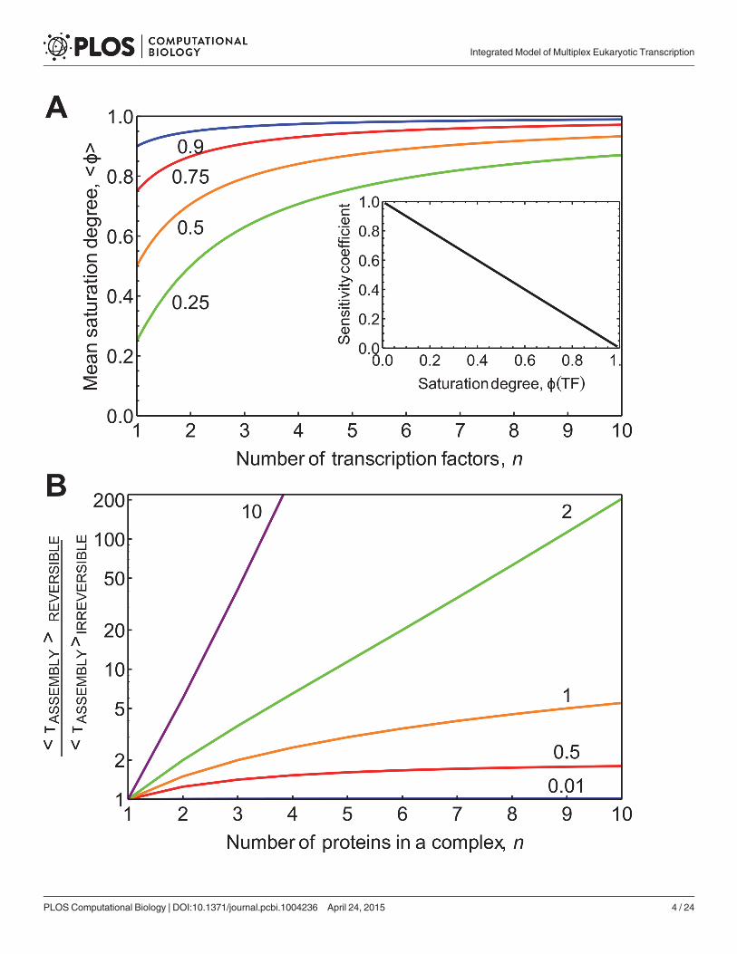
maximal gene activity, i.e.F(T) = 0.75, each individual site would have to be virtually saturated,e.g. ϕi(Ti) = 0.98, which is unrealistic. The tendency of requiring enhanced saturation of indi-vidual binding sites is shown in Fig 1A.
Integrated Model of Multiplex Eukaryotic Transcription
PLOS Computational Biology | DOI:10.1371/journal.pcbi.1004236 April 24, 2015 3 / 24
Integrated Model of Multiplex Eukaryotic Transcription
PLOS Computational Biology | DOI:10.1371/journal.pcbi.1004236 April 24, 2015 4 / 24
Gene regulation by many regulatory factors based on equilibrium binding is also limited interms of responsiveness. The sensitivity of the transcription rate to a specific TF depends onthe extent of saturation of the gene with that TF: @ ln v
@ ln Ti¼ 1� �iðTiÞ ¼ 0:02 for ϕi(Ti) = 0.98.
This analysis shows that increasing the single-site saturation, which is sufficient to produce sig-nificant activation of transcription, causes the gene to loose sensitivity to regulators. Thus, incase of the equilibrium binding mechanism, genes become progressively harder to regulatewhen the number of regulatory factors increases. This finding is highly relevant as the numberof TF-binding sites actively regulating many eukaryotic genes is readily around ten or more. Agenome-wide analysis of data from ENCODE project shows in Drosophila that the number offunctional TF-binding sites per enhancer sequence is between 2 and 15 with the averagearound 8 [31]. Combined evidence from several studies of individual promoter studies sup-ports similar conclusions [32–34]. Additionally, many transcription co-factors and histonemodifiers are regulated by signaling and metabolic pathways [35], increasing the number ofpotential regulatory inputs.
We have also considered whether cooperative binding of the TFs can overcome the deficien-cies of the equilibrium-binding model (see S1 Text). We find that for a high number of TFsunrealistically high levels of cooperativity between regulatory factors are required to solve theproblem of sensitivity or saturation loss (S1 Fig). We will further discuss alternative mecha-nisms for eukaryotic gene regulation that do not suffer from this limitation.
Reversible assembly of large protein complexes can take tens ofminutesThe mean assembly time for a protein complex of n factors, with identical kinetics, depends onthe reversibility of complex formation (K 0
D) and the first-order association rate constant (k0F) as:
tðnÞ ¼ nk0FþPn�1
i¼1ik0FðK 0
DÞn�i. The irreversible assembly time equals n=k0F for n identical factors;
if factors have different association rate constants, this time is given byPn
i¼11=k0F;i. In these
equations, k0F denotes the effective first order rate constant (unit: min-1) for the binding of aregulatory factor to a partially assembled complex. It equals the multiplication of a (diffusion-limited) second-order rate constant for binding (unit: nM-1 min-1) with the (nuclear) concen-tration of the regulatory factor (unit: nM).
Using these equations, one can estimate both the irreversible and the reversible assemblytimes of a protein complex composed out of 10 components. We take the cell nucleus volumeto be 1.2 pL given that the measured cell volume is 3–4 pL [36] and that 30% of this volume isoccupied by the nucleus [37]. The average copy number of sequence–specific TFs per cell, asfound in whole-cell proteomic studies, is around 2500 (S2 Fig), which results in a concentrationof 3.5 nM [38–41]. Similar average concentrations are found for the chromatin modifiers (S2Fig). Protein-chromatin association rates are found to be about two orders of magnitude belowthe diffusion limit [42], which can be directly calculated to be around 0.01 nM-1s-1 (see S3Table). With these numbers, k0F is approximately 2 min-1, i.e. transcription activation couldtake place twice per minute if it would depend on just a single TF. For a complex of 10 TFs,
Fig 1. The relationship between gene saturation, sensitivity to regulation, and the dependence of protein complex assembly on reversibility. A. Toachieve a fixed saturation degree of the entire gene (Φ(T, n); values considered 0.25, 0.5, 0.75, and 0.9 are indicated next to the respective lines) therequired average saturation degree per TF site, hϕi, is plotted as function of n, the number of TFs. The sensitivity of the binding sites to the TF, RF
Tj, is
inversely proportional to the saturation degree as indicated in the inset. B. The influence of the complex size and reversibility of complex formation on theassembly time for an ordered-sequential and reversible protein complex formation mechanism. The assembly times of the reversible mechanism relative tothat of the irreversible mechanism are plotted. The different lines correspond to different values for the effective dissociation constant K 0
D as indicated by thenumbers above the lines.
doi:10.1371/journal.pcbi.1004236.g001
Integrated Model of Multiplex Eukaryotic Transcription
PLOS Computational Biology | DOI:10.1371/journal.pcbi.1004236 April 24, 2015 5 / 24

assembling irreversibly in a defined sequence, assembly should then take 5 min. Ficz et al. [43]measured an off-rate constant (kB) of 2 min-1 for Polycomb group proteins from chromatin.This sets the dissociation equilibrium constant (K 0
D) close to 1, which is consistent with ouranalysis of the requirements for the promoter sensitivity to TFs. Assuming the dissociation rateconstant, kB, to be equal to k0F and substituting these numbers into the equation for τ(n), sup-plied above, gives for complexes of 2, 5, 7, 10, 12 and 24 TFs assembly times of 1.5, 7.5, 14, 27.5,39 and 150 min, respectively, i.e. much longer than the times for irreversible assembly (39 ver-sus 5 min for a complex of size 10). Notably, for the complex size of 10 the assembly time ismore than twice as high as the assembly time of a complex of five proteins (7.5 min). While theexact timing of the complex assembly depends on the values of the protein copy number andthe rate constants, which may vary, the overall conclusion that the reversible mechanism isconsiderably slower at high protein copy number is always correct for effective KD around 1 orhigher. These numbers indicate that protein complex formation on chromatin can become po-tentially rate limiting for transcription rate. The delay in reversible assembly is due to ‘hesita-tion’, i.e. frequent disassembly of a partially assembled complex before full assembly. Divisionof the reversible assembly time, τ(n), by the time for irreversible assembly of the complex, τ(n)/(n/kF), indicates the influence of reversibility and complex size on assembly time (Fig 1B): largecomplexes have very long assembly times and this effect becomes dramatic if the processes aremore than half reversible, i.e. when the effective dissociation constant favours dissociation. Foran effective dissociation equilibrium constant of 2 and an on-rate constant of 2 min-1 the as-sembly for a decameric complex is delayed by a factor of 200; i.e. the process is hesitating allthe time. Then transcription activation would take 17 h rather than 5 min. This clearly pointsout that, if a substantial number of TFs are involved in transcription activation, the reversiblemechanism may well become too slow for appropriate timing of transcription. The assemblytime also depends on the precise mechanism of assembly, as discussed in S2 and S3 Texts. Inparticular, we find that, in general, random assembly mechanisms are faster than the sequentialones (S3 Fig) and that direct assembly on the chromatin is faster compared to the pre-assemblyin the nucleoplasm (S4 Fig). In summary, our analysis indicates that for many eukaryotic genesmechanism of reversible association of all TFs becomes too slow.
Batch assembly of partial complexes speeds up transcription initiationFig 1B shows that for a K 0
D of 2, the reversible assembly of 9 TFs is 100-times slower than the ir-reversible assembly, whilst for a complex size of 3 it is only about 3-times slower. This suggeststhat a sequence of two trimeric association processes could be faster than a single 6-proteins as-sociation process. Likewise, for an effective dissociation constant of 1, the reversible continuousformation of a complex composed of 24 proteins should be 5-times slower than the corre-sponding assembly of 6 complexes, each of which is composed of 4 in sequence, i.e. τ(24)/6τ(4) = 5 (for KD = 1 and 140 for KD = 1.33). These calculations indicate that the batch-wise as-sembly of multi-factor complexes can reduce their otherwise long assembly times. But for this,the partial complex established in the first phase should not dissociate whilst the protein com-plex is being formed.
This conclusion brings us to an important finding: to be fast enough, the process of sequen-tial formation of small protein complexes requires irreversible marking of progress (which weshall refer to as ‘ticking’), for instance, by way of covalent histone modification of nucleosomes.The transcription activation process should therefore resemble a molecular ratchet: it can ‘hesi-tate’ during the reversible assembly of the small protein complex but cannot return to a statewhere the previous set of proteins were assembling due to the histone ticking. This batch-wise
Integrated Model of Multiplex Eukaryotic Transcription
PLOS Computational Biology | DOI:10.1371/journal.pcbi.1004236 April 24, 2015 6 / 24

mechanism has been suggested before [22] but it has never been deduced as essential for eu-karyotic transcription to be fast and regulative enough (see below).
Revertibility of transcription activation requires a cycleOne way to make the process of transcription activation revertible is to make it thermodynami-cally reversible. This corresponds to the equilibrium transcription activation mechanism dis-cussed above. S5A Fig shows this mechanism for the case where 10 TFs reversibly bind to thechromatin, producing the activated transcription complex. In the S4 Text we detail why thistype of mechanism and 4 others (S5 Fig), proceeding in reverse order through the transcriptionactivation pathway, are either too slow or not revertible.
An alternative to reversing on a linear scheme is a combination of a forward linear pathwayand a separate reverse linear pathway; together constituting a cycle. In Fig 2A, four TFs bind ir-reversibly to and (a number of steps later) dissociate irreversibly from their DNA-binding site,referred to as a response element. The covalent histone modifications are shown by the varioussymbols in the rectangles. In the simplest irreversible cyclic mechanism (Fig 2A) the regulatoryfactors dissociate according to a last-in, first-out principle. In the S4 Text we show that thismechanism corresponds to the one Fig 2B and that this mechanism is too linear to be effective.A mechanism in which TFs dissociate irreversibly on the basis of a first-in, first-out principle(Fig 2C) is able to attain much higher transcriptional activity than the equilibrium bindingmodel, whilst transition times between active and inactive gene states are also much shorter.Although every step in Fig 2C is irreversible, the cycle could also operate batch-wise (leading toa considerable advantage, see above): one or a few regulatory factors could bind reversibly afterwhich one would bind really strongly (nearly irreversibly; so, the bound state has a long life-time), thereby fixing the information that all regulatory factors have bound (see Fig 2D). Amechanism in which chromatin is ‘ticked’ or ‘stamped’ upon encountering a TF, which subse-quently dissociates (Fig 2E), is equally competent kinetically. We call this the ‘ticking mecha-nism’. We note that the most realistic mechanism for leaving marks on the chromatin,following the activity of a particular short-lived protein complex, is via histone modifications,as we doubt that many regulatory proteins can stay bound to chromatin for tens of minuteswithout covalent interactions.
Our analysis thus far has used criteria of regulatory sensitivity and the speed of regulation oftranscription to show that a ‘ticking’ transcription cycle mechanism is an attractive mechanismfor eukaryotic genes that are regulated by multiple TFs. There is considerable experimental evi-dence that shows that this mechanism is indeed operative (S5 Text).
Experimental evidence for the ticking mechanismThe ticking mechanism described above is consistent with the wealth of experimental evidenceon the role of chromatin covalent-modification and remodelling in regulation of eukaryoticgene transcription [3, 5–7, 9, 10, 22, 44]. There are many examples of histone modification re-cruiting new modifiers, and subsequent modifications recruiting new proteins actively involvedin transcription induction or cessation. For instance, during promoter activation, H4R3 meth-ylation, mediated by the methylase PRMT1, increases the affinity of histone acetyltransferasep300 for chromatin [7]. Some acetylation marks, e.g. H3K14Ac, H4K16Ac, increase binding ofchromatin remodelling complexes in various experimental systems [45, 46] while others(H3K9Ac, H4K12Ac, H4K8Ac) attract basal TFs required for initiation [47, 48]. This could po-tentially create an orderly sequence of modifications of a chromatin site thus providing theticking mechanism of the gene activation process [22].
Integrated Model of Multiplex Eukaryotic Transcription
PLOS Computational Biology | DOI:10.1371/journal.pcbi.1004236 April 24, 2015 7 / 24

There is also ample evidence for TFs forming complexes with each other and with co-factorsthat have “ticking activity”. One example is the PPARγ-RXRα heterodimer that forms a com-plex with the co-activator PGC1α and the histone acetyltransferases CBP and SRC1 (Fig 3A)[49]. Another example is the repressive complex Mi-2/NurD that includes the chromatin
Fig 2. Cyclic mechanisms for transcription activation/inactivation, illustrated for four TFs. A. Cyclic mechanism with irreversible association anddissociation of the TFs, first-in, last-out.B. The linear mechanism identical to A. C. Feasible cyclic mechanism with sequential irreversible association anddissociation (first-in, last-out) of four TFs, each accompanied by the creation and later removal of a specific covalent modification of the chromatin. D. Batch-wise cyclic mechanism requiring only half the covalent chromatin modifications. E. Ticking mechanism, in which transient association of TFs suffices to tickthe chromatin irreversibly.
doi:10.1371/journal.pcbi.1004236.g002
Integrated Model of Multiplex Eukaryotic Transcription
PLOS Computational Biology | DOI:10.1371/journal.pcbi.1004236 April 24, 2015 8 / 24

remodeller Mi-2, the histone deacetylases 1 and 2, and the methyl CpG-binding protein MBD,which can also bind repressive TFs [50]. Fig 3B shows the possible transitions’ sequence be-tween ‘ON’ and ‘OFF’ phases of the promoter cycle based on literature data (see S5 Text for fulldescription and references). The ‘ON’ state is attained through three successive ticks, or irre-versible modifications: arginine methylation, lysine acetylation and chromatin remodellingthat make the transcription site accessible to basal TFs. This is the state during which RNA po-lymerases are assembled into ranscriptionally competent complexes, and start the elongationof an mRNA transcript. The RNA polymerase II-associated complexes then add a fourthmark—methylation of a lysine group(s). The inactivation of the TF-binding site follows a simi-lar mechanism of four successive protein complex formations, now accompanied by the re-moval of ticks from the chromatin. The transcriptionally active state of chromatin can in
Fig 3. A tickingmechanism for gene switching between OFF and ON states. A. An example of a transition in gene (in)activation is given by the assemblyof the protein complex composed of the two TFs, PPARγ and RXRα, the co-activator PGC1α and the histone acetyltransferases SRC1 and p300 [49]. Theassembly follows a preferentially randommechanism and is followed by modifications of the histone H3 residues K3 and K9.B. The cycle of chromatintransitions associated with gene-expression switching in mammalian cells is show, this cycle is driven by covalent histone modifications and nucleosomeremodelling. The proposed sequence was reconstructed from literature (see S5 Text). C. During the period of gene switching from the ON to OFF state, RNApolymerase II can bind to the transcription start site (TSS) and subsequently escape and initiate elongation. Given that the duration of the effective ON stateis longer than RNA polymerase II binding and TSS escape, multiple polymerase molecules can leave the TSS per ON phase [20, 51, 52].
doi:10.1371/journal.pcbi.1004236.g003
Integrated Model of Multiplex Eukaryotic Transcription
PLOS Computational Biology | DOI:10.1371/journal.pcbi.1004236 April 24, 2015 9 / 24

principle persist during de-ticking, depending on the order of the deactivating events. Eachtransition in the cycle could be dependent on DNA-binding TFs, which would regulate thetranscription rate by changing the duration of ‘ON’ or ‘OFF’ states.
In the proposed mechanism, the sequence of chromatin modifications proceeds in a definedorder. When the activation and inactivation routes would cross however, as would happen in arandom mechanism, this would obviously disturb the progress marking, causing the system toerroneously move backward or forward, and skip steps. These considerations may explain thepreference for a unique order of histone mark addition/removal and specificity of each partiallyassembled complex to a chromatin state.
Precise transcription cycle times, despite inherent molecular noise, cancause transient transcriptional oscillations at the population levelThe mean waiting time for a single TF-binding event equals its standard deviation, making itstiming very imprecise. The time to complete a defined sequence of n identical first-order reac-tions follows an Erlang distribution [30]. The variance in the duration of such a sequence, de-noted by hδ2τi, equals n/k2. The noise in the duration of the sequence, defined as hδ2τi/hτi2equals 1/n. Therefore, the noise equals 1 for the single step mechanism but it is much less for asequence of such steps: a sequence of reactions with identical duration has a more precise com-pletion time than any of its component reactions. This conclusion holds true if a sufficientnumber of the steps in the cycle have different but comparable durations, but falters shouldone reaction be much slower than all the others, then the noise increases.
We modelled a sequence of nine chromatin-state transitions as approximation of an entiretranscription cycle. Each of the transitions involved the reversible assembly of a complex offive proteins followed by irreversible histone modification (Fig 4A). In total 45 regulatory pro-teins were involved. We considered a batch process, i.e. a preferentially random-assemblymechanism on chromatin for protein complex formation, with a strictly ordered sequence ofthe chromatin modifications guiding the sequence of complex assemblies and sensitization ofthe chromatin for the assembly of the next protein complex. We chose realistic values for therate constants and protein abundances (see S3 Text and S3–S5 Tables). Fig 4C shows cyclingthrough several promoter states by four individual cells after transcription was activated ineach of them at the same time. We observe that, for example, state 2 occurs in the first cycle inall cells almost simultaneously; and in subsequent cycles the occurrence of this state slowlyloses synchrony across the four cells, as indicated by the increasing dispersion of the occur-rence time. This means that the cells are progressing through the transcription cycle slowly (ascompared to the TF binding time scale) and synchronously in the beginning, but desynchro-nize over time. This can be further illustrated by plotting the probability of observing each ofthe four states in a population as function of time (Fig 4B).
To illustrate how the stochasticity of assembly times leads to transient de-synchronizationof a population of cells, we will consider what occurs with increasing number of cycles of nsteps. The overall waiting time distribution to complete Nth cycle becomes narrower with an in-
creasing number (N) of cycles: hd2tN i
htN 2i ¼ ðn � NÞ�1. To quantify the effect of de-synchronization,
we consider the noise in the timing of the end of the N-th cycle relative to the mean duration of
a single cycle, i.e. using htNi ¼ Nhti : hd2tN ihti2 ¼ N
n. This relation shows that cells will progressively
become asynchronous with increasing number of completed transcription cycles, and thatphase-noise in the Nth cycling time increases linearly with N. At the same time, it shows thatmore transitions per transcription cycle, n, tend to prolong the persistence of population levelsynchrony. These relations explain the behaviour calculated in Fig 4.
Integrated Model of Multiplex Eukaryotic Transcription
PLOS Computational Biology | DOI:10.1371/journal.pcbi.1004236 April 24, 2015 10 / 24

Fig 4. Precision of transcription cycle duration leads to transient population level transcription cycles. A.We consider a transcription cycle of 9chromatin states. Each transition involves the formation of a single protein complex consisting of 5 proteins following a particular semi-random assemblymechanism followed by an irreversible histone modification event. B. Probabilities to observe states 2, 4, 6, and 8 in the population as function of time,obtained by modelling 2,500 individual cells that started in promoter state 1. C. Durations of the 4 chromatin states calculated for 4 cells from the population.
Integrated Model of Multiplex Eukaryotic Transcription
PLOS Computational Biology | DOI:10.1371/journal.pcbi.1004236 April 24, 2015 11 / 24

We simulated the stochastic dynamics for a population of 1,000 cells that had simultaneous-ly started transcription activation of two gene copies. Fig 4D shows the fractions of cells thatare in a given state at any moment in time (also shown for a single cell in Fig 4C).
Fig 4D shows that population level transcriptional cycles are observable for around 100 minand decrease in amplitude over time due to de-synchronization as expected. The transcription-al cycling time that we predict with this realistically parameterized model is approximately54 min. The predicted waiting time distribution of the cycling time indeed peaks (Fig 5A), asexpected for a multi-step sequential process.
Bursts in the systemThe number of RNA polymerase molecules that initiate elongation per transcription cycle de-pends on the tendency of genes to engage in a phenomenon called re-initiation [20, 51–53].Many experimental studies have shown that mRNA is produced in bursts [14, 54–56] of vari-able size [57, 58]. We incorporated RNA polymerase II binding and promoter escape duringthe permissive ON state into the model. We considered promoter state 5 to be transcriptionallypermissive. The overall lifetime of the state is around 6 min (Fig 5A, orange line). However, aswe assumed competition between polymerase and deactivating complex, the effective permis-sive state only occurs while the deactivating complex is not bound and its lifetime is close to1 min (Fig 5A inset, red line). The polymerase concentration in the model was taken higherthan the concentrations of other TFs, estimate supported by the experimental copy numberdata (S2 Fig). Therefore, polymerase binding and promoter escape time had an average of0.1 min (Fig 5A inset, black line), a much shorter time than the lifetime of the effective ONstate, resulting in multiple transcripts being initiated during most of the permissive periods.The average burst size should then be 10 mRNAs/cycle and the distribution of burst size canbe shown to be geometric; which is indeed the case for calculated burst size distributions inFig 5B. This finding is in good agreement with experimental observations that mRNA burst-size distributions for regulated genes across a cell population are often geometric [54, 55]. Thecontribution of the stochasticity of transcriptional bursts, as generated by the ratchet mecha-nism, to the total stochasticity of transcript concentrations in a cell depends also on factors, in-dependent of the ratchet mechanism [59].
Whether bursting will occur in synchrony across cells depends on whether the cells were in-duced at the same time. Fig 5C and 5D show the prediction of mRNA trajectories in five indi-vidual cells for the 9-state ratchet cycle model that were induced at the same time (Fig 4A). Themodel predicted mRNA bursting: all five modelled cells fired mRNAs periodically and for thefirst 2 h they did this in fair synchrony. When mRNA decay was rapid (Fig 5C), most of themRNA was degraded during the promoter OFF period. For multiple cells, this led to the pre-diction of population level transient oscillation in mRNA levels (Fig 5E). If no mRNA degrada-tion occurred on the considered timescale the bursts led to step-wise accumulation of mRNAin individual cells (Fig 5D) as well as on the cell population level (Fig 5F).
D. Stochastic simulation of the transcriptional cycling performed for 2,000 gene copies. The probability of the presence of the four successive states wascalculated as a function of time; colours correspond to the state as labelled inB.
doi:10.1371/journal.pcbi.1004236.g004
Integrated Model of Multiplex Eukaryotic Transcription
PLOS Computational Biology | DOI:10.1371/journal.pcbi.1004236 April 24, 2015 12 / 24

Fig 5. Stochastic dynamics of mRNA production in the 9-state transcription ratchet model. A.Overall cycle time distribution function PCYCLE(τ) with anaverage time of 54 min (dark red line; as produced by the ratchet model for transcription activation) and the time distribution of the chromatin state permissiveto polymerase binding with an average of 6 min (orange line). Inset: waiting time distribution function PON(τ) for the ON state (red line); and for regulatoryregion escape event PESCAPE(τ) (black line), which have an average of approximately 1 and 0.1 min and are exponentially distributed, respectively. B.
Integrated Model of Multiplex Eukaryotic Transcription
PLOS Computational Biology | DOI:10.1371/journal.pcbi.1004236 April 24, 2015 13 / 24

A transcription (in)activation cycle model with realistic kinetics canreproduce experimentally observed population-level transcriptionalcyclingThe model developed in the previous sections explains a number of experimental observationsdealing with cyclical changes in protein occupancy, nucleosome modifications, looping of eu-karyotic regulatory regions, and transcriptional bursts. The transcriptional cycling periods thathave been observed experimentally vary between 40 and 90 min [24, 25, 27, 28, 60], in line withthe above predictions for the 9-state ratchet cycle model.
The most detailed dataset available is for the regulatory region of the estrogen-responsivetrefoil factor 1 (TFF1) gene [27]. It exhibits the orderliness of the binding events that is a com-ponent of cyclic ratchet model: first, co-activator complexes containing histone acetyltrans-ferases and histone methyltransferases bind, then the basal TFs and RNA polymerase II, andfinally the de-activator complexes, containing remodelling/HDAC activity. This experimentaldata (Fig 6B) can be reproduced fairly well by a 9-state cycle model with realistic kinetic param-eters (Fig 6A). The de-synchronization observed in the experiment was however slower than inthe model simulations. This could be explained by an even higher number of proteins andchromatin modifications involved serially in the initiation process than currently known exper-imentally, by more peaked waiting time distributions for individual cycle transitions, whichcould again be due to multi-step serial processes, or by differences in kinetic parameters. Aclear example where the de-synchronization predicted by our model is observed experimental-ly has been provided by the measurement of the TF occupancy on chromatin, using fluores-cence-based methods in single cells [24].
Population level oscillations in mRNA concentrations as predicted by our cycle model(Fig 5E) have also been observed experimentally (Fig 6D). The yeast metallothionein (CUP1)gene, displayed 50-min transcriptional cycles of its main TF, Ace1p, upon activation and theCUP1mRNA was oscillating at the same frequency [24]. Adjustment of the protein concentra-tions and the consideration of two promoter states (PR2 and PR3), which can each be tran-scriptionally permissive, allows for qualitative correspondence of the model simulations andexperimental data.
Evidence for reversible looping between a response element and the TSS has been found inthe regulatory region of the human cyclin-dependent kinase inhibitor 1A (CDKN1A) gene,which is regulated by several REs for the TF VDR [28]. Some of the co-regulators were shownto oscillate at the population level with a 60-min period. The experimental data (Fig 6F) can bereproduced by our model (Fig 6E) if the looping event between the distant RE and TSS in themodel occurs during the chromatin state corresponding to RNA polymerase II binding.
DiscussionIn this study, we asked how transcription regulation of conditionally active genes in eukaryotescould be organized so that it is both fast enough, given diffusion limitations, and sensitiveenough towards a large number of TFs. We showed that neither an equilibrium binding mech-anism nor a more irreversible binding mechanism, reversing its steps upon inactivation, couldbe fast and sensitive enough. We then showed that the next simplest, competent mechanism isa batch-like multi-step process of transcription activation, followed by a separate batch-like
Distribution function of burst sizes p(b) produced by the model with a calculated average of ~11 molecules per burst. C. Due to the fairly precise waitingdistribution of the cycling times, five cells that are activated simultaneously will display synchrony in mRNA production for some time.D. A similar picture isobserved in the case of lack of degradation where bursts result in a step-wise increase of mRNA. E. Population level response of mRNA in the presence ofmRNA degradation. F. Population level response of mRNA in the absence of mRNA degradation.
doi:10.1371/journal.pcbi.1004236.g005
Integrated Model of Multiplex Eukaryotic Transcription
PLOS Computational Biology | DOI:10.1371/journal.pcbi.1004236 April 24, 2015 14 / 24

Fig 6. The transcription clockmodel can explain existing experimental data. The single regulatory region model with nine chromatin transitions(presented in Fig 4) with adjusted protein concentrations can qualitatively reproduce the ordered cyclic co-regulator binding as observed in variousexperiments reported in literature. A. Time courses of the average regulatory region occupancy on the cell population level. The plotted regulatory regionoccupancy states were chosen to fit the data profiles (see S6 Table). Inset shows the schematic representation of the cycle of chromatin states, colourscorrespond to plotted states.B. Experimental data, simulated in panel A, reproduced from [27]: blue: the TF estrogen receptor, red: RNA polymerase II,purple: co-regulator CARM1, dark blue: histone acetyltransferase CBP, green: HDAC1, dark green: chromatin remodelling factor Brg1.C. The same model,with corrected protein concentrations to match cycle duration, extended with mRNA synthesis and degradation, reproduces population level mRNA cyclingas observed experimentally and shown in: D. Data reproduced from [24]. E. Simulation of the model modified to include the TSS and a RE, which form a loopduring the initiation phase (red complex). This model can reproduce looping (black line) and polymerase-binding data (red line) as observed experimentallyfor the regulatory region of theCDKN1A gene by [28]; experimental data shown in F. The arbitrary units of regulatory region looping data (between TSS andRE 3) are re-scaled by a factor of 10,000 for ease of visualization. The parameter adjustments are documented in the S4 and S5 Tables.
doi:10.1371/journal.pcbi.1004236.g006
Integrated Model of Multiplex Eukaryotic Transcription
PLOS Computational Biology | DOI:10.1371/journal.pcbi.1004236 April 24, 2015 15 / 24

multi-step inactivation process, together constituting a transcription (in)activation cycle. Eachstep would correspond to the reversible association of a limited number of TFs leading to irre-versible marking of the transcription activation complex, after which the TFs dissociate. Thistranscription (in)activation cycle should not be confused with the more restricted use of theterm transcription cycle when it refers to the process of an RNA polymerase producing an en-tire mRNA. We have a much more involved process of gene activation and transcription initia-tion in mind.
If one were to specify that histone modification is the process that marks the completion ofa step in the multi-step process and sensitizes the local chromatin region for the assembly ofthe next protein complex, this model comes close to the accepted view of transcription activa-tion in eukaryotes [7, 10, 22, 44]. The difference is that we derived this view as a requirementfor delivering kinetic competence, revertibility and sensitivity, whereas the accepted view isbased on experimental observations of the transcription activation process. All in all, our studysuggests that we have found a plausible explanation as to why transcription (in)activation ofmany regulated genes in eukaryotes is organized the way it is, and why it is different from theactivation of transcription that is regulated by a small number of TFs, such as in prokaryotes,or the house-keeping genes.
Metivier et al. [27] proposed a branched mechanism for the transcription cycle at the TFF1promoter. Our model can straightforwardly be extended with such details. We considered suchan extension in a minimalist manner when we incorporated the transcription re-initiationmechanism to allow for variable burst size of the transcription cycle.
In our model, a sequence of protein complex assemblies intermitted by covalent-modifica-tion of histones to mark the phase of the transcription cycle, leads to a fairly deterministic du-ration of the active transcription state. This has two consequences. Firstly, our model explainshow the duration of the entire transcription cycle could be close to deterministic, i.e. clock-like.Secondly, in the transcription re-initiation formulation of the model the number of transcrip-tion (re-)initiations per cycle becomes more deterministic. This means that the eukaryotic tran-scription cycle can be both clock-like in terms of duration and quantal in terms of its activity.This is within the limit of many molecular events in series with similar reaction rates. To whatextent real genes function within this limit is unclear. Recent experiments indicate that the ONand OFF durations of genes can have non-exponential waiting time distributions, which is inagreement with our predictions [58, 61, 62]. The clock-like nature of the model underlies thetendency of cells in a population to display transiently synchronous transcription activity uponsimultaneous activation from the same initial state. The model does not, however, exclude apossibility of the transcription cycle times being less precise, due, for instance, to the presenceof a very slow step in one or more of the cycle transitions, or a possibility of variable gene in-duction times due to heterogeneity of the initial promoter states in a cell population.
We have shown that our model explains precise durations of the entire transcription cycleof about 1 hour on the basis of molecular processes that are faster than 1 per minute. This is be-cause of a sequence of several multi-step processes, each of which is consolidated by irreversiblemarking (ticking). This ticking corresponds to a ratchet mechanism, but the overall processcorresponds to a clock mechanism as already proposed by Reid et al. [22]. A mechanical clockhas the same type of ratchet mechanism and is similarly accurate at longer time scales: the tim-ing of its individual ticks is not accurate, but the timing of large numbers (for example 60) ofits ticks is. From a coarse-grained perspective, the transcription activation cycle model assumesthat eukaryotic genes switch between transcription active ON states and ditto, inactive OFFstates. The duration of these phases could be under the control of some regulator [56, 63, 64].The same applies to burst size. From this perspective, transcription rates can be controlled indifferent ways. A suitable definition of transcription rate would be the mean burst size divided
Integrated Model of Multiplex Eukaryotic Transcription
PLOS Computational Biology | DOI:10.1371/journal.pcbi.1004236 April 24, 2015 16 / 24

by the mean cycle time: hbihtON iþhtOFF i. Control of transcription rate can then be achieved via modu-
lation of burst frequency (cycle duration; the FMmode) or burst size (the AMmode). Skupskyet al. [65] found evidence for regulation of burst size rather than frequency. The transcriptioncycle models we presented can accommodate both mechanisms.
Not all protein complexes involved at the various stages of the transcription cycle assembleon the chromatin, some may already be assembled in the nucleoplasm, such as mediator com-plex [66]. These pre-assembled complexes do not affect the total duration of the transcriptioncycle and its precision, as they bind in one step. However, even if all protein complexes wouldassemble in the nucleoplasm, which we know is not the case, histone marking would be still ad-vantageous to make sure that they bind in the correct order.
In our model we propose that histone modification function as marks of the progress of thetranscription cycle, because they have a longer lifetime than protein complexes—proteins re-side on chromatin only for several tens of seconds. This does not mean that all histone marksshould be remembered along the entire transcription cycle. We expect the minimal life of a his-tone mark to be related to the time that it takes to form the next-in-line protein complex. So, ifprotein complex i has been formed after which mark i is added, the lifetime of mark i should belong enough to make sure that the protein complex i+1 has had its time to form and leavemark i+1. Since marks can always be removed by accident by an enzyme, or fall off spontane-ously, it makes sense that a short memory of previous marks should be present on chromatin,say in addition to mark i, mark i-1 and i-2, to make sure that if mark i gets removed by accidentthe transcription process does not erroneously reset to its resting state. Considering that theformation of a protein complex on chromatin takes several minutes, we would therefore expectthat histone modifications that mark the progress of the transcription cycle stay for about 10minutes or more on chromatin.
Transcription dynamics differ between prokaryotes and eukaryotes [14, 19–21, 54, 55, 67–69]. Eukaryotes tend to display transcription bursts, which in prokaryotes have only beenfound under conditions of leaky transcription repression [56, 64, 70], and occur infrequentlyacross the majority of genes in Escherichia coli [71]; in contrast to what is found in Saccharo-myces cerevisiae [72, 73]. RNA polymerase II assembly, and its escape from the regulatory re-gion, only take place during a fraction of the entire transcription cycle, suggesting that manyeukaryotic genes are prone to transcriptional bursting even under conditions of high transcrip-tion activity [24, 27, 54, 55]. The mechanisms for transcriptional bursts in bacteria [64, 74, 75]are likely very different in molecular detail from the mechanisms in eukaryotes, even thoughthey can be coarse-grained to a similar mechanism giving rise to a variable number of RNA po-lymerases that initiate transcription during a single ‘ON’ state of the gene [59].
Transcriptional cycling differs from regular oscillations induced by a negative feedbackloop, as found, for example, in NADH fluorescence and glycolytic activity in yeast [76], the dy-namics of calcium concentrations [77], and the activities of NF-κB [78] or the kinase ERK [79].In the latter examples the dynamics of metabolite or signal transduction factor pools are cou-pled through nonlinear kinetics. In some of these cases sustained oscillations have been ob-served, and for one of these the requisite active synchronization mechanism requiring dynamiccommunication between individual cells, has been elucidated [80]. The mechanism we proposefor the transient oscillations at the population level is entirely cell-autonomous, i.e. no activecommunication between cells is involved. For transcriptional cycling, we propose that the tran-sient cyclic dynamics at the population level are the consequence of a simultaneous start andaccurate durations.
Integrated Model of Multiplex Eukaryotic Transcription
PLOS Computational Biology | DOI:10.1371/journal.pcbi.1004236 April 24, 2015 17 / 24
ModelsThe mass balances, rate equations and parameters for protein-assembly and complete tran-scription initiation models were generated by using custom algorithms implemented in Mathe-matica 7.0. ODE system simulation were done using NDSolve function; stochastic simulations—by implementing direct-method of Gillespie algorithm [81]. For detailed description of themodels’ structures, parameters and initial conditions see S3 Text, S1–S6 Tables and S6 Fig. AllMathematica files are available as part of Supporting Information (S1 Dataset).
Supporting InformationS1 Fig. Effect of TF interaction coefficient on the sensitivity of the transcription rate to asingle TF concentration. The value of the interaction coefficient, β, that minimizes (F(T, n, β)−ϕ(T))2 for a range of values for the TF concentration (T; from 0 to 20-times the affinity con-stant, K) is plotted as function of the number of TFs, n. The inset shows the dependency of thesquared difference between F(T, n, β) and ϕ(T) as function of β and shows that a minimalvalue for β, the optimal value, exists for each value of n (ranging from 2 to 20). The optimal
value of β is defined as bopt ¼ argmin½P40
i¼0ðFðiDa; n; boptÞ � �ðiDaÞÞ2� with Δα = 0.5; hence,
α = T / K ranges from 0 to 20. This notation works as follows: “argmin[f(λ)]” returns the valueof variable λ that minimizes the function f(λ).(PDF)
S2 Fig. Copy number distributions found experimentally for different types of proteins in-volved in eukaryotic transcription. Distribution of copy numbers were calculated for se-quence specific TFs (blue), histone modifiers (green) and general TFs (red) based on theproteomic data from two mouse cell(line)s (Azimifar et al. [38] and Schwanhausser et al. [41]and two human cell lines (Beck et al [39]. and Nagaraj et al. [40] Calculated means of distribu-tions are indicated by vertical lines. Lists of proteins with specific functions (sequence specificTFs, histone modifiers, GTFs), were created based on relevant GO-terms and further curatedto ensure that only proteins for which experimental evidence for their functions is providedwere included. The corresponding UniProt IDs lists were checked against the correspondingID lists provided in the proteomic measurement publications and copy number measurementsfor proteins found used to calculate the distributions. The curated lists of Gene Names/UniProtIDs used as well as the lists of all copy numbers used for the distribution calculations are pro-vided as part of Supporting Information (Modeling files.zip).(PDF)
S3 Fig. Assembly time of protein complexes depending on the mechanism. The proteincomplex assembly time as function of the dimensionless dissociation constant of trimer forma-tion for assembly mechanisms that differ in organization. In addition to the ordered irrevers-ible assembly mechanism, four reversible mechanisms are considered for the assembly of atrimeric complex on chromatin as indicated by the diagrams on the right. They are comparedto the irreversible ordered-sequential mechanism illustrated on top. The tendency to fall apartis captured by the apparent dissociation constant K 0
D. The inset gives the probability distribu-tion of assembly times for the sequential-ordered reversible mechanism (at an apparent KD =0.1). The red dot gives the mean assembly time.(PDF)
S4 Fig. Mechanisms of protein complex assembly and their duration depending on the se-quence and association rate constant. A. All complexes that can be formed in the N = 5 ran-dom mechanism (both in RE and in cytoplasm) that is used in the models are presented; the
Integrated Model of Multiplex Eukaryotic Transcription
PLOS Computational Biology | DOI:10.1371/journal.pcbi.1004236 April 24, 2015 18 / 24

complexes that can bind RE are shown as bound forms. The lists of complexes that are used inpreferentially random and sequential assembly mechanism are given in S2 Table. B. Half-timeof promoter modification for a range of kon
eff values (product of protein concentrations andtheir kon) for nine different protein assembly mechanisms (sequential—Seq; preferentially ran-dom—PR; random—Ran; on chromatin—Chr; nucleoplasmic—Nuc) involving 5 proteins asindicated. In all instances Keq = 109 M (here defined as kon/koff; koff is variable depending onthe value of kon
eff). The rate constant for the final irreversible acetylation of lysine 9 and lysine14 kmod = 60 min-1, i.e. much faster than the dissociation constant. C. Fraction of promoterbinding at equilibrium as function of the kon
eff for the same set of mechanisms. In all simula-tions, the RE became ready for complex assembly at time zero. All models were simulated so asto be at equilibrium prior to time zero. At time zero the RE becomes available for binding.(PDF)
S5 Fig. Various mechanisms for linear transcription mechanism by 10 TFs, with their effi-cacy. A. Inactive (unless mass action irreversible) but then irrevertible; B. active, but irreverti-ble; C. active, forward hesitant (unless mass action irreversible) but then irrevertible, becauseof hesitation on the reverse route;D. Active, forward hesitation removed, but again irrevertible;E. Active, revertible; but with re-emerging hesitation problem.(PDF)
S6 Fig. Structures of detailed initiation models. Chromatin state transition scheme for themodels presented—a basic 9-state promoter model (also used for simulating the Metivier et al.and Karpova et al. data) (A), a modified model with looping between distant RE and TSS (B)used to simulate the Saramäki et al. data. The chromatin states in which RNA polymerase IIbinding/mRNA production occurs are marked in colour. In scheme A, PR5 is considered inON state for generic 9-state chromatin model, while for simulating Karpova et al. PR1+PR2are considered permissive. Specific chromatin states plotted in Figs 4 and 6 in the main text aredescribed in S6 Table. In every model a single chromatin transition corresponds to formationof an N = 5 protein complex on the PR/RE by preferentially random mechanism followed by achromatin modification step (C) or an N = 4 protein complex on TSS likewise followed by achromatin modification step (E). For simulating Karpova et al. complexes formed on PR2 con-tain polymerase and lead to transition to PR3 (D); likewise, PR3 can be bound to the sameRNA polymerase II containing complex and its own de-activation complex. Each successfulformation of an RNA polymerase II complex leads to the start of elongation. In model B (simu-lation of Saramäki et al.) the loop is formed between partially bound RE3 and RNA polymeraseII bound to TSS3, leading to modification of polymerase that is ready for elongation (F). In allmodels elongation is modeled as a 30-step process that leads to formation of mature mRNAand release of the RNA polymerase II.(PDF)
S1 Table. Interaction matrix of protein complexes. A realistic interaction scheme for a com-plex of five proteins is presented in the form of an interaction matrix. The interaction matrixwas used to produce the mass balance and rate vectors for the random assembly model.(PDF)
S2 Table. Complexes formed in preferential random and sequential assembly mechanisms.A customary algorithm was used to remove reactions involving complexes formed only in fullyrandom mechanisms.(PDF)
Integrated Model of Multiplex Eukaryotic Transcription
PLOS Computational Biology | DOI:10.1371/journal.pcbi.1004236 April 24, 2015 19 / 24
S3 Table. Example of parameter calculation. A customary algorithm was used to calculaterate constants for the corresponding protein association and dissociation reactions.(PDF)
S4 Table. Parameters for transcription in 9-state promoter model. Protein association anddissociation rate constants and concentrations were calculated in the same way as shown in S3Table. The modification rate constants were chosen in a way to provide the same effective ratesas a single protein-binding step to reduce the noise in waiting times. The values are in therange for kcat of chromatin modifying enzymes measured in vitro [s35-37]. Elongation and ex-port times were estimated from data available in literature [s38-41] and modeled as a multi-step process (N = 30). A 30 min life-time was assumed; degradation was modeled as multi-stepprocess with (N = 20). For models used to simulate data, additional parameters as well as pa-rameters different from the main model are given. For simulation of the Karpova et al. data,the initiation rate constant was taken to be faster than the RNA polymerase II off-rate. Therates of mRNA elongation (modeled as N = 30 process) and degradation (modeled as N = 20process) were adjusted to fit the data.(PDF)
S5 Table. Parameters for the promoter model with looping between RE and TSS for simu-lating data from Saramäki et al. Protein association and dissociation rate constants and con-centrations were calculated in the same way as shown in S3 Table. Only the new mRNAsynthesis was modelled. As the number of states is lower in this model, a lower degree of re-versibility is required to reach comparable passive synchronization as in the 9-state model,hence the lower off rates. The on-rate of loop formation was taken as an estimation from datapresented by Polikanov et al. (2007) [s42], which suggests that the average rate for DNA loopformation of comparable length in vitro is at least faster than 60 seconds. The loop was as-sumed to be stable (Kd = 1012) due to a big protein interaction surface. The polymerase modifi-cation step and promoter step were modelled explicitly. The protein concentrations wereadjusted to fit experimental data.(PDF)
S6 Table. Summary of information on different promoter models. Specifications (structuresand parameters) of each model are described, and the model states plotted in the main text fig-ures are listed.(PDF)
S1 Text. Promoter sensitivity to multiple TFs in the equilibrium binding model.(PDF)
S2 Text. Mean time of protein complex assembly reversible versus irreversible mechanisms.(PDF)
S3 Text. Timing of the protein assembly mechanisms.(PDF)
S4 Text. Revertible linear mechanism of transcription (in)activation.(PDF)
S5 Text. Evidence for chromatin modification-driven promoter cycling mechanism.(PDF)
S6 Text. Supporting Information references.(PDF)
Integrated Model of Multiplex Eukaryotic Transcription
PLOS Computational Biology | DOI:10.1371/journal.pcbi.1004236 April 24, 2015 20 / 24

S1 Dataset. Modeling files.(ZIP)
AcknowledgmentsWe thank L. Anink, M. Dobrzynski, D. Piebes, R. van Driel and P. Verschure forinsightful discussions.
Author ContributionsWrote the paper: KNR FJB HVW. Collection and analysis of the literature data: AT MJM CC.Model definition: KNR FJB HVW. Model implementation, simulation and analysis: KNR. An-alytical derivations: FJB HVW.
References1. Perissi V, Rosenfeld MG. Controlling nuclear receptors: the circular logic of cofactor cycles. Nat Rev
Mol Cell Biol. 2005; 6(7):542–54. doi: 10.1038/nrm1682 PMID: 15957004
2. ThomasMC, Chiang CM. The general transcription machinery and general cofactors. Crit Rev BiochemMol Biol. 2006; 41(3):105–78. doi: 10.1080/10409230600648736 PMID: 16858867
3. Mellor J. The dynamics of chromatin remodeling at promoters. Mol Cell. 2005; 19(2):147–57. doi: 10.1016/j.molcel.2005.06.023 PMID: 16039585
4. Narlikar GJ, Fan HY, Kingston RE. Cooperation between complexes that regulate chromatin structureand transcription. Cell. 2002; 108(4):475–87. doi: 10.1016/S0092-8674(02)00654-2 PMID: 11909519
5. Saha A, Wittmeyer J, Cairns BR. Chromatin remodelling: the industrial revolution of DNA around his-tones. Nat Rev Mol Cell Biol. 2006; 7(6):437–47. doi: 10.1038/nrm1945 PMID: 16723979
6. Berger SL. The complex language of chromatin regulation during transcription. Nature. 2007; 447(7143):407–12. doi: 10.1038/nature05915 PMID: 17522673
7. Li B, Carey M, Workman JL. The role of chromatin during transcription. Cell. 2007; 128(4):707–19. doi:10.1016/j.cell.2007.01.015 PMID: 17320508
8. Barnard A, Wolfe A, Busby S. Regulation at complex bacterial promoters: how bacteria use differentpromoter organizations to produce different regulatory outcomes. Curr Opin Microbiol. 2004; 7(2):102–8. doi: 10.1016/j.mib.2004.02.011 PMID: 15063844
9. Hager GL, McNally JG, Misteli T. Transcription Dynamics. Molecular cell. 2009; 35(6):741–53. doi: 10.1016/j.molcel.2009.09.005 PMID: 19782025
10. Weake VM, Workman JL. Inducible gene expression: diverse regulatory mechanisms. Nat Rev Genet.2010; 11(6):426–37. doi: 10.1038/nrg2781 PMID: 20421872
11. Ben-Tabou de-Leon S, Davidson EH. Gene Regulation: Gene Control Network in Development. AnnuRev Biophys Biomol Struct. 2007; 36:191–212. doi: 10.1146/annurev.biophys.35.040405.102002PMID: 17291181
12. van Driel R, Fransz PF, Verschure PJ. The eukaryotic genome: a system regulated at different hierar-chical levels. J Cell Sci. 2003; 116(Pt 20):4067–75. doi: 10.1242/jcs.00779 PMID: 12972500
13. Davidson EH. The regulatory genome: gene regulatory networks in development and evolution. NewYork: Academic Press; 2006.
14. Kim HD, O'Shea EK. A quantitative model of transcription factor-activated gene expression. Nat StructMol Biol. 2008; 15(11):1192–8. doi: 10.1038/nsmb.1500 PMID: 18849996
15. Mao C, Brown CR, Falkovskaia E, Dong S, Hrabeta-Robinson E, Wenger L, et al. Quantitative analysisof the transcription control mechanism. Mol Syst Biol. 2010; 6:431. doi: 10.1038/msb.2010.83 PMID:21081924
16. Yen K, Vinayachandran V, Batta K, Koerber RT, Pugh BF. Genome-wide Nucleosome Specificity andDirectionality of Chromatin Remodelers. Cell. 2012; 149(7):1461–73. doi: 10.1016/j.cell.2012.04.036PMID: 22726434
17. Wang Z, Zang C, Rosenfeld JA, Schones DE, Barski A, Cuddapah S, et al. Combinatorial patterns ofhistone acetylations and methylations in the human genome. Nat Genet. 2008; 40(7):897–903. doi: 10.1038/ng.154 PMID: 18552846
Integrated Model of Multiplex Eukaryotic Transcription
PLOS Computational Biology | DOI:10.1371/journal.pcbi.1004236 April 24, 2015 21 / 24

18. Barski A, Cuddapah S, Cui K, Roh TY, Schones DE, Wang Z, et al. High-resolution profiling of histonemethylations in the human genome. Cell. 2007; 129(4):823–37. doi: 10.1016/j.cell.2007.05.009 PMID:17512414
19. BlakeWJ, Balazsi G, Kohanski MA, Isaacs FJ, Murphy KF, Kuang Y, et al. Phenotypic consequencesof promoter-mediated transcriptional noise. Mol Cell. 2006; 24(6):853–65. doi: 10.1016/j.molcel.2006.11.003 PMID: 17189188
20. BlakeWJ, KAM, Cantor CR, Collins JJ. Noise in eukaryotic gene expression. Nature. 2003; 422(6932):633–7. doi: 10.1038/nature01546 PMID: 12687005
21. Lam FH, Steger DJ, O'Shea EK. Chromatin decouples promoter threshold from dynamic range. Nature.2008; 453(7192):246–50. doi: 10.1038/nature06867 PMID: 18418379
22. Reid G, Gallais R, Métivier R. Marking time: The dynamic role of chromatin and covalent modification intranscription. The International Journal of Biochemistry & Cell Biology. 2009; 41(1):155–63. doi: 10.1016/j.biocel.2008.08.028 PMID: 18805503
23. Kangaspeska S, Stride B, Metivier R, Polycarpou-Schwarz M, Ibberson D, Carmouche RP, et al. Tran-sient cyclical methylation of promoter DNA. Nature. 2008; 452(7183):112–5. doi: 10.1038/nature06640PMID: 18322535
24. Karpova TS, Kim MJ, Spriet C, Nalley K, Stasevich TJ, Kherrouche Z, et al. Concurrent Fast and SlowCycling of a Transcriptional Activator at an Endogenous Promoter. Science. 2008; 319(5862):466–9.doi: 10.1126/science.1150559 PMID: 18218898
25. Kim S, Shevde NK, Pike JW. 1,25-Dihydroxyvitamin D3 stimulates cyclic vitamin D receptor/retinoid Xreceptor DNA-binding, co-activator recruitment, and histone acetylation in intact osteoblasts. J BoneMiner Res. 2005; 20(2):305–17. doi: 10.1359/JBMR.041112 PMID: 15647825
26. Metivier R, Gallais R, Tiffoche C, Le Peron C, Jurkowska RZ, Carmouche RP, et al. Cyclical DNAmeth-ylation of a transcriptionally active promoter. Nature. 2008; 452(7183):45–50. doi: 10.1038/nature06544 PMID: 18322525
27. Metivier R, Penot G, Hubner MR, Reid G, Brand H, Kos M, et al. Estrogen receptor a directs ordered,cyclical, and combinatorial recruitment of cofactors on a natural target promoter. Cell. 2003; 115(6):751–63. doi: 10.1016/S0092-8674(03)00934-6 PMID: 14675539
28. Saramaki A, Diermeier S, Kellner R, Laitinen H, Vaisanen S, Carlberg C. Cyclical Chromatin Loopingand Transcription Factor Association on the Regulatory Regions of the p21 (CDKN1A) Gene in Re-sponse to 1a,25-Dihydroxyvitamin D3. Journal of Biological Chemistry. 2009; 284(12):8073–82. doi:10.1074/jbc.M808090200 PMID: 19122196
29. Hager GL, Elbi C, Becker M. Protein dynamics in the nuclear compartment. Current Opinion in Genetics& Development. 2002; 12(2):137–41. doi: 10.1016/S0959-437X(02)00278-2 PMID: 11893485
30. Lemaire V, Lee CF, Lei J, Metivier R, Glass L. Sequential recruitment and combinatorial assembling ofmultiprotein complexes in transcriptional activation. Phys Rev Lett. 2006; 96(19):198102. doi: 10.1103/PhysRevLett.96.198102 PMID: 16803143
31. Negre N, Brown CD, Ma L, Bristow CA, Miller SW,Wagner U, et al. A cis-regulatory map of the Dro-sophila genome. Nature. 2011; 471(7339):527–31. doi: 10.1038/nature09990 PMID: 21430782
32. Bai L, Ondracka A, Cross FR. Multiple sequence-specific factors generate the nucleosome-depleted re-gion on CLN2 promoter. Mol Cell. 2011; 42(4):465–76. doi: 10.1016/j.molcel.2011.03.028 PMID:21596311
33. Crispín JC, Tsokos GC. Transcriptional regulation of IL-2 in health and autoimmunity. AutoimmunityReviews. 2009; 8(3):190–5. doi: 10.1016/j.autrev.2008.07.042 PMID: 18723131
34. Jitrapakdee S. Transcription factors and coactivators controlling nutrient and hormonal regulation of he-patic gluconeogenesis. The International Journal of Biochemistry & Cell Biology. 2012; 44(1):33–45.doi: 10.1016/j.biocel.2011.10.001 PMID: 22004992
35. Badeaux AI, Shi Y. Emerging roles for chromatin as a signal integration and storage platform. Nat RevMol Cell Biol. 2013; 14(4):211–24. doi: 10.1038/nrm3545 PMID: 23524488
36. Devlin AM, Solban N, Tremblay S, Gutkowska J, SchurchW, Orlov SN, et al. HCaRG is a novel regula-tor of renal epithelial cell growth and differentiation causing G2M arrest. American journal of physiologyRenal physiology. 2003; 284(4):F753–62. doi: 10.1152/ajprenal.00252.2002 PMID: 12620924
37. Swanson JA, Lee M, Knapp PE. Cellular dimensions affecting the nucleocytoplasmic volume ratio. JCell Biol. 1991; 115(4):941–8. PMID: 1955464
38. Azimifar SB, Nagaraj N, Cox J, Mann M. Cell-Type-Resolved Quantitative Proteomics of Murine Liver.Cell Metabolism. 2014; 20(6):1076–87. doi: 10.1016/j.cmet.2014.11.002 PMID: 25470552
39. Beck M, Schmidt A, Malmstroem J, Claassen M, Ori A, Szymborska A, et al. The quantitative proteomeof a human cell line. Mol Syst Biol. 2011; 7:549. doi: 10.1038/msb.2011.82 PMID: 22068332
Integrated Model of Multiplex Eukaryotic Transcription
PLOS Computational Biology | DOI:10.1371/journal.pcbi.1004236 April 24, 2015 22 / 24

40. Nagaraj N, Wisniewski JR, Geiger T, Cox J, Kircher M, Kelso J, et al. Deep proteome and transcriptomemapping of a human cancer cell line. Mol Syst Biol. 2011; 7:548. doi: 10.1038/msb.2011.81 PMID:22068331
41. Schwanhausser B, Busse D, Li N, Dittmar G, Schuchhardt J, Wolf J, et al. Global quantification of mam-malian gene expression control. Nature. 2011; 473(7347):337–42. doi: 10.1038/nature10098 PMID:21593866
42. Geertz M, Shore D, Maerkl SJ. Massively parallel measurements of molecular interaction kinetics on amicrofluidic platform. Proc Natl Acad Sci U S A. 2012; 109(41):16540–5. doi: 10.1073/pnas.1206011109 PMID: 23012409
43. Ficz G, Heintzmann R, Arndt-Jovin DJ. Polycomb group protein complexes exchange rapidly in livingDrosophila. Development. 2005; 132(17):3963–76. doi: 10.1242/dev.01950 PMID: 16079157
44. Fuda NJ, Ardehali MB, Lis JT. Defining mechanisms that regulate RNA polymerase II transcription invivo. Nature. 2009; 461(7261):186–92. doi: 10.1038/nature08449 PMID: 19741698
45. Chandy M, Gutierrez JL, Prochasson P, Workman JL. SWI/SNF Displaces SAGA-Acetylated Nucleo-somes. Eukaryotic Cell. 2006; 5(10):1738–47. doi: 10.1128/ec.00165-06 PMID: 17030999
46. DiRenzo J, Shang Y, Phelan M, Sif S, Myers M, Kingston R, et al. BRG-1 Is Recruited to Estrogen-Re-sponsive Promoters and Cooperates with Factors Involved in Histone Acetylation. Mol Cell Biol. 2000;20(20):7541–9. doi: 10.1128/mcb.20.20.7541–7549.2000 PMID: 11003650
47. Kanno T, Kanno Y, Siegel RM, Jang MK, Lenardo MJ, Ozato K. Selective Recognition of AcetylatedHistones by Bromodomain Proteins Visualized in Living Cells. Molecular Cell. 2004; 13(1):33–43. doi:10.1016/S1097-2765(03)00482-9 PMID: 14731392
48. Sawa C, Nedea E, Krogan N, Wada T, Handa H, Greenblatt J, et al. Bromodomain Factor 1 (Bdf1) IsPhosphorylated by Protein Kinase CK2. Mol Cell Biol. 2004; 24(11):4734–42. doi: 10.1128/mcb.24.11.4734–4742.2004 PMID: 15143168
49. Puigserver P, Adelmant G, Wu Z, Fan M, Xu J, O'Malley B, et al. Activation of PPARgamma coactiva-tor-1 through transcription factor docking. Science. 1999; 286(5443):1368–71. doi: 10.1126/science.286.5443.1368 PMID: 10558993
50. Denslow SA, Wade PA. The human Mi-2/NuRD complex and gene regulation. Oncogene. 2007; 26(37):5433–8. doi: 10.1038/sj.onc.1210611 PMID: 17694084
51. Yudkovsky N, Ranish JA, Hahn S. A transcription reinitiation intermediate that is stabilized by activator.Nature. 2000; 408(6809):225–9. doi: 10.1038/35041603 PMID: 11089979
52. Yean D, Gralla JD. Transcription reinitiation rate: a potential role for TATA box stabilization of the TFIID:TFIIA:DNA complex. Nucleic Acids Res. 1999; 27(3):831–8. doi: 10.1093/nar/27.3.831 PMID: 9889280
53. Yean D, Gralla J. Transcription reinitiation rate: a special role for the TATA box. Mol Cell Biol. 1997; 17(7):3809–16. PMID: 9199314
54. Raj A, Peskin CS, Tranchina D, Vargas DY, Tyagi S. Stochastic mRNA Synthesis in Mammalian Cells.PLoS Biol. 2006; 4(10):e309. doi: 10.1371%2Fjournal.pbio.0040309 PMID: 17048983
55. Chubb JR, Trcek T, Shenoy SM, Singer RH. Transcriptional pulsing of a developmental gene. CurrBiol. 2006; 16(10):1018–25. doi: 10.1016/j.cub.2006.03.092 PMID: 16713960
56. Golding I, Paulsson J, Zawilski SM, Cox EC. Real-time kinetics of gene activity in individual bacteria.Cell. 2005; 123(6):1025–36. PMID: 16360033
57. Zenklusen D, Larson DR, Singer RH. Single-RNA counting reveals alternative modes of gene expres-sion in yeast. Nat Struct Mol Biol. 2008; 15(12):1263–71. doi: doi: 10.1038/nsmb.1514 PMID:19011635
58. Suter DM, Molina N, Gatfield D, Schneider K, Schibler U, Naef F. Mammalian genes are transcribedwith widely different bursting kinetics. Science. 2011; 332(6028):472–4. doi: 10.1126/science.1198817PMID: 21415320
59. Schwabe A, Rybakova Katja N, Bruggeman Frank J. Transcription Stochasticity of Complex Gene Reg-ulation Models. Biophysical Journal. 103(6):1152–61. doi: 10.1016/j.bpj.2012.07.011 PMID: 22995487
60. Kang Z, Pirskanen A, Janne OA, Palvimo JJ. Involvement of proteasome in the dynamic assembly ofthe androgen receptor transcription complex. J Biol Chem. 2002; 277(50):48366–71. doi: 10.1074/jbc.M209074200 PMID: 12376534
61. Harper CV, Finkenstadt B, Woodcock DJ, Friedrichsen S, Semprini S, Ashall L, et al. Dynamic analysisof stochastic transcription cycles. PLoS Biol. 2011; 9(4):e1000607. doi: 10.1371/journal.pbio.1000607PMID: 21532732
62. Kandhavelu M, Hakkinen A, Yli-Harja O, Ribeiro AS. Single-molecule dynamics of transcription of thelar promoter. Phys Biol. 2012; 9(2):026004. doi: 10.1088/1478-3975/9/2/026004 PMID: 22475987
Integrated Model of Multiplex Eukaryotic Transcription
PLOS Computational Biology | DOI:10.1371/journal.pcbi.1004236 April 24, 2015 23 / 24

63. Ozbudak EM, Thattai M, Kurtser I, Grossman AD, van Oudenaarden A. Regulation of noise in the ex-pression of a single gene. Nat Genet. 2002; 31(1):69–73. doi: 10.1038/ng869 PMID: 11967532
64. Yu J, Xiao J, Ren X, Lao K, Xie XS. Probing gene expression in live cells, one protein molecule at atime. Science. 2006; 311(5767):1600–3. doi: 10.1126/science.1119623 PMID: 16543458
65. Skupsky R, Burnett JC, Foley JE, Schaffer DV, Arkin AP. HIV promoter integration site primarily modu-lates transcriptional burst size rather than frequency. PLoS Comput Biol. 2010; 6(9). doi: 10.1371/journal.pcbi.1000952 PMID: 20941390
66. Poss ZC, Ebmeier CC, Taatjes DJ. The Mediator complex and transcription regulation. Critical Reviewsin Biochemistry and Molecular Biology. 2013; 48(6):575–608. doi: 10.3109/10409238.2013.840259PMID: 24088064
67. Buetti-Dinh A, Ungricht R, Kelemen JZ, Shetty C, Ratna P, Becskei A. Control and signal processing bytranscriptional interference. Mol Syst Biol. 2009; 5:300. doi: 10.1038/msb.2009.61 PMID: 19690569
68. Louis M, Becskei A. Binary and graded responses in gene networks. Sci STKE. 2002; 2002(143):pe33.doi: 10.1126/stke.2002.143.pe33 PMID: 12149512
69. Raser JM, O'Shea EK. Control of stochasticity in eukaryotic gene expression. Science. 2004; 304(5678):1811–4. doi: 10.1126/science.1098641 PMID: 15166317
70. Cai L, Friedman N, Xie XS. Stochastic protein expression in individual cells at the single molecule level.Nature. 2006; 440(7082):358–62. doi: 10.1038/nature04599 PMID: 16541077
71. Taniguchi Y, Choi PJ, Li GW, Chen H, Babu M, Hearn J, et al. Quantifying E. coli proteome and tran-scriptome with single-molecule sensitivity in single cells. Science. 329(5991):533–8. doi: 10.1126/science.1188308 PMID: 20671182
72. Bar-Even A, Paulsson J, Maheshri N, Carmi M, O'Shea E, Pilpel Y, et al. Noise in protein expressionscales with natural protein abundance. Nat Genet. 2006; 38(6):636–43. doi: 10.1038/ng1807 PMID:16715097
73. Newman JR, Ghaemmaghami S, Ihmels J, Breslow DK, Noble M, DeRisi JL, et al. Single-cell proteomicanalysis of S. cerevisiae reveals the architecture of biological noise. Nature. 2006; 441(7095):840–6.doi: 10.1038/nature04785 PMID: 16699522
74. Choi PJ, Cai L, Frieda K, Xie XS. A stochastic single-molecule event triggers phenotype switching of abacterial cell. Science. 2008; 322(5900):442–6. doi: 10.1126/science.1161427 PMID: 18927393
75. Elf J, Li GW, Xie XS. Probing transcription factor dynamics at the single-molecule level in a living cell.Science. 2007; 316(5828):1191–4. doi: 10.1126/science.1141967 PMID: 17525339
76. Teusink B, Larsson C, Diderich J, Richard P, van Dam K, Gustafsson L, et al. Synchronized heat fluxoscillations in yeast cell populations. J Biol Chem. 1996; 271(40):24442–8. doi: 10.1074/jbc.271.40.24442 PMID: 8798702
77. Schuster S, Marhl M, Hofer T. Modelling of simple and complex calcium oscillations—From single-cellresponses to intercellular signalling. European Journal of Biochemistry. 2002; 269(5):1333–55. doi: 10.1046/j.0014-2956.2001.02720.x PMID: 11874447
78. Nelson DE, Ihekwaba AE, Elliott M, Johnson JR, Gibney CA, Foreman BE, et al. Oscillations in NF-kap-paB signaling control the dynamics of gene expression. Science. 2004; 306(5696):704–8. doi: 10.1126/science.1099962 PMID: 15499023
79. Shankaran H, Ippolito DL, Chrisler WB, Resat H, Bollinger N, Opresko LK, et al. Rapid and sustainednuclear-cytoplasmic ERK oscillations induced by epidermal growth factor. Mol Syst Biol. 2009; 5:332.doi: 10.1038/msb.2009.90 PMID: 19953086
80. Richard P, Bakker BM, Teusink B, Van Dam K, Westerhoff HV. Acetaldehydemediates the synchroni-zation of sustained glycolytic oscillations in populations of yeast cells. Eur J Biochem. 1996; 235(1–2):238–41. doi: 10.1111/j.1432-1033.1996.00238.x PMID: 8631335
81. Gillespie DT. A general method for numerically simulating the stochastic time evolution of coupledchemical reactions. Journal of Computational Physics. 1976; 22(4):403–34.
Integrated Model of Multiplex Eukaryotic Transcription
PLOS Computational Biology | DOI:10.1371/journal.pcbi.1004236 April 24, 2015 24 / 24