CADAVERIC SMALL BOWEL AND SMALL BOWEL-LIVER TRANSPLANTATION IN HUMANS 1,2
SMALL INTESTINAL TRANSPLANTATION IN HUMANS WITH ORWITHOUT THE COLON,1,2
Satoru Todo, Andreas Tzakis, Jorge Reyes, Kareem Abu-Elmagd, Hiroyuki Furukawa, BakrNour, Adrian Casavilla, Kenjiro Nakamura, John Fung, Anthony J. Demetris3, and ThomasStarzl4The Departments of Surgery and Pathology, Pittsburgh Transplantation Institute; University ofPittsburgh School of Medicine; and the Veterans Administration Medical Center, Pittsburgh,Pennsylvania
AbstractUnder FK506-based immunosuppression, 16 cadaveric small bowel transplantations were performedin 15 recipients with (n = 5) or without (n = 11) the large bowel. Twelve (80%) patients are aliveafter 1.5 to 19 months, 11 bearing their grafts, of which 4 include colon. The actuarial one-year patientand graft survivals are 87.5% and 65.9%, respectively. Five grafts were lost to acute (n = 4) or chronic(n = 1) rejection, and 3 of these patients subsequently died after 376, 440, and 776 days total survival.Six recipients developed severe CMV infection that was strongly associated with seronegative statuspreoperatively and receipt of grafts from CMV positive donors; 3 died, and the other 3 requiredprolonged hospitalization. Currently, 9 patients are free from TPN 1–18 months postoperatively, 2require partial TPN, and one has returned to TPN after graft removal. The results show the feasibilityof small bowel transplantation but emphasize the difficulty of managing these recipients not onlyearly but long after their operation.
Until recently, patients with irreversible intestinal failure had only the socially restrictive optionof parenteral nutrition, which is beset by annoying as well as life-threatening complications.Past experience with the potential alternative of isolated intestinal transplantation was notencouraging, because of the inability to control rejection, graft-versus-host disease or both(1).
Two years ago, we reported on the first five recipients of the small bowel treated with the newimmunosuppressive agent, FK506; 4 of the intestines were in combination with the liver andone was alone (2). We describe here our experience with 16 isolated intestinal transplantationsin 15 patients. In several cases, part of the colon also was included.
1Presented at the 19th Annual Meeting of the American Society of Transplant Surgeons, May 19–21, 1993, Houston, TX.2This work was supported by Research Grants from the Veterans Administration and by Project Grant DK-29961 from the NationalInstitute of Health, Bethesda, MD.Copyright © 1994 by Williams & Wilkins4Address correspondence to Thomas E. Starzl, M.D., Ph.D., Pittsburgh Transplantation Institute, 5C Falk Clinic, 3601 Fifth Ave.,Pittsburgh, PA 15213.3Department of Pathology.
NIH Public AccessAuthor ManuscriptTransplantation. Author manuscript; available in PMC 2010 November 10.
Published in final edited form as:Transplantation. 1994 March 27; 57(6): 840–848.
NIH
-PA Author Manuscript
NIH
-PA Author Manuscript
NIH
-PA Author Manuscript
MATERIALS AND METHODSThe recipients
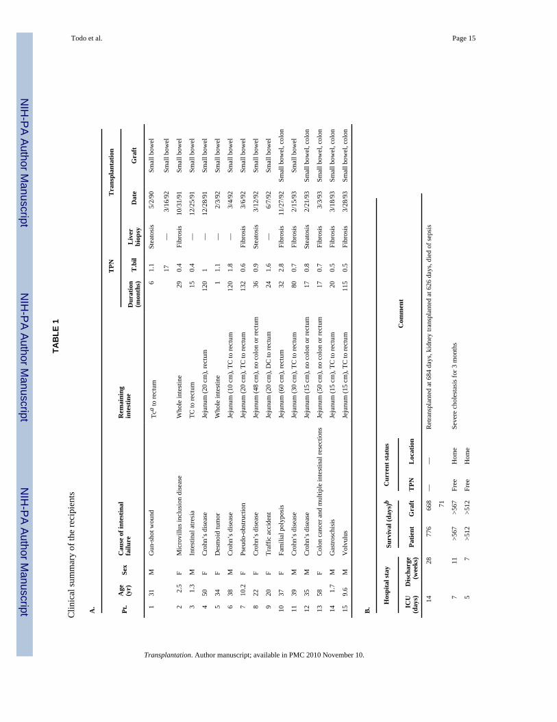
Five recipients were children and 10 were adults with a mean age of 5.1 ± 4.4 years and 36.5± 11.4 years, respectively. Indication for isolated intestinal transplantation was short-bowelsyndrome in 13 patients and uncorrectable intestinal disease in 2. All of the recipients had beenmanaged by total parenteral nutrition (TPN*) from 1 to 132 months preoperatively and hadexperienced more than one episode of TPN-related complications. Bacterial and/or fungalsepsis and multiple line replacements were seen in all but one patient (case 5). Eight had majorvessel thrombosis that had already made venous access extremely difficult.
Abnormal histopathology of the liver was found in all 10 recipients from whom a biopsy wasavailable, consisting of mild steatosis (n = 3) or mild portal fibrosis (n = 7). Five patients hadbeen hospitalized and the remaining 10 were home-bound. These and other features of therecipients are summarized in Table 1.
Donor operationThe 16 cadaveric donors had the same ABO blood type as the recipients and were slightlylarger (n = 5), similar (n = 8), or smaller (n = 3) in size. All lymphocytotoxic crossmatcheswere negative, and HLA matching was universally poor. Selective bacterial decontaminationof the donor was started immediately after the donor was accepted, as described before (3,4).The grafts for patient 1 through patient 9 consisted of the entire small intestine except for shortsegments distal to the ligament of Treitz and proximal to the ileocecal valve (Fig. 1A). Becausethese patients tended to have high postoperative stomal output and diarrhea, the ascendingcolon, with or without the transverse colon, was included with the small bowel in patients 10,12, 13, 14, and 15 to slow intestinal transit and facilitate water absorption (Fig. 1B). All graftsexcept one were flushed with 1 L University of Wisconsin solution via the abdominal aorta,and preserved for a mean duration of 6.5 ± 2.0 hr (ranging from 2.8 to 9.8 hr). The exceptionwas the first graft for patient 1, which was excised and simply immersed in an ice bath for 10.5hr until transplantation. Luminal flushing was performed only with grafts that consisted of bothsmall bowel and colon. Depletion of immunocytes was not attempted in any of the grafts.
Recipient operationThe technique for isolated intestinal transplantation was essentially the same as describedbefore (5) but with modifications for venous reconstruction and restoration of gastrointestinaltract continuity. Arterial reconstruction was performed exclusively by end-to-side anastomosisof the graft superior mesenteric artery to the recipient infrarenal abdominal aorta. The portalvein at the hepatic hilum was chosen for venous reconstruction by mesenteric piggybackmethod in most of the cases (9/16) (6), but the distal end of the recipient superior mesentericvein (4/16), or its confluence with the splenic vein (2/16) was also selected if feasible.Mesocaval anastomosis was needed on only one occasion at the time of retransplantation.
In reestablishing GI tract continuity, 4 different types of enterostomies were used fordecompression and monitoring of the transplant. Initially, both ends of the graft wereexteriorized by the chimney method (the first graft of patient 1, Fig. 2A). For the second graftof patient 1, patients 2–9, and patient 11, the proximal enterostomy was eliminated and replacedby a tube jejunostomy (Fig. 2B). In patients 10 and 12–14, a distal enterostomy was made withthe transverse colon (Fig. 2C). Patient 15 had a distal ileal loop exteriorized by the Bishop-Koop method (Fig. 2D). A tube jejunostomy was added for intestinal decompression and
*Abbreviation: TPN, total parenteral nutrition.
Todo et al. Page 2
Transplantation. Author manuscript; available in PMC 2010 November 10.
NIH
-PA Author Manuscript
NIH
-PA Author Manuscript
NIH
-PA Author Manuscript
enteral feeding in all recipients except after the first transplantation in patient 1.Cholecystectomy always was performed.
ImmunosuppressionFK506, steroids, and prostaglandin E1 (Prostin) (Fig. 3) were used for postoperativeimmunosuppression, as is routine at our center for liver recipients (7). Immediately after graftreperfusion, FK506 at 0.1 to 0.15 mg/kg/day was given intravenously, and then switched tooral FK506, 0.3 mg/kg/day, or usually less, when the recipients became tolerant to enteralfeedings. To ensure enteric absorption, oral and i.v. doses of FK506 were allowed to overlapfor several days with gradual weaning of the i.v. route. FK506 dose was adjusted to maintaintrough plasma levels of 2–3 ng/ml for the first month, 1–2 ng/ml until the third month, and 1ng/ml thereafter. These target levels are higher than for liver transplantation. The FK506 dosewas lowered if toxic side effects appeared. Methylprednisolone 1 g in adults or hydrocortisonein children was given intravenously in the operating room, and followed by rapid tapering ofprednisone over 5 days after transplantation. Maintenance steroids were given thereafter orstopped when possible. Prostaglandin E1 was started at 0.2 µg/kg/hr soon after the graft wasrevascularized, gradually increased to 0.6 to 0.8 µg/kg/hr if the recipient was stable, andcontinued until intravenous FK506 was stopped.
Graft rejection was monitored by a combination of clinical findings, endoscopic observation,and histopathologic examination of endoscope-guided mucosal biopsies. The treatment of graftrejection, based upon clinical, endoscopic, and mucosal biopsy findings, has been describedelsewhere (5,8).
Nutritional managementMethods for nutritional management after intestinal transplantation have been describedelsewhere (9). In brief, total parenteral nutrition was continued postoperatively. Afterconfirming the integrity of gastrointestinal reconstruction by an upper GI series (usually at 7–10 postoperative days), enteral feeding with an isoosmolar elemental diet (Peptamen)containing peptides and a small amount of long-chain triglycerides was begun via ajejunostomy tube. Enteral feedings and oral intake were gradually increased with a reciprocaldecrease in parenteral nutrition.
Prophylaxis of infectionSelective decontamination of the GI tract was continued for 4–6 weeks postoperatively andsupplemented with i.v. ampicillin and cefotaxin for the first 5 days. Frequent cultures of theblood, stool, wound, urine, sputum, and ostomy discharge were obtained to monitor changesin flora and to detect evidence suggesting translocation. Ganciclovir was continued for 3 to 6months for prophylaxis of cytomegalovirus infection. If severe CMV infection occurred,foscarnet or CMV immunoglobulin was added to the ganciclovir treatment.
Assessment of graft functionBody weight, volume of stomal output, and frequency and nature of the stool were commonindices used to evaluate intestinal function. D-xylose absorption test and 72-hr fecal fatsecretion were also studied periodically.
RESULTSPostoperative course
Postoperative recovery was uneventful for most of the recipients, with a median ICU stay of6 days (4 to 72 days, Table 1). The postoperative course of two adult recipients was complicated
Todo et al. Page 3
Transplantation. Author manuscript; available in PMC 2010 November 10.
NIH
-PA Author Manuscript
NIH
-PA Author Manuscript
NIH
-PA Author Manuscript
by bouts of rejection, sepsis, and renal failure. Patient 1 had repeated episodes of rejectionfollowing drug noncompliance. Patient 12 suffered from a poor pretransplant general conditiondue to his 25-year history of Crohn’s disease. Enteral feeding was started a median of 9 dayspostoperatively (range 3 to 17), and TPN was discontinued at a median of 30.5 days (range 18to 300) with one exception. Patients were discharged after 4 to 28 weeks, as shown in Table 1for each case. After discharge, the patients with a >1-month follow-up were readmitted amedian of 3 times for a median stay of 15 days. High stomal output and diarrhea resulting indehydration were the leading indications for readmission, but CMV infection and rejectionwere also prevalent indications. Patients were in the hospital a median of 32% of the time aftertheir transplant admission.
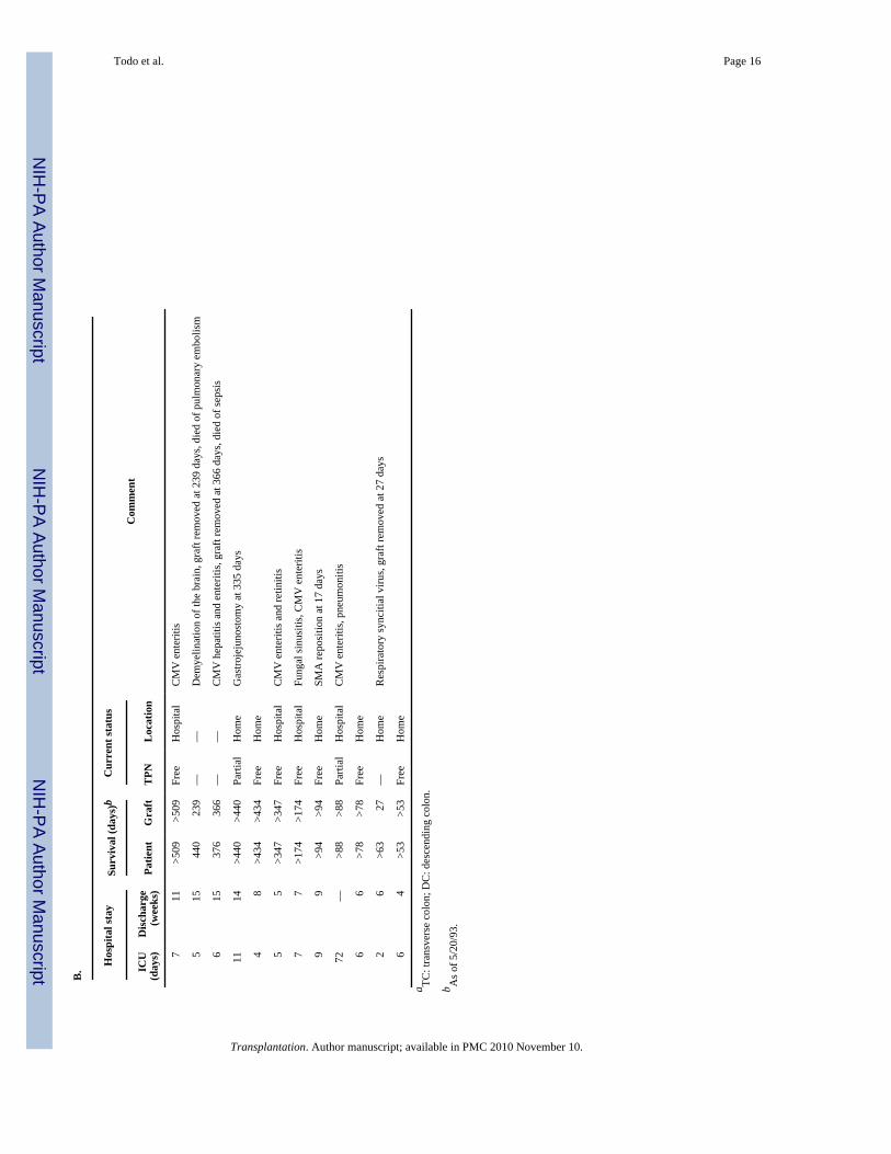
SurvivalThe 5 pediatric recipients are alive at home after 53 to 567 days, and 7 of the 10 adult patientsare alive after 78 to 499 days. Three of the surviving 7 adult patients are at home while 4 arecurrently hospitalized for the treatment of CMV infection (n = 3) or fungal sinusitis (n = 1).Thus patient survival is 12/15 (80%). By a life-table analysis using the Kaplan-Mayer method,actuarial patient survival rates at 6, 12, and 18 months are 100%, 87.5%, and 70%, respectively(Fig. 4).
Causes of graft loss and mortalityThe grafts were removed before the deaths of the 3 adult recipients (patients 1, 5, and 6), andin patient 1 the mortality followed subsequent retransplantation. A pediatric patient (patient14) who underwent graft removal at 27 days is alive after returning to TPN. Thus, graft survivalis 11/16 (68.8%). Actuarial graft survivals at 6, 12, and 18 months are 86.5%, 65.9%, and65.9% respectively (Fig. 4).
The 5 graft losses were from refractory rejection. Patient 1, who had a stormy course duringthe immediate postoperative period, lost his graft at 668 days from chronic rejection afterseveral episodes of drug noncompliance. A second graft was lost to acute rejection 71 dayslater and he died of sepsis 22 days after its removal, for a total survival of 776 days. The graftof patient 5 was removed 239 days after transplantation because of acute rejection that followedwithdrawal of immunosuppression because of a neurologic syndrome caused by demyelinationof the white matter of the brain. She was discharged from the hospital on TPN but died 201days later from a pulmonary embolism that occurred during an operation to replace the TPNline.
The postoperative course of patient 6 was uneventful until he developed recurrent Crohn’sdisease in his native colon 300 days after transplantation, and then acute rejection of theintestinal graft. This patient died of sepsis at 376 days, 10 days after the graft was removed.Patient 14, who received an intestine and colon transplant with a colostomy (Fig. 2C),developed clinical signs of graft rejection on postoperative day 12, but histopathologicconfirmation of the diagnosis and initiation of treatment were delayed because an ileal biopsycould not be obtained. Biopsies of the transplanted colon remained normal until rejectionbecame advanced. By this time, respiratory syncitial virus infection precluded augmentationof immunosuppression. The graft was removed at 27 days, and this child is alive on TPN.
RejectionOf the 16 grafts, only one (patient 3) developed no evidence of rejection. Thus, the overallincidence of graft rejection was 93.8% (15/16). The risk of acute rejection was highest at 87.5%(14/16) during the first month and decreased to 28.6% (4/14) at 3 months and 36.4% (4/11) at6 months, but was still high at 42.9% (3/7) after 12 months (Fig. 5). Moderate-to-severe acutegraft rejection was treated by OKT3. Patient 1 received two courses of OKT3 for his first graft
Todo et al. Page 4
Transplantation. Author manuscript; available in PMC 2010 November 10.
NIH
-PA Author Manuscript
NIH
-PA Author Manuscript
NIH
-PA Author Manuscript
and one for the second. Patients 10, 14, and 15 also received a course of OKT3 treatment.Chronic rejection was seen in two removed grafts (the first graft of patient 1 and patient 5).Characteristics of clinical course and angiographic and pathological changes have beendescribed elsewhere (5). Graft-versus-host disease was not seen in any of these patients.
InfectionAll except patient 8 experienced more than one episode of postoperative bacterial or fungalinfection: line infection (n = 7), abdominal wound (n = 5), peritonitis (n = 2), evidence oftranslocation (n = 2), and others (n = 6).
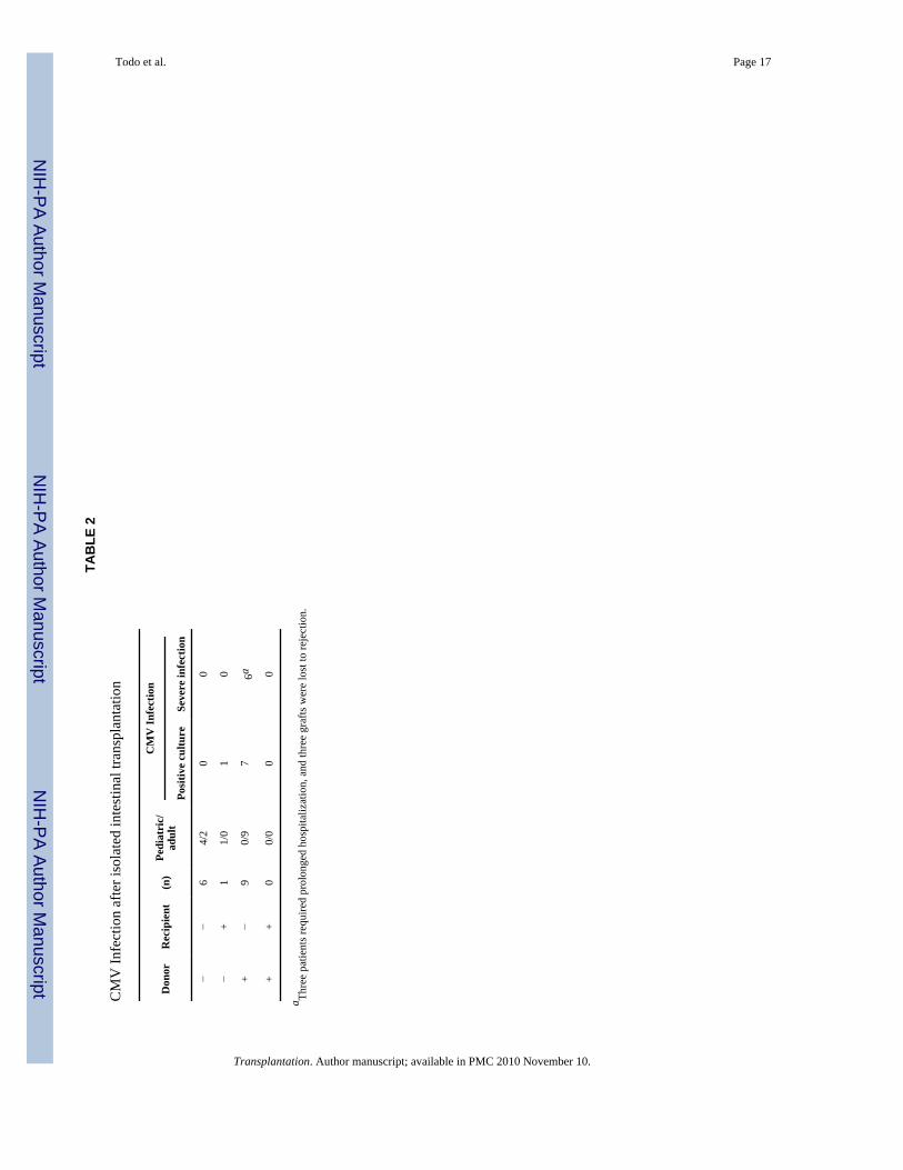
Cytomegalovirus infection after transplantation was strongly influenced by the preoperativeserological status (Table 2). For example, 7 of the 9 adult patients, who were negative for CMVpreoperatively and received grafts from CMV-positive donors developed severe clinicaldisease at 1.5 to 4 months after transplantation. Three patients required frequent hospitalreadmission for CMV infection treatment. Five patients with CMV infection developed graftrejection due to reduction of immunosuppression, and 3 of the 5 went on to graft removal. Onepediatric recipient who was seropositive before transplant and received the graft from aseronegative donor (patient 2) had positive culture of CMV in urine and sputum, but developedno clinical symptoms. In contrast, there were no episodes of CMV infection in the 6 CMV-seronegative patients who received CMV-seronegative intestinal grafts.
Graft functionNine of the 11 surviving patients with functioning grafts are free of TPN and on an unrestrictedoral diet. One patient (patient 7), who was transplanted for pseudoobstruction, had agastrojejunostomy placed surgically 335 days after transplantation and requires partialnutritional support by TPN because of dysmotility of the stomach. Patient 12 is currentlyundergoing weaning from parenteral to enteral feeding.
All recipients except for patient 1 gained or maintained body weight after 3 months (Fig. 6)and had improved D-xylose absorption.
None of the four Crohn’s disease patients has developed disease recurrence in the transplantedintestine to date. One patient (case 4) had recurrence of Crohn’s disease to the native rectum12 months after transplantation and was medically treated. One patient with desmoid tumor(case 5) also has had no recurrence for 14 months.
DISCUSSIONBefore the introduction of cyclosporine, the longest survival of an intestinal transplant was 76days (10) using immunosuppressive regimens that included the combination of azathioprineand prednisone, to which antilymphocyte globulin or thoracic duct drainage were added insome cases (1,11,12). After the advent of cyclosporine, isolated intestinal grafts were lost torejection in Toronto (n = 1) (13), Chicago (n = 1) (14), Paris (n = 7) (15), Kiel (n = 2) (16,17), London, Ontario (n = 1) (18), and Uppsala (n = 1) (19). However, the other 2 recipients,one of an intestinal segment from a living-related donor (17) and the other of a full small bowelfrom a cadaveric donor (20), are currently alive and maintained totally by an unrestricted oraldiet. In our series with FK506 immunosuppression, patient and graft survival rates improvedto 80% and 68.8%, respectively. Although the follow-up period is still limited, this experienceindicates that FK506 has moved isolated intestinal transplantation toward clinical practicality.
Because of tolerogenicity induced by the liver, Grant et al. (21) suggested that the intestinalgraft be transplanted together with the liver even in patients who have normal liver function.We confine the indication for isolated intestinal transplantation only to patients who have
Todo et al. Page 5
Transplantation. Author manuscript; available in PMC 2010 November 10.
NIH
-PA Author Manuscript
NIH
-PA Author Manuscript
NIH
-PA Author Manuscript
irreversible intestinal failure without hepatic abnormalities. However, since it is still in thedevelopmental stage, we perform this procedure in highly selected patients, such as thosewhose venous accesses are running out from major vessel thrombosis or those who have a longhistory of active Crohn’s disease that is refractory to any conventional treatments. In spite ofintensive management by TPN, no microvillus inclusion disease patients survived for morethan a few years (22), compared with our patient (case 2) who is well with an unrestricted oraldiet for 19 months after transplantation.
Although isolated intestinal transplantation has thus become feasible under improvedimmunosuppression, this achievement has been far from easy. The occurrence of severediarrhea and high stomal output have been particularly troublesome during the first 3 to 6months after transplantation, necessitating frequent readmissions for dehydration. The causeof diarrhea after intestinal transplantation is multifactorial, and is enhanced by decreasedintestinal transit time, increased osmolarity of luminal contents, increased water and electrolytesecretion, bacterial overgrowth, and steatorrhea (23). Intestinal denervation, ischemic damage,interruption of lymphatics, malabsorption, and rejection also could be factors of cumulativeimpact.
Antidiarrheal medications, such as opiates, loperamide, and kaolin-pectin mixture, were notalways effective, and in 5 recent cases an attempt was made to reduce the problem by includingthe ileocecal valve and at least the ascending colon in the graft. This approach was mentionedby Lillehei 25 years ago (24), and has been used in our multivisceral recipients (4,25). Althoughstill inconclusive because of the small number of patients, small bowel and colon transplantrecipients have tended to have less stomal output and more semiformed stool. However, theuse of a colostomy instead of a distal ileostomy has made passage of the endoscope difficultfor monitoring by ileal biopsies and was responsible for one graft loss in a child whose colonbiopsies failed to reflect the more proximal acute rejection. In the last patient to receive a smalland large bowel graft, the distal colostomy was replaced with a Bishop-Koop ileostomy (at thesuggestion of Dr. Adrian Bianchi, Manchester, England). This permitted easy access to theileum and did not increase the amount of stomal discharge.
Several (26–28) but not all (29) experimental studies have suggested the metabolic and/orimmunologic advantages of draining the venous effluent of the intestinal graft through the liver(mesoportal reconstruction) rather than resorting to a mesocaval shunt, which may be easier.In the 11 historical cases in which information on the operative procedures was available (1,11), portal drainage was used in only two instances, all others having a mesosystemic shunt.We have been able to routinely accomplish the more physiologic mesoportal reconstruction,reserving the mesosystemic shunt anastomosis for the eventuality of retransplantation. Thesignificance of the method of venous drainage remains unresolved. No serious metaboliccomplications have developed from mesocaval shunt in one of our liver-intestine recipients atalmost 3 years postoperatively (5) or in the long-surviving isolated intestinal recipients instudies by Kiel (17) and Paris (20).
CMV has been the most frequent cause of serious infectious complications in our patients, witha specific risk for those who converted to CMV-positive serology after transplantation. Six ofthe 8 developed severe CMV disease, 5 had episodes of rejection, 3 required prolongedhospitalization, and 3 underwent graft removal and eventually died. Prophylaxis and activetherapy were ineffective. In contrast, the patients who did not develop CMV infection had asmooth recovery and stable postoperative course. Posttransplant disease can occur byreactivation of preexisting CMV or by infection from blood products—and perhaps mostimportant, by transmission from infected organs.
Todo et al. Page 6
Transplantation. Author manuscript; available in PMC 2010 November 10.
NIH
-PA Author Manuscript
NIH
-PA Author Manuscript
NIH
-PA Author Manuscript
This experience recapitulates that with other kinds of organ transplants. The incidence of CMVpneumonia in seronegative recipients after transplantation of the lung from seropositive donorswas 80%, with mortality exceeding 50% (30). When seropositive grafts were given toseropositive recipients, the incidence of CMV pneumonitis decreased to 20%. After kidneytransplantation with prophylaxis with acyclovir and hyperimmunoglobulin, CMV diseaseoccurred in 10% of the seronegative patients receiving CMV-infected organs, while itdecreased to 0.8% when CMV-negative grafts were given to seronegative patients (31). Thesesame trends, but in an exaggerated form, were seen in our transplant recipients. These findingssuggest the advisability of avoiding transplantation of isolated intestinal grafts fromseropositive donors to seronegative recipients. However, the shortage of otherwise suitabledonors and the urgent need of some of the recipients requires case-by-case decision making.
Our experience has addressed the question of whether the liver should be transplantedsimultaneously with the intestine if recipient liver function is normal in order to exploit the socalled hepatic tolerogenicity that extends to concomitantly transplanted organ(s) from the samedonor (32). Although this has been a matter for discussion since the first successful combinedintestine and liver transplantation by Grant et al. (21), our earlier report comparing the evolutionof isolated intestinal versus composite grafts (containing both liver and intestine) showed betterresults with the intestine alone (4). This trend has continued with our subsequent experience.Therefore, we perform combined liver and intestinal transplantation only for patients who havefailure of both organs. Further observations will be required to establish the validity of thispolicy, particularly because its application selects the sickest patients for the compositeprocedure and therefore biases the results for comparison.
REFERENCES1. Kirkman RT. Small bowel transplantation. Transplantation 1984;37:429. [PubMed: 6729949]2. Todo S, Tzakis AG, Abu-Elmagd K, et al. Cadaveric small bowel and small bowel-liver transplantation
in humans. Transplantation 1992;53:369. [PubMed: 1738932]3. Casavilla A, Selby R, Abu-Elmagd K, et al. Logistics and technique for combined hepatic-intestinal
retrieval. Ann Surg 1992;216:85.4. Starzl TE, Todo S, Tzakis A, et al. The many faces of multivisceral transplantation. Surg Gynecol
Obstet 1991;172:335. [PubMed: 2028370]5. Todo S, Tzakis AG, Abu-Elmagd K, et al. Intestinal transplantation in composite visceral grafts or
alone. Ann Surg 1992;216:223. [PubMed: 1384443]6. Tzakis AG, Todo S, Reyes J, Nour B, Fung JJ, Starzl TE. Piggyback orthotopic intestinal
transplantation. Surg Gynecol Obstet 1993;176:297. [PubMed: 8438205]7. Takaya S, Iwaki Y, Starzl TE. Liver transplantation in positive cytotoxic crossmatch cases using
FK506, high-dose steroids, and prostaglandin E1. Transplantation 1992;54:927. [PubMed: 1279851]8. Abu-Elmagd K, Tzakis A, Todo S, et al. Monitoring and treatment of intestinal allograft rejection in
humans. Transplant Proc 1993;25:1202. [PubMed: 7680150]9. Reyes J, Tzakis AG, Todo S, et al. Nutritional management of intestinal transplant recipients.
Transplant Proc 1993;25:1200. [PubMed: 7680149]10. Fortner JG, Sichuk G, Litwin SD, Beattie EJ Jr. Immunological responses to an intestinal allograft
with HL-A-identical donor-recipient. Transplantation 1972;14:531. [PubMed: 4263654]11. Okumura M, Mester M. The coming age of small bowel transplantation: a historical perspective.
Transplant Proc 1992;24:1241. [PubMed: 1604601]12. Grant D. Intestinal transplantation: current status. Transplant Proc 1989;21:2869. [PubMed: 2650391]13. Cohen Z, Silverman RE, Wassaf R, et al. Small intestinal transplantation using cyclosporine: report
of a case. Transplantation 1986;42:613. [PubMed: 3787703]14. Tattersall C, Gebel H, Haklin M, Hartsell W, Williams J. Lymphocyte responsiveness after irradiation
in canine and human intestinal allografts. Curr Surg 1989;46:16. [PubMed: 2721232]
Todo et al. Page 7
Transplantation. Author manuscript; available in PMC 2010 November 10.
NIH
-PA Author Manuscript
NIH
-PA Author Manuscript
NIH
-PA Author Manuscript
15. Revillon Y, Jan D, Goulet O, Ricour C. Small bowel transplantation in seven children: preservationtechnique. Transplant Proc 1991;23:2350. [PubMed: 1926384]
16. Hansmann ML, Deltz E, Gundlach M, Schroeder P, Radzun HJ. Small bowel transplantation in achild. Am J Clin Pathol 1989;920:686. [PubMed: 2816825]
17. Deltz E, Schroeder P, Gebhardt H, et al. Successful clinical small bowel transplantation: report of acase. Clin Transplant 1989;3:89.
18. Grant D, Sommerauer J, Mimeault R, et al. Treatment with continuous high-dose intravenouscyclosporine following intestinal transplantation: a case report. Transplantation 1989;48:151.[PubMed: 2749888]
19. Wallander J, Ewald U, Lackgren G, Tufveson G, Wahlberg J, Meurling S. Extreme short bowelsyndrome in neonates: an indication for small bowel transplantation. Transplant Proc 1992;24:1230.[PubMed: 1604598]
20. Goulet O, Revillon Y, Brousse N, et al. Successful small bowel transplantation in an infant.Transplantation 1992;53:940. [PubMed: 1533072]
21. Grant D, Wall W, Mimeault R, et al. Successful small-bowel/liver transplantation. Lancet1990;335:181. [PubMed: 1967664]
22. Cutz E, Rhoads JM, Drumm B, Sherman PM, Durie PR, Forstner GG. Microvillus inclusion disease:an inherited defect of brush-border assembly and differentiation. New Engl J Med 1989;320:646.[PubMed: 2537465]
23. Quigley EMM, Thompson JS, Rose SG. The long-term function of canine jejunoileal autotransplants—insights into allograft physiology. Transplant Proc 1992;24:1105. [PubMed: 1604538]
24. Lillehei RC, Idezuki Y, Feemster JA, et al. Transplantation of stomach, intestine and pancreas:experimental and clinical observations. Surgery 1967;62:721. [PubMed: 4862242]
25. Starzl TE, Rowe M, Todo S, et al. Transplantation of multiple abdominal viscera. JAMA1989;26:1449. [PubMed: 2918640]
26. Schraut WH, Abraham VS, Loe KKW. Portal versus caval venous drainage of small bowel allografts:technical and metabolic consequences. Surgery 1986;99:193. [PubMed: 3484845]
27. Koltun WA, Madara JL, Smith RJ, Kirkman RL. Metabolic aspects of small bowel transplantationin inbred rats. J Surg Res 1987;42:341. [PubMed: 3494883]
28. Murase N, Demetris AJ, Furuya T, et al. Comparison of the small intestine after multivisceraltransplantation and the small intestine transplanted with portal versus caval drainage. Transplant Proc1992;24:1143. [PubMed: 1604559]
29. Shaffer D, Diflo T, Love W, Clowes GHA, Maki T, Monaco AP. Immunologic and metabolic effectsof caval versus portal venous drainage in small-bowel transplantation. Surgery 1988;104:518.[PubMed: 3261896]
30. Hutter JA, Scott JA, Wreghitt T, et al. The importance of cytomegalovirus in heart-lung transplantrecipients. Chest 1989;95:627. [PubMed: 2537711]
31. Nicol D, McDonald AS, Belitsky P, et al. Reduction by combination prophylactic therapy with CMVhyperimmune globulin and acyclovir of the risk of primary CMV disease in renal transplantrecipients. Transplantation 1993;55:841. [PubMed: 8386404]
32. Calne RY, Sells RA, Pena JR, et al. Induction of immunological tolerance by porcine liver allografts.Nature 1969;223:472. [PubMed: 4894426]
Todo et al. Page 8
Transplantation. Author manuscript; available in PMC 2010 November 10.
NIH
-PA Author Manuscript
NIH
-PA Author Manuscript
NIH
-PA Author Manuscript
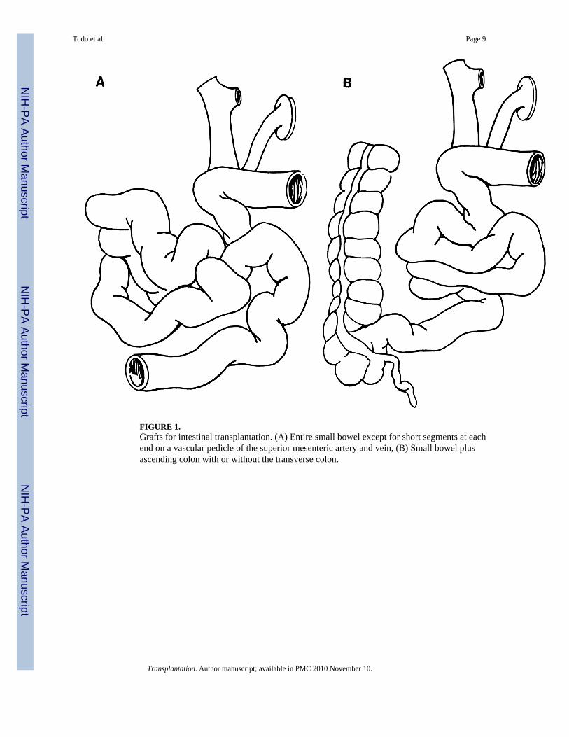
FIGURE 1.Grafts for intestinal transplantation. (A) Entire small bowel except for short segments at eachend on a vascular pedicle of the superior mesenteric artery and vein, (B) Small bowel plusascending colon with or without the transverse colon.
Todo et al. Page 9
Transplantation. Author manuscript; available in PMC 2010 November 10.
NIH
-PA Author Manuscript
NIH
-PA Author Manuscript
NIH
-PA Author Manuscript
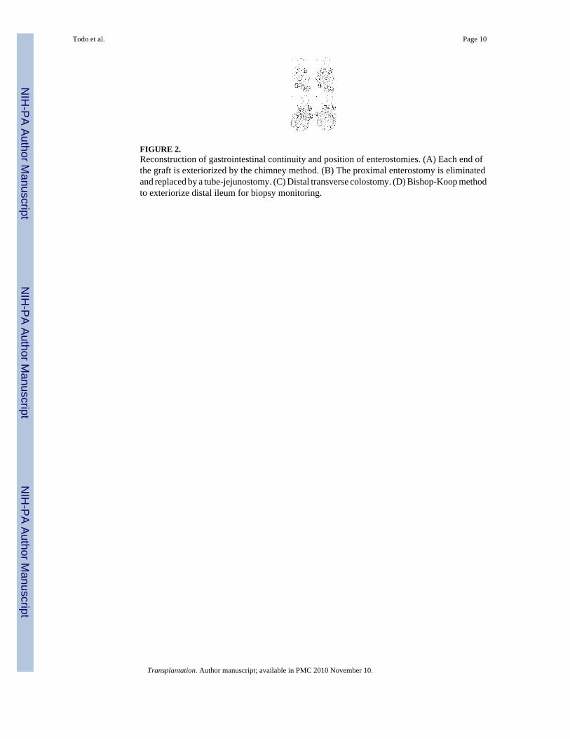
FIGURE 2.Reconstruction of gastrointestinal continuity and position of enterostomies. (A) Each end ofthe graft is exteriorized by the chimney method. (B) The proximal enterostomy is eliminatedand replaced by a tube-jejunostomy. (C) Distal transverse colostomy. (D) Bishop-Koop methodto exteriorize distal ileum for biopsy monitoring.
Todo et al. Page 10
Transplantation. Author manuscript; available in PMC 2010 November 10.
NIH
-PA Author Manuscript
NIH
-PA Author Manuscript
NIH
-PA Author Manuscript
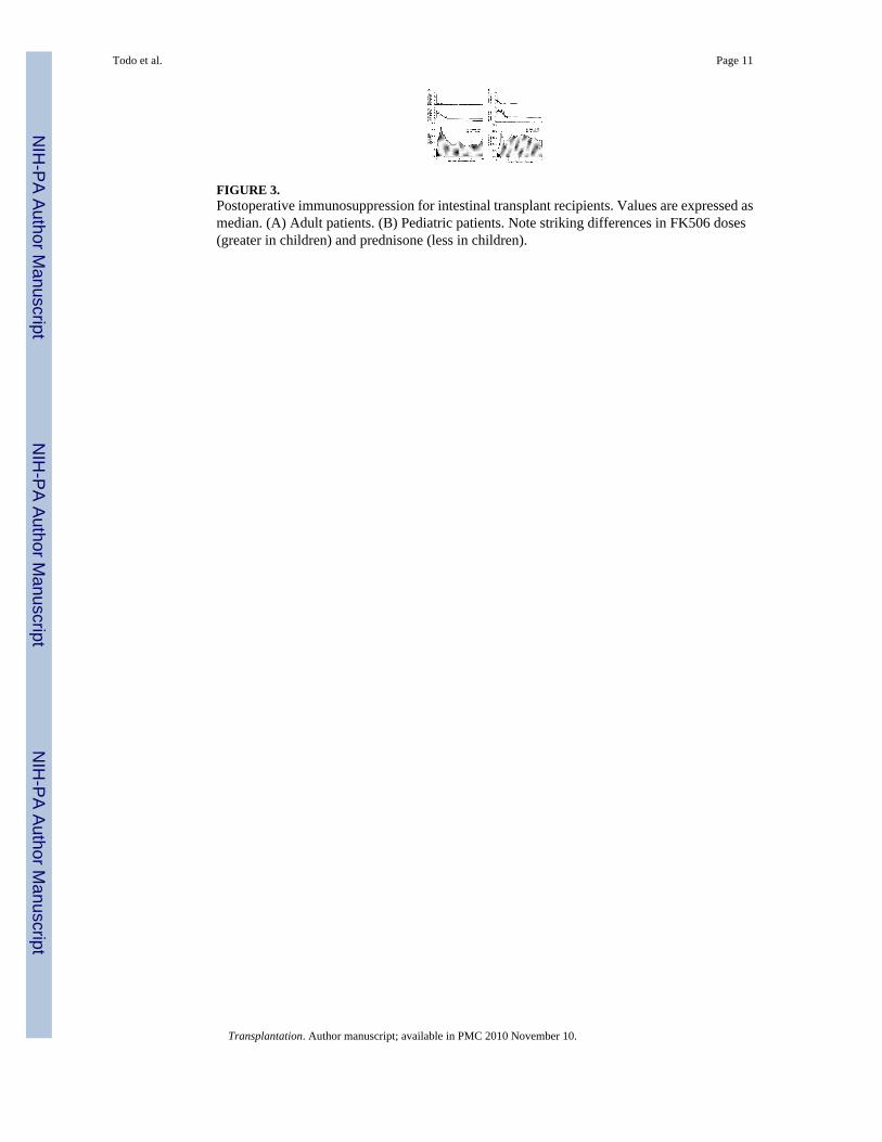
FIGURE 3.Postoperative immunosuppression for intestinal transplant recipients. Values are expressed asmedian. (A) Adult patients. (B) Pediatric patients. Note striking differences in FK506 doses(greater in children) and prednisone (less in children).
Todo et al. Page 11
Transplantation. Author manuscript; available in PMC 2010 November 10.
NIH
-PA Author Manuscript
NIH
-PA Author Manuscript
NIH
-PA Author Manuscript
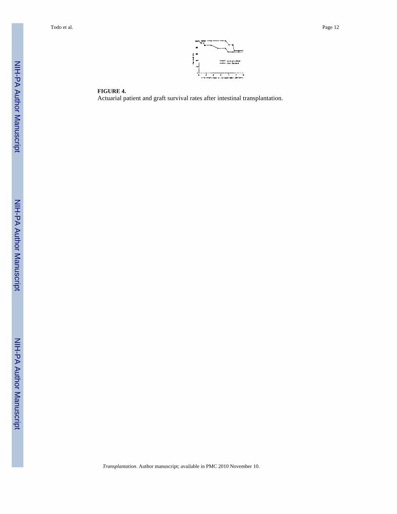
FIGURE 4.Actuarial patient and graft survival rates after intestinal transplantation.
Todo et al. Page 12
Transplantation. Author manuscript; available in PMC 2010 November 10.
NIH
-PA Author Manuscript
NIH
-PA Author Manuscript
NIH
-PA Author Manuscript
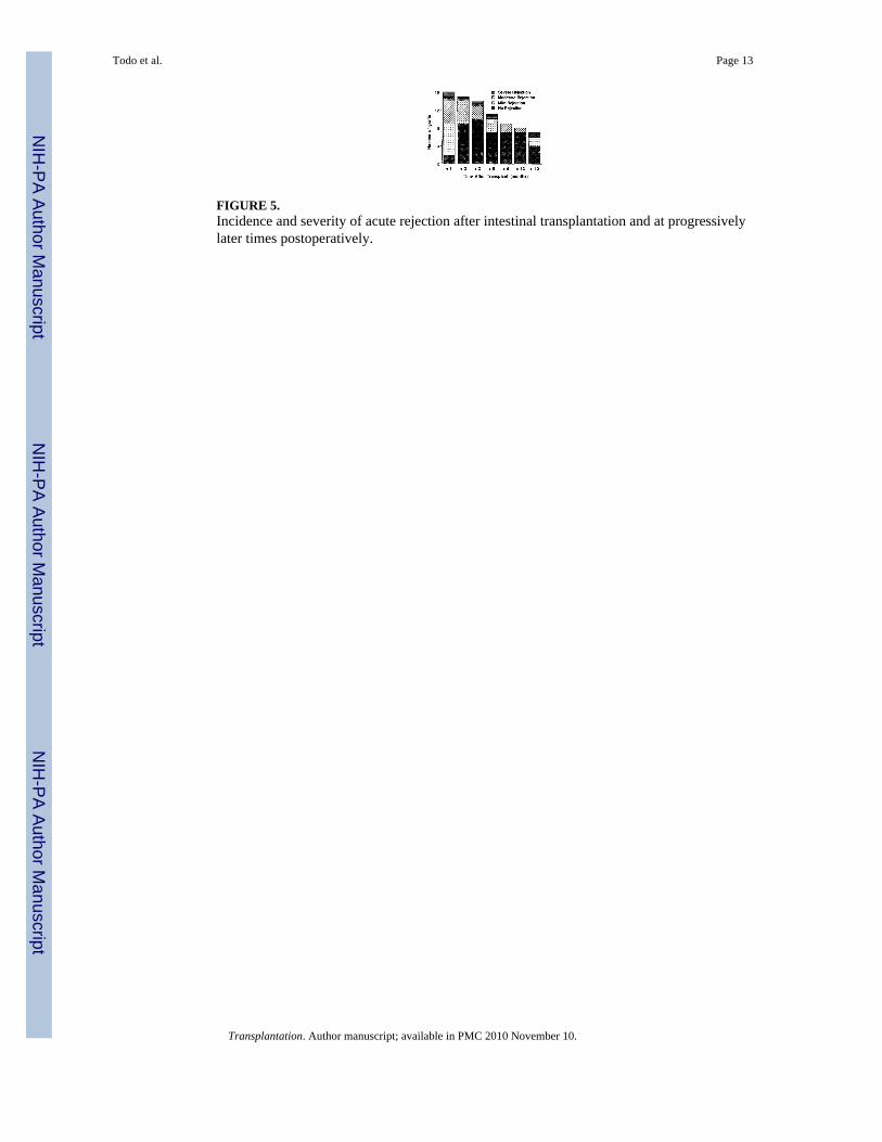
FIGURE 5.Incidence and severity of acute rejection after intestinal transplantation and at progressivelylater times postoperatively.
Todo et al. Page 13
Transplantation. Author manuscript; available in PMC 2010 November 10.
NIH
-PA Author Manuscript
NIH
-PA Author Manuscript
NIH
-PA Author Manuscript
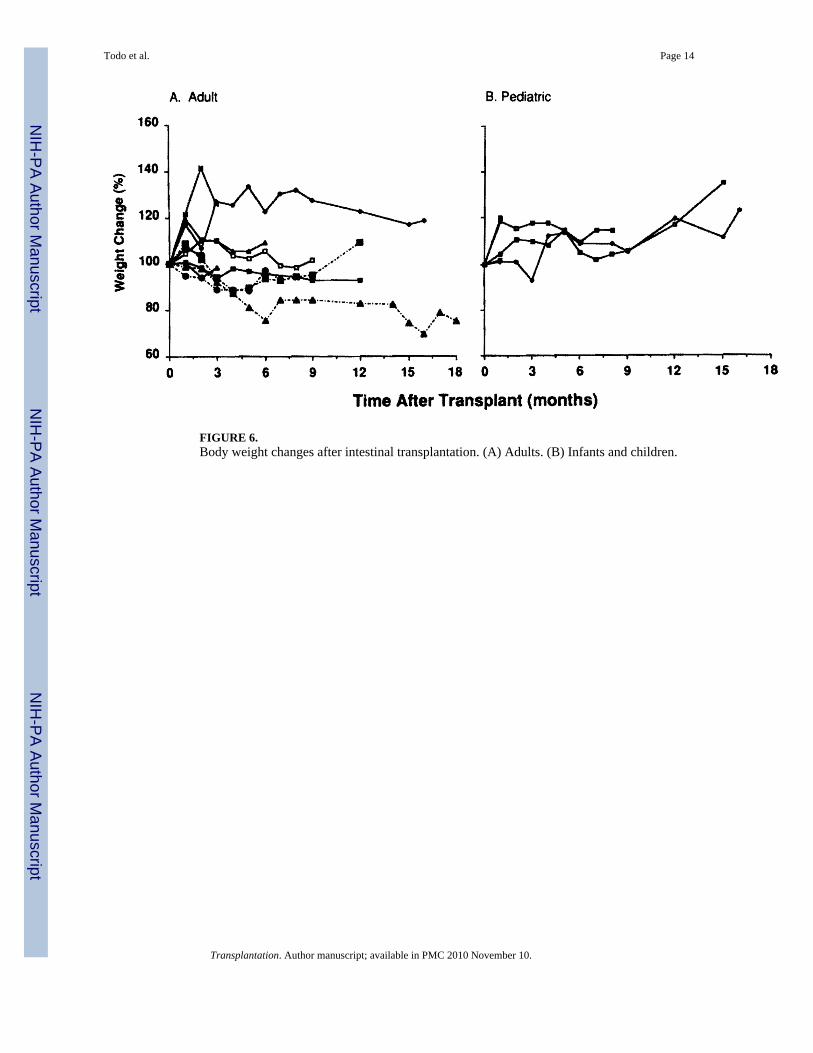
FIGURE 6.Body weight changes after intestinal transplantation. (A) Adults. (B) Infants and children.
Todo et al. Page 14
Transplantation. Author manuscript; available in PMC 2010 November 10.
NIH
-PA Author Manuscript
NIH
-PA Author Manuscript
NIH
-PA Author Manuscript
NIH
-PA Author Manuscript
NIH
-PA Author Manuscript
NIH
-PA Author Manuscript
Todo et al. Page 15
TAB
LE 1
Clin
ical
sum
mar
y of
the
reci
pien
ts
A.
Pt.
Age
(yr)
Sex
Cau
se o
f int
estin
alfa
ilure
Rem
aini
ngin
test
ine
TPN
Tra
nspl
anta
tion
Dur
atio
n(m
onth
s)T
.bil
Liv
erbi
opsy
Dat
eG
raft
131
MG
un-s
hot w
ound
Tca
to re
ctum
61.
1St
eato
sis
5/2/
90Sm
all b
owel
17—
3/16
/92
Smal
l bow
el
2 2
.5F
Mic
rovi
llus i
nclu
sion
dis
ease
Who
le in
test
ine
290.
4Fi
bros
is10
/31/
91Sm
all b
owel
3 1
.3M
Inte
stin
al a
tresi
aTC
to re
ctum
150.
4—
12/2
5/91
Smal
l bow
el
450
FC
rohn
’s d
isea
seJe
junu
m (2
0 cm
), re
ctum
120
1—
12/2
8/91
Smal
l bow
el
534
FD
esm
oid
tum
orW
hole
inte
stin
e1
1.1
—2/
3/92
Smal
l bow
el
638
MC
rohn
’s d
isea
seJe
junu
m (1
0 cm
), TC
to re
ctum
120
1.8
—3/
4/92
Smal
l bow
el
710
.2F
Pseu
do-o
bstru
ctio
nJe
junu
m (2
0 cm
), TC
to re
ctum
132
0.6
Fibr
osis
3/6/
92Sm
all b
owel
822
FC
rohn
’s d
isea
seJe
junu
m (4
8 cm
), no
col
on o
r rec
tum
360.
9St
eato
sis
3/12
/92
Smal
l bow
el
920
FTr
affic
acc
iden
tJe
junu
m (2
0 cm
), D
C to
rect
um24
1.6
—6/
7/92
Smal
l bow
el
1037
FFa
mili
al p
olyp
osis
Jeju
num
(60
cm),
rect
um32
2.8
Fibr
osis
11/2
7/92
Smal
l bow
el, c
olon
1139
MC
rohn
’s d
isea
seJe
junu
m (3
0 cm
), TC
to re
ctum
800.
7Fi
bros
is2/
15/9
3Sm
all b
owel
1235
MC
rohn
’s d
isea
seJe
junu
m (1
5 cm
), no
col
on o
r rec
tum
170.
8St
eato
sis
2/21
/93
Smal
l bow
el, c
olon
1358
FC
olon
can
cer a
nd m
ultip
le in
test
inal
rese
ctio
nsJe
junu
m (5
0 cm
), no
col
on o
r rec
tum
170.
7Fi
bros
is3/
3/93
Smal
l bow
el, c
olon
14 1
.7M
Gas
trosc
hisi
sJe
junu
m (1
5 cm
), TC
to re
ctum
200.
5Fi
bros
is3/
18/9
3Sm
all b
owel
, col
on
159.
6M
Vol
vulu
sJe
junu
m (1
5 cm
), TC
to re
ctum
115
0.5
Fibr
osis
3/28
/93
Smal
l bow
el, c
olon
B. H
ospi
tal s
tay
Surv
ival
(day
s)b
Cur
rent
stat
us
Com
men
tIC
U(d
ays)
Dis
char
ge(w
eeks
)Pa
tient
Gra
ftT
PNL
ocat
ion
1428
776
668
——
Ret
rans
plan
ted
at 6
84 d
ays,
kidn
ey tr
ansp
lant
ed a
t 626
day
s, di
ed o
f sep
sis
71
711
>567
>567
Free
Hom
eSe
vere
cho
lest
asis
for 3
mon
ths
57
>512
>512
Free
Hom
e
Transplantation. Author manuscript; available in PMC 2010 November 10.
NIH
-PA Author Manuscript
NIH
-PA Author Manuscript
NIH
-PA Author Manuscript
Todo et al. Page 16B
. Hos
pita
l sta
ySu
rviv
al (d
ays)
bC
urre
nt st
atus
Com
men
tIC
U(d
ays)
Dis
char
ge(w
eeks
)Pa
tient
Gra
ftT
PNL
ocat
ion
711
>509
>509
Free
Hos
pita
lC
MV
ent
eriti
s
515
440
239
——
Dem
yelin
atio
n of
the
brai
n, g
raft
rem
oved
at 2
39 d
ays,
died
of p
ulm
onar
y em
bolis
m
615
376
366
——
CM
V h
epat
itis a
nd e
nter
itis,
graf
t rem
oved
at 3
66 d
ays,
died
of s
epsi
s
1114
>440
>440
Parti
alH
ome
Gas
troje
juno
stom
y at
335
day
s
48
>434
>434
Free
Hom
e
55
>347
>347
Free
Hos
pita
lC
MV
ent
eriti
s and
retin
itis
77
>174
>174
Free
Hos
pita
lFu
ngal
sinu
sitis
, CM
V e
nter
itis
99
>94
>94
Free
Hom
eSM
A re
posi
tion
at 1
7 da
ys
72—
>88
>88
Parti
alH
ospi
tal
CM
V e
nter
itis,
pneu
mon
itis
66
>78
>78
Free
Hom
e
26
>63
27—
Hom
eR
espi
rato
ry sy
nciti
al v
irus,
graf
t rem
oved
at 2
7 da
ys
64
>53
>53
Free
Hom
e
a TC: t
rans
vers
e co
lon;
DC
: des
cend
ing
colo
n.
b As o
f 5/2
0/93
.
Transplantation. Author manuscript; available in PMC 2010 November 10.
NIH
-PA Author Manuscript
NIH
-PA Author Manuscript
NIH
-PA Author Manuscript
Todo et al. Page 17
TAB
LE 2
CM
V In
fect
ion
afte
r iso
late
d in
test
inal
tran
spla
ntat
ion
Don
orR
ecip
ient
(n)
Pedi
atri
c/ad
ult
CM
V In
fect
ion
Posi
tive
cultu
reSe
vere
infe
ctio
n
−−
64/
20
0
−+
11/
01
0
+−
90/
97
6a
++
00/
00
0
a Thre
e pa
tient
s req
uire
d pr
olon
ged
hosp
italiz
atio
n, a
nd th
ree
graf
ts w
ere
lost
to re
ject
ion.
Transplantation. Author manuscript; available in PMC 2010 November 10.
Copyright © 2022 FDOKUMEN