
SFTA3, a novel protein of the lung: three-dimensional structure, characterisation and immune...
10
SFTA3, a novel protein of the lung: three- dimensional structure, characterisation and immune activation Martin Schicht 1,7 , Felix Rausch 1,7 , Susetta Finotto 2 , Martina Mathews 2 , Anja Mattil 2 , Melanie Schubert 2 , Beate Koch 2 , Maximilian Traxdorf 3 , Christopher Bohr 3 , Dieter Worlitzsch 4 , Wolfgang Brandt 5 , Fabian Garreis 1 , Saadettin Sel 6 , Friedrich Paulsen 1,8 and Lars Bra ¨uer 1,8 Affiliations: 1 Institute of Anatomy, Dept II, Friedrich Alexander University Erlangen-Nuremberg, Erlangen, Germany. 2 Dept of Molecular Pneumology, Friedrich Alexander University Erlangen-Nuremberg, Erlangen, Germany. 3 Dept of Otorhinolaryngology, Head and Neck Surgery, Friedrich Alexander University Erlangen- Nuremberg, Erlangen, Germany. 4 Dept of Hygiene of the Medical Faculty, Martin Luther University of Halle- Wittenberg, Halle (Saale), Germany. 5 Dept of Bioorganic Chemistry, Leibniz Institute of Plant Biochemistry, Halle, Germany. 6 Dept of Ophthalmology, Ruprecht-Karls University Heidelberg, Heidelberg, Germany. 7 These two authors contributed equally to this work. 8 These two authors contributed equally to this work. Correspondence: M. Schicht, Dept of Anatomy II, University of Erlangen-Nuremberg, Universita ¨tsstrabe 19, 91054 Erlangen, Germany. E-mail: [email protected] ABSTRACT The lung constantly interacts with numerous pathogens. Thus, complex local immune defence mechanisms are essential to recognise and dispose of these intruders. This work describes the detection, characterisation and three-dimensional structure of a novel protein of the lung (surfactant- associated protein 3 (SFTA3/SP-H)) with putative immunological features. Bioinformatics, biochemical and immunological methods were combined to elucidate the structure and function of SFTA3. The tissue-specific detection and characterisation was performed by using electron microscopy as well as fluorescence imaging. Three-dimensional structure generation and analysis led to the development of specific antibodies and, as a consequence, to the localisation of a novel protein in human lung under consideration of cystic fibrosis, asthma and sepsis. In vitro experiments revealed that lipopolysaccharide induces expression of SFTA3 in the human lung alveolar type II cell line A549. By contrast, the inflammatory cytokines interleukin (IL)-1b and IL-23 inhibit expression of SFTA3 in A549. Sequence- and structure-based prediction analysis indicated that the novel protein is likely to belong to the family of lung surfactant proteins. The results suggest that SFTA3 is an immunoregulatory protein of the lung with relevant protective functions during inflammation at the mucosal sites. @ERSpublications SFTA3: a novel lung protein with putative protective and immunological functions during inflammation in lung diseases http://ow.ly/tsnne This article has supplementary material available from www.erj.ersjournals.com Received: Oct 15 2013 | Accepted after revision: Jan 25 2014 Support statement: This study was supported by the German Research Foundation (DFG, Programme Grants 1329/12-1 BR and BR 3681/2-1), European Grant PreDicta, ‘‘Post-infectious immune reprogramming and its association with persistence and chronicity of respiratory allergic diseases’’ at the UK-ER, Erlangen and the Wilhelm Roux Programme, Halle, Germany (Programme Grants FKZ 17/20). Conflict of interest: None declared. Copyright ßERS 2014 ORIGINAL ARTICLE IN PRESS | CORRECTED PROOF Eur Respir J 2014; in press | DOI: 10.1183/09031936.00179813 1 . Published on April 17, 2014 as doi: 10.1183/09031936.00179813 ERJ Express Copyright 2014 by the European Respiratory Society.
-
Upload
uni-erlangen -
Category
Documents
-
view
0 -
download
0
Transcript of SFTA3, a novel protein of the lung: three-dimensional structure, characterisation and immune...

SFTA3, a novel protein of the lung: three-dimensional structure, characterisationand immune activation
Martin Schicht1,7, Felix Rausch1,7, Susetta Finotto2, Martina Mathews2,Anja Mattil2, Melanie Schubert2, Beate Koch2, Maximilian Traxdorf3,Christopher Bohr3, Dieter Worlitzsch4, Wolfgang Brandt5, Fabian Garreis1,Saadettin Sel6, Friedrich Paulsen1,8 and Lars Brauer1,8
Affiliations: 1Institute of Anatomy, Dept II, Friedrich Alexander University Erlangen-Nuremberg, Erlangen,Germany. 2Dept of Molecular Pneumology, Friedrich Alexander University Erlangen-Nuremberg, Erlangen,Germany. 3Dept of Otorhinolaryngology, Head and Neck Surgery, Friedrich Alexander University Erlangen-Nuremberg, Erlangen, Germany. 4Dept of Hygiene of the Medical Faculty, Martin Luther University of Halle-Wittenberg, Halle (Saale), Germany. 5Dept of Bioorganic Chemistry, Leibniz Institute of Plant Biochemistry,Halle, Germany. 6Dept of Ophthalmology, Ruprecht-Karls University Heidelberg, Heidelberg, Germany. 7Thesetwo authors contributed equally to this work. 8These two authors contributed equally to this work.
Correspondence: M. Schicht, Dept of Anatomy II, University of Erlangen-Nuremberg, Universitatsstrabe 19,91054 Erlangen, Germany. E-mail: [email protected]
ABSTRACT The lung constantly interacts with numerous pathogens. Thus, complex local immune
defence mechanisms are essential to recognise and dispose of these intruders. This work describes the
detection, characterisation and three-dimensional structure of a novel protein of the lung (surfactant-
associated protein 3 (SFTA3/SP-H)) with putative immunological features.
Bioinformatics, biochemical and immunological methods were combined to elucidate the structure and
function of SFTA3. The tissue-specific detection and characterisation was performed by using electron
microscopy as well as fluorescence imaging.
Three-dimensional structure generation and analysis led to the development of specific antibodies and, as
a consequence, to the localisation of a novel protein in human lung under consideration of cystic fibrosis,
asthma and sepsis. In vitro experiments revealed that lipopolysaccharide induces expression of SFTA3 in the
human lung alveolar type II cell line A549. By contrast, the inflammatory cytokines interleukin (IL)-1b and
IL-23 inhibit expression of SFTA3 in A549. Sequence- and structure-based prediction analysis indicated that
the novel protein is likely to belong to the family of lung surfactant proteins.
The results suggest that SFTA3 is an immunoregulatory protein of the lung with relevant protective
functions during inflammation at the mucosal sites.
@ERSpublications
SFTA3: a novel lung protein with putative protective and immunological functions duringinflammation in lung diseases http://ow.ly/tsnne
This article has supplementary material available from www.erj.ersjournals.com
Received: Oct 15 2013 | Accepted after revision: Jan 25 2014
Support statement: This study was supported by the German Research Foundation (DFG, Programme Grants 1329/12-1BR and BR 3681/2-1), European Grant PreDicta, ‘‘Post-infectious immune reprogramming and its association withpersistence and chronicity of respiratory allergic diseases’’ at the UK-ER, Erlangen and the Wilhelm Roux Programme,Halle, Germany (Programme Grants FKZ 17/20).
Conflict of interest: None declared.
Copyright �ERS 2014
ORIGINAL ARTICLEIN PRESS | CORRECTED PROOF
Eur Respir J 2014; in press | DOI: 10.1183/09031936.00179813 1
. Published on April 17, 2014 as doi: 10.1183/09031936.00179813ERJ Express
Copyright 2014 by the European Respiratory Society.

IntroductionThe lung surface is lined with a complex mixture of phospholipids and proteins known as pulmonary
surfactant. It is essential for normal respiratory mechanics and reduces the surface tension of the air–liquid
interface and, as a consequence, prevents collapse of the alveoli. A lack of surfactant leads to respiratory
failure [1]. Beyond this function, the proteins of the pulmonary surfactant, surfactant proteins (SP),
contribute to the innate immune defence against inhaled pathogens and moreover act as anti-inflammatory
substances [2]. Lung SPs are mainly secreted by alveolar type II cells.
Four different lung SPs have been described to date: two hydrophobic proteins, SP-B and SP-C, and two
collectin-like proteins, SP-A and SP-D. These proteins have been described in detail in relation to their lung
surface activity and immunological defence function [2].
SP-A and SP-D are representatives of the C-type lectin family, which according to current understanding
are able to bind to specific carbohydrates of bacteria, protozoans, fungi and viruses [3]. This function
facilitates opsonisation of and accelerates immune defence reactions towards these microorganisms [4].
In contrast to SP-A and SP-D, the small and extremely hydrophobic proteins SP-B and SP-C are essential
components during formation of surfactant monolayers and stabilisation of air–fluid interfaces [5, 6].
Similar to SP-A and SP-D, the presence of SP-B and SP-C has already been demonstrated in a variety of
tissues and humours, including tissues of the nasolacrimal apparatus and ocular surface, in tear fluid, in
salivary glands, in the gingiva and nasal mucosa [7–10]. Due to the homology to amoebapores, an
immunological function of SP-B has been also been discussed [11]. Recent publications demonstrate the
role of SP-B in the activation of alveolar macrophages in the innate immune response in the lung [11].
Recently, another new surfactant protein SP-G has been identified and described [12]. SP-G has
physicochemical properties similar to SP-B and SP-C. The possibility of interactions with lipid systems and
a potential surface-regulatory function of SP-G have been discussed. Alveolar macrophages constitute an
important defence against infection because they clear pathogens. Maintaining normal and consistent
amounts of alveolar surfactant is in part dependent on clearance of surfactant by alveolar macrophages [13].
While working with the four known SPs, our attention was attracted to another putative surfactant protein.
It was theoretically identified by bioinformatics and named surfactant protein H (SP-H) or surfactant-
associated protein 3 (SFTA3) [14]. The official gene symbol is SFTA3 (‘‘Surfactant associated 3’’; HUGO
Nomenclature Committee). This putative SP shows no significant sequential or structural similarities to
other SPs or other known proteins in general. The protein (SFTA3 or SP-H) is encoded on the human
chromosome 14, its primary translation product consists of 94 amino acid residues with a molecular weight
of ,10 kDa. The aim of the study was the first detection and characterisation of this novel protein of the lung.
Methods summaryAll tissue samples were obtained from cadavers donated to the Dept of Anatomy and Cell Biology, Martin
Luther University of Halle-Wittenberg, Halle, Germany. Bronchoalveolar lavage (BAL) and sputum samples
were obtained from patients of the Dept of Otorhinolaryngology-Head and Neck Surgery, Friedrich
Alexander University Erlangen-Nuremberg, Erlangen, Germany and of the Dept of Hygiene, Martin Luther
University of Halle-Wittenberg, in accordance with the Declaration of Helsinki, the study was approved by
the Institutional Review Board (Martin Luther University Halle-Wittenberg) as well as the responsible
ethics committee. The acquisition of BAL and cystic fibrosis (CF)-sputum is further described in the online
supplementary material.
RNA preparation, cDNA synthesis and PCR analysis was performed using the primers (see the online
supplementary material) previously described by SCHICHT et al. [10]. Quantitative real-time RT-PCR using
cultivated as well as stimulated A549 cells was performed as previously described by REPPERT et al. [15].
Western blot, ELISA (USCN Life Science, Wuhan, PRC) and immunohistochemical detection of the protein
was performed as described by SCHICHT et al. 2013 [10] (see the online supplementary material). The
protocol for immunoelectron transmission electron microscopy, as well as for cutting 50 nm ultrathin
sections using a transmission electron microscope (Zeiss 900; Carl Zeiss Meditec AG, Jena, Germany), is
described in the online supplementary material.
The three-dimensional structural model for SP-H was obtained from the Robetta online server (http://
robetta.bakerlab.org). The prediction of post-translational modifications was carried out using various
online tools (online supplementary material) and the model stability was assessed by YASARA simulations
(www.yasara.org/). The molecular dynamics (MD) simulations in the lipid environment were performed
with the GROMACS program package (www.gromacs.org/). These modelling steps were performed as
described in detail by RAUSCH et al. [12].
LUNG BIOLOGY | M. SCHICHT ET AL
DOI: 10.1183/09031936.001798132

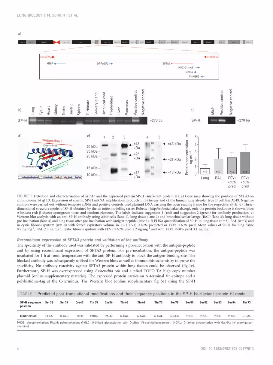
ResultsCharacteristics of the human SFTA3 gene and expression of specific RNA amplification productsSFTA3 is a single copy gene on chromosome 14 q13.3. The transcript comprises 1500 bp with 5 exons in the
total genomic region (fig. 1a). SFTA3 is positioned close to the NKX2 genes. Different tissues were analysed
by PCR and SP-H mRNA was shown to be expressed especially at the mucosal sites. The human lung
alveolar type II cell line A549 also expressed SP-H mRNA (fig. 1b–c). The detected PCR bands are in
accordance with the expected sequences for SFTA3 in gene bank data.
SP-H protein structure model and post-translational modificationsA structural model for SP-H was created by the ab initio structure prediction server Robetta. After minor
refinements, the model showed a native-like fold and very good stereochemical quality according to the
quality assessment tools PROSA II (https://prosa.services.came.sbg.ac.at/prosa.php) and PROCHECK
(www.ebi.ac.uk/thornton-srv/software/PROCHECK/). A 20 ns MD simulation in a water box showed the
stability of the final model. Scanning the SP-H sequence with sequence-based prediction tools for post-
translational modifications (PTMs) gave the results summarised in table 1. All the predicted PTMs were
manually added to the SP-H model and again a 20 ns MD simulation showed the stability of the modified
model. While the PTMs had nearly no effect on the model stability, they influenced protein surface properties.
They could form polar or hydrophobic areas (depending on the PTM type) on the protein surface with a
significant impact on water solubility or the protein–lipid interaction potential of SP-H. Two stable SP-H
models were obtained which could be used for more sophisticated simulation studies. For more information
about the modelling process, the protein model and PTMs, please see the online supplementary material.
Generation of a specific SP-H antibody and SP-H localisation in lung tissueWith the help of the protein structure model obtained for SP-H, two potential antigen sequences could be
identified (fig. 1d). The first suggestion covers the very stable N-terminal a-helix from position 7 to 31
(DFQLIRDQVLFLQDQAQRLTEWLQL) and the second suggestion comprises the amino acid positions 35
to 51 (ENPVSESTTLCLREREK). Both regions contain various amino acids with functional groups, which
would allow specific binding of an antibody. But whereas there are no predicted PTMs within the sequence
of the first suggestion, the predicted O-glycosylation at position 39 and palmitoylation at position 45 could
interfere with a proper binding of the antibody to the second suggested area. Furthermore, the spatial
proximity of the palmitoylation on position 56 could cause steric hindrance. Therefore, the antibodies for
SP-H were produced solely based on a shorter version of the first suggestion (DFQLIRDQVLFLQDQAQ). A
BLAST-search (http://blast.ncbi.nlm.nih.gov/Blast.cgi) with this peptide sequence showed no hits for
another human protein sequence. Therefore, it was considered as unique in the human proteome.
Anti-peptide antibodies were generated against a specific region of the human SP-H sequence
(CDFQLIRDQVLFLQDQAQE). The peptide was synthesised by SeqLab (Gottingen, Germany). The
specificity of the antibody was verified by Western blot analysis. The specificity of the resulting antibody was
tested with protein from lung tissue and BAL (30 mg) (fig. 1e). The purified antibody shows distinct protein
bands in lung for SP-H at 13 kDa, 26 kDa and 43 kDa. Lung tissue was used as a specific positive control
for SPs. The analysis of BAL showed distinct bands at 13 kDa, 26 kDa and 43 kDa, no distinct bands above
43 kDa could be observed.
The quantification of SP-H in lung and BAL show a concentration between 0.06–1.83 ng?mg-1 in lung
(n53) and between 0.15–4.87 ng?mg-1 in BAL (n53) (fig. 1f). The analysis of sputum from CF patients
showed a small, nonsignificant increase in SP-H with severity of CF (stratified by forced expiratory volume
in 1 s (FEV1)) (fig. 1f). The concentration of SP-H varied between 0.25–3.47 ng?mg-1 for FEV1 .60%
predicted CF sputum and 0.78–6.51 ng?mg-1 FEV1 ,60% pred CF sputum.
The quantification of SP-H in lung tissue and BAL shows different protein concentrations, but they are
similar to the protein concentrations observed for the known SPs. It is known that the protein
concentration is very variable and influenced by infection, smoking and by respiratory effects. The detection
of SP-H with in BAL indicates that the cells of the lung secrete the protein. The ELISA analysis with samples
from CF patients shows a no regulation effect of SP-H in severe CF. The result is in accord with microarray
date and a big variance between the SP-H mRNA concentrations in dependence of CF [16]. It seems that the
different results from distinct time points of infection, drug treatment and patient age, Similar effects are
known from CF where the protein concentration of SP is increased on the one hand or down regulated on
the other hand [17].
Interestingly, SFTA3 seems to be involved in the neonatal respiratory distress syndrome with intact NKX2-1
gene [18].
LUNG BIOLOGY | M. SCHICHT ET AL
DOI: 10.1183/09031936.00179813 3
Recombinant expression of SFTA3 protein and validation of the antibodyThe specificity of the antibody used was validated by performing a pre-incubation with the antigen-peptide
and by using recombinant expression of SFTA3 protein. For pre-incubation, the antigen-peptide was
incubated for 1 h at room temperature with the anti-SP-H antibody to block the antigen-binding-site. The
blocked antibody was subsequently utilised for Western blots as well as immunohistochemistry to prove the
specificity. No antibody reactivity against SFTA3 protein within lung tissues could be observed (fig 1e).
Furthermore, SP-H was overexpressed using Escherichia coli and a pBad TOPO TA high copy number
plasmid (online supplementary material). The expressed protein carries an N-terminal V5-epitope and a
polyhistidine-tag at the C-terminus. The Western blot (online supplementary fig. S1) using the SP-H
40 kDa
540
35
25
15
10
4321e)d)
b)
a)
c)
SP-H
Lung
Eyel
id
Hea
rt
Kid
ney
Test
is
Gas
tric
Sple
en
Panc
reas
Posi
tive
cont
rol
Neg
ativ
e co
ntro
l
A549
Posi
tive
cont
rol
Neg
ativ
e co
ntro
l
Turb
inat
e
Saliv
ary
glan
d
Um
bilic
al c
ord
Trop
hobl
ast
Live
r
35 kDa25 kDa
15 kDa
10 kDa
≈43 kDa
≈26 kDa
≈13 kDa
≈43 kDa
≈270 bp≈270 bp SP-H
≈26 kDa
≈13 kDa
C-terminus
N-terminus
suggestion 1
suggestion 2
5f)
3
4
2
1
0Prot
ein
conc
entr
atio
nng
·mL-
1
Lung
n=3 n=3 n=4 n=6
BAL FEV1>60% pred
FEV1<60% pred
3700355836767764
SFTA3
PHKBP2
NKX-2
NKX-2-1-AS1DPPA3P2MBIP
q32.3314q32.2q32.1214q31.314q31.114q24.314q24.2q24.1q23.3q23.2q22.3 14q23.114q22.114q21.314q21.214q21.113.114q1214q11.214p11.214p1214p13
FIGURE 1 Detection and characterisation of SFTA3 and the expressed protein SP-H (surfactant protein H). a) Gene map showing the position of SFTA3 onchromosome 14 q13.3. Expression of specific SP-H mRNA amplification products in b) tissues and c) the human lung alveolar type II cell line A549. Negativecontrols were carried out without template cDNA and positive controls used plasmid DNA carrying the open reading frame for the respective SP-H. d) Three-dimensional structure model of SP-H obtained by the ab initio modelling server Robetta (http://robetta.bakerlab.org), only the protein backbone is shown; blue:a-helices; red: b-sheets; cyan/green: turns and random elements. The labels indicate suggestion 1 (red) and suggestion 2 (green) for antibody production. e)Western blot analysis with an anti-SP-H antibody using A549 cells (lane 1), lung tissue (lane 2) and bronchoalveolar lavage (BAL) (lane 3); lung tissue withoutpre-incubation (lane 4) and lung tissue after pre-incubation with antigen-peptide (lane 5). f) ELISA quantification of SP-H in lung tissue (n53), BAL (n53) andin cystic fibrosis sputum (n510) with forced expiratory volume in 1 s (FEV1) .60% predicted or FEV1 ,60% pred. Mean values of SP-H for lung tissue0.7 ng?mg-1, BAL 2.0 ng?mg-1, cystic fibrosis sputum with FEV1 .60% pred 2.2 ng?mg-1 and with FEV1 ,60% pred 3.1 ng?mg-1.
TABLE 1 Predicted post-translational modifications and their sequence positions in the SP-H (surfactant protein H) model
SP-H sequenceposition
Ser32 Ser39 Cys45 Thr55 Cys56 Thr66 Thr69 Thr75 Ser78 Ser80 Ser82 Ser83 Ser84 Thr93
Modification PHOS O-GLC PALM PHOS PALM O-GAL O-GAL O-GAL O-GLC PHOS PHOS PHOS PHOS O-GAL
PHOS: phosphorylation; PALM: palmitoylation; O-GLC: O-linked glycosylation with GlcNAc (N-acetylglucosamine); O-GAL: O-linked glycosylation with GalNAc (N-acetylgalact-osamine).
LUNG BIOLOGY | M. SCHICHT ET AL
DOI: 10.1183/09031936.001798134

antibody displays distinct bands at ,18 kDa and ,43 kDa. After subtracting the molecular weight of the
V5-epitope and the polyhistidine-tag the bands of the recombinant protein are in accordance to the
molecular weight of the SFTA3 detected in tissues and BAL. Performing Western blot analysis using an anti-
V5 antibody the same distinct protein bands were detected, indicating a specific reaction of the anti-SP-H
antibody on the one hand and proper recombinant expression on the other hand.
Simulation of the protein models in a lipid environmentThe SP-H model without PTMs was calculated for 50 ns in four independent MD simulations and showed a
stable root mean square deviation (RMSD) after approximately 10 ns. To produce different protein–lipid
interaction scenarios, the orientation of the protein with respect to the lipid surface was manually altered for
each simulation start. In all cases, the model started to interact with the dipalmitoylphosphatidylcholine
(DPPC) monolayer after 2–10 ns. In the simulation with the highest interaction potential, the protein was
fixed on the lipid surface mostly by interactions of the polar residues of a-helix spanning residues 7–31
(fig. 2b). The progression of the RMSD (fig. 2a, black plot) underlines the stability of this complex. The
hydrophobic amino acids of the helix caused a hollow on the monolayer surface, which enabled immersion
of the C-terminal parts of the protein below the head group region. This reduced the lipid ordering and
layer stability, as indicated by the increased fluctuations in the area per lipid plot (fig. 2c, black plot). For
the SP-H model with attached PTMs, MD simulations with the same setup were performed, and the results
showed different interaction sites. In the most stable simulation, the a-helix spanning residues 7–31 was
positioned in a nearly perpendicular orientation (fig. 2d). Only the N-terminus (residues 1–6) and major
parts of the C-terminus (residues 75–94) were interacting with the lipids. Whereas the unmodified model
showed an immersion into the lipid layer involving mainly hydrophobic interactions of the C-terminus, the
interactions of the modified model were mostly driven by hydrogen bonds. The palmitoylations were also in
close proximity to the monolayer surface, but were not interacting with lipids. They were flexibly attached
to the protein, forming hydrophobic spots on the surface, which were shielded from the surrounding water
during the MD by hydrophilic modifications of the flexible loop spanning residues 75–84. The whole
protein structure was stable during the contact formation between protein and monolayer (fig. 2a, red
RMSD plot). However, the interaction may not have reached its final state at the end of the simulation,
because an influence on monolayer characteristics or stability was not evident from the very stable area per
lipid plot (fig. 2c, red plot). With a longer MD simulation, a deeper immersion and interactions, especially
involving the attached palmitoylations, with significant effect on the lipid system could be observed.
Nevertheless, the simulations showed that SP-H, without and with PTMs, has the potential to interact with
lipid systems (fig 2b–d).
Detection and distribution of SP-H in lung and in A549 cells by means of immunohistochemistry andimmunogold electron microscopyAll investigated lung tissue samples showed antibody reactivity against SP-H (fig. 3a). Within lung tissue, it
could be demonstrated that SP-H is distributed especially in the alveolar cells (I and II), in alveolar
macrophages and is cytoplasmic in the epithelium.
The cellular distribution is similar to the known SPs; it is known that SPs are localised in the cytosol of
epithelial cells as granular structures [9, 19]. SP-H is secreted as a layer in the bronchioles (fig. 3a) and also
showed reactivity in Western blot experiments on BAL samples (fig. 1e–f). Immunocytochemical
investigations on A549 cells showed that SP-H is localised on the outer cell membrane (fig. 3b). The
results obtained from electron microscopy indicate that the protein is also secreted and presented on the
membrane of the ciliated cells, as demonstrated by positive immunoreactivity of the gold-labelled
antibodies (red arrows and black dots in figure 3c). This indicates that SP-H remains cytoplasmic because
of its physicochemical properties and is secreted after modification. In theoretical studies, SP-H showed
palmitoylation potential and, if this is the case, it would be able to interact with a lipid membrane similarly
to SP-B and SP-C [20]. To determine if SP-H can stabilise the liquid surface, further functional studies are
needed. However, these experiments support the results of the MD simulations, which showed theoretical
interaction potential for SP-H with a lipid system (fig. 3d).
Influence of cytokines and lipopolysaccharide on SP-H production by A549 as well as SP-Hexpression in case of bacterial sepsis and asthmaBecause it is difficult to obtain and cultivate primary lung cells, A549 cells were used and incubated to
investigate whether SP-H is regulated during infections. This cell line is well established and known to
produce surfactant proteins in response to stimulation with lipopolysaccharide (LPS) of the bacterial
membrane wall. A significant increase in SP-H mRNA concentration could be detected after 3–24 h
stimulation with LPS (fig. 4a). By contrast, the dose–response analysis with interleukin (IL)-1b showed a
downregulation of SP-H expression (fig. 4b). Similarly, IL-23 downregulated SP-H mRNA in A549 cells
LUNG BIOLOGY | M. SCHICHT ET AL
DOI: 10.1183/09031936.00179813 5

(fig. 4c). The immunohistochemical fluorescence analysis revealed similar results (fig. 4d). The stimulation
with IL-1b, IL-23 and both IL-1b and IL-23 showed a weak signal for SP-H protein within the cytoplasm
and on the endoplasmic reticulum. In contrast, SP-H could not be detected on the cell membrane but
within the nucleolus (green spots in fig. 4d). Within tissue from patients suffering from asthma or bacterial
sepsis SP-H could be detected especially in alveolar cells (I and II) as well as in alveolar macrophages and the
cytoplasm of the epithelial cells.
The subcellular distribution is comparable to the known SPs, namely as granular structures within the
cytosol of the epithelial cells [9, 21]. Moreover, the lung tissue of patients suffering from bacterial sepsis
showed an increased expression of SP-H corresponding to the real-time RT-PCR experiment shown in
figure 4e. By contrast, the expression of SP-H is decreased in tissue affected by asthma, also corresponding
to the real-time RT-PCR experiments shown in figure 4e.
Abnormal accumulation of surfactant has been described in humans since 1958 as a pulmonary disorder
called pulmonary alveolar proteinosis, which is associated with accumulation of surfactant lipids and
proteins in the airspaces [22]. Abnormal surfactant catabolism by alveolar macrophages could contribute to
this disorder [21]. Considering that patients with pulmonary alveolar proteinosis have an increased risk for
viral, bacterial and fungal infections, regulation of SP production by alveolar macrophages and epithelial
type II cells is very important to avoid infections.
The present study shows that cytokines IL-1b, IL-23 and a combination of both downregulate the
expression of SP-H. The addition of LPS, a component of the outer membrane component of Gram-
negative bacteria, by contrast, induces an increase in SP-H mRNA concentration. LPS induces acute
pulmonary inflammation and changes in the composition of the surfactant, and type and activation of the
lung resident cell population. In response to immune stimulation, alveolar epithelial type II cells can
synthesise SPs which maintain the mucosal integrity. In particular, SP-A, the most abundant pulmonary SP,
can bind to LPS, viruses and fungal cell wall components. However, overproduction of SPs in the lung
might lead to chronic diseases [23]. This could indicate that LPS simultaneously induces inflammatory
cytokines like IL-1b and IL-23 and SP-H production by resident alveolar macrophages via Toll-like receptor
5.0 without PTMswith PTMs
without PTMswith PTMs
a)4.54.03.53.02.52.01.51.00.50.0
0 5 10 15 20 25Time ns
30 35 40 45 50
0 5 10 15 20 25Time ns
30 35 40 45 50
Bac
kbon
e R
MSD
Å
60c)
b)
d)59585756555453525150
Area
per
lipi
d Å2
FIGURE 2 a) Root-mean-square deviation (RMSD) plot, c) area per lipid plot and structures after molecular dynamicssimulations (50 ns) of the SP-H (surfactant protein H) protein model b) without and d) with post-translationalmodifications (PTMs) in the presence of a DPPC monolayer. In both the SP-H protein model with and without PTMs astable protein structure and lipid surface is indicated. The protein in b) and d) is shown in ribbon presentation (a-helices:blue; b-sheets: red; turns: green; coil: cyan) and the lipids with a yellow van der Waals surface. PTMs with tightinteractions to lipids are shown as a red van der Waals surface in (d).
LUNG BIOLOGY | M. SCHICHT ET AL
DOI: 10.1183/09031936.001798136

(TLR) [24]. Thus, Gram-negative bacterial infection induces SP-H production by alveolar epithelial cells
and inflammatory cytokines like IL-1b and IL-23 produced by alveolar macrophages limit this process
(fig. 5). The downregulation of SP-H mRNAs caused by proinflammatory cytokines could play an
important role during the pathogenesis of respiratory infection. As shown by others [25] and us, cytokine-
induced changes in SP mRNA are reflected in the levels of the proteins. In small premature infants
developing chronic lung disease [26] and in acute respiratory distress syndrome (ARDS) [27],
proinflammatory cytokines are increased in the airways. The surfactant defects evident in chronic lung
disease and in ARDS include deficiencies in SP-B [28] and SP-A [29]. It is known that the regulation of SPs
by microbial proteins is mediated by cytokines [25].
TLRs are type I transmembrane proteins. In alveolar epithelial cells, LPS can specifically activate TLR4,
which then activates mitogen-activated protein kinase family proteins, nuclear factor (NF)-kB and activator
protein (AP)-1 [30]. Both IL-23 and IL-1b activate NF-kB via the canonical pathway involving degradation
of IkB. Aberrant activation of NF-kB has been linked to inflammatory and autoimmune diseases, septic
shock, viral infection and improper immune function and plays a role in cancer development. It is, thus,
possible that inflammation has a protective effect to limit an LPS-induced overproduction of SPs by alveolar
epithelial type II cells. Understanding of the basic mechanisms of SP-A, -D, -G and -H action might lead to
new therapeutic strategies for the cure of several lung diseases.
b)
c)
d)B
ronchiolesAlveolar duct
Alveoli
a)
1 2
3 4
5 6
7 8
9 10
FIGURE 3 a) Immunohistochemical detection of SP-H (surfactant protein H) (red, AEC (3-amino-9-ethylcarbazole) staining) in lung tissue. Bronchioles:cytoplasmic protein distribution in bronchioles (panels 1–4); alveolar duct: protein distribution as a superficial layer (panels 5–6); alveoli: detection in themembrane of alveolar type II cells (panels 7–10). Insets show magnifications. Panels 1, 3, 5, 7 and 9 scale bars5100 mm; panels 2, 4, 6, 8 and 10 scale bars550 mm.b) Immunofluorescence of lung cell line A549 showing localisation of SP-H within the membrane (SP-H: green; DAPI (4’,6-diamidino-2-phenylindole): blue).Scale bar520 mm. c) Immunogold electron microscopy of bronchial epithelium. Positive antibody reactivity is indicated by black dots and red arrows, especiallyon cilia. Scale bar51 mm. d) Simplified representation of the SP-H protein model interacting with the surface of a lipid monolayer after a 50 ns moleculardynamics simulation.
LUNG BIOLOGY | M. SCHICHT ET AL
DOI: 10.1183/09031936.00179813 7

Bac
teri
al lu
ng s
epsi
sCh
roni
c as
thm
a lu
ngN
orm
al lu
ng
e)
d) Control 100 ng·mL-1 LPS
50 ng·mL-1 IL-23 50 ng·mL-1 IL-1β + IL-23
50 ng·mL-1 IL-1β
1 2
4 5
3
1 2 3
4 5 6
7 8 9
2.5a)
2.0
1.0
1.5
0.5
0.0
SP-H
gen
e ex
pres
sion
Control 3 h LPS 24 h LPS
*
*
1.5c)
0.5
1.0
0.0
SP-H
gen
e x-
fold
of
con
trol
Control IL-1β IL-23 IL-1β +IL-23
**
#
1.2b)
1.0
0.6
0.4
0.8
0.2
0.0
SP-H
gen
e x-
fold
of
con
trol
Cont
rol
IL-1β
12.5
ng·
mL-
1
IL-1β
25 n
g·m
L-1
IL-1β
50 n
g·m
L-1
IL-1β
100
ng·m
L-1
FIGURE 4 Immunoregulation of SP-H (surfactant protein H): real-time RT-PCR on human lung alveolar type II cell lineA549. a) Stimulation for 3 and 24 h with 100 ng?mL-1 lipopolysaccharide (LPS). b) Stimulation for 24 h with 12.5, 25,50 and 100 ng?mL-1 of recombinant interleukin (IL)-1b. c) Stimulation for 24 h with 50 ng?mL-1 of IL-1b, IL-23 andboth. The regulation of SP-H transcript levels was expressed as mean¡SEM, n53. *: pf0.05; #: pf0.005. d)Immunofluorescence with anti-SP-H antibody in A549 cells showing localisation of SP-H. Cells were stimulated for withLPS, IL-1b, IL-23, IL-1b + IL23 for 24 h. SP-H: green; DAPI (4’,6-diamidino-2-phenylindole): blue. Scale bars: 10 mm. e)Detection of SP-H (red, AEC (3-amino-9-ethylcarbazole) staining) in lung diseases: bacterial sepsis (panels 1–3), asthma(panels 4–6) and normal lung (panels 7–9). Regional effects in pathological diseases (panels 2, 3, 5 and 6). Insets showmagnifications of the selected regions. Scale bars550 mm.
LUNG BIOLOGY | M. SCHICHT ET AL
DOI: 10.1183/09031936.001798138

DicussionWe detected and characterised SP-H (SFTA3) a novel secretory surfactant protein expressed and secreted by
different human cells and tissues (especially the lung, but also for example kidney and testis) for the first
time. The proposed physicochemical properties of SP-H show similarity to the known SP-B and SP-C. In
this context the protein modelling studies and molecular dynamics simulations performed indicate surface
activity of SP-H. Despite the fact that no structurally conserved immune regulatory domains could be
identified in SP-H, this protein might play an important role during inflammation processes and immune
defence. SP-H seems to be a new lung surfactant protein with different functions and properties. Based on
the current knowledge about the four known surfactant proteins, SP-G and SP-H are the only two novel
proteins considered to be SPs. A protein formerly named SP-J proved to be a claudin and, therefore, no
longer belongs to the ‘‘HUGO surfactant proteins’’ [31]. Nevertheless, more proteins with functions similar
to the SPs are supposed to exist and be part of interphases and surfaces.
AcknowledgementsThe authors would like to thank S. Beileke, H. Nguyen, A. Fischer, G. Link and J. Pekarsky (Institute of Anatomy,Erlangen, Germany) for excellent technical assistance.
References1 Halliday HL. Surfactants: past, present and future. J Perinatol 2008; 28: Suppl. 1, S47–S56.2 Wright JR. Immunoregulatory functions of surfactant proteins. Nat Rev Immunol 2005; 5: 58–68.3 Hartshorn KL, Crouch E, White MR, et al. Pulmonary surfactant proteins A and D enhance neutrophil uptake of
bacteria. Am J Physiol 1998; 274: L958–L969.4 Kishore U, Greenhough TJ, Waters P, et al. Surfactant proteins SP-A and SP-D: structure, function and receptors.
Mol Immunol 2006; 43: 1293–1315.
TLR-2/4 IL-1R
IL-23
AM
LPS
LPS
LPS
IL-1β
SP-H
IL-23RMAPKs
SP-H
NF-κB/AP1
FIGURE 5 Regulation of SP-H. The bacterial lipopolysaccharide (LPS) induces the expression of SP-H via mitogen-activated protein kinase (MAPK) signalling and nuclear transcription factors (nuclear factor (NF)-kB and activatorprotein (AP)-1). In addition, LPS induces production of interleukin (IL)-1b and IL-23 by alveolar macrophages that limitSP-H production by alveolar type II cells. IL-1R: IL-1 receptor; IL-23R: IL-23 receptor; TLR-2/4: Toll-like receptor 2/4;AM: alveolar macrophage.
LUNG BIOLOGY | M. SCHICHT ET AL
DOI: 10.1183/09031936.00179813 9

5 Ding J, Takamoto DY, von Nahmen A, et al. Effects of lung surfactant proteins, SP-B and SP-C, and palmitic acidon monolayer stability. Biophys J 2001; 80: 2262–2272.
6 Cochrane CG, Revak SD. Pulmonary surfactant protein B (SP-B): structure-function relationships. Science 1991;254: 566–568.
7 Brauer L, Johl M, Borgermann J, et al. Detection and localization of the hydrophobic surfactant proteins B and C inhuman tear fluid and the human lacrimal system. Curr Eye Res 2007; 32: 931–938.
8 Brauer L, Moschter S, Beileke S, et al. Human parotid and submandibular glands express and secrete surfactantproteins A, B, C and D. Histochem Cell Biol 2009; 132: 331–338.
9 Brauer L, Schicht M, Stengl C, et al. Detection of surfactant proteins A, B, C and D in human gingiva and saliva.Biomed Tech (Berl) 2012; 57: 59–64.
10 Schicht M, Knipping S, Hirt R, et al. Detection of surfactant proteins A, B, C, and D in human nasal mucosa andtheir regulation in chronic rhinosinusitis with polyps. Am J Rhinol Allergy 2013; 27: 24–29.
11 Yang L, Johansson J, Ridsdale R, et al. Surfactant protein B propeptide contains a saposin-like protein domain withantimicrobial activity at low pH. J Immunol 2010; 184: 975–983.
12 Rausch F, Schicht M, Paulsen F, et al. ‘‘SP-G’’, a putative new surfactant protein – tissue localization and 3Dstructure. PLoS One 2012; 7: e47789.
13 Wright JR. Clearance and recycling of pulmonary surfactant. Am J Physiol 1990; 259: L1–L12.14 Heilig R, Eckenberg R, Petit JL, et al. The DNA sequence and analysis of human chromosome 14. Nature 2003; 421:
601–607.15 Reppert S, Boross I, Koslowski M, et al. A role for T-bet-mediated tumour immune surveillance in anti-IL-17A
treatment of lung cancer. Nat Commun 2011; 2: 600.16 Wright JM, Merlo CA, Reynolds JB, et al. Respiratory epithelial gene expression in patients with mild and severe
cystic fibrosis lung disease. Am J Respir Cell Mol Biol 2006; 35: 327–336.17 Noah TL, Murphy PC, Alink JJ, et al. Bronchoalveolar lavage fluid surfactant protein-A and surfactant protein-D
are inversely related to inflammation in early cystic fibrosis. Am J Respir Crit Care Med 2003; 168: 685–691.18 Barnett CP, Mencel JJ, Gecz J, et al. Choreoathetosis, congenital hypothyroidism and neonatal respiratory distress
syndrome with intact NKX2-1. Am J Med Genet A 2012; 158A: 3168–3173.19 Madsen J, Kliem A, Tornøe I, et al. Localization of lung surfactant protein D on mucosal surfaces in human tissues.
J Immunol 2000; 164: 5866–5870.20 Parra E, Alcaraz A, Cruz A, et al. Hydrophobic pulmonary surfactant proteins SP-B and SP-C induce pore
formation in planar lipid membranes: evidence for proteolipid pores. Biophys J 2013; 104: 146–155.21 Golde DW, Territo M, Finley TN, et al. Defective lung macrophages in pulmonary alveolar proteinosis. Ann Intern
Med 1976; 85: 304–309.22 Rosen SH, Castleman B, Liebow AA. Pulmonary alveolar proteinosis. N Engl J Med 1958; 258: 1123–1142.23 McCormack FX, Whitsett JA. The pulmonary collectins, SP-A and SP-D, orchestrate innate immunity in the lung.
J Clin Invest 2002; 109: 707–712.24 Muzio M, Bosisio D, Polentarutti N, et al. Differential expression and regulation of toll-like receptors (TLR) in
human leukocytes: selective expression of TLR3 in dendritic cells. J Immunol 2000; 164: 5998–6004.25 Vayrynen O, Glumoff V, Hallman M. Regulation of surfactant proteins by LPS and proinflammatory cytokines in
fetal and newborn lung. Am J Physiol Lung Cell Mol Physiol 2002; 282: L803–L810.26 Speer CP. Inflammatory mechanisms in neonatal chronic lung disease. Eur J Pediatr 1999; 158: Suppl. 1, S18–S22.27 Hybertson BM, Lee YM, Repine JE. Phagocytes and acute lung injury: dual roles for interleukin-1. Ann N Y Acad Sci
1997; 832: 266–273.28 Gregory TJ, Longmore WJ, Moxley MA, et al. Surfactant chemical composition and biophysical activity in acute
respiratory distress syndrome. J Clin Invest 1991; 88: 1976–1981.29 Hallman M, Lappalainen U, Bry K. Clearance of intra-amniotic lung surfactant: uptake and utilization by the fetal
rabbit lung. Am J Physiol 1997; 273: L55–L63.30 Ben-Neriah Y, Karin M. Inflammation meets cancer, with NF-kB as the matchmaker. Nat Immunol 2011; 12: 715–723.31 Niimi T, Nagashima K, Ward JM, et al. Claudin-18, a novel downstream target gene for the T/EBP/NKX2.1
homeodomain transcription factor, encodes lung- and stomach-specific isoforms through alternative splicing. MolCell Biol 2001; 21: 7380–7390.
LUNG BIOLOGY | M. SCHICHT ET AL
DOI: 10.1183/09031936.0017981310