Refractory remodeling of the microenvironment by abnormal type V collagen, apoptosis, and immune...
10
Original contribution Refractory remodeling of the microenvironment by abnormal type V collagen, apoptosis, and immune response in non-small cell lung cancer ☆ Paola Souza MD a , Fabrizio Rizzardi a , Gustavo Noleto a , Marcelo Atanazio a , Osmar Bianchi a , Edwin Roger Parra MD, PhD a , Walcy Rosolia Teodoro PhD c , Solange Carrasco c , Ana Paula Pereira Velosa PhD c , Sandra Fernezlian a , Alexandre Muxfeldt Ab'Saber MD, PhD a , Leila Antonângelo MD, PhD a , Tereza Takagaki MD, PhD b , Cláudia Goldenstein Schainberg MD, PhD c , Natalino Hajime Yoshinari MD, PhD c , Vera Luiza Capelozzi MD, PhD a, ⁎ a Departament of Pathology, Faculdade de Medicina da Universidade de São Paulo 01246-903, Brazil b Oncology, Faculdade de Medicina da Universidade de São Paulo, Brazil c Discipline of Rheumatology, Faculdade de Medicina da Universidade de São Paulo, Brazil Received 30 May 2009; revised 22 July 2009; accepted 23 July 2009 Keywords: Collagen V; Apoptosis; Caspase 9; In situ hybridization; Lung cancer; Survival; Morphometry; Immunohistochemistry; Immunofluorescence; Tridimensional reconstruction; Real-time polymerase chain reaction Summary Collagen V shows promise as an inducer of the death response via caspases. Remodeling of the microenvironment by collagen V, tumoral/vascular apoptosis, and the immune response were evaluated, based on the prognosis of 65 patients with surgically excised non-small cell lung cancer. Immunofluorescence, immunohistochemistry, morphometry, tridimensional reconstruction, and a real- time polymerase chain reaction were used to evaluate the amount, structure, and molecular chains of collagen V, tumoral and vascular apoptosis, immune cells, and microvessel density. The impact of these markers was tested on follow-up until death from recurrent lung cancer occurred. A decreased and abnormal synthesis of collagen V was found to lead to increased angiogenesis due to a low endothelial death rate and a low immune response. A Cox model analysis, controlled for the lymph node stage, demonstrated that only collagen V and vascular apoptosis variables were significantly associated with survival time. A point at the median for collagen V and vascular apoptosis divided patients into 2 groups, each with a distinctive prognosis. Those with a collagen V higher than 9.40% and vascular apoptosis higher than 1.09% had a low risk of death (0.27 and 0.41, respectively) compared to those with a collagen V lower than 9.40% and vascular apoptosis lower than 1.09%. Collagen V and vascular apoptosis in resected non-small cell lung cancer was strongly related to the prognosis, suggesting that ☆ This study was supported by the following Brazilian agencies: the National Council for Scientific and Technological Development; the Foundation for the Support of Research of the State of São Paulo; and the Laboratories for Medical Research, Hospital das Clinicas, University of São Paulo Medical School. Silver Sponsorship by Oral Presentation at the 2009 Vienna European Respiratory Congress. Oral Presentation at the 2009 22nd European Congress of Pathology. ⁎ Corresponding author. Departamento de Patologia, Faculdade de Medicina da Universidade de São Paulo, São Paulo, SP 01296-903, Brazil. E-mail address: [email protected] (V. L. Capelozzi). www.elsevier.com/locate/humpath 0046-8177/$ – see front matter © 2010 Elsevier Inc. All rights reserved. doi:10.1016/j.humpath.2009.07.018 Human Pathology (2010) 41, 239–248
-
Upload
independent -
Category
Documents
-
view
1 -
download
0
Transcript of Refractory remodeling of the microenvironment by abnormal type V collagen, apoptosis, and immune...

www.elsevier.com/locate/humpath
Human Pathology (2010) 41, 239–248
Original contribution
Refractory remodeling of the microenvironment byabnormal type V collagen, apoptosis, and immuneresponse in non-small cell lung cancer☆
Paola Souza MDa, Fabrizio Rizzardia, Gustavo Noletoa, Marcelo Atanazioa,Osmar Bianchia, Edwin Roger Parra MD, PhDa, Walcy Rosolia Teodoro PhD c,Solange Carrascoc, Ana Paula Pereira Velosa PhD c, Sandra Fernezliana,Alexandre Muxfeldt Ab'Saber MD, PhDa, Leila Antonângelo MD, PhDa,Tereza Takagaki MD, PhDb, Cláudia Goldenstein Schainberg MD, PhD c,Natalino Hajime Yoshinari MD, PhD c, Vera Luiza Capelozzi MD, PhDa,⁎
aDepartament of Pathology, Faculdade de Medicina da Universidade de São Paulo 01246-903, BrazilbOncology, Faculdade de Medicina da Universidade de São Paulo, BrazilcDiscipline of Rheumatology, Faculdade de Medicina da Universidade de São Paulo, Brazil
Received 30 May 2009; revised 22 July 2009; accepted 23 July 2009
S
0d
Keywords:Collagen V;Apoptosis;Caspase 9;In situ hybridization;Lung cancer;Survival;Morphometry;Immunohistochemistry;Immunofluorescence;Tridimensionalreconstruction;
Real-time polymerasechain reaction
Summary Collagen V shows promise as an inducer of the death response via caspases. Remodeling ofthe microenvironment by collagen V, tumoral/vascular apoptosis, and the immune response wereevaluated, based on the prognosis of 65 patients with surgically excised non-small cell lung cancer.Immunofluorescence, immunohistochemistry, morphometry, tridimensional reconstruction, and a real-time polymerase chain reaction were used to evaluate the amount, structure, and molecular chains ofcollagen V, tumoral and vascular apoptosis, immune cells, and microvessel density. The impact of thesemarkers was tested on follow-up until death from recurrent lung cancer occurred. A decreased andabnormal synthesis of collagen V was found to lead to increased angiogenesis due to a low endothelialdeath rate and a low immune response. A Cox model analysis, controlled for the lymph node stage,demonstrated that only collagen V and vascular apoptosis variables were significantly associated withsurvival time. A point at the median for collagen V and vascular apoptosis divided patients into 2groups, each with a distinctive prognosis. Those with a collagen V higher than 9.40% and vascularapoptosis higher than 1.09% had a low risk of death (0.27 and 0.41, respectively) compared to thosewith a collagen V lower than 9.40% and vascular apoptosis lower than 1.09%. Collagen V and vascularapoptosis in resected non-small cell lung cancer was strongly related to the prognosis, suggesting that
☆ This study was supported by the following Brazilian agencies: the National Council for Scientific and Technological Development; the Foundation for theupport of Research of the State of São Paulo; and the Laboratories for Medical Research, Hospital das Clinicas, University of São Paulo Medical School.Silver Sponsorship by Oral Presentation at the 2009 Vienna European Respiratory Congress.Oral Presentation at the 2009 22nd European Congress of Pathology.⁎ Corresponding author. Departamento de Patologia, Faculdade de Medicina da Universidade de São Paulo, São Paulo, SP 01296-903, Brazil.E-mail address: [email protected] (V. L. Capelozzi).
046-8177/$ – see front matter © 2010 Elsevier Inc. All rights reserved.oi:10.1016/j.humpath.2009.07.018

240 P. Souza et al.
strategies aimed at preventing low collagen V synthesis, or local responses to low vascular apoptosismay have a greater impact in lung cancer treatment.© 2010 Elsevier Inc. All rights reserved.
1. Introduction
The extracellular matrix (ECM) contains a significantnumber of “informational”molecules whose interaction withscattered cells is of primary importance in the control ofcellular proliferation and differentiation in cancer develop-ment [1]. One of the commonest features of most highlyinvasive tumors, such as non-small cell lung cancer(NSCLC), is a massive degradation of the basementmembrane [2]; on the other hand, marked qualitative andquantitative modifications of the interstitial collagen com-ponent have been described [2-4]. Collagen V is acomponent of the ECM located in the pulmonary interstitiumand capillary basement membranes and is surrounded byvascular smooth cells [5]. It is a highly preserved proteinfound in many different animal species, maintaining the NH2
terminal region and is highly immunogenic [6]. Normally,this protein is not found to be exposed in the ECM, as it ishidden among collagen I and III, together comprisingheterotypic fibers [7]. Studies by Luparello and Sirchia [8]have shown that collagen V, a minor component of normalbreast stroma and accounting for about 1% of total collagens,was overdeposited in the ECM of breast carcinoma,increasing to up to 10% of extractable collagens. Subse-quently, in vitro breast cancer experiments [9] demonstrateda direct binding of collagen V epitopes to a nonintegrinreceptor recognized by an anti–elastin/laminin-bindingprotein antibody, resulting in a prominent slowing of growthrate and an inhibition of motility and invasion [10].Interestingly, more recent results on breast cancer providedevidence that collagen V was able to trigger apoptoticpromotion via caspases [8].
Despite advances in surgery, chemotherapy, and radio-therapy in the last 2 decades, the death rate from lung cancerhas remained little changed, the main cause being metastasis[11]. There is therefore great interest in finding ways toidentify which tumors are likely to invade and metastasize,thereby shortening the patient's life, and to optimizetherapeutic efforts for each individual patient. In this regard,our group has been studying morphometric and biologicalmarkers in resected lung cancer and its correspondentlymphatic and hematogenousmetastasis [12-20]. Lung canceris the phenotypic consequence of the interactions betweencellular and microenvironment factors, such as blood supplyand ECM, in terms of growth, invasion, and metastasis, andcurrently much interest is focused on the influence of thesefactors on programmed cell death, or apoptosis.
In the light of previous data suggesting an interactionbetween collagen V, growth-restraining, and cell death rate
factors, the aim of the present study was to (1) determinewhether collagen V is overdeposited in the ECM of NSCLC;(2) determine the relationship between type I, III, and Vcollagens; (3) determine whether the collagen is associatedwith angiogenesis, vascular apoptosis via caspase and theimmune response; (4) verify the impact on the survival of thepatients. The hypothesis was that in NSCLC, one of mosthighly invasive tumors, there is an interaction betweencollagen V, vascular apoptosis and the immune response.
It was found that a decreased and abnormal synthesis ofcollagen V leads to increased angiogenesis from a lowendothelial death rate and a low immune response resultingin high probability of death from lung cancer. Therefore, ourfindings support the idea that collagen V may be a promisingmarker for new therapeutic approaches.
2. Methods
2.1. Patients and tumor tissues
The patients of this study comprised 65 patients withcompletely resected NSLCL. All were clinically staged to beT1-4N0M0 and were considered to have tumors potentiallycurable by surgical resection. Clinical staging comprisedroutine chest x-ray, bronchoscopy, computed tomography ofthe thorax and upper abdomen, abdominal ultrasound, andbone scans. Mediastinoscopy and lymph node biopsy wereadditionally performed on patients whose lymph nodes had ashort-axis diameter of more than 1 cm. Other details aboutthese patients are summarized in Table 1.
Tumor tissue used in this study was derived from routineformalin-fixed pathologic samples taken from the resectedlung specimens after routine pathologic studies had beencompleted. Two of us (P. S. and V. L. C.) reviewed andverified the histologic diagnosis to be squamous cell,adenocarcinoma, and large cell carcinoma types, classifiedaccording to the WHO tumor classification [21], and weselected a sample of the paraffin-embedded tumor for furtherobservations of the tumor.
2.2. Immunohistochemistry andin situ hybridization
To evaluate caspase-9 apoptosis by the endothelial cells,angiogenesis (CD34), and immune cells (CD3, CD4, CD8,CD1a, S100), we used immunostaining using the NovolinkMax Polymer (Novocastra Laboratories LTDA), pressure

Table 1 Patient characteristics
Total no. of patients 65Age (y) 62 (34-82) a
Sex 39 males, 26 femalesOverall stageI 14II 33III 12IV 6
T stage1 102 443 54 6
N stage0 391 172 9
Histologic typeSquamous cell carcinoma 20Adenocarcinoma 28Large cell carcinoma 17Follow-up (mo) 23 (1-151) a
Patients censored for survival analysisat last time of follow-up
21
a Values represent median (range).
241Type V collagen, caspase and lung cancer
cooking antigen retrieval, biotinylated rabbit antimouseimmunoglobulin G (Dako, Carpinteria, CA, USA; dilution,1:400), streptavidin combined in vitro with biotinylatedhorseradish peroxidase (Dako; dilution, 1:1000), diamino-benzidine tetrahydrochloride, and counterstaining withhematoxylin. The antibodies used were caspase 9 (ChemiconMillipore Corporation, Temecula, CA, USA; dilution,1:6000), anti-CD34 monoclonal antibody (NovocastraLaboratory; dilution, 1:25), CD3 (Dako; dilution, 1:500),CD4 (Dako Corp, Inc; dilution 1:500), CD8 (Dako; dilution1:500), CD1a (dilution 1:20), and S100 (Dako; dilution,1:600). Brownish cytoplasmic staining was considered to beevidence of antigen expression. In situ hybridization wasused to identify apoptosis in tumoral and surroundingenvironment nuclear expression by deoxynucleotidyl trans-ferase (TdT) method of end labeling (TUNEL) (BoehringerMannheim, Mannheim, Germany). This method involves theaddition of deoxyuridine triphosphate labeled with fluores-cein to the ends of the DNA fragments by the catalytic actionof TdT. Paraffin wax–embedded sections (3-4 μm) werelayered on to glass slides. The tissue sections were dewaxedwith xylene and rehydrated with graded dilutions of ethanolin water. The slides were then washed 4 times with double-distilled water for 2 minutes and immersed in TdT buffer(Boehringer Mannheim). The sections were then coveredwith TdT (0.3 U/μL) and fluorescein-labeled deoxyuridinetriphosphate in TdT buffer, and the samples were incubatedin a humid atmosphere at 37°C for 60 minutes. For negativecontrols, TdT was eliminated from the reaction mixture. The
sections were then incubated with an antibody specific forfluorescein conjugated to peroxidase. The staining wasobserved with a substrate system, in which nuclei with DNAfragmentation stained brown. The reaction was terminatedby washing the sections twice in phosphate-buffered saline.The nuclei without DNA fragmentation stained blue as aresult of counterstaining with hematoxylin. In addition, wequantified the staining in a tumor and normal microenviron-ment using the conventional stereological method of pointsampling [22]. At ×400 magnification, we used an eyepiecesystematic point sampling grid with 100 points and 50 linesto count the fraction of points overlying positively stainedstructures. We averaged this over 10 microscopic fields toobtain a final result as the percentage of staining structures.
2.3. Immunofluorescence
To characterize type I, III, and V collagen, 3-μmparaffin-embedded sections were mounted on a methacry-loxypropyltri-methoxysilane (Sigma Chemical Co; StLouis, Mo) slides. The sections were dewaxed in xyleneand dehydrated in graded ethanol. Antigen retrieval wasdetermined by enzymatic treatment of lungs with bovinepepsin (10 000 units tissue dry) (Sigma Chemical Co) inacetic acid buffer (pH 2.2) (4 mg/mL) for 30 minutes at37°C and subsequent incubation with milk 5% inphosphate buffer, pH 7.0. The slides were then incubatedovernight with mouse polyclonal antihuman type I (1:100)and V (1:2000) and monoclonal antihuman type IIIcollagen (1:100) [23,24]. For negative controls, sectionswere incubated with fetal bovine serum instead of theprimary antibody [23]. The same tissue treatment wascarried out for immunofluorescence detection. The sectionswere incubated with the same primary antibody dilutedwith phosphate-buffered saline plus bovine serum albumin1% overnight at 4°C in a humid atmosphere. The sectionswere then incubated with a fluorescein isothiocyanate(FITC)-conjugated goat antimouse and antirabbit immuno-globulin (Sigma Chemical Co; dilution 1:50) as asecondary antibody and mounted with an aqueousmounting medium. The different types of collagen fibers(I, III, V) were quantitatively evaluated in tumor andnormal microenvironments by image analysis. Briefly, theimage analysis system consisted of an Olympus camera,attached to an Olympus microscope, from where theimages were sent to a monitor LG by means of a digitizingsystem (Oculus TCX; Coreco Inc, St Laurent, Quebec,Canada) inserted in a computer (Pentium 1330Mhz). Theimages were processed using Imagem Pro-Pus 6.0software. For each lung specimen, tumor and normalmicroenvironments were analyzed at a magnification of×200. The collagen fiber density in each category wasmeasured and expressed as a relation between thequantities of fibers divided by the total area studied. Thefinal results express the amount of different collagenousfibers (in area) per total [25].

242 P. Souza et al.
2.4. Tridimensional reconstruction
The microenvironment of the tumors was evaluated usingconfocal microscopy by anti–collagen V fluorescenceimmunohistochemistry. Tumors were incubated with anti–collagen V (polyclonal antihuman type V collagen antibody(1:50) [26], followed by double staining with fluorescein andrhodamine (rhodamine-conjugated goat antimouse immuno-globulin G receptor; dilution, 1:40; Santa Cruz Biotechnol-ogy, Santa Cruz, CA). Images were obtained using a ZeissLSM-410 laser-scanning confocal microscope. Serial opticalsections were performed with Simple 32 C-imagingcomputer software (LSM Image Browser software, CarlZeiss). Z-series sections were collected at 0.6 μm with a ×60Plan Apo lens and a scan zoom of ×2. All images werecollected at the same photomultiplier tube settings. Imageswere processed and reconstructed using NIH Imagesoftware, and contrast and color levels were adjusted withAdobe Photoshop 7.0.
2.5. Tumor fibroblast culture
The primary fibroblast culture was developed using the“explant” method [27] straight after the material collection.The tumor specimens were sectioned in small fragments,transferred to a large 25-cm2 plastic bottle (Costar, EUAmanufacturer), containing a DMEM medium, supplementedwith 20% of fetal bovine serum, 1% of L-glutamine, and 1%of penicillin + streptomycin and incubated at 37°C in a 5%carbon dioxide atmosphere, until cell confluence wasreached. After 0.2% trypsin treatment, the cells weretransferred to 75-cm2 bottles and treated as describedabove. For types I, III, and V collagen immunostaining,some fragments were placed on coverslips, placed on 24-well plaques, and fibroblasts were cultivated until theconfluence.
2.6. Real-time polymerase chain reaction
The biomolecular profile of α1 (collagen V) and α2(collagen V) chains of collagen V was evaluated by real-timepolymerase chain reaction (PCR). Tissue specimens (ap-
Fig. 1 Tumoral specimens obtained from 3 patients, respectively, with sstained with HE (A, B, C), immunohistochemistry for caspase 9 (D, E, F),microscopy (J, K, L), and confocal microscopy (M, N, O). In squamous cethe strong green birefringence of type V collagen fibers were organized inmalignant cells (J), and coincided with the HE phenotype (A). Adenocarcincells (H), and a distortion of the tumoral stroma by a diffuse increase of greof thin fibers involving malignant glands, visualized under HE stainingbirefringence of collagen V fibers showing an irregular texture with the thicell carcinoma (C). By collagen V 3-dimensional reconstruction, NSCLCand thickness. In squamous cell carcinoma, these features are more proobserved in large cell carcinoma (O). By contrast, greenish collagendisorganization in adenocarcinomas (N).
proximately 100 mg) were pulverized (Bio-Pulverizer,BioSpec Products Inc, Bartlesville, OK, USA) under liquidnitrogen, and total RNA was isolated using Trizol reagent(Invitrogen Corporation, Carlsbad, CA, USA), according tothe Manufacturer's protocol. RNA quality and integrity wereverified by the absorbance 260 nm/280 nm ratio (A260/280),which varied between 1.78 and 2.0, and through observationof 18S/28S rRNA using agarose gel (1%) electrophoresis, indenaturant conditions with visualization using ethidiumbromide (ratio N1.0). Total RNA (4 g) was reverse-transcribed using a hexamer primer (0.5 g/L) (GE HealthcareLife Sciences, Little Chalfont, St Giles, UK) and SuperscriptIII (Invitrogen Corporation). Real-time RT-PCR was carriedout using SYBR-green I (Sigma) in a Rotor-gene system(Corbett Research, Mortlake, Australia). Amplificationreactions were carried out using 5-μL (125 ng) cDNA(final volume, 20 μL), 1.25 U Platinum Taq Polymerase(Invitrogen Corporation), 1× polymerase buffer (InvitrogenCorporation), 2.0 mmol/L MgCl2, 200 μmol/L each dNTP,0.3 mol/L each primer, 5% dimethyl sulfoxide, and 0.1 μLSYBR Green. Amplification conditions consisted of dena-turation at 95°C for 15 seconds followed by 40 cycles ofannealing at 56°C for 60 s, and extension at 72°C for 60seconds. Primer sets were designed based on the codingregion closer to the 3′ end of the gene using Primer3 (http://frodo.wi.mit.edu/cgi-bin/primer3/primer3_www.cgi) α1(V)chain left GTGGCACAGAATTGCTCTCA, rightTCACCCTCAAACACCTCCTC; α2(V) chain leftG G A A A T G T G G G C A A G A C T G T , r i g h tTTGATGGTGGTGCTCATTGT. Sequences, present indifferent exons preferentially separated by long introns,were selected according to sequences deposited at http://www.ncbi.nlm.nih.gov/nucleotide. To avoid nonspecificproduct formation, BLAST analysis (www.ncbi.nlm.nih.gov/blast) was carried out. To minimize self- and cross-dimer hairpin formation, homodimer melting temperatureswere verified using the program OligoTech version 1.00,Copyright (1995) (Oligos Etc Inc and Oligo TherapeuticsInc, Invitrogen Corporation). All samples were tested induplicates and analyzed by the software Rotor-Gene 6System (Corbett Research, Mortlake, Australia). Resultsdisplaying a CT variation of less than 1.5 between them werefurther used to calculate average values. Data were expressed
quamous cell carcinoma, adenocarcinoma, and large cell carcinoma,CD8-immune cells (G, H, I), collagen V observed under fluorescentll carcinomas (A), the caspase expression (D), the CD8 cells (G), anda dense and irregular fibrillar pattern around of individual or groupedomas (B) show endothelial expression of caspase (E), CD8-immuneen birefringence of collagen V (K), organized in a prominent texture. Endothelial caspase expression (F), CD8 cells (I), and the greenn fibers in individual large groups of malignant cells are seen in largeshows a disorganization of fibrillar collagen with a diverse disarrayminent than in tumoral stroma (M). A similar characterization istype V shows a marked thickness and dramatic morphological

243Type V collagen, caspase and lung cancer

244 P. Souza et al.
as CT values (the cycle number at which logarithmic PCRplots cross a calculated threshold line). Relative expressionof genes of interest was normalized to that of GAPDH, andgene expression in each sample was then compared withexpression in pool cells. The comparative CT method (CT)was used for quantification of gene expression and relativeexpression was calculated as 2−CT [28].
2.7. Statistical analysis
Initial analyses were performed using Kaplan-Meiercurves, and final multivariate analyses were carried outusing the Cox proportional hazard model. In addition,paired-samples t test and the general linear model were usedto test the relationship between one continuous variable andseveral others, and the residuals were examined to ensurethat they were approximately normally distributed. Allanalyses were carried out using SPSS statistical software(Inc, Chicago, IL). The threshold for statistical significancewas chosen as P = .05.
Table 2 Summary of morphometric results a
Mean Range
Tumor microenvironmentEndothelium caspase 9+ (% of points) 2.7 0.0-12.0TUNEL (% of points) 7.2 0.0-26.9Microvessels (no. per area) 10.6 0.0-35.5CD3 (% of points) 7.7 0.9-34.3CD4 (% of points) 21.5 0.0-160.0CD8 (% of points) 1.7 0.0-7.2CD1a (% of points) 1.8 0.0-21.9S100 (% of points) 1.4 0.0-6.9Collagen I 3.4 0.2-9.2Collagen III 8.5 0.01-31.8Collagen V 10.8 0.02-39.4
Normal microenvironmentEndothelium caspase 9+ (% of points) 2.3 0.0-17.0TUNEL (% of points) 10.2 0.0-29.2Microvessels (no. per area) 41.9 16.6-93.1CD3 (% of points) 2.5 0.3-5.9CD4 (% of points) 13.6 0.0-113.9CD8 (% of points) 1.7 0.2-4.5CD1a (% of points) 0.2 0.0-2.0S100 (% of points) 0.2 0.0-1.09Collagen I 6.4 0.2-25.5Collagen III 18.4 1.2-47.3Collagen V 17.3 0.2-35.8
a The units of “% of points” indicate the number of points overlyingthe phenomena of interest divided by the total number of pointsoverlying tumor. In morphometry, this is called a point fraction and isoften symbolyzed as Pp. Pp has been shown to approximate thevolume fraction or Vv [30].
3. Results
Squamous cell carcinoma, adenocarcinoma, and large cellcarcinoma are respectively represented in Fig. 1 stained byhematoxylin-eosin (HE) (A, B, C), immunohistochemistryfor caspase 9 (D, E, F), CD8 (G, H, I), collagen V observedunder fluorescent microscopy (J, K, L), and confocalmicroscopy (M, N, O). Squamous cell carcinomas (A)show caspase-9 expression (D), CD8-immune cells (G), anda green birefringence of collagen V fibers observed underfluorescent microscopy (J), which are organized in a denseand irregular fibrillar pattern around individual malignantcells, cells in squamous membranaceous differentiation, aswell as those around new vessels of the tumoral microen-vironment coincident with the HE phenotype (A). Bycontrast, adenocarcinomas (B) show endothelial caspaseexpression (E), CD8-immune cells (H), and a distortion ofthe tumoral microenvironment and a diffuse green birefrin-gence of collagen V fibers observed under fluorescentmicroscopy (K). Collagen V fibers are organized in aprominent texture of thin fibers consisting of a complexglandular proliferation, the stroma and immune cellsvisualized under HE staining. For large cell carcinoma (C),equally prominent were endothelial caspase expression (F),the presence of CD8-immune cells (I), and a greenbirefringence of collagen V fibers, observed under fluores-cent microscopy, forming an irregular texture with the thinfibers in individual large groups of malignant cells (L).Collagen V tridimensional reconstruction in NSCLC wascharacterized by a morphological disorganization of fibrillarcollagen with different thickness according to the histologictype. In these different subtypes, collagen V was character-ized by an increase in fiber thickness and disorganization. In
squamous cell carcinoma, these features were more prom-inent in tumoral stroma (M). A similar characterization wasobserved in large cell carcinoma, where the increase in fiberthickness and disorganization were again more prominent intumoral stroma (O). By contrast, greenish collagen V showeda marked thickness and a dramatic morphological disorga-nization in adenocarcinomas (N).
Table 2 summarizes the morphometric results for thetumor and normal microenvironments. The endotheliumcaspase 9+ apoptosis index was 2.7% in the tumoralmicroenvironment and 2.3% in the normal microenviron-ment (P = .2). Epithelial TUNEL apoptosis occurred in7.2% of the tumoral microenvironments and in 10.2% ofthe normal microenvironment (P = .01). The microvesselsoccupied 41.9% of the total area in the normal microen-vironment, whereas 10.6% were inside of the tumoralmicroenvironment (P b .01). The percentage of CD3lymphocytes in the tumoral microenvironment was 7.7%and 2.5% in the normal microenvironment (P b .01). Thelymphocyte CD4 percentage in tumoral and normalmicroenvironments was 21.5% and 13.6% (P = .2),respectively. The immune cell CD8 density was equal intumoral and normal areas (1.7% in each, P = .5). Immaturedendritic cells CD1a+ represented 1.8% of the total in

Fig. 2 Abox plot shows the [α1 (collagenV)] and [α2 (collagenV)]mRNA expression gene in a normal and tumoral microenvironment.
245Type V collagen, caspase and lung cancer
tumoral parenchyma and 0.2% in the normal microenvi-ronment (P = .01), respectively, whereas the value for thefraction of S100+ cells in tumoral parenchyma and thenormal microenvironment was 1.4% and 0.2%, respectively(P = .03). The level of collagen V was higher than that ofcollagen I or III in tumoral and normal microenvironments(P b .05). For collagen V in the tumoral microenvironment,the level was 10.8%, whereas it was 17.3% for the normalmicroenvironment (P b .01). For collagen III, the level intumoral stroma was 8.5% and was 18.4% in the normalmicroenvironment (P = .001). Finally, for collagen I, thelevel in tumoral areas was 3.4%, whereas it was 6.4% in thenormal microenvironment (P = .5). In addition, we foundthat the level of collagen V was related to several factorsconnected with the phenotype of the tumor and the tissueenvironment. A multivariate model demonstrated that thelevel of collagen V related weakly to the pathologic tumorstage (R = 0.67; P = .06), collagen I (R = 0.21; P = .07),
Table 3 Cox proportional hazard model analysis of survival time (m
β coefficient SE Wal
N2 stage 22.0N0 stage −2.57 0.60 18.0N1 stage −0.24 0.55 0.2Large cell carcinoma 1.2Squamous cell carcinoma 0.57 0.68 0.7Adenocarcinoma 0.62 0.56 1.2Collagen V −1.32 0.53 6.2Endothelium apoptosis −0.90 0.47 3.6Microvessel density 0.29 0.42 0.4Epithelial apoptosis 0.01 0.41 0.0
collagen III (R = 0.09; P = .06), to adenocarcinomahistologic subtype (R = 0.25; P = .06), CD3 (R = 0.47; P =.07), CD4 (R = 0.16; P = .08), S100 (R = 0.35; P = .07),and most strongly with the pathologic N lymph node stage(R = 0.24; P b .01), endothelium apoptosis caspase-9+ (R =0.9; P = .05), microvessel density (R = 0.24; P = .05), CD8(R = 0.65; P = .01), and Cd1a (R = 0.60; P = .02). Inaddition, according to a univariate analysis, collagen V wasnegatively related to the amount of microvessels andpositively with endothelium apoptosis caspase 9+. In otherwords, lower levels of tumor with collagen V wereassociated with higher fractions of microvessels in thestroma and a low fraction of endothelium apoptosiscaspase 9+.
The [α1 (collagen V)] and [α2 (collagen V)] mRNAexpression gene levels were lower in the tumor, comparedwith the normal microenvironment, suggesting a low rate ofsynthesis of this collagen type. Significantly (P = .046), wefound a lower expression of [α1 (collagen V)] (0.41 ± 0.25)in tumor when compared with [α1 (collagen V)] (2.27 ± 1.4)in the normal microenvironment (Fig. 2).
3.1. Survival analysis
The results of the Cox model analysis appear in Table 3.Only three variables were significantly associated withsurvival time: pathologic N stage, collagen V, endotheliumapoptosis caspase-9+. Once these three variables wereaccounted for, none of the others related to survival. Forexample, we found that individual N stages were signifi-cantly related to survival time, in the absence of collagen Vand endothelium apoptosis caspase 9+ (P = .001); however,when collagen V and endothelium apoptosis caspase 9+ werepresent as covariates, its relationship to survival was muchstronger. Multivariate analyses showed a low risk of deathfor high-density collagen V (OR = 0.27) and lowendothelium apoptosis caspase expression (OR = 0.41).Regression plots of survival probability versus follow-uptime in months, for patients with NSCLC, show that the
odel likelihood ratio, 188.40; χ2 = 26.58, P = .002)
d Significance Exp(B) 95% confidenceinterval for Exp(B)
Lower Upper
9 0.003 0.00 0.07 0.02 0.250 0.65 0.78 0.26 2.308 0.532 0.39 1.78 0.47 6.704 0.26 1.86 0.62 5.528 0.01 0.27 0.09 0.754 0.05 0.41 0.16 1.048 0.48 1.33 0.58 3.029 0.77 0.88 0.39 1.98
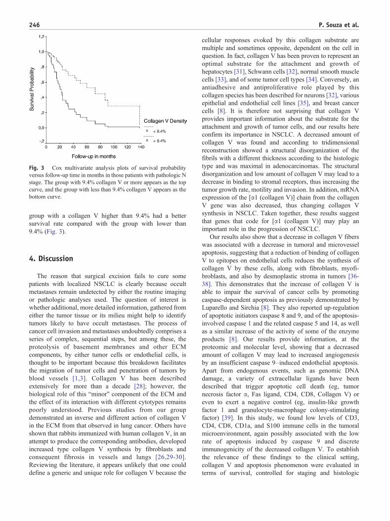
Fig. 3 Cox multivariate analysis plots of survival probabilityversus follow-up time in months in those patients with pathologic Nstage. The group with 9.4% collagen V or more appears as the topcurve, and the group with less than 9.4% collagen V appears as thebottom curve.
246 P. Souza et al.
group with a collagen V higher than 9.4% had a bettersurvival rate compared with the group with lower than9.4% (Fig. 3).
4. Discussion
The reason that surgical excision fails to cure somepatients with localized NSCLC is clearly because occultmetastases remain undetected by either the routine imagingor pathologic analyses used. The question of interest iswhether additional, more detailed information, gathered fromeither the tumor tissue or its milieu might help to identifytumors likely to have occult metastases. The process ofcancer cell invasion and metastases undoubtedly comprises aseries of complex, sequential steps, but among these, theproteolysis of basement membranes and other ECMcomponents, by either tumor cells or endothelial cells, isthought to be important because this breakdown facilitatesthe migration of tumor cells and penetration of tumors byblood vessels [1,3]. Collagen V has been describedextensively for more than a decade [28]; however, thebiological role of this “minor” component of the ECM andthe effect of its interaction with different cytotypes remainspoorly understood. Previous studies from our groupdemonstrated an inverse and different action of collagen Vin the ECM from that observed in lung cancer. Others haveshown that rabbits immunized with human collagen V, in anattempt to produce the corresponding antibodies, developedincreased type collagen V synthesis by fibroblasts andconsequent fibrosis in vessels and lungs [26,29-30].Reviewing the literature, it appears unlikely that one coulddefine a generic and unique role for collagen V because the
cellular responses evoked by this collagen substrate aremultiple and sometimes opposite, dependent on the cell inquestion. In fact, collagen V has been proven to represent anoptimal substrate for the attachment and growth ofhepatocytes [31], Schwann cells [32], normal smooth musclecells [33], and of some tumor cell types [34]. Conversely, anantiadhesive and antiproliferative role played by thiscollagen species has been described for neurons [32], variousepithelial and endothelial cell lines [35], and breast cancercells [8]. It is therefore not surprising that collagen Vprovides important information about the substrate for theattachment and growth of tumor cells, and our results hereconfirm its importance in NSCLC. A decreased amount ofcollagen V was found and according to tridimensionalreconstruction showed a structural disorganization of thefibrils with a different thickness according to the histologictype and was maximal in adenocarcinomas. The structuraldisorganization and low amount of collagen V may lead to adecrease in binding to stromal receptors, thus increasing thetumor growth rate, motility and invasion. In addition, mRNAexpression of the [α1 (collagen V)] chain from the collagenV gene was also decreased, thus changing collagen Vsynthesis in NSCLC. Taken together, these results suggestthat genes that code for [α1 (collagen V)] may play animportant role in the progression of NSCLC.
Our results also show that a decrease in collagen V fiberswas associated with a decrease in tumoral and microvesselapoptosis, suggesting that a reduction of binding of collagenV to epitopes on endothelial cells reduces the synthesis ofcollagen V by these cells, along with fibroblasts, myofi-broblasts, and also by desmoplastic stroma in tumors [36-38]. This demonstrates that the increase of collagen V isable to impair the survival of cancer cells by promotingcaspase-dependent apoptosis as previously demonstrated byLuparello and Sirchia [8]. They also reported up-regulationof apoptotic initiators caspase 8 and 9, and of the apoptosis-involved caspase 1 and the related caspase 5 and 14, as wellas a similar increase of the activity of some of the enzymeproducts [8]. Our results provide information, at theproteomic and molecular level, showing that a decreasedamount of collagen V may lead to increased angiogenesisby an insufficient caspase 9–induced endothelial apoptosis.Apart from endogenous events, such as genomic DNAdamage, a variety of extracellular ligands have beendescribed that trigger apoptotic cell death (eg, tumornecrosis factor α, Fas ligand, CD4, CD8, Collagen V) oreven to exert a negative control (eg, insulin-like growthfactor 1 and granulocyte-macrophage colony-stimulatingfactor) [39]. In this study, we found low levels of CD3,CD4, CD8, CD1a, and S100 immune cells in the tumoralmicroenvironment, again possibly associated with the lowrate of apoptosis induced by caspase 9 and discreteimmunogenicity of the decreased collagen V. To establishthe relevance of these findings to the clinical setting,collagen V and apoptosis phenomenon were evaluated interms of survival, controlled for staging and histologic

247Type V collagen, caspase and lung cancer
types. Clearly, multivariate analyses revealed a low risk ofdeath for high collagen V synthesis and high endotheliumapoptosis caspase expression.
It was also found that collagen V was associated withother collagen types. For example, a decrease in the densityof fibers of collagen V in tumoral stroma was associated witha decrease in collagens I and III. The findings are opposingbecause collagen I and III are both collagen components,which are usually up-regulated in any type of lungremodeling. In the normal lung, small amounts of collagenV are expressed as very thin fibrils in the basementmembranes for binding with stromal collagen, this beingimportant for cell adhesion and the matrix repair process [5].In the current work, the decreased amount of fibers ofcollagens I, III, and V found in tumors was associated withlow desmoplasia, probably due to a low co-polymerizationamong the fibers, thus resulting in a heterotypic assemblyincapable of controlling fibril structure, collagen fibrillogen-esis, and tumoral growth [5,25,26,28,29,40,41]. From theliterature, data can be found showing an inverse correlationof the extent of the desmoplastic stroma reaction with theprogression of carcinomas, which looks similar to thedecrease of collagen V found in our study [2-4]. Anotherquestion that could be resolved is the relationship betweenlow collagen V synthesis, poor desmoplastic stroma, andincreased blood vessel density, which leads to reducedhypoxia and increased nutrient flow. Thus, a decreasedtumor expression of collagen V may be more of a primaryevent while a decreased apoptosis may be more of asecondary event. Regardless of the mechanism, staining ofthe tumor for collagen V and apoptosis provides importantinformation in NSCLC.
We conclude that collagen V fiber density and theapoptosis index in tumors offers the potential to controlgrowth rate, motility, and invasion, suggesting that strategiesaimed at preventing the low collagen V synthesis, or localresponses to low cell apoptosis, may have a useful impact inlung cancer treatment. To realize this will require a larger-scale study in a randomized and prospective trial.
References
[1] Aznavoorian S, Murphy AN, Stetler-Stevenson WG, Liotta LA.Molecular aspects of tumor cell invasion and metastasis. Cancer(Phila) 1993;71:1368-83.
[2] Pinto CA, Carvalho PEO, Antonangelo L, et al. Morphometricevaluation of tumor matrix metalloproteinase 9 predicts survival aftersurgical resection of adenocarcinoma of the lung. Clin Cancer Res2003;9:3098-104.
[3] Liotta LA, Rao CN, Barsky SH. Tumor invasion and the extracellularmatrix. Lab Invest 1983;49:636-49.
[4] Demarchi LM, Reis MM, Palomino SAP, et al. Prognostic values ofstroma proportion and PCNA, Ki-67 and p53 proteins in patients withresected adenocarcinoma of the lung. Mod Pathol 2000;13:511-20.
[5] Mayne R, Burgeson R. Structure and function of collagen types. In:Ulecham R, editor. Biology of Extracellular Matrix, 20th ed. London:Academic Press; 1993. p. 1-37.
[6] Mares DC, Heidler KM, Smith GN, et al. Collagen V modulatesalloantigen-induced pathology and immunology in the lung. Am JRespir Cell Mol Biol 2000;23:62-70.
[7] Linsenmayer TF, Fitch JM, Birk DE. Heterotypic collagen fibrils andstabilizing collagens. Controlling elements in corneal morphogenesis?Ann N Y Acad Sci 1990;580:143-60.
[8] Luparello C, Sirchia R. Type V collagen regulates the expression ofapoptotic and stress response genes by breast cancer cells. J CellPhysiol 2005;202:411-21.
[9] Minafra S, Morello V, Glorioso F, et al. A new line (8701-BC) fromprimary ductal infiltrating carcinoma of human breast. Br J Cancer1989;60:185-92.
[10] Minafra S, Luparello C, Pucci-Minafra I, Sobel ME, Garbisa S.Adhesion of 8701-BC breast cancer cells to type V collagen and 67-kDreceptor. J Cell Sci 1992;102:323-8.
[11] Schrump D, Altorki N, Henschke C, Carter D, Turrisi A, Gutierrez M.Non-small cell lung cancer. In: De Vita V, Hellman S, Rosenberg S,editors. Cancer: principles and practice of oncology. 7th ed.Philadelphia: Lippincott Williams & Wilkins; 2005. p. 753-4.
[12] Bernardi FD, Capelozzi VL, Takagaki T, Younes R, Saldiva PH.Usefulness of morphometric evaluation of histopathological slides isuseful in predicting long-term outcome of patients with squamous cellcarcinoma of the lung. A preliminary report. Chest 1995;107:614-20.
[13] Bernardi FDC, Antonângelo L, Beyruti R, Takagaki T, Saldiva PHN,Capelozzi VL. Clinical, pathological and biological markers ofprognosis in surgically resected squamous cell carcinoma of thelung. Mod Pathol 1997;10:992-1000.
[14] Carvalho HA, Takagaki TY, Saldiva PHN, Capelozzi V. Stereologicalestimates of the nuclear/cytoplasmic ratio and star volume onfibreoptic biopsies are of prognostic value for survival in a preliminarystudy of advanced squamous cell carcinoma of the lung. Histopathol-ogy 1997;31:420-9.
[15] Antonangelo L, Bernardi FDC, Capelozzi VL, et al. Morphometricevaluation of argyrophilic nucleolar organizer region (AgNOR) isuseful in predicting long-term survival in squamous cell carcinoma ofthe lung. Chest 1997;111:110-4.
[16] Rodrigues OR, Antonângelo L, Franco Filho EC, Capelozzi VL,Saldiva PHN. Prognostic significance of argyrophilic nucleolarorganizer region (AgNOR) staining in mediastinal lymph nodesmetastasis and ressected non-small cell lung cancer. Jpn J Clin Oncol1997;27:298-304.
[17] Minamoto H, Antonângelo L, Silva AGP, et al. Different cell andmicroenvironment profiles in metastatic and non-metastatic non-smallcell lung carcinomas. Histopathology 2003;43:427-43.
[18] Matheus RS, Bernardi FD, Gallo CP, et al. Nuclear markers (starvolume, mitotic index, AgNOR and Ki-67) of the primary tumor andits metastasis in non-small cell lung carcinomas. Pathol Res Pract2004;200:13-23.
[19] Saito DM, Franco MR, Parra ER, et al. Cell cycle regulator in primarylung adenocarcinoma and its haematogenous metastases. Histopathol-ogy 2007;50:525-7.
[20] Parra ER, Park J, Rizzardi F, et al. Prognostic indice expression ofcyclin-d1, c-erbB-2 and VEGF: metastases vs corresponding primarycancers and metastatic vs non-metastatic adenocarcinomas. HistolHistopathol 2008;23:987-93.
[21] Travis W, Brambilla E, Hermelink-Muller HK, Harris CC, editors.Pathology and genetics of tumours of the lung, pleura, thymus andheart. Lyon: IARC Press; 2004. p. 26-50.
[22] Weibel ER. Morphometry: stereological theory and practical methods.In: Gil J, editor. Models of lung disease—microscopy and structuralmethods. New York: Marcel Dekker; 1990. p. 199-247.
[23] Miller EJ, Rhodes RK. Preparation and characterization of the differenttypes of collagen. Methods Enzymol 1982;82(Pt A):33-64.
[24] Lane D, Harlow E. Antibodies: a laboratory manual. Cold SpringHarbor, NY: Cold Spring Harbor Laboratory Press; 1988. p. 56-81.
[25] Parra ER, Kairalla RA, de Carvalho CR, Capelozzi VL. Abnormaldeposition of collagen/elastic vascular fibres and prognostic

248 P. Souza et al.
significance in idiopathic interstitial pneumonias. Thorax 2007;62:428-37.
[26] Teodoro WR, Velosa AP, Witzel SS, et al. Architectural remodellingin lungs of rabbits induced by type V collagen immunization: apreliminary morphologic model to study diffuse connective tissuediseases. Pathol Res Pract 2004;200:681-91.
[27] Livak KJ, Schmittgen TD. Analysis of relative gene expression datausing real-time quantitative PCR and the 2(Delta Delta C (T)) Method.Methods 2001;25:402-8.
[28] Bateman JF, Lamandè SR, Ramshaw JAM. Collagen superfamily. In:Comper WD, editor. Extracellular matrix volume 2, molecular compo-nents and interaction. Amsterdam: Harwood Acad. Public; 1996. p. 22-67.
[29] Velosa AP, Teodoro WR, de Oliveira CC, et al. Collagen V nasaltolerance in experimental model of systemic sclerosis. Arch DermatolRes 2007;299:177-89.
[30] Callado MR, Viana VS, Vendramini MB, et al. Autoantibody profile inthe experimental model of scleroderma induced by type V humancollagen. Immunology 2007;122:38-46.
[31] Takai KK, Hattori S, Irie S. Type V collagen distribution in liver isreconstructed in coculture system of hepatocytes and stellate cells; thepossible functions of type V collagen in liver under normal andpathological conditions. Cell Struct Funct 2001;26:289-302.
[32] Chernousev MA, Stahl RC, Carey DJ. Schwann cell type V collageninhibits axonal outgrowth and promotes Schwann cell migration viadistinct adhesive activities of the collagen and noncollagen domains.J Neurosci 2001;21:6125-35.
[33] Grotendorst GR, Seppã HE, Kleimanman HK, Martin GR.Attachment of smooth muscle cells to collagen and their migration
toward platelet-derived growth factor. Proc Natl Acad Sci USA1981;78:3669-72.
[34] Ruggiero F, Champliaud MF, Garrone R, Aumailley M. Interactionsbetween cells and collagen V molecules or single chains involvedistinct mechanisms. Exp Cell Res 1994;210:215-23.
[35] Parekh TV, Wang XW, Makri-Werzen DM, Greenspan DS, NewmanMJ. Type V collagen is an epithelial cell cycle inhibitor that is inducedby and mimics the effects of transforming growth factor β1. CellGrowth Differ 1998;9:423-33.
[36] Konomi H, Toshihiko H, Kiyoo N, Masataka A. Localization of type Vcollagen and type IV collagen in human cornea, lung and skin.immunohistochemical evidence by anti-collagen antibodies character-ized by immunoelectroblotting. Am J Pathol 1984;116:417-26.
[37] Reiser KM, Last JA. Type V collagen. Quantitation in normal lungsand in lungs of rats with bleomycin-induced pulmonary fibrosis. J BiolChem 1983;258:269-75.
[38] Para ER, Teodoro WR, Velosa AP, de Oliveira CC, Yoshinari NH,Capelozzi VL. Interstitial and vascular type V collagen morphologicdisorganization in usual interstitial pneumonia. J Histochem Cytochem2006;54:1315-25.
[39] Sgonc R. The vascular perspective of systemic sclerosis: of chickens,mice and men. Int Arch Allergy Immunol 1999;120:169-76.
[40] Bezerra MC, Teodoro WR, de Oliveira CC, et al. Scleroderma-likeremodeling induced by type V collagen. Arch Dermatol Res 2006;298:51-7.
[41] Ogido LT, Teodoro WR, Velosa AP, et al. Abnormal collagendeposition in synovia after collagen type V immunization in rabbits.Histol Histopathol 2008;23:263-9.