Leaving the Heliosphere: A Nuclear Powered Interstellar Probe
Chemistry & Biology
Article
Selective and Sensitive Monitoringof Caspase-1 Activityby a Novel Bioluminescent Activity-Based ProbeMaik Kindermann,1,4 Heidi Roschitzki-Voser,2,4 Dejan Cagli�c,3,4 Ur�ska Repnik,3 Catherine Miniejew,1 Peer R.E. Mittl,2
Gregor Kosec,3 Markus G. Grutter,2 Boris Turk,3,* and K. Ulrich Wendt1,*1Sanofi-Aventis Deutschland GmbH, R&D Chemical and Analytical Sciences, Industriepark Park Hoechst, 65926 Frankfurt, Germany2University of Zurich, Department of Biochemistry, Winterthurerstrasse 190, 8057 Zurich, Switzerland3Jo�zef Stefan Institute, Department of Biochemistry, and Molecular and Structural Biology, Jamova 39, 1000 Ljubljana, Slovenia4These authors contributed equally to this work
*Correspondence: [email protected] (B.T.), [email protected] (K.U.W.)
DOI 10.1016/j.chembiol.2010.07.011
SUMMARY
The role of caspase-1 in inflammation has beenstudied intensely over recent years. However, theresearch of caspase-1 has remained difficult mainlydue to the lack of sensitive and selective toolsto monitor not only its abundance but also its activ-ity. Here we present a bioluminescent activity-based probe (ABP) for caspase-1, developed by theReverse Design concept, where chemically opti-mized protease inhibitors are turned into selectivesubstrate ABPs. The probe exhibits excellent selec-tivity for caspase-1 and�1000-fold increase in sensi-tivity compared to available fluorogenic peptidic cas-pase-1 substrates. Moreover, we have been ableto monitor and quantify specific caspase-1 activitydirectly in cell lysates. The activity correlated wellwith processing of prointerleukin-1b and prointerleu-kin-18 in phorbol 12-myristate 13-acetate (PMA)-stimulated cells. A detectable caspase-1 activitywas present also in nonstimulated cells, consistentwith processing of constitutively expressed prointer-leukin-18.
INTRODUCTION
Caspase-1 (interleukin-1b converting enzyme [ICE]) belongs to
the caspase family of cysteine endopeptidases (Alnemri et al.,
1996) that play a key role in the initiation and execution of
apoptosis and inflammation. Caspase-1 was the first caspase
identified and represents the prototypic member of the family
of proinflammatory caspases (Black et al., 1989; Kostura et al.,
1989). The main substrate of caspase-1 is the proinflammatory
cytokine interleukin-1b (IL-1b). On induction with proinflamma-
tory stimuli, proIL-1b is expressed as inactive cytoplasmic
precursor (p35) that must be proteolytically processed to
generate the mature active form (p17) (Thornberry et al., 1992).
Another important caspase-1 substrate, prointerleukin-18
(proIL-18), which is structurally similar to proIL-1b, is also
Chemistry & Biology 17, 999–10
synthesized as a biologically inactive precursor that is, how-
ever, constitutively expressed in a variety of cells (Dinarello,
1999).
Like most proteolytic enzymes, caspase-1 is synthesized as
an inactive zymogen, which requires activation. Caspase-1,
carrying a long N-terminal prodomain, is suggested to belong
to the family of apical caspases together with related proapop-
totic initiator caspases-2, -8, -9, and -10 (Baliga et al., 2004;
Boatright et al., 2003; Donepudi et al., 2003; Wang et al.,
2001). On the basis of the initial studies of caspase-8 and cas-
pase-9 activation, it was suggested that they are activated by
oligomerization, which is assisted by multiprotein platforms
such as DISC or apoptosome and not by proteolytic cleavage
within the linker region (Salvesen and Dixit, 1997). Internal prote-
olysis was initially believed not to activate these caspases but
was rather a secondary event and resulted in partial stabilization
of the active dimer (Boatright et al., 2003). However, a recent
report demonstrated that neither dimerization nor cleavage of
caspase-8 alone was sufficient to activate caspase-8 (Keller
et al., 2010; Oberst et al., 2010). The discovery of inflammasome,
a multiprotein scaffold that assists caspase-1 oligomerization,
paved the way to better understanding of caspase-1 activation
in cells challenged with specific microbial or danger signals
(Martinon et al., 2002; Martinon and Tschopp, 2004; Srinivasula
et al., 2002). Several inflammatory diseases are linked to cas-
pase-1 activity such as septic shock, inflammatory bowel
disease, familial cold auto-inflammatory syndrome, rheumatoid
arthritis, osteoarthritis, and gout (Cornelis et al., 2007; Joshi
et al., 2002; Martinon et al., 2006). On the other hand, cas-
pase-1 deficiency leads to increased susceptibility for bacterial
infection (Lara-Tejero et al., 2006), demonstrating the impor-
tance of caspase-1 activation for host defense (Faustin et al.,
2007).
However, only a very minor portion of caspase-1 is usually
found to be proteolytically processed after inflammatory stimula-
tion (Ayala et al., 1994). Moreover, because activation of procas-
pase-1 is likely not critically dependent on enzyme processing
but multiprotein platform-assisted oligomerization, immunode-
tection with antibodies or proteomics methods may likely fail to
distinguish between the expression level (physical abundance)
of caspase-1 and its actual proteolytic activity in vivo. This
inherent limitation in protease biochemistry would be overcome
07, September 24, 2010 ª2010 Elsevier Ltd All rights reserved 999

Figure 1. Reverse Design of CM-269
The scaffold of the bioluminescent substrate
CM-269 is based on the caspase-1 inhibitor Pral-
nacasan 1 (molecule 1). The concept of Reverse
Design transferred the selectivity profile of the
optimized inhibitor into the bioluminescent
substrate CM-269 (molecule 2) by replacing the
warhead of the inhibitor with a cleavable peptide
bond and subsequently attaching amino-luciferin
as reporter group (see also Supplemental Scheme
and Figure S1 for details). Enzymatic turnover of
CM-269 was tested in a coupled enzymatic assay
containing recombinant firefly luciferase as the
reporter enzyme. Cleavage of the luciferin-
coupled substrate generated free amino-luciferin
which was subsequently oxidized by luciferase
resulting in emission of light.
Chemistry & Biology
Bioluminescent Activity-Based Probe for Caspase-1
by the use of sensitive and selective activity-based probes
(ABPs) enabling a direct detection of caspase-1 activity, and
would contribute to the elucidation of the molecular processes
in which caspase-1 has been shown to play a role, and would
open venues for novel therapeutic interventions.
We recently proposed that highly selective substrate ABPs
with reduced peptidic character can be obtained by a simple
chemical concept, which we referred to as Reverse Design
(Watzke et al., 2008). The concept is based on turning highly
selective protease inhibitors with chemically optimized struc-
tures into selective ABPs. We successfully applied this concept
to transfer the selectivity profile of two selective cathepsin inhib-
itors into selective ABPs by replacing the substrate-mimicking
warhead of the inhibitors with a cleavable peptide bond and
subsequently attaching appropriate reporter groups (Watzke
et al., 2008).
In the present work, we have applied the chemical concept
of Reverse Design to turn the highly selective caspase-1 inhib-
itor Pralnacasan (Figure 1, molecule 1) (Siegmund and Zeitz,
2003) into a bioluminescent caspase-1 selective ABP, referred
to as CM-269 (Figure 1, molecule 2). CM-269 was validated
using recombinant human caspases and whole-cell lysates.
The probe exhibits good kinetic properties and superior
selectivity toward caspase-1 and is sensitive enough to allow
direct monitoring of caspase-1 activity in complex proteomic
samples. We were able to detect caspase-1 activity in whole-
cell lysates of stimulated and nonstimulated monocytic cells.
Our results demonstrate that considerable caspase-1 activity
is present already in nonstimulated immune cells, which corre-
lates with processing of constitutively expressed proIL-18 in
nonstimulated cells. Using CM-269 we were able to further
show that in phorbol 12-myristate 13-acetate (PMA)-stimulated
cells the activity of caspase-1 was significantly increased,
which may be required for efficient processing of its natural
substrate proIL-1b.
1000 Chemistry & Biology 17, 999–1007, September 24, 2010 ª2010 Elsevier Ltd All rights re
RESULTS
Synthesis and In VitroCharacterization of theBioluminescent ABP for Caspase-1The highly selective caspase-1 inhibitor
Pralnacasan (Linton, 2005; Siegmund
and Zeitz, 2003) provided an attractive scaffold for the Reverse
Design of a bioluminescent ABP for caspase-1. The resulting
bioluminescent CM-269 was synthesized by solid-phase pep-
tide chemistry on a 2-chlorotrityl resin (see Supplemental Exper-
imental Procedures available online). Enzymatic turnover of
CM-269 was tested in a coupled enzymatic assay containing
recombinant firefly luciferase as a reporter enzyme. Cleavage
of the luciferin-coupled substrate generated free amino-luciferin,
which was subsequently oxidized by luciferase resulting in emis-
sion of light (Figure 1). Maximum signal was detected when the
steady state between the caspase-1 and luciferase reactions
was achieved. Different from fluorescent measurement, where
the signal of the accumulating product can be displayed as
a velocity (e.g., relative fluorescence over time), the intensity of
the emitted light in the steady state of a bioluminescent assay
reaches a plateau. A set of control experiments was carried
out to obtain optimal conditions resulting in a maximum signal
of the coupled assay (Figures S2–S4). A stable signal could be
obtained within 20 min for caspase-1 concentrations of 0.01
to 10 nM (Figure S3D). To determine the kinetic parameters
(Km, kcat) of probe hydrolysis, the coupled assay was ‘‘un-
coupled’’ and the two reaction steps, cleavage of the probe by
caspase-1 and the subsequent oxidation of amino-luciferin by
luciferase, were carried out consecutively. In this experiment,
CM-269 was shown to be efficiently cleaved by caspase-1
with a kcat value of 0.96 s�1 and a Km value of 4.33 3 10�6 M.
Even though product accumulation in fluorescent assays
generally results in higher intensities than for bioluminescent
readouts, more sensitive signals are achieved in biolumines-
cence due to the very low background levels (Troy et al.,
2004). To compare the sensitivity of the CM-269 bioluminescent
assay with the sensitivity of fluorescent measurement using the
commercially available caspase-1 substrate Ac-WEHD-AMC,
both the peptidic substrate and the ABP were assayed with cas-
pase-1 ranging from 0.005 nM to 100 nM. As shown in Figure 2,
served

Figure 2. CM-269 Exhibits Improved Sensitivity over Standard Fluo-rogenic Substrate Ac-WEHD-AMC
Sensitivity of bioluminescent CM-269 (circles; left y axis) and fluorescent
substrate Ac-WEHD-AMC (triangles; right y axis) were tested at substrate
saturation (30 mM) with 5 pM to 100 nM active caspase-1. To verify the line-
arity, the results were plotted on a logarithmic scale. Each measurement
was carried out in triplicate. Error bars indicate standard deviations from the
mean value. See also Figure S2 and Figure S3 for optimization of coupled
assay conditions.
Figure 4. Caspase-1 Activity Increases in PMA-Stimulated Cells
The bioluminescent signal of CM-269 (3 mM) is shown as bars on the left
(THP-1 in black and U937 in gray) and the fluorescent signal of Ac-DEVD-
AFC (10 mM) as line plot (THP-1 dashed line, U937 solid line) on the right axis.
In both cell lines, phorbol 12-myristate 13-acetate (PMA)-stimulation resulted
in increased cleavage of CM-269, whereas no increase in Ac-DEVD-AFC
cleavage was observed. At least three independent experiments were carried
out. Error bars indicate standard deviations from the mean value.
Chemistry & Biology
Bioluminescent Activity-Based Probe for Caspase-1
the detection limit of active caspase-1 using CM-269 was at
about 5 pM, whereas significantly higher enzyme concentrations
(1 nM) were required for the detection of active caspase-1 when
using the conventional fluorogenic substrate Ac-WEHD-AMC.
To test the in vitro selectivity of the probe, CM-269 was
profiled against a panel of recombinant human caspases and
recombinant human granzyme B, a proapoptotic serine pro-
tease. The substrate binding site of granzyme B is similar to
that of the caspase family of proteases and in both cases
substrate hydrolysis occurs after an aspartic acid (Thornberry
et al., 1997). The selectivity, expressed as ðS=NÞmax=K, was
derived from the slope of a S/N versus [E]t[S] plot (see Experi-
mental Procedures for details). As depicted in Figure 3, the
rate of substrate hydrolysis of CM-269 was highest for cas-
pase-1, being about ten times higher than for any other caspase
tested in this experiment or granzyme B. Selectivity of CM-269,
although bearing a rather largemoiety enabling a bioluminescent
Figure 3. CM-269 Is Selective for Recombinant Caspase-1
In vitro selectivity of CM-269 was tested at Km (4 mM) against a panel of
recombinant human caspases and recombinant human granzyme B.
All enzymes were applied at 10 nM active protein concentrations. (S/N)max/Km
values (see Equation 5) derived from the slope of the S/N versus [E]t[S] plot
denoting the selectivity parameters (see Experimental Procedures for details).
Each measurement was carried out in triplicate. Error bars indicate standard
deviations from the mean value. See also Figure S4 and Figure S5.
Chemistry & Biology 17, 999–100
readout, was similar to the selectivity of Ac-WEHD-AMC
(Figure S5), which has been shown to be cleaved only by proin-
flammatory caspases-1, -4, and -5 and not by the proapoptotic
caspases (McStay et al., 2008; Pereira and Song, 2008; Thorn-
berry, 1997).
Cellular Characterization of Caspase-1 ActivityUsing Caspase-1 Selective ABPHaving found that CM-269 is highly selective for caspase-1,
we investigated in the next step whether the high in vitro selec-
tivity and sensitivity of CM-269 are sufficient for functional
cellular studies. Therefore THP-1 and U937 monocytic leukemia
cell lines, either nonstimulated or PMA-stimulated, were used
as cellular models of monocytes or activated macrophages,
respectively (Tsuchiya et al., 1982). Samples were probed for
caspase-1 activity, whereas expression and processing of pro-
caspase-1 and its natural substrates proIL-1b and proIL-18
were analyzed by western blotting. With CM-269 caspase-1
activity was detected in both nonstimulated and PMA-stimulated
cells with significantly higher activity (2–4-fold, depending on the
experiment) detected in PMA-stimulated cells (shown as bars
in Figure 4). In contrast, western blot analysis of caspase-1
showed only little differences in its expression and processing
upon PMA-treatment (Figure 5A). Processing of proinflammatory
proIL-1b by caspase-1 is often used as a read-out of its activity
(Scott and Saleh, 2007) as there are no sensitive and selective
small-molecule caspase-1 substrates. However, the activity of
IL-1b is regulated on many levels, including gene expression
(Dinarello, 1997), as various inflammatory stimulators like endo-
toxins or PMA induce the synthesis of IL-1b (Martinon and
Tschopp, 2004). In agreement with the tight regulation of its
expression, we were able to detect IL-1b only in PMA-stimulated
cells, whereas nonstimulated cells lacked the protein completely
(Figure 5B) or its concentration was below detection limit of
the western blot. In addition to the proform (p35), lysates of
PMA-stimulated cells also contained the processed, mature
form (p17) of IL-1b, correlating with the observed caspase-1
activity in PMA-stimulated lysates (bars in Figure 4). Because
7, September 24, 2010 ª2010 Elsevier Ltd All rights reserved 1001
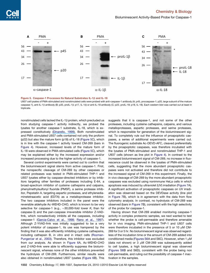
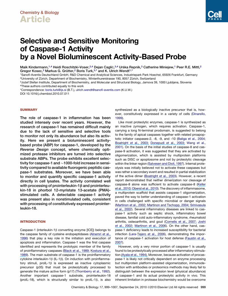
Figure 5. Caspase-1 Processes Its Natural Substrates IL-1b and IL-18
U937 cell lysates of PMA-stimulated and nonstimulated cells were probed with anti-caspase-1 antibody (A; p45, procaspase-1; p20, large subunit of the mature
caspase-1), anti-IL-1b antibody (B; p35, proIL-1b; p17, IL-1b) or anti-IL-18 antibody (C; p22, proIL-18; p18, IL-18). Each western blot was carried out at least in
triplicate.
Chemistry & Biology
Bioluminescent Activity-Based Probe for Caspase-1
nonstimulated cells lacked the IL-1b protein, which precluded us
from studying caspase-1 activity indirectly, we probed the
lysates for another caspase-1 substrate, IL-18, which is ex-
pressed constitutively (Dinarello, 1999). Both nonstimulated
and PMA-stimulated U937 cells contained not only the proform
(p22) but also the mature form (p18) of IL-18 (Figure 5C), which
is in line with the caspase-1 activity toward CM-269 (bars in
Figure 4). However, increased levels of the mature form of
IL-18 were observed in PMA-stimulated cells (Figure 5C), which
may be explained either by the increased expression and/or
increased processing due to the higher activity of caspase-1.
Several control experiments were carried out to confirm that
the bioluminescent signal stems from active caspase-1. First,
the nonspecific cleavage of CM-269 by other caspases or
related proteases was tested in PMA-stimulated THP-1 and
U937 lysates either by caspase-directed inhibitors or by inhib-
itors targeting other families of proteases including E-64, a
broad-spectrum inhibitor of cysteine cathepsins and calpains,
phenylmethylsulfonyl fluoride (PMSF), a serine protease inhib-
itor, Pepstatin A, targeting aspartic proteases, and ethylenedia-
minetetraacetic acid (EDTA) as a metalloprotease inhibitor.
The two caspase inhibitors included in the panel were the
reversible aldehyde Ac-WEHD-CHO, which is known to be very
selective for caspase-1 (e.g., �30,000-fold lower Ki than for
caspase-3) and the pan-caspase irreversible inhibitor Z-VAD-
fmk, which nonselectively inhibits all the caspases, including
caspase-1 (Garcia-Calvo et al., 1998; Rano et al., 1997).
Although Z-YVAD-fmk was initially suggested to be a very
potent inhibitor of caspase-1, its use was hampered by the
finding that it was also efficiently inhibiting cysteine cathepsins,
including cathepsin B, in vitro and in intact cells (Rozman-
Punger�car et al., 2003). Therefore, this inhibitor was excluded
from our analysis. As shown in Figure 6A, Ac-WEHD-CHO
and Z-VAD-fmk were able to efficiently suppress the biolumi-
nescent signal, whereas none of the other inhibitors prevented
the hydrolysis of CM-269. Furthermore, similar results were
also obtained in nonstimulated U937 lysates (Figure 6B). This
1002 Chemistry & Biology 17, 999–1007, September 24, 2010 ª2010
suggests that it is caspase-1, and not some of the other
proteases, including cysteine cathepsins, calpains, and various
metalloproteases, aspartic proteases, and serine proteases,
which is responsible for generation of the bioluminescent sig-
nal. To completely rule out the influence of proapoptotic cas-
pases, a series of additional experiments were carried out.
The fluorogenic substrate Ac-DEVD-AFC, cleaved preferentially
by the proapoptotic caspases, was therefore incubated with
the lysates of PMA-stimulated and nonstimulated THP-1 and
U937 cells (shown as line plot in Figure 4). In contrast to the
increased bioluminescent signal of CM-269, no increase in fluo-
rescence could be observed in the lysates of PMA-stimulated
cells, suggesting that the more abundant proapoptotic cas-
pases were not activated and therefore did not contribute to
the increased signal of CM-269 in this experiment. Finally, the
in vivo cleavage of CM-269 by the more abundant proapoptotic
caspases was excluded using nonimmune HeLa cells in which
apoptosis was induced by ultraviolet (UV) irradiation (Figure 7A).
A significant activation of proapoptotic caspases on UV irradi-
ation was observed based on the DEVDase activity (line plot
in Figure 7B), which is in agreement with the data from flow
cytometry analysis. In contrast, no hydrolysis of CM-269 was
observed (bars in Figure 7B), consistent with the high selectivity
of the probe for caspase-1.
Having shown that CM-269 enabled monitoring caspase-1
activity in complex proteomic samples, we next wanted to test
whether the probe is cell-permeable and therefore amenable
for in vivo imaging. PMA-stimulated THP-1 and U937 cells
were therefore incubated in the presence of 5 or 10 mM CM-
269 for 3 or 5 hr. No bioluminescent signal was observed regard-
less of the incubation time or the amount of CM-269 added into
conditioned media (Figures S6A and S6C). However, when 1 mM
(data not shown) or 3 mM CM-269 was subsequently added
to cell lysates, a high bioluminescent signal was observed
(Figures S6B and S6D), consistent with the probe being non
cell-permeable, and ruling out the possibility of caspase-1 inac-
tivation in the samples.
Elsevier Ltd All rights reserved
Figure 6. Caspase-1 Inhibitor Ac-WEHD-CHO Suppresses Activity
toward CM-269
(A) Cleavage of CM-269 in PMA-stimulated THP-1 (black) and PMA-stimulated
U937 (gray) cell lysates was suppressed by the caspase-1 inhibitor Ac-WEHD-
CHO (30 mM) and the pan-caspase inhibitor Z-VAD-fmk (30 mM), whereas
broad-spectrum inhibitors of cysteine cathepsins (E-64; 30 mM), serine prote-
ases (phenylmethylsulfonyl fluoride [PMSF]; 1 mM), aspartic proteases (Pep-
statin A; 1 mM), metalloproteases (ethylenediaminetetraacetic acid [EDTA],
5 mM), or the carrier (dimethyl sulfoxide [DMSO]; 1%) did not inhibit the signal.
(B) Cleavage of CM-269 in nonstimulated U937 cell lysates was suppressed by
the caspase-1 inhibitor Ac-WEHD-CHO (30 mM) and the pan-caspase inhibitor
Z-VAD-fmk (30 mM), whereas broad-spectrum inhibitors of cysteine cathep-
sins (E-64; 30 mM), aspartic proteases (Pepstatin A; 1 mM), metalloproteases
(EDTA, 5 mM), or the carrier (DMSO, 1%) did not inhibit the signal. Three inde-
pendent experiments were performed for each panel. Error bars indicate stan-
dard deviations from the mean value.
Figure 7. CM-269 Does Not Cross-React with Proapoptotic
Caspases in Apoptotic HeLa Cells
(A) Proportion of apoptotic and live HeLa cells irradiated by ultraviolet (UV)
light. Annexin V-negative and propidium iodide (PI)-negative cells represent
population of live cells. Apoptotic cells are represented as annexin V-positive
and PI-negative population of cells. Error bars indicate standard deviations
from the mean value.
(B) The bioluminescent signal of CM-269 (3 mM) is shown as bars on the left and
the fluorescent signal of Ac-DEVD-AFC (10 mM) as line plot on the right axis.
Activity monitoring with Ac-DEVD-AFC showed significant activation of the
apoptotic caspases on UV irradiation, whereas CM-269 remained uncleaved.
Three independent experiments were carried out. Error bars indicate standard
deviations from the mean value.
Chemistry & Biology
Bioluminescent Activity-Based Probe for Caspase-1
DISCUSSION
Research in life sciences increasingly requires ABPs that are
selective for a particular enzyme and allowmonitoring of enzyme
activities in a proteomic context. This concept is of particular
relevance for protease research, as their specific activation
makes direct activity monitoring highly advantageous over
detection of enzyme abundance. Until now, small-molecule
ABPs for proteases have been designed either as irreversible
inhibitors, covalently modifying and inactivating the catalytic
site of target enzymes (covalent labeling ABPs) for the subse-
quent labeling, enrichment and identification of specific enzymes
in different physiological samples (Blum et al., 2005, 2007; Evans
andCravatt, 2006), or as peptidic substrates (reviewed in Baruch
et al. (2004) and Blum (2008)). Both types of probes exhibit
limitations for the monitoring of enzymes in low abundance
and with low activity. Covalent labeling ABPs inactivate the
Chemistry & Biology 17, 999–100
enzyme on a single turnover thereby preventing signal amplifica-
tion. Peptidic substrates offer high cleavage rates, but they
frequently suffer from limited selectivity and nonspecific cleav-
age when applied in proteomic studies. This is particularly rele-
vant for caspases, as cleavage sites of different caspases have
largely overlapping motifs, and that is the reason for promis-
cuous processing of peptidic substrates (McStay et al., 2008).
For example, caspase-3, the major executioner caspase, has
been shown to cleave the peptidic substrates of other caspases
more efficiently than the enzymes to which the substrates were
originally designed to (McStay et al., 2008; Pereira and Song,
2008). The highly selective and sensitive monitoring of cas-
pase-1 in complex proteomic samples therefore requires a
chemically different type of ABPs, which is (1) not an irreversible
inhibitor but a substrate; (2) exhibits exquisite selectivity; and (3)
provides high sensitivity in terms of the read-out signal.
In this study, we have applied the Reverse Design concept
using the scaffold of selective caspase-1 inhibitor Pralnacasan
(1). Pralnacasan selectively inhibits caspase-1 with an IC50 value
of 1.3 nM, compared to IC50 values of 2.3 mM and 0.12 mM for
7, September 24, 2010 ª2010 Elsevier Ltd All rights reserved 1003

Chemistry & Biology
Bioluminescent Activity-Based Probe for Caspase-1
caspase-3 and caspase-8, respectively (Linton, 2005). Redesign
of the aldehyde warhead of 1 into a caged amino-luciferin-
coupled peptide bond provided the bioluminescent CM-269.
It has been shown previously that appropriate chemical modifi-
cation of the 6-amino group of amino-luciferin is an effective
way to approach bioluminescent assays for enzymes of interest
(Zhou et al., 2008). With regard to kinetic properties, CM-269
was very efficiently cleaved by recombinant caspase-1, with
catalytic properties in the range of commercially available, but
nonselective fluorogenic substrates (Garcia-Calvo et al., 1999).
Moreover, in these in vitro experiments CM-269 exhibited excel-
lent selectivity for caspase-1 with insignificant cleavage by
other caspases or by granzyme B. The selectivity of CM-269
was confirmed in complex proteomic samples, because none
of the inhibitors targeting serine, aspartic, metallo, or cysteine
proteases reduced the CM-269 signal. In our final test of
CM-269 selectivity, the probe was added to lysates of apoptotic
HeLa cells, which have a high proteolytic potential due to
the large amounts of activated proapoptotic caspases, but
CM-269 signal did not increase. Collectively, these results
demonstrate that CM-269, despite the exchange of the aldehyde
warhead with the scissile peptide bond, retained chemically
optimized selectivity of the caspase-1 inhibitor Pralnacasan,
although the cell permeability was lost. This probe has therefore
a significant advantage over the existing fluorescent covalent
probes based on the fluoromethylketone warhead (Grabarek
et al., 2002), which suffer from the lack of selectivity, and target
also other cysteine proteases including the very abundant
cysteine cathepsins (Rozman-Punger�car et al., 2003).
In addition to the excellent selectivity, we have shown
that CM-269 exerts high sensitivity. CM-269 was cleaved by
recombinant caspase-1 at enzyme concentrations of only about
5-10 pM, which represents a sensitivity increase of about three
orders of magnitude compared to Ac-WEHD-AMC, currently
the best fluorogenic peptidic substrate for this enzyme (Thorn-
berry et al., 1997). This superior sensitivity proved useful in anal-
yses of complex whole-cell lysates, where activity of caspase-1
could be studied by one-third of the sample required for immu-
nological detection of caspase-1. However, even high amounts
(150 mg of total protein) of the same whole-cell lysates were
not sufficient to allow monitoring caspase-1 activity with the
fluorogenic substrate Ac-WEHD-AMC (data not shown).
In recent years, there has been a major progress toward eluci-
dation of the mechanism of caspase-1 activation in response to
specific stimuli, such as various pathogen-associated molecular
patterns (lipopolysaccharide, muramyl dipeptide, flagellin from
certain Gram-negative bacteria, etc.; reviewed in Franchi et al.
(2009)), temperature shift in buffer with a low concentration of
potassium (Martinon et al., 2002) or uric acid crystals (Martinon
et al., 2006). In these studies, the assembly of largemacromolec-
ular complex, termed inflammasome, was identified as a critical
step in caspase-1 activation. Generally, the appearance of p20
subunit (large subunit of the mature caspase-1) (Fernandes-
Alnemri et al., 2009; Mehta et al., 2001) or N-terminal CARD
fragment (Martinon et al., 2002, 2006) has been a read-out for
its activation. Although caspase-1 is expressed constitutively
and its proform can be readily detected by immunodetection, it
is muchmore difficult to detect its p20 subunit even in stimulated
cells due to incomplete processing of the zymogen (Ayala et al.,
1004 Chemistry & Biology 17, 999–1007, September 24, 2010 ª2010
1994). However, using large amounts of whole-cell lysates we
were able to detect the p20 subunit not only in PMA-stimulated
but also in nonstimulated cells. This, together with the detectable
caspase-1 activity toward CM-269 in nonstimulated cells, raises
a question about limited caspase-1 activation in the apparent
absence of proinflammatory stimuli. However, in a number of
immune cells, the components of inflammasome are expressed
constitutively (Fernandes-Alnemri et al., 2009; Martinon et al.,
2002, 2006), whereas the minimal composition of the functional
inflammasome is still unknown (Faustin et al., 2007). Another
argument in favor of active caspase-1 being present in nonsti-
mulated cells is that IL-18, a substrate of caspase-1, is constitu-
tively expressed and processed to the mature p18 form not only
in stimulated but also in nonstimulated cells. Upon PMA stimula-
tion, which is taken as a surrogate for the in vivo activation of
immune cells, the expression of pro-inflammatory cytokine
IL-1b is induced and the expression of IL-18 is upregulated. At
the same time, the activity of caspase-1 is increased, which
may be explained by the requirement of the cell to process newly
expressed cytokines and thereby mount a potent response to
a microbial or danger signal.
In conclusion, our results demonstrate that the concept of
Reverse Design represents an efficient strategy for the design
of selective protease ABPs starting from chemically optimized
protease inhibitors. Different from the known covalent labeling
ABPs, Reverse Design ABPs are substrates of the target
enzyme, and thus take advantage of signal amplification by the
activity of the enzyme of interest. With a sensitive substrate
ABP, selective for caspase-1, we have been able to directly
monitor and quantify the activity of caspase-1 in complex pro-
teomic samples, such as whole-cell lysates, but not in intact
cells. Our results demonstrate that considerable caspase-1
activity is present already in nonstimulated immune cells, which
raises questions about activation of caspase-1 in the absence
of microbial or danger signals and about potential roles of cas-
pase-1 in physiological processes other than inflammation.
SIGNIFICANCE
Caspase-1 is themajor proinflammatory caspase that is crit-
ically involved in the activation of proIL-1b and proIL-18,
thereby playing a major role in inflammation. Caspase-1,
produced as inactive zymogen, gets activated on amultipro-
tein scaffold, inflammasome, in cells challenged with spe-
cific microbial or danger signals. However, complete under-
standing of caspase-1 activation and its role in inflammation
has remained difficult mainly due to the lack of tools to
monitor not only its abundance but also its activity. Particu-
larly in complex proteomic samples, selective and sensitive
activity-based probes (ABPs) would be advantageous over
fluorogenic peptidic substrates and immunodetection.
In the present study, we have applied the concept of
Reverse Design, where chemically optimized protease
inhibitors are turned into selective substrate ABPs (Watzke
et al., 2008), to turn the caspase-1 inhibitor Pralnacasan
(Siegmund and Zeitz, 2003) into a novel bioluminescent
ABP for caspase-1 (CM-269). The probe retained its excel-
lent selectivity and exhibited about three orders of magni-
tude increased sensitivity compared to the available
Elsevier Ltd All rights reserved

Chemistry & Biology
Bioluminescent Activity-Based Probe for Caspase-1
fluorogenic peptidic caspase-1 substrates. Moreover, CM-
269 offers a significant advantage over the existing fluoro-
methylketone-based probes that suffer from the lack of
selectivity and target also the cathepsins. Its sensitivity
allows direct monitoring of caspase-1 activity in complex
samples such as whole-cell lysates of monocytes and offers
a significant advantage over the traditional, indirect way of
monitoring caspase-1 activity based on proteolytic process-
ing of its major substrate, proIL-1b. Using CM-269 we were
able to detect caspase-1 activity already in nonstimulated
immune cells, raising a question about its physiological
role. Therefore, the novel ABP represents a useful and highly
sensitive tool to address not only questions related with the
role of active caspase-1 in inflammation, but also about the
activation of caspase-1 in the absence of microbial or
danger signals and about potential roles of caspase-1 in
physiological processes other than inflammation.
EXPERIMENTAL PROCEDURES
Synthesis of CM-269
CM-269 was synthesized using a combination of solid-support and solution-
phase synthesis as previously described (Watzke et al., 2008). In brief, Fmoc
protected 6-amino-D-luciferin 5 (see Supplemental Experimental Procedures,
Supplemental Scheme, and Figure S1 for details) was synthesized in solution
and coupled via the free carboxy group to the solid support. Likewise the pyr-
idazine building block 8 was prepared as previously published and used after
Fmoc-Asp-(Boc)-OH coupling on the solid support. After cleavage from the
solid support, the molecule was fully deprotected with 50% TFA/CH2Cl2 to
yield the final CM-269.
Enzymes
All human caspases were purified according to the published protocols
(Garcia-Calvo et al., 1999; Scheer et al., 2006; Stennicke and Salvesen,
1999), which were further optimized. Recombinant human granzyme B was
purchased from Alexis Biochemicals (San Diego, CA). Active protein concen-
trations were determined by active-site titration using the following inhibitors:
Ac-YVAD-cmk (Peptanova, Sandhausen, Germany) for caspase-1, -4, and -5,
Ac-VDVAD-CHO (Peptanova) for caspase-2, and Ac-DEVD-cmk (R&D
Systems, Palo Alto, CA) for caspase-3, -7, and -8.
Coupled Enzymatic Assay for Bioluminescence Measurements
In vitro bioluminescent experiments were carried out in white 96-well COSTAR
CellStar plates. The measurements were performed as a coupled enzymatic
assay, using QuantiLum recombinant firefly luciferase (Promega, Madison,
WI) as reporter enzyme. Bioluminescence was detected with a Tecan Safire2
plate reader in the luminescence mode (l = 230–850 nm) at 25�C. No signal
was observed for uncleaved substrates under standard assay conditions.
CM-269, caspases, and luciferase were diluted in 100 mM HEPES buffer,
pH 7.4, containing 50 mMMgSO4, 2.5 mM CaCl2, 0.4 mM ATP, 0.6% Prionex,
0.4% Tergitol NP-9, 0.1% silicone antifoam, and 40 mM dithiothreitol (DTT) to
the concentrations indicated below. The final dimethyl sulfoxide (DMSO)
concentration in the assays did not exceed 1% (v/v).
Kinetic Characterization of CM-269 with Caspase-1
For determination of the kinetic parameters cleavage of the CM-269 by cas-
pase-1 and subsequent oxidation of amino-luciferin by luciferase were carried
out consecutively. Caspase-1 (1 nM final concentration) and CM-269
(0.4–25 mM final concentrations) were mixed and incubated.
In two independent experiments an excess of the inhibitor Ac-YVAD-CHO
(Bachem, Bubendorf, Switzerland) at a final concentration of 1 mMwas added
to stop the reaction after 5 min and after 10 min respectively. Concentration of
free amino-luciferin produced during the course of caspase-1 cleavage was
measured by adding firefly luciferase (1 mg/ml final concentration). The relative
light units (RLU) were quantified using an amino-luciferin standard curve
Chemistry & Biology 17, 999–100
(50 nM–1300 nM final concentrations). Kinetic data were fitted by nonlinear
least-squares regression analysis according to Equation 1, where y is the initial
velocity, V the limiting rate, Kms the Michaelis constant (considering substrate
inhibition), [S] the substrate concentration and Kis the substrate inhibition
constant:
y=½S�Vmax�
Kms + 1+ ½S�Kis
�½S�
: (1)
Sensitivity Measurement of CM-269 with Caspase-1
Thirty micromolars CM-269 was mixed with 50 mg/ml (final concentration)
firefly luciferase. Reaction was started by adding different concentrations of
caspase-1 (5 pM to 10 nM final concentrations). Signal-to-noise (S/N) ratios
were calculated according to Equation 2 (Zhang et al., 1999), and the results
were plotted on a logarithmic scale to verify the linearity:
S=N=mean signal �mean background
standard deviation background: (2)
In the fluorescent assay, caspase-1 was diluted in 40 mM PIPES buffer,
pH 7.5, containing 100 mM NaCl, 0.1% CHAPS, 10% sucrose, and 10 mM
DTT to final concentrations of 0.5–40 nM. The DMSO-dissolved substrate
Ac-WEHD-AMC (Peptanova) was added at a concentration of 30 mM, fluores-
cence measured (lex = 360 nm, lem = 465 nm) and steady-state kinetic data
(RFU/s) were taken. The final DMSO concentration in the assay did not exceed
1% (v/v).
Selectivity Measurement of CM-269 with Different Recombinant
Human Caspases
Different concentrations of CM-269 (1–130 mM final concentrations) were
mixed with recombinant firefly luciferase (10 mg/ml final concentration) and
the reaction was started by adding recombinant human caspases and
recombinant human granzyme B, all enzymes at 10 nM final concentrations.
The reaction velocity under steady-state condition is given by
y=Vmax½S�Km + ½S�=
kcat½E�t ½S�Km + ½S� ; (3)
for [S] < < Km Equation 3 can be simplified as:
yzkcatKm
½E�t ½S�; (4)
where kcat/Km represents the specificity constant.
According to the properties of the bioluminescent measurement, Equation 4
was modified to:
S=NzS=Nmax
Km
½E�t ½S�; (5)
with ðS=NÞmax=Km becoming the specificity constant. This value can be
derived from the slope of the S/N versus [E]t[S] plot.
PMA-Differentiation of U937 and THP-1 Cells and Western
Blot Analysis
U937 and THP-1 cells were grown in RPMI 1640mediumwith 10% fetal bovine
serum (FBS) at 37�C in a humidified incubator with a 5% CO2, 95% air atmo-
sphere. Cells were seeded at a density of 0.5 3 106 cells/ml in 10-cm culture
dishes. Differentiation was induced by 30 ng/ml of phorbol 12-myristate
13-acetate (PMA) over 48 hr. Cells were then grown in the absence of PMA
for additional 48 hr. Whole-cell lysates were prepared in 50 mMHEPES buffer,
pH 7.5, containing 200 mM NaCl, 10% sucrose, 0.1% CHAPS, 5 mM MgCl2,
0.02% BSA, 0.5% Triton X-100, 1% NP-40, and 100 mM sodium malonate.
Equal amounts of whole-cell lysates (75 mg of total cell protein) were applied
on a 15% SDS-PAGE, electro-blotted, and probed with anti-caspase-1 anti-
body (ab17820; Abcam, Cambridge, UK), anti-IL-1b antibody (3ZD; NCI Bio-
logical Resources Branch, Bethesda, USA) or anti-IL-18 antibody (ab37640;
Abcam). Whole-cell lysates (75 mg of total cell protein) were also measured
for activity toward CM-269 (3 mM final concentration) by complementing the
buffer with recombinant firefly luciferase (10 mg/ml final concentration) and
ATP (0.2 mM final concentration) and toward Ac-DEVD-AFC (Bachem,
7, September 24, 2010 ª2010 Elsevier Ltd All rights reserved 1005
Chemistry & Biology
Bioluminescent Activity-Based Probe for Caspase-1
10 mM final concentration), to detect the activity of apoptotic caspases. Fluo-
rescence wasmeasured (lex = 400 nm, lem = 505 nm) and steady-state kinetic
data (RFU/s) were taken. For bioluminescent measurements, S/N-ratios were
calculated as described above.
Inhibition Study of CM-269 in Cell Lysates
PMA-stimulated U937 and THP-1 cell lysates (75 mg of total cell protein) were
incubated with 1% DMSO (Sigma), 30 mM inhibitor Z-VAD-fmk (Calbiochem,
San Diego, CA), 30 mM Ac-WEHD-CHO (Bachem), 30 mM E-64 (Peptide
Institute, Osaka, Japan), 1 mM PMSF (Sigma), 1 mMPepstatin A (Calbiochem),
or 5 mM EDTA (Sigma) for 1 hr at 25�C. The cleavage of CM-269 was deter-
mined as described above.
UV-Induced Apoptosis in HeLa cells
HeLa cells were cultured in DMEMwith 10% FBS at 37�C in a humidified incu-
bator with a 5% CO2, 95% air atmosphere. Two days before the experiment,
cells were seeded at a density of 5.0 3 105 cells/ml into 6-cm culture dishes.
UV-irradiation-induced apoptosis was initiated by exposing the cells to UVC
light for 45 s. After 18 hr incubation, whole-cell lysates were prepared as
described above. Equal amounts of whole-cell lysates (75 mg of total cell
protein) were measured for activity toward Ac-DEVD-AFC and CM-269 under
the conditions described above.
SUPPLEMENTAL INFORMATION
Supplemental Information includes Supplemental Experimental Procedures,
Supplemental Scheme, and six figures, and can be found with this article
online at doi:10.1016/j.chembiol.2010.07.011.
ACKNOWLEDGMENTS
We gratefully acknowledge Aimo Kant and Volker Jeske for excellent technical
support and fruitful discussions.We thank Antonio Baici and Patricia Schenker
for assistance regarding the kinetic evaluation. This work was supported by
the European Commission Framework 6 Program (CAMP project, LSHG-
2006-018830) and by the European Commission Framework 7 Program
(FP7-Health-2009-241919-LIVIMODE project). The authors declare no conflict
of interest.
Received: February 2, 2010
Revised: July 5, 2010
Accepted: July 14, 2010
Published: September 23, 2010
REFERENCES
Alnemri, E.S., Livingston, D.J., Nicholson, D.W., Salvesen, G., Thornberry,
N.A., Wong, W.W., and Yuan, J. (1996). Human ICE/CED-3 protease nomen-
clature. Cell 87, 171.
Ayala, J.M., Yamin, T.T., Egger, L.A., Chin, J., Kostura, M.J., and Miller, D.K.
(1994). IL-1 beta-converting enzyme is present in monocytic cells as an inac-
tive 45-kDa precursor. J. Immunol. 153, 2592–2599.
Baliga, B.C., Read, S.H., and Kumar, S. (2004). The biochemical mechanism of
caspase-2 activation. Cell Death Differ. 11, 1234–1241.
Baruch, A., Jeffery, D.A., and Bogyo, M. (2004). Enzyme activity—it’s all about
image. Trends Cell Biol. 14, 29–35.
Black, R.A., Kronheim, S.R., and Sleath, P.R. (1989). Activation of interleukin-1
beta by a co-induced protease. FEBS Lett. 247, 386–390.
Blum, G. (2008). Use of fluorescent imaging to investigate pathological
protease activity. Curr. Opin. Drug Discov. Devel. 11, 708–716.
Blum, G., Mullins, S.R., Keren, K., Fonovic, M., Jedeszko, C., Rice, M.J.,
Sloane, B.F., and Bogyo, M. (2005). Dynamic imaging of protease activity
with fluorescently quenched activity-based probes. Nat. Chem. Biol. 1,
203–209.
1006 Chemistry & Biology 17, 999–1007, September 24, 2010 ª2010
Blum, G., von Degenfeld, G., Merchant, M.J., Blau, H.M., and Bogyo, M.
(2007). Noninvasive optical imaging of cysteine protease activity using fluores-
cently quenched activity-based probes. Nat. Chem. Biol. 3, 668–677.
Boatright, K.M., Renatus, M., Scott, F.L., Sperandio, S., Shin, H., Pedersen,
I.M., Ricci, J.E., Edris, W.A., Sutherlin, D.P., Green, D.R., et al. (2003). A unified
model for apical caspase activation. Mol. Cell 11, 529–541.
Cornelis, S., Kersse, K., Festjens, N., Lamkanfi, M., and Vandenabeele, P.
(2007). Inflammatory caspases: targets for novel therapies. Curr. Pharm.
Des. 13, 367–385.
Dinarello, C.A. (1997). Interleukin-1. Cytokine Growth Factor Rev. 8, 253–265.
Dinarello, C.A. (1999). Interleukin-18. Methods 19, 121–132.
Donepudi, M., Mac Sweeney, A., Briand, C., and Grutter, M.G. (2003). Insights
into the regulatory mechanism for caspase-8 activation. Mol. Cell 11, 543–549.
Evans, M.J., and Cravatt, B.F. (2006). Mechanism-based profiling of enzyme
families. Chem. Rev. 106, 3279–3301.
Faustin, B., Lartigue, L., Bruey, J.M., Luciano, F., Sergienko, E., Bailly-Maitre,
B., Volkmann, N., Hanein, D., Rouiller, I., and Reed, J.C. (2007). Reconstituted
NALP1 inflammasome reveals two-step mechanism of caspase-1 activation.
Mol. Cell 25, 713–724.
Fernandes-Alnemri, T., Yu, J.W., Datta, P., Wu, J., and Alnemri, E.S. (2009).
AIM2 activates the inflammasome and cell death in response to cytoplasmic
DNA. Nature 458, 509–513.
Franchi, L., Eigenbrod, T., Munoz-Planillo, R., and Nunez, G. (2009). The
inflammasome: a caspase-1-activation platform that regulates immune
responses and disease pathogenesis. Nat. Immunol. 10, 241–247.
Garcia-Calvo, M., Peterson, E.P., Leiting, B., Ruel, R., Nicholson, D.W., and
Thornberry, N.A. (1998). Inhibition of human caspases by peptide-based and
macromolecular inhibitors. J. Biol. Chem. 273, 32608–32613.
Garcia-Calvo, M., Peterson, E.P., Rasper, D.M., Vaillancourt, J.P., Zamboni,
R., Nicholson, D.W., and Thornberry, N.A. (1999). Purification and catalytic
properties of human caspase family members. Cell Death Differ. 6, 362–369.
Grabarek, J., Amstad, P., and Darzynkiewicz, Z. (2002). Use of fluorescently
labeled caspase inhibitors as affinity labels to detect activated caspases.
Hum. Cell 15, 1–12.
Joshi, V.D., Kalvakolanu, D.V., Hebel, J.R., Hasday, J.D., and Cross, A.S.
(2002). Role of caspase 1 in murine antibacterial host defenses and lethal
endotoxemia. Infect. Immun. 70, 6896–6903.
Keller, N., Grutter, M.G., and Zerbe, O. (2010). Studies of the molecular mech-
anism of caspase-8 activation by solution NMR. Cell Death Differ. 17, 710–718.
Kostura, M.J., Tocci, M.J., Limjuco, G., Chin, J., Cameron, P., Hillman, A.G.,
Chartrain, N.A., and Schmidt, J.A. (1989). Identification of a monocyte specific
pre-interleukin 1 beta convertase activity. Proc. Natl. Acad. Sci. USA 86, 5227–
5231.
Lara-Tejero, M., Sutterwala, F.S., Ogura, Y., Grant, E.P., Bertin, J., Coyle, A.J.,
Flavell, R.A., and Galan, J.E. (2006). Role of the caspase-1 inflammasome in
Salmonella typhimurium pathogenesis. J. Exp. Med. 203, 1407–1412.
Linton, S.D. (2005). Caspase inhibitors: a pharmaceutical industry perspective.
Curr. Top. Med. Chem. 5, 1697–1717.
Martinon, F., and Tschopp, J. (2004). Inflammatory caspases: linking an
intracellular innate immune system to autoinflammatory diseases. Cell 117,
561–574.
Martinon, F., Burns, K., and Tschopp, J. (2002). The inflammasome: a molec-
ular platform triggering activation of inflammatory caspases and processing of
proIL-beta. Mol. Cell 10, 417–426.
Martinon, F., Petrilli, V., Mayor, A., Tardivel, A., and Tschopp, J. (2006). Gout-
associated uric acid crystals activate the NALP3 inflammasome. Nature 440,
237–241.
McStay, G.P., Salvesen, G.S., and Green, D.R. (2008). Overlapping cleavage
motif selectivity of caspases: implications for analysis of apoptotic pathways.
Cell Death Differ. 15, 322–331.
Mehta, V.B., Hart, J., and Wewers, M.D. (2001). ATP-stimulated release of
interleukin (IL)-1beta and IL-18 requires priming by lipopolysaccharide and is
independent of caspase-1 cleavage. J. Biol. Chem. 276, 3820–3826.
Elsevier Ltd All rights reserved

Chemistry & Biology
Bioluminescent Activity-Based Probe for Caspase-1
Oberst, A., Pop, C., Tremblay, A.G., Blais, V., Denault, J.B., Salvesen, G.S.,
and Green, D.R. (2010). Inducible dimerization and inducible cleavage reveal
a requirement for both processes in caspase-8 activation. J. Biol. Chem.
285, 16632–16642.
Pereira, N.A., and Song, Z. (2008). Some commonly used caspase substrates
and inhibitors lack the specificity required to monitor individual caspase
activity. Biochem. Biophys. Res. Commun. 377, 873–877.
Rano, T.A., Timkey, T., Peterson, E.P., Rotonda, J., Nicholson, D.W., Becker,
J.W., Chapman, K.T., and Thornberry, N.A. (1997). A combinatorial approach
for determining protease specificities: application to interleukin-1beta con-
verting enzyme (ICE). Chem. Biol. 4, 149–155.
Rozman-Punger�car, J., Kopitar-Jerala, N., Bogyo, M., Turk, D., Vasiljeva, O.,
Stefe, I., Vandenabeele, P., Bromme, D., Puizdar, V., Fonovi�c, M., et al.
(2003). Inhibition of papain-like cysteine proteases and legumain by caspase-
specific inhibitors: when reaction mechanism is more important than speci-
ficity. Cell Death Differ. 10, 881–888.
Salvesen, G.S., and Dixit, V.M. (1997). Caspases: intracellular signaling by
proteolysis. Cell 91, 443–446.
Scheer, J.M., Romanowski, M.J., and Wells, J.A. (2006). A common allosteric
site and mechanism in caspases. Proc. Natl. Acad. Sci. USA 103, 7595–7600.
Scott, A.M., and Saleh, M. (2007). The inflammatory caspases: guardians
against infections and sepsis. Cell Death Differ. 14, 23–31.
Siegmund, B., and Zeitz, M. (2003). Pralnacasan (Vertex Pharmaceuticals).
IDrugs 6, 154–158.
Srinivasula, S.M., Poyet, J.L., Razmara, M., Datta, P., Zhang, Z., and Alnemri,
E.S. (2002). The PYRIN-CARD protein ASC is an activating adaptor for
caspase-1. J. Biol. Chem. 277, 21119–21122.
Stennicke, H.R., and Salvesen, G.S. (1999). Caspases: preparation and char-
acterization. Methods 17, 313–319.
Chemistry & Biology 17, 999–100
Thornberry, N.A. (1997). The caspase family of cysteine proteases. Br. Med.
Bull. 53, 478–490.
Thornberry, N.A., Bull, H.G., Calaycay, J.R., Chapman, K.T., Howard, A.D.,
Kostura, M.J., Miller, D.K., Molineaux, S.M., Weidner, J.R., Aunins, J., et al.
(1992). A novel heterodimeric cysteine protease is required for interleukin-1
beta processing in monocytes. Nature 356, 768–774.
Thornberry, N.A., Rano, T.A., Peterson, E.P., Rasper, D.M., Timkey, T., Garcia-
Calvo, M., Houtzager, V.M., Nordstrom, P.A., Roy, S., Vaillancourt, J.P., et al.
(1997). A combinatorial approach defines specificities of members of the
caspase family and granzyme B. Functional relationships established for key
mediators of apoptosis. J. Biol. Chem. 272, 17907–17911.
Troy, T., Jekic-McMullen, D., Sambucetti, L., and Rice, B. (2004). Quantitative
comparison of the sensitivity of detection of fluorescent and bioluminescent
reporters in animal models. Mol. Imaging 3, 9–23.
Tsuchiya, S., Kobayashi, Y., Goto, Y., Okumura, H., Nakae, S., Konno, T., and
Tada, K. (1982). Induction ofmaturation in cultured humanmonocytic leukemia
cells by a phorbol diester. Cancer Res. 42, 1530–1536.
Wang, J., Chun, H.J., Wong, W., Spencer, D.M., and Lenardo, M.J. (2001).
Caspase-10 is an initiator caspase in death receptor signaling. Proc. Natl.
Acad. Sci. USA 98, 13884–13888.
Watzke, A., Kosec, G., Kindermann, M., Jeske, V., Nestler, H.P., Turk, V., Turk,
B., and Wendt, K.U. (2008). Selective activity-based probes for cysteine
cathepsins. Angew. Chem. Int. Ed. Engl. 47, 406–409.
Zhang, J.H., Chung, T.D., and Oldenburg, K.R. (1999). A simple statistical
parameter for use in evaluation and validation of high throughput screening
assays. J. Biomol. Screen. 4, 67–73.
Zhou, W., Andrews, C., Liu, J., Shultz, J.W., Valley, M.P., Cali, J.J., Hawkins,
E.M., Klaubert, D.H., Bulleit, R.F., and Wood, K.V. (2008). Self-cleavable bio-
luminogenic luciferin phosphates as alkaline phosphatase reporters. Chem-
BioChem 9, 714–718.
7, September 24, 2010 ª2010 Elsevier Ltd All rights reserved 1007
Copyright © 2022 FDOKUMEN