Peptide hydrolysis and the uptake of dipeptides by phytoplankton
Upload
jagiellonianCategory
view
0download
0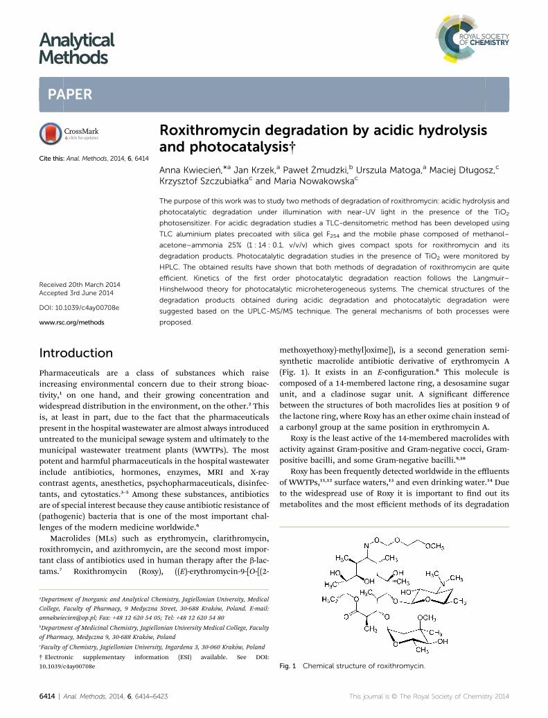
AnalyticalMethods
PAPER
Roxithromycin d
aDepartment of Inorganic and Analytical Ch
College, Faculty of Pharmacy, 9 Medyczna
[email protected]; Fax: +48 12 620 54 0bDepartment of Medicinal Chemistry, Jagiell
of Pharmacy, Medyczna 9, 30-688 Krakow,cFaculty of Chemistry, Jagiellonian Universi
† Electronic supplementary informa10.1039/c4ay00708e
Cite this: Anal. Methods, 2014, 6, 6414
Received 20th March 2014Accepted 3rd June 2014
DOI: 10.1039/c4ay00708e
www.rsc.org/methods
6414 | Anal. Methods, 2014, 6, 6414–64
egradation by acidic hydrolysisand photocatalysis†
Anna Kwiecien,*a Jan Krzek,a Paweł Zmudzki,b Urszula Matoga,a Maciej Długosz,c
Krzysztof Szczubiałkac and Maria Nowakowskac
The purpose of this work was to study two methods of degradation of roxithromycin: acidic hydrolysis and
photocatalytic degradation under illumination with near-UV light in the presence of the TiO2
photosensitizer. For acidic degradation studies a TLC-densitometric method has been developed using
TLC aluminium plates precoated with silica gel F254 and the mobile phase composed of methanol–
acetone–ammonia 25% (1 : 14 : 0.1, v/v/v) which gives compact spots for roxithromycin and its
degradation products. Photocatalytic degradation studies in the presence of TiO2 were monitored by
HPLC. The obtained results have shown that both methods of degradation of roxithromycin are quite
efficient. Kinetics of the first order photocatalytic degradation reaction follows the Langmuir–
Hinshelwood theory for photocatalytic microheterogeneous systems. The chemical structures of the
degradation products obtained during acidic degradation and photocatalytic degradation were
suggested based on the UPLC-MS/MS technique. The general mechanisms of both processes were
proposed.
Introduction
Pharmaceuticals are a class of substances which raiseincreasing environmental concern due to their strong bioac-tivity,1 on one hand, and their growing concentration andwidespread distribution in the environment, on the other.2 Thisis, at least in part, due to the fact that the pharmaceuticalspresent in the hospital wastewater are almost always introduceduntreated to the municipal sewage system and ultimately to themunicipal wastewater treatment plants (WWTPs). The mostpotent and harmful pharmaceuticals in the hospital wastewaterinclude antibiotics, hormones, enzymes, MRI and X-raycontrast agents, anesthetics, psychopharmaceuticals, disinfec-tants, and cytostatics.3–5 Among these substances, antibioticsare of special interest because they cause antibiotic resistance of(pathogenic) bacteria that is one of the most important chal-lenges of the modern medicine worldwide.6
Macrolides (MLs) such as erythromycin, clarithromycin,roxithromycin, and azithromycin, are the second most impor-tant class of antibiotics used in human therapy aer the b-lac-tams.7 Roxithromycin (Roxy), ((E)-erythromycin-9-[O-[(2-
emistry, Jagiellonian University, Medical
Street, 30-688 Krakow, Poland. E-mail:
5; Tel: +48 12 620 54 80
onian University Medical College, Faculty
Poland
ty, Ingardena 3, 30-060 Krakow, Poland
tion (ESI) available. See DOI:
23
methoxyethoxy)-methyl]oxime]), is a second generation semi-synthetic macrolide antibiotic derivative of erythromycin A(Fig. 1). It exists in an E-conguration.8 This molecule iscomposed of a 14-membered lactone ring, a desosamine sugarunit, and a cladinose sugar unit. A signicant differencebetween the structures of both macrolides lies at position 9 ofthe lactone ring, where Roxy has an ether oxime chain instead ofa carbonyl group at the same position in erythromycin A.
Roxy is the least active of the 14-membered macrolides withactivity against Gram-positive and Gram-negative cocci, Gram-positive bacilli, and some Gram-negative bacilli.9,10
Roxy has been frequently detected worldwide in the effluentsof WWTPs,11,12 surface waters,13 and even drinking water.14 Dueto the widespread use of Roxy it is important to nd out itsmetabolites and the most efficient methods of its degradation
Fig. 1 Chemical structure of roxithromycin.
This journal is © The Royal Society of Chemistry 2014

Paper Analytical Methods
before it reaches the environment. Oxidative dealkylation of thealkylether side chain, hydrolytic cleavage of the sugar cladinose,and O-demethylation at the oxime side chain are the majormetabolic pathways of Roxy in humans.8 Metabolic isomeriza-tion of the oxime is stereoselective leading to the formation ofZ-oxime isomers.15,16
The acidic hydrolysis of Roxy in the simulated gastric uid(SGF) has been already reported.17 Moreover, the degradation ofclarithromycin and Roxy adsorbed onto iron(III) and man-ganese(IV) oxides was studied.7 It was found that the degrada-tion of these MLs proceeds by the hydrolysis of both thecladinose sugar and the lactone ring, with the N-dealkylation ofthe amino sugar being the minor pathway. Sorption andtransformation of MLs on clays have been also described.18
Biodegradation studies of Roxy in an activated sludge processhave shown that hydrolysis of Roxy is negligible and the anti-biotic removal proceeds mainly through adsorption.19
The aim of this work was to study the degradation of Roxy byacidic hydrolysis and sensitized photodegradation as possiblemethods of its treatment before it is transferred into the envi-ronment. We have performed a detailed analysis of the degra-dation pathway under these conditions. We also propose thealternative degradationmethod, a photocatalytic degradation ofRoxy using a TiO2 dispersion. To the best of our knowledge nopapers presenting UV-light induced photocatalytic degradationof Roxy sensitized by suspended TiO2 are available in the liter-ature, although systems containing TiO2 coated on carriers suchas cenospheres have been reported.20
ExperimentalApparatus
The degraded Roxy samples were analyzed using a UPLC-MS/MSsystem – an ACQUITY UPLC and TQD mass spectrometer(Waters Corporation, Milford, MA, USA) and an HPLC systemcomposed of a Waters 515 HPLC pump, a 2996 PhotodiodeArray Detector, and a 717 Plus Autosampler (Waters Corpora-tion, Milford, MA, USA). TLC analyses were performed using aTLC Scanner 3 densitometer with Cats 4 soware (Camag,Muttenz, Switzerland). Irradiation was performed using aRayonet RPR-100 photoreactor (Southern New England Ultra-violet Company, Brandford, CT, USA). Samples were dried usinga drying oven with natural convection – a Binder ED 115/E2(Binder GmbH, Tuttlingen, Germany) and a muffle furnace FCF5S HM (Czylok, Jastrzebie Zdroj, Poland).
Dynamic light scattering
Dynamic light scattering (DLS) of TiO2 dispersed in water wasused to estimate the size of the average TiO2 particle. Themeasurement was carried out using a Malvern Zetasizer Nano-ZS. The anatase refractive index of 2.55 was used in thecalculations.
UV-Vis reectance spectroscopy
The reectance spectrum of the TiO2 powder was acquiredusing an Ocean Optics USB2G5726 spectrophotometer
This journal is © The Royal Society of Chemistry 2014
equipped with an AvaLight-DHS lamp and an integrationsphere. The bandgap energy was estimated using a Tauc plot.
Powder X-ray diffraction (PXRD)
The PXRD analyses were carried out to assess the crystalstructure of the obtained TiO2. The measurement was carriedout using an X'PERT PRO diffractometer. The Scherrer equationwas used to approximate the crystallite size:
L ¼ Kl
b1=2 cos q
where: L – average crystallite size, l – the wavelength of X-rayradiation, K – shape constant equal to 0.9 for spherical particles,b1/2 – the line broadening at half the maximum intensity, and q
– Bragg angle.
Materials and reagents
Roxithromycin – (3R,4S,5S,6R,7R,9R,11S,12R,13S,14R)-6-{[(2S,3R,4S,6R)-4-(dimethylamino)-3-hydroxy-6-methyloxan-2-yl]oxy}-14-ethyl-7,12,13-trihydroxy-4-{[(2R,4R,5S,6S)-5-hydroxy-4-methoxy-4,6-dimethyloxan-2-yl]oxy}-3,5,7,9,11,13-hexamethyl-10-(2,4,7-trioxa-1-azaoctan-1-ylidene)-1-oxacyclotetradecan-2-one – was purchased from LGC Standards, EPR 1500000, Ted-dington, London, England. The following commercial Roxypreparations were used: Rulid 150 mg (Sano-Aventis, Paris,France), Xitrocin 150 mg (Polfa Pabianice, Pabianice, Poland),Rolicyn 150 mg (Polfa Tarchomin, Warsaw, Poland), and Reni-cin 150 mg (Sandoz, Kundl, Austria). Ethanol (POCh), methanol(Merck), acetone (Chempur), ammonia 25% (POCh), sulfuricacid 95% (POCh), hydrochloric acid (POCh), water (Merck),acetonitrile (Merck), formic acid (Merck), titanium(IV) isoprop-oxide 97% (Sigma-Aldrich), and KH2PO4 (POCh) were ofanalytical or HPLC purity and used as received.
Standard solutions
Standard solutions were prepared by dissolving an appropriateamount of the standard substance of Roxy in ethanol to give aconcentration of 0.025% (w/v) for linearity and precision studiesand 0.0025% (w/v) for the determination of LOD and LOQ. Astandard solution of Roxy at a concentration of 0.6% (w/v) wasprepared for the degradation studies in acidic solutions.
TLC-densitometric conditions
A standard solution of Roxy was applied in 8 mm bands, 10 mmapart, and 10 mm from the lower edge to TLC silica gel 60 F254plates (10� 15 cm) using a Camag sample applicator Linomat 4equipped with a 100 mL syringe. Chromatograms were devel-oped over a distance of 145 mm using methanol–acetone–ammonia 25% (1 : 14 : 0.1, v/v/v) in a chromatographicchamber saturated for 10 min with mobile phase vapor at roomtemperature. The mobile phase was chosen experimentally aerchecking 20 different mobile phases. Aer development, theTLC plates were dried at room temperature and spots weredetected with a freshly prepared concentrated sulfuric acid–ethanol (1 : 4, v/v) mixture. Aer drying the plates were sprayed
Anal. Methods, 2014, 6, 6414–6423 | 6415

Analytical Methods Paper
with the reagent, and then heated in an oven at 100 �C for 5min.Aer spot detection, the plates were scanned in the visible rangeat 483 nm, which was chosen on the basis of an absorptionspectrum recorded directly from the chromatogram. Theanalytical wavelength was the same for Roxy and its degradationproducts. Densitometric scanning was carried out using aCamag TLC Scanner 3 equipped with a tungsten lamp in thelinear absorbance-reectance mode controlled by Cats 4 so-ware resident in the system. The slit dimensions were 8 � 0.6mm, the scanning speed was 20 mm s�1, and the data resolu-tion was 100 mm per step. Retardation factors (RF values) wereused to identify Roxy and its possible degradation products. Forquantitative determination of Roxy in the degradation studies,the scan areas of appropriate peaks were recorded and therelative percent concentrations computed by employingthe internal normalization method, according to the formula:i [%]¼ (xi/
Px) 100, where i [%] is the constituent concentration,
xi is the peak area for the determined constituent, andP
x is thesum of peak scan areas in the chromatogram.
Fig. 2 The kinetic data for the acidic hydrolysis of Roxy for pH of (a)2.30, (b) 1.70, and (c) 1.30 at (-) 40, (C) 50, and (:) 60 �C.
Determination of Roxy in commercial pharmaceuticalpreparations
Three mL of the Roxy standard solution and the analyzed solu-tions at a concentration of 0.025% (w/v) were applied to thechromatographic plates with a Linomat in the form of a band 8mm in width. Chromatograms were developed under theconditions described in the TLC-densitometric conditionssection. The content of antibiotics in drugs was computed bycomparing the peak areas for standard and tested solutions.The results of determination for preparations under investiga-tion are listed in Table S2 (see the ESI†).
Acid-induced hydrolysis of Roxy
Hydrolysis of Roxy was carried out at pH 1.30, 1.70, and 2.30 at40, 50 and 60 �C. In an 8 mL borosilicate glass vial 2 mL of 0.6%(w/v) Roxy standard solution was mixed with 2 mL of anappropriate acid (0.1, 0.04, and 0.01 mol L�1 HCl) and heated inan oven to the desired temperature. Then, 500 mL of incubatedsolutions were taken for the analysis at time intervals given inFig. 2. Aer cooling the solutions were mixed with ethanol at a1 : 1 ratio. 10 mL aliquots of resultant solutions were applied tothe TLC plates in triplicate. The TLC-densitometric method wascarried out according to the developed method. For quantitativedetermination purposes, the surface areas of appropriate peakswere recorded, and the percentage concentrations of Roxy werecomputed by using an internal normalization method. Pre-sented results are the mean values of three measurements.
Kinetic testing
To characterize the degradation processes of Roxy, based on therelationship log(c) ¼ f(t), where c is concentration and t standsfor time, the order of the degradation reaction was determined;then the rate constants k and t0.1, t0.5, Ea, and DH++ werecalculated.21
6416 | Anal. Methods, 2014, 6, 6414–6423
Photocatalytic degradation studies of Roxy in the presenceof TiO2
Synthesis of TiO2. 2mL of titanium(IV) isopropoxide 97%wasadded to 10 mL of ethanol. Then, the mixture of 2 mL of waterin 10 mL of ethanol was added in 0.1 mL portions while stirringvigorously. The mixture was le on a magnetic stirrer for 2 h,then 30 mL of water was added and the mixture was centri-fuged. The obtained solid was washed with water and dried inan oven at 60 �C for 12 h. The obtained dry powder was heatedin an oven for 2 h at 80 �C and then for 2 h at 500 �C. The dryproduct was triturated in an agate mortar.
Photocatalytic degradation studies of Roxy. 5 mg of Roxy wasdissolved in 50 mL of 0.01 M HCl to obtain 0.1 mg mL�1 solu-tion of pH ¼ 2 (HCl was used to increase solubility of Roxy).25 mg of synthesized TiO2 was introduced to 20 mL of thesolution. The obtained dispersion was placed on a magneticstirrer and was intensively bubbled with oxygen for 30 min.Then, the mixture was irradiated for 30 min in the Rayonetreactor equipped with 6 lamps (8 W each) at l ¼ 350 nm. 4 mLsamples were taken for analysis and the mixture was constantlybubbled with oxygen during irradiation. In order to separateTiO2 from the Roxy solution the samples were centrifuged for
This journal is © The Royal Society of Chemistry 2014

Paper Analytical Methods
10 min at 18 000 rpm. Concentration of Roxy was monitored bythe HPLC method, using a methanol : 0.03 M KH2PO4 (50 : 50,v/v) mixture as the eluent at a ow rate of 0.7 mL min�1 and a
Table 1 Kinetic and thermodynamic parameters calculated for Roxyacidic hydrolysisa
pH
Temperature (�C)
Ea (kJ mol�1) DH++ (kJ mol�1)40 �C 50 �C 60 �C
2.30 k (h�1) 0.02 0.08 0.12 77.64 74.95t0.5 (h) 38.45 8.75 5.99t0.1 (h) 5.84 1.33 0.91
1.70 k (h�1) 0.17 0.29 0.59 53.92 51.23t0.5 (h) 4.06 2.43 1.18t0.1 (h) 0.62 0.37 0.18
1.30 k (h�1) 0.27 0.60 1.29 67.76 65.08t0.5 (h) 2.59 1.16 0.54t0.1 (h) 0.39 0.18 0.08
a k – reaction rate constant, t0.5 – half-life time, t0.1 – time in whichconcentration of Roxy is reduced by 10%, Ea – activation energy,DH++ – activation enthalpy.
Fig. 3 Densitograms registered for roxithromycin after hydrolysis at pH1 – roxithromycin, peaks 2 and 3 – degradation products.
This journal is © The Royal Society of Chemistry 2014
Biobasic-8 (250 mm � 4 mm, particle size: 5 mm) ThermoScientic column. To nd out if the adsorption processes ofRoxy on the surface of the photosensitizer occur the sameexperiment was conducted in the dark. To nd out if the processof direct photolysis of Roxy takes place the same experiment wasconducted in the absence of the photosensitizer.
Studies of the degradation products using UPLC-MS/MS
The UPLC-MS/MS system consisted of a Waters ACQUITY®UPLC® (Waters Corporation, Milford, MA, USA) coupled to aWaters TQD mass spectrometer (electrospray ionization modeESI-tandem quadrupole). Chromatographic separations werecarried out using the Acquity UPLC BEH (bridged ethyl hybrid)C18 column: 2.1 � 100 mm, and 1.7 mm particle size. Thecolumn was maintained at 40 �C, and eluted under gradientconditions from 95% to 0% of eluent A over 10 min, at a owrate of 0.3 mL min�1. Eluent A: formic acid–water (0.1%, v/v)and eluent B: formic acid–acetonitrile (0.1%, v/v). Chromato-grams were recorded using a Waters el PDA detector. Spectrawere analyzed in the 200–700 nm range with 1.2 nm resolutionand a sampling rate of 20 points/s. MS detection settings of a
¼ 1.70 at 60 �C after 0 (a), 30 (b), 60 (c), 90 (d), and 120 (e) min. Peak
Anal. Methods, 2014, 6, 6414–6423 | 6417
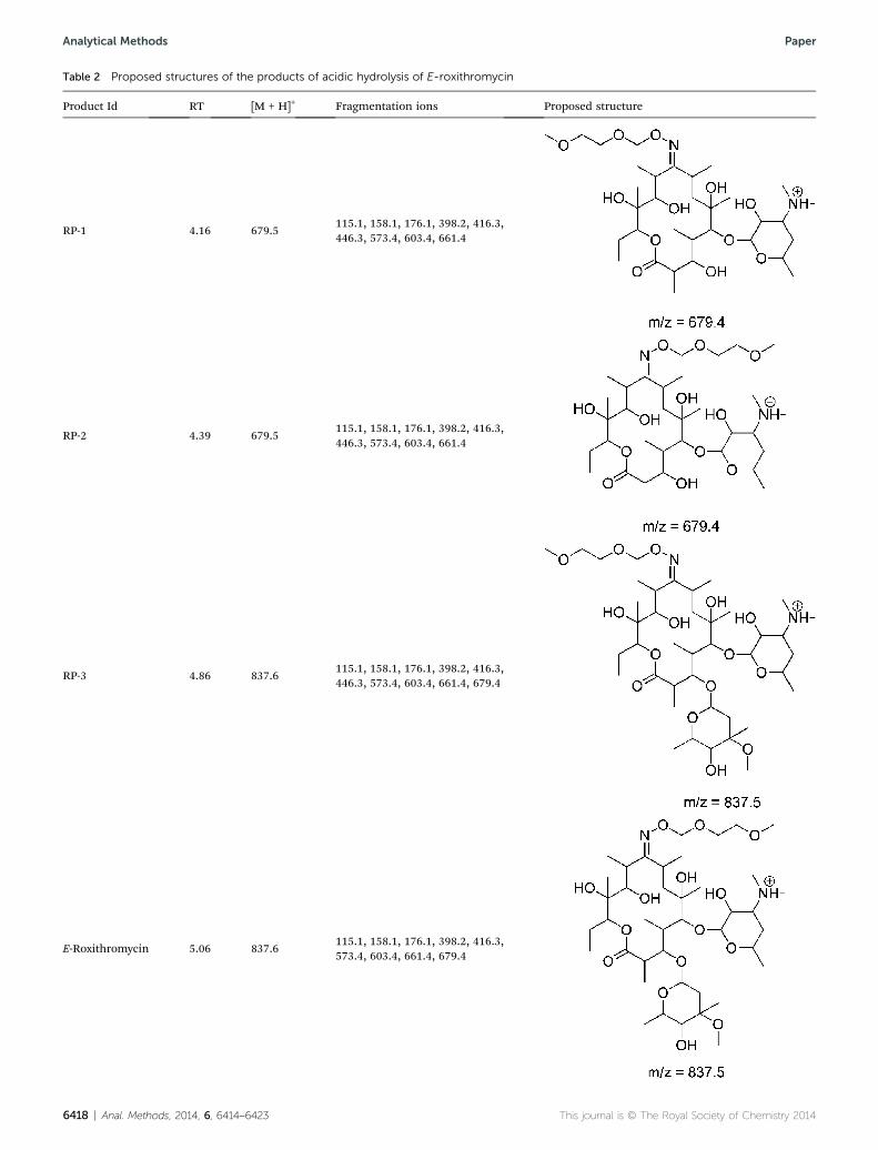
Table 2 Proposed structures of the products of acidic hydrolysis of E-roxithromycin
Product Id RT [M + H]+ Fragmentation ions Proposed structure
RP-1 4.16 679.5115.1, 158.1, 176.1, 398.2, 416.3,446.3, 573.4, 603.4, 661.4
RP-2 4.39 679.5115.1, 158.1, 176.1, 398.2, 416.3,446.3, 573.4, 603.4, 661.4
RP-3 4.86 837.6115.1, 158.1, 176.1, 398.2, 416.3,446.3, 573.4, 603.4, 661.4, 679.4
E-Roxithromycin 5.06 837.6115.1, 158.1, 176.1, 398.2, 416.3,573.4, 603.4, 661.4, 679.4
6418 | Anal. Methods, 2014, 6, 6414–6423 This journal is © The Royal Society of Chemistry 2014
Analytical Methods Paper

Table 2 (Contd. )
Product Id RT [M + H]+ Fragmentation ions Proposed structure
L-Cladinose 6.16 177.1161.1, 147.1, 149.1, 129.1, 119.1,103.1, 101.1, 85.1, 75.1
Paper Analytical Methods
Waters TQD mass spectrometer were as follows – sourcetemperature: 150 �C, desolvation temperature: 350 �C, des-olvation gas ow rate: 600 L h�1, cone gas ow: 100 L h�1,capillary potential: 3.00 kV and cone potential: 20 V. Nitrogenwas used as both nebulizing and drying gas. The data wereobtained in a scan mode ranging from 50 to 1000 m/z at 0.5 sintervals. Collisionally activated dissociation (CAD) analyseswere carried out with the energy of 40 eV, and all the frag-mentations were observed in the source. Consequently, the ionspectra were obtained by scanning from 50 to 1000 m/z range.Data acquisition soware was MassLynx V 4.1 (Waters).
Results and discussion
The aim of this paper was to study the degradation of Roxy inacidic solutions and its photocatalytic degradation process inUV light in the presence of the TiO2 photosensitizer. For bothmethods the degradation products of Roxy were identied.
Acidic hydrolysis of roxithromycin
Acid-induced hydrolysis of Roxy was carried out by the devel-oped chromatographic-densitometric method. Using TLC F254silica gel plates as a stationary phase and methanol–acetone–ammonia 25% (1 : 14 : 0.1, v/v/v) as the mobile phase well-developed and resolved peaks of Roxy and its degradation
Fig. 4 Photodegradation of roxithromycin in the absence and in thepresence of a TiO2 photosensitizer.
This journal is © The Royal Society of Chemistry 2014
products were obtained. The spots were visualized with sul-phuric acid in ethanol and scanned densitometrically at483 nm. The method was validated according to ICH require-ments (see the ESI, method validation, Table S1†).
Acidic hydrolysis of Roxy was performed at three differenttemperatures, i.e., at 40, 50, and 60 �C, and for three differentpH of 2.30, 1.70, and 1.30 (Fig. 2). The relationship betweenRoxy concentration and time follows the kinetics of a rst orderreaction, consistent with the results obtained for hydrolysiscarried out in SGF.17 To obtain full characteristics of thedegradation process, kinetic parameters such as k, t0.1, and t0.5values describing Roxy acidic degradation rates under differentconditions were calculated together with the thermodynamicparameters such as Ea and DH++ (Table 1).
An increase in temperature and a decrease in pH result in anincrease in the rate of Roxy hydrolysis.
TLC analysis of the acidic solutions revealed two degradationproducts with RF values of 0.82 and 0.87 besides the TLC peak ofRoxy with RF ¼ 0.42 (Fig. 3). The obtained degradation productswere subjected to UPLC-MS/MS analysis in order to proposetheir molecular weights and structures. The identication of thedegradation products of E-Roxy induced by acidic conditionswas performed on the basis of UPLC/MS analysis and supportedwith the fragmentation patterns obtained from MS/MS experi-ments. The possible degradation products are shown in Table 2.The acidic degradation was found to affect mainly the L-cladi-nose substituent, causing its detachment from the macrolidecore, and to bring about isomerization of the oximemoiety fromthe native E form to the Z form.
Structures of the presented stable products of the degrada-tion were proposed based on Collisionally Activated Dissocia-tion (CAD) experiments. The fragmentation pattern involved theloss of the L-cladinose substituent (�176.1) of E- and Z-roxi-thromycin. In the next step decladinose derivatives lost H2O(�18) and subsequently the methoxyethene moiety (�58). Theseproducts underwent further decomposition by two routes: bydeformylation (�30) and subsequently the loss of the D-desos-amine substituent, or the loss of N,N,2-trimethyl-3,4-dihydro-2H-pyran-4-amine (�157.1), subsequent deformylation (�30)and loss of H2O (�18) (see ESI Scheme S1†).
D-Desosamine and its decomposition products were alsoobserved. D-Desosamine underwent dehydration (�18) andsubsequent loss of the N-methylenemethanamine fragment(�43) (see ESI Scheme S2†). The main route of fragmentation of
Anal. Methods, 2014, 6, 6414–6423 | 6419

Table 3 Proposed products of photocatalytic degradation of E-roxithromycin in the presence of TiO2
Product Id RT [M + H]+ Fragmentation ions Proposed structure
RP-UV-1 3.54 693.4 115.1, 158.1, 376.2, 500.3, 569.3, 587.4, 675.4
RP-UV-2 3.70 695.4 115.1, 158.1, 571.3, 677.4
RP-UV-3 3.88 665.4 115.1, 158.1, 490.3, 647.4
RP-UV-4 4.38 649.4 115.1, 158.1, 398.2, 416.3, 446.3
RP-UV-5 4.59 711.4 115.1, 158.1, 398.2, 470.3, 488.3, 675.4, 693.4
6420 | Anal. Methods, 2014, 6, 6414–6423 This journal is © The Royal Society of Chemistry 2014
Analytical Methods Paper

Table 3 (Contd. )
Product Id RT [M + H]+ Fragmentation ions Proposed structure
RP-UV-6 4.74 853.5 115.1, 158.1, 398.2, 446.3, 573.4, 661.4, 679.4
RP-UV-7 5.05 869.5 115.1, 158.1, 398.2, 679.4, 851.5
RP-UV-8 5.48 835.5 115.1, 158.1, 176.1, 398.2, 446.3, 573.4, 603.4, 661.4, 679.4
Paper Analytical Methods
L-cladinose involved the loss of carbon monoxide (�28) andsubsequent deformylation (�30) and dehydration (�18). Otherobserved routes involved deformylation (�30) with subsequentdehydration (�18) and loss of the acetaldehyde moiety (�44),demethylation (�14), or decarbonylation (�28) with thesubsequent loss of the acetaldehyde moiety (�44) and decar-bonylation (�28) (see ESI Scheme S3†).
This journal is © The Royal Society of Chemistry 2014
Sensitized by TiO2 photodegradation of roxithromycin
Although acidic hydrolysis of Roxy is quite efficient, it requiresvery low pH (<3) and elevated temperatures; hence it may not bealways applicable for Roxy hydrolytic degradation.7 Therefore,we propose also an alternative method of Roxy degradation:TiO2 sensitized photocatalytic degradation. The photosensitizerin the form of a powder can be obtained in a facile way by the
Anal. Methods, 2014, 6, 6414–6423 | 6421

Analytical Methods Paper
hydrolysis of titanium(IV) isopropoxide and can be easily sus-pended in water.
To nd out which isomorphic form is the main constituentof the TiO2 powder obtained the Powder X-Ray Diffractometry(PXRD) measurement was performed. The presence of Braggpeaks originating only from the anatase crystal structure in thePXRD diffractograms conrms that it is the dominant crystalstructure of the photocatalyst (see Fig. S1 in the ESI†). There areno peaks originating from rutile or brookite indicating thatthese phases are absent; however, the presence of amorphousTiO2 cannot be ruled out. Assuming spherical particle geometry,the average crystallite size calculated from the XRD linebroadening is around 43 nm, thereby conrming its nano-structural morphology.
The diameters of TiO2 particles in a water colloid found inthe dynamic light scattering measurements are around 900 nm(Fig. S2 in the ESI†). It indicates that TiO2 crystallites formlarger clusters. This effect might be attributed to the tempera-ture induced sintering of the grains during the calcination step.
The bandgap energy estimated from the reectance UV-Visspectra equals to 3.13 eV which further conrms the crystalstructure of anatase. The wavelength of the photocatalystabsorption band closely overlaps with the wavelength of themaximum intensity of the Rayonet lamp's emission. On theother hand, the absorption band of Roxy, which ends at about240 nm, is separated from the emission band of the lamps (seeFig. S3 in the ESI†). This implies that the lamp radiation isstrongly absorbed by the photocatalyst and its absorption byRoxy can be neglected.
Photocatalytic degradation studies of Roxy revealed that thedrug studied undergoes photodegradation when irradiated withlight at 350 nm (near UV) in the presence of TiO2. Kinetics of therst order reaction follows the suggested Langmuir–Hinshel-wood theory for photocatalytic microheterogeneous systems(Fig. 4) with the reaction rate constant of 0.00768 min�1. SinceRoxy does not absorb the light used for the irradiation the directphotolysis of Roxy did not occur.
The adsorption process of Roxy on the surface of TiO2 doesnot have a signicant impact on the course of the experiment,as evidenced by minimal changes of the Roxy concentration inthe experiment carried out in the dark, which also excludedacidic hydrolysis at this temperature.
The possible photodegradation products are shown inTable 3.
The photocatalytic degradation was found to affect mainlythe L-cladinose substituent, causing its hydroxylation (RP-UV-6and RP-UV-7) or oxidation of the hydroxy substituent (RP-UV-8).
In the case of decladinose roxithromycin photocatalyticdegradation seems to involve oxidation of the 2-methoxyethoxy-methyl moiety (products RP-UV-4 and RP-UV-5), demethylationof the 2-methoxyethoxymethyl moiety (product RP-UV-3) oroxidation of the core of the antibiotic (RP-UV-1 and RP-UV-2).
Structures of the presented stable products of photocatalyticdegradation were proposed based on collisionally activateddissociation (CAD) experiments. The fragmentation patterns ofproducts RP-UV-3, RP-UV-4 and RP-UV-6–RP-UV-8 were similarto that observed for products of acidic degradation of Roxy.
6422 | Anal. Methods, 2014, 6, 6414–6423
Additionally for RP-UV-7 fragmentation ions with m/z ¼ 851.5were observed, which were probably the product of dehydrationof the 4-methoxy-2,4-dimethyltetrahydro-2H-pyran-2,3,5,6-tet-raol moiety (2,5-dihydroxy-L-cladinose) to 5,6-dihydroxy-4-methoxy-2,4-dimethyldihydro-2H-pyran-3(4H)-one (2-hydroxy-4-oxo-L-cladinose). The fragmentation patterns for RP-UV-1 andRP-UV-2 are similar to those previously described, but due to thepresence of an additional hydroxyl moiety in the roxithromycincore additional dehydration of fragmentation ions is involved.The fragmentation path of RP-UV-1 is presented in Scheme S4(see the ESI†). In the case of RP-UV-5, possessing additionalhydroxy substituents attached to the 2-methoxyethoxymethylmoiety, fragmentation involves dehydration of this moiety,besides previously mentioned transitions. The fragmentationpath for this product is presented in Scheme S5 (see the ESI†).D-Desosamine and its decomposition products were alsoobserved.
Conclusions
Detailed studies on the acidic hydrolytic degradation and TiO2
sensitized photodegradation of roxithromycin have shown thatboth processes are quite efficient. Although the mechanisms ofthese processes are quite different, in both of them the L-cla-dinose substituent is primarily involved. The structures of thedegradation products and the mechanisms of the processesstudied were proposed. Both types of the processes can be usedfor degradation of Roxy; however, the photocatalytic degrada-tion seems to be more promising because it can occur undermild conditions.
References
1 K. Kummerer, J. Environ. Manage., 2009, 90(8), 2354–2366.2 D. W. Kolpin, E. T. Furlong, M. T. Meyer, E. M. Thurman,S. D. Zaugg, L. B. Barber and H. T. Buxton, Environ. Sci.Technol., 2002, 36(6), 1202–1211.
3 B. Pauwels and W. Verstraete, J. Water Health, 2006, 4(4),405–416.
4 F. Sacher, F. T. Lange, H. J. Brauch, et al., J. Chromatogr. A,2001, 938(1–2), 199–210.
5 T. A. Ternes, TrAC, Trends Anal. Chem., 2001, 20(8), 419–434.6 D. Debabov, Appl. Biochem. Microbiol., 2013, 49(8), 665–671.7 J. Feitosa-Felizzola, K. Hanna and S. Chiron, Environ. Pollut.,2009, 157(4), 1317–1322.
8 D. Zhong, X. Li, A. Wang, Y. Xu and S. Wu, Drug Metab.Dispos., 2000, 28, 552–559.
9 M. Ostrowski, E. Wilkowska and T. Baczek, Cent. Eur. J. Med.,2010, 5, 83–90.
10 W. Yam and H. Wahab, J. Chem. Inf. Model., 2009, 49, 1558–1567.
11 M. Gros, M. Petrovic and D. Barcelo, Environ. Toxicol. Chem.,2007, 26(8), 1553–1562.
12 X. Miao, F. Bishay, M. Chen and C. D. Metcalfe, Environ. Sci.Technol., 2004, 38(13), 3533–3541.
13 D. Calamari, E. Zuccato, S. Castiglioni, R. Bagnati andR. Fanelli, Environ. Sci. Technol., 2003, 37(7), 1241–1248.
This journal is © The Royal Society of Chemistry 2014
Paper Analytical Methods
14 Z. Ye, H. S. Weinberg and M. T. Meyer, Anal. Chem., 2007,79(3), 1135–1144.
15 X. Li, D. Zhong, S. Wu, A. Wang, L. Tian and H. Yu, ActaPharmacol. Sin., 1999, 34, 53–58.
16 S. Q. Zhang, L. F. Zhang, J. Xing and D. F. Zhong, Acta Pharm.Sin., 2003, 38, 374–379.
17 Sh. Zhang, J. Xing and D. Zhong, J. Pharm. Sci., 2004, 93,1300–1309.
This journal is © The Royal Society of Chemistry 2014
18 Y. Kim, T. M. Heinze, S. Kim and C. E. Cerniglia, J. Environ.Qual., 2004, 33(1), 257–264.
19 B. Li and T. Zhang, Environ. Sci. Technol., 2010, 44(9), 3468–3473.
20 P. Huo, Y. Yan, S. Li, H. Li and W. Huang, Appl. Surf. Sci.,2010, 256, 3380–3385.
21 A. Molski, Introduction to Kinetics, WNT, Warsaw,2001.
Anal. Methods, 2014, 6, 6414–6423 | 6423
Copyright © 2022 FDOKUMEN