Role of Endogenous IFN-γ In Macrophage Programming Induced by IL-12 and IL-18
12
JOURNAL OF INTERFERON & CYTOKINE RESEARCH 27:399–410 (2007) © Mary Ann Liebert, Inc. DOI: 10.1089/jir.2007.0128 Role of Endogenous IFN- in Macrophage Programming Induced by IL-12 and IL-18 KARINA R.B. BASTOS, RENATO BARBOZA, LUIZ SARDINHA, MOMTCHILO RUSSO, JOSÉ M. ALVAREZ, and MARIA REGINA D’IMPÉRIO LIMA ABSTRACT Besides the established role of interleukin-12 (IL-12) and IL-18 on interferon- (IFN-) production by nat- ural killer (NK), T, and B cells, the effects of these cytokines on macrophages are largely unknown. Here, we investigated the role of IL-12/IL-18 on nitric oxide (NO) and tumor necrosis factor- (TNF-) production by CD11b adherent peritoneal cells, focusing on the involvement of endogenously produced IFN-. C57BL/6 cells released substantial amounts of NO when stimulated with IFN- or lipopolysaccharide (LPS), but failed to respond to IL-12 or IL-18 or both. However, IL-12/IL-18 pretreatment was able to program these cells to release 6–8-fold more NO and TNF- in response to LPS or Trypanosoma cruzi stimulation, with NO levels directly correlating with macrophage resistance to intracellular parasite growth. Analysis of IL-12/IL-18- primed cells from mice deficient in IFN-, IFNGR, and IFN regulatory factor-1 (IRF-1) revealed that these molecules were essential for LPS-induced NO release, but TNF- production was IFN- independent. Con- versely, the myeloid differentiation factor 88 (MyD88)-dependent pathway was indispensable for IL-12/IL-18- programmed LPS-induced TNF- production, but not for NO release. Contaminant T and NK cells largely modulated the IL-12/IL-18 programming of LPS-induced NO response through IFN- secretion. Neverthe- less, a small population of IFN- cells with a macrophage phenotype was also identified, particularly in the peritoneum of chronically T. cruzi-infected mice, reinforcing the notion that macrophages can be an alterna- tive source of IFN-. Taken together, our data contribute to elucidate the molecular basis of the IL-12/IL-18 autocrine pathway of macrophage activation, showing that endogenous IFN- plays an important role in pro- gramming the NO response, whereas the TNF- response occurs through an IFN--independent pathway. 399 INTRODUCTION M ACROPHAGES, MONOCYTES, AND DENDRITIC CELLS (DCs) are the major sources of interleukin-12 (IL-12), 1–3 a het- erodimeric cytokine composed of p35 and p40 subunits. The central role of this cytokine in the development of immune re- sponses was evidenced by data showing that treatment of mice with rIL-12 or IL-12 cDNA induces and sustains in vivo gen- erated effector/memory Th1 cells, 4 upregulates the synthesis of antigen-specific complement-fixing antibodies, 5 and protects against tumors and infectious diseases. 6,7 Conversely, IL-12p40 gene knockout (IL-12p40KO) mice have inadequate Th1 re- sponses 8 and increased susceptibility to infections in which pro- tection is primarily mediated by interferon- (IFN-), such as leishmaniasis, 9 Chagas’ disease, 10 and tuberculosis. 11 The abil- ity of IL-12 to direct the differentiation pattern of T cells indi- cates that this cytokine bridges innate and adaptive immunity, influencing the development of immune responses and, there- fore, the degree of susceptibility to infection. 12 It is generally accepted that the central role of IL-12 in host defense against many intracellular pathogens arises from its ca- pacity to stimulate IFN- secretion by natural killer (NK) and T cells, which in turn activates phagocytes to control parasite growth. 13 Nonetheless, in recent years, macrophages have been recognized as competent cells regarding the ability to respond to IL-12, which has led to the notion that this cytokine can in- duce macrophage activation through an autocrine pathway. In- deed, it has been shown that macrophages not only express 1 and 2 chains from IL-12 receptor (IL-12R), but also respond to IL-12 by producing IFN-, tumor necrosis factor- (TNF- Department of Immunology, Institute of Biomedical Sciences, University of São Paulo, São Paulo, SP, Brazil.
-
Upload
independent -
Category
Documents
-
view
2 -
download
0
Transcript of Role of Endogenous IFN-γ In Macrophage Programming Induced by IL-12 and IL-18
JOURNAL OF INTERFERON & CYTOKINE RESEARCH 27:399–410 (2007)© Mary Ann Liebert, Inc.DOI: 10.1089/jir.2007.0128
Role of Endogenous IFN-� in Macrophage ProgrammingInduced by IL-12 and IL-18
KARINA R.B. BASTOS, RENATO BARBOZA, LUIZ SARDINHA, MOMTCHILO RUSSO, JOSÉ M. ALVAREZ, and MARIA REGINA D’IMPÉRIO LIMA
ABSTRACT
Besides the established role of interleukin-12 (IL-12) and IL-18 on interferon-� (IFN-�) production by nat-ural killer (NK), T, and B cells, the effects of these cytokines on macrophages are largely unknown. Here, weinvestigated the role of IL-12/IL-18 on nitric oxide (NO) and tumor necrosis factor-� (TNF-�) production byCD11b� adherent peritoneal cells, focusing on the involvement of endogenously produced IFN-�. C57BL/6cells released substantial amounts of NO when stimulated with IFN-� or lipopolysaccharide (LPS), but failedto respond to IL-12 or IL-18 or both. However, IL-12/IL-18 pretreatment was able to program these cells torelease 6–8-fold more NO and TNF-� in response to LPS or Trypanosoma cruzi stimulation, with NO levelsdirectly correlating with macrophage resistance to intracellular parasite growth. Analysis of IL-12/IL-18-primed cells from mice deficient in IFN-�, IFNGR, and IFN regulatory factor-1 (IRF-1) revealed that thesemolecules were essential for LPS-induced NO release, but TNF-� production was IFN-� independent. Con-versely, the myeloid differentiation factor 88 (MyD88)-dependent pathway was indispensable for IL-12/IL-18-programmed LPS-induced TNF-� production, but not for NO release. Contaminant T and NK cells largelymodulated the IL-12/IL-18 programming of LPS-induced NO response through IFN-� secretion. Neverthe-less, a small population of IFN-�� cells with a macrophage phenotype was also identified, particularly in theperitoneum of chronically T. cruzi-infected mice, reinforcing the notion that macrophages can be an alterna-tive source of IFN-�. Taken together, our data contribute to elucidate the molecular basis of the IL-12/IL-18autocrine pathway of macrophage activation, showing that endogenous IFN-� plays an important role in pro-gramming the NO response, whereas the TNF-� response occurs through an IFN-�-independent pathway.
399
INTRODUCTION
MACROPHAGES, MONOCYTES, AND DENDRITIC CELLS (DCs)are the major sources of interleukin-12 (IL-12),1–3 a het-
erodimeric cytokine composed of p35 and p40 subunits. Thecentral role of this cytokine in the development of immune re-sponses was evidenced by data showing that treatment of micewith rIL-12 or IL-12 cDNA induces and sustains in vivo gen-erated effector/memory Th1 cells,4 upregulates the synthesis ofantigen-specific complement-fixing antibodies,5 and protectsagainst tumors and infectious diseases.6,7 Conversely, IL-12p40gene knockout (IL-12p40KO) mice have inadequate Th1 re-sponses8 and increased susceptibility to infections in which pro-tection is primarily mediated by interferon-� (IFN-�), such asleishmaniasis,9 Chagas’ disease,10 and tuberculosis.11 The abil-
ity of IL-12 to direct the differentiation pattern of T cells indi-cates that this cytokine bridges innate and adaptive immunity,influencing the development of immune responses and, there-fore, the degree of susceptibility to infection.12
It is generally accepted that the central role of IL-12 in hostdefense against many intracellular pathogens arises from its ca-pacity to stimulate IFN-� secretion by natural killer (NK) andT cells, which in turn activates phagocytes to control parasitegrowth.13 Nonetheless, in recent years, macrophages have beenrecognized as competent cells regarding the ability to respondto IL-12, which has led to the notion that this cytokine can in-duce macrophage activation through an autocrine pathway. In-deed, it has been shown that macrophages not only express �1and �2 chains from IL-12 receptor (IL-12R), but also respondto IL-12 by producing IFN-�, tumor necrosis factor-� (TNF-
Department of Immunology, Institute of Biomedical Sciences, University of São Paulo, São Paulo, SP, Brazil.
�), and nitric oxide (NO).14–24 IL-12 has also been implicatedin programming the macrophage response to lipopolysaccha-ride (LPS) by upregulating the production of TNF-�.25 IL-18,a cytokine secreted by several cell types, including macro-phages, originally designated as IFN-�-inducing factor(IGIF),26 has been shown to act in synergism with IL-12 tostimulate IFN-� production by T cells,27 NK cells,28 B cells,27
macrophages,16,18,21 and DCs.29,30 Although IL-18 per se doesnot seem to induce IFN-� secretion by these cells, it can im-prove the response to IL-12 in different ways. In macrophages,the synergic effect of IL-18 depends on nuclear translocationof Stat4 that is attained only in the presence of both cytokines,18
whereas in DCs, IL-18 upregulates the activity of p38, a mem-ber of the MAP kinase (MAPK) superfamily, culminating withIFN-� secretion.29
Another feature attributed to IL-12 is the ability to down-regulate the expression of transforming growth factor-�1 (TGF-�1) mRNA in monocytes and bone marrow cells.31 Overall, IL-12 directly influences the macrophage activation profile, drivingthem to react against foreign stimuli with a response dominatedby proinflammatory cytokines. In this context, we have shownpreviously that macrophages from IL-12p40KO mice have anactivation bias, spontaneously secreting large amounts of TGF-�, and responding with weak NO production to rIFN-�.32,33
Moreover, IL-12p40KO macrophages are more permissive tothe growth of the intracellular protozoan Trypanosoma cruzithan are wild-type cells and have an impaired ability to ingestopsonized Plasmodium chabaudi-infected erythrocytes. Thus,IL-12 might affect host defense against pathogens by at leasttwo distinct pathways: (1) inducing IFN-� production by sev-eral cell types and (2) programming macrophages prior to theonset of immune responses, rendering these cells prone to re-act with an effector (NO-dominant) rather than a tolerant (TGF-�-dominant) profile.
In the attempt to further characterize the effects of IL-12 andIL-18 on macrophages, we have focused our attention on therole of endogenously produced IFN-� in the ability of IL-12/IL-18 to program the responses of CD11b� adherent peritonealcells (CD11b� aPECs). Taken together, our data highlight theIL-12/IL-18 autocrine pathway of macrophage activation andreinforce the notion that at least in some circumstances, mac-rophages can be an alternative source of IFN-�.
MATERIALS AND METHODS
Mice and parasites
Six to eight-week-old C57BL/6, 129/SV, IFN-�KO, IFN-GRKO, IL-12p40KO, MyD88KO, IRF-1KO, CD14KO, andiNOSKO male mice were bred in our animal facilities at theUniversity of São Paulo, under standard pathogen-free condi-tions. IFN-�KO, IFNGRKO, IL-12p40KO, iNOSKO, andCD14KO mice are originally from Jackson Laboratories (BarHarbor, ME), MyD88KO34 mice were kindly provided by DrBernard Ryffel (Centre National de la Recherche Scientifique,Orléans, France), and IRF-1KO35 mice were kindly providedby Dr Luiz Fernando Reis (Ludwig Institute for Cancer Re-search, São Paulo, Brazil). T. cruzi trypomastigotes of theSylvio-X10/4 strain were purified from a monkey epithelial cell
line (LLC-MK2). C57BL/6 mice were infected i.p. with 103 try-pomastigotes 3 months before the experiments.
PEC suspensions
Four to six mice were injected i.p. with 5 mL of 3% starch(Sigma, St. Louis, MO). Five days later, cells were obtained byperitoneal lavage with chilled RPMI 1640 (Sigma).
Purification of cell populations
CD11b� PECs were purified by magnetic beads coupledwith anti-CD11b (Mac-1, M1/70) monoclonal antibodies(mAb), according to the manufacturer’s instructions (MiltenyiBiotec GMBH, Bergisch Gladbach, Germany). Cells with size(FSC) and granularity (SSC) characteristics of macrophageswere sorted by flow cytometry using a FacsVantage (BectonDickinson, Mountain View, CA) with CELLQUEST software(Becton Dickinson). CD3�NK1.1� and CD3�NK1.1� cellswere purified with phycoerythrin (PE)-labeled anti-NK1.1(PK136) and anti-CD3 (145-2C11) mAbs (PharMingen, SanDiego, CA), followed by magnetic beads coupled with anti-PEmAb (Miltenyi Biotec).
Phenotypic analysis of purified macrophages
Cells (106) stained with FITC, PE, or Cy-Chrome-labeledmAb to CD11b (Mac-1, M1/70), CD3 (145-2C11), and NK1.1(PK136) (PharMingen) and F4/80 mAb (Caltag Laboratories,Burlingame, CA) were analyzed by flow cytometry using a Fac-scalibur (Becton Dickinson) with CELLQUEST software.
CD11b� aPEC culture conditions
Purified CD11b� PECs (2 � 105) were incubated for 4 h inRPMI 1640 supplemented with penicillin (100 U/mL), strepto-mycin (100 �g/mL), 2-mercaptoethanol (50 �M), L-glutamine(2 mM), sodium pyruvate (1 mM), and 3% heat-inactivated fe-tal bovine serum (FBS). All supplements were purchased fromLife Technologies (Rockville, MD). Nonadherent cells were re-moved by three vigorous washes with warm and FBS-freemedium. To evaluate the direct effect of LPS, IFN-� IL-12, orIL-18, cells were cultured with 0.1–50 �g/mL LPS (Sigma),2.5–20 ng/mL rIFN-� (PharMingen), or 0.5–10 ng/mL rIL-12(PharMingen) with or without 0.5–10 ng/mL rIL-18 (MBL In-ternational Corporation, Woburn, MA), and the supernatantswas harvested 48 h later. To evaluate the priming effect of IL-12, IL-18, and IFN-�, cells were cultured for 18 h with rIL-12(2.5 ng/mL), rIL-18 (2.5 ng/mL), or rIFN-� (2.5 ng/mL),washed with medium, and then stimulated for additional 48 hwith LPS (1 �g/mL) or T. cruzi (5 parasites/macrophage).
Detection of NO and TNF-� in culture supernatants
Culture supernatants were assayed for NO by the Griess re-action. Briefly, 50 �L supernatant was incubated with 50 �LGriess reagent for 5 min at ambient temperature, and the NO�
2
concentration was determined by measuring the optical density(OD) at 550 nm in reference to a standard NaNO2 solution. Amodified bioassay was used to quantify TNF-�.36 TNF-�-sen-sitive L929 cells (ATCC, Rockville, MD) were incubated insupplemented RPMI 1640 overnight at 37°C in a 5% CO2 atmo-
BASTOS ET AL.400
sphere at a density of 5.5 � 104 cells in flat-bottomed 96-wellplates. After medium removal, 100 �L of serial dilutions of cellculture supernatants (diluted from 1:2 to 1:1024) were addedto each well. Four hours later, 10 �L medium containing actin-omycin D was added to each well at a final concentration of 5�g/mL. After 20 h in culture, viable L929 cells were stainedwith 20 �L/well of 0.75% crystal violet in 30% acetic acid for15 min, rinsed, and dried. Methanol was used to solubilize thecrystal violet, and the absorbance was read at 630 nm with aVmax-Kinetic Microplate Reader (Molecular Devices, Sunny-vale, CA). Percent cytotoxicity was calculated by reference tocontrol monolayers incubated in medium only. One cytotoxicunit corresponded to the sample dilution in which 50% of L929cells were killed. To obtain TNF-�-positive serum, mice wereinfected with BCG, and 2 weeks later 20 �g LPS was given in-travenously (iv). TNF-�-rich serum was collected 90 min afterLPS inoculation.
Killing of intracellular T. cruzi parasites
The killing assay for intracellular T. cruzi parasites has beendescribed previously.32 CD11b� PECs (2 � 105) were addedto tissue culture chambers (Lab-Tek Chamber Slide, Nunc,Rochester, NY) and incubated for 4 h in supplemented RPMImedium. Adherent cells were then cultured for 24 h with rIL-12 (2.5 ng/mL), rIL-18 (2.5 ng/mL), rIFN-� (2.5 ng/mL), ormedium, infected with T. cruzi at 5:1 ratio for 120 min, andwashed six times to remove extracellular parasites. Cultureswere kept for an additional 48 h with cytokines or medium. Af-ter this period, cells were stained with Giemsa to count intra-cellular amastigotes.
Detection of intracellular IFN-�
CD11b� aPECs (106) from normal and chronically T. cruzi-infected C57BL/6 mice were cultured for 48 h at 37°C in a 5%CO2 atmosphere, according to the manufacturer’s instructions,in the presence or absence of rIL-12 and rIL-18 (10 ng/mL ofeach). Monensin-containing Golgistop (PharMingen) wasadded in the last 6 h of culture. After washing, cells were sur-face stained with FITC-conjugated or Cy-Chrome-conjugatedmAbs to CD3 and NK1.1 (PharMingen) and F4/80 mAbs (Cal-tag). Cells were then fixed with the Cytofix/Cytoperm buffer(PharMingen) and incubated with PE-labeled mAb to IFN-�(XMG-1.2) (PharMingen) diluted in Perm/Wash buffer(PharMingen). Cells were analyzed by flow cytometry using aFacscalibur with CELLQUEST software.
Confocal microscopy
For analysis of intracellular IFN-� and TNF-�, CD11b�
PECs (2 � 105) were added to tissue culture chambers (Lab-Tek Chamber Slide) and incubated for 4 h in supplementedRPMI medium. Adherent cells were then cultured for 18 h withrIL-12 (2.5 ng/mL), rIL-18 (2.5 ng/mL), or medium, washed,and then stimulated for an additional 6 h with LPS (1 �g/mL)in the presence of monensin-containing Golgistop at 37°C in a5% CO2 atmosphere, according to the manufacturer’s instruc-tions. After washing, cells were surface stained with FITC-con-jugated mAbs to CD11b and MHC II. Cells were then fixedwith the Cytofix/Cytoperm buffer and incubated with PE-la-
beled mAb to IFN-� (XMG-1.2) and TNF-� (MP6-XT22) di-luted in Perm/Wash buffer. All reagents were purchased fromPharMingen. A CLSM 410 confocal microscopy (Carl ZeissInc., Oberlochen, Germany) was used to reveal the surface andintracellular staining with Carl Zeiss CLSM Image Browsersoftware (version 3.1).
Statistical analysis
Statistical analysis was performed by unpaired ANOVA andTukey’s multiple comparison tests using GraphPad Prism 3software. Differences between two groups were considered sig-nificant when the p value was �0.05 (5%).
RESULTS
Role of endogenous IFN-� in NO response of CD11b� aPECs
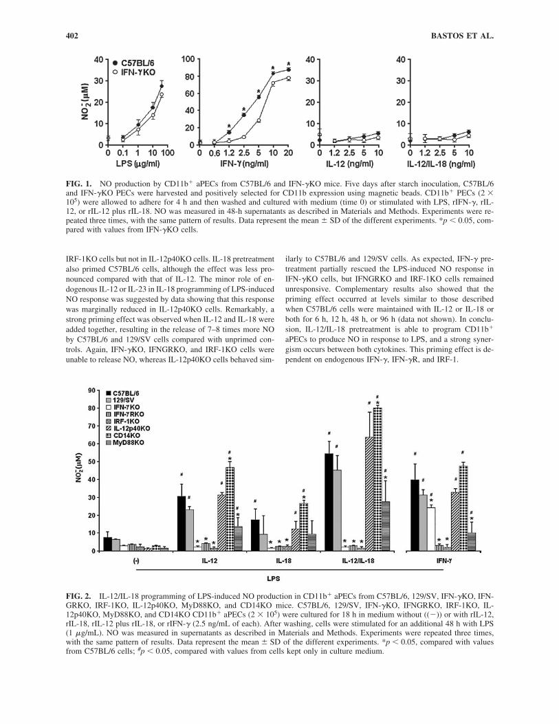
Initially, we compared the NO production of CD11b� aPECsobtained from C57BL/6 and IFN-�KO mice. The rationale ofthese experiments was to determine the influence of endoge-nously produced IFN-� in the ability of these cells to releaseNO in response to different stimuli. Therefore, CD11b� aPECsof both mouse strains were cultured for 48 h with increasingconcentrations of LPS, rIFN-�, rIL-12, or rIL-12 plus rIL-18.As shown in Figure 1, C57BL/6 and IFN-�KO cells releasedsignificant amounts of NO in response to IFN-�. At low con-centrations of IFN-�, however, higher NO production occurredwith C57BL/6 cells compared with IFN-�KO cells. When LPSwas used as a stimulus, C57BL/6 and IFN-�KO cells respondedat similar levels. In this case, however, an optimal response wasachieved only with high LPS doses. In contrast, IL-12 was un-able to induce NO release, even at high concentrations and inthe presence of IL-18. We concluded that IL-12 alone or com-bined with IL-18 is not capable of stimulating NO productionby peritoneal macrophages, a population that under our exper-imental conditions comprises 98%–99% of CD11b� aPECs(data not shown). In addition, our data have shown that en-dogenous IFN-� is not required for NO release by these cellswhen the stimulus is LPS or exogenous IFN-�.
Role of endogenous IFN-� in IL-12/IL-18programming of LPS-induced NO response
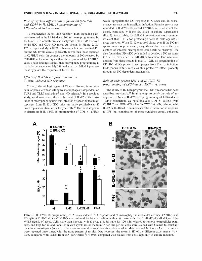
Next, we evaluated the role of IL-12 and IL-18 in macro-phage programming for LPS-induced NO response, focusingagain on endogenous IFN-�. Therefore, NO levels were quan-tified in culture supernatants of CD11b� aPECs from C57BL/6,129/SV, IFN-�KO and IFNGRKO mice treated with rIL-12 orrIL-18 or both for 18 h and then stimulated for 48 h with 1�g/mL LPS, a dose that induces CD11b� aPECs to release lowamounts of NO (Fig. 1). Additionally, the NO response wasevaluated in IL-12p40KO and IRF-1KO cells to estimate therole of endogenous IL-12 and IL-23 and the contribution of IFNregulatory factor-1 (IRF-1), a transcriptional activator of genesinvolved in the IFN responses.37 Data in Figure 2 showed thatIL-12 programming of C57BL/6 and 129/SV cells increased theLPS-induced NO production by 3–4-fold. Conversely, this re-sponse was completely abolished in IFN-�KO, IFNGRKO, and
ENDOGENOUS IFN-� IN MACROPHAGE PROGRAMMING BY IL-12/IL-18 401
IRF-1KO cells but not in IL-12p40KO cells. IL-18 pretreatmentalso primed C57BL/6 cells, although the effect was less pro-nounced compared with that of IL-12. The minor role of en-dogenous IL-12 or IL-23 in IL-18 programming of LPS-inducedNO response was suggested by data showing that this responsewas marginally reduced in IL-12p40KO cells. Remarkably, astrong priming effect was observed when IL-12 and IL-18 wereadded together, resulting in the release of 7–8 times more NOby C57BL/6 and 129/SV cells compared with unprimed con-trols. Again, IFN-�KO, IFNGRKO, and IRF-1KO cells wereunable to release NO, whereas IL-12p40KO cells behaved sim-
ilarly to C57BL/6 and 129/SV cells. As expected, IFN-� pre-treatment partially rescued the LPS-induced NO response inIFN-�KO cells, but IFNGRKO and IRF-1KO cells remainedunresponsive. Complementary results also showed that thepriming effect occurred at levels similar to those describedwhen C57BL/6 cells were maintained with IL-12 or IL-18 orboth for 6 h, 12 h, 48 h, or 96 h (data not shown). In conclu-sion, IL-12/IL-18 pretreatment is able to program CD11b�
aPECs to produce NO in response to LPS, and a strong syner-gism occurs between both cytokines. This priming effect is de-pendent on endogenous IFN-�, IFN-�R, and IRF-1.
BASTOS ET AL.402
FIG. 1. NO production by CD11b� aPECs from C57BL/6 and IFN-�KO mice. Five days after starch inoculation, C57BL/6and IFN-�KO PECs were harvested and positively selected for CD11b expression using magnetic beads. CD11b� PECs (2 �105) were allowed to adhere for 4 h and then washed and cultured with medium (time 0) or stimulated with LPS, rIFN-�, rIL-12, or rIL-12 plus rIL-18. NO was measured in 48-h supernatants as described in Materials and Methods. Experiments were re-peated three times, with the same pattern of results. Data represent the mean � SD of the different experiments. *p � 0.05, com-pared with values from IFN-�KO cells.
FIG. 2. IL-12/IL-18 programming of LPS-induced NO production in CD11b� aPECs from C57BL/6, 129/SV, IFN-�KO, IFN-GRKO, IRF-1KO, IL-12p40KO, MyD88KO, and CD14KO mice. C57BL/6, 129/SV, IFN-�KO, IFNGRKO, IRF-1KO, IL-12p40KO, MyD88KO, and CD14KO CD11b� aPECs (2 � 105) were cultured for 18 h in medium without ((�)) or with rIL-12,rIL-18, rIL-12 plus rIL-18, or rIFN-� (2.5 ng/mL of each). After washing, cells were stimulated for an additional 48 h with LPS(1 �g/mL). NO was measured in supernatants as described in Materials and Methods. Experiments were repeated three times,with the same pattern of results. Data represent the mean � SD of the different experiments. *p � 0.05, compared with valuesfrom C57BL/6 cells; #p � 0.05, compared with values from cells kept only in culture medium.
Role of myeloid differentiation factor 88 (MyD88) and CD14 in IL-12/IL-18 programming of LPS-induced NO response
To characterize the toll-like receptor (TLR) signaling path-way involved in the LPS-induced NO response programmed byIL-12 or IL-18 or both, we also analyzed CD11b� aPECs fromMyD88KO and CD14KO mice. As shown in Figure 2, IL-12/IL-18-primed MyD88KO cells were able to respond to LPS,but the NO levels were significantly lower than those obtainedin C57BL/6 cells. In contrast, the amounts of NO released byCD14KO cells were higher than those produced by C57BL/6cells. These findings suggest that macrophage programming ispartially dependent on MyD88 and that IL-12/IL-18 pretreat-ment bypasses the requirement for CD14.
Effects of IL-12/IL-18 programming on T. cruzi-induced NO response
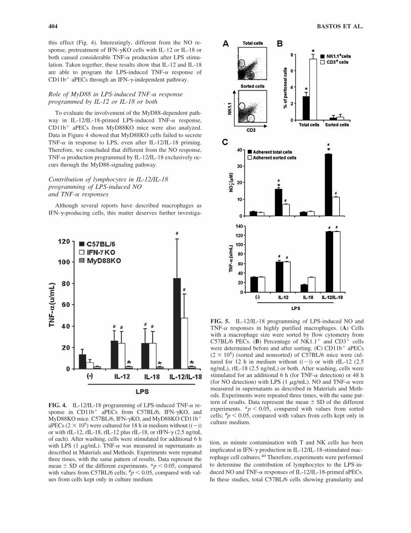
T. cruzi, the etiologic agent of Chagas’ disease, is an intra-cellular parasite whose killing by macrophages is dependent onTLR2 and TLR9 activation38 and NO release.39 In a previousstudy, we demonstrated the involvement of IL-12 in the resis-tance of macrophage against this infection by showing that mac-rophages from IL-12p40KO mice are more permissive to T.cruzi replication than are wild-type cells.32 Our next step wasto determine if IL-12/IL-18 programming of CD11b� aPECs
would upregulate the NO response to T. cruzi and, in conse-quence, restrain the intracellular infection. Parasite growth wasinhibited in IL-12/IL-18-primed C57BL/6 cells, an effect thatclearly correlated with the NO levels in culture supernatants(Fig. 3). Remarkably, IL-12/IL-18 pretreatment was even moreefficient than IFN-� for protecting C57BL/6 cells against T.cruzi infection. When IL-12 was used alone, even if the NO re-sponse was less pronounced, a significant decrease in the per-centage of infected macrophages could still be observed. Wealso found that IFN-�KO cells failed to develop a NO responseto T. cruzi, even after IL-12/IL-18 pretreatment. Our main con-clusion from these results is that IL-12/IL-18 programming ofCD11b� aPECs protects macrophages from T. cruzi infection.Endogenous IFN-� mediates this protective effect probablythrough an NO-dependent mechanism.
Role of endogenous IFN-� in IL-12/IL-18programming of LPS-induced TNF-� response
The ability of IL-12 to program the TNF-� response has beendescribed previously.25 In an attempt to verify the role of en-dogenous IFN-� in IL-12/IL-18 programming of LPS-inducedTNF-� production, we have analyzed CD11b� aPECs fromC57BL/6 and IFN-�KO mice. In C57BL/6 cells, priming withIL-12 or IL-18 led to an increased TNF-� secretion in responseto LPS, but combination of these cytokines greatly enhanced
ENDOGENOUS IFN-� IN MACROPHAGE PROGRAMMING BY IL-12/IL-18 403
FIG. 3. IL-12/IL-18 programming of T. cruzi-induced NO response and of macrophage microbicidal activity. C57BL/6 andIFN-�KO CD11b� aPECs (2 � 105) were cultured for 24 h in medium without ((�)) or with rIL-12, rIL-12 plus rIL-18, or rIFN-� (2.5 ng/mL of each). Cells were then infected with T. cruzi at a 5:1 ratio for 120 min, washed to remove extracellular para-sites, and kept for an additional 48 h with cytokines or medium. After this period, cells were stained with Giemsa to count in-tracellular amastigotes (A and B). NO was measured in supernatants as described in Materials and Methods (A). Experimentswere repeated three times, with the same pattern of results. Data represent the mean � SD of the different experiments. *p �0.05, compared with values from IFN-�KO cells; #p � 0.05, compared with values from cells kept only in culture medium.
this effect (Fig. 4). Interestingly, different from the NO re-sponse, pretreatment of IFN-�KO cells with IL-12 or IL-18 orboth caused considerable TNF-� production after LPS stimu-lation. Taken together, these results show that IL-12 and IL-18are able to program the LPS-induced TNF-� response ofCD11b� aPECs through an IFN-�-independent pathway.
Role of MyD88 in LPS-induced TNF-� responseprogrammed by IL-12 or IL-18 or both
To evaluate the involvement of the MyD88-dependent path-way in IL-12/IL-18-primed LPS-induced TNF-� response,CD11b� aPECs from MyD88KO mice were also analyzed.Data in Figure 4 showed that MyD88KO cells failed to secreteTNF-� in response to LPS, even after IL-12/IL-18 priming.Therefore, we concluded that different from the NO response,TNF-� production programmed by IL-12/IL-18 exclusively oc-curs through the MyD88-signaling pathway.
Contribution of lymphocytes in IL-12/IL-18programming of LPS-induced NO and TNF-� responses
Although several reports have described macrophages asIFN-�-producing cells, this matter deserves further investiga-
tion, as minute contamination with T and NK cells has beenimplicated in IFN-� production in IL-12/IL-18-stimulated mac-rophage cell cultures.40 Therefore, experiments were performedto determine the contribution of lymphocytes to the LPS-in-duced NO and TNF-� responses of IL-12/IL-18-primed aPECs.In these studies, total C57BL/6 cells showing granularity and
BASTOS ET AL.404
FIG. 4. IL-12/IL-18 programming of LPS-induced TNF-� re-sponse in CD11b� aPECs from C57BL/6, IFN-�KO, andMyD88KO mice. C57BL/6, IFN-�KO, and MyD88KO CD11b�
aPECs (2 � 105) were cultured for 18 h in medium without ((�))or with rIL-12, rIL-18, rIL-12 plus rIL-18, or rIFN-� (2.5 ng/mLof each). After washing, cells were stimulated for additional 6 hwith LPS (1 �g/mL). TNF-� was measured in supernatants asdescribed in Materials and Methods. Experiments were repeatedthree times, with the same pattern of results. Data represent themean � SD of the different experiments. *p � 0.05, comparedwith values from C57BL/6 cells; #p � 0.05, compared with val-ues from cells kept only in culture medium
FIG. 5. IL-12/IL-18 programming of LPS-induced NO andTNF-� responses in highly purified macrophages. (A) Cellswith a macrophage size were sorted by flow cytometry fromC57BL/6 PECs. (B) Percentage of NK1.1� and CD3� cellswere determined before and after sorting. (C) CD11b� aPECs(2 � 105) (sorted and nonsorted) of C57BL/6 mice were cul-tured for 12 h in medium without ((�)) or with rIL-12 (2.5ng/mL), rIL-18 (2.5 ng/mL) or both. After washing, cells werestimulated for an additional 6 h (for TNF-� detection) or 48 h(for NO detection) with LPS (1 �g/mL). NO and TNF-� weremeasured in supernatants as described in Materials and Meth-ods. Experiments were repeated three times, with the same pat-tern of results. Data represent the mean � SD of the differentexperiments. *p � 0.05, compared with values from sortedcells; #p � 0.05, compared with values from cells kept only inculture medium.
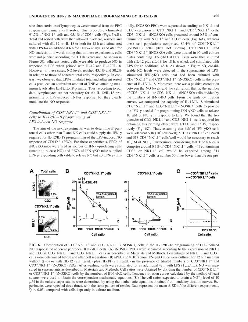
size characteristics of lymphocytes were removed from the PECsuspensions using a cell sorter. This procedure eliminated91.7% of NK1.1� cells and 95.1% of CD3� cells (Figs. 5A,B).Total and sorted cells were then allowed to adhere, washed, andcultured with rIL-12 or rIL-18 or both for 18 h and stimulatedwith LPS for an additional 6 h for TNF-� analysis and 48 h forNO analysis. It is worth noting that in these experiments, cellswere not purified according to CD11b expression. As shown inFigure 5C, adherent sorted cells were able to produce NO inresponse to LPS when primed with IL-12 and IL-12/IL-18.However, in these cases, NO levels reached 43.1% and 30.2%in relation to those of adherent total cells, respectively. In con-trast, we observed that LPS-stimulated total and adherent sortedcells produced an equivalent amount of TNF-�, attaining max-imum levels after IL-12/IL-18 priming. Thus, according to ourdata, lymphocytes are not necessary for the IL-12/IL-18 pro-gramming of LPS-induced TNF-� response, but they clearlymodulate the NO response.
Contribution of CD3�NK1.1� and CD3�NK1.1�
cells to IL-12/IL-18 programming of LPS-induced NO response
The aim of the next experiments was to determine if peri-toneal cells other than T and NK cells could supply the IFN-�required for IL-12/IL-18 programming of the LPS-induced NOresponse of CD11b� aPECs. For these experiments, PECs ofiNOSKO mice were used as sources of IFN-�-producing cells(unable to release NO) and PECs of IFN-�KO mice suppliedIFN-�-responding cells (able to release NO but not IFN-�). Ini-
tially, iNOSKO PECs were separated according to NK1.1 andCD3 expression in CD3�NK1.1� and CD3�/NK1.1� cells.CD3�NK1.1� (iNOSKO) cells presented around 0.3% of con-tamination with NK1.1� and CD3� cells (Fig. 6A), whereasthese cell populations comprised 86.4% of CD3�NK1.1�
(iNOSKO) cells (data not shown). CD3�NK1.1� andCD3�NK1.1� (iNOSKO) cells were titrated in 96-well cultureplates containing IFN-�KO aPECs. Cells were then culturedwith rIL-12 plus rIL-18 for 18 h, washed, and stimulated withLPS for an additional 48 h. As shown in Figure 6B, consid-erable NO levels were detected in the supernatants of LPS-stimulated IFN-�KO cells that had been cultured withCD3�NK1.1� and CD3�NK1.1� (iNOSKO) cells in the pres-ence of IL-12/IL-18. Moreover, there was a positive correlationbetween the NO levels and the cell ratios, that is, the numberof CD3�NK1.1� or CD3�NK1.1� (iNOSKO) cells divided bythe numbers of IFN-�KO cells. From the tendency titrationcurves, we compared the capacity of IL-12/IL-18-stimulatedCD3�NK1.1� and CD3�NK1.1� (iNOSKO) cells to providethe IFN-� needed for programming IFN-�KO cells to release10 �M of NO�
2 in response to LPS. We found that the fre-quencies of CD3�NK1.1� and CD3�NK1.1� cells required forobtaining this priming effect were 1/1731 and 1/319, respec-tively (Fig. 6C). Thus, assuming that half of IFN-�KO cellswere adherent cells (105 cells/well), 58 CD3�NK1.1� cells/welland 313 CD3�NK1.1� cells/well would be necessary to reach10 �M of NO�
2. Furthermore, considering that T or NK cellscomprise around 0.3% of CD3�NK1.1� cells, �1 contaminantCD3� or NK1.1� cell would be expected among 313CD3�NK1.1� cells, a number 50 times lower than the one pre-
ENDOGENOUS IFN-� IN MACROPHAGE PROGRAMMING BY IL-12/IL-18 405
FIG. 6. Contribution of CD3�NK1.1� and CD3�NK1.1� (iNOSKO) cells in the IL-12/IL-18 programming of LPS-inducedNO response of adherent peritoneal IFN-�KO cells. (A) iNOSKO PECs were separated according to the expression of NK1.1and CD3 in CD3�NK1.1� and CD3�NK1.1� cells as described in Materials and Methods. Percentages of NK1.1� and CD3�
cells were determined before and after cell separation. (B) aPECs (2 � 105) from IFN-�KO mice were cultured for 12 h in mediumwithout ((�)) or with rIL-12 (2.5 ng/mL) plus rIL-18 (2.5 ng/mL) in the presence of titrated numbers of CD3�NK1.1� andCD3�NK1.1� (iNOSKO) PECs. After washing, cells were stimulated for an additional 48 h with LPS (1 �g/mL). NO was mea-sured in supernatants as described in Materials and Methods. Cell ratios were obtained by dividing the number of CD3�NK1.1�
or CD3�NK1.1� (iNOSKO) cells by the numbers of IFN-�KO cells. Tendency titration curves calculated by the method of leastsquares were used to obtain the correspondent mathematic equations. (C) The cell ratios expected to attain a NO�
2 level of 10�M in the culture supernatants were determined by using the mathematic equations obtained from tendency titration curves. Ex-periments were repeated three times, with the same pattern of results. Data represent the mean � SD of the different experiments.#p � 0.05, compared with cells kept only in culture medium.
dictable if the priming effect of CD3�NK1.1� cells were dueto CD3�NK1.1� cell contamination. Besides this, consideringthe tendency titration curves, a very high CD3�NK1.1� cellcontamination (of around 8%) would be necessary to attain 20�M of NO�
2. These data corroborate the notion that cell pop-ulations other than T and NK cells may contribute to the IFN-� production required for programming IFN-�KO cells to re-lease NO in response to LPS.
Analysis of IFN-� production by IL-12/IL-18-stimulated macrophages
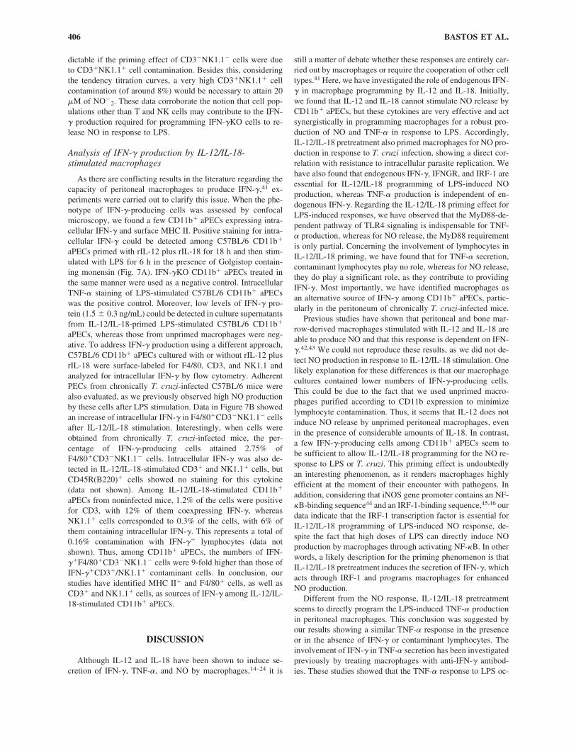
As there are conflicting results in the literature regarding thecapacity of peritoneal macrophages to produce IFN-�,41 ex-periments were carried out to clarify this issue. When the phe-notype of IFN-�-producing cells was assessed by confocal microscopy, we found a few CD11b� aPECs expressing intra-cellular IFN-� and surface MHC II. Positive staining for intra-cellular IFN-� could be detected among C57BL/6 CD11b�
aPECs primed with rIL-12 plus rIL-18 for 18 h and then stim-ulated with LPS for 6 h in the presence of Golgistop contain-ing monensin (Fig. 7A). IFN-�KO CD11b� aPECs treated inthe same manner were used as a negative control. IntracellularTNF-� staining of LPS-stimulated C57BL/6 CD11b� aPECswas the positive control. Moreover, low levels of IFN-� pro-tein (1.5 � 0.3 ng/mL) could be detected in culture supernatantsfrom IL-12/IL-18-primed LPS-stimulated C57BL/6 CD11b�
aPECs, whereas those from unprimed macrophages were neg-ative. To address IFN-� production using a different approach,C57BL/6 CD11b� aPECs cultured with or without rIL-12 plusrIL-18 were surface-labeled for F4/80, CD3, and NK1.1 andanalyzed for intracellular IFN-� by flow cytometry. AdherentPECs from chronically T. cruzi-infected C57BL/6 mice werealso evaluated, as we previously observed high NO productionby these cells after LPS stimulation. Data in Figure 7B showedan increase of intracellular IFN-� in F4/80�CD3�NK1.1� cellsafter IL-12/IL-18 stimulation. Interestingly, when cells wereobtained from chronically T. cruzi-infected mice, the per-centage of IFN-�-producing cells attained 2.75% ofF4/80�CD3�NK1.1� cells. Intracellular IFN-� was also de-tected in IL-12/IL-18-stimulated CD3� and NK1.1� cells, butCD45R(B220)� cells showed no staining for this cytokine (data not shown). Among IL-12/IL-18-stimulated CD11b�
aPECs from noninfected mice, 1.2% of the cells were positivefor CD3, with 12% of them coexpressing IFN-�, whereasNK1.1� cells corresponded to 0.3% of the cells, with 6% ofthem containing intracellular IFN-�. This represents a total of0.16% contamination with IFN-�� lymphocytes (data notshown). Thus, among CD11b� aPECs, the numbers of IFN-��F4/80�CD3�NK1.1� cells were 9-fold higher than those ofIFN-��CD3�/NK1.1� contaminant cells. In conclusion, ourstudies have identified MHC II� and F4/80� cells, as well asCD3� and NK1.1� cells, as sources of IFN-� among IL-12/IL-18-stimulated CD11b� aPECs.
DISCUSSION
Although IL-12 and IL-18 have been shown to induce se-cretion of IFN-�, TNF-�, and NO by macrophages,14–24 it is
still a matter of debate whether these responses are entirely car-ried out by macrophages or require the cooperation of other celltypes.41 Here, we have investigated the role of endogenous IFN-� in macrophage programming by IL-12 and IL-18. Initially,we found that IL-12 and IL-18 cannot stimulate NO release byCD11b� aPECs, but these cytokines are very effective and actsynergistically in programming macrophages for a robust pro-duction of NO and TNF-� in response to LPS. Accordingly,IL-12/IL-18 pretreatment also primed macrophages for NO pro-duction in response to T. cruzi infection, showing a direct cor-relation with resistance to intracellular parasite replication. Wehave also found that endogenous IFN-�, IFNGR, and IRF-1 areessential for IL-12/IL-18 programming of LPS-induced NOproduction, whereas TNF-� production is independent of en-dogenous IFN-�. Regarding the IL-12/IL-18 priming effect forLPS-induced responses, we have observed that the MyD88-de-pendent pathway of TLR4 signaling is indispensable for TNF-� production, whereas for NO release, the MyD88 requirementis only partial. Concerning the involvement of lymphocytes inIL-12/IL-18 priming, we have found that for TNF-� secretion,contaminant lymphocytes play no role, whereas for NO release,they do play a significant role, as they contribute to providingIFN-�. Most importantly, we have identified macrophages asan alternative source of IFN-� among CD11b� aPECs, partic-ularly in the peritoneum of chronically T. cruzi-infected mice.
Previous studies have shown that peritoneal and bone mar-row-derived macrophages stimulated with IL-12 and IL-18 areable to produce NO and that this response is dependent on IFN-�.42,43 We could not reproduce these results, as we did not de-tect NO production in response to IL-12/IL-18 stimulation. Onelikely explanation for these differences is that our macrophagecultures contained lower numbers of IFN-�-producing cells.This could be due to the fact that we used unprimed macro-phages purified according to CD11b expression to minimizelymphocyte contamination. Thus, it seems that IL-12 does notinduce NO release by unprimed peritoneal macrophages, evenin the presence of considerable amounts of IL-18. In contrast,a few IFN-�-producing cells among CD11b� aPECs seem tobe sufficient to allow IL-12/IL-18 programming for the NO re-sponse to LPS or T. cruzi. This priming effect is undoubtedlyan interesting phenomenon, as it renders macrophages highlyefficient at the moment of their encounter with pathogens. Inaddition, considering that iNOS gene promoter contains an NF-�B-binding sequence44 and an IRF-1-binding sequence,45,46 ourdata indicate that the IRF-1 transcription factor is essential forIL-12/IL-18 programming of LPS-induced NO response, de-spite the fact that high doses of LPS can directly induce NOproduction by macrophages through activating NF-�B. In otherwords, a likely description for the priming phenomenon is thatIL-12/IL-18 pretreatment induces the secretion of IFN-�, whichacts through IRF-1 and programs macrophages for enhancedNO production.
Different from the NO response, IL-12/IL-18 pretreatmentseems to directly program the LPS-induced TNF-� productionin peritoneal macrophages. This conclusion was suggested byour results showing a similar TNF-� response in the presenceor in the absence of IFN-� or contaminant lymphocytes. Theinvolvement of IFN-� in TNF-� secretion has been investigatedpreviously by treating macrophages with anti-IFN-� antibod-ies. These studies showed that the TNF-� response to LPS oc-
BASTOS ET AL.406
curs independently of IFN-�,15 whereas this cytokine is requiredfor TNF-� production in response to IL-12/IL-18.20 Thus, itlooks as if both TLR4-dependent and IFN-�-dependent path-ways participate in TNF-� secretion. In some circumstances,the IFN-� pathway seems to predominate, as suggested by datashowing abrogation of the synergistic effect of IL-12 on TNF-� secretion in BCG-infected IFN-�KO macrophages.22 Con-versely, in the priming effect described here, IL-12/IL-18 pre-
treatment could enhance the amount of TNF-� induced by LPSin an IFN-�-independent manner by upregulating TNF tran-scripts, which could result from transcriptional and posttran-scriptional modifications, as previously described for phorbol-activated human monocytes.47
Macrophage activation through TLR4 causes NF-�B activa-tion by the MyD88-dependent pathway and in IRF-3 activationby the TRIF (TOLL/IL-IR[TIR] domain containing adapter in-
ENDOGENOUS IFN-� IN MACROPHAGE PROGRAMMING BY IL-12/IL-18 407
FIG. 7. IFN-� production by CD11b� aPECs. (A) CD11b� aPECs (2 � 105) from C57BL/6 and IFN-�KO mice were culturedin medium without ((�)) or with rIL-12 and rIL-18 (2.5 ng/mL of each) for 18 h. After washing, cells were cultured for 6 h withLPS (1 �g/mL) in the presence of Golgistop containing monensin. Fixed cells were stained with fluorochrome-labeled antibod-ies to CD11b, MHC II, TNF-�, and IFN-� and analyzed by confocal microscopy. (B) CD11b� aPECs (2 � 105) from nonin-fected and chronically T. cruzi-infected C57BL/6 mice were kept in culture medium without ((�)) or with rIL-12 plus rIL-18(2.5 ng/mL of each) for 48 h. Golgistop containing monensin was added in the last 6 h. Fixed cells were stained with fluo-rochrome-labeled antibodies to F4/80, CD3, NK1.1, and IFN-� and analyzed by flow citometry. Gated F4/80�CD3�NK1.1�
cells are shown. Experiments were repeated three times, with the same pattern of results.
ducing IFN-�)-dependent pathway.48 TLR4, upon recognitionof LPS, can induce the production of proinflammatory cyto-kines, such as TNF-�, IL-1�, and IL-6, in an MyD88-depen-dent manner, and in turn, these cytokines might modulate NOproduction. TLR4 signaling also leads to rapid IRF-3 activa-tion, resulting in IFN-�/� release and to delayed NF-�B acti-vation with production of NO and several chemokines.49 Ourresults confirm the major role of an MyD88-dependent path-way for the LPS-induced TNF-� response programmed by IL-12/IL-18, whereas for NO release, both arms of the TLR4 sig-naling pathway seem to cooperate. NF-�B and IRF-1transcription factors may act together on their specific bindingsequences in the iNOS gene promoter for optimal NO produc-tion.45,46 Our data showing MyD88-independent LPS-inducedNO production in IL-12/IL-18-primed macrophages could beexplained either by the delayed NF-�B activation or by the se-cretion of IFN-�/�. However, our results clearly showed thatsignaling via IFNGR is essential for NO production, indicatingthe major role of IFN-� in this model. Thus, the involvementof IFN-�/� in NO production might be indirect in this processprobably by enhancing IFN-� production via Stat1 activation.50
In addition, although CD14 is known to improve the interac-tion of LPS with TLR4,48 in our experiments, this coreceptorwas not required for the LPS-induced NO response of IL-12/IL-18-primed macrophages. A possible explanation for this find-ing is that IL-12/IL-18 priming increases the number or avid-ity of TLR4 molecules in macrophages, bypassing therequirement for CD14.
The low numbers of IFN-�-producing cells within IL-12/IL-18-stimulated CD11b� aPECs make their identification a hardtask.41 Here, we have addressed this issue by three different ap-proaches. The first approach, in which lymphocytes were elim-inated from cell preparations, clearly showed a significant role for these cells in NO production. The second approach,which compared the priming capacity of CD3�NK1.1� andCD3�NK1.1� cells, indicated that the IFN-� supplied by cellsother than T and NK cells contributes to enhancing the NO re-sponse programmed by IL-12/IL-18. Moreover, by evaluatingintracellular IFN-� in IL-12/IL-18-stimulated CD11b� aPECs,we have identified a small fraction of IFN-�-producing cellswith a macrophage phenotype, which was significantly in-creased in chronically T. cruzi-infected mice. Thus, it seemsthat in some circumstances, as the one shown here, macro-phages could also secrete IFN-�. Classic sources of IFN-� areNK cells, CD4� cells, and CD8� T cells, but NK T cells, ��T cells, and B cells have also been shown to secrete this cyto-kine.41 More recently, myeloid cells, such as macrophages andDCs, were also reported to produce IFN-�.40 Thus, in princi-ple, macrophages, DCs, and B cells from the peritoneal cavitycould be alternative sources of IFN-� after IL-12/IL-18 stimu-lation. We have found that the small fraction of CD3�NK1.1�
cells expressing intracellular IFN-� was positive for F4/80, amacrophage marker not expressed by DCs and B cells. Although MHC II� IFN-�� cells visualized by confocal microscopy could still be B cells, the few contaminantCD45R (B220)� cells did not show intracellular IFN-�, asrevealed by flow cytometry. Based on these considerations,we concluded that a small proportion of macrophages amongCD11b� aPECs is able to secrete IFN-� when stimulated withIL-12/IL-18.
Our finding that IFN-��F4/80�CD3�NK1.1� cells were in-creased in chronically T. cruzi-infected mice not only corrobo-rates the idea that in some situations macrophages can produceIFN-� but also indicates that this capacity can be acquired un-der conditions of infection. This is an interesting possibility, asIFN-� mRNA and protein have been detected in macrophagesobtained from mice and humans infected with mycobacteria,Legionella, Salmonella, and Chlamydia.15,19,51,52 Unknown ad-ditional signals generated in vivo could be required for makingmacrophages capable of secreting IFN-� in response to IL-12/IL-18. As a consequence of this, the proportion of IFN-�-secreting cells within IL-12/IL-18-stimulated peritoneal mac-rophages would vary according to the immunologic status ofthe animals. This is an interesting point because it could be thereason why in our experiments only a minor fraction of IL-12/IL-18-stimulated F4/80� cells showed intracellular IFN-�.This hypothesis could explain the fact that this population is in-creased in chronically T. cruzi-infected mice. Regarding the ad-ditional signals that could be necessary to differentiate macro-phages in IFN-�-secreting cells, IL-2 and IL-15 emerge asreliable possibilities. IL-2 has been shown to induce expressionand secretion of IFN-� by peritoneal macrophages, an effectsynergized by simultaneous activation with IL-12.24 IL-15, inassociation with IL-12, is a potent stimulus for IFN-� produc-tion by a recently described population of DCs showing cyto-toxic activity.53 The necessity of IL-2 or IL-15 or both for IFN-� secretion by macrophages could be an alternative explanationfor the results of Schleicher et al.,40 showing the absence ofIFN-� protein, with detectable levels of IFN-� mRNA, in IL-12/IL-18-stimulated PECs from mice lacking the common �chain, which is shared by both IL-2 and IL-15 receptors. An-other additional signal that could be implicated in macrophagedifferentiation to IFN-� production is the one provided by theCD40–CD40L costimulatory pathway, which results from in-teraction with lymphocytes and other cell types. This possibil-ity is suggested by data showing that i.p. injection of anti-CD40antibodies elicits intracellular IFN-� expression by 55%–75%of F4/80� peritoneal macrophages.54
The results presented here reinforce the notion that IL-12, insynergism with IL-18, can program macrophages through anautocrine pathway,14–24 corroborating our previous observationthat IL-12 or IL-23 or both directly influence the macrophageactivation profile.32,33 In addition, the present study suggeststhat at least in some circumstances, macrophages can be an al-ternative source of IFN-�. The biologic relevance of intracel-lular IFN-� in macrophages is still under discussion.41 Our re-sults indicate that by producing minute quantities of IFN-�,which could be insufficient for directly activating effector func-tions, macrophages can be programmed for an optimized re-sponse to a future encounter with pathogens. Based on thesefindings, we consider the elucidation of the molecular basis in-volved in macrophage differentiation an exciting field for thenear future.
ACKNOWLEDGMENTS
This study was supported by grants from FAPESP and CNPq(Brazil). We thank Rogério Nascimento and Eliane Costa fortechnical assistance.
BASTOS ET AL.408
REFERENCES
1. Kobayashi M, Fitz L, Ryan M, Hewick RM, Clark SC, Chan S,Loudon R, Sherman F, Perussia B, Trinchieri G. Identification andpurification of natural killer cell stimulatory factor (NKSF), a cy-tokine with multiple biologic effects on human lymphocytes. J.Exp. Med. 1989;170:827–845.
2. Heufler C, Koch F, Stanzl U, Topar G, Wysocka M, Trinchieri G,Enk A, Steinman RM, Romani N, Schuler G. Interleukin-12 is pro-duced by dendritic cells and mediates T helper 1 development aswell as interferon-� production by T helper 1 cells. Eur. J. Im-munol. 1996;26:659–668.
3. D’Andrea A, Rengaraju M, Valiante NM, Chehimi J, Kubin M,Aste M, Chan SH, Kobayashi M, Young D, Nickbarg E. Produc-tion of natural killer cell stimulatory factor (interleukin 12) by pe-ripheral blood mononuclear cells. J. Exp. Med. 1992;176:1387–1398.
4. Stobie L, Gurunathan S, Prussin C, Sacks DL, Glaichenhaus N,Wu CY, Seder RA. The role of antigen and IL-12 in sustainingTh1 memory cells in vivo: IL-12 is required to maintain mem-ory/effector Th1 cells sufficient to mediate protection to an infec-tious parasite challenge. Proc. Natl. Acad. Sci. USA 2000;97:8427–8432.
5. Germann T, Bongartz M, Dlugonska H, Hess H, Schmitt E, KolbeL, Kolsch E, Podlaski FJ, Gately MK, Rude E. Interleukin-12 pro-foundly up-regulates the synthesis of antigen-specific complement-fixing IgG2a, IgG2b and IgG3 antibody subclasses in vivo. Eur. J.Immunol. 1995;25:823–829.
6. Meko JB, Yim JH, Tsung K, Norton JA. High cytokine productionand effective antitumor activity of a recombinant vaccinia virus en-coding murine interleukin 12. Cancer Res. 1995;55:4765–4770.
7. Heinzel FP, Schoenhaut DS, Rerko RM, Rosser LE, Gately MK.Recombinant interleukin 12 cures mice infected with Leishmaniamajor. J. Exp. Med. 1993;177:1505–1509.
8. Magram J, Connaughton SE, Warrier RR, Carvajal DM, Wu CY,Ferrante J, Stewart C, Sarmiento U, Faherty DA, Gately MK. IL-12-deficient mice are defective in IFN gamma production and type1 cytokine responses. Immunity 1996;4:471–481.
9. Mattner F, Magram J, Ferrante J, Launois P, Di Padova K, BehinR, Gately MK, Louis JA, Alber G. Genetically resistant mice lack-ing interleukin-12 are susceptible to infection with Leishmania ma-jor and mount a polarized Th2 cell response. Eur. J. Immunol.1996;26:1553–1559.
10. Aliberti JCS, Cardoso MA, Martins GA, Gazzinelli RT, Vieira LQ,Silva JS. Interleukin-12 mediates resistance to Trypanosoma cruziin mice and is produced by murine macrophages in response to livetrypomastigotes. Infect. Immun. 1996;64:1961–1967.
11. Cooper AM, Magram J, Ferrante J, Orme IM. Interleukin 12 (IL-12) is crucial to the development of protective immunity in miceintravenously infected with Mycobacterium tuberculosis. J. Exp.Med. 1997;186:39–45.
12. Trinchieri G. Interleukin-12: a proinflammatory cytokine with im-munoregulatory functions that bridge innate resistance and antigen-specific adaptive immunity. Annu. Rev. Immunol. 1995;13:251–276.
13. Trinchieri G. Cytokines acting on or secreted by macrophages dur-ing intracellular infection (IL-10, IL-12, IFN-gamma). Curr. Opin.Immunol. 1997;9:17–23.
14. Puddu P, Fantuzzi L, Borghi P, Varano B, Rainaldi G, GuillemardE, Malorni W, Nicaise P, Wolf SF, Belardelli F, Gessani S. IL-12induces IFN-gamma expression and secretion in mouse peritonealmacrophages. J. Immunol. 1997;159:3490–3497.
15. Fenton M, Vermeulen MW, Kim S, Burdick M, Strieter RM, Ko-rnfield H. Induction of interferon production in human alveolarmacrophages by Mycobacterium tuberculosis. Infect. Immun.1997;65:5149–5156.
16. Munder M, Mallo M, Eichmann K, Modolell M. Murine macrophages secrete interferon gamma upon combined stimu-lation with interleukin (IL)-12 and IL-18: a novel pathway ofautocrine macrophage activation. J. Exp. Med. 1998;187:2103–2108.
17. Salkowski CA, Kopydlowski K, Blanco J, Cody MJ, McNally R,Vogel SN. IL-12 is dysregulated in macrophages from IRF-1 andIRF-2 knockout mice. J. Immunol. 1999;163:1529–1536.
18. Schindler H, Lutz MB, Rollinghoff M, Bogdan C. The productionof IFN-gamma by IL-12/IL-18-activated macrophages requiresStat4 signaling and is inhibited by IL-4. J. Immunol. 2001;166:3075–3082.
19. Wang J, Wakeham J, Harkness R, Xing Z. Macrophages are a sig-nificant source of type 1 cytokines during mycobacterial infection.J. Clin. Invest. 1999;103:1023–1029.
20. Kawakami K, Qureshi MH, Koguchi Y, Zhang T, Okamura H, Ku-rimoto M, Saito A. Role of TNF-alpha in the induction of fungi-cidal activity of mouse peritoneal exudate cells against Crypto-coccus neoformans by IL-12 and IL-18. Cell. Immunol. 1999;193:9–16.
21. Fantuzzi L, Puddu P, Varano B, Del Corno M, Belardelli F, Ges-sani S. IFN-alpha and IL-18 exert opposite regulatory effects onthe IL-12 receptor expression and IL-12-induced IFN-gamma pro-duction in mouse macrophages: novel pathways in the regulationof the inflammatory response of macrophages. J. Leukoc. Biol.2000;68:707–714.
22. Xing Z, Zganiacz A, Santosuosso M. Role of IL-12 in macrophageactivation during intracellular infection: IL-12 and mycobacteriasynergistically release TNF-alpha and nitric oxide from macro-phages via IFN-gamma induction. J. Leukoc. Biol. 2000;68:897–902.
23. Grohmann U, Belladonna ML, Vacca C, Bianchi R, Fallarino F,Orabona C, Fioretti M, Puccetti P. Positive regulatory role of IL-12 in macrophages and modulation by IFN-�. J. Immunol. 2001;167:221–227.
24. Puddu P, Carollo M, Pietraforte I, Spadaro F, Tombesi M, RamoniC, Belardelli F, Gessani S. IL-2 induces expression and secretionof IFN-gamma in murine peritoneal macrophages. J. Leukoc. Biol.2005;78:686–695.
25. Shnyra A, Brewington R, Alipio A, Amura C, Morrison DC. Re-programming of lipopolysaccharide-primed macrophages is con-trolled by a counterbalanced production of IL-10 and IL-12. J. Im-munol. 1998;160:3729–3736.
26. Okamura H, Tsutsi H, Komatsu T, Yutsudo M, Hakura A, Tani-moto T, Torigoe K, Okura T, Nukada Y, Hattori K, Akita K, NambaM, Tanabe F, Konishi K, Fukuda S, Kurimoto M. Cloning of a newcytokine that induces IFN-gamma production by T cells. Nature1995;378:88–91.
27. Yoshimoto T, Takeda K, Tanaka T, Ohkusu K, Kashiwamura S,Okamura H, Akira S, Nakanishi K, Tsutsui H. IL-12 up-regulatesIL-18 receptor expression on T cells, Th1 cells, and B cells: syn-ergism with IL-18 for IFN-gamma production. J. Immunol. 1998;161:3400–3407.
28. Takeda K, Tsutsui H, Yoshimoto T, Adachi O, Yoshida N, Kishi-moto T, Okamura H, Nakanishi K, Akira S. Defective NK cell ac-tivity and Th1 response in IL-18-deficient mice. Immunity 1998;8:383–390.
29. Fukao T, Matsuda S, Koyasu S. Synergistic effects of IL-4 and IL-18 on IL-12-dependent IFN-gamma production by dendritic cells.J. Immunol. 2000;164:64–71.
30. Stober D, Schirmbeck R, Reimann J. IL-12/IL-18-dependent IFN-gamma release by murine dendritic cells. J. Immunol. 2001;167:957–965.
31. Wilkinson KA, Aung H, Wu M, Toossi Z. Modulation of trans-forming growth factor beta-1 gene expression by interleukin-12.Scand. J. Immunol. 2000;52:271–277.
ENDOGENOUS IFN-� IN MACROPHAGE PROGRAMMING BY IL-12/IL-18 409
32. Bastos KR, Alvarez JM, Marinho CR, Rizzo LV, Lima MR. Mac-rophages from IL-12p40-deficient mice have a bias toward the M2activation profile. J. Leukoc. Biol. 2002;71:271–278.
33. Bastos KR, Barboza R, Elias RM, Sardinha LR, Grisotto MG, Mar-inho CR, Amarante-Mendes GP, Alvarez JM, Lima MR. Impairedmacrophage responses may contribute to exacerbation of blood-stage Plasmodium chabaudi chabaudi malaria in interleukin-12-deficient mice. J. Interferon Cytokine Res. 2002;22:1191–1199.
34. Kawai T, Adachi O, Ogawa T. Unresponsiveness of MyD88-defi-cient mice to endotoxin. Immunity 1999;11:15–122.
35. Reis LFL, Ruffner H, Stark G, Aguet M, Weissmann C. Mice de-void of interferon regulatory factor 1 (IRF-1) show normal ex-pression of type I interferon genes. EMBO J. 1994;13:4798–4806.
36. Flick DA, Gifford GE. Comparison of in vitro cell cytotoxic assaysfor tumor necrosis factor. J. Immunol. Methods 1984;68:167–175.
37. Taniguchi T, Ogasawara K, Takaoka A, Tanaka N. IRF family oftranscription factors as regulators of host defense. Annu. Rev. Im-munol. 2001;19:623–655.
38. Bafica A, Santiago HC, Goldszmid R, Ropert C, Gazzinelli RT,Sher A. Cutting edge: TLR9 and TLR2 signaling together accountfor MyD88-dependent control of parasitemia in Trypanosoma cruziinfection. J. Immunol. 2006;177:3515–3519.
39. Gazzinelli RT, Oswald IP, Hieny S, James SL, Sher A. Themicrobicidal activity of interferon-gamma-treated macrophagesagainst Trypanosoma cruzi involves an L-arginine-dependent, ni-trogen oxide-mediated mechanism inhibitable by interleukin-10and transforming growth factor-beta. Eur. J. Immunol. 1992;22:2501–2506.
40. Schleicher U, Hesse A, Bogdan C. Minute numbers of contami-nant CD8� T cells or CD11b�CD11c� NK cells are the source ofIFN-gamma in IL-12/IL-18-stimulated mouse macrophage popu-lations. Blood 2005;105:1319–1328.
41. Bogdan C, Schleicher U. Production of interferon-gamma bymyeloid cells—fact or fancy? Trends Immunol. 2006;27:282–290.
42. Golab J, Zagozdzon R, Stoklosal T, Kaminski R, Kozar K, Jako-bisiak M. Direct stimulation of macrophages by IL-12 and IL-18—a bridge too far? Immunol. Lett. 2000;72:153–157.
43. Matsuura M, Saito S, Hirai Y, Okamura H. A pathway through in-terferon-� is the main pathway of nitric oxide upon stimulationwith bacterial lipopolysaccharide in mouse peritoneal cells. Eur. J.Biochem. 2003;270:4016–4025.
44. Lowenstein CJ, Alley EW, Raval P, Snowman AM, Snyder SH, Rus-sell SW, Murphy WJ. Macrophage nitric oxide synthase gene: two up-stream regions mediate induction by interferon � and lipopolysaccha-ride. Proc. Natl. Acad. Sci. USA 1993;90:9730–9734.
45. Kamijo R, Harada H, Matsuyama T, Bosland M, Gerecitano J,Shapiro D, Le J, Koh SI, Kimura T, Green SJ, Mak TW, TaniguchiT, Vilcek J. Requirement for transcription factor IRF-1 in NO syn-thase induction in macrophages. Science 1994;263:1612–1615.
46. Martin E, Nathan C, Xie QW. Role of interferon regulatory factor1 in induction of nitric oxide synthase. J. Exp. Med. 1994;180:977–984.
47. Sariban E, Imamura K, Luebbers R, Kufe DJ. Transcriptional andposttranscriptional regulation of tumor necrosis factor gene ex-pression in human monocytes. J. Clin. Invest. 1998;81:1506–1510.
48. O’Neill LA. How toll-like receptors signal: what we know and whatwe don’t know. Curr. Opin. Immunol. 2006;18:3–9.
49. Hoebe K, Du X, Georgel P, Janssen E, Tabeta K, Kim SO, GoodeJ, Lin P, Mann N, Mudd S, Crozat K, Sovath S, Han J, Beutler B.Identification of Lps2 as a key transducer of MyD88-independentTLR signalling. Nature 2003;424:743–748.
50. Takaoka A, Yanai H. Interferon signalling networks in innate de-fence. Cell Microbiol. 2006;8:907–922.
51. Salins S, Newton C, Widen R, Klein TW, Friedman H. Differen-tial induction of gamma interferon in Legionella pneumophila-in-fected macrophages from BALB/c and A/J mice. Infect. Immun.2001;69:3605–3610.
52. Rothfuchs AG, Gigliotti D, Palmblad K, Andersson U, Wigzell H,Rottenberg ME. IFN-alpha beta-dependent, IFN-gamma secretionby bone marrow-derived macrophages controls an intracellular bac-terial infection. J. Immunol. 2001;167:6453–6461.
53. Chan CW, Crafton E, Fan HN, Flook J, Yoshimura K, Skarica M,Brockstedt D, Dubensky TW, Stins MF, Lanier LL, Pardoll DM,Housseau F. Interferon-producing killer dendritic cells provide alink between innate and adaptive immunity. Nat. Med. 2006;12:207–213.
54. Buhtoiarov IN, Lum H, Berke G, Paulnock DM, Sondel PM,Rakhmilevich AL. CD40 ligation activates murine macrophagesvia an IFN-gamma-dependent mechanism resulting in tumor celldestruction in vitro. J. Immunol. 2005;174:6013–6022.
Address reprint requests or correspondence to:Dr. Karina R. B. Bastos
Departamento de ImunologiaICB
Av. Prof. Lineu Prestes, 1730Universidade de São Paulo
São Paulo, SPBrazil
CEP-05508-000
Tel: (55) (11) 30917367Fax: (55) (11) 30917224E-mail: [email protected]
Received 22 August 2006/Accepted 21 November 2006
BASTOS ET AL.410