The macrophage: a therapeutic target in HIV-1 infection
15
REVIEW Open Access The macrophage: a therapeutic target in HIV-1 infection Amit Kumar 1 and Georges Herbein 1,2* Abstract Human immunodeficiency virus (HIV) is still a serious global health concern responsible for more than 25 million deaths in last three decades. More than 34 million people are living with HIV infection. Macrophages and CD4+ T cells are the principal targets of HIV-1. The pathogenesis of HIV-1 takes different routes in macrophages and CD4+ T cells. Macrophages are resistant to the cytopathic effect of HIV-1 and produce virus for longer periods of time. In addition, macrophages being present in every organ system thus can disseminate virus to the different anatomical sites leading to the formation of viral sanctuaries. Complete cure of HIV-1 needs better understanding of viral pathogenesis in these reservoirs and implementation of knowledge into robust therapeutic products. In this review we will focus on the unique relationship between HIV-1 and macrophages. Furthermore, we will describe how successful antiretroviral therapy (ART) is in suppressing HIV and novel molecular and cellular strategies against HIV-1 in macrophages. Keywords: HIV-1, Macrophages, Nef, Tat, Vpr, Antiretroviral therapy, Latency Introduction Human immunodeficiency virus type 1 (HIV-1) can in- fect several types of immune cells, however macrophages and CD4+ T lymphocytes cells are the principal targets of HIV-1 in human body [1,2]. Macrophages are termin- ally differentiated immune cells which play an important role in the clearing of pathogens and cellular debris by phagocytosis. Besides, they also act as the antigen pre- senting cells and present processed pathogen antigen peptides to the CD4+ T cells via MHC II pathway [3,4]. This exchange of information between macrophages and CD4+ T cells also has important role in the transmission of HIV-1 from macrophage to CD4+ T cells [5-7]. In addition, HIV-infected macrophages release soluble cytotoxic factors that can promote the apoptosis of by- stander cells for example CD4+ and CD8+ T cells [8,9]. HIV-1 infection results in the lysis of T lymphocytes (CD4+ T and CD8+ T cells) leading to their depletion, a hallmark of HIV-1 pathogenesis. On the contrary, macrophages are relatively less prone to the cytopathic effect of the virus [10,11]. Since the life span of HIV-1 in- fected macrophage is long, thus they act as a source of virus production for longer period of time in infected patients [12]. In addition, macrophages are virtually present in every organ system (although with different names), thus can dis- seminate HIV-1 throughout the body of infected persons including brain [13]. Therefore, how HIV-1 interacts with macrophages and governs its life cycle in macrophage environment is very important. In this review we will summarize the interplay of HIV-1 and macrophages and therapeutic interventions against HIV-1 in macrophages. Review HIV-1 replication in the macrophage HIV-1 entry into macrophages First step of HIV-1 entry into target host cells involves virus ligand (virus surface glycoprotein gp120) and its interaction with CD4 receptor which is present in both T cells as well as in macrophages [14,15] (Figure 1). Second step involves the fusion of viral envelope with host cell membrane which is governed by the engagement of the co-receptors (CCR5 or CXCR4) (Figure 1). Earlier it was * Correspondence: [email protected] 1 Department of Virology, UPRES EA4266 Pathogens & Inflammation, University of Franche-Comte, SFR FED 4234, F-25030 Besançon, France 2 Department of Virology, Hôpital Saint-Jacques, CHRU Besançon, 2 place Saint-Jacques, F-25030 Besançon cedex, France © 2014 Kumar and Herbein; licensee BioMed Central Ltd. This is an Open Access article distributed under the terms of the Creative Commons Attribution License (http://creativecommons.org/licenses/by/2.0), which permits unrestricted use, distribution, and reproduction in any medium, provided the original work is properly credited. The Creative Commons Public Domain Dedication waiver (http://creativecommons.org/publicdomain/zero/1.0/) applies to the data made available in this article, unless otherwise stated. Kumar and Herbein Molecular and Cellular Therapies 2014, 2:10 http://www.molcelltherapies.com/content/2/1/10
-
Upload
independent -
Category
Documents
-
view
3 -
download
0
Transcript of The macrophage: a therapeutic target in HIV-1 infection
Kumar and Herbein Molecular and Cellular Therapies 2014, 2:10http://www.molcelltherapies.com/content/2/1/10
REVIEW Open Access
The macrophage: a therapeutic target in HIV-1infectionAmit Kumar1 and Georges Herbein1,2*
Abstract
Human immunodeficiency virus (HIV) is still a serious global health concern responsible for more than 25 milliondeaths in last three decades. More than 34 million people are living with HIV infection. Macrophages and CD4+T cells are the principal targets of HIV-1. The pathogenesis of HIV-1 takes different routes in macrophages and CD4+T cells. Macrophages are resistant to the cytopathic effect of HIV-1 and produce virus for longer periods of time.In addition, macrophages being present in every organ system thus can disseminate virus to the different anatomicalsites leading to the formation of viral sanctuaries. Complete cure of HIV-1 needs better understanding of viralpathogenesis in these reservoirs and implementation of knowledge into robust therapeutic products. In this reviewwe will focus on the unique relationship between HIV-1 and macrophages. Furthermore, we will describe howsuccessful antiretroviral therapy (ART) is in suppressing HIV and novel molecular and cellular strategies against HIV-1in macrophages.
Keywords: HIV-1, Macrophages, Nef, Tat, Vpr, Antiretroviral therapy, Latency
IntroductionHuman immunodeficiency virus type 1 (HIV-1) can in-fect several types of immune cells, however macrophagesand CD4+ T lymphocytes cells are the principal targetsof HIV-1 in human body [1,2]. Macrophages are termin-ally differentiated immune cells which play an importantrole in the clearing of pathogens and cellular debris byphagocytosis. Besides, they also act as the antigen pre-senting cells and present processed pathogen antigenpeptides to the CD4+ T cells via MHC II pathway [3,4].This exchange of information between macrophages andCD4+ T cells also has important role in the transmissionof HIV-1 from macrophage to CD4+ T cells [5-7]. Inaddition, HIV-infected macrophages release solublecytotoxic factors that can promote the apoptosis of by-stander cells for example CD4+ and CD8+ T cells [8,9].HIV-1 infection results in the lysis of T lymphocytes
(CD4+ T and CD8+ T cells) leading to their depletion,a hallmark of HIV-1 pathogenesis. On the contrary,
* Correspondence: [email protected] of Virology, UPRES EA4266 Pathogens & Inflammation,University of Franche-Comte, SFR FED 4234, F-25030 Besançon, France2Department of Virology, Hôpital Saint-Jacques, CHRU Besançon, 2 placeSaint-Jacques, F-25030 Besançon cedex, France
© 2014 Kumar and Herbein; licensee BioMedCreative Commons Attribution License (http:/distribution, and reproduction in any mediumDomain Dedication waiver (http://creativecomarticle, unless otherwise stated.
macrophages are relatively less prone to the cytopathiceffect of the virus [10,11]. Since the life span of HIV-1 in-fected macrophage is long, thus they act as a source of virusproduction for longer period of time in infected patients[12]. In addition, macrophages are virtually present in everyorgan system (although with different names), thus can dis-seminate HIV-1 throughout the body of infected personsincluding brain [13]. Therefore, how HIV-1 interacts withmacrophages and governs its life cycle in macrophageenvironment is very important. In this review we willsummarize the interplay of HIV-1 and macrophages andtherapeutic interventions against HIV-1 in macrophages.
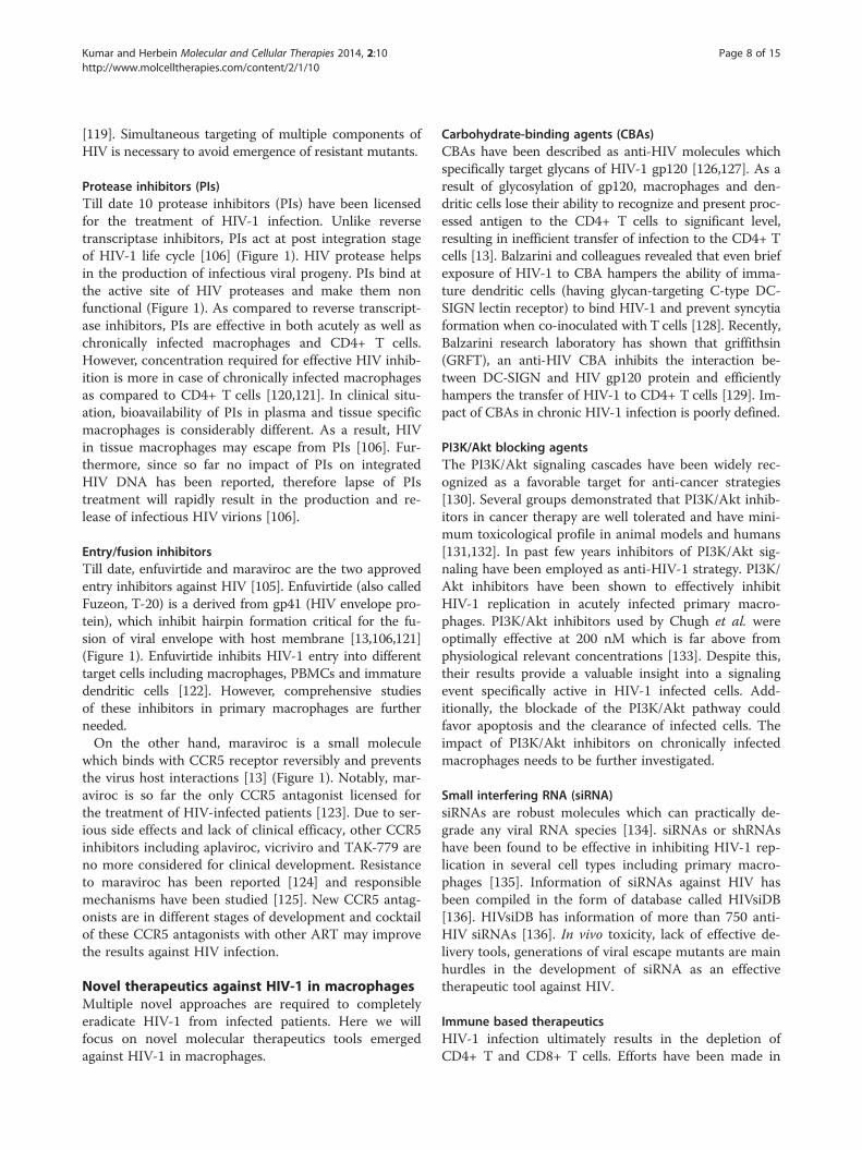
ReviewHIV-1 replication in the macrophageHIV-1 entry into macrophagesFirst step of HIV-1 entry into target host cells involvesvirus ligand (virus surface glycoprotein gp120) and itsinteraction with CD4 receptor which is present in both Tcells as well as in macrophages [14,15] (Figure 1). Secondstep involves the fusion of viral envelope with host cellmembrane which is governed by the engagement of theco-receptors (CCR5 or CXCR4) (Figure 1). Earlier it was
Central Ltd. This is an Open Access article distributed under the terms of the/creativecommons.org/licenses/by/2.0), which permits unrestricted use,, provided the original work is properly credited. The Creative Commons Publicmons.org/publicdomain/zero/1.0/) applies to the data made available in this
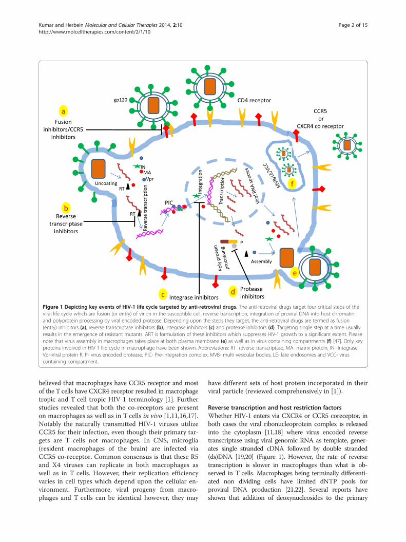
Figure 1 Depicting key events of HIV-1 life cycle targeted by anti-retroviral drugs. The anti-retroviral drugs target four critical steps of theviral life cycle which are fusion (or entry) of virion in the susceptible cell, reverse transcription, integration of proviral DNA into host chromatinand polyprotein processing by viral encoded protease. Depending upon the steps they target, the anti-retroviral drugs are termed as fusion(entry) inhibitors (a), reverse transcriptase inhibitors (b), integrase inhibitors (c) and protease inhibitors (d). Targeting single step at a time usuallyresults in the emergence of resistant mutants. ART is formulation of these inhibitors which suppresses HIV-1 growth to a significant extent. Pleasenote that virus assembly in macrophages takes place at both plasma membrane (e) as well as in virus containing compartments (f) [47]. Only keyproteins involved in HIV-1 life cycle in macrophage have been shown. Abbreviations: RT- reverse transcriptase, MA- matrix protein, IN- Integrase,Vpr-Viral protein R, P- virus encoded protease, PIC- Pre-integration complex, MVB- multi vesicular bodies, LE- late endosomes and VCC- viruscontaining compartment.
Kumar and Herbein Molecular and Cellular Therapies 2014, 2:10 Page 2 of 15http://www.molcelltherapies.com/content/2/1/10
believed that macrophages have CCR5 receptor and mostof the T cells have CXCR4 receptor resulted in macrophagetropic and T cell tropic HIV-1 terminology [1]. Furtherstudies revealed that both the co-receptors are presenton macrophages as well as in T cells in vivo [1,11,16,17].Notably the naturally transmitted HIV-1 viruses utilizeCCR5 for their infection, even though their primary tar-gets are T cells not macrophages. In CNS, microglia(resident macrophages of the brain) are infected viaCCR5 co-receptor. Common consensus is that these R5and X4 viruses can replicate in both macrophages aswell as in T cells. However, their replication efficiencyvaries in cell types which depend upon the cellular en-vironment. Furthermore, viral progeny from macro-phages and T cells can be identical however, they may
have different sets of host protein incorporated in theirviral particle (reviewed comprehensively in [1]).
Reverse transcription and host restriction factorsWhether HIV-1 enters via CXCR4 or CCR5 coreceptor, inboth cases the viral ribonucleoprotein complex is releasedinto the cytoplasm [11,18] where virus encoded reversetranscriptase using viral genomic RNA as template, gener-ates single stranded cDNA followed by double stranded(ds)DNA [19,20] (Figure 1). However, the rate of reversetranscription is slower in macrophages than what is ob-served in T cells. Macrophages being terminally differenti-ated non dividing cells have limited dNTP pools forproviral DNA production [21,22]. Several reports haveshown that addition of deoxynucleosides to the primary
Kumar and Herbein Molecular and Cellular Therapies 2014, 2:10 Page 3 of 15http://www.molcelltherapies.com/content/2/1/10
human macrophage culture remarkably enhances therate of HIV-1 reverse transcription proving that dNTPpool is an important rate limiting factor in macro-phages [21,23,24].Additionally, macrophages possess certain inhibitory
factors which interfere with viral life cycle and are termedas host restriction factors [25,26]. These host restrictionfactors include tetherin, APOBEC3G and recently identi-fied sterile alpha motif (SAM) domain and HD domain-containing protein 1 (SAMHD1) [25-27]. APOBEC3G isknown to trigger G-to-A hypermutation in nascent DNA.Tetherin (also called CD317/BST-2) hinders the release ofviral progeny from infected cells [26]. HIV-1 employsseveral strategies to overcome these restriction factors(reviewed in [28,29]). HIV-1 accessory protein Vif andVpu counteracts the APOBEC3G and tetherin respectively[4,25,26]. Even there are reports describing tetherin antag-onism by HIV-1 Nef protein [30,31].SAMHD1 is a macrophage specific host restriction
factor which has triphosphohydrolase activity resultingin hydrolysis of dNTPs into nucleosides and triphos-phates. Thus SAMHD1 reduces the dNTPs pool in mac-rophages to a certain level resulting in the inefficientreverse transcription of HIV-1 genomic RNA intoproviral DNA [32]. However, Vpx protein of HIV-2 in-duces proteasome-dependent degradation of SAMHD1through CRL4DCAF1 E3 ubiquitin ligase [27]. RecentlyMcKnight research group, in order to search for hostrestriction factors, screened several human genes andidentified 114 genes with significant impact on HIV-1replication. Furthermore, their studies revealed that in-hibition of all members of PAF1 family resulted in in-crease in HIV-1 replication. Notably PAF1 is notrestricted to macrophages only, they are also expressedin primary monocytes and T-lymphocytes, suggestingexhaustive list of restriction factors against HIV-1 [33].Recently Allouch and colleagues showed that cyclin-dependent kinase inhibitor p21 inhibits HIV-1 replica-tion in monocyte-derived macrophages (MDMs) byinterfering with reverse transcription of the viral genomeby a mechanism independent of SAMHD1. Additionally,they demonstrated that p21 curtails the dNTP synthesisthrough the down regulation of the expression of RNR2(a subunit of ribonucleotide reductase) necessary for thebiosynthesis of dNTPs [34].
Nuclear transportNewly synthesized HIV dsDNA is imported to the nucleusas pre-integration complex (PIC) (Figure 1). Unlike T cells,in macrophages PIC transport to the nucleus is indepen-dent of cell division. PIC comprises of viral proteins whichincludes reverse transcriptase, Vpr, integrase (IN), matrix(MA, p17) and capsid protein (CA) in addition to newlysynthesized dsDNA. However, CA dissociates from PIC
prior to the nuclear entry. Vpr, IN and MA direct thetransport PIC through nuclear pore mediated by importinα/β [35,36] (Figure 1). However, precise function of theseproteins in PIC nuclear transport is still a matter of debate[11]. Unlike IN and MA, Vpr lacks nuclear localizationsignal [37,38]. In addition, interaction between importinα and Vpr is critical not only for the nuclear transportof PIC but also for the replication of HIV-1 in macro-phages [39]. Furthermore, in primary macrophages, hostcell protein emerin (an integral nuclear inner membraneprotein) plays an indispensible role in integration ofviral DNA into the chromatin [40,41]. Primary macro-phages lacking emerin have poor rate of HIV proviralDNA integration into the host chromatin however,lack of emerin does not inhibit PIC entry into the nu-cleus [40]. In addition, binding partners of emerin, theLEM (LAP2 (lamina-associated polypeptide 2)/emerin/MAN1) is necessary for the interaction of viral cDNAwith emerin and capability of emerin to support HIV-1infection in macrophages [40]. However, Shun and col-leagues demonstrated that HIV-1 can efficiently infectdividing cells despite of the absence of emerin, sugges-ting the role of emerin in HIV-1 infection restricted toonly macrophages [42]. Besides several other host fac-tors are involved in the HIV life cycle in macrophageshave been reviewed recently [43].
HIV-1 transcriptionHIV-1 transcription is governed by binding of viral pro-teins and host factors to the long terminal repeat (LTR)of the virus, which functions as viral promoter [44]. Hostfactors include nuclear factor kappa B (NF-κB) family,AP-1 (activator protein 1), Sp family, C/EBP (CCAATenhancer binding protein and NFAT (nuclear factor ofactivated T cells). These host factors have specific bind-ing sites present on LTR. On the other hand, viral pro-teins Tat and Vpr also bind to the LTR to govern HIV-1transcription [20,44]. Worth mentioning, host factorscould be cell type specific, for example C/EBP proteinsand their binding sites are critical for HIV-1 replicationin macrophages but not in CD4+ T cells [45]. In addition,primary macrophages infected with HIV-1 having mutationin C/EBP binding sites does not support HIV-1 replication.On the other hand, primary CD4+ T cells, Jurkat and H9cells support the replication of HIV-1C/EBP mutants [45].
HIV-1 assembly in macrophagesIn case of primary CD4+ T cells, HIV-1 assembly takesplace at the plasma membrane [46]. On the other hand,the corresponding site in macrophages is not yet fullycharacterized [47]. Initial studies demonstrated the pre-sence of HIV-1 virion particles in multivesicular bodies(MVBs) or late endosomes (LEs) like structures [47,48](Figure 1). Even immuno-electron microscopy studies

Kumar and Herbein Molecular and Cellular Therapies 2014, 2:10 Page 4 of 15http://www.molcelltherapies.com/content/2/1/10
supported latter finding as their studies revealed the pres-ence of MVB specific markers (for example CD53, CD9,tetraspanins, CD81 and MHC II) in those structures[47,49-51]. In addition, HIV-1 progeny released from in-fected macrophages also possess these markers, furtherstrengthening the view that macrophages are releasedfrom LEs or MVBs [47,50,52]. However, several studies re-vealed that structures harboring HIV-1 in infected macro-phages have some distinct characters which are notcharacteristics of LEs or MVBs. These unique characteris-tics include tubular connection to the extracellular spaceand neutral pH [53]. The term ‘virus containing compart-ments’ (VCCs) has been assigned to the structures whichact as the site for the virus assembly in macrophages [47](Figure 1). Interestingly, these VCCs are also present inuninfected macrophages however, they become moreprominent upon HIV-1 infection [51,53]. Worth mention-ing, VCCs have limited access to the innate and adaptiveimmune effector molecules [47]. In contrast, several stu-dies are in the favor of budding of HIV-1 progeny fromplasma membrane in infected macrophages [54]. Takentogether, these contrasting studies indicate that there is afair possibility that HIV-1 may bud from plasma mem-brane as well as from VCCs (Figure 1). VCCs may act as asafe house for HIV-1 in macrophages leading to HIV-1reservoirs. However, elegant experiments are furtherrequired to support this hypothesis.
Interplay between HIV proteins and cell signalingin macrophagesAmong HIV-1 proteins, the viral proteins Tat, Vpr andNef interfere with signaling pathways in macrophages.
TatThe trans-activator of transcription (Tat) protein is a86–101aa virus encoded pleiotropic protein which di-rectly or indirectly modulates several steps of HIV lifecycle including replication, transcription and progeny re-lease by regulating both cellular as well as viral gene ex-pression [20,55-57]. In addition, Tat has been detected insera of HIV infected patients as well in cell culture set-tings indicating its role as a modulator of cellular func-tion in infected cells and also to target bystander cells[20,58]. Furthermore, monocytes, macrophages andmicroglia are activated by Tat protein [20]. In addition,Tat is known to trigger the expression of HIV corecep-tors (CXCR4, CCR5 and CCR3) in macrophages in adose-dependent manner which might positively influ-ence HIV-1 infection [59]. Furthermore, Tat acts as a po-tent chemoattractant for monocytes, macrophages anddendritic cells [60,61]. Tat induces the production andrelease of tumor necrosis factor alpha (TNF-α) frommacrophages [62]. Further, Tat mediated TNF-α induc-tion was NF-kappa B (NF-κB) dependent and mediated
through activation of signaling cascades including PLC(phospholipase C), protein kinase A and protein tyrosinekinase [20]. In addition, Tat enhances the endogenouslevels of Ca2+ in macrophages which may subsequently leadto the production of chemokines and pro-inflammatorycytokines [63]. Latter events may be responsible forHIV-1 induced neuropathogenesis and inflammation [64].
Viral protein R (Vpr)Vpr is a virion-associated protein dispensable for viralreplication in T cells however is indispensible for viralreplication in macrophages [65]. Vpr has been localizedin cytoplasm as well as in nucleus of the infected cells[66]. Vpr is a multifunctional protein which regulatesviral replication, cellular events like NF-κB-mediatedtranscription, apoptosis and cytokine production [20,67].Effect of recombinant Vpr (rVpr) has been demonstratedin macrophages. Although high concentration of rVprresulted in significant cytotoxicity in macrophages how-ever, at lower concentration rVpr has been shown to in-crease the biological activity of several transcriptionfactors including NF-κB, c-Jun and AP-1 in promonocy-tic cells and primary macrophages [68]. In addition rVprstimulates HIV-1 replication in acutely infected primarymacrophages. Furthermore, infection of macrophageswith Vpr-deficient viral mutants resulted in decreasedproduction of p24 which can be corrected by addition ofrVpr [69]. Moreover, Vpr independently enhances theexpression of cyclin-dependent kinase inhibitor 1A(CDKN1A/p21) in macrophages whereas Vpr mutantsexhibit lack of upregulation of p21 and display reducedviral replication [70]. Taken together, data strongly sug-gest that Vpr enhances the viral replication in acutelyand latently infected macrophages.
NefNef is expressed during early life cycle of HIV-1. Nef is a27 kDa myristoylated protein required for efficient viralreplication in infected cells [71,72]. In addition, Nef en-hances the survival of infected cells which helps in theexpansion of infectious viral population. Furthermore,Nef hampers the immune system of infected patients byseveral mechanisms including down-regulating the ex-pression of MHC I, MHC II, CD28, CD4 [73,74] and byactivating PI3K [75]. Nef down-regulates the expressionof CD4 receptor in macrophages which serves two pur-poses. Firstly, CD4 down-regulation in infected cells maypromote the release of viral progeny by avoiding seques-tration of viral envelope by CD4 [76]. Secondly, it helpsin avoiding superinfection which otherwise could lead topremature cell death [71,76].In monocyte derived macrophages (MDMs) exogen-
ously added recombinant Nef (rNef) regulates the ex-pression of several genes in a short time span (2 hours).

Kumar and Herbein Molecular and Cellular Therapies 2014, 2:10 Page 5 of 15http://www.molcelltherapies.com/content/2/1/10
These findings indicate a robust transcriptional pro-gramming governed by Nef protein leading to the pro-duction and secretion of soluble factors which in turnactivates STAT1 and STAT3 in primary monocytes/mac-rophages [20,77]. Similarly, addition of rNef to theMDMs cultures resulted in the rapid induction of tran-scription factors NF-κB, AP-1, and c-Jun N-terminalkinase and enhanced HIV-1 transcription. Furthermore,in vitro treatment of macrophages with rNef has beenreported to trigger IKK/NF-κB, MAPK and IRF-3 signal-ing cascades. Additionally, Nef induces robust phosphor-ylation of MAPKs, including ERK1/2, JNK, and p38
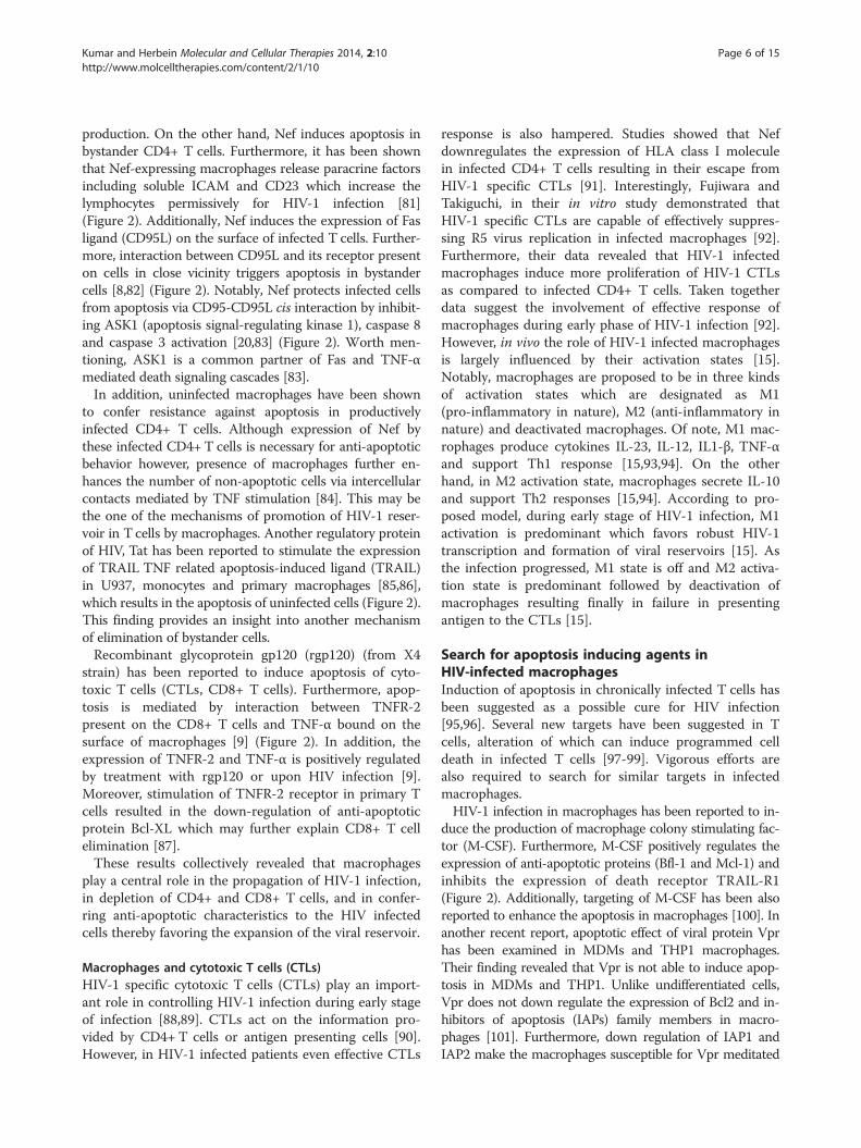
Figure 2 Relationship between macrophages and T lymphocytes in HHIV pathogenesis. Nef stimulates the release of soluble factors ICAM and Cinfection, thereby favoring the expansion of the viral reservoir (a). In additioHIV-infected cells. Interaction of CD95L and its receptor (Fas) present on uninfected CD4+ T cells, Nef inhibits the expression of proteins involved in apCD4+ T cells from cell death and further expands the viral reservoir. HIV regfrom the infected macrophages. TRAIL binds with its receptor (DR5) presengp120 interaction with CXCR4 receptor increases the expression of TNF-α oThis interaction results in the down regulation of the anti-apoptotic proteinin macrophages is known to induce macrophage colony stimulating factorand upregulates the expression of anti-apoptotic proteins (f), favoring theM-CSF has been suggested to increase apoptosis in infected macrophages
[20,78]. Notably, the role of Nef in HIV-HCV coinfectedmacrophages has been recently described [79].
Contribution of macrophages to HIV-1pathogenesisHIV-1 pathogenesis is characterized by progressive celldepletion involved in adaptive immunity including CD4+T and CD8+ T cells [8,9]. Not only HIV-infected CD4+T cells are lysed but uninfected CD4+ T cells more pro-minently undergo apoptosis [80] (Figure 2). Nef plays dualrole in HIV-1 pathogenesis. On one hand, Nef protectsHIV-infected cells from cell death to favor efficient viral
IV-1 infection. Macrophages harboring HIV-1 play an important role inD23 which makes uninfected CD4+ T cells more susceptible to HIVn, Nef induces the expression of Fas ligand (FasL, CD95L) oninfected CD4+ T cells results in apoptosis (b). On the other hand inoptosis including ASK1, caspase 8 and caspase 3 (c), protects infectedulatory protein Tat stimulates the production and release of TRAILt on uninfected CD4+ T cells and induces apoptosis (d). Furthermore,n macrophages which interacts with TNFR2 present on CD8+ T cells.Bcl-XL and ultimately leads to apoptosis (e). Moreover, HIV infection(M-CSF) which inhibits the expression of TRAILR1 on macrophagesresistance to apoptosis of infected macrophages. Therefore, targeting.
Kumar and Herbein Molecular and Cellular Therapies 2014, 2:10 Page 6 of 15http://www.molcelltherapies.com/content/2/1/10
production. On the other hand, Nef induces apoptosis inbystander CD4+ T cells. Furthermore, it has been shownthat Nef-expressing macrophages release paracrine factorsincluding soluble ICAM and CD23 which increase thelymphocytes permissively for HIV-1 infection [81](Figure 2). Additionally, Nef induces the expression of Fasligand (CD95L) on the surface of infected T cells. Further-more, interaction between CD95L and its receptor presenton cells in close vicinity triggers apoptosis in bystandercells [8,82] (Figure 2). Notably, Nef protects infected cellsfrom apoptosis via CD95-CD95L cis interaction by inhibit-ing ASK1 (apoptosis signal-regulating kinase 1), caspase 8and caspase 3 activation [20,83] (Figure 2). Worth men-tioning, ASK1 is a common partner of Fas and TNF-αmediated death signaling cascades [83].In addition, uninfected macrophages have been shown
to confer resistance against apoptosis in productivelyinfected CD4+ T cells. Although expression of Nef bythese infected CD4+T cells is necessary for anti-apoptoticbehavior however, presence of macrophages further en-hances the number of non-apoptotic cells via intercellularcontacts mediated by TNF stimulation [84]. This may bethe one of the mechanisms of promotion of HIV-1 reser-voir in T cells by macrophages. Another regulatory proteinof HIV, Tat has been reported to stimulate the expressionof TRAIL TNF related apoptosis-induced ligand (TRAIL)in U937, monocytes and primary macrophages [85,86],which results in the apoptosis of uninfected cells (Figure 2).This finding provides an insight into another mechanismof elimination of bystander cells.Recombinant glycoprotein gp120 (rgp120) (from X4
strain) has been reported to induce apoptosis of cyto-toxic T cells (CTLs, CD8+ T cells). Furthermore, apop-tosis is mediated by interaction between TNFR-2present on the CD8+ T cells and TNF-α bound on thesurface of macrophages [9] (Figure 2). In addition, theexpression of TNFR-2 and TNF-α is positively regulatedby treatment with rgp120 or upon HIV infection [9].Moreover, stimulation of TNFR-2 receptor in primary Tcells resulted in the down-regulation of anti-apoptoticprotein Bcl-XL which may further explain CD8+ T cellelimination [87].These results collectively revealed that macrophages
play a central role in the propagation of HIV-1 infection,in depletion of CD4+ and CD8+ T cells, and in confer-ring anti-apoptotic characteristics to the HIV infectedcells thereby favoring the expansion of the viral reservoir.
Macrophages and cytotoxic T cells (CTLs)HIV-1 specific cytotoxic T cells (CTLs) play an import-ant role in controlling HIV-1 infection during early stageof infection [88,89]. CTLs act on the information pro-vided by CD4+ T cells or antigen presenting cells [90].However, in HIV-1 infected patients even effective CTLs
response is also hampered. Studies showed that Nefdownregulates the expression of HLA class I moleculein infected CD4+ T cells resulting in their escape fromHIV-1 specific CTLs [91]. Interestingly, Fujiwara andTakiguchi, in their in vitro study demonstrated thatHIV-1 specific CTLs are capable of effectively suppres-sing R5 virus replication in infected macrophages [92].Furthermore, their data revealed that HIV-1 infectedmacrophages induce more proliferation of HIV-1 CTLsas compared to infected CD4+ T cells. Taken togetherdata suggest the involvement of effective response ofmacrophages during early phase of HIV-1 infection [92].However, in vivo the role of HIV-1 infected macrophagesis largely influenced by their activation states [15].Notably, macrophages are proposed to be in three kindsof activation states which are designated as M1(pro-inflammatory in nature), M2 (anti-inflammatory innature) and deactivated macrophages. Of note, M1 mac-rophages produce cytokines IL-23, IL-12, IL1-β, TNF-αand support Th1 response [15,93,94]. On the otherhand, in M2 activation state, macrophages secrete IL-10and support Th2 responses [15,94]. According to pro-posed model, during early stage of HIV-1 infection, M1activation is predominant which favors robust HIV-1transcription and formation of viral reservoirs [15]. Asthe infection progressed, M1 state is off and M2 activa-tion state is predominant followed by deactivation ofmacrophages resulting finally in failure in presentingantigen to the CTLs [15].
Search for apoptosis inducing agents inHIV-infected macrophagesInduction of apoptosis in chronically infected T cells hasbeen suggested as a possible cure for HIV infection[95,96]. Several new targets have been suggested in Tcells, alteration of which can induce programmed celldeath in infected T cells [97-99]. Vigorous efforts arealso required to search for similar targets in infectedmacrophages.HIV-1 infection in macrophages has been reported to in-
duce the production of macrophage colony stimulating fac-tor (M-CSF). Furthermore, M-CSF positively regulates theexpression of anti-apoptotic proteins (Bfl-1 and Mcl-1) andinhibits the expression of death receptor TRAIL-R1(Figure 2). Additionally, targeting of M-CSF has been alsoreported to enhance the apoptosis in macrophages [100]. Inanother recent report, apoptotic effect of viral protein Vprhas been examined in MDMs and THP1 macrophages.Their finding revealed that Vpr is not able to induce apop-tosis in MDMs and THP1. Unlike undifferentiated cells,Vpr does not down regulate the expression of Bcl2 and in-hibitors of apoptosis (IAPs) family members in macro-phages [101]. Furthermore, down regulation of IAP1 andIAP2 make the macrophages susceptible for Vpr meditated

Kumar and Herbein Molecular and Cellular Therapies 2014, 2:10 Page 7 of 15http://www.molcelltherapies.com/content/2/1/10
apoptosis. Altering IAP activity has been suggested as apossible way to induce apoptosis in infected macro-phages [101].
Conventional therapies against HIV-1 inmacrophagesCurrently, combinatorial antiretroviral therapy (ART) iswidely used in suppressing HIV-1 infection to a signifi-cant level [102,103]. ART has made a remarkable contri-bution in improving and enhancing life span of infectedpatients [104]. HIV-1 growth kinetics is different in mac-rophages and T cells suggesting varied impact of anti-retroviral drugs against HIV-1 in these target cells. Herewe will briefly describe the potential contribution ofART in HIV-infected macrophages.
Reverse transcriptase inhibitors (RTIs)More than 25 compounds have been licensed for treat-ing HIV in infected patients [105]. Out of them nearlyfifty percent are reverse transcriptase inhibitors (RTIs)[105]. RTIs are of two types which are nucleoside reversetranscriptase inhibitors (NRTIs) and non nucleoside re-verse transcriptase inhibitors (NNRTIs) [13].
Nucleoside reverse transcriptase inhibitors (NRTIs)NRTIs target reverse transcriptase enzyme which is respon-sible for conversion of HIV genomic RNA into cDNA, animportant step in the life cycle of HIV (Figure 1). NRTIs in-clude emtricitabine, tenofovir, abacavir, lamivudine, stavu-dine, zalcitabine, didanosine and didovudine [105].NRTIs mimic and compete with natural nucleotides
pool for incorporation into growing chain of nascentHIV DNA. Notably, NRTIs require intracellular phos-phorylation for conversion into functional inhibitors ofHIV. Since most of NRTIs lacks 3′ OH moiety, thereforetheir incorporation into nascent HIV DNA leads to ter-mination of DNA chain formation. Efficacy of theseNRTIs majorly depends upon the levels of dNTPs pools[13,24]. As discussed earlier, macrophages being termin-ally differentiated non dividing cells have limited poolsof dNTPs as compared to actively dividing cells [13,106].Therefore, theoretically in this scenario, NRTIs will faceless competition with natural dNTPs in macrophages.That may be the one of the reasons for better efficacy ofNRTIs in macrophages as compared to CD4+ T cells[21,107,108]. In fact NRTIs have shown promising re-sults in reducing the neuropathological consequences ofHIV encephalitis in the CNS and onset of HIV-associated dementia (HAD) [108-110]. Notably, in CNS,macrophages represent the major HIV infected popula-tion [101]. In addition, NRTIs treatments in macro-phages result in fewer emergences of resistant HIVmutants as compared to lymphocytes [111].
Strikingly, NRTIs efficacy is remarkably different inacutely and chronically infected macrophages. Exactmechanism responsible for such observation is poorlyunderstood. Since chronically infected cells possess inte-grated HIV DNA into host chromatin, HIV RNA pro-duced via integrated DNA using transcription by hostRNA polymerase is therefore not susceptible to NRTIs.Besides this, there must be several other mechanisms re-sponsible for the difference in the efficacy of NRTIs be-tween chronically and acutely infected macrophages[13,108]. Notably, NRTIs are associated with several un-desirable effects including their interference with cellcycle and mitochondrial environment and also induceapoptosis [112,113].
Non nucleoside reverse transcriptase inhibitors (NNRTIs)Licensed NNRTIs include rilpivirine, etravirine, delavir-dine, efavirenz and nevirapine. Unlike NRTIs, NNRTIs donot require phosphorylation nor compete with naturaldNTPs pools for their action. NNRTIs act by binding tothe hydrophobic pocket near the reverse transcriptase ac-tive site resulting in the inhibition of polymerization reac-tion [13,106]. Since NNRTIs efficacy does not dependupon the cellular dNTPs pools, therefore their impact onacutely infected macrophages and CD4+ T cells is notsignificantly different. Furthermore, macrophage colonystimulating factor which positively regulates the dNTPspool, have no effect on the NNRTIs efficacy against HIV[106]. Notably, NNRTIs have less adverse effects as com-pared to NRTIs. However, Badley research group hasstudied the side effects of NNRTI in Jurkat T cells andPBMCs. They observed the induction of caspase andmitochondrial dependent apoptosis by NNRTIs [114].Like NRTIs, NNRTIs anti-HIV activities remarkably dif-
fer between acutely infected and chronically infected mac-rophages. To be more precise, EC50 of NNRTIs againstacutely infected macrophages varies from 10 to 50 nM.On the other hand, their effect is negligible against chro-nically infected macrophages [13,108]. Reasons for theseobservations are incompletely understood.
Integrase inhibitorsChronic HIV infection is mostly characterized by integra-tion of proviral DNA into the host chromatin (Figure 1).This process called strand transfer is governed by HIVencoded enzyme called integrase and is indispensible forthe establishment of latency [115,116] (Figure 1). Till datethree integrase inhibitors (raltegravir, elvitegravirs anddolutegravir) have been approved for clinical use. Efficacyof integrase inhibitors has been studied in MDMs andlymphocytes and showed similar results [117]. Notably,even single point mutation in integrase confers resistanceagainst the integrase inhibitor raltegravir [118]. However,other integrase inhibitors are still effective in that situation
Kumar and Herbein Molecular and Cellular Therapies 2014, 2:10 Page 8 of 15http://www.molcelltherapies.com/content/2/1/10
[119]. Simultaneous targeting of multiple components ofHIV is necessary to avoid emergence of resistant mutants.
Protease inhibitors (PIs)Till date 10 protease inhibitors (PIs) have been licensedfor the treatment of HIV-1 infection. Unlike reversetranscriptase inhibitors, PIs act at post integration stageof HIV-1 life cycle [106] (Figure 1). HIV protease helpsin the production of infectious viral progeny. PIs bind atthe active site of HIV proteases and make them nonfunctional (Figure 1). As compared to reverse transcript-ase inhibitors, PIs are effective in both acutely as well aschronically infected macrophages and CD4+ T cells.However, concentration required for effective HIV inhib-ition is more in case of chronically infected macrophagesas compared to CD4+ T cells [120,121]. In clinical situ-ation, bioavailability of PIs in plasma and tissue specificmacrophages is considerably different. As a result, HIVin tissue macrophages may escape from PIs [106]. Fur-thermore, since so far no impact of PIs on integratedHIV DNA has been reported, therefore lapse of PIstreatment will rapidly result in the production and re-lease of infectious HIV virions [106].
Entry/fusion inhibitorsTill date, enfuvirtide and maraviroc are the two approvedentry inhibitors against HIV [105]. Enfuvirtide (also calledFuzeon, T-20) is a derived from gp41 (HIV envelope pro-tein), which inhibit hairpin formation critical for the fu-sion of viral envelope with host membrane [13,106,121](Figure 1). Enfuvirtide inhibits HIV-1 entry into differenttarget cells including macrophages, PBMCs and immaturedendritic cells [122]. However, comprehensive studiesof these inhibitors in primary macrophages are furtherneeded.On the other hand, maraviroc is a small molecule
which binds with CCR5 receptor reversibly and preventsthe virus host interactions [13] (Figure 1). Notably, mar-aviroc is so far the only CCR5 antagonist licensed forthe treatment of HIV-infected patients [123]. Due to ser-ious side effects and lack of clinical efficacy, other CCR5inhibitors including aplaviroc, vicriviro and TAK-779 areno more considered for clinical development. Resistanceto maraviroc has been reported [124] and responsiblemechanisms have been studied [125]. New CCR5 antag-onists are in different stages of development and cocktailof these CCR5 antagonists with other ART may improvethe results against HIV infection.
Novel therapeutics against HIV-1 in macrophagesMultiple novel approaches are required to completelyeradicate HIV-1 from infected patients. Here we willfocus on novel molecular therapeutics tools emergedagainst HIV-1 in macrophages.
Carbohydrate-binding agents (CBAs)CBAs have been described as anti-HIV molecules whichspecifically target glycans of HIV-1 gp120 [126,127]. As aresult of glycosylation of gp120, macrophages and den-dritic cells lose their ability to recognize and present proc-essed antigen to the CD4+ T cells to significant level,resulting in inefficient transfer of infection to the CD4+ Tcells [13]. Balzarini and colleagues revealed that even briefexposure of HIV-1 to CBA hampers the ability of imma-ture dendritic cells (having glycan-targeting C-type DC-SIGN lectin receptor) to bind HIV-1 and prevent syncytiaformation when co-inoculated with T cells [128]. Recently,Balzarini research laboratory has shown that griffithsin(GRFT), an anti-HIV CBA inhibits the interaction be-tween DC-SIGN and HIV gp120 protein and efficientlyhampers the transfer of HIV-1 to CD4+ T cells [129]. Im-pact of CBAs in chronic HIV-1 infection is poorly defined.
PI3K/Akt blocking agentsThe PI3K/Akt signaling cascades have been widely rec-ognized as a favorable target for anti-cancer strategies[130]. Several groups demonstrated that PI3K/Akt inhib-itors in cancer therapy are well tolerated and have mini-mum toxicological profile in animal models and humans[131,132]. In past few years inhibitors of PI3K/Akt sig-naling have been employed as anti-HIV-1 strategy. PI3K/Akt inhibitors have been shown to effectively inhibitHIV-1 replication in acutely infected primary macro-phages. PI3K/Akt inhibitors used by Chugh et al. wereoptimally effective at 200 nM which is far above fromphysiological relevant concentrations [133]. Despite this,their results provide a valuable insight into a signalingevent specifically active in HIV-1 infected cells. Add-itionally, the blockade of the PI3K/Akt pathway couldfavor apoptosis and the clearance of infected cells. Theimpact of PI3K/Akt inhibitors on chronically infectedmacrophages needs to be further investigated.
Small interfering RNA (siRNA)siRNAs are robust molecules which can practically de-grade any viral RNA species [134]. siRNAs or shRNAshave been found to be effective in inhibiting HIV-1 rep-lication in several cell types including primary macro-phages [135]. Information of siRNAs against HIV hasbeen compiled in the form of database called HIVsiDB[136]. HIVsiDB has information of more than 750 anti-HIV siRNAs [136]. In vivo toxicity, lack of effective de-livery tools, generations of viral escape mutants are mainhurdles in the development of siRNA as an effectivetherapeutic tool against HIV.
Immune based therapeuticsHIV-1 infection ultimately results in the depletion ofCD4+ T and CD8+ T cells. Efforts have been made in
Kumar and Herbein Molecular and Cellular Therapies 2014, 2:10 Page 9 of 15http://www.molcelltherapies.com/content/2/1/10
the direction of boosting immunity against HIV-1 [137].For example, in various studies the application of IL-2,IL7, IL-12 and growth hormone have been reported toresult in increase in CD4+ T counts in HIV-1 infectedindividuals [138-141]. Interestingly, IL-2 along with ARTsignificantly reduces HIV-1 replication in infected pa-tients as compared to ART only treated patients. How-ever, upon treatment cessation virus bounce backindicating the inability of IL-2 to enhance immunity forthe longer period of time [96,138]. In addition, role ofIL-15 has been suggested in improving functionality ofanti-HIV CTLs and natural killer (NK) cells in vitro[142]. Moreover, IL-15 enhances simian immunodefi-ciency virus (SIV) specific CD8+ T cells, NK cells anddecreases the number of SIV infected cells in lymphnode in infected rhesus macaque [143]. Surprisingly,viral load was found to be increased more than two foldupon IL-15 treatment [143]. Notably, IL-21 treatment inSIV infected macaques resulted in increase in granzymesB and perforins in NK cells and CD8+ T cells [96,144].Benefits of such transient immunity evoke by interleu-kins and impact of continuous use of such immunebased therapeutics on the health of HIV-1 infected indi-viduals need to be carefully addressed.
IL-27, an anti-HIV cytokineIL-27 is a cytokine belonging to the IL-12 cytokine fam-ily and plays important roles in innate and adaptive im-munity [145]. IL-27 is produced by epithelial cells,dendritic cells and macrophages [146]. Several researchgroups have documented the anti-HIV properties of IL-27in MDMs, CD4+ T cells, immature and mature dendriticcells [147]. Mechanistic details of anti-HIV cytokine IL-27have been recently revealed. IL-27 down regulates theexpression of SPTBN1 (spectrin β nonerythrocyte 1), oneof the host factor required for HIV-1 infection in macro-phages [148]. Furthermore, IL-27 down-regulates theexpression of SPTBN1 via TAK-1-mediated MAPK signal-ing cascade [148]. Importantly, their results indicate thatSPTBN1 is a critical host component which can be targetedto inhibit HIV-1 replication in one of the principal HIV-1reservoirs, the macrophages.
Macrophage targeted carriersEffective therapeutic agent must be complimented witheffective delivery tools for the successful delivery of re-sults. Nanotechnology has made it possible to deliverthe therapeutic agents to specific cell types or anatom-ical location which otherwise are not accessible by con-ventional delivery methods [149]. It is assumed thatanti-HIV drugs delivered via nano-carrier can be select-ively accumulate in infected cell types while uninfectedcells will have much lower concentration of drugs there-fore, will have less side effects [150]. Wan and colleagues
have developed nano-carrier based system for drug de-livery in macrophages using formyl methionine-leucine-phenylalanine (fMLF) peptide-PEG derivatives [151].fMLF are employed because fMLF receptors are specific-ally present on phagocytic cells including macrophagesand fMLF binds to the receptors present on macro-phages with high affinity [151,152]. Bio-distribution offMLF-PEG nano-carrier was studied in vivo, revealed thegreater accumulation of fMLF-PEG into macrophages ofkidneys, spleen and liver as compared to only PEG[152]. Results are encouraging and suggest the feasibilityof specifically targeting HIV-1 reservoir in macrophages.
Myeloid cells of central nervous system (CNS) andHIV-1ART has significantly reduced morbidity and mortalityburden associated with HIV-1. However, despite of thatsignificant number of the patients receiving ART de-velops HIV-1 associated CNS disorders [153,154]. Not-ably, Zink and colleagues demonstrated that ART is ableto reduce the viral load in cerebrospinal fluid of ma-caques infected with simian immunodeficiency virus(SIV). However, they observed the presence of SIV DNAin CNS [155]. In CNS, major reservoirs of HIV-1 are thecells of myeloid origin which include meningeal macro-phages, microglia and perivascular cells. Therefore, theinterplay between these cells and HIV-1 is of utmost im-portance. Recently role of HIV-1 Tat protein has beenshown in disrupting synaptical architecture in vitro aswell in vivo [156-158]. Lu and colleagues have furtherdemonstrated the involvement of CNS resident myeloidcells in deteriorating the synaptical architecture in re-sponse to Tat [157].In addition, recently role of cathepsin B secreted by
HIV-1 infected macrophages in neural apoptosis hasbeen also described [159]. Notably, low level of cathep-sin B has been detected in the post-mortem brain tissueof HIV-1 individual with HAD but not in normal indi-vidual or HIV-1 infected individual with normal cogni-tion. Their results suggest the involvement of cathepsinB in HAD [159]. Altogether above findings provide avaluable insight into the mechanism of HIV-1 associatedCNS disorder which involves myeloid cells, their secre-tome and viral proteins. These novel findings will helpin generating new targets for managing HAD.
HIV-1 latency in macrophages and reactivation:the “flushing out” therapyAlthough highly active retroviral therapy (ART) has sig-nificantly reduced viral levels (50 copies/ml) in infectedpatients however, interruption of ART results in rapidincrease in viremia. HIV infection leads to the rapid de-pletion of CD4+ T and CD8+ T cells. Despite there iscertain percent of cells where virus integrate with host
Kumar and Herbein Molecular and Cellular Therapies 2014, 2:10 Page 10 of 15http://www.molcelltherapies.com/content/2/1/10
chromatin. These cells do not produce virus in restingcondition, however produce it upon activation [160,161].These cells represent a pool of latent infection and are amain obstacle in complete eradication of HIV-1 from in-fected patients [96,116,162]. Besides resting CD4+ T cells,it is suggested that monocytes, macrophages, dendriticcells and hematopoietic stem cells can be latently infectedwith HIV [163-165]. There are experimental evidences inthe support of latency in monocytes [163,166].Role of macrophages in dissemination of virus and
expanding viral reservoir especially in T lymphocytes hasbeen discussed elsewhere in this review. Prolonged lifespan and resistance to HIV cytopathic effects make mac-rophages as unique viral reservoirs. However, associationbetween HIV-1 latency and macrophages is less clear. HIVinfected patients on ART treatment are reported to haveonly few macrophages infected in lymph nodes howeverundergoes reactivation in case of opportunistic infections[167]. Interestingly, FDA approved amphotericin B (an an-tifungal drug) has been reported to reactivate HIV-1 inTHP89GFP cells (a model cell line for the HIV-1 latencyin macrophages) but not in T lymphocytes [168]. How-ever, when amphotericin B induced THP89GFP cells areco-cultured with J89GFP (latently infected T cells), theyactivate latent HIV in latter cells [168]. In addition, re-cently role of polybacterial challenge in activating latentHIV-1 in the cells of monocyte/macrophage lineage hasbeen shown in vitro [169,170]. These findings indicate thatmacrophages may be a site of HIV-1 latent infection. Un-like CD4+ T cells, pre-integration latency in macrophagesmay contribute to the viral reservoir formation to a sig-nificant extent [171]. Mechanism/s responsible for postintegration latency in macrophages is poorly understood.However, presence of host transcriptional repressors,anti-HIV microRNA and lack of functional Tat could playsignificant role in establishing post-integration latency ininfected macrophage [171]. For example host factorC/EBPb is known to repressor HIV-1 transcription inmacrophages which may contribute to HIV latency. Inaddition, in human microglial cells, CTIP2 (a highlyexpressed transcriptional repressor in brain) is knownto inhibit the HIV-1 replication mediated by recruit-ment of chromatin modifying complex involvingHDAC1, HDAC2 and methylase SUV39H1 [172]. Roleof CTIP2 has been suggested in post integration latencyin microglia cells [165,172].Current efforts have been made in the direction of re-
activation of HIV from latent reservoir followed by theircomplete removal by ART [96]. According to this hy-pothesis, cells in which latency is reactivated should dieeither due to viral cytopathic effect or due to recognitionby cytotoxic T cells [96,115]. Furthermore, the fresh in-fection by viral progeny (released from lysed cells) willbe inhibited by ART.
Several kinds of new approaches have been employed inreactivating HIV including the use of histone deacetylaseinhibitors (HDACi) such as valproic acid (VPA), trichosta-tin (TSA), suberoylanilide hydroxyamic acid (SAHA) andsodium butyrate, methylation inhibitors including BIX-01294, 5-aza-2′deoxycytidine (Aza-CdR) and chaetocin,NFκB activators for example TNF-α and bryostatin andprotein kinase C modulators and immune modulators in-cluding IL-7 and IL-15 [96,105,116,173]. These new com-pounds have shown significant results in reactivatinglatency in CD4+ T cells and are at different stages of de-velopment. For example first successful clinical trial hasbeen reported with HDACi, valproic acid (VPA)[165,174]. However, these findings are not confirmed inother trials [175,176].Regarding efficacy of these novel compounds in reacti-
vating latency in macrophages, not many reports areavailable. However, several HDACi have been tested inACH2 and U1 cell lines and found to be equally effectivein both cell lines [177]. Recently, Matalon and colleaguetested ITF2357 (givinostat) and VPA in ACH2 and U1cell line. Their data revealed that ITF2357 is more po-tent in activating latency as compared to VPA [178].Notably, givinostat has been found to be safe in healthyindividuals in phase I trial [179]. Altogether data fromin vitro studies suggest that agents used in reactivatinglatency in T cells have similar effects in cells of mono-cyte/macrophage lineage. However, in clinical trials viralload has been mainly determined in T lymphocytes. Im-portantly, isolation of monocytes followed by productionof monocyte derived macrophages is rather a lengthyprocess as compared to isolation of T lymphocytes. Inaddition, brain resident macrophages represent the ana-tomical sanctuaries where drug penetration is poor anddetermination of drug efficacy in these sanctuaries is ra-ther a difficult task [180,181]. Furthermore, the presenceof efflux pumps and array of metabolic enzymes in bloodbrain barrier further put the efficacy of drugs in a diffi-cult proposition. CNS resident macrophages play an im-portant role in HAD, a severe morbidity of HIV-1 infection.Treating HIV-1 needs holistic view where besides T lym-phocytes cells of monocyte/macrophage lineage must betaken into consideration. Ignoring one or other viral reser-voir will not result in any favorable outcome.
ConclusionMacrophages are among the early targets of HIV-1. Theyalso act as chronic and latent viral reservoirs. AlthoughART has suppressed viremia in most of infected pa-tients, complete eradication is not possible withoutclearance of HIV-1 from latent reservoirs. Novel thera-peutics options have emerged against these reservoirs.However, delivery of therapeutic molecules in vivo is stilla major challenge. In the future, combinatorial therapies
Kumar and Herbein Molecular and Cellular Therapies 2014, 2:10 Page 11 of 15http://www.molcelltherapies.com/content/2/1/10
equipped with precise delivery tools can fulfill the scien-tific dream of the complete eradication of HIV-1 frominfected patients.
Competing interestsGH is a member of the editorial board of Molecular and Cellular Therapies.No other competing interests are declared.
Authors’ contributionsAK and GH wrote the manuscript. Both authors read and approved thismanuscript.
AcknowledgementsThis work was supported by grants from the University of Franche-Comté(UFC) and the Région Franche-Comté (RECH-FON12-000013) to G.H. A.K. is arecipient of a postdoctoral fellowship of the Region Franche-Comté.
Received: 27 October 2013 Accepted: 27 January 2014Published: 2 April 2014
References1. Iordanskiy S, Santos S, Bukrinsky M: Nature, nurture and HIV: the effect of
producer cell on viral physiology. Virology 2013, 443:208–213.2. Abbas W, Herbein G: T-cell signaling in HIV-1 infection. Open Virol J 2013,
7:57–71.3. Ackerman AL, Cresswell P: Cellular mechanisms governing
cross-presentation of exogenous antigens. Nat Immunol 2004, 5:678–684.4. Koppensteiner H, Brack-Werner R, Schindler M: Macrophages and their
relevance in Human Immunodeficiency Virus Type I infection.Retrovirology 2012, 9:82.
5. Crowe SM, Mills J, Kirihara J, Boothman J, Marshall JA, McGrath MS:Full-length recombinant CD4 and recombinant gp120 inhibit fusionbetween HIV infected macrophages and uninfected CD4-expressingT-lymphoblastoid cells. AIDS Res Hum Retroviruses 1990, 6:1031–1037.
6. Crowe SM, Mills J, Elbeik T, Lifson JD, Kosek J, Marshall JA, Engleman EG,McGrath MS: Human immunodeficiency virus-infected monocyte-derivedmacrophages express surface gp120 and fuse with CD4 lymphoid cellsin vitro: a possible mechanism of T lymphocyte depletion in vivo. ClinImmunol Immunopathol 1992, 65:143–151.
7. Groot F, Welsch S, Sattentau QJ: Efficient HIV-1 transmission frommacrophages to T cells across transient virological synapses. Blood 2008,111:4660–4663.
8. Badley AD, Dockrell D, Simpson M, Schut R, Lynch DH, Leibson P, Paya CV:Macrophage-dependent apoptosis of CD4+ T lymphocytes fromHIV-infected individuals is mediated by FasL and tumor necrosis factor.J Exp Med 1997, 185:55–64.
9. Herbein G, Mahlknecht U, Batliwalla F, Gregersen P, Pappas T, Butler J,O'Brien WA, Verdin E: Apoptosis of CD8+ T cells is mediated bymacrophages through interaction of HIV gp120 with chemokinereceptor CXCR4. Nature 1998, 395:189–194.
10. Gendelman HE, Orenstein JM, Martin MA, Ferrua C, Mitra R, Phipps T,Wahl LA, Lane HC, Fauci AS, Burke DS, Meltzer MS: Efficient isolation andpropagation of human immunodeficiency virus on recombinant colony-stimulating factor 1-treated monocytes. J Exp Med 1988, 167:1428–1441.
11. Carter CA, Ehrlich LS: Cell biology of HIV-1 infection of macrophages.Annu Rev Microbiol 2008, 62:425–443.
12. Kelly J, Beddall MH, Yu D, Iyer SR, Marsh JW, Wu Y: Human macrophagessupport persistent transcription from unintegrated HIV-1 DNA. Virology2008, 372:300–312.
13. Gavegnano C, Schinazi RF: Antiretroviral therapy in macrophages:implication for HIV eradication. Antivir Chem Chemother 2009, 20:63–78.
14. Kwong PD, Wyatt R, Robinson J, Sweet RW, Sodroski J, Hendrickson WA:Structure of an HIV gp120 envelope glycoprotein in complex with theCD4 receptor and a neutralizing human antibody. Nature 1998,393:648–659.
15. Herbein G, Varin A: The macrophage in HIV-1 infection: from activation todeactivation? Retrovirology 2010, 7:33.
16. Zaitseva M, Blauvelt A, Lee S, Lapham CK, Klaus-Kovtun V, Mostowski H,Manischewitz J, Golding H: Expression and function of CCR5 and CXCR4on human Langerhans cells and macrophages: implications for HIVprimary infection. Nat Med 1997, 3:1369–1375.
17. Yi Y, Rana S, Turner JD, Gaddis N, Collman RG: CXCR-4 is expressed byprimary macrophages and supports CCR5-independent infection bydual-tropic but not T-tropic isolates of human immunodeficiency virustype 1. J Virol 1998, 72:772–777.
18. Abbas W, Herbein G: Plasma membrane signaling in HIV-1 infection.Biochim Biophys Acta 2013. doi:10.1016/j.bbamem.2013.06.020.
19. Mougel M, Houzet L, Darlix JL: When is it time for reverse transcription tostart and go? Retrovirology 2009, 6:24.
20. Herbein G, Gras G, Khan KA, Abbas W: Macrophage signaling in HIV-1infection. Retrovirology 2010, 7:34.
21. Gavegnano C, Kennedy EM, Kim B, Schinazi RF: The impact of macrophagenucleotide pools on HIV-1 reverse transcription, viral replication, and thedevelopment of novel antiviral agents. Mol Biol Int 2012, 2012:625983.
22. Schmidtmayerova H, Alfano M, Nuovo G, Bukrinsky M: Humanimmunodeficiency virus type 1 T-lymphotropic strains entermacrophages via a CD4- and CXCR4-mediated pathway: replication isrestricted at a postentry level. J Virol 1998, 72:4633–4642.
23. Furge LL, Guengerich FP: Analysis of nucleotide insertion and extensionat 8-oxo-7,8-dihydroguanine by replicative T7 polymerase exo- andhuman immunodeficiency virus-1 reverse transcriptase usingsteady-state and pre-steady-state kinetics. Biochemistry 1997,36:6475–6487.
24. Diamond TL, Roshal M, Jamburuthugoda VK, Reynolds HM, Merriam AR,Lee KY, Balakrishnan M, Bambara RA, Planelles V, Dewhurst S, Kim B:Macrophage tropism of HIV-1 depends on efficient cellular dNTPutilization by reverse transcriptase. J Biol Chem 2004, 279:51545–51553.
25. Sheehy AM, Gaddis NC, Choi JD, Malim MH: Isolation of a human genethat inhibits HIV-1 infection and is suppressed by the viral Vif protein.Nature 2002, 418:646–650.
26. Neil SJ, Zang T, Bieniasz PD: Tetherin inhibits retrovirus release and isantagonized by HIV-1 Vpu. Nature 2008, 451:425–430.
27. Hrecka K, Hao C, Gierszewska M, Swanson SK, Kesik-Brodacka M, Srivastava S,Florens L, Washburn MP, Skowronski J: Vpx relieves inhibition of HIV-1infection of macrophages mediated by the SAMHD1 protein. Nature2011, 474:658–661.
28. Vicenzi E, Poli G: Novel factors interfering with human immunodeficiencyvirus-type 1 replication in vivo and in vitro. Tissue Antigens 2013, 81:61–71.
29. Goila-Gaur R, Strebel K: HIV-1 Vif, APOBEC, and intrinsic immunity.Retrovirology 2008, 5:51.
30. Sauter D, Kirchhoff F: Tetherin antagonism by primate lentiviral nefproteins. Curr HIV Res 2011, 9:514–523.
31. Zhang F, Wilson SJ, Landford WC, Virgen B, Gregory D, Johnson MC, Munch J,Kirchhoff F, Bieniasz PD, Hatziioannou T: Nef proteins from simian immunodeficiencyviruses are tetherin antagonists. Cell Host Microbe 2009, 6:54–67.
32. Lahouassa H, Daddacha W, Hofmann H, Ayinde D, Logue EC, Dragin L,Bloch N, Maudet C, Bertrand M, Gramberg T, Pancino G, Priet S, Canard B,Laguette N, Benkirane M, Transy C, Landau NR, Kim B, Margottin-Goguet F:SAMHD1 restricts the replication of human immunodeficiency virus type1 by depleting the intracellular pool of deoxynucleoside triphosphates.Nat Immunol 2012, 13:223–228.
33. Liu L, Oliveira NM, Cheney KM, Pade C, Dreja H, Bergin AM, Borgdorff V,Beach DH, Bishop CL, Dittmar MT, McKnight A: A whole genome screenfor HIV restriction factors. Retrovirology 2011, 8:94.
34. Allouch A, David A, Amie SM, Lahouassa H, Chartier L, Margottin-Goguet F,Barré-Sinoussi F, Kim B, Sáez-Cirión A, Pancino G: p21-mediated RNR2repression restricts HIV-1 replication in macrophages by inhibiting dNTPbiosynthesis pathway. Proc Natl Acad Sci U S A 2013, 110:E3997–E4006.
35. Bukrinsky MI, Sharova N, Dempsey MP, Stanwick TL, Bukrinskaya AG,Haggerty S, Stevenson M: Active nuclear import of humanimmunodeficiency virus type 1 preintegration complexes. Proc Natl AcadSci U S A 1992, 89:6580–6584.
36. Bukrinsky MI, Sharova N, McDonald TL, Pushkarskaya T, Tarpley WG, StevensonM: Association of integrase, matrix, and reverse transcriptase antigens ofhuman immunodeficiency virus type 1 with viral nucleic acids followingacute infection. Proc Natl Acad Sci U S A 1993, 90:6125–6129.
37. Gallay P, Stitt V, Mundy C, Oettinger M, Trono D: Role of the karyopherinpathway in human immunodeficiency virus type 1 nuclear import.J Virol 1996, 70:1027–1032.
38. Gallay P, Hope T, Chin D, Trono D: HIV-1 infection of nondividing cellsthrough the recognition of integrase by the importin/karyopherinpathway. Proc Natl Acad Sci U S A 1997, 94:9825–9830.
Kumar and Herbein Molecular and Cellular Therapies 2014, 2:10 Page 12 of 15http://www.molcelltherapies.com/content/2/1/10
39. Nitahara-Kasahara Y, Kamata M, Yamamoto T, Zhang X, Miyamoto Y, MunetaK, Iijima S, Yoneda Y, Tsunetsugu-Yokota Y, Aida Y: Novel nuclear import ofVpr promoted by importin alpha is crucial for human immunodeficiencyvirus type 1 replication in macrophages. J Virol 2007, 81:5284–5293.
40. Jacque JM, Stevenson M: The inner-nuclear-envelope protein emerinregulates HIV-1 infectivity. Nature 2006, 441:641–645.
41. Kobiler O, Drayman N, Butin-Israeli V, Oppenheim A: Virus strategies forpassing the nuclear envelope barrier. Nucleus 2012, 3:526–539.
42. Shun MC, Daigle JE, Vandegraaff N, Engelman A: Wild-type levels ofhuman immunodeficiency virus type 1 infectivity in the absence ofcellular emerin protein. J Virol 2007, 81:166–172.
43. Cobos-Jimenez V, Booiman T, Hamann J, Kootstra NA: Macrophages andHIV-1. Curr Opin HIV AIDS 2011, 6:385–390.
44. Kilareski EM, Shah S, Nonnemacher MR, Wigdahl B: Regulation of HIV-1transcription in cells of the monocyte-macrophage lineage.Retrovirology 2009, 6:118.
45. Henderson AJ, Calame KL: CCAAT/enhancer binding protein (C/EBP) sites arerequired for HIV-1 replication in primary macrophages but not CD4(+)T cells. Proc Natl Acad Sci U S A 1997, 94:8714–8719.
46. Frank I, Stoiber H, Godar S, Stockinger H, Steindl F, Katinger HW, Dierich MP:Acquisition of host cell-surface-derived molecules by HIV-1. AIDS 1996,10:1611–1620.
47. Tan J, Sattentau QJ: The HIV-1-containing macrophage compartment: aperfect cellular niche? Trends Microbiol 2013, 21:405–412.
48. Orenstein JM, Meltzer MS, Phipps T, Gendelman HE: Cytoplasmic assemblyand accumulation of human immunodeficiency virus types 1 and 2 inrecombinant human colony-stimulating factor-1-treated humanmonocytes: an ultrastructural study. J Virol 1988, 62:2578–2586.
49. Raposo G, Moore M, Innes D, Leijendekker R, Leigh-Brown A, Benaroch P,Geuze H: Human macrophages accumulate HIV-1 particles in MHC IIcompartments. Traffic 2002, 3:718–729.
50. Pelchen-Matthews A, Kramer B, Marsh M: Infectious HIV-1 assembles inlate endosomes in primary macrophages. J Cell Biol 2003, 162:443–455.
51. Deneka M, Pelchen-Matthews A, Byland R, Ruiz-Mateos E, Marsh M: Inmacrophages, HIV-1 assembles into an intracellular plasma membranedomain containing the tetraspanins CD81, CD9, and CD53. J Cell Biol2007, 177:329–341.
52. Kramer B, Pelchen-Matthews A, Deneka M, Garcia E, Piguet V, Marsh M: HIVinteraction with endosomes in macrophages and dendritic cells.Blood Cells Mol Dis 2005, 35:136–142.
53. Welsch S, Groot F, Krausslich HG, Keppler OT, Sattentau QJ: Architectureand regulation of the HIV-1 assembly and holding compartment inmacrophages. J Virol 2011, 85:7922–7927.
54. Welsch S, Keppler OT, Habermann A, Allespach I, Krijnse-Locker J, KrausslichHG: HIV-1 buds predominantly at the plasma membrane of primaryhuman macrophages. PLoS Pathog 2007, 3:e36.
55. Buonaguro L, Barillari G, Chang HK, Bohan CA, Kao V, Morgan R, Gallo RC,Ensoli B: Effects of the human immunodeficiency virus type 1 Tat proteinon the expression of inflammatory cytokines. J Virol 1992, 66:7159–7167.
56. Ott M, Emiliani S, Van Lint C, Herbein G, Lovett J, Chirmule N, McCloskey T,Pahwa S, Verdin E: Immune hyperactivation of HIV-1-infected T cellsmediated by Tat and the CD28 pathway. Science 1997, 275:1481–1485.
57. Jeang KT, Xiao H, Rich EA: Multifaceted activities of the HIV-1transactivator of transcription, Tat. J Biol Chem 1999, 274:28837–28840.
58. Ensoli B, Buonaguro L, Barillari G, Fiorelli V, Gendelman R, Morgan RA,Wingfield P, Gallo RC: Release, uptake, and effects of extracellular humanimmunodeficiency virus type 1 Tat protein on cell growth and viraltransactivation. J Virol 1993, 67:277–287.
59. Huang L, Bosch I, Hofmann W, Sodroski J, Pardee AB: Tat protein induceshuman immunodeficiency virus type 1 (HIV-1) coreceptors andpromotes infection with both macrophage-tropic and T-lymphotropicHIV-1 strains. J Virol 1998, 72:8952–8960.
60. Albini A, Benelli R, Giunciuglio D, Cai T, Mariani G, Ferrini S, Noonan DM:Identification of a novel domain of HIV tat involved in monocytechemotaxis. J Biol Chem 1998, 273:15895–15900.
61. Campbell GR, Loret EP: What does the structure-function relationship ofthe HIV-1 Tat protein teach us about developing an AIDS vaccine?Retrovirology 2009, 6:50.
62. Chen P, Mayne M, Power C, Nath A: The Tat protein of HIV-1 inducestumor necrosis factor-alpha production. Implications for HIV-1-associatedneurological diseases. J Biol Chem 1997, 272:22385–22388.
63. Mayne M, Holden CP, Nath A, Geiger JD: Release of calcium from inositol1,4,5-trisphosphate receptor-regulated stores by HIV-1 Tat regulatesTNF-alpha production in human macrophages. J Immunol 2000,164:6538–6542.
64. Nath A: Human immunodeficiency virus (HIV) proteins in neuropathogenesisof HIV dementia. J Infect Dis 2002, 186(Suppl 2):S193–S198.
65. Subbramanian RA, Kessous-Elbaz A, Lodge R, Forget J, Yao XJ, Bergeron D,Cohen EA: Human immunodeficiency virus type 1 Vpr is a positive regulatorof viral transcription and infectivity in primary human macrophages. J ExpMed 1998, 187:1103–1111.
66. Jacquot G, Le RE, David A, Mazzolini J, Bouchet J, Bouaziz S, Niedergang F,Pancino G, Benichou S: Localization of HIV-1 Vpr to the nuclear envelope:impact on Vpr functions and virus replication in macrophages.Retrovirology 2007, 4:84.
67. Bukrinsky M, Adzhubei A: Viral protein R of HIV-1. Rev Med Virol 1999, 9:39–49.68. Varin A, Decrion AZ, Sabbah E, Quivy V, Sire J, Van Lint C, Roques BP,
Aggarwal BB, Herbein G: Synthetic Vpr protein activates activatorprotein-1, c-Jun N-terminal kinase, and NF-kappaB and stimulates HIV-1transcription in promonocytic cells and primary macrophages. J BiolChem 2005, 280:42557–42567.
69. Eckstein DA, Sherman MP, Penn ML, Chin PS, De Noronha CM, Greene WC,Goldsmith MA: HIV-1 Vpr enhances viral burden by facilitating infectionof tissue macrophages but not nondividing CD4+ T cells. J Exp Med 2001,194:1407–1419.
70. Vazquez N, Greenwell-Wild T, Marinos NJ, Swaim WD, Nares S, Ott DE, Schubert U,Henklein P, Orenstein JM, Sporn MB, Wahl SM: Human immunodeficiencyvirus type 1-induced macrophage gene expression includes the p21 gene,a target for viral regulation. J Virol 2005, 79:4479–4491.
71. Das SR, Jameel S: Biology of the HIV Nef protein. Indian J Med Res 2005,121:315–332.
72. Lamers SL, Fogel GB, Singer EJ, Salemi M, Nolan DJ, Huysentruyt LC,McGrath MS: HIV-1 Nef in macrophage-mediated disease pathogenesis.Int Rev Immunol 2012, 31:432–450.
73. Lama J, Mangasarian A, Trono D: Cell-surface expression of CD4 reducesHIV-1 infectivity by blocking Env incorporation in a Nef- andVpu-inhibitable manner. Curr Biol 1999, 9:622–631.
74. Foster JL, Garcia JV: HIV-1 Nef: at the crossroads. Retrovirology 2008, 5:84.75. Tachado SD, Li X, Swan K, Patel N, Koziel H: Constitutive activation of
phosphatidylinositol 3-kinase signaling pathway down-regulatesTLR4-mediated tumor necrosis factor-alpha release in alveolarmacrophages from asymptomatic HIV-positive persons in vitro.J Biol Chem 2008, 283:33191–33198.
76. Lama J: The physiological relevance of CD4 receptor down-modulationduring HIV infection. Curr HIV Res 2003, 1:167–184.
77. Mangino G, Percario ZA, Fiorucci G, Vaccari G, Acconcia F, Chiarabelli C,Leone S, Noto A, Horenkamp FA, Manrique S, Romeo G, Polticelli F, GeyerM, Affabris E: HIV-1 Nef induces proinflammatory state in macrophagesthrough its acidic cluster domain: involvement of TNF alpha receptorassociated factor 2. PLoS One 2011, 6:e22982.
78. Mangino G, Percario ZA, Fiorucci G, Vaccari G, Manrique S, Romeo G,Federico M, Geyer M, Affabris E: In vitro treatment of human monocytes/macrophages with myristoylated recombinant Nef of humanimmunodeficiency virus type 1 leads to the activation of mitogen-activated protein kinases, IkappaB kinases, and interferon regulatoryfactor 3 and to the release of beta interferon. J Virol 2007,81:2777–2791.
79. Khan KA, Abbas W, Varin A, Kumar A, Di Martino V, Dichamp I, Herbein G:HIV-1 Nef interacts with HCV core, recruits TRAF2, TRAF5 and TRAF6, andstimulates HIV-1 replication in macrophages. J Innate Immun 2013,5:639–656.
80. Finkel TH, Tudor-Williams G, Banda NK, Cotton MF, Curiel T, Monks C, BabaTW, Ruprecht RM, Kupfer A: Apoptosis occurs predominantly in bystandercells and not in productively infected cells of HIV- and SIV-infectedlymph nodes. Nat Med 1995, 1:129–134.
81. Swingler S, Brichacek B, Jacque JM, Ulich C, Zhou J, Stevenson M: HIV-1 Nefintersects the macrophage CD40L signalling pathway to promoteresting-cell infection. Nature 2003, 424:213–219.
82. Oyaizu N, Adachi Y, Hashimoto F, McCloskey TW, Hosaka N, Kayagaki N,Yagita H, Pahwa S: Monocytes express Fas ligand upon CD4 cross-linkingand induce CD4+ T cells apoptosis: a possible mechanism of bystandercell death in HIV infection. J Immunol 1997, 158:2456–2463.
Kumar and Herbein Molecular and Cellular Therapies 2014, 2:10 Page 13 of 15http://www.molcelltherapies.com/content/2/1/10
83. Geleziunas R, Xu W, Takeda K, Ichijo H, Greene WC: HIV-1 Nefinhibits ASK1-dependent death signalling providing a potentialmechanism for protecting the infected host cell. Nature 2001,410:834–838.
84. Mahlknecht U, Deng C, Lu MC, Greenough TC, Sullivan JL, O’Brien WA,Herbein G: Resistance to apoptosis in HIV-infected CD4+ T lymphocytesis mediated by macrophages: role for Nef and immune activation in viralpersistence. J Immunol 2000, 165:6437–6446.
85. Zhang M, Li X, Pang X, Ding L, Wood O, Clouse K, Hewlett I, Dayton AI:Identification of a potential HIV-induced source of bystander-mediatedapoptosis in T cells: upregulation of trail in primary human macrophagesby HIV-1 tat. J Biomed Sci 2001, 8:290–296.
86. Yang Y, Tikhonov I, Ruckwardt TJ, Djavani M, Zapata JC, Pauza CD, SalvatoMS: Monocytes treated with human immunodeficiency virus Tat killuninfected CD4(+) cells by a tumor necrosis factor-related apoptosis-induced ligand-mediated mechanism. J Virol 2003, 77:6700–6708.
87. Lin RH, Hwang YW, Yang BC, Lin CS: TNF receptor-2-triggered apoptosis isassociated with the down-regulation of Bcl-xL on activated T cells andcan be prevented by CD28 costimulation. J Immunol 1997, 158:598–603.
88. Koup RA, Safrit JT, Cao Y, Andrews CA, McLeod G, Borkowsky W, Farthing C,Ho DD: Temporal association of cellular immune responses with theinitial control of viremia in primary human immunodeficiency virus type1 syndrome. J Virol 1994, 68:4650–4655.
89. Ogg GS, Jin X, Bonhoeffer S, Dunbar PR, Nowak MA, Monard S, Segal JP,Cao Y, Rowland-Jones SL, Cerundolo V, Hurley A, Markowitz M, Ho DD,Nixon DF, McMichael AJ: Quantitation of HIV-1-specific cytotoxic T lymphocytes and plasma load of viral RNA. Science 1998, 279:2103–2106.
90. Pozzi LA, Maciaszek JW, Rock KL: Both dendritic cells and macrophagescan stimulate naive CD8 T cells in vivo to proliferate, develop effectorfunction, and differentiate into memory cells. J Immunol 2005,175:2071–2081.
91. Collins KL, Chen BK, Kalams SA, Walker BD, Baltimore D: HIV-1 Nefprotein protects infected primary cells against killing by cytotoxicT lymphocytes. Nature 1998, 391:397–401.
92. Fujiwara M, Takiguchi M: HIV-1-specific CTLs effectively suppressreplication of HIV-1 in HIV-1-infected macrophages. Blood 2007,109:4832–4838.
93. Mills CD, Kincaid K, Alt JM, Heilman MJ, Hill AM: M-1/M-2 macrophagesand the Th1/Th2 paradigm. J Immunol 2000, 164:6166–6173.
94. Cassol E, Cassetta L, Rizzi C, Alfano M, Poli G: M1 and M2a polarization ofhuman monocyte-derived macrophages inhibits HIV-1 replication bydistinct mechanisms. J Immunol 2009, 182:6237–6246.
95. Badley AD, Pilon AA, Landay A, Lynch DH: Mechanisms of HIV-associatedlymphocyte apoptosis. Blood 2000, 96:2951–2964.
96. Badley AD, Sainski A, Wightman F, Lewin SR: Altering cell death pathwaysas an approach to cure HIV infection. Cell Death Dis 2013, 4:e718.
97. Schnepple DJ, Shepard B, Bren GD, Cummins NW, Natesampillai S, TrushinS, Algeciras-Schimnich A, Meng XW, Sainski AM, Rizza SA, Kaufmann SH,Badley AD: Isolation of a TRAIL antagonist from the serum ofHIV-infected patients. J Biol Chem 2011, 286:35742–35754.
98. Sainski AM, Natesampillai S, Cummins NW, Bren GD, Taylor J, Saenz DT,Poeschla EM, Badley AD: The HIV-1-specific protein Casp8p41 inducesdeath of infected cells through Bax/Bak. J Virol 2011, 85:7965–7975.
99. Cummins NW, Badley AD: Anti-apoptotic mechanisms of HIV: lessons andnovel approaches to curing HIV. Cell Mol Life Sci 2013, 70:3355–3363.
100. Swingler S, Mann AM, Zhou J, Swingler C, Stevenson M: Apoptotic killingof HIV-1-infected macrophages is subverted by the viral envelopeglycoprotein. PLoS Pathog 2007, 3:1281–1290.
101. Busca A, Saxena M, Kumar A: Critical role for antiapoptotic Bcl-xL andMcl-1 in human macrophage survival and cellular IAP1/2 (cIAP1/2) inresistance to HIV-Vpr-induced apoptosis. J Biol Chem 2012, 287:15118–15133.
102. Pomerantz RJ, Horn DL: Twenty years of therapy for HIV-1 infection. NatMed 2003, 9:867–873.
103. Anderson AM, Lennox JL: Antiretroviral therapy: when to start and whichdrugs to use. Curr Infect Dis Rep 2008, 10:332–339.
104. Palella FJ Jr, Delaney KM, Moorman AC, Loveless MO, Fuhrer J, Satten GA,Aschman DJ, Holmberg SD: Declining morbidity and mortality amongpatients with advanced human immunodeficiency virus infection. HIVOutpatient Study Investigators. N Engl J Med 1998, 338:853–860.
105. Abbas W, Herbein G: Molecular understanding of HIV-1 latency. Adv Virol2012, 2012:574967.
106. Perno CF, Svicher V, Schols D, Pollicita M, Balzarini J, Aquaro S: Therapeuticstrategies towards HIV-1 infection in macrophages. Antiviral Res 2006,71:293–300.
107. Aquaro S, Calio R, Balestra E, Bagnarelli P, Cenci A, Bertoli A, Tavazzi B, DiPierro D, Francesconi M, Abdelahad D, Perno CF: Clinical implications ofHIV dynamics and drug resistance in macrophages. J Biol Regul HomeostAgents 1998, 12:23–27.
108. Aquaro S, Svicher V, Schols D, Pollicita M, Antinori A, Balzarini J, Perno CF:Mechanisms underlying activity of antiretroviral drugs in HIV-1-infectedmacrophages: new therapeutic strategies. J Leukoc Biol 2006, 80:1103–1110.
109. Haworth SJ, Christofalo B, Anderson RD, Dunkle LM: A single-dose study toassess the penetration of stavudine into human cerebrospinal fluid inadults. J Acquir Immune Defic Syndr Hum Retrovirol 1998, 17:235–238.
110. Limoges J, Persidsky Y, Poluektova L, Rasmussen J, Ratanasuwan W,Zelivyanskaya M, McClernon DR, Lanier ER, Gendelman HE: Evaluation ofantiretroviral drug efficacy for HIV-1 encephalitis in SCID mice. Neurology2000, 54:379–389.
111. Aquaro S, Svicher V, Ceccherini-Silberstein F, Cenci A, Marcuccilli F, GiannellaS, Marcon L, Caliò R, Balzarini J, Perno CF: Limited development andprogression of resistance of HIV-1 to the nucleoside analogue reversetranscriptase inhibitor lamivudine in human primary macrophages.J Antimicrob Chemother 2005, 55:872–878.
112. Chariot P, Monnet I, Gherardi R: Cytochrome c oxidase reaction improveshistopathological assessment of zidovudine myopathy. Ann Neurol 1993,34:561–565.
113. Viora M, Di GG, Rivabene R, Malorni W, Fattorossi A: Interference with cellcycle progression and induction of apoptosis by dideoxynucleosideanalogs. Int J Immunopharmacol 1997, 19:311–321.
114. Pilon AA, Lum JJ, Sanchez-Dardon J, Phenix BN, Douglas R, Badley AD:Induction of apoptosis by a nonnucleoside human immunodeficiencyvirus type 1 reverse transcriptase inhibitor. Antimicrob Agents Chemother2002, 46:2687–2691.
115. Siliciano RF, Greene WC: HIV latency. Cold Spring Harb Perspect Med 2011,1:a007096.
116. Van Lint C, Bouchat S, Marcello A: HIV-1 transcription and latency: anupdate. Retrovirology 2013, 10:67.
117. Scopelliti F, Pollicita M, Ceccherini-Silberstein F, Di SF, Surdo M, Aquaro S,Perno CF: Comparative antiviral activity of integrase inhibitors in humanmonocyte-derived macrophages and lymphocytes. Antiviral Res 2011,92:255–261.
118. Marsden MD, Avancena P, Kitchen CM, Hubbard T, Zack JA: Singlemutations in HIV integrase confer high-level resistance to raltegravir inprimary human macrophages. Antimicrob Agents Chemother 2011,55:3696–3702.
119. Canducci F, Ceresola ER, Saita D, Castagna A, Gianotti N, Underwood M,Burioni R, Lazzarin A, Clementi M: In vitro phenotypes to elvitegravir anddolutegravir in primary macrophages and lymphocytes of clonalrecombinant viral variants selected in patients failing raltegravir.J Antimicrob Chemother 2013, 68:2525–2532.
120. Perno CF, Aquaro S, Rosenwirth B, Balestra E, Peichl P, Billich A, Villani N,Caliò R: In vitro activity of inhibitors of late stages of the replication ofHIV in chronically infected macrophages. J Leukoc Biol 1994, 56:381–386.
121. Perno CF, Newcomb FM, Davis DA, Aquaro S, Humphrey RW, Calio R,Yarchoan R: Relative potency of protease inhibitors in monocytes/macrophages acutely and chronically infected with humanimmunodeficiency virus. J Infect Dis 1998, 178:413–422.
122. Yi Y, Loftin L, Wang L, Ratcliffe SJ, Isaacman-Beck J, Collman RG: Entrycoreceptor use and fusion inhibitor T20 sensitivity of dual-tropic R5X4HIV-1 in primary macrophage infection. J Acquir Immune Defic Syndr 2008,47:285–292.
123. Dorr P, Westby M, Dobbs S, Griffin P, Irvine B, Macartney M, Mori J, RickettG, Smith-Burchnell C, Napier C, Webster R, Armour D, Price D, Stammen B,Wood A, Perros M: Maraviroc (UK-427,857), a potent, orally bioavailable,and selective small-molecule inhibitor of chemokine receptor CCR5 withbroad-spectrum anti-human immunodeficiency virus type 1 activity.Antimicrob Agents Chemother 2005, 49:4721–4732.
124. Moore JP, Kuritzkes DR: A piece de resistance: how HIV-1 escapes smallmolecule CCR5 inhibitors. Curr Opin HIV AIDS 2009, 4:118–124.
125. Anastassopoulou CG, Ketas TJ, Klasse PJ, Moore JP: Resistance to CCR5inhibitors caused by sequence changes in the fusion peptide of HIV-1gp41. Proc Natl Acad Sci U S A 2009, 106:5318–5323.
Kumar and Herbein Molecular and Cellular Therapies 2014, 2:10 Page 14 of 15http://www.molcelltherapies.com/content/2/1/10
126. Balzarini J: Carbohydrate-binding agents: a potential future cornerstonefor the chemotherapy of enveloped viruses? Antivir Chem Chemother2007, 18:1–11.
127. Pollicita M, Schols D, Aquaro S, Peumans WJ, Van Damme EJ, Perno CF,Balzarini J: Carbohydrate-binding agents (CBAs) inhibit HIV-1 infection inhuman primary monocyte-derived macrophages (MDMs) and efficientlyprevent MDM-directed viral capture and subsequent transmission toCD4+ T lymphocytes. Virology 2008, 370:382–391.
128. Balzarini J, Van HY, Vermeire K, Vanham G, Schols D: Carbohydrate-bindingagents efficiently prevent dendritic cell-specific intercellular adhesionmolecule-3-grabbing nonintegrin (DC-SIGN)-directed HIV-1 transmissionto T lymphocytes. Mol Pharmacol 2007, 71:3–11.
129. Hoorelbeke B, Xue J, LiWang PJ, Balzarini J: Role of the carbohydrate-binding sites of griffithsin in the prevention of DC-SIGN-mediatedcapture and transmission of HIV-1. PLoS One 2013, 8:e64132.
130. El-Deiry WS: Akt takes centre stage in cell-cycle deregulation. Nat Cell Biol2001, 3:E71–E73.
131. Morgensztern D, McLeod HL: PI3K/Akt/mTOR pathway as a target forcancer therapy. Anticancer Drugs 2005, 16:797–803.
132. Knowling M, Blackstein M, Tozer R, Bramwell V, Dancey J, Dore N, Matthews S,Eisenhauer E: A phase II study of perifosine (D-21226) in patients with previouslyuntreated metastatic or locally advanced soft tissue sarcoma: a National CancerInstitute of Canada Clinical Trials Group trial. Invest New Drugs 2006, 24:435–439.
133. Chugh P, Bradel-Tretheway B, Monteiro-Filho CM, Planelles V, Maggirwar SB,Dewhurst S, Kim B: Akt inhibitors as an HIV-1 infected macrophage-specific anti-viral therapy. Retrovirology 2008, 5:11.
134. Leonard JN, Schaffer DV: Antiviral RNAi therapy: emerging approaches forhitting a moving target. Gene Ther 2006, 13:532–540.
135. Song E, Lee SK, Dykxhoorn DM, Novina C, Zhang D, Crawford K, Cerny J,Sharp PA, Lieberman J, Manjunath N, Shankar P: Sustained small interferingRNA-mediated human immunodeficiency virus type 1 inhibition in primarymacrophages. J Virol 2003, 77:7174–7181.
136. Tyagi A, Ahmed F, Thakur N, Sharma A, Raghava GP, Kumar M: HIVsirDB:a database of HIV inhibiting siRNAs. PLoS One 2011, 6:e25917.
137. Deeks SG, Autran B, Berkhout B, Benkirane M, Cairns S, Chomont N, ChunTW, Churchill M, Di Mascio M, Katlama C, Lafeuillade A, Landay A, LedermanM, Lewin SR, Maldarelli F, Margolis D, Markowitz M, Martinez-Picado J,Mullins JI, Mellors J, Moreno S, O'Doherty U, Palmer S, Penicaud MC, PeterlinM, Poli G, Routy JP, Rouzioux C, Silvestri G, Stevenson M: Towards an HIVcure: a global scientific strategy. Nat Rev Immunol 2012, 12:607–614.
138. Chun TW, Davey RT Jr, Engel D, Lane HC, Fauci AS: Re-emergence of HIVafter stopping therapy. Nature 1999, 401:874–875.
139. Jacobson MA, Hardy D, Connick E, Watson J, DeBruin M: Phase 1 trial of asingle dose of recombinant human interleukin-12 in humanimmunodeficiency virus-infected patients with 100–500 CD4 cells/microL. J Infect Dis 2000, 182:1070–1076.
140. Napolitano LA, Lo JC, Gotway MB, Mulligan K, Barbour JD, Schmidt D, GrantRM, Halvorsen RA, Schambelan M, McCune JM: Increased thymic mass andcirculating naive CD4 T cells in HIV-1-infected adults treated with growthhormone. AIDS 2002, 16:1103–1111.
141. Levy Y, Lacabaratz C, Weiss L, Viard JP, Goujard C, Lelievre JD, Boué F,Molina JM, Rouzioux C, Avettand-Fénoêl V, Croughs T, Beq S, Thiébaut R,Chêne G, Morre M, Delfraissy JF: Enhanced T cell recovery in HIV-1-infected adults through IL-7 treatment. J Clin Invest 2009, 119:997–1007.
142. Seder RA, Grabstein KH, Berzofsky JA, McDyer JF: Cytokine interactions inhuman immunodeficiency virus-infected individuals: roles of interleukin(IL)-2, IL-12, and IL-15. J Exp Med 1995, 182:1067–1077.
143. Mueller YM, Do DH, Altork SR, Artlett CM, Gracely EJ, Katsetos CD, Legido A,Villinger F, Altman JD, Brown CR, Lewis MG, Katsikis PD: IL-15 treatmentduring acute simian immunodeficiency virus (SIV) infection increasesviral set point and accelerates disease progression despite the inductionof stronger SIV-specific CD8+ T cell responses. J Immunol 2008,180:350–360.
144. Pallikkuth S, Rogers K, Villinger F, Dosterii M, Vaccari M, Franchini G, PahwaR, Pahwa S: Interleukin-21 administration to rhesus macaques chronicallyinfected with simian immunodeficiency virus increases cytotoxic effectormolecules in T cells and NK cells and enhances B cell function withoutincreasing immune activation or viral replication. Vaccine 2011,29:9229–9238.
145. Vignali DA, Kuchroo VK: IL-12 family cytokines: immunologicalplaymakers. Nat Immunol 2012, 13:722–728.
146. Hunter CA: New IL-12-family members: IL-23 and IL-27, cytokines withdivergent functions. Nat Rev Immunol 2005, 5:521–531.
147. Chen Q, Swaminathan S, Yang D, Dai L, Sui H, Yang J, Hornung RL, Wang Y,Huang da W, Hu X, Lempicki RA, Imamichi T: Interleukin-27 is a potent inhibitorof cis HIV-1 replication in monocyte-derived dendritic cells via a type Iinterferon-independent pathway. PLoS One 2013, 8:e59194.
148. Dai L, Lidie KB, Chen Q, Adelsberger JW, Zheng X, Huang D, Yang J,Lempicki RA, Rehman T, Dewar RL, Wang Y, Hornung RL, Canizales KA,Lockett SJ, Lane HC, Imamichi T: IL-27 inhibits HIV-1 infection in humanmacrophages by down-regulating host factor SPTBN1 during monocyteto macrophage differentiation. J Exp Med 2013, 210:517–534.
149. Park K: Nanotechnology: what it can do for drug delivery.J Control Release 2007, 120:1–3.
150. Langer R: Drug delivery. Drugs on target. Science 2001, 293:58–59.151. Wan L, Pooyan S, Hu P, Leibowitz MJ, Stein S, Sinko PJ: Peritoneal macrophage
uptake, pharmacokinetics and biodistribution of macrophage-targetedPEG-fMLF (N-formyl-methionyl-leucyl-phenylalanine) nanocarriers forimproving HIV drug delivery. Pharm Res 2007, 24:2110–2119.
152. Vandamme AM, Van VK, De CE: Anti-human immunodeficiency virus drugcombination strategies. Antivir Chem Chemother 1998, 9:187–203.
153. Clifford DB: HIV-associated neurocognitive disease continues in theantiretroviral era. Top HIV Med 2008, 16:94–98.
154. Heaton RK, Clifford DB, Franklin DR Jr, Woods SP, Ake C, Vaida F, Ellis RJ,Letendre SL, Marcotte TD, Atkinson JH, Rivera-Mindt M, Vigil OR, Taylor MJ,Collier AC, Marra CM, Gelman BB, McArthur JC, Morgello S, Simpson DM,McCutchan JA, Abramson I, Gamst A, Fennema-Notestine C, Jernigan TL,Wong J, Grant I; CHARTER Group: HIV-associated neurocognitive disorderspersist in the era of potent antiretroviral therapy: CHARTER Study.Neurology 2010, 75:2087–2096.
155. Zink MC, Brice AK, Kelly KM, Queen SE, Gama L, Li M, Adams RJ, Bartizal C,Varrone J, Rabi SA, Graham DR, Tarwater PM, Mankowski JL, Clements JE:Simian immunodeficiency virus-infected macaques treated with highlyactive antiretroviral therapy have reduced central nervous system viralreplication and inflammation but persistence of viral DNA. J Infect Dis2010, 202:161–170.
156. Kim HJ, Martemyanov KA, Thayer SA: Human immunodeficiency virusprotein Tat induces synapse loss via a reversible process that is distinctfrom cell death. J Neurosci 2008, 28:12604–12613.
157. Lu SM, Tremblay ME, King IL, Qi J, Reynolds HM, Marker DF, Varrone JJ,Majewska AK, Dewhurst S, Gelbard HA: HIV-1 Tat-induced microgliosis andsynaptic damage via interactions between peripheral and centralmyeloid cells. PLoS One 2011, 6:e23915.
158. Bagashev A, Sawaya BE: Roles and functions of HIV-1 Tat protein in theCNS: an overview. Virol J 2013, 10:358.
159. Rodriguez-Franco EJ, Cantres-Rosario YM, Plaud-Valentin M, Romeu R,Rodriguez Y, Skolasky R, Meléndez V, Cadilla CL, Melendez LM: Dysregulationof macrophage-secreted cathepsin B contributes to HIV-1-linked neuronalapoptosis. PLoS One 2012, 7:e36571.
160. Chun TW, Finzi D, Margolick J, Chadwick K, Schwartz D, Siliciano RF:In vivo fate of HIV-1-infected T cells: quantitative analysis of the transitionto stable latency. Nat Med 1995, 1:1284–1290.
161. Chun TW, Carruth L, Finzi D, Shen X, DiGiuseppe JA, Taylor H, HermankovaM, Chadwick K, Margolick J, Quinn TC, Kuo YH, Brookmeyer R, Zeiger MA,Barditch-Crovo P, Siliciano RF: Quantification of latent tissue reservoirsand total body viral load in HIV-1 infection. Nature 1997, 387:183–188.
162. Eisele E, Siliciano RF: Redefining the viral reservoirs that prevent HIV-1eradication. Immunity 2012, 37:377–388.
163. Zhu T, Muthui D, Holte S, Nickle D, Feng F, Brodie S, Hwangbo Y, Mullins JI,Corey L: Evidence for human immunodeficiency virus type 1 replicationin vivo in CD14(+) monocytes and its potential role as a source of virusin patients on highly active antiretroviral therapy. J Virol 2002,76:707–716.
164. Alexaki A, Wigdahl B: HIV-1 infection of bone marrow hematopoieticprogenitor cells and their role in trafficking and viral dissemination.PLoS Pathog 2008, 4:e1000215.
165. Redel L, Le Douce V, Cherrier T, Marban C, Janossy A, Aunis D, Van Lint C, Rohr O,Schwartz C: HIV-1 regulation of latency in the monocyte-macrophage lineageand in CD4+ T lymphocytes. J Leukoc Biol 2010, 87:575–588.
166. McElrath MJ, Steinman RM, Cohn ZA: Latent HIV-1 infection in enrichedpopulations of blood monocytes and T cells from seropositive patients.J Clin Invest 1991, 87:27–30.
Kumar and Herbein Molecular and Cellular Therapies 2014, 2:10 Page 15 of 15http://www.molcelltherapies.com/content/2/1/10
167. Caselli E, Galvan M, Cassai E, Caruso A, Sighinolfi L, Di LD: Humanherpesvirus 8 enhances human immunodeficiency virus replication inacutely infected cells and induces reactivation in latently infected cells.Blood 2005, 106:2790–2797.
168. Jones J, Kosloff BR, Benveniste EN, Shaw GM, Kutsch O: Amphotericin-B-mediated reactivation of latent HIV-1 infection. Virology 2005,331:106–116.
169. Gonzalez OA, Li M, Ebersole JL, Huang CB: HIV-1 reactivation induced by theperiodontal pathogens Fusobacterium nucleatum and Porphyromonasgingivalis involves Toll-like receptor 2 [corrected] and 9 activation inmonocytes/macrophages. Clin Vaccine Immunol 2010, 17:1417–1427.
170. Huang CB, Alimova YV, Strange S, Ebersole JL: Polybacterial challengeenhances HIV reactivation in latently infected macrophages anddendritic cells. Immunology 2011, 132:401–409.
171. Le Douce V, Herbein G, Rohr O, Schwartz C: Molecular mechanisms ofHIV-1 persistence in the monocyte-macrophage lineage. Retrovirology2010, 7:32.
172. Marban C, Redel L, Suzanne S, Van Lint C, Lecestre D, Chasserot-Golaz S, LeidM, Aunis D, Schaeffer E, Rohr O: COUP-TF interacting protein 2 represses theinitial phase of HIV-1 gene transcription in human microglial cells. NucleicAcids Res 2005, 33:2318–2331.
173. Kumar A, Abbas W, Herbein G: TNF and TNF receptor superfamilymembers in HIV infection: new cellular targets for therapy? MediatorsInflamm 2013, 2013:484378.
174. Lehrman G, Hogue IB, Palmer S, Jennings C, Spina CA, Wiegand A, LandayAL, Coombs RW, Richman DD, Mellors JW, Coffin JM, Bosch RJ, Margolis DM:Depletion of latent HIV-1 infection in vivo: a proof-of-concept study.Lancet 2005, 366:549–555.
175. Siliciano JD, Lai J, Callender M, Pitt E, Zhang H, Margolick JB, Gallant JE,Cofrancesco J Jr, Moore RD, Gange SJ, Siliciano RF: Stability of the latentreservoir for HIV-1 in patients receiving valproic acid. J Infect Dis 2007,195:833–836.
176. Sagot-Lerolle N, Lamine A, Chaix ML, Boufassa F, Aboulker JP, Costagliola D,Goujard C, Pallier C, Delfraissy JF, Lambotte O; ANRS EP39 study: Prolongedvalproic acid treatment does not reduce the size of latent HIV reservoir.AIDS 2008, 22:1125–1129.
177. Savarino A, Mai A, Norelli S, El DS, Valente S, Rotili D, Altucci L, Palamara AT,Garaci E: “Shock and kill” effects of class I-selective histone deacetylaseinhibitors in combination with the glutathione synthesis inhibitorbuthionine sulfoximine in cell line models for HIV-1 quiescence.Retrovirology 2009, 6:52.
178. Matalon S, Palmer BE, Nold MF, Furlan A, Kassu A, Fossati G, Mascagni P,Dinarello CA: The histone deacetylase inhibitor ITF2357 decreases surfaceCXCR4 and CCR5 expression on CD4(+) T-cells and monocytes and issuperior to valproic acid for latent HIV-1 expression in vitro. J AcquirImmune Defic Syndr 2010, 54:1–9.
179. Furlan A, Monzani V, Reznikov LL, Leoni F, Fossati G, Modena D, Mascagni P,Dinarello CA: Pharmacokinetics, safety and inducible cytokine responsesduring a phase 1 trial of the oral histone deacetylase inhibitor ITF2357(givinostat). Mol Med 2011, 17:353–362.
180. Dallas S, Miller DS, Bendayan R: Multidrug resistance-associated proteins:expression and function in the central nervous system. Pharmacol Rev2006, 58:140–161.
181. Shikuma CM, Nakamoto B, Shiramizu B, Liang CY, DeGruttola V, Bennett K,Paul R, Kallianpur K, Chow D, Gavegnano C, Hurwitz SJ, Schinazi RF, ValcourVG: Antiretroviral monocyte efficacy score linked to cognitiveimpairment in HIV. Antivir Ther 2012, 17:1233–1242.
doi:10.1186/2052-8426-2-10Cite this article as: Kumar and Herbein: The macrophage: a therapeutictarget in HIV-1 infection. Molecular and Cellular Therapies 2014 2:10.
Submit your next manuscript to BioMed Centraland take full advantage of:
• Convenient online submission
• Thorough peer review
• No space constraints or color figure charges
• Immediate publication on acceptance
• Inclusion in PubMed, CAS, Scopus and Google Scholar
• Research which is freely available for redistribution
Submit your manuscript at www.biomedcentral.com/submit