Rohitukine inhibits in vitro adipogenesis arresting mitotic clonal expansionand improves...
42
1 Rohitukine inhibits in vitro adipogenesis arresting mitotic clonal expansion and improves dyslipidemia in vivo Salil Varshney 1 , Kripa Shankar 1 , Muheeb Beg 1 , Vishal M. Balaramnavar 2 , Sunil Kumar Mishra 2 , Pankaj Jagdale 3 , Shishir Srivastava 2 , Yashpal S. Chhonker 4 , Vijai Lakshmi 5 , Bhushan P. Chaudhari 3 , Rabi Shankar Bhatta 4 , Anil Kumar Saxena 2 and Anil Nilkanth Gaikwad 1* 1 Division of Pharmacology, CSIR-Central Drug Research Institute, Sector 10, Jankipuram Extension, Lucknow- 226031 UP, India, 2 Medicinal and Process Chemistry Division, CSIR-Central Drug Research Institute, Sector 10, Jankipuram Extension, Lucknow-226031 UP India, 3 Regulatory Toxicology Group, CSIR-Indian Institute of Toxicology Research, M.G. Road, Lucknow-226001, 4 Pharmacokinetics Division, CSIR-Central Drug Research Institute, 10/1, Sector 10, Jankipuram Extension, Lucknow-226031, 5 Department of Biochemistry, King George's Medical University, Chowk Area Lucknow-226003. Running Title: Anti-adipogenic and anti-dyslipidemic effects of rohitukine * Corresponding author Anil N. Gaikwad, MS (Pharm) PhD Division of Pharmacology CSIR- Central Drug Research Institute, Sector 10, Jankipuram Extension, Lucknow-226031 UP. India. Phone: 0091-522-2771940 Ext: 4876 Fax: 0091-522-2771941 E-mail: [email protected] by guest, on March 20, 2014 www.jlr.org Downloaded from
-
Upload
independent -
Category
Documents
-
view
4 -
download
0
Transcript of Rohitukine inhibits in vitro adipogenesis arresting mitotic clonal expansionand improves...

1
Rohitukine inhibits in vitro adipogenesis arresting mitotic clonal expansion
and improves dyslipidemia in vivo
Salil Varshney1, Kripa Shankar1, Muheeb Beg1, Vishal M. Balaramnavar2, Sunil Kumar Mishra2, Pankaj Jagdale3, Shishir Srivastava2, Yashpal S. Chhonker4, Vijai Lakshmi5, Bhushan P. Chaudhari3, Rabi Shankar Bhatta4, Anil Kumar Saxena2 and Anil Nilkanth Gaikwad1* 1Division of Pharmacology, CSIR-Central Drug Research Institute, Sector 10, Jankipuram Extension, Lucknow-226031 UP, India, 2Medicinal and Process Chemistry Division, CSIR-Central Drug Research Institute, Sector 10, Jankipuram Extension, Lucknow-226031 UP India, 3Regulatory Toxicology Group, CSIR-Indian Institute of Toxicology Research, M.G. Road, Lucknow-226001, 4 Pharmacokinetics Division, CSIR-Central Drug Research Institute, 10/1, Sector 10, Jankipuram Extension, Lucknow-226031, 5Department of Biochemistry, King George's Medical University, Chowk Area Lucknow-226003. Running Title: Anti-adipogenic and anti-dyslipidemic effects of rohitukine * Corresponding author Anil N. Gaikwad, MS (Pharm) PhD Division of Pharmacology CSIR- Central Drug Research Institute, Sector 10, Jankipuram Extension, Lucknow-226031 UP. India. Phone: 0091-522-2771940 Ext: 4876 Fax: 0091-522-2771941 E-mail: [email protected]
by guest, on March 20, 2014
ww
w.jlr.org
Dow
nloaded from
2
Abbreviations: CFPMA, Common feature pharmacophore model using known anti-adipogenic compounds PPARγ, Peroxisome proliferator activated receptor γ; C/EBP, CCAAT/enhancer binding protein; aP2, Adipocyte fatty acid binding protein; MDI, cDMEM media containing IBMX, Dexamethasone, and Insulin ORO, Oil Red O HBAL, hydrogen bond acceptor lipid RA, Ring aromatic HBD, Hydrogen bond donor TC, Total Cholesterol TG, Triglyceride LDL-c, Low density lipoprotein cholesterol HDL-c, High density lipoprotein cholesterol
Keywords: Adipogenesis; Mitotic clonal expansion; S-phase arrest; Dyslipidemia; Rohitukine;
Dysoxylum binacteriferum hook. F.; 3T3-L1; C3H10T1/2.
by guest, on March 20, 2014
ww
w.jlr.org
Dow
nloaded from

3
Abstract We developed a common feature pharmacophore model using known anti-adipogenic
compounds (CFPMA). We identified rohitukine, a reported chromone anti-cancer alkaloid as
potential hit through in silico mapping of in-house natural product library on CFPMA. Studies
were designed to assess anti-adipogenic potential of rohitukine. Rohitukine was isolated
from Dysoxylum binacteriferum Hook. to > 95% purity. As predicted by CFPMA, rohitukine
indeed found to be anti-adipogenic molecule. Rohitukine inhibited lipid accumulation and
adipogenic differentiation in concentration and exposure time dependent manner in 3T3-L1
and C3H10T1/2 cells. Rohitukine down-regulated expression of PPARγ, C/EBPα, aP2, FAS
and GLUT4. It also suppressed mRNA expression of LPL, SREBP-1c, FAS and aP2, the
downstream targets of PPARγ. Rohitukine arrests cells in S-phase during mitotic clonal
expansion.
Rohitukine was bio-available and 25.7% of orally administered compound reached in
systemic circulation. We evaluated effect of rohitukine on high fat diet induced dyslipidemia
in the hamster model. Rohitukine increased hepatic expression of LXRα and decreased
expression of SREBP2 and associated targets. Rohitukine decreased hepatic and gonadal
lipid accumulation and ameliorated dyslipidemia significantly.
In summary, our strategy to identify novel anti-adipogenic molecule using CFPMA
successfully resulted in identification of rohitukine, which confirmed anti-adipogenic activity
and also exhibited in vivo anti-dyslipidemic activity.
by guest, on March 20, 2014
ww
w.jlr.org
Dow
nloaded from

4
Introduction
Obesity and associated disorders now crossed epidemic state and entered pandemic
proportions (1). Although factors responsible for obesity are still unclear, increase in lipid
content of adipocytes is considered relevant. These cells consist of roughly 1/6th of body
weight and are involved in both catabolic and anabolic processes. By virtue of its capacity to
secret various adipokines and cytokines, adipocytes gained status of an important endocrine
tissue (2). In constant renewal process approximately 50% sub-cutaneous adipocytes are
replaced every 8 years (3), thus it is a good target for pharmacological intervention of
obesity. Many natural products like genstein, conjugated linolenic acid, docosahexanoic
acid, epigallocatechingallate, quercetin, resveratrol, ajoene affects adipogenic
differentiation at various life cycle stages of adipocytes (4-10). These effects are exerted
either by induction of apoptosis, inhibition of adipogenesis, stimulating lipolysis or
combinations thereof (11). Though many compounds are reported for anti-adipogenic
activity, systematic analysis of structure and activity relationship (SAR) based on common
feature is still a knowledge gap. This can be performed by different molecular modeling
tools such as SYBYL/comparative molecular field analysis (CoMFA) and comparative
molecular similarity indices analysis (CoMSIA); Catalyst (HypoGen Module for quantitative
analysis and Hiphop Module for qualitative pharmacophore analysis)(12-14).
Pharmacophore based virtual screening methods have added advantage over CoMFA,
CoMSIA or docking in terms of screening speed and its tendency to retrieve more
structurally diverse leads. Therefore, exploiting the literature information on diverse anti-
adipogenic compounds, we developed and validated a Hiphop model to find common
pharmacophore properties of anti-adipogenic molecules (CFPMA). We used this model to
perform virtual screening of an in-house natural compound library in compliance to (a)
by guest, on March 20, 2014
ww
w.jlr.org
Dow
nloaded from

5
feature requirements of pharmacophore (b) not reported earlier for anti-adipogenic effects.
We identified rohitukine as a potential hit candidate. Rohitukine, a chromone alkaloid
reported to be isolated from Dysoxylum binectariferum Hook. f (maliaceae) and Amoora
rohituka. It had earlier been reported for various biological activities viz. anti-inflammatory,
anti-cancer, immuno-modulatory and anti-ulcer properties (15-20). Two of its analogues
Flavopiridol and P-276-00 are currently being evaluated in advanced phase II clinical trials
for potential anti-cancer therapy (15). Many natural compounds like Epigallocatechin
(EGCG), Genistein, Kaempferol which show anti-adipogenic potential has also found to be
anti-cancer agents (21-23). Lately there are reports which establish an association between
obesity and cancer (24-26). PI3K/AKT/mTOR pathway is key signaling pathway links both
obesity and cancer. As anti-adipogenic activity of rohitukine was not reported earlier, It was
intriguing to assess its anti-adipogenic potential.
In the present study, we investigated effects of rohitukine on adipocyte differentiation in
3T3-L1 and C3H10T1/2 cells under influence of hormonal inducers. We measured effects in
terms of lipid accumulation, changes in gene and protein level expression for various
adipogenesis associated markers. Moreover, we studied rohitukine effect in high fat diet
(HFD) fed Syrian hamster model of dyslipidemia. Our studies conclude, that rohitukine
affects the early phase of adipogenic differentiation by inhibiting AKT-mTOR
phosphorylation and also leads to S- phase cell cycle arrest during mitotic clonal expansion
(MCE). Rohitukine, at a later time- points, downregulates expression of PPARγ, C/EBPα, aP2,
FAS, GLUT-4 and leads to inhibition of adipogenesis. Rohitukine also exhibited in vivo anti-
dyslipidemic effects in HFD fed hamster model.
by guest, on March 20, 2014
ww
w.jlr.org
Dow
nloaded from

6
Materials and Methods
Common feature pharmacophore model
The set of sixteen diverse were used as a training set(27). Pharmacophore was
generatedusing the Hiphop algorithm of CATALYST implemented in the Discovery Studio 2.0
software (DS 2.0). All compounds used in the study were built using ISIS Draw 2.5 and
imported to DS 2.0 Windows. These compounds were optimized using the CHARMm force
field (14, 28, 29).The module of DS generated diverse 255 conformations of molecules with
the energy cut off range of 20 kcal/mol above the global minimum. Most potent compound
(Rutin) in training set inhibited 83% while least active compound (Sinapinic acid) inhibited
2.3% of total lipid accumulation increased during adipogenesis(27). In the DS, the principal
value of 2 and maximum omit feature were assigned to 0 for the most active compound
from the training set, whereas 1 and 0 were assigned to all other compounds to assign them
as moderately active compounds. Minimum inter-feature distance was set to 2 Å from the
default value of 2.97 Å in order to consider the functional groups that are present in the
training set compounds as closely as 2 Å. This has been done to include the closely related
functional groups present in the training set compounds (C=O and –OH) in carboxylic acid
which are present in most of the training set compounds involved in the 3T3-L1 inhibition.
Feature mapping protocol was used to identify the common features present in the training
set compounds. As predicted, hydrogen bond acceptor lipid (HBAL, min. 1 - max. 5), ring
aromatic (RA, min. 1- max. 5), and hydrogen bond donor (HBD, min. 1 - max. 5) were
selected during the pharmacophore generation.
by guest, on March 20, 2014
ww
w.jlr.org
Dow
nloaded from

7
Extraction, fractionation and isolation of pure compounds
Rohitukine was isolated and characterized as mentioned previously (16). Briefly, Air-dried,
powdered stem bark of Dysoxylumbinectariferum (2.0 kg) extracted with distilled ethanol
(5×5.0L) by cold percolation. The combined extracts were filtered and concentrated under
reduced pressure below 50°C to a brown viscous mass. It was dried under high vacuum to
remove the last traces of solvent yielding 80 g crude extract. Twenty grams of ethanolic
extract macerated and fractionated into n-hexane soluble (0.8g), chloroform soluble (10.2
g), n-butanol soluble (4.2 g) and n-butanol insoluble (4.8 g) fractions. The most active
chloroform soluble fraction on repeated chromatography yielded a known alkaloid
rohitukine [5,7-dihydroxy-2-methyl-8-[4-(3-hydroxy-1-methyl) -piperidinyl] -4H-1-
benzopyran- 4-one)]. The compound, then characterized using IR, NMR, mass,
derivatization, and comparison with available literatures. Purity was determined by HPLC.
Purity was determined by HPLC and found to be >95%.
Differentiation of 3T3-L1 and C3H10T1/2 adipocytes
3T3-L1 and C3H10T1/2 cell lines were purchased from the American Type Culture Collection.
Cells were cultured in a humidified atmosphere at 37°C and 5% CO2 in Dulbecco’s modified
Eagle’s medium containing 10% (v/v) heat-inactivated fetal bovine serum and Penicillin-
streptomycin antibiotics (cDMEM). For adipogenic induction cells were seeded in 24-multi-
well plates. Two day post-confluence, culture media was replaced with differentiation
medium (MDI) (medium containing Insulin 5 µg/ml, IBMX 0.5 mM and Dexamethasone 250
nM). This media was replaced after 48 hours with medium containing 5 µg/ml Insulin. This
medium was replaced after 48 hours and cells were maintained in cDMEM. Lipid droplet
started appearing from day 4th and >90% cells show lipid globules after 6-8 days after
by guest, on March 20, 2014
ww
w.jlr.org
Dow
nloaded from

8
induction. To study effect of rohitukine on adipogenic differentiation, cells were
differentiated at given concentrations and/or time exposed to rohitukine as indicated in the
figures.
Oil Red O (ORO) staining
Differentiated 3T3-L1 (with or without rohitukine) adipocyte were rinsed in phosphate
buffered saline (PBS) (pH 7.4). The adipocyte lipid globules were stained with ORO (0.36% in
60% Isopropanol) for 20 min. Accumulated dye was extracted using 100% Isopropanol and
measured absorbance at 492 nm.
Western blotting
Cells were lysed in mammalian lysis buffer containing protease and phosphatase inhibitors
(Roche). Similary tissues were freeze thawed and triturated in liquid nitrogen and protein
lysates were prepared using lysis buffer as mentioned above.Protein lysates were denatured
and equal quantity of protein was resolved by SDS-PAGE. It was electro-transferred to
Nitrocellulose paper. The membranes were blocked with 5% skimmed milk in Tris-buffered
saline containing 0.05% Tween-20 (TBS-T). After washing with TBS-T, the membranes were
incubated with target protein specific antibodies for 14 hours at 4°C, followed by incubation
with HRP-conjugated secondary antibodies for 1hour. The target proteins were detected
using chemiluminescent substrate (Millipore) using Chemidoc (GE). For equal loading and
normalization purpose, β-actin was used as internal control.
Real Time PCR
by guest, on March 20, 2014
ww
w.jlr.org
Dow
nloaded from

9
Total RNA was isolated from 3T3-L1 cells and tissues using TRIZOL reagent (Invitrogen CA,
USA). First strand cDNA synthesis performed using Megascript reverse transcriptase kit
(Applied Bio-systems) and subsequently used for quantitative real time PCR analysis on Light
Cycler 480 (Roche Diagnostics). Statistical analysis of the quantitative real time PCR obtained
using the (2-∆∆Ct) method, which calculates the relative changes in gene expression of the
target, normalized to an endogenous reference (GAPDH in in vitro and β-actin for in vivo
experiments) and relative to a calibrator that serves as a control group. Gene specific primer
pairs used in these studies are enlisted in Table 3.
Cell cycle analysis using flow Cytometry
Two day post-confluent 3T3-L1 preadipocytes were incubated in adipogenesis MDI with or
without Rohitukine at different concentrations. The cells were harvested after 24 hours,
washed and re-suspended in PBS. Then, cells were fixed at 70% ice-cold ethanol. Pelleted
cells were suspended in Propidium iodide for 30 minutes at room temperature. At least
10000 events were acquired per sample on flow cytometer (BD, FACSCalibur). Analysis was
performed using Modfit software to determine the relative number of cells in G1, S and
G2/M phase.
Cell proliferation assay/ [3H] Thymidine uptake assay
To assess cell proliferation, [3H] thymidine incorporation was measured after induction of
differentiation of 3T3-L1 with or without rohitukine (20µM) in the presence of 1 mCi of [3H]
thymidine for 48 h. After that cells were washed with PBS, once with ice-cold 10% TCA, and
twice more with PBS. Cells were lysed in 2 N NaOH and samples were analyzed after 24 h
by scintillation counter.
by guest, on March 20, 2014
ww
w.jlr.org
Dow
nloaded from

10
Animals and diets
High fat diet (Cat. No: D12451, 45%kCal) was procured from Research Diets Inc. (USA) to
induce dyslipidemia in 8 weeks old (100-120g body weight.) Golden Syrian hamsters, i.e.
Mesocricetus auratus. All experimental procedures were carried out as per guidelines of
CPCSEA, India and approved by the IAEC.
A group of 5 animals was kept in controlled conditions. The animals with identification
marks were acclimatized for seven days before experimentation. The control group of
hamster was fed with normal chow diet. All other groups like HFD, PFD (Pair fed diet) and
rohitukine treated were fed with 45 % kcal HFD, for 10 days (day 1–day 10). All groups were
having free access to diet and water. Fresh HFD kept at 4 °C was administered daily (40 g
diet/cage) at same fixed time. The leftover diet of the previous day was weighed for the net
food intake before being discarded. Likewise, body weight of the animals was recorded daily
before feeding and drug administration. Rohitukine was suspended in the 0.1% gum acacia
solution and gavaged orally, once daily at a fixed time for seven consecutive days (day 4-10)
to treated group and vehicle to the parallel control.
Blood collection, plasma analysis and histological analysis
The blood withdrawn from the retro-orbital plexus of anesthetized animals was collected in
citrate-phosphate-dextrose containing micro-centrifuge tubes and plasma were separated.
Total cholesterol (TC), HDL Cholesterol, LDL cholesterol and Triglycerides (TG) were
estimated from blood plasma using Cobas Integra 400TM Clinical Bio-analyzer (Roche
Diagnostics). The sections of epididymal adipose tissue, pancreas and liver were fixed in 10%
by guest, on March 20, 2014
ww
w.jlr.org
Dow
nloaded from

11
formalin, dehydrated, and embedded in paraffin. Adipose tissue and liver sections were
stained with haematoxylin and eosin (HE) to examine the morphology.
Pharmacokinetics studies- Bioavailability estimation
In vivo pharmacokinetics study was performed in male Golden Syrian hamster (n=3, weight
range 120.0 ± 10 gm). Oral and intra-venous (i.v.) pharmacokinetics of rohitukine were
carried out at 50 mg/kg and 5 mg/kg respectively for bioavailability estimation. Animals
were kept on fasting overnight prior to dosing. Blood samples were collected from the
retro-orbital plexus into micro-centrifuge tubes containing heparin (20 IU/ml) as anti-
coagulant at 0.25-48.0 hours post oral dosing and 0.08 – 24 hour post i.v. dosing. Plasma
separation performed by centrifuging the blood samples at 2000 × g for 5 min. Plasma
samples (300 μL) were extracted by solid phase extraction (SPE) method using 1 cc, C-18
(DSC-18, Supelco) cartridge. Cartridges were conditioned with 2 ml methanol and followed
by 2 ml 10mM Sodium acetate buffer pH 5.5 (SAB). Plasma samples (300 µL) were diluted to
1 ml with SAB and then loaded into the cartridges. Then cartridges were washed with 2.0 ml
of SAB. Analytes were eluted with 2 ml of methanol. The eluents were collected in glass
tubes and evaporated to dryness under nitrogen in a water bath set at 40°C. These dry
residues were finally reconstituted in 100 µl methanol 50 µl supernatant was injected to
HPLC analysis. Rohitukine resolution and better peak shape were achieved on a Phenomex
C18 column (4.6 ×250 mm, particle size 5 µm) protected with a Phenomenex C18 guard
column. Phenacetin was used as internal standard (IS). The system was analyzed in isocratic
mode with a mobile phase consisting of methanol : 10mM sodium acetate buffer (pH 5.5)
38: 62, at a flow rate of 1.0 ml/min. The absorption wavelength was set to 257 nm and 240
nm for rohitukine and IS, respectively. The pharmacokinetic parameters of plasma
by guest, on March 20, 2014
ww
w.jlr.org
Dow
nloaded from
12
concentration-time profile were determined using the standard non compartmental analysis
module of WinNonlin (version 1.5, Pharsight, Mountain View, CA).
Statistical analysis
For in vivo studies, data were expressed as mean+SEM and for in vitro study data were
expressed as mean+S. D., the Student's t-test was used for comparisons of measured
parameters. A probability value of P<0.05 (*), 0.01 (**) and 0.0001 (***) was used as a
measure of statistical significance. Data was analyzed on Graph Pad Prism (Version3.00,
Graph pad Software Inc. San Diego, CA, USA).
by guest, on March 20, 2014
ww
w.jlr.org
Dow
nloaded from

13
Results
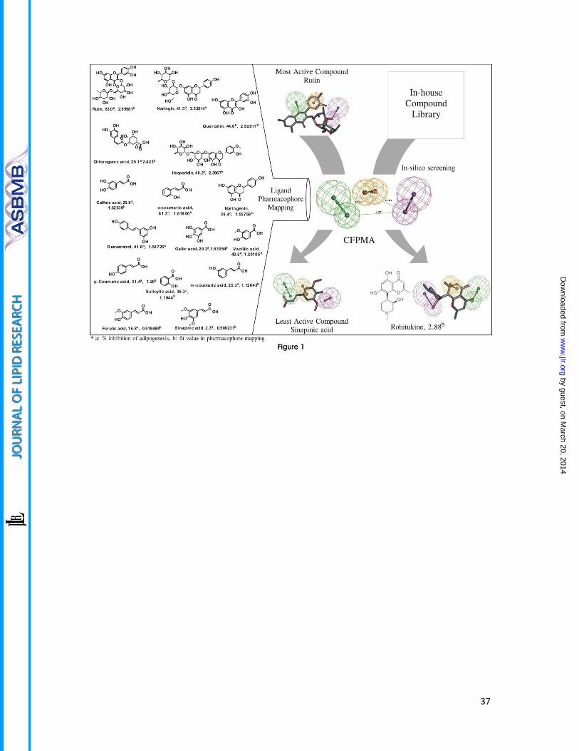
Rohitukine maps on common feature pharmacophore model of anti-adipogenic
compounds
The developed common feature pharmacophore models consisted mainly 3 main features
viz. HBAL, RA and HBD. Among the resulted 10 pharmacophore models with ranking scores
ranging from 85.23-80.90 (Table 1), the top five pharmacophore models were analyzed for
the best fit values of the training set molecules. Four out of the first five pharmacophore
models had same features and inter-feature distances except first hypothesis with RA [1]
and HBAL [2] had different orientations of groups in 3D space. To select the appropriate
model the predictability of this first hypothesis (Hypo-1) was analyzed against the rest four
hypotheses which revealed that the third hypotheses (Hypo-3) predicted the active,
moderately active and least active compounds better than the other four hypotheses
(Figure 1, fit value). Thus, on the basis of the predictability of the third hypothesis (Hypo-3)
and the mapping of the most (MA) and the least (LA) active compounds (Figure 1) was
selected for further validation studies. In hypothesis validation studies, an external test set
of 20 compounds (Supplemental data: Figure 1) which were not included in the training set
and have also been reported active by this model. The Hypo-3 predicted well the external
test set significantly (Supplemental data: Table1). The Hypo-3 perfectly maps MA compound
(Rutin) with phenyl ring maps the RA function of the pharmacophore, the oxygen atom of
4H-chroman maps one HBA and the hydroxyl group at the 3-[α-L-rhamnopyranosyl-(1→6) -
β-D-glucopyranosyloxy] maps one HBD function. Further, this model was used as a
contrivance for in-house library based virtual screening of compounds resulted in
identification of top ranked lead, rohitukine; which was selected for further studies of the
targeted activity. Rohitukine mapped to highest fit values (2.88318) on Hypo-3 (Figure 1) as
by guest, on March 20, 2014
ww
w.jlr.org
Dow
nloaded from
14
compared to the most active compound (Rutin) fit value (2.99981) of the training set. Thus,
Rohitukine maps well to the pharmacophore model (Hypo-3) where its hydroxyl (-OH) group
of 3-hydroxy-1-methylpiperidin function present at 8 position of the basic chroman ring
corresponds to the one HBD, the aromatic ring of its basic chroman ring feature as RA
feature and its carbonyl (C=O) function at C-4 position of the chroman ring (chromen-4-one)
fulfills the requirement of one HBA function.
Rohitukine inhibits adipogenesis
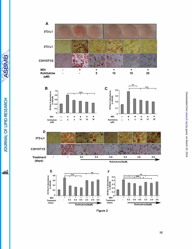
Varying concentrations of rohitukine (0-20 µM) was added to the MDI during differentiation.
Microscopic observation showed rohitukine decreased lipid droplet accumulation in
differentiated 3T3-L1 and C3H10T1/2 derived adipocyte in concentration dependent
manner (Figure 2A). Absorbance of extracted ORO accumulated in lipid droplets confirms
that rohitukine inhibits adipogenesis significantly at 5µM concentration and more than 80%
MDI-induced increased lipid accumulation is inhibited at a 20µM concentration (Figure 2B.
2C). At this concentration, rohitukine found to be nontoxic in 3T3-L1 cells using MTT assay
(Data not shown), Rohitukine (20µM) was added during day 0-2, 0-4, 0-6, 2-4, 2-6 and 4-6 of
adipogenesis. At every rohitukine exposure condition, it leads to decreased lipid
accumulation in adipocyte with a maximum inhibition at 0-6 day. Minimum exposure of 0-2
day was enough to yield significant decrease (P<0.001) in lipid accumulation and
adipogenesis in 3T3-L1 adipocyte (Figure 2 D, E, F). Consistent with these results, we
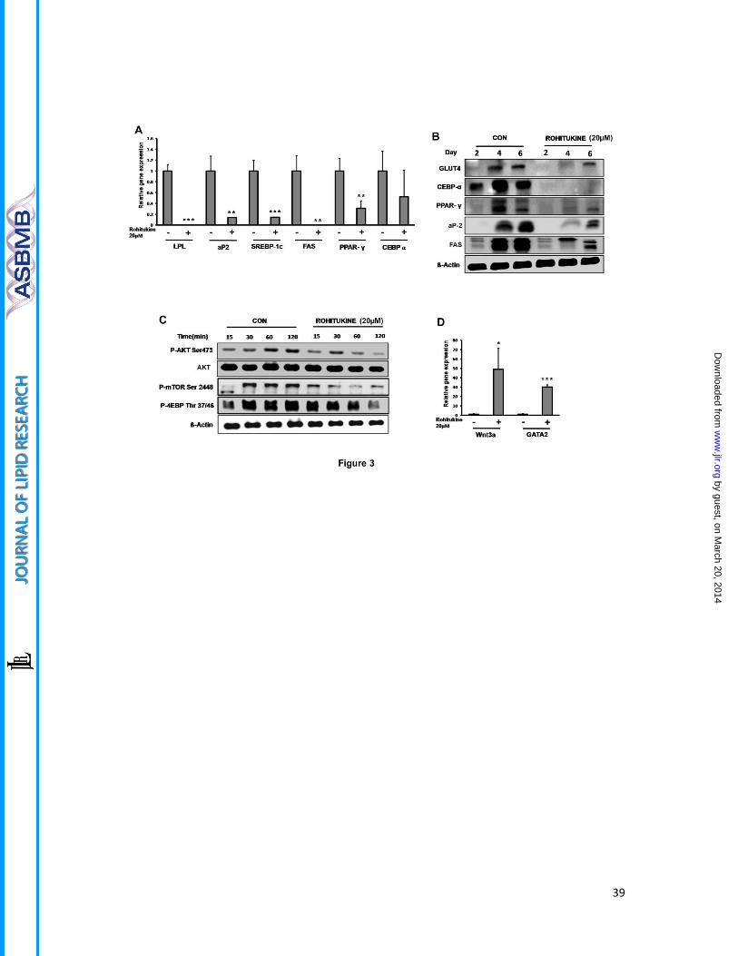
observed that at 20 µM concentration, mRNA level expression of adipogenesis associated
genes that are PPARγ, FABP4, SREBP-1c, FAS and LPL were significantly reduced (P<0.01),
while non-significant, but the decreasing trend was observed in case of C/EBPα in real time
PCR analysis (Figure 3A). This was also supported by the fact that protein level expression of
by guest, on March 20, 2014
ww
w.jlr.org
Dow
nloaded from

15
PPARγ, C/EBPα, aP2, FAS and GLUT-4 expression was found to be suppressed significantly as
compared to control during late phase i.e. day2, day 4 and Day 6 of adipogenesis induction
(Figure 3 B). AKT phsophorylation, mTOR phosphorylation and its target phosphorylation of
4EBP is reduced significantly in early rohitukine exposure (0-2 hour) during adipogenesis
(Figure 3C). The anti-adipogenic transcription factors GATA2 and Wnt3a gene expression
were increased significantly 30 and 50 fold by 48 hours of rohitukine exposure (Figure 3D).
Collectively, these results indicate that, rohitukine possesses an anti-adipogenic potential in
vitro without any cytotoxic effect and early 48 hour exposure is sufficient for significant
suppression of adipogenesis.
Rohitukine arrest mitotic clonal expansion in S- phase
When MDI is added to growth arrested confluent cells, it imposes cells to enter into 2-3
rounds of cell cycles i.e. MCE which is prerequisite for adipogenesis. Addition of rohitukine
to MDI perturbed MCE (Figure 4A). At 20μM concentrations of rohitukine where maximum
suppression of adipogenesis is observed, 41.15% cells were in S-phase compared to that of
only 19.28% cells in MDI alone treated cells (Figure 4B). This highly significant increase
demonstrates that cells either get delayed entry into S-phase or get arrested in S-phase.
MCE arrest was further evidenced in immuno-blot expression analysis of cell cycle
regulatory proteins performed at 16 and 24 hour time-points. Rohitukine causes reduction
of Cyclin-D, CDK-6, CDK4, Cyclin-E, CDK2 protein expression. These proteins required to
accomplish MCE in response to MDI induction. Furthermore, addition of rohitukine in MDI
causes rescue of degradation of P27, as well as a reduced expression of C/EBPβ, that can be
attributed to inhibition of CDKs and MCE arrest respectively (Figure 4C). MCE blockade was
also confirmed by [3H] -thymidine uptake assay. Rohitukine significantly inhibited MDI-
by guest, on March 20, 2014
ww
w.jlr.org
Dow
nloaded from

16
induced 3H-thymidine incorporation after 48 hours exposure (Figure 4D) at 20µM
concentration.
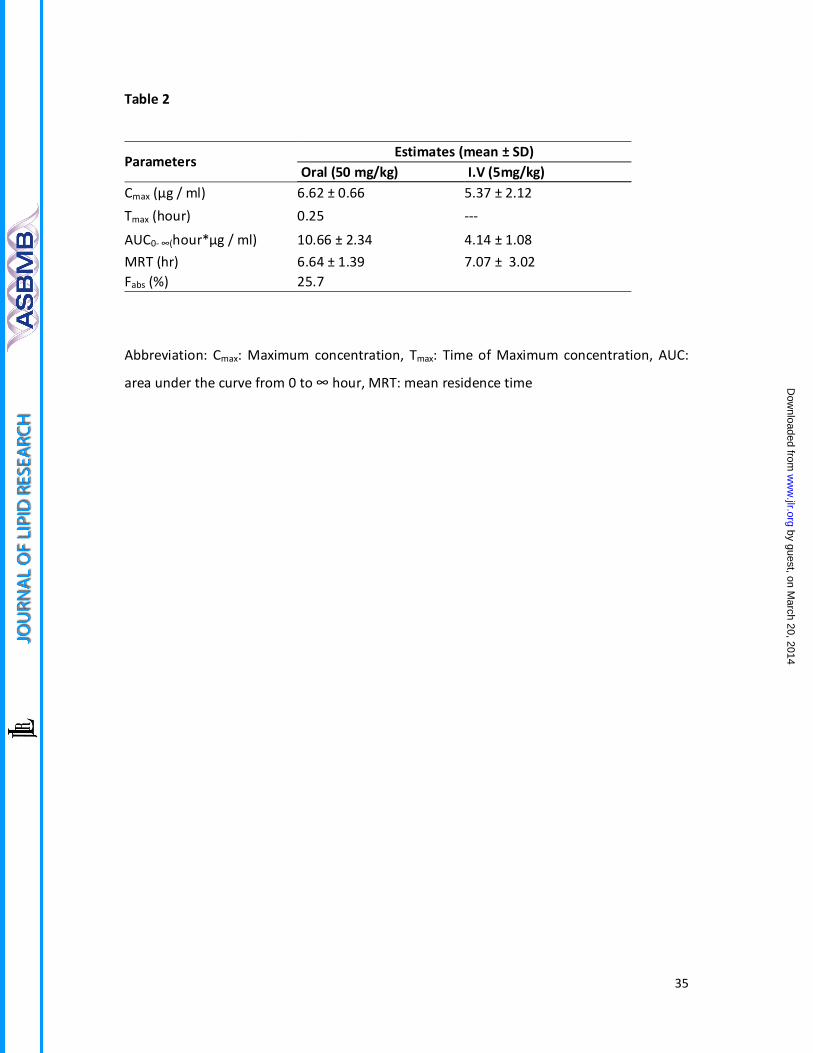
Oral bioavailability of rohitukine in hamster
The therapeutic response correlates with drug availability in systemic circulation.
Bioavailability studies of rohitukine has not been reported yet. Oral bioavailability (50
mg/kg) was carried out in Syrian Golden hamster. The studies shows that rohitukine is
readily bioavailable and approximately 25.7 % rohitukine gets absorbed into systemic
circulation when administered orally. It could be detected in plasma within 15 min post oral
dose and remained in circulation for more than 24 hours (Table 2).
Rohitukine inhibits high fat diet induced dyslipidemia in the Syrian Golden Hamster model
Initial experiments were designed to evaluate in vivo effects of rohitukine at 30mg/Kg and
100 mg/kg dose. We noticed major effects: (i) Very significant reduction of all lipid
parameters like total triglycerides, total cholesterol, LDL cholesterol, (ii) reduced diet
consumption as well as significant body weight loss (Supplemental data: Figure IV: A-H).
These observations indicated that the lipid parameter reduction might be associated with
decreased food intake due to anorexia, central regulation of appetite or any other reason.
To remove this variable, we planned pair fed group study to evaluate the true contribution
of rohitukine towards associated lipid parameter changes. Compound dosing was initiated
in 4 days of HFD fed hamsters and continued till day 10. The pair fed group was fed with an
equivalent quantity of diet that was consumed by the rohitukine treated group but
significant reduction in body weight gain was observed in rohitukine treatment group
(Figure 5A). The serum from pair fed group were collected a day later and comparative lipid
by guest, on March 20, 2014
ww
w.jlr.org
Dow
nloaded from
17
profiling were done between pair fed and rohitukine treated groups. We observed
significant reduction in TG, TC and LDL-c levels (Figure 5B, 5C, 5D). We observed reduction
in HDL levels in rohitukine treatment group similar to earlier mentioned lipid parameters,
but HDL-c/ TC ratio was signficiantly increased (Figure 5E and 5F). We collected liver,
pancreas and adipose tissues of hamsters. Rohitukine increased hepatic LXRα mRNA
expression approximately to 4 fold while expression levels of LDL receptor (LDL R) , HMG-
CoA reductase (HMGC R) and Sterol Regulatory Binding Protein-2 (SREBP-2) were reduced
significantly. However, we did not observe significant changes in the hepatic mRNA
expression levels of PPARα and lipoprotein lipase (LPL, Figure5H). Epididymal fat tissue
weight was significantly increased in the HFD treated group while it was reduced very
significantly in rohitukine treated group (Figure 5 I). Protein level expression studies in
adipose tissue showed PPARγ expression reduced slightly while significant reduction was
observed in Fatty acid synthase (FAS), fatty acid binding protein (aP2) and Glucose
transporter (GLUT-4) expression exhibiting rohitukine effects on fatty acid synthesis (Figure
5J). Tissues were subjected to histological sectioning and staining. Careful observation
showed that HFD fed hamster adipocytes are hypertrophied compared to rohitukine treated
group. We also observed more lipid droplet accumulation in the liver as usually seen in
hepatic steatosis in HFD fed animals. But, these droplets were absent in rohitukine treated
animals. In pancreatic tissue also we observed more lipid/fat droplet accumulation in HFD
fed animals which was reduced in rohitukine treated group (Figure 5K).
by guest, on March 20, 2014
ww
w.jlr.org
Dow
nloaded from

18
Discussion
In this study, we for the first time developed common feature pharmacophore model using
known anti-adipogenic compounds (CFPMA). We validated its predictability. Exploiting the
potential of this model, we screened an in-house library of natural compounds and
identified rohitukine as a top ranking lead molecule. Indeed, rohitukine showed anti-
adipogenic activity in more than one cell models of in vitro adipogenesis. Rohitukine
inhibition is prominent when added in the early phase of differentiation, possibly by
affecting S-phase arrest in mitotic clonal expansion. In vivo Studies of rohitukine showed
anti-dyslipidemic activity potentially affecting both liver and adipocyte metabolism.
Pre-adipocyte fibroblast clonal cell line 3T3-L1 is considered as an excellent model system to
identify molecules affecting adipogenesis. Many phytochemicals were studied in great detail
for their potential to inhibit adipogenesis and/or lipid accumulation capacities and have
been reviewed comprehensively (11). Similarly, many phenolic and flavanoid compounds
are able to inhibit lipogenesis and adipogenesis (27). In this study, we are reporting a
pharmacophore model built using set of 16 chemically diverse compounds of natural and
synthetic origin capable of inhibiting adipogenesis (CFPMA). Our virtual screen efforts
yielded 10 natural molecules out of which we screened the top ranking hit at an initial
concentration of 20 μM. Rohitukine showed adipogenic inhibition in our primary assay. In
subsequent investigations, Rohitukine exhibited concentration dependent as well as
exposure time dependent decrease in ORO accumulation during 3T3-L1 and C3H10T1/2
adipogenic differentiation (Figure 2).
by guest, on March 20, 2014
ww
w.jlr.org
Dow
nloaded from
19
In general, PPARγ is considered as master regulator of adipogenic programming. C/EBPα and
PPARγ cross regulate each other through positive feedback loops and trans-activate
downstream target genes such as fatty acid binding protein-4 (aP2), Lipoprotein lipase (LPL),
and Sterol regulatory element binding protein 1c (SREBP-1c; 30, 31). GATA2 and GATA3
negatively regulate PPARγ and C/EBPα expression and thus responsible for decreased
adipogenesis (32, 33). Similarly Wnt pathway is also negative regulator of adipogenic
induction (34). In general rohitukine addition to differentiation media supresses
proadipogenic mRNA and protein expression and increases anti-adipogenic gene expression
(Figure 3). It is noteworthy that (i) maximum inhibition can be achieved at concentration 20
µM where total lipid accumulation is equivalent of undifferentiated cells and (ii) The activity
at this concentration is limited but not exclusive to exposure of compound in early
adipogenic phase. So, at this concentration of maximum suppression, we studied early
signaling events during adipogenesis.
PI3K-AKT pathway is involved in wide variety of metabolic actions (35). AKT pathway gets
up-regulated in cancer(36). Rohitukine analogue flavopiridol inhibit AKT signaling pathway
and exert its anti-cancer activity (37). AKT/mTOR activation is necessary for adipogenesis
programming (38, 39). Adipogenic stimuli strongly induce AKT phosphorylation, which is
sustained up to 2 hours. Rohitukine (20µM) co-incubation leads to significant suppression of
AKT phosphorylation at Ser-473 residue. It was also found that mTOR phosphorylation at
Ser-2448 is also reduced significantly. Rohitukine abrogates phosphorylation of substrate of
AKT/mTOR pathway, 4EBP at Thr37/46 which is necessary for protein synthesis during
adipogenesis. ERK signaling has also been reported for responsible for early adipogenic
programming and has been the cause of inhibition of adipogenesis by CTRP11,
by guest, on March 20, 2014
ww
w.jlr.org
Dow
nloaded from

20
suphoraphane mediated adipogenic inhibition (40, 41). In our experiments we could not see
any rohitukine mediated alteration in this pathway. Thus, rohitukine may exerts anti-
adipogenic effect in very early phase signaling through PI3K/AKT/mTOR pathway.
Phytocompound such as Piceatannol and Resveratrol affected early insulin signaling events
which ultimately translated in inhibition of mitotic clonal expansion (42-44).These changes
in early signaling upon hormonal induction are of an importance which allow fully confluent
cells to re-enter in clonal expansion. In view of early blocking in adipogenesis (Figure 2D-F),
we performed cell cycle regulation studies. As anticipated, we found concentration
dependent cell number increase in S-phase post 24 hour of MDI-induction (Figure 4 A and
B).
Cell cycle and its regulation play important role in completion of MCE which is prerequisite
for early adipogenesis. Activation and assembly of Cyclin-D to CDK4 and 6, Cyclin-E with
CDK2, and degradation of the CDK inhibitor required for G1/S-phase progression and re-
enter in MCE. Many natural compounds have been reported to inhibit adipogenesis by
affecting MCE. Sulforaphane arrests cells in G0-G1 phase (41), Widdrol causes cell cycle
arrest in G1-S-phase transition(45), genistein, piceatannol, curcumin arrests cells in S-G2/M
phase(46), garcinol and pterostilbene causes G2/M phase arrest (47). Rohitukine decreases
CDK4, CDK6, CDK2, Cyclin-D and Cyclin-E and stabilizes P27 levels (Figure 4C) which can be
corroborated to obseved S-phase arrest in cell cycle analysis by flow cytometry.
Furthermore decreased expression of C/EBPβ expression further strengthens adipogenesis
inhibition phenomenon in early stage. Thus, to summarize, rohitukine modulated early
phase adipogenesis and caused MCE arrest by modulation of cell cycle regulatory protein.
by guest, on March 20, 2014
ww
w.jlr.org
Dow
nloaded from
21
Once established anti-adipogenic effects of rohitukine, we wanted to evaluate its in vivo
effects. Incidentally, most of the compounds on which CFPMA was developed, had also
been reported for anti-dyslipidemic or hypo-lipidemic activity. Although these activities are
distinct from each other, there is observed link between them. The mechanisms of their
interdependence are yet not very clear. Since adipocyte and liver are major organs
contributing overall lipid turnover in vivo, one can possibly corroborate anti-adipogenic and
anti-dyslipidemic activity interdependence.The most active anti-adipogenic compound, rutin
was found to be anti-dyslipidemic when studied in HFD-hamster model (27, 48). Naringin
was proved to be hypolipidemic in diet induced hypercholesterolemia hamster model (49).
Quercetin and Resveratrol protect hypercholesterolemic hamsters diet against aortic fatty
streak accumulation (50). Resveratrol analogue Pterostilbene also have been reported to
reduce LDL/HDL-c ratio significantly in HFD fed hypercholesterolemic hamster (51).
Mulberry water extracts which contains chlorogenic acid, runin, gallic acid and anthocyanins
was found to be hypolipidemic in hamster model (52). Mixture of hespiridine and naringin
have shown hypolipidemic activity in diet induced hypercholesteromic hamsters (49). The
least active compound Sinapinic acid had not been reported yet for anti-dyslipidemic or
hypolipidemic activity. Besides this, well known anti-adipogenic compounds, to mention
curcumin(46), cordycepin(39), berberine(53), piceatennol(43) and daidzein(54) also showed
anti-dyslipidemic and hypolipidemic activity in hamster model (51, 55-58). Most of the
studies were conducted using Golden Syrian hamster, as it is a very sensitive model system
for changes in lipid profile when fed with high fat diet. Secondly, they have closer similarity
to the human lipoprotein profile in comparisons with rat, mice (59). Thus, we decided to
evaluate effect of rohitukine in HFD fed hamster model. In view of notable significant
decrease in food consumption of high dose treated as an experiment progressed to eighth
by guest, on March 20, 2014
ww
w.jlr.org
Dow
nloaded from

22
day it was inevitable for us to perform studies with pair fed group (Supplemental data and
Figure IV). We initially checked oral bioavailability and it is readily bioavailable with more
than 25.7% absorbed within 30 min duration and remains in circulation for more than 24
hours. We then modified dose regimen to 50 mg/kg body weight. To evaluate the true
contribution of rohitukine independent of diet consumption on changes in lipid parameters,
we put one animal group on pair fed. As shown in results, there is a significant increase in
TG, TC, LDL-c, HDL-c in HFD as well as PFD group when compared to normal chow.
Rohitukine significantly decreased all above parameters at 50mg/kg dose (Figure 5B-5D) and
significantly increased HDL-c/TC ratio (Figure 5F). LXRα is the main regulator of lipid
controlling genes (60). It is also known that LXRα is a sensor of cholesterol excess (61) and
its activation in dyslipidemic hamster led to increase in reverse cholesterol transport (62),
and reduced lipid accumulation in dyslipidemic hamsters (63). SREBP-2 is involved in
cholesterol synthesis (64) which in turn regulates expression of HMGC R and LDL R (65). All
these gene expression levels in liver were found to be reduced in rohitukine treated groups
compared to HFD treated group (Figure 5H), similar to that observed with fenofibrate, the
standard anti-dyslipidemic agent (66). In adipocyte, 10 days HFD intervention shown
adipocyte hypertrophy as well as epididymal weight gain, which was reduced in the
rohitukine treated group. Rohitukine also reduced expression of PPARγ, FAS, aP2 and GLUT-
4 proteins in gonadal fat tissue, indicating decreased fat turnover (Figure 5 G, I, J).
Histological sections show that lipid accumulation was increased hepatocytes in HFD and
PFD diet groups while rohitukine treatment decreased lipid accumulation and brings it back
to near normal (Figure 5K). Although it is beyond the scope of this manuscript to pinpoint
the exact mechanism, similar to earlier demonstrated anti-adipogenic molecules exhibiting
anti-dyslipidemic activity, rohitukine also demonstrated in vivo anti-dyslipidemic effects.
by guest, on March 20, 2014
ww
w.jlr.org
Dow
nloaded from

23
To further explore the utility of this pharmacophore model, known molecular entities
analogous derivative of rohitukine flavopiridol and P-276-00 were predicted by this
pharmacophore model. The two compounds from clinical trials with a similar basic core or
analogues structures of rohitukine were predicted by this pharmacophore model. The two
compounds like flavopiridol and P-276-00 were mapped well on Hypo-3 with the fit values
of 2.791 and 2.969 respectively. The pharmacophore mapping of these compounds was
represented in the supporting information (Supplemental data: Figure II). The results may
encourage the researchers to explore this new activity for these under trial anti-cancer
compounds.
In summary, Our studies lead to the development of the first pharmacophore model of anti-
adipogenic compounds for successful identification of rohitukine as lead molecule. We
showed rohitukine possess both anti-adipogenic in vitro and anti-dyslipidemic activity in
vivo. Although, clinical development of rohitukine for these indications and chronic
treatment is dependent on many other factors. Nevertheless, rohitukine is parent
pharmacophore of two leading molecules in anti-cancer drug development pipeline
Flavopiridol and P-276-00 and both of them also map well for feature requirements of anti-
adipogenic compounds. It would be interesting to evaluate the anti-adipogenic potential of
these molecules and its impact on clinical outcome of clinical trials that currently being
conducted.
by guest, on March 20, 2014
ww
w.jlr.org
Dow
nloaded from
24
Conflict of Interest:
The authors declare no conflicts of interest.
Acknowledgments:
Research work is supported by CSIR-CDRI Network project, "Towards holistic understanding
of Complex diseases: Unraveling threads of complex disease (Thunder) Project No.
BSC0102" and partly by Department of Biotechnology (DBT) project (GAP0079). Financial
fellowship/support for SV :Thunder, MB: DBT-SRF, KS: UGC-JRF, VB and YSC : ICMR-SRF,
SKM and SS : CSIR-SRF. Support from Flow cytometry facility of SAIF CSIR-CDRI is
acknowledged.
by guest, on March 20, 2014
ww
w.jlr.org
Dow
nloaded from

25
References:
1. Ginsberg, H. N., and P. R. MacCallum. 2009. The obesity, metabolic syndrome, and type 2 diabetes mellitus pandemic: Part I. Increased cardiovascular disease risk and the importance of atherogenic dyslipidemia in persons with the metabolic syndrome and type 2 diabetes mellitus. J Cardiometab Syndr 4: 113-119.
2. Adamczak, M., and A. Wiecek. 2013. The adipose tissue as an endocrine organ. Semin Nephrol 33: 2-13.
3. Taillan, B., J. G. Fuzibet, G. Garnier, A. Pesce, H. Vinti, M. C. Saint-Paul, and P. Dujardin. 1991. [Hepatic involvement in human immunodeficiency virus infection. 104 histologically documented cases]. Ann Med Interne (Paris) 142: 226-227.
4. Zhang, M., K. Ikeda, J. W. Xu, Y. Yamori, X. M. Gao, and B. L. Zhang. 2009. Genistein suppresses adipogenesis of 3T3-L1 cells via multiple signal pathways. Phytother Res 23: 713-718.
5. Noto, A., P. Zahradka, N. Yurkova, X. Xie, H. Truong, E. Nitschmann, M. R. Ogborn, and C. G. Taylor. 2007. Dietary conjugated linoleic acid decreases adipocyte size and favorably modifies adipokine status and insulin sensitivity in obese, insulin-resistant rats. Metabolism 56: 1601-1611.
6. Kim, H. K., M. Della-Fera, J. Lin, and C. A. Baile. 2006. Docosahexaenoic acid inhibits adipocyte differentiation and induces apoptosis in 3T3-L1 preadipocytes. J Nutr 136: 2965-2969.
7. Kim, H., and K. Sakamoto. 2012. (-)-Epigallocatechin gallate suppresses adipocyte differentiation through the MEK/ERK and PI3K/Akt pathways. Cell Biol Int 36: 147-153.
8. Yang, J. Y., M. A. Della-Fera, S. Rayalam, S. Ambati, D. L. Hartzell, H. J. Park, and C. A. Baile. 2008. Enhanced inhibition of adipogenesis and induction of apoptosis in 3T3-L1 adipocytes with combinations of resveratrol and quercetin. Life Sci 82: 1032-1039.
9. Rayalam, S., J. Y. Yang, S. Ambati, M. A. Della-Fera, and C. A. Baile. 2008. Resveratrol induces apoptosis and inhibits adipogenesis in 3T3-L1 adipocytes. Phytother Res 22: 1367-1371.
10. Ambati, S., J. Y. Yang, S. Rayalam, H. J. Park, M. A. Della-Fera, and C. A. Baile. 2009. Ajoene exerts potent effects in 3T3-L1 adipocytes by inhibiting adipogenesis and inducing apoptosis. Phytother Res 23: 513-518.
11. Rayalam, S., M. A. Della-Fera, and C. A. Baile. 2008. Phytochemicals and regulation of the adipocyte life cycle. J Nutr Biochem 19: 717-726.
12. Klebe, G., and U. Abraham. 1999. Comparative molecular similarity index analysis (CoMSIA) to study hydrogen-bonding properties and to score combinatorial libraries. Journal of computer-aided molecular design 13: 1-10.
13. Klebe, G., U. Abraham, and T. Mietzner. 1994. Molecular similarity indices in a comparative analysis (CoMSIA) of drug molecules to correlate and predict their biological activity. Journal of medicinal chemistry 37: 4130-4146.
14. Catalyst. Version 4.1 ed. 2006.. Accelrys Inc., San Diego, CA. (USA).
by guest, on March 20, 2014
ww
w.jlr.org
Dow
nloaded from

26
15. Jain, S. K., S. B. Bharate, and R. A. Vishwakarma. 2012. Cyclin-dependent kinase inhibition by flavoalkaloids. Mini Rev Med Chem 12: 632-649.
16. Singh, N., P. Singh, S. Shrivastva, S. K. Mishra, V. Lakshmi, R. Sharma, and G. Palit. 2012. Gastroprotective effect of anti-cancer compound rohitukine: possible role of gastrin antagonism and H(+) K (+)-ATPase inhibition. Naunyn Schmiedebergs Arch Pharmacol 385: 277-286.
17. Mohana Kumara, P., S. Zuehlke, V. Priti, B. T. Ramesha, S. Shweta, G. Ravikanth, R. Vasudeva, T. R. Santhoshkumar, M. Spiteller, and R. Uma Shaanker. 2012. Fusarium proliferatum, an endophytic fungus from Dysoxylum binectariferum Hook.f, produces rohitukine, a chromane alkaloid possessing anti-cancer activity. Antonie Van Leeuwenhoek 101: 323-329.
18. Keshri, G., R. M. Oberoi, V. Lakshmi, K. Pandey, and M. M. Singh. 2007. Contraceptive and hormonal properties of the stem bark of Dysoxylum binectariferum in rat and docking analysis of rohitukine, the alkaloid isolated from active chloroform soluble fraction. Contraception 76: 400-407.
19. Lakshmi, V., K. Pandey, A. Kapil, N. Singh, M. Samant, and A. Dube. 2007. In vitro and in vivo leishmanicidal activity of Dysoxylum binectariferum and its fractions against Leishmania donovani. Phytomedicine 14: 36-42.
20. Arguello, F., M. Alexander, J. A. Sterry, G. Tudor, E. M. Smith, N. T. Kalavar, J. F. Greene, Jr., W. Koss, C. D. Morgan, S. F. Stinson, T. J. Siford, W. G. Alvord, R. L. Klabansky, and E. A. Sausville. 1998. Flavopiridol induces apoptosis of normal lymphoid cells, causes immunosuppression, and has potent antitumor activity In vivo against human leukemia and lymphoma xenografts. Blood 91: 2482-2490.
21. Du, G. J., Z. Zhang, X. D. Wen, C. Yu, T. Calway, C. S. Yuan, and C. Z. Wang. 2012. Epigallocatechin Gallate (EGCG) is the most effective cancer chemopreventive polyphenol in green tea. Nutrients 4: 1679-1691.
22. Hormann, V., J. Kumi-Diaka, M. Durity, and A. Rathinavelu. 2012. Anticancer activities of genistein-topotecan combination in prostate cancer cells. J Cell Mol Med 16: 2631-2636.
23. Kang, J. W., J. H. Kim, K. Song, S. H. Kim, J. H. Yoon, and K. S. Kim. 2010. Kaempferol and quercetin, components of Ginkgo biloba extract (EGb 761), induce caspase-3-dependent apoptosis in oral cavity cancer cells. Phytother Res 24 : S77-82.
24. Vucenik, I., and J. P. Stains. 2012. Obesity and cancer risk: evidence, mechanisms, and recommendations. Ann N Y Acad Sci 1271: 37-43.
25. Aballay, L. R., A. R. Eynard, P. Diaz Mdel, A. Navarro, and S. E. Munoz. 2013. Overweight and obesity: a review of their relationship to metabolic syndrome, cardiovascular disease, and cancer in South America. Nutr Rev 71: 168-179.
26. Ito, K., R. Ishigamori, M. Mutoh, T. Ohta, T. Imai, and M. Takahashi. 2013. The A allele promotes azoxymethane-induced colorectal carcinogenesis via macrophage migration in hyperlipidemic/diabetic KK mice. Cancer Sci 104: 835-843.
27. Hsu, C. L., and G. C. Yen. 2007. Effects of flavonoids and phenolic acids on the inhibition of adipogenesis in 3T3-L1 adipocytes. J Agric Food Chem 55: 8404-8410.
by guest, on March 20, 2014
ww
w.jlr.org
Dow
nloaded from
27
28. Brooks, R. B., R. E. Bruccoleri, B. D. Olafson, D. J. States, S. Swaminathan, and M. Karplus. 1983. Charmmea program for macromolecular energy, minimization, and dynamics calculations. J Comp Chem 4: 187-217.
29. Smellie, A., S. L. Teig, and P. Towbin. 1995. Poling: promoting conformational variation. J Comp Chem 16: 171-187.
30. Wu, Z., E. D. Rosen, R. Brun, S. Hauser, G. Adelmant, A. E. Troy, C. McKeon, G. J. Darlington, and B. M. Spiegelman. 1999. Cross-regulation of C/EBP alpha and PPAR gamma controls the transcriptional pathway of adipogenesis and insulin sensitivity. Mol Cell 3: 151-158.
31. Rosen, E. D., C. H. Hsu, X. Wang, S. Sakai, M. W. Freeman, F. J. Gonzalez, and B. M. Spiegelman. 2002. C/EBPalpha induces adipogenesis through PPARgamma: a unified pathway. Genes Dev 16: 22-26.
32. Hu, Y., and G. E. Davies. 2009. Berberine increases expression of GATA-2 and GATA-3 during inhibition of adipocyte differentiation. Phytomedicine 16: 864-873.
33. Horikawa, T., T. Shimada, Y. Okabe, K. Kinoshita, K. Koyama, K. Miyamoto, K. Ichinose, K. Takahashi, and M. Aburada. 2012. Polymethoxyflavonoids from Kaempferia parviflora induce adipogenesis on 3T3-L1 preadipocytes by regulating transcription factors at an early stage of differentiation. Biol Pharm Bull 35: 686-692.
34. Bennett, C. N., S. E. Ross, K. A. Longo, L. Bajnok, N. Hemati, K. W. Johnson, S. D. Harrison, and O. A. MacDougald. 2002. Regulation of Wnt signaling during adipogenesis. J Biol Chem 277: 30998-31004.
35. Avruch, J. 1998. Insulin signal transduction through protein kinase cascades. Mol Cell Biochem 182: 31-48.
36. Morgan, T. M., T. D. Koreckij, and E. Corey. 2009. Targeted therapy for advanced prostate cancer: inhibition of the PI3K/Akt/mTOR pathway. Curr Cancer Drug Targets 9: 237-249.
37. Wu, K., C. Wang, M. D'Amico, R. J. Lee, C. Albanese, R. G. Pestell, and S. Mani. 2002. Flavopiridol and trastuzumab synergistically inhibit proliferation of breast cancer cells: association with selective cooperative inhibition of cyclin D1-dependent kinase and Akt signaling pathways. Mol Cancer Ther 1: 695-706.
38. Zhang, H. H., J. Huang, K. Duvel, B. Boback, S. Wu, R. M. Squillace, C. L. Wu, and B. D. Manning. 2009. Insulin stimulates adipogenesis through the Akt-TSC2-mTORC1 pathway. PLoS One 4: e6189.
39. Takahashi, S., M. Tamai, S. Nakajima, H. Kato, H. Johno, T. Nakamura, and M. Kitamura. 2012. Blockade of adipocyte differentiation by cordycepin. Br J Pharmacol 167: 561-575.
40. Wei, Z., M. M. Seldin, N. Natarajan, D. C. Djemal, J. M. Peterson, and G. W. Wong. 2013. C1q/TNF-related protein 11 (CTRP11), a novel adipose stroma-derived regulator of adipogenesis. J Biol Chem 288: 10214-10229.
41. Choi, K. M., Y. S. Lee, D. M. Sin, S. Lee, M. K. Lee, Y. M. Lee, J. T. Hong, Y. P. Yun, and H. S. Yoo. 2012. Sulforaphane inhibits mitotic clonal expansion during adipogenesis through cell cycle arrest. Obesity (Silver Spring) 20: 1365-1371.
by guest, on March 20, 2014
ww
w.jlr.org
Dow
nloaded from
28
42. Kwon, J. Y., S. G. Seo, S. Yue, J. X. Cheng, K. W. Lee, and K. H. Kim. 2012. An inhibitory effect of resveratrol in the mitotic clonal expansion and insulin signaling pathway in the early phase of adipogenesis. Nutr Res 32: 607-616.
43. Kwon, J. Y., S. G. Seo, Y. S. Heo, S. Yue, J. X. Cheng, K. W. Lee, and K. H. Kim. 2012. Piceatannol, natural polyphenolic stilbene, inhibits adipogenesis via modulation of mitotic clonal expansion and insulin receptor-dependent insulin signaling in early phase of differentiation. J Biol Chem 287: 11566-11578.
44. Tidhar, Y., H. Weissman, S. G. Wolf, A. Gulino, and B. Rybtchinski. 2011. Pathway-dependent self-assembly of perylene diimide/peptide conjugates in aqueous medium. Chemistry 17: 6068-6075.
45. Yun, H. J., J. H. Kim, H. Y. Jeong, H. H. Ji, S. W. Nam, E. W. Lee, B. W. Kim, and H. J. Kwon. 2012. Widdrol blocks 3T3-L1 preadipocytes growth and differentiation due to inhibition of mitotic clonal expansion. J Microbiol Biotechnol 22: 806-813.
46. Kim, C. Y., T. T. Le, C. Chen, J. X. Cheng, and K. H. Kim. 2011. Curcumin inhibits adipocyte differentiation through modulation of mitotic clonal expansion. J Nutr Biochem 22: 910-920.
47. Hsu, C. L., Y. J. Lin, C. T. Ho, and G. C. Yen. 2012. Inhibitory effects of garcinol and pterostilbene on cell proliferation and adipogenesis in 3T3-L1 cells. Food Funct 3: 49-57.
48. Kalgaonkar, S., H. B. Gross, W. Yokoyama, and C. L. Keen. 2010. Effects of a flavonol-rich diet on select cardiovascular parameters in a Golden Syrian hamster model. J Med Food 13: 108-115.
49. Kurowska, E. M., and J. A. Manthey. 2004. Hypolipidemic effects and absorption of citrus polymethoxylated flavones in hamsters with diet-induced hypercholesterolemia. J Agric Food Chem 52: 2879-2886.
50. Auger, C., P. L. Teissedre, P. Gerain, N. Lequeux, A. Bornet, S. Serisier, P. Besancon, B. Caporiccio, J. P. Cristol, and J. M. Rouanet. 2005. Dietary wine phenolics catechin, quercetin, and resveratrol efficiently protect hypercholesterolemic hamsters against aortic fatty streak accumulation. J Agric Food Chem 53: 2015-2021.
51. Rimando, A. M., R. Nagmani, D. R. Feller, and W. Yokoyama. 2005. Pterostilbene, a new agonist for the peroxisome proliferator-activated receptor alpha-isoform, lowers plasma lipoproteins and cholesterol in hypercholesterolemic hamsters. J Agric Food Chem 53: 3403-3407.
52. Peng, C. H., L. K. Liu, C. M. Chuang, C. C. Chyau, C. N. Huang, and C. J. Wang. 2011. Mulberry water extracts possess an anti-obesity effect and ability to inhibit hepatic lipogenesis and promote lipolysis. J Agric Food Chem 59: 2663-2671.
53. Huang, C., Y. Zhang, Z. Gong, X. Sheng, Z. Li, W. Zhang, and Y. Qin. 2006. Berberine inhibits 3T3-L1 adipocyte differentiation through the PPARgamma pathway. Biochem Biophys Res Commun 348: 571-578.
54. Kim, M. H., J. S. Park, M. S. Seo, J. W. Jung, Y. S. Lee, and K. S. Kang. 2010. Genistein and daidzein repress adipogenic differentiation of human adipose tissue-derived mesenchymal stem cells via Wnt/beta-catenin signalling or lipolysis. Cell Prolif 43: 594-605.
55. Jang, E. M., M. S. Choi, U. J. Jung, M. J. Kim, H. J. Kim, S. M. Jeon, S. K. Shin, C. N. Seong, and M. K. Lee. 2008. Beneficial effects of curcumin on hyperlipidemia and insulin resistance in high-fat-fed hamsters. Metabolism 57: 1576-1583.
by guest, on March 20, 2014
ww
w.jlr.org
Dow
nloaded from

29
56. Guo, P., Q. Kai, J. Gao, Z. Q. Lian, C. M. Wu, C. A. Wu, and H. B. Zhu. 2010. Cordycepin prevents hyperlipidemia in hamsters fed a high-fat diet via activation of AMP-activated protein kinase. J Pharmacol Sci 113: 395-403.
57. Kong, W., J. Wei, P. Abidi, M. Lin, S. Inaba, C. Li, Y. Wang, Z. Wang, S. Si, H. Pan, S. Wang, J. Wu, Y. Wang, Z. Li, J. Liu, and J. D. Jiang. 2004. Berberine is a novel cholesterol-lowering drug working through a unique mechanism distinct from statins. Nat Med 10: 1344-1351.
58. Song, T., S. O. Lee, P. A. Murphy, and S. Hendrich. 2003. Soy protein with or without isoflavones, soy germ and soy germ extract, and daidzein lessen plasma cholesterol levels in golden Syrian hamsters. Exp Biol Med (Maywood) 228: 1063-1068.
59. Zhang, Z., H. Wang, R. Jiao, C. Peng, Y. M. Wong, V. S. Yeung, Y. Huang, and Z. Y. Chen. 2009. Choosing hamsters but not rats as a model for studying plasma cholesterol-lowering activity of functional foods. Mol Nutr Food Res 53: 921-930.
60. Nuclear Receptor Nomenclature Committee. 1999. A unified nomenclature system for the nuclear receptor superfamily. Cell 97: 161-163.
61. Chawla, A., J. J. Repa, R. M. Evans, and D. J. Mangelsdorf. 2001. Nuclear receptors and lipid physiology: opening the X-files. Science 294: 1866-1870.
62. Briand, F., M. Treguier, A. Andre, D. Grillot, M. Issandou, K. Ouguerram, and T. Sulpice. 2010. Liver X receptor activation promotes macrophage-to-feces reverse cholesterol transport in a dyslipidemic hamster model. Journal of lipid research 51: 763-770.
63. Mukherjee, R., K. T. Locke, B. Miao, D. Meyers, H. Monshizadegan, R. Zhang, D. Search, D. Grimm, M. Flynn, K. M. O'Malley, L. Zhang, J. Li, Y. Shi, L. J. Kennedy, M. Blanar, P. T. Cheng, J. Tino, and R. A. Srivastava. 2008. Novel peroxisome proliferator-activated receptor alpha agonists lower low-density lipoprotein and triglycerides, raise high-density lipoprotein, and synergistically increase cholesterol excretion with a liver X receptor agonist. The Journal of pharmacology and experimental therapeutics 327: 716-726.
64. Konig, B., A. Koch, J. Spielmann, C. Hilgenfeld, G. I. Stangl, and K. Eder. 2007. Activation of PPARalpha lowers synthesis and concentration of cholesterol by reduction of nuclear SREBP-2. Biochemical pharmacology 73: 574-585.
65. Van Rooyen, D. M., and G. C. Farrell. 2011. SREBP-2: a link between insulin resistance, hepatic cholesterol, and inflammation in NASH. Journal of gastroenterology and hepatology 26: 789-792.
66. Guo, Q., P. R. Wang, D. P. Milot, M. C. Ippolito, M. Hernandez, C. A. Burton, S. D. Wright, and Y. Chao. 2001. Regulation of lipid metabolism and gene expression by fenofibrate in hamsters. Biochimica et biophysica acta 1533: 220-232.
by guest, on March 20, 2014
ww
w.jlr.org
Dow
nloaded from

30
Figure/Table captions:
Table 1.The summary of hypothesis run.
Table 2. Pharmacokinetic profile of Rohitukine after oral (50 mg/kg) and I.V. (5 mg/kg)
administration (n=3).
Table 3. Primer sequences used for real time PCR gene expression studies.
Figure 1. The CFPMA pharmacophore model and in silico virtual screen
The common feature pharmacophore model of was generated using a diverse set of anti-
adipogenic compounds. CFPMA is shown along with the inter-feature distance. The
pharmacophore mapping of most active (rutin), least active (Sinapinic acid) and virtual
screen identified hit rohitukine has been shown in panel. The actual % inhibition of MDI-
induced lipid accumulation by molecule (a) that was used in model generation as well as Fit
value (b) generated on each molecule in pharmacophore mapping shown next to the
compound name.
Figure 2. Rohitukine inhibits adipogenesis
(A) 3T3–L1 preadipocytes and C3H10T1/T2 were cultured in the DMEM and MDI medium
containing 0, 5, 10, 15 and 20 μM rohitukine for 8 days. Culture dish images were acquired
after lipid droplet staining with with Oil Red O (ORO), microscopic images were taken with
Nikon eclipse TI. Absorbance of extracted ORO accumulated in lipid droplets of (B) 3T3-L1
(C) C3H10T1/2 derived adipocytes were measured spectrophotometrically at 490 nm. (D)
3T3–L1 preadipocytes and C3H10T1/T2 were cultured in the MDI medium with 20 μM
rohitukine for 0-2, 0-4, 0-6, 2-4, 2-6 and 4- 6 days. The lipid droplets were stained with Oil
Red O (ORO) and microscopic images were taken with Nikon eclipse TI. Absorbance of
by guest, on March 20, 2014
ww
w.jlr.org
Dow
nloaded from

31
extracted ORO accumulated in lipid droplets of (E) 3T3-L1 (F) C3H10T1/2 derived adipocytes
were measured spectrophotometrically at 490 nm
All values are presented as the mean ± s.d. of three experiments performed in triplicate.
Statistical significance: * p < 0.05 **P < 0.01, ***P < 0.001.
Figure 3. Molecular markers of adipogenic inhibition
(A) Relative mRNA abundance of LPL, aP2, SREBP-1c, FAS, PPARγ, and C/EBPα on day 6 of
adipocyte differentiation were measured by Real time PCR in the absence and presence of
20μM rohitukine in MDI. (B) Rohitukine addition to MDI significantly down-regulate the late
phase protein level expression of GLUT-4, C/EBPα, PPARγ. aP2 and FAS in 3T3-L1 adipocytes
i.e. on day 2, day 4 and day 6 of differentiation. (C) Rohitukine inhibits early phosphorylation
states of p-AKT (ser473), mTOR (Ser2448) and 4EBP (Thr37/46) observed by western
blotting. (D) Rohitukine increases the relative mRNA abundance of Wnt3a and GATA2 on
day 2 measured by real time PCR. The results were verified by three independent
experiments, mean ± s.d.Significance of difference between the MDI-induced control and
rohitukine treated groups (*P<0.05, **P<0.01, ***P<0.001).
Figure 4. Effects of rohitukine on MDI-induced cell cycle progression in 3T3-L1 preadipocytes
(A) Rohitukine arrested MDI-induced cell cycle progression in S-phase. (B) The population of
cells in each stage of the cell cycle was quantified and showed concentration dependent
arrest in S-phase. (C)Post-confluent of 3T3-L1 preadipocytes cells were incubated with MDI
containing 20 μM rohitukine for 0, 16 and 24hours , rohitukine down-regulated the
expression of Cyclin-D, CDK6,CDK4, Cyclin-E, , CDK2,C/EBPβ but stabilized P27 protein as
by guest, on March 20, 2014
ww
w.jlr.org
Dow
nloaded from

32
observed by western blotting. Represented cut blots were run, transferred and exposed
same time but separated away by unrelated samples. (D) Rohitukine inhibited the
incorporation of [3H]Thymidine into newly synthesized DNA after 48 hour significantly,
measured by using scintillation counter. The data are representative of three independent
experiments that give similar results. Significance of difference between the MDI-induced
control and rohitukine treated groups (*P<0.05,**P<0.01,***P<0.001).
Figure 5. Effect of rohitukine administration on normal chow diet (ND) High fat diet (HFD) ,
pair fed died (PFD) fed dyslipidemic Syrian golden hamsters
(A) Mean body weight gain of different groups showed rohitukine significantly decreased
body weight gain by end experiment. Rohitukine showed significantly decreased (B) total
cholesterol (C) Total triglyceride (D) LDL-cholesterol and (E) HDL-c compared to pair fed diet.
Rohitukine significantly increased (F) HDL-c/TC ratio. (G) Rohitukine reduced gonadal fat
mass as compared to HFD fed group (H) Heptaic mRNA expression level in HFD and HFD+
rohitukine (50 mg/Kg) fed groups were analyzed at the end of the experiment. LXRα
expression was increased significantly to 4 fold while significant reduction was observed in
LDL R, HMGC R, SREBP-2 in HFD+rohitukine fed group. (I) Significantly increased epidydemal
fat weight in HFD fed group, is reduced significantly in the rohitukine fed animal group. (J)
Protein level expression of PPARγ, FAS, aP2 and GLUT4 in epididymal fat tissue in Control,
HFD and HFD+ rohitukine fed groups (K) Haematoxylin and eosin staining of liver, adipose
and pancreas tissues. Images in the inbox are magnified images from the same section.
Rohitukine decreased the lipid accumulation in liver compared to HFD fed animals. Each bar
represents mean ±SEM. with significance expressed : ***P < 0.0001 , **P < 0.001 , *P <
0.05.
by guest, on March 20, 2014
ww
w.jlr.org
Dow
nloaded from

33
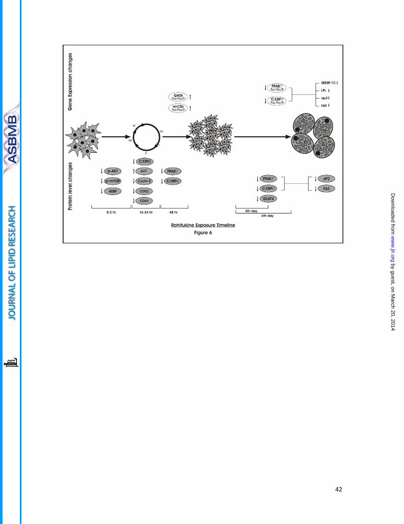
Figure 6.Schematic presentation of time dependent gene and protein level expression
changes imparted by rohitukine addition to MDI during adipogenesis
by guest, on March 20, 2014
ww
w.jlr.org
Dow
nloaded from

34
Table 1
Hypo. Featuresa Rank Direct Hit Partial Hit Max Fit
1 RAA 85.024 1111111111111111 0000000000000000 3 2 RDA 84 1111111111111111 0000000000000000 3 3 RDA 83.013 1111111111111111 0000000000000000 3 4 RDA 82.187 1111111111111111 0000000000000000 3 5 RDA 82.185 1111111111111111 0000000000000000 3 6 RAA 82.18 1111111111111111 0000000000000000 3 7 RDA 81.838 1111111111111111 0000000000000000 3 8 RAA 81.231 1111111111111111 0000000000000000 3 9 RDA 81.213 1111111111111111 0000000000000000 3
10 RDA 80.891 1111111111111111 0000000000000000 3
Direct hitmaskindicates[1] or (0) not a training setmolecule mapped every feature. Partial
hit mask indicates whether [1] or (0) not a molecule mapped all but one feature. aH;
hydrophobic, A; hydrogen bond acceptor lipid (HBA) and R; ring aromatic
by guest, on March 20, 2014
ww
w.jlr.org
Dow
nloaded from
35
Table 2
Abbreviation: Cmax: Maximum concentration, Tmax: Time of Maximum concentration, AUC:
area under the curve from 0 to ∞ hour, MRT: mean residence time
Parameters Estimates (mean ± SD)
Oral (50 mg/kg) I.V (5mg/kg) Cmax (µg / ml) 6.62 ± 0.66 5.37 ± 2.12 Tmax (hour) 0.25 ---
AUC0- ∞(hour*µg / ml) 10.66 ± 2.34 4.14 ± 1.08 MRT (hr) 6.64 ± 1.39 7.07 ± 3.02 Fabs (%) 25.7
by guest, on March 20, 2014
ww
w.jlr.org
Dow
nloaded from

36
Table 3
Gene Name Primer pairs Mouse Specific primer pairs
LPL F 5’tttgtgaaatgccatgacaag3’ R 5’cagatgctttcttctcttgtttgt3’
aP2 F 5’gaaaacgagatggtgacaacg3 R 5’ gccctttcataaactcttgtgg3
SREBP-1c F 5’ttcctcagactgtaggcaaatct3’ R 5’agcctcagtttacccactcct3’
FAS- F 5’caacatgggacaccctgag3’ R 5’gttgtggaagtgcaggttagg3’
PPARγ- F 5’aagacaacggacaaatcacaa3’ R 5’gggggtgatatgtttgaacttg3’
C/EBPα- F 5’aaacaacgcaacgtggaga3’ R 5’gcggtcattgdcactggtc3’
Wnt3a - F 5’gagacatggggacacagtca3’ R 5’gggaatcagatgggtcctg3’
GATA2- F 5’cacccctatcccgtgaatc3’ R 5’cagcagtagagagtaagagacacca3’
Hamster specific primer pairs
LXRα F 5’tcagcatcttctctgcagaccgg3’ R 5’tcattagcatccgtgggaaca3’
LDL R F 5’gcagtgtttctgtggctgacac3’ R 5’gccatgcacagggtcca3’
HMGC R F 5’gagctacatttgtgcttggcg3’ R 5’ttcattaggccgaggctcac3’
SREBP-2 F 5’gcaaggtgttcctgcatgaa3’ R 5’tggtgttctgactggtacgcc3’
LPL F 5’gattcacttttctgggactga3’ R 5’ gccactgtgccgtacagaga3’
PPARα F 5’ggccaatggcatccaaaata3’ R 5’ ccttggcgaattctgtgagc3’
β Actin F 5’ tgctgtccctgtatgcctctg3’ R 5’ agggagagcgtagccctcat3’
by guest, on March 20, 2014
ww
w.jlr.org
Dow
nloaded from
37
by guest, on March 20, 2014
ww
w.jlr.org
Dow
nloaded from
38
by guest, on March 20, 2014
ww
w.jlr.org
Dow
nloaded from
39
by guest, on March 20, 2014
ww
w.jlr.org
Dow
nloaded from

40
by guest, on March 20, 2014
ww
w.jlr.org
Dow
nloaded from

41
by guest, on March 20, 2014
ww
w.jlr.org
Dow
nloaded from
42
by guest, on March 20, 2014
ww
w.jlr.org
Dow
nloaded from