Calcium dysregulation in heart diseases: Targeting calcium ...
Upload
independentCategory
view
0download
0
Riboflavin and Vitamin E Increase Brain Calciumand Antioxidants, and Microsomal Calcium-ATP-ase Valuesin Rat Headache Models Induced by Glyceryl Trinitrate
Ayse Butun • Mustafa Nazıroglu • Serpil Demirci •
Omer Celik • Abdulhadi Cihangir Uguz
Received: 26 August 2014 / Accepted: 18 November 2014
� Springer Science+Business Media New York 2014
Abstract The essential use of riboflavin is the prevention
of migraine headaches, although its effect on migraines is
considered to be associated with the increased mitochon-
drial energy metabolism. Oxidative stress is also important
in migraine pathophysiology. Vitamin E is a strong anti-
oxidant in nature and its analgesic effect is not completely
clear in migraines. The current study aimed to investigate
the effects of glyceryl trinitrate (GTN)-sourced exogen
nitric oxide (NO), in particular, and also riboflavin and/or
vitamin E on involved in the headache model induced via
GTN-sourced exogen NO on oxidative stress, total brain
calcium levels, and microsomal membrane Ca2?-ATPase
levels. GTN infusion is a reliable method to provoke
migraine-like headaches in experimental animals and
humans. GTN resulted in a significant increase in brain
cortex and microsomal lipid peroxidation levels although
brain calcium, vitamin A, vitamin C, and vitamin E, and
brain microsomal-reduced glutathione (GSH), glutathione
peroxidase (GSH-Px), and plasma-membrane Ca2?-ATP-
ase values decreased through GTN. The lipid peroxidation,
GSH, vitamin A, b-carotene, vitamin C, and vitamin E, and
calcium concentrations, GSH-Px, and the Ca2?-ATPase
activities were increased both by riboflavin and vitamin E
treatments. Brain calcium and vitamin A concentrations
increased through riboflavin only. In conclusion, riboflavin
and vitamin E had a protective effect on the GTN-induced
brain injury by inhibiting free radical production, regula-
tion of calcium-dependent processes, and supporting the
antioxidant redox system. However, the effects of vitamin
E on the values seem more important than in riboflavin.
Keywords Migraine � Antioxidants � Oxidative stress �Calcium � Vitamin E � Riboflavin
Abbreviations
[Ca2?]i Intracellular Ca2?
CSD Cortical spreading depression
DMSO Dimethyl sulfoxide
GSH Reduced glutathione
GSH-Px Glutathione peroxidase
LP Lipid peroxidation
MMCA Microsomal membrane Ca2?-ATPase
NO Nitric oxide
PMCA Plasma-membrane Ca2?-ATPase
ROS Reactive oxygen species
VGCC Voltage-gated calcium channels
Introduction
Migraine is one of the most common neurological condi-
tions with underlying pathophysiological mechanisms that
remain unclear (Reuter et al. 2002). It has been suggested
to be a neurovascular disease resulting from dysfunction of
the trigeminovascular system (Yan and Dussor 2014). The
blockade of presynaptic P/Q-, N-, and L-type calcium
A. Butun � S. Demirci
Department of Neurology, Faculty of Medicine, Suleyman
Demirel University, Isparta, Turkey
M. Nazıroglu � O. Celik � A. C. Uguz
Department of Biophysics, Faculty of Medicine, Suleyman
Demirel University, Isparta, Turkey
M. Nazıroglu (&) � S. Demirci � O. Celik � A. C. Uguz
Neuroscience Research Center, Suleyman Demirel University,
32260 Isparta, Turkey
e-mail: [email protected]
123
J Membrane Biol
DOI 10.1007/s00232-014-9758-5

channels present in trigeminovascular neurons inhibit the
release of the calcitonin gene-related peptide and conse-
quently the dilatation of dural blood vessels (Amrutkar
et al. 2011). The clinical observations and genetic studies
suggest that high-threshold voltage-gated calcium channels
(VGCC) play an important role in the pathogenesis of
migraine (Yan and Dussor 2014). Many cellular functions
are regulated directly or indirectly via free cytosolic cal-
cium-ion (Ca2?) concentration (Nazıroglu 2007; Kumar
et al. 2014). The intracellular Ca2? levels are maintained
by the plasma-membrane Ca2?-ATPase (PMCA) and
microsomal membrane Ca2?-ATPase (MMCA) in an
equilibrium state. The Ca2? current is considered to be
associated with the release of neurotransmitters via VGCCs
(Akerman et al. 2003).
It has been proven with certainty that nitric oxide (NO)
plays a role as a neurotransmitter in the neurological
transmission process induced by glutamate (Dawson 1995).
NO inhibits mitochondrial respiration and leads to the
release of glutamate by glutamate carriers, the activation of
NMDA receptors and Ca2? current into the cells (Brown and
Bal-Price 2003). During cortical spreading depression
(CSD), extracellular K? increases, while levels of extra-
cellular Na? and Ca2? decrease remarkably. It is considered
that Ca2? waves play primary role in CSD (Eikermann-
Haerter and Ayata 2010). Since a typical headache, which is
more common in subjects with migraines than those without
migraine, develops after 4–6 h following the administration
of nitroglycerin (GTN), the effect of nitroglycerin as a NO
donor has been evaluated over several experimental studies
(Ramachandran et al. 2014).
Oxidative stress is defined as the overproduction of
reactive oxygen species (ROS) (e.g., superoxide radicals,
hydrogen peroxide, and NO) (Nazıroglu 2007; Paredes and
Reiter 2010). The generation of ROS is ubiquitous since
ROS are generated during aerobic metabolism, such as
mitochondrial oxidations and other monoamine oxidants
(Daiber et al. 2013). The results of recent studies indicated
that oxidative stress is the main source in the etiology of
migraines (Yilmaz et al. 2011; Ishii et al. 2011). In order to
scavenge, various ROS defense systems exist in the brain.
Glutathione peroxidase (GSH-Px) is responsible for the
reduction of hydro and organic peroxides in the presence of
reduced glutathione (GSH) (Nazıroglu and Yurekli 2013;
Yurekli and Nazıroglu 2013). GSH is the most abundant
thiol antioxidant in mammalian cells and maintains thiol
redox in the cells. GSH depletion has been implicated in
the neurobiology of neurons (Nazıroglu et al. 2011).
Vitamin E, alpha-tocopherol, is the most important anti-
oxidant in the lipid phase of cells and it modulates also
Ca2? entry in neurons (Nazıroglu and Ozgul 2013). Vita-
min C, as well as being a free radical scavenger, also
transforms vitamin E to its active form. As such, the
positive effect of vitamin E is well-known on systemic
diseases such as diabetes and cardiovascular diseases and
neurological diseases such as epilepsy and depression
although its effects on migraine prophylaxis is not clearly
determined.
Riboflavin is an agent used in the prophylactic treatment
of migraine. It has been reported in preclinical studies that
riboflavin may reduce pain in mice (Franca et al. 2001).
Riboflavin is a water-soluble vitamin that works as a
coenzyme in the mitochondrial electron transport chain
(Colombo et al. 2014). Mitochondria are one of main
sources of ROS in cells, although there are no reports on
oxidative stress and the subject of riboflavin on migraines.
It is considered that it exhibits its positive functional effects
by increasing the activities of complexes I and II and also
mitochondrial energy metabolism (Markley 2012). How-
ever, this issue is still controversial (Colombo et al. 2014).
It has not been studied whether vitamin E and riboflavin
modify the alterations in the antioxidant enzyme system
and lipid peroxidation levels in the brain and microsomal in
rats. Hence, we aimed to evaluate whether there would be a
protective effect of vitamin E and riboflavin on oxidative
stress and enzymatic antioxidants, MMCA, and calcium
values in GTN-induced brain injury in experimental
migraine rat models.
Materials and Methods
Animals
The current study included 60 female Wistar-Albino rats
aged 8–12 weeks and weighing 200–250 g. The approval
was obtained from the Ethics Committee of the Medical
Faculty of Suleyman Demirel University to use experi-
mental animals for scientific purposes. All of the rats were
held under standard conditions of light (12 h of daylight/
12 h of darkness) and heat (*25 8C). All of the rats were
given enough water and standard rat food pellets and put
into separate chambers of the cage.
Experimental Design and Induction of Migraine
The rats were distributed randomly into four groups
including 15 rats in each group. The groups were deter-
mined as Group 1: Control Group; Group 2: Glyceryl
trinitrate (GTN) Group; Group 3: GTN ? RBF (riboflavin)
Group; Group 4: Riboflavin ? GTN ? vitamin E Group.
Control Group
Rats in Group 1 received a placebo over 10 days of the
study.
A. Butun et al.: Migraine, Riboflavin and Vitamin E
123

GTN Group
Animals in the groups received a placebo over 10 days of
the study and then GTN was administered to the animals.
Riboflavin ? GTN Group
Rats in Group 3 received 100 mg/kg/day oral riboflavin
(Eczacibasi Pharmaceuticals Corporation, Istanbul, Tur-
key) (Granados-Soto et al. 2004) for 10 days before GTN
administration.
Riboflavin ? GTN ? Vitamin E Group
Rats in the Group 4 received oral riboflavin (100 mg/kg/
day) plus 100 mg/kg/vitamin E (Evigen, Eras Inc., Istan-
bul, Turkey) intraperitoneally every other day (i.p.) for
10 days before GTN administration (Nazıroglu et al. 2004).
We used the vitamin E and riboflavin in pretreatment
groups for 10 days before GTN administrations. Twenty-
four hours following the last medicinal treatment, an
experimental headache model was induced with a three-
hour-infusion of 10 mg/kg i.p. GTN to Groups 2, 3, and 4
(Ramachandran et al. 2012). Group 1 was established as
the control group via administrating the same amount of
physiological serum i.p. One hour after GTN infusion, rats
showed decelerated motion and decreased reaction to
pulling test of rat-tail (Ramachandran et al. 2012). GTN
(Sigma, Istanbul, Turkey), dissolved in saline, alcohol, and
propylene glycol was injected i.p. at a dose of 10 mg/kg.
Anesthesia and Preparation of Brain Samples
The animals, whose feeding was interrupted the previous
night, were administered mixture of ketamine hydrochlo-
ride (50 mg/kg) and xylazine (5 mg/kg) i.p. 1 h after the
3-h GTN infusion. Then, all of the rats were sacrificed and
their brain cortexes were removed. The removed tissues of
brain cortex samples were washed twice with cold physi-
ological saline. They were held in glass bottles in a deep
freeze (-30 �C) for a specified period (maximum 10 h).
Next, cortex samples were minced on the ice. They was
homogenized at 5,000 rpm for two minutes using cold-ice
Tris–HCl buffer (50 mM, pH 7.4) in a Teflon homogenizer.
Microsomes were isolated from the remaining samples of
the brain cortex. All of the samples were stored at ?4 �C.
Isolation of Brain Microsomes
Brain samples were cleaned and minced and then homog-
enized in six volumes of freshly prepared buffer A con-
taining: 0.3 mol/l sucrose, 10 mmol/l Hepes- HCl buffer
pH 7.4, and 2 mmol/l dithiothreitol. The material was
homogenized with the glass Teflon homogenizer. The
homogenate was centrifuged (MS 80, Sanyo Inc.) at
85,0009g (Sorvall, Teknolab A.S, Ankara, Turkey) for
75 min. The supernatant was discarded and the pellet was
re-suspended in the original volume of buffer A containing
0.6 mol/l KCl using four strokes of the pestle and centri-
fuged again at 85,0009g for 75 min. The pellets were re-
suspended in the original volume of buffer A. After cen-
trifugation at 85,0009g for 75 min, the pellet was sus-
pended in buffer A using four strokes of the pestle to
produce a protein concentration of 2–7 mg/ml. The com-
plete procedure took 10–12 h for eight samples. The
samples were frozen and stored at -33 �C until assayed.
The isolation procedure was carried out at ?4 �C (Nazır-oglu et al. 2009; Caliskan et al. 2010).
Measurement of Microsomal Ca2?-ATPase Activity
The Ca2?-ATPase activity was measured spectrophoto-
metrically using the method of Burette et al. (2003). The
assay medium contained: 120 mmol/l KCl, 60 mmol/l
Hepes buffer, pH 7 (at 37 �C), 1 mmol/l MgCl2, 0.5 mmol/
l K2-ATP, 0.2 mmol/l NADH, 0.5 mmol/PEPA, 1 IU/l
pyruvate kinase, 1 IU/l LDH, and 500 mmol/l EGTA.
After pre-incubation of the assay medium (total volume of
1 ml) for 4 min at 37 �C, 50 mg of the microsomal
homogenate was added to the medium. After 2 min, the
reaction was initiated by the addition of CaCl2 (600 mmol/
l). The ATPase activity as oxidation of NADH was fol-
lowed by continuously measuring the absorbance at
340 nm.
Based on the extinction coefficient for NADH,
e = 6.2 9 106 M-1, the amount of NADH oxidized was
equivalent to the hydrolyzed amount of ATP. Values were
expressed as IU/mg protein.
Measurement of Total Brain Calcium Concentrations
Microsomal calcium levels were analyzed with a plasma
optic emission atomic absorption spectrophotometer (ICP-
OES, Optima 4300 DV, Perkin Elmer Life and Analytical
Sciences, Inc. Waltham, Massachusetts, USA) by follow-
ing the wet ashing procedure with nitric acid (Nazıroglu
et al. 2008). Ca values were measured at 422.7 nm in the
atomic absorption spectrophotometer.
Lipid Peroxidation Level Determinations
Lipid peroxidation levels in the brain homogenate and
microsomal samples were measured with the thiobarbitu-
ric-acid reaction by the method of Placer et al. (1966). The
quantification of thiobarbituric-acid reactive substances
was determined by comparing the absorption to the
A. Butun et al.: Migraine, Riboflavin and Vitamin E
123
standard curve for malondialdehyde (MDA) equivalents
generated by acid-catalyzed hydrolysis of 1,1,3,3-tetra-
methoxypropane. The values of lipid peroxidation in the
brain and microsomal were expressed as lmol/g protein.
Brain Cortex and Microsomal-Reduced Glutathione
(GSH), Glutathione Peroxidase (GSH-Px) and Protein
Assay
The GSH contents of the brain homogenate and microsome
were measured at 412 nm using the method of Sedlak and
Lindsay (1968) as described in the previous studies
(Nazıroglu et al. 2004, 2008). GSH-Px activities of the
brain homogenate and microsome were measured spec-
trophotometrically at 37 �C and 412 nm according to
Lawrence and Burk (1976). The protein content in the
brain cortex and microsomes was measured by method of
Lowry et al. (1951) with bovine serum albumin as the
standard.
Brain b-carotene, Vitamins A, C, and E Analyses
Vitamins A (retinol) and E (a-tocopherol) were determined
in the brain samples by a modification of the method
described by Desai (1984) and Suzuki and Katoh (1990).
Brain samples of approximately 0.25 g were saponified by
the addition of 0.3 ml 60 percent (w/v in water) KOH and
2 ml of one percent (w/v in ethanol) ascorbic acid, fol-
lowed by heating at 70 �C for 30 min. After cooling the
samples on ice, 2 ml of water and 1 ml of n-hexane were
added and mixed with the samples and then allowed to rest
for 10 min to allow for phase separation. An aliquot of
0.5 ml of n-hexane extract was taken and vitamin A con-
centrations were measured at 325 nm. Then reactants were
added and the absorbance value of hexane was measured in
a spectrophotometer at 535 nm. Calibration was performed
using standard solutions of all-trans retinol and a-tocoph-
erol in hexane.
The concentrations of b-carotene in brain samples were
determined according to the method of Suzuki and Katoh
(1990). Two ml of hexane were mixed with 0.25 g brain.
The concentration of b-carotene in hexane was measured at
453 nm in a spectrophotometer.
Statistical Analyses
The statistical analyses were performed using the pocket
software of SPSS 15.0 for Windows. The Mann–Whitney
U-test was used for the comparison between the groups
with respect to Ca?2-ATPase and total brain calcium
concentration. The results were expressed as mean
value ± SD. p \ 0.05 was accepted as significant.
Results
Results of Brain Microsomal Ca2?-ATPase Activities
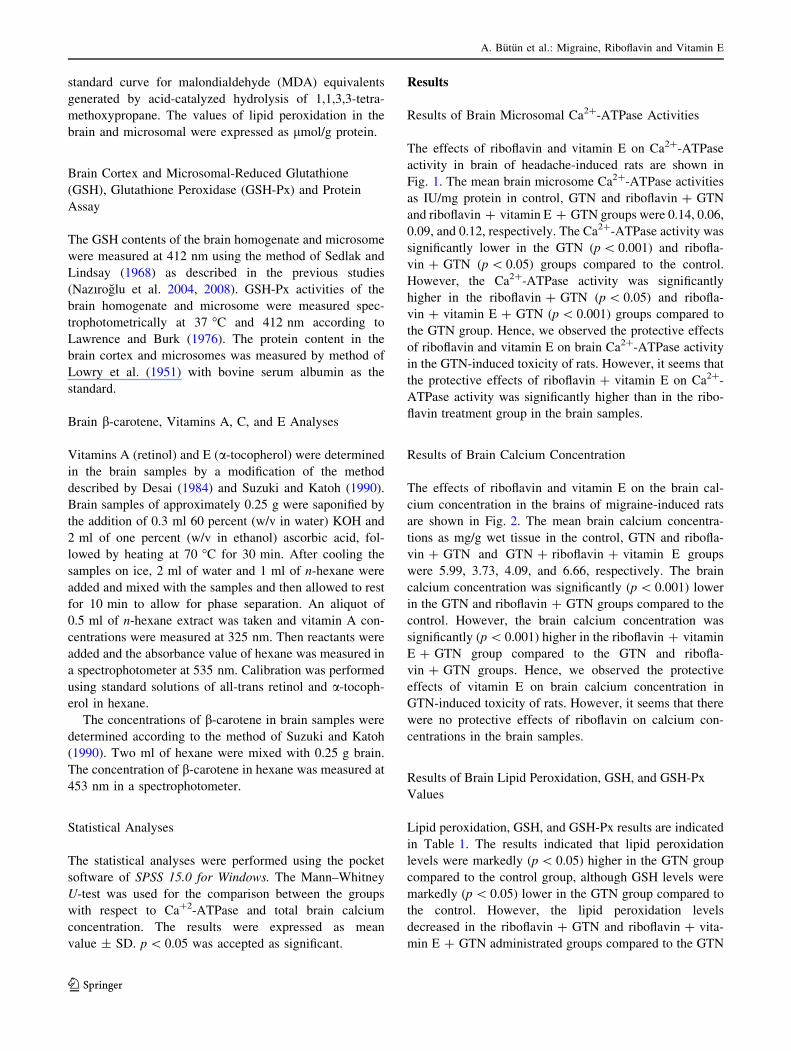
The effects of riboflavin and vitamin E on Ca2?-ATPase
activity in brain of headache-induced rats are shown in
Fig. 1. The mean brain microsome Ca2?-ATPase activities
as IU/mg protein in control, GTN and riboflavin ? GTN
and riboflavin ? vitamin E ? GTN groups were 0.14, 0.06,
0.09, and 0.12, respectively. The Ca2?-ATPase activity was
significantly lower in the GTN (p \ 0.001) and ribofla-
vin ? GTN (p \ 0.05) groups compared to the control.
However, the Ca2?-ATPase activity was significantly
higher in the riboflavin ? GTN (p \ 0.05) and ribofla-
vin ? vitamin E ? GTN (p \ 0.001) groups compared to
the GTN group. Hence, we observed the protective effects
of riboflavin and vitamin E on brain Ca2?-ATPase activity
in the GTN-induced toxicity of rats. However, it seems that
the protective effects of riboflavin ? vitamin E on Ca2?-
ATPase activity was significantly higher than in the ribo-
flavin treatment group in the brain samples.
Results of Brain Calcium Concentration
The effects of riboflavin and vitamin E on the brain cal-
cium concentration in the brains of migraine-induced rats
are shown in Fig. 2. The mean brain calcium concentra-
tions as mg/g wet tissue in the control, GTN and ribofla-
vin ? GTN and GTN ? riboflavin ? vitamin E groups
were 5.99, 3.73, 4.09, and 6.66, respectively. The brain
calcium concentration was significantly (p \ 0.001) lower
in the GTN and riboflavin ? GTN groups compared to the
control. However, the brain calcium concentration was
significantly (p \ 0.001) higher in the riboflavin ? vitamin
E ? GTN group compared to the GTN and ribofla-
vin ? GTN groups. Hence, we observed the protective
effects of vitamin E on brain calcium concentration in
GTN-induced toxicity of rats. However, it seems that there
were no protective effects of riboflavin on calcium con-
centrations in the brain samples.
Results of Brain Lipid Peroxidation, GSH, and GSH-Px
Values
Lipid peroxidation, GSH, and GSH-Px results are indicated
in Table 1. The results indicated that lipid peroxidation
levels were markedly (p \ 0.05) higher in the GTN group
compared to the control group, although GSH levels were
markedly (p \ 0.05) lower in the GTN group compared to
the control. However, the lipid peroxidation levels
decreased in the riboflavin ? GTN and riboflavin ? vita-
min E ? GTN administrated groups compared to the GTN
A. Butun et al.: Migraine, Riboflavin and Vitamin E
123

group (p \ 0.05) only. There was no statistical change in
GSH-Px activity in the four groups.
Results of Brain Microsome Lipid Peroxidation, GSH,
and GSH-Px Values
Lipid peroxidation, GSH, and GSH-Px value results of the
brain microsome in the four groups are shown in Table 2.
The results indicated that lipid peroxidation levels were
markedly (p \ 0.05) higher in the GTN group compared to
the control group, although GSH-Px activity and GSH levels
were markedly (p \ 0.05) lower in the GTN group compared
to the control group. However, the lipid peroxidation levels
were significantly lower in the riboflavin ? GTN (p \ 0.05)
and riboflavin ? vitamin E ? GTN (p \ 0.05) groups
compared to the GTN group alone.
Results of Brain Antioxidant Vitamin Concentrations
Vitamin A, vitamin E, vitamin C, and b-carotene concen-
trations in the total brain in the four groups are shown in
Table 3. Vitamin A (p \ 0.05), vitamin C (p \ 0.05), and
vitamin E (p \ 0.01) concentrations were markedly lower
in the GTN group compared to the control. However, the
Fig. 1 Effects of riboflavin
(RBF) and vitamin E (VE) on
Ca2?-ATPase activity in the
brain microsomes of migraine-
induced rats (n = 15 and
mean ± SD). ap \ 0.001 andbp \ 0.05 versus the control.cp \ 0.05 and dp \ 0.001
versus the GTN group.ep \ 0.05 versus the
GTN ? RBF group
Fig. 2 The effects of riboflavin
(RBF) and vitamin E (VE) on
brain calcium concentration in
the brains of migraine-induced
rats (n = 15 and mean ± SD).ap \ 0.001 versus the control.bp \ 0.001 versus the GTN
group. cp \ 0.001 versus the
GTN ? RBF group
Table 1 The effects of riboflavin (RBF) and vitamin E (VE) on brain lipid peroxidation (LP), glutathione peroxidase (GSH-Px) and reduced
glutathione (GSH) values in GTN-induced brain injury in rats (mean ± SD)
Parameters Control
(n = 15)
GTN
(n = 15)
RBF ? GTN
(n = 15)
RBF ? VE ? GTN
(n = 15)
GSH-Px (IU/g protein) 27.49 ± 4.22 26.00 ± 2.99 28.94 ± 4.38 27.67 ± 4.38
GSH (lmol/g protein) 10.90 ± 1.78 9.48 ± 1.08* 11.40 ± 1.32a 11.46 ± 1.04a
LP (lmol/g protein) 13.39 ± 2.09 14.14 ± 3.11* 12.32 ± 1.48a 11.51 ± 1.48a
* p \ 0.05 versus the controla p \ 0.05 versus the GTN group
A. Butun et al.: Migraine, Riboflavin and Vitamin E
123

b-carotene, vitamin C, and vitamin E concentrations were
significantly (p \ 0.05) higher in the riboflavin ? GTN
group compared to the GTN group. The vitamin A
(p \ 0.05), b-carotene (p \ 0.01), vitamin C (p \ 0.01),
and vitamin E (p \ 0.05) concentrations were also signif-
icantly higher in the riboflavin ? vitamin E ? GTN group
compared to the GTN group. The effects of vitamin E on
the vitamin concentrations in the brain samples seem most
significant compared to the riboflavin group.
Discussion
We observed that the brain and microsomal lipid per-
oxidation levels were increased by GTN administration,
although brain and microsomal GSH, brain calcium,
vitamin A, vitamin C, and vitamin E concentrations, and
MMCA activities decreased. Hence, GTN administrations
in animals are characterized by increased oxidative stress
and decreased MMCA, calcium, GSH, and antioxidant
vitamin values. The administration of riboflavin and
vitamin E induced a decrease in calcium and brain and
microsomal lipid peroxidation levels, although GSH,
vitamin A, b-carotene, vitamin E, and vitamin C con-
centrations and MMCA activity increased. A limited
number of in vivo or in vitro studies in the brain of
experimental animals have been reported regarding the
effects of riboflavin and vitamin E on antioxidant
enzymatic system, lipid peroxidation, and MMCA values
(Hassan et al. 2013; Das Evcimen et al. 2004). To the
best of our knowledge, the current study is the first to
compare riboflavin and vitamin with particular reference
to their effects on oxidative stress and the antioxidant
redox system in GTN-induced brain injury in a migraine
rat model.
Inactivation of ROS can be conducted by antioxidant
vitamins (Nazıroglu 2007b). Vitamin E (a-tocopherol) is
the most important antioxidant in the lipid phase of cells.
Vitamin E acts to protect cells against the effects of ROS,
which are potentially damaging byproducts of the body’s
metabolism (Nazıroglu et al. 2004; Nazıroglu and Ozgul
2013). Vitamin C and GSH, in addition to being two free
radical scavengers, also transform vitamin E to its active
form (Nazıroglu 2007; Daiber et al. 2013). It is well known
that CSD induces oxidative stress in the brain and tri-
geminal nociceptive system (Shatillo et al. 2013). Ribo-
flavin is a well-known nutritional supplement that has been
shown to exhibit antioxidant properties and protect the
brain from oxidative damage (Hassan et al. 2013). The
antioxidant levels in the brain were considerably low.
Therefore, low antioxidant levels and high content of
PUFA result in limited antioxidant defense in the brain.
Vitamin A, vitamin C, and vitamin E concentrations in the
brain cortex decreased in the GTN group, although their
concentrations in the brain cortex increased in the ribofla-
vin and vitamin E treatment groups. The increased
Table 2 The effects of riboflavin (RBF) and vitamin E (VE) on brain microsomal lipid peroxidation (LP), glutathione peroxidase (GSH-Px), and
reduced glutathione (GSH) values in GTN-induced brain injury in rats (mean ± SD)
Parameters Control
(n = 15)
GTN
(n = 15)
RBF ? GTN
(n = 15)
RBF ? VE ? GTN
(n = 15)
GSH-Px (IU/g protein) 16.28 ± 2.56 14.05 ± 4.33* 15.26 ± 4.09a 15.52 ± 4.09a
GSH (lmol/g protein) 7.20 ± 1.98 7.54 ± 1.74* 12.00 ± 2.86a 10.38 ± 2.04a
LP (lmol/g protein) 21.29 ± 1.88 24.65 ± 2.72* 20.49 ± 1.96 18.35 ± 1.96b
* p \ 0.05 versus the controla p \ 0.05 and b p \ 0.01 versus the GTN group
Table 3 The effects of riboflavin (RBF) and vitamin E (VE) on antioxidant vitamin values in GTN-induced brain injury in rats (mean ± SD)
Parameters Control
(n = 15)
GTN
(n = 15)
RBF ? GTN
(n = 15)
RBF ? VE ? GTN
(n = 15)
Vitamin A (lmol/g brain) 3.22 ± 0.56 2.78 ± 0.46* 2.89 ± 0.10 3.24 ± 0.53a,c
b-carotene (lmol/g brain) 2.37 ± 0.28 2.27 ± 0.38 2.48 ± 0.12a 2.61 ± 0.14b,a
Vitamin C (lmol/g brain) 1.31 ± 0.15 1.04 ± 0.27* 1.25 ± 0.25a 2.00 ± 0.38b,d
Vitamin E (lmol/g brain) 12.11 ± 1.51 7.10 ± 1.34** 8.20 ± 1.16a 10.64 ± 1.24b,d
* p \ 0.05 and ** p \ 0.01 versus the controla p \ 0.05 and b p \ 0.01 versus the GTN groupc p \ 0.05 and d p \ 0.01 versus the GTN ? RBF group
A. Butun et al.: Migraine, Riboflavin and Vitamin E
123
concentrations of the antioxidant vitamins could be due to
its depletion or inhibition as a result of the increased pro-
duction of free radicals. The increase in the brain cortex
vitamin A, vitamin C, and vitamin E values in animals
during riboflavin and vitamin E treatments has been
attributed to the inhibition of free radicals and lipid per-
oxidation (Dyatlov et al. 1998; Erol et al. 2010; Ozkaya
et al. 2011; Ceylan et al. 2011; Sanches et al. 2014).
The selenium-dependent GSH-Px antioxidant enzyme is
responsible for the reduction of hydro and organic perox-
ides in the presence of GSH (Nazıroglu and Yurekli 2013;
Yurekli and Nazıroglu 2013). GSH is the most abundant
thiol antioxidant in mammalian cells and maintains thiol
redox in the cells. GSH depletion has been implicated in
the neurobiology of neurons (Nazıroglu et al. 2011). The
results of recent papers indicated that vitamin E (Ozkaya
et al. 2011; Ceylan et al. 2011) and riboflavin (Sanches
et al. 2014) supplementation induced an increase of GSH
and GSH-Px values in animal and human studies due to the
modulation of mitochondrial ROS production process
(Siler-Marsiglio et al. 2005; Xie et al. 2013). In the current
study, we observed brain GSH, brain and microsomal GSH,
and GSH-Px values were low in the GTN group, although
they increased with vitamin E and riboflavin supplemen-
tation and the effects of vitamin E and riboflavin might be
induced by the modulation of the mitochondrial ROS
production process. To our knowledge, there have been no
reports on the effects of vitamin E and riboflavin in GTN-
induced brain injury in human and experimental animals.
However, there are a few reports on the antioxidant
enzyme values in patients with migraines. Erol et al. (2010)
and Vurucu et al. (2013) reported that GSH-Px activity was
low in adult and pediatric patients with migraines. Hence,
GSH-Px results of the current study were supported by the
results of Erol et al. (2010) and Vurucu et al. (2013).
Mitochondria were reported to accumulate Ca2? pro-
vided cytosolic Ca2? rises or provided mitochondrial
uptake dominates mitochondrial Ca2? extrusion, thereby
leading to the depolarization of mitochondrial membranes
(Kovacs et al. 2002; Espino et al. 2010). The uptake of
Ca2? into mitochondria stimulates the tricarboxylate cycle,
resulting in an augmented reduction of pyridine nucleo-
tides, which may be one of the mechanisms of the coupling
of neuronal and metabolic activity (Kumar et al. 2014). On
the other hand, the exposure of mitochondria to high
cytosolic free Ca2? was shown to increase the formation of
ROS (Nazıroglu et al. 2014). It has been reported that
vitamin E and riboflavin induced the modulator role on
ROS production and mitochondrial functions (Siler-
Marsiglio et al. 2005; Xie et al. 2013) and also reduced
cytosolic Ca2? levels by the regulation of VGCC and TRP
cation channels (Huang et al. 2000; Nazıroglu and Ozgul
2013). In the current study, the brain cortex and
microsomal lipid peroxidation and MMCA values were
lower in the riboflavin and vitamin E groups compared to
the GTN group. Modulation of VGCC and TRP channels in
the brain cells by means of the treatment with riboflavin
and vitamin E might has caused a decrease in mitochon-
drial ROS productions and Ca2? entry.
The fluctuations in the levels of intracellular calcium
levels affect the physiological, chemical, and biological
processes (Kumar et al. 2014). CSD is the transient sup-
pression of neuronal activity resulting from the temporary
disruption of local brain ionic homeostasis (Eikermann-
Haerter and Ayata 2010). The production of NO and entry
of Ca2? in cytosol increase during CSD (Dawson 1995;
Akerman et al. 2003). NO inhibits mitochondrial respira-
tion and leads to the release of glutamate via glutamate
carriers (Eikermann-Haerter and Ayata 2010). Exogen NO
facilitates CSD that occurs due to excitatory neurotrans-
mitters (Dawson 1995). Similar to CSD, extracellular cal-
cium decreased 1 mmol (Jing et al. 1993), while
intracellular calcium increased less than 0.2 lmol (Wang
et al. 2001) at 37 �C along hypoxic spreading depression.
Therefore, reduction in the extracellular calcium levels are
higher than the elevation in the intracellular calcium levels
along CSD. In this case, the reduction in total calcium
levels along CSD is possible. The results of our study
support this hypothesis. In our study, total brain calcium
levels significantly decreased when compared to the con-
trol group in the rats in which an experimental headache
model was induced via GTN-sourced exogen NO.
These results caused us consider that riboflavin and/or
vitamin E improved the disrupted calcium-ion balance in
the rats in which headache was induced by GTN.
Riboflavin inhibits guanylate cyclase (Galagan et al.
1991) and the inhibition of guanylate cyclase decreases
the presynaptic release of NO and consequently reduces
the NMDA-mediated calcium current into the cell.
Finally, it was demonstrated that riboflavin inhibits the
release of glutamate from the cerebrocortical nerve ends
at a significant ratio and suppress VGCCs. In the current
study, total brain calcium levels significantly increased in
the group in which riboflavin was treated compared to
the GTN group. The findings obtained in the current
study may be interpreted in favor of the inhibition of
guanylate cyclase or the suppression of VGCC by ribo-
flavin. However, the effects and the action mechanism of
riboflavin over brain calcium homeostasis in migraines is
not completely clear yet. In this study, significantly
increased total brain calcium levels were observed in the
riboflavin ? vitamin E group when compared with the
riboflavin group. NO, as a free radical, increased the
activation of NMDA receptors. Vitamin E may affect the
NMDA-mediated calcium current; however, various
findings have been reported in the studies on this issue.
A. Butun et al.: Migraine, Riboflavin and Vitamin E
123

Nazıroglu and Luckhoff (2008) and Nazıroglu and Ozgul
(2013) reported that vitamin E, well-known for its
membrane stabilizing effect, decreases the permeability
of cell membrane against various ions including Ca2?;
whereas, Dyatlov et al. (1998) demonstrated that
a-tocopherol increased the Ca2? current significantly in
the cells with low energy. The findings obtained in the
current study also supported these results, in which
Vitamin E increased total brain calcium levels.
In conclusion, the current study’s brain results in the
GTN group are consistent with a generalized antioxidant
abnormality in different tissues of animals and humans
with migraines and headaches. However, riboflavin and
vitamin E supplementation induced a protective effect on
the oxidative stress and antioxidant redox system in the
brain cortex and microsomes. The beneficial effect of
riboflavin and vitamin E on enzymatic antioxidant systems
was the regulation of GSH, antioxidant vitamins, MMCA
activities, and lipid peroxidation levels in the brain. Hence,
use of the riboflavin with vitamin E could be a potential
approach in arresting or inhibiting the headache genesis
caused by excitotoxic agents.
Acknowledgments The study was partially supported by the Sci-
entific Research Project Unit of Suleyman Demirel University (BAP-
1595-TU-07).
Conflict interest None of the authors have any conflicts to disclose.
All authors approved the final manuscript.
References
Akerman S, Williamson DJ, Goadsby PJ (2003) Voltage-dependent
calcium channels are involved in neurogenic dural vasodilatation
via a presynaptic transmitter release mechanism. Br J Pharmacol
140:558–566
Amrutkar DV, Ploug KB, Olesen J, Jansen-Olesen I (2011) Role for
voltage gated calcium channels in calcitonin gene-related
peptide release in the rat trigeminovascular system. Neurosci-
ence 172:510–517
Brown GC, Bal-Price A (2003) Inflammatory neurodegeneration
mediated by nitric oxide, glutamate, and mitochondria. Mol
Neurobiol 27:325–355
Burette A, Rockwood JM, Strehler EE, Weinberg RJ (2003) Isoform-
specific distribution of the PMCA in the rat brain. J Comp
Neurol 467:464–476
Caliskan AM, Nazıroglu M, Uguz AC, Ovey IS, Sutcu R, Bal R,
Caliskan S, Ozcankaya R (2010) Acamprosate modulates
alcohol-induced hippocampal NMDA receptors and brain micro-
somal Ca2?-ATPase but induces oxidative stress in rat. J Membr
Biol 237:51–58
Ceylan BG, Nazıroglu M, Uguz AC, Barak C, Erdem B, Yavuz L
(2011) Effects of vitamin C and E combination on element and
oxidative stress levels in the blood of operative patients under
desflurane anesthesia. Biol Trace Elem Res 141(1–3):16–25
Colombo B, Saraceno L, Comi G (2014) Riboflavin and migraine: the
bridge over troubled mitochondria. Neurol Sci 35(Suppl
1):141–144
Daiber A, Mader M, Stamm P, Zinßius E, Kroller-Schon S, Oelze M,
Munzel T (2013) Oxidative stress and vascular function. Cell
Membr Free Radic Res 5:221–231
DasEvcimen N, Ulusu NN, Karasu C, Dogru B (2004) Adenosine
triphosphatase activity of streptozotocin-induced diabetic rat
brain microsomes. Effect of vitamin E. Gen Physiol Biophys
23(3):347–355
Dawson VL (1995) Nitric oxide: role neurotoxicity. Clin Exp
Pharmacol Physiol 122:305–308
Desai ID (1984) Vitamin E analysis methods for animal tissues.
Methods Enzymol 105:138–147
Dyatlov VA, Makovetskaıa VV, Leonhardt R, Lawrence DA,
Carpenter DO (1998) Vitamin E enhances Ca?2-mediated
vulnerability of immature cerebellar granule cells to ischemia.
Free Radic Biol Med 25:793–802
Eikermann-Haerter K, Ayata C (2010) Cortical spreading depression
and migraine. Curr Neurol Neurosci Rep 10:167–173
Erol I, Alehan F, Aldemir D, Ogus E (2010) Increased vulnerability to
oxidative stress in pediatric migraine patients. Pediatr Neurol
43:21–24
Espino J, Bejarano I, Redondo PC, Rosado JA, Barriga C, Reiter RJ,
Pariente JA, Rodrıguez AB (2010) Melatonin reduces apoptosis
induced by calcium signaling in human leukocytes: evidence for
the involvement of mitochondria and Bax activation. J Membr
Biol 233:105–118
Franca DS, Souza AL, Almeida KR, Dolabella SS, Martinelli C,
Coelho MM (2001) B vitamins induce an antinociceptive effect
in the asetic acid and formaldehyde models of nociception in
mice. Eur J Pharmacol 421:157–164
Galagan ME, Mordvintcev PI, Vanin AF (1991) Effect of riboflavin
on hypotensive activity of dinitrosyl iron complex with thiosul-
phate. Eur J Pharmacol 203:325–326
Granados-Soto V, Teran-Rosales F, Rocha-Gonzalez HI, Reyes-
Garcıa G, Medina-Santillan R, Rodrıguez-Silverio J, Flores-
Murrieta FJ (2004) Riboflavin reduces hyperalgesia and inflam-
mation but not tactile allodynia in the rat. Eur J Pharmacol
492:35–40
Hassan I, Chibber S, Khan AA, Naseem I (2013) Cisplatin-induced
neurotoxicity in vivo can be alleviated by riboflavin under
photoillumination. Cancer Biother Radiopharm 28:160–168
Huang HM, Ou HC, Hsieh SJ (2000) Antioxidants prevent amyloid
peptide-induced apoptosis and alteration of calcium homeostasis
in cultured cortical neurons. Life Sci 66:1879–1892
Ishii M, Iizuka R, Kiuchi Y, Mori Y, Shimizu S (2011) Neuropro-
tection by lomerizine, a prophylactic drug for migraine, against
hydrogen peroxide-induced hippocampal neurotoxicity. Mol Cell
Biochem 358:1–11
Jing J, Aitken PG, Somjen GG (1993) Role of calcium channels in
spreading depression in rat hippocampal slices. Brain Res
604:251–259
Kovacs R, Schuchmann S, Gabriel S, Kann O, Kardos J, Heinemann
U (2002) Free radical-mediated cell damage after experimental
status epilepticus in hippocampal slice cultures. J Neurophysiol
88:2909–2918
Kumar VS, Gopalakrishnan A, Naziroglu M, Rajanikant GK (2014)
Calcium ion: the key player in cerebral ischemia. Curr Med
Chem 21:2065–2075
Lawrence RA, Burk RF (1976) Glutathione peroxidase activity in
selenium-deficient rat liver. Biochem Biophys Res Commun
71:952–958
Lowry OH, Rosebrough NJ, Farr AL, Randall RJ (1951) Protein
measurement with the folin- phenol reagent. J Biol Chem
193:265–275
Markley HG (2012) CoEnzyme Q10 and riboflavin: the mitochondrial
connection. Headache 52(Suppl 2):81–87
A. Butun et al.: Migraine, Riboflavin and Vitamin E
123

Nazıroglu M (2007) New molecular mechanisms on the activation of
TRPM2 channels by oxidative stress and ADP-ribose. Neuro-
chem Res 32:1990–2001
Nazıroglu M, Luckhoff A (2008) Effects of antioxidants on calcium
influx through TRPM2 channels in transfected cells activated by
hydrogen peroxide. J Neurol Sci 270:152–158
Nazıroglu M, Ozgul C (2013) Vitamin E modulates oxidative stress
and protein kinase C activator (PMA)-induced TRPM2 channel
gate in dorsal root ganglion of rats. J Bioenerg Biomembr
45:541–549
Nazıroglu M, Yurekli VA (2013) Effects of antiepileptic drugs on
antioxidant and oxidant molecular pathways: focus on trace
elements. Cell Mol Neurobiol 33:589–599
Nazıroglu M, Karaoglu A, Aksoy AO (2004) Selenium and high dose
vitamin E administration protects cisplatin-induced oxidative
damage to renal, liver and lens tissues in rats. Toxicology
195:221–230
Nazıroglu M, Kutluhan S, Yilmaz M (2008) Selenium and topiramate
modulates brain microsomal oxidative stress values, Ca2?-
ATPase activity, and EEG records in pentylentetrazol-induced
seizures in rats. J Membr Biol 225:39–49
Nazıroglu M, Uguz AC, Kocak A, Bal R (2009) Acetaminophen at
different doses protects brain microsomal Ca2?-ATPase and the
antioxidant redox system in rats. J Membr Biol 231:57–64
Nazıroglu M, Ozgul C, Cig B, Dogan S, Uguz AC (2011) Glutathione
modulates Ca(2?) influx and oxidative toxicity through TRPM2
channel in rat dorsal root ganglion neurons. J Membr Biol
242:109–118
Nazıroglu M, Senol N, Ghazizadeh V, Yuruker V (2014) Neuropro-
tection Induced by N-acetylcysteine and selenium against
traumaticb injury-induced apoptosis and calcium entry in
hippocampus of rat. Cell Mol Neurobiol 34:895–903
Ozkaya D, Nazıroglu M, Armagan A, Demirel A, Koroglu BK,
Colakoglu N, Kukner A, Sonmez TT (2011) Dietary vitamin C
and E modulates oxidative stress induced-kidney and lens injury
in diabetic aged male rats through modulating glucose homeo-
stasis and antioxidant systems. Cell Biochem Funct 29:287–293
Paredes SD, Reiter RJ (2010) Melatonin: helping cells cope with
oxidative disaster. Cell Membr Free Radic Res 2:99–111
Placer ZA, Cushman L, Johnson BC (1966) Estimation of products of
lipid peroxidation (malonyl dialdehyde) in biological fluids.
Anal Biochem 16:359–364
Ramachandran R, Bhatt DK, Ploug KB, Olesen J, Jansen-Olesen I,
Hay-Schmidt A, Gupta S (2012) A naturalistic glyceryl trinitrate
infusion migraine model in the rat. Cephalalgia 32:73–84
Ramachandran R, Bhatt DK, Ploug KB, Hay-Schmidt A, Jansen-
Olesen I, Gupta S, Olesen J (2014) Nitric oxide synthase,
calcitonin gene-related peptide and NK-1 receptor mechanisms
are involved in GTN-induced neuronal activation. Cephalalgia
34:136–147
Reuter U, Chiarugi A, Bolay H, Moskowitz MA (2002) Nuclear
factor-j B as a molecular target for migraine therapy. Ann
Neurol 51:507–516
Sanches SC, Ramalho LN, Mendes-Braz M, Terra VA, Cecchini R,
Augusto MJ, Ramalho FS (2014) Riboflavin (vitamin B-2)
reduces hepatocellular injury following liver ischaemia and
reperfusion in mice. Food Chem Toxicol 67:65–71
Sedlak J, Lindsay RHC (1968) Estimation of total, protein bound and
non-protein sulfhydryl groups in tissue with Ellmann’ s reagent.
Anal Biochem 25:192–205
Shatillo A, Koroleva K, Giniatullina R, Naumenko N, Slastnikova
AA, Aliev RR, Bart G, Atalay M, Gu C, Khazipov R, Davletov
B, Grohn O, Giniatullin R (2013) Cortical spreading depression
induces oxidative stress in the trigeminal nociceptive system.
Neuroscience 253:341–349
Siler-Marsiglio KI, Pan Q, Paiva M, Madorsky I, Khurana NC, Heaton
MB (2005) Mitochondrially targeted vitamin E and vitamin E
mitigate ethanol-mediated effects on cerebellar granule cell
antioxidant defense systems. Brain Res 1052(2):202–211
Suzuki J, Katoh N (1990) A simple and cheap method for measuring
vitamin A in cattle using only a spectrophotometer. Jpn J Vet Sci
52:1282–1284
Vurucu S, Karaoglu A, Paksu MS, Yesilyurt O, Oz O, Unay B, Akin
R (2013) Relationship between oxidative stress and chronic daily
headache in children. Hum Exp Toxicol 32:113–119
Wang J, Chambers G, Cottrell JE, Kass IS (2001) Differential fall in
ATP accounts for effects of temperature on hypoxic damage in
rat hippocampal slices. J Neurophysiol 83:3462–3471
Xie Y, Zhong C, Zeng M, Guan L, Luo L (2013) Effect of hexavalent
chromium on electron leakage of respiratory chain in mitochon-
dria isolated from rat liver. Cell Physiol Biochem 31(2–3):
473–485
Yan J, Dussor G (2014) Ion channels and migraine. Headache
54:619–639
Yilmaz N, Aydin O, Yegin A, Tiltak A, Eren E (2011) Increased
levels of total oxidant status and decreased activity of arylest-
erase in migraineurs. Clin Biochem 44:832–837
Yurekli VA, Nazıroglu M (2013) Selenium and topiramate attenuates
blood oxidative toxicity in patients with epilepsy: a clinical pilot
study. Biol Trace Elem Res 152:180–186
A. Butun et al.: Migraine, Riboflavin and Vitamin E
123
Copyright © 2022 FDOKUMEN