Recurrent Signature Patterns in HIV-1 B Clade Envelope Glycoproteins Associated with either Early or...
19
Recurrent Signature Patterns in HIV-1 B Clade Envelope Glycoproteins Associated with either Early or Chronic Infections S. Gnanakaran 1 , Tanmoy Bhattacharya 1,2 , Marcus Daniels 1 , Brandon F. Keele 3,4 , Peter T. Hraber 1 , Alan S. Lapedes 1 , Tongye Shen 1,5 , Brian Gaschen 1 , Mohan Krishnamoorthy 1 , Hui Li 4 , Julie M. Decker 4 , Jesus F. Salazar-Gonzalez 4 , Shuyi Wang 4 , Chunlai Jiang 6,7 , Feng Gao 7 , Ronald Swanstrom 8 , Jeffrey A. Anderson 8 , Li-Hua Ping 8 , Myron S. Cohen 8 , Martin Markowitz 9 , Paul A. Goepfert 4 , Michael S. Saag 4 , Joseph J. Eron 8 , Charles B. Hicks 7 , William A. Blattner 10 , Georgia D. Tomaras 7 , Mohammed Asmal 11 , Norman L. Letvin 11,12 , Peter B. Gilbert 13 , Allan C. DeCamp 13 , Craig A. Magaret 13 , William R. Schief 14 , Yih-En Andrew Ban 14,15 , Ming Zhang 1,16 , Kelly A. Soderberg 7 , Joseph G. Sodroski 17 , Barton F. Haynes 7 , George M. Shaw 4 , Beatrice H. Hahn 4 , Bette Korber 1,2 * 1 Theoretical Biology, Los Alamos National Laboratory, Los Alamos, New Mexico, United States of America, 2 Santa Fe Institute, Santa Fe, New Mexico, United States of America, 3 SAIC-Frederick, National Cancer Institute, Frederick, Maryland, United States of America, 4 Departments of Medicine and Microbiology, University of Alabama at Birmingham, Birmingham, Alabama, United States of America, 5 Center for Molecular Biophysics and Department of Biochemistry, Cellular & Molecular Biology, University of Tennessee, Knoxville, Tennessee, United States of America, 6 National Engineering Laboratory of AIDS Vaccine School of Life Science, Jilin University, Changchun, China, 7 Duke University Medical Center, the Departments of Medicine and Surgery, and the Duke Human Vaccine Institute, Duke University, Durham, North Carolina, United States of America, 8 Department of Biochemistry and Biophysics and the Division of Infectious Diseases Center for AIDS Research, University of North Carolina at Chapel Hill, Chapel Hill, North Carolina, United States of America, 9 Aaron Diamond AIDS Research Center, an affiliate of the Rockefeller University, New York, New York, United States of America, 10 Institute of Human Virology, University of Maryland, School of Medicine, Baltimore, Maryland, United States of America, 11 Beth Israel Deaconess Medical Center, Boston, Massachusetts, United States of America, 12 Division of Viral Pathogenesis, Department of Medicine, Harvard Medical School, Boston, Massachusetts, United States of America, 13 Vaccine Infectious Disease Division, Fred Hutchinson Cancer Research Center, Seattle, Washington, United State of America, 14 Department of Biochemistry, University of Washington, Seattle, Washington, United States of America, 15 Arzeda Corporation, Seattle, Washington, United States of America, 16 Department of Epidemiology and Biostatistics, College of Public Health, University of Georgia, Athens, Georgia, United States of America, 17 Dana-Farber Cancer Institute, Department of Cancer Immunology and AIDS, Boston, Massachusetts, United States of America Abstract Here we have identified HIV-1 B clade Envelope (Env) amino acid signatures from early in infection that may be favored at transmission, as well as patterns of recurrent mutation in chronic infection that may reflect common pathways of immune evasion. To accomplish this, we compared thousands of sequences derived by single genome amplification from several hundred individuals that were sampled either early in infection or were chronically infected. Samples were divided at the outset into hypothesis-forming and validation sets, and we used phylogenetically corrected statistical strategies to identify signatures, systematically scanning all of Env. Signatures included single amino acids, glycosylation motifs, and multi-site patterns based on functional or structural groupings of amino acids. We identified signatures near the CCR5 co-receptor- binding region, near the CD4 binding site, and in the signal peptide and cytoplasmic domain, which may influence Env expression and processing. Two signatures patterns associated with transmission were particularly interesting. The first was the most statistically robust signature, located in position 12 in the signal peptide. The second was the loss of an N-linked glycosylation site at positions 413–415; the presence of this site has been recently found to be associated with escape from potent and broad neutralizing antibodies, consistent with enabling a common pathway for immune escape during chronic infection. Its recurrent loss in early infection suggests it may impact fitness at the time of transmission or during early viral expansion. The signature patterns we identified implicate Env expression levels in selection at viral transmission or in early expansion, and suggest that immune evasion patterns that recur in many individuals during chronic infection when antibodies are present can be selected against when the infection is being established prior to the adaptive immune response. Citation: Gnanakaran S, Bhattacharya T, Daniels M, Keele BF, Hraber PT, et al. (2011) Recurrent Signature Patterns in HIV-1 B Clade Envelope Glycoproteins Associated with either Early or Chronic Infections. PLoS Pathog 7(9): e1002209. doi:10.1371/journal.ppat.1002209 Editor: John A. T. Young, The Salk Institute for Biological Studies, United States of America Received December 5, 2010; Accepted June 26, 2011; Published September 29, 2011 This is an open-access article, free of all copyright, and may be freely reproduced, distributed, transmitted, modified, built upon, or otherwise used by anyone for any lawful purpose. The work is made available under the Creative Commons CC0 public domain dedication. Funding: This work was funded by the a grant from the Division of AIDS, NIAID, NIH for the Center for HIV/AIDS Vaccine Immunology (CHAVI) AI06785. This study was undertaken as part of our response to the CHAVI call, however, and in this sense it was at the request of the NIH that we initiated this work, but it was implemented, details were designed, and the specific experiments and analyses undertaken by CHAVI consortium. The supercomputing facility at Los Alamos National Laboratory also contributed computational resources. The funders had no role in study design, data collection and analysis, decision to publish, or preparation of the manuscript. Competing Interests: The authors have declared that no competing interests exist. * E-mail: [email protected] PLoS Pathogens | www.plospathogens.org 1 September 2011 | Volume 7 | Issue 9 | e1002209
Transcript of Recurrent Signature Patterns in HIV-1 B Clade Envelope Glycoproteins Associated with either Early or...

Recurrent Signature Patterns in HIV-1 B Clade EnvelopeGlycoproteins Associated with either Early or ChronicInfectionsS. Gnanakaran1, Tanmoy Bhattacharya1,2, Marcus Daniels1, Brandon F. Keele3,4, Peter T. Hraber1, Alan S.
Lapedes1, Tongye Shen1,5, Brian Gaschen1, Mohan Krishnamoorthy1, Hui Li4, Julie M. Decker4, Jesus F.
Salazar-Gonzalez4, Shuyi Wang4, Chunlai Jiang6,7, Feng Gao7, Ronald Swanstrom8, Jeffrey A. Anderson8,
Li-Hua Ping8, Myron S. Cohen8, Martin Markowitz9, Paul A. Goepfert4, Michael S. Saag4, Joseph J. Eron8,
Charles B. Hicks7, William A. Blattner10, Georgia D. Tomaras7, Mohammed Asmal11, Norman L.
Letvin11,12, Peter B. Gilbert13, Allan C. DeCamp13, Craig A. Magaret13, William R. Schief14, Yih-En Andrew
Ban14,15, Ming Zhang1,16, Kelly A. Soderberg7, Joseph G. Sodroski17, Barton F. Haynes7, George M.
Shaw4, Beatrice H. Hahn4, Bette Korber1,2*
1 Theoretical Biology, Los Alamos National Laboratory, Los Alamos, New Mexico, United States of America, 2 Santa Fe Institute, Santa Fe, New Mexico, United States of
America, 3 SAIC-Frederick, National Cancer Institute, Frederick, Maryland, United States of America, 4 Departments of Medicine and Microbiology, University of Alabama at
Birmingham, Birmingham, Alabama, United States of America, 5 Center for Molecular Biophysics and Department of Biochemistry, Cellular & Molecular Biology, University
of Tennessee, Knoxville, Tennessee, United States of America, 6 National Engineering Laboratory of AIDS Vaccine School of Life Science, Jilin University, Changchun, China,
7 Duke University Medical Center, the Departments of Medicine and Surgery, and the Duke Human Vaccine Institute, Duke University, Durham, North Carolina, United
States of America, 8 Department of Biochemistry and Biophysics and the Division of Infectious Diseases Center for AIDS Research, University of North Carolina at Chapel
Hill, Chapel Hill, North Carolina, United States of America, 9 Aaron Diamond AIDS Research Center, an affiliate of the Rockefeller University, New York, New York, United
States of America, 10 Institute of Human Virology, University of Maryland, School of Medicine, Baltimore, Maryland, United States of America, 11 Beth Israel Deaconess
Medical Center, Boston, Massachusetts, United States of America, 12 Division of Viral Pathogenesis, Department of Medicine, Harvard Medical School, Boston,
Massachusetts, United States of America, 13 Vaccine Infectious Disease Division, Fred Hutchinson Cancer Research Center, Seattle, Washington, United State of America,
14 Department of Biochemistry, University of Washington, Seattle, Washington, United States of America, 15 Arzeda Corporation, Seattle, Washington, United States of
America, 16 Department of Epidemiology and Biostatistics, College of Public Health, University of Georgia, Athens, Georgia, United States of America, 17 Dana-Farber
Cancer Institute, Department of Cancer Immunology and AIDS, Boston, Massachusetts, United States of America
Abstract
Here we have identified HIV-1 B clade Envelope (Env) amino acid signatures from early in infection that may be favored attransmission, as well as patterns of recurrent mutation in chronic infection that may reflect common pathways of immuneevasion. To accomplish this, we compared thousands of sequences derived by single genome amplification from severalhundred individuals that were sampled either early in infection or were chronically infected. Samples were divided at theoutset into hypothesis-forming and validation sets, and we used phylogenetically corrected statistical strategies to identifysignatures, systematically scanning all of Env. Signatures included single amino acids, glycosylation motifs, and multi-sitepatterns based on functional or structural groupings of amino acids. We identified signatures near the CCR5 co-receptor-binding region, near the CD4 binding site, and in the signal peptide and cytoplasmic domain, which may influence Envexpression and processing. Two signatures patterns associated with transmission were particularly interesting. The first wasthe most statistically robust signature, located in position 12 in the signal peptide. The second was the loss of an N-linkedglycosylation site at positions 413–415; the presence of this site has been recently found to be associated with escape frompotent and broad neutralizing antibodies, consistent with enabling a common pathway for immune escape during chronicinfection. Its recurrent loss in early infection suggests it may impact fitness at the time of transmission or during early viralexpansion. The signature patterns we identified implicate Env expression levels in selection at viral transmission or in earlyexpansion, and suggest that immune evasion patterns that recur in many individuals during chronic infection when antibodiesare present can be selected against when the infection is being established prior to the adaptive immune response.
Citation: Gnanakaran S, Bhattacharya T, Daniels M, Keele BF, Hraber PT, et al. (2011) Recurrent Signature Patterns in HIV-1 B Clade Envelope GlycoproteinsAssociated with either Early or Chronic Infections. PLoS Pathog 7(9): e1002209. doi:10.1371/journal.ppat.1002209
Editor: John A. T. Young, The Salk Institute for Biological Studies, United States of America
Received December 5, 2010; Accepted June 26, 2011; Published September 29, 2011
This is an open-access article, free of all copyright, and may be freely reproduced, distributed, transmitted, modified, built upon, or otherwise used by anyone forany lawful purpose. The work is made available under the Creative Commons CC0 public domain dedication.
Funding: This work was funded by the a grant from the Division of AIDS, NIAID, NIH for the Center for HIV/AIDS Vaccine Immunology (CHAVI) AI06785. This study wasundertaken as part of our response to the CHAVI call, however, and in this sense it was at the request of the NIH that we initiated this work, but it was implemented, detailswere designed, and the specific experiments and analyses undertaken by CHAVI consortium. The supercomputing facility at Los Alamos National Laboratory alsocontributed computational resources. The funders had no role in study design, data collection and analysis, decision to publish, or preparation of the manuscript.
Competing Interests: The authors have declared that no competing interests exist.
* E-mail: [email protected]
PLoS Pathogens | www.plospathogens.org 1 September 2011 | Volume 7 | Issue 9 | e1002209

Introduction
It has proven to be very difficult to elicit protective immunity
through an HIV vaccine [1], although a recent vaccine trial in
Thailand, RV144, yielded encouraging results [2]. A protective
vaccine will need to elicit immune responses that interact
effectively with the spectrum of circulating viral strains, and
HIV is a remarkably diverse virus [3,4,5]. Against this backdrop of
variation, if viruses sampled early in infection exhibit a more
constrained pattern of diversity at than chronic viruses, i.e. exhibit
statistically enriched signature patterns related to transmission or
establishing infection, then designing vaccines that incorporate
such signatures may be beneficial, and such signatures may yield
insight into the biology of viral transmission and disease
progression.
Several aspects of the biology of sexual transmission of HIV
motivated this systematic search for early versus chronic infection
signatures. First was the genetic bottleneck at transmission. It has
long been apparent that HIV-1 undergoes extensive diversification
during the course of an infection [6,7,8,9,10], and that viruses
sampled from early in infection are less diverse than chronic
samples [11,12,13,14,15]. Improved sampling, modeling strate-
gies, and experimental methods have added greater clarity to this,
and recent studies indicate new infections are established by a
single virus in approximately 80% of HIV-1 heterosexual trans-
mission cases [16,17,18,19,20]. By an infection being established
by a single virus, we mean that only one lineage is apparent in the
viral population sampled early in infection, and that the sampled
data is fully consistent with a single founder virus that was
transmitted and that expanded in accord with a model of early
viral diversification using established parameters for HIV mutation
rates and generation time [17,21,22]. In addition, the estimated
time of infection in homogeneous infections based on experimen-
tally defined Fiebig staging is consistent with estimated times to the
most recent common ancestor based on viral diversity [17,18]. In
these cases, the virus that established the infection and was
presumably transmitted can be modeled and reconstructed from
sequences sampled in early infection, and synthesized for further
study [23]. The appropriateness of these models has been
confirmed experimentally in macaques where the inoculum,
infecting strains and time of infection were known [24,25]. The
rates of multi-variant transmission in men who have sex with men
(MSM) [26] and in individuals with inflammatory genital infection
[20] are higher, indicating that barriers to transmission may be
reduced in these circumstances. The high mutation and repli-
cation rates of the virus in a newly infected host provides the
baseline for acquisition of genetic diversity, enabling escape from
host cytotoxic T lymphocyte (CTL) [27,28,29,30,31] and antibody
[30,32,33] responses, and adaptation in a rapidly changing
landscape of in vivo selection pressures.
Our second motivation for this study was that a sequence
pattern associated with early viruses had already been defined, so a
systematic extended search for more patterns seemed likely to yield
results. The known pattern was that hypervariable loops of HIV-1
Env tend to be shorter and to carry fewer potential N-linked
glycosylation sites (PNLGs) than their chronic counterparts
[34,35]. One hypothesis to explain this is that while larger loops
may mask epitopes recognized by neutralizing antibodies, and so
may be acquired during the course of infection under immune
pressure, these same variable loop insertions may reduce CD4
receptor or CCR5 co-receptor access, and be disfavored at
transmission [6,36]. Our third motivation was the evidence for
phenotypic trait selection at transmission: Viruses isolated during
acute infection almost exclusively use the CCR5 co-receptor, while
during progression HIV-1 can utilize different co-receptors, most
commonly CXCR4 [17,23,26,37]. In addition, cloned early
viruses replicate efficiently in activated human CD4+T cells, but
not in monocyte-derived macrophages [23,26].
Here we performed a search for amino acids in Env sequences
to discern patterns in amino acid substitutions (signatures) that
were statistically associated either with transmission or with
frequent recurrence across individuals during viral diversification
in the chronic phase of the infection. We based our analyses on
thousands of sequences from several hundred subjects (summa-
rized in Table 1, with subjects individually described in Table S1).
The analyses involved a series of strategies to identify signatures in
single sites or sets of functionally related sites. By putting the
signatures in a structural, functional, and immunological context,
we then discuss what is known about the sites and the protein
regions they are embedded in, to raise hypothesis regarding their
possible modes of action.
Results
Sequence dataAll sequences were derived using single genome amplification
(SGA) methods [38] from individuals with sexually transmitted
subtype B infections. We assembled as many well-characterized
Author Summary
A single virus most often establishes HIV-1 infection. As aconsequence, virus sampled early in infection is usuallyvery homogeneous. A few months into the infection, thevirus begins to accumulate mutations as it evolves toevade HIV-specific immune responses mounted by theinfected host. During chronic infection, the viral popula-tion diversifies, reflecting the history of mutations thatarose within that infected individual. We hypothesized thatparticular amino acids might confer a selective advantageduring transmission or early infection, and others mightrecur during chronic infection because they providecommon and effective strategies of immune escape. Wecompared a large number of viral sequences from severalhundred infected people sampled soon after transmissionor during chronic infection to identify such infection-status‘‘signature’’ patterns. A particularly robust signature wasidentified in the signal peptide of Envelope, a region thatregulates its expression. Other signatures were found inregions of Envelope that interact with its cellular receptors,or are implicated in immune escape.
Table 1. Number of subjects and SGA sequences used in thisstudy.
Dataset Stage Total Number
Subjects Sequences
Original Early 48 1340
Chronic 43 892
Holdout Early 43 1375
Chronic 43 1230
Plasma Donors Early 44 1466
LANL Database Chronic 54 760
doi:10.1371/journal.ppat.1002209.t001
HIV-1 Early versus Chronic Infection Signatures
PLoS Pathogens | www.plospathogens.org 2 September 2011 | Volume 7 | Issue 9 | e1002209
samples as we could that met these criteria, with contributions
from many groups, with the goal of making this study as well
powered as possible. Most samples were collected within the
United States, with a subset from Trinidad. The demographic and
clinical information relating to the subjects and samples are
described in Supplement Table S1. Sequences were separated into
two data sets: the ‘original’ hypothesis-raising set, and the ‘holdout’
hypothesis-validating set. Data sets were matched as described in
the methods. In a second series of hypothesis-forming analyses, to
increase our sample size and statistical power, we also generated a
third set of sequences from acute/early infections, from infected
plasma donors, and added additional sequences reported to be
sampled during chronic infection from the Los Alamos database,
and combined them with the original set.
Analyses strategiesWe performed a series of exploratory tests to identify signatures
that were significantly associated with Env protein sequences from
either viruses sampled in early infection or viruses collected during
chronic infection. We used an approach that accounts for the non-
independence of the sequences due to phylogenetic relationships
and adjusts for multiple tests (see the results and methods sections
for more details) [39]. By ‘‘signature’’ we mean a mutational
pattern that compared to expectations from unselected inheritance
either (i) is enriched among the early virus, or (ii) recurs in chronic
infection and yet is rare among the early variants,. We began with
a search for statistically significant enrichment of single amino
acids found at each position in the Env alignment. We next
grouped small sets of alignment positions based on their
contribution to a potential N-linked glycosylation site (PNLG)
motif, membership in an inferred functional domain (functional
groups), or spatial proximity defined using structural models
(contact sets). We then systematically looked for signatures based
on combinations of amino acid changes within these three groups,
enabling us to identify additional patterns that were significantly
different between early and chronic sequences.
The first approach we used tested for correlations between early
versus chronic status and the amino acids found in the consensus
sequences derived from individual patients, using the same
methods as we have used previously [39,40]. A consensus sequ-
ence represents the most common amino acid found at each
alignment position within an individual. Consensus sequences
from homogeneous early infection cases generally correspond to
the modeled transmitted virus [17,18]. The second approach we
used included all sequences from each subject, modifying our
earlier published methods to enable inclusion of multiple
sequences per subject, as illustrated in Supplement Fig. S1. Fig.
S1 shows the phylogenetic tree based on all of the available data,
highlights characteristic phylogenetic patterns from examples of
early and chronic infection, and illustrates the strategy we used to
incorporate all sequences from every subject into the signature
analysis. We initially required associations both be statistically
supported in the ‘‘test’’ data set with a q-value of ,0.2, and that
they show a consistent association in a separate analysis in the
‘‘holdout’’ data test set. A q-value is a false discovery rate [41] that
adjusts for multiple tests, critical in this study as thousands of tests
were conducted. We chose a relatively high q-value cut off in our
initial analysis; thus we expect approximately 20% of our sites
from our first round of analysis to be by chance. We then used
then conservative strategy of requiring validation in a completely
separate holdout set to minimize false positives (Type I errors).
This was very stringent, and we only found a small number of
signatures. Therefore, we subsequently did an analysis combining
data from all subjects, test and holdout and plasma donors, using a
cross-validation strategy to test the statistical robustness of the
observed signature sites. This provided an alternate view of the
data that minimizes false negatives (Type II errors).
Identification of a signature at position 12 in theEnvelope signal peptide
Using just the consensus sequences from each subject, only one
signature amino acid at position 12 in Env was identified through
an analysis of all amino acids found at each single alignment
position in Env in both the test and holdout sets. Mutating away
from His at position 12 (expressed here as !H12) was statistically
enriched in chronic viruses, while a stable His was enriched in
early viruses (p = 0.001, for details see Table 2). The distribution of
amino acids at position 12 for each subject is shown in Supplement
Fig. S2A. H12 is the most common amino acid among both early
and chronic viruses, but it was enriched among early sequences.
This was true for the within-subject consensus sequences (74% in
early versus 57% in chronics were His), as well as all of the natural
sequences (3114/4181, 74%, of early sequences were His, as
compared to 1150/2122, 54%, of chronic sequences). Thus H12 is
enriched among early infection relative to chronic sequences (odds
ratio = 2.5, 95% CI 2.2–2.8, Fisher’s p,2610216). However, as
demonstrated in Bhattacharya et al. [39], a simple analysis testing
for enrichment can be profoundly biased by lineage effects, as
sequences are not independent but related by shared phylogenetic
history. Thus without a phylogenetic correction even such
apparently strong associations should be viewed with caution. In
the case of the !H12 chronic signature we have such support
(Table 2), and in all of the other signature identification strategies
employed here (Tables 3–5) we have used a phylogenetic
correction.
We did not see significant increases in changes towards H12 in
early Envs when using a phylogenetic correction, only the
reciprocal signature, away from H12 (!H12), in chronics. This
could be because these two tests, both based on frequencies of
changes from ancestral states, not just simple counts, had different
powers in our dataset. The statistic that captures inferred H12 to
!H12 changes in the phylogenetic tree in chronic infection was
powered by H being the most common amino acid in this position,
and so the most commonly inferred ancestral amino acid. In
contrast, a statistic looking for changes towards H12 in early
sequences required the relatively rare !H12 as an ancestor. In
other words, we were statistically better powered to see changes
away from His in chronics than towards His in early infection, and
this simple explanation may account for the lack of significant
association with changes towards H in early infection despite a
high level of significance for !H12 in chronic infection.
Identification of a transmission signature at position 415,near the CCR5-binding site
After detecting only a single signature in our first analysis of
consensus sequences, we were concerned that we did not have
adequate power to detect potentially important but subtle
signatures. Thus, to improve our power in the hypothesis-raising
context, we extended our original data set with the set of samples
from acute and early infection plasma donors, and a set of chronic
samples from the Los Alamos database (www.hiv.lanl.gov); our
holdout set remained the same (Table 1). A factor complicating
our analysis was that although 80% of early patients were
productively infected with only one HIV-1 strain, the rest were
clearly infected by multiple transmitted viruses. Given that this
latter group might have multiple transmissions because of a less
restricted transmission bottleneck, we next analyzed only the
HIV-1 Early versus Chronic Infection Signatures
PLoS Pathogens | www.plospathogens.org 3 September 2011 | Volume 7 | Issue 9 | e1002209

subset of the early infection cases that were established by a single
virus [17]. When one consensus sequence per patient was analyzed
after excluding heterogeneous acute infections, a signature pattern
of not having a Thr at HXB2 position 415 (!T415), was found to
be enriched in acute infection samples (Table 3). This position is
part of a PNLG sequon at N413, lies at the end of the flexible part
of the gp120 V4 loop, and is in the conformationally conserved
part of the outer domain. It is structurally proximal to three
regions of potential interest: the binding site of several CD4-
binding site (CD4bs) antibodies (Fig. 1a) [42]; two sites that have
been implicated in co-receptor binding by mutational studies,
positions 419 and 444 [43,44]; and two key residue for mannose
addition for the 2G12 epitope, N295 and N332 [45,46]. We
therefore checked if there was a correlation between the presence
or absence of T415 and neutralizing antibody (NAb) IC50 scores
that were available for a set of SGA-derived pseudotyped Envs
(Table S3). !T415 (Envs lacking the PNLG) was associated with
increased b12 neutralization sensitivity (p = 0.0001, Wilcoxon rank
test). In contrast, neutralization by sCD4 was not significantly
correlated with the !T415 signature (p = 0.2756, Wilcoxon rank
test). Detectable neutralization by the CD4-inducible (CD4i)
monoclonal antibody 17b, or by 17b with sCD4, was extremely
rare in this dataset and observed only 3/113 times. In all three
cases, a T415 was present, suggesting that its presence did not
inhibit access to the 17b binding site, but this result was not
statistically significant. Finally, this site was not significantly
correlated with neutralization susceptibility to monoclonal anti-
body 2G12, which critically depends on other nearby PNLG sites
in Env [45,46].
Analysis combining consensus data from all subjectsusing cross-validation
In a hypotheses-raising framework, we also did an exploratory
signature test on consensus sequences across all positions,
combining the subjects listed in Table 1 to further increase our
power. For this analysis we compared consensus sequences
representing the 135 acute or early infection subjects to the 86
chronic infection subjects sampled and sequenced through this
project. To further minimize Type II error and be inclusive in a
hypothesis-raising framework, a liberal q-value of 0.5 was used.
As stated above, this analysis, with a larger N but without a strict
separation of hypothesis generating and validation sets, is not as
statistically robust as the original analysis with a distinct
validation set. We used a stratified 10-fold cross-validation test
as an assessment of the robustness of the predictor. Ten
potentially interesting signatures were identified with this
strategy, including continuing support for the signatures !H12
and !T415 with a range of cross-validation support, with the
signature at position 12 yielding a high degree of support
(Table 4). 2 of these 10 associations were early infection
signatures (!T415 and F712), the other 8 were chronic. We also
performed an additional 10-fold cross validation analysis to
reduce the possibility that the observed signatures were the result
of an alignment artifact (see methods for alignment details). Our
primary alignment for our original analysis was created using the
Genecutter alignment tool coupled with a HMMER model [47];
we we repeated the procedures on a second distinct alignment
generated with the alignment program MAFFT [48,49]. 8/10 of
the signature sites defined using the HMMER alignment were
also found in the MAFFT alignment; the two that were not found
in the MAFFT alignment also had only low level support in the
cross-validation test.
Identification of signatures using all sequence data fromindividuals
We also systematically explored the complete Env glycoprotein
using all available sequences from individuals, not just the per-
individual consensus sequences. To do this, the sequence at the
node preceding the ancestral node within each subject in the
reconstructed phylogenetic tree was estimated by maximizing
Table 2. Summary statistics for the only single-site signature found in Env based on within-subject consensus sequence analysis,His at position 12.
Data Analysis HXB2 Pos Align Pos Original HoldoutFiebigstage Direction
ChangeEarly Stasis Early
p-value q-value p-value q-valueChangeChronic Stasis Chronic
Consensus Tree 12 H 12 0.001 0.07 0.12 0.30 F1–F5 H -.!H 2 35
chronic 13 21
Full Tree, strong 12 H 12 461029 961028 961025 0.0005 F1–F5 H -.!H 8 67
chronic signatures chronic 57 54
Full Tree 12 H 12 861025 0.0007 0.08 0.19 F1–F6 !R -.R 2 14
chronic 20 6
Full Tree 12 H 12 161025 0.0002 ns ns F1–F6 !P -.P 0 91
chronic 20 127
The full tree analysis and summary of common changes in position 12 support this signature, and are also provided. The direction indicates the signature amino acids,and H -.!H is read as H changes to ‘‘not His’’ (i.e. any other amino acid). The Fiebig stage indicates the group included in the comparison that gave the p-value shown.For example, F1-F5 means that Fiebig stages F1–F5 were included in the early group, and the p-values for this set are given, as they have the lowest p- and q-values.Five increasing inclusive levels of Fiebig stages were compared, however; all 5 groupings of Fiebig stages had a trend indicting support of this signature, although notalways meeting the q-value threshold. The contingency table on the right of each row indicates the number of times the ML tree indicated a change between theancestral state immediately preceding the consensus sequence, versus when the amino state did not change. Thus H is enriched among transmitted variants. In theconsensus tree, it mutates away from H in only 2/37 times in acute/early, versus and 13/34 times in the chronic cases (5% in acutes versus 38% in chronics). In the fulltree including all of the sequences, the distinction was similarly pronounced, changing 8/75 in acute cases and 57/111 in chronics (10% versus 51%). H most frequentlymutates to R or P during the course of an infection; changes to P were statistically not supported (ns) in the holdout set.doi:10.1371/journal.ppat.1002209.t002
HIV-1 Early versus Chronic Infection Signatures
PLoS Pathogens | www.plospathogens.org 4 September 2011 | Volume 7 | Issue 9 | e1002209

the marginal likelihood [39,50], and the number of times each
ancestral amino-acid was estimated to have changed between that
node and the sampled sequences within each subject was
calculated (See Fig. S1 for an illustration of the strategy). As with
our first exploration of the consensus sequences we validated the
results from the test data with the holdout data. Position 12 was
again found to commonly mutate away from H, most often to R or
P, during chronic infection (Fisher’s exact p-value of 461029,
Table 2). Although we found changes specifically associated with
early signatures at a number of positions in the original test set,
none of these associations were also supported in the holdout
validation set. In contrast, many chronic signatures (specific
changes found repeatedly in chronic patients) were supported in
both the test and holdout sets. 25 signature patterns were found
that were indicative of recurrent change during chronic infection,
using the criteria of q,0.2 in the test set and q,0.3 in the holdout
set; these signatures are listed in Supplement Table S2.
Interestingly, 8 of these 25 chronic signatures, including !H12,
were found in either the signal peptide or the cytoplasmic tail,
supporting the possibility that modulation of Env expression levels
may play a role in selection at transmission, and lowered Env
expression levels may be important for immune evasion during
chronic infection.
The interpretation of chronic signatures identified by analyzing
the full-sequence alignment, not just one sequence per person, is
complicated by the fact that chronic sequences are inherently
more heterogeneous, and hence display more changes than acute
sequences, and we can not distinguish between associations arising
due to repeated mutations in a small number of very complex
chronic infections, and a pattern repeated across multiple patients.
Thus we did one further computational experiment to help
interpret our observed levels of significance. Since we were
interested in identifying recurring patterns across multiple patients,
we performed a shuffling test where we randomized the acute/
chronic classification categories and redid the signature analysis 10
times (these analyses are extremely computationally intensive, so it
was only feasible to do 10 such randomizations for this study). This
randomization should maintain significance if it arose as recurrent
pattern that was distributed across many distinct infections, but
would remove the signal if it was an anomaly resulting from a
single or very small set of complex patients. The results of this re-
sampling experiment showed that while low p-values did indeed
occur even after randomization, p-values of less than 1028 were
not found in the analyses of these randomly classified data (Fig. 2).
Four of the chronic mutational signatures were found to both be
significant in the test data with p-values of less than 1028, and also
were supported in the holdout data: !H12, !N397, !T399, and
!N362 (Tables 2 and 3). Thus, these 4 signatures were singled out
as being the most robust. Like the consensus signature analysis, the
full tree signature captured the !H12 chronic infection signature
(Table 2). Two additional full tree chronic signatures at position 12
shown in Table 2 (12R and 12P); they represent the most common
amino acid substitutions in position 12 as it mutates away from
His. The other three robust chronic amino acid signature patterns
all impact PNLGs: positions 397 and 399 are part of the same
PNLG, and 362 is in a PNLG in the C3 region.
Next, associations between the presence or absence of intact
PNLG motifs with early versus chronic sequences were examined.
Glycans can play an important role in immune escape and
immunogenicity, can contribute to transmissibility and impact cell
entry [51,52,53], and several of the single site signatures already
described are part of PNLGs. We identified six PNLG motifs (N-
X-[ST], where X is any amino acid other than Pro) that were
significantly associated with a repeated pattern of loss during
chronic infection (Table 5). These PNLGs spatially mapped on an
X-ray structure of gp120 are shown in Figure 1b. The per-subject
Table 3. Summary statistics additional signatures identified with additional searches, using the combined original and PD/DB setsto identify potential signatures and comparing to the holdout set. For legend see table 2.
Data AnalysisaHXB2Pos
AlignPos Original+PD/DB Holdout
Fiebigstage Direction
ChangeAcute Stasis Acute
p-value q-value p-value q-valueChangeChronic
StasisChronic
Homogeneous 415 525 0.003 0.40 0.05 0.11 F1–F2 T -.!T 14 30
Early, consensus acute 9 78
Full tree, strongchronic signatures
397 487 3610211 561029 161029 661028 F1–F4 N -.!N 6 146
66 154
Full tree, strongchronic signatures
399 489 5610211 561029 361026 361026 F1–F6 T -.!T 17 184
77 148
Full tree, strongchronic signatures
362 445 6610211 161028 161028 161026 F1–F2 N -.!N 11 82
130 138
Consensus Tree Ref 1 Ref 2 0.007 0.23 0.01 0.28 F1–F5 L[IV]---N -. 0 36
CCR5 model setb !L[IV]---N 8 35
aOne new acute signature site was identified through restricting the search to just the homogeneous early infection samples, !T415. This association was significant onlyfor a grouping of the earliest samples, from Fiebig stages 1 and 2. Three sites in addition to site 12 (already included in Table 2) were strongly supported signatures ofrecurrent change in the chronic subjects using full tree analyses. One combination of sites was found through more intensive examination of the functional domainsets. It was found in the CCR5 CoRbs model, defined based on a heavy-atom based distance criterion to identify the proximal amino acids to the CCR5 CoRbs.
bRegion explored for Ref-1 HXB2 amino acid and positions, complex signature positions in bold; Ref-2 refers to the alignment position given in parenthesis. Q114(133), L122 (141), I201 (271), Q203 (273), A204 (274), S209 (279), N377 (463), Y384 (470), A436 (546) and P437 (547)
doi:10.1371/journal.ppat.1002209.t003
HIV-1 Early versus Chronic Infection Signatures
PLoS Pathogens | www.plospathogens.org 5 September 2011 | Volume 7 | Issue 9 | e1002209
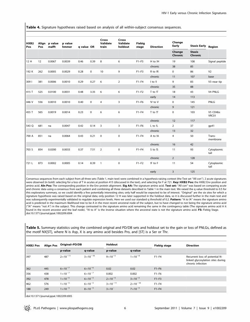
Table 4. Signature hypotheses raised based on analysis of all within-subject consensus sequences.
HXB2Pos
AlignPos
p valuemafft
p valuehmmer q value OR
CrossValidatetrain
CrossValidateholdout
Fiebigstage Direction
ChangeEarly Stasis Early Region
ChangeChronic
StasisChronic
12 H 12 0.0067 0.0039 0.46 0.39 8 6 F1–F5 H to !H 19 108 Signal peptide
chronic 38 85
192 K 262 0.0005 0.0029 0.28 0 10 9 F1–F3 R to !R 0 86 V2
chronic 11 107 base
309 I 381 0.0006 0.0010 0.29 0.27 6 2 F1–F4 I to !I 9 83 V3 near tip
chronic 35 88
415 T 525 0.0100 0.0031 0.48 3.35 6 6 F1–F2 T to !T 18 43 V4 PNLG
early 14 113
446 V 556 0.0010 0.0010 0.40 0 4 3 F1–F6 !V to V 0 145 PNLG
chronic 9 121
455 T 565 0.0019 0.0014 0.23 0 6 6 F1–F4 T to !T 0 103 V5 CD4bsVRC01
chronic 12 117
543 Q 681 na 0.0047 0.42 0.14 3 3 F1–F6 L to !L 2 37 gp41
chronic 13 32
700 A 851 na 0.0064 0.43 0.21 0 0 F1–F4 A to !A 4 50 Trans-membrane
chronic 16 42
703 S 854 0.0200 0.0033 0.37 7.51 2 0 F1–F4 S to !S 11 93 Cytoplasmictail
chronic 2 128
721 L 873 0.0002 0.0005 0.14 8.39 1 0 F1–F2 !F to F 11 54 Cytoplasmictail
early 3 125
Consensus sequences from each subject from all three sets (Table 1, main text) were combined in a hypothesis-raising context (the Test set ‘‘All con’’). 2 acute signatureswere observed (in bold): selecting for a loss of T in acutes at position 415 (discussed in the text), and selecting for F at 721. Key: HXB2 Pos: the HXB2 Env position andamino acid. Aln Pos: The corresponding position in the Env protein alignment. Sig AA: The signature amino acid. Test set: ‘‘All con’’ was based on comparing acuteand chronic data using a consensus from each patient and combining all three datasets described in Table 1 in the main text. We raised the q value threshold to 0.5 forthis exploratory summary, so we could identify a few potentially interesting sites; only half would be expected to be of interest. ‘‘Original’’ are the six sites for which asignature hypothesis was raised based on the original data; only position 12 H was later supported in the holdout data, so it is discussed further in the main text andwas subsequently experimentally validated to regulate expression levels. Here we used our standard q threshold of 0.2. Pattern: ‘‘A to !A’’ means the signature aminoacid is predicted in the maximum likelihood tree to be A in the most recent ancestral node of the subject, but to have changed to not being the signature amino acid(‘‘!A’’ means ‘‘not A’’) in the subject. This change contrasted to the signature amino acid remaining the same in the contingency table (The signature amino acid A itfound in the recent ancestor and the leaf node). ‘‘!A to A’’ is the inverse situation where the ancestral state is not the signature amino acid. FS: Fiebig Stage.doi:10.1371/journal.ppat.1002209.t004
Table 5. Summary statistics using the combined original and PD/DB sets and holdout set to the gain or loss of PNLGs, defined asthe motif NX[ST], where N is Asp, X is any amino acid besides Pro, and [ST] is a Ser or Thr.
HXB2 Pos Align Pos Original+PD/DB Holdout Fiebig stage Direction
p-value q-value p-value q-value
397 487 2610211 3610210 961025 161024 F1–F4 Recurrent loss of potential N-linked glycosylation sites duringchronic infection
362 445 661027 661026 0.02 0.02 F1–F6
356 438 161027 661027 0.002 0.002 F1–F6
392 478 161025 661025 261025 361025 F1–F3
462 576 161025 661025 3610211 2610210 F1–F4
188 249 161025 861025 36102 761025 F1–F4
doi:10.1371/journal.ppat.1002209.t005
HIV-1 Early versus Chronic Infection Signatures
PLoS Pathogens | www.plospathogens.org 6 September 2011 | Volume 7 | Issue 9 | e1002209

frequency of one of these patterns, the PNLG motif at position
397–399, is illustrated in Supplement Figure S2B–the PNLG at
position 397 was conserved overall (Fig. S2B), although it was
more likely to be present early in infection (Table 5, q-
value = 3610210 in the original data, 0.0001 in the holdout data).
One of the PNLG signatures, that enables glycosylation at position
392, is part of the monoclonal antibody 2G12 epitope [45,46,54].
Experimental data from Nab IC50 scores 2G12 from 113 clones
representing SGA clones from early transmission cases (Table S3),
and confirmed that the glycosylation motif at position 392 was
highly correlated with 2G12 neutralization (p = 0.006, Wilcoxon
rank test).
Identification of a complex signature near the CCR5Coreceptor-binding site (CCR5 CoRbs)
Clearly, analysis of single amino acid positions may miss
complex mutational patterns in functionally or conformationally
important regions. Given the vast number of combinations of
alignment positions and the range of different amino acids at each
position, we are limited in our ability to look at arbitrary
combinations of sites and amino acids across the full Env
sequence, , due to multiple test issues and limited power due to
sampling constraints (Table 1) compounded by computational
feasibility. Thus, we performed a focused in depth exploration for
signatures based on a small number of combinations of sites,
including only amino acids within narrowly defined sets of
functionally related sites [3] (Table S4). How extensively we
searched combinations of sites within these sets was determined
dynamically as described in the methods; however, at a minimum,
all combinations of up to 3 amino acids at each of 2 positions were
searched within each functional region, using a sliding window
approach to span different amino acid subsets and combinations
within each functional domain. These functional regions included:
the CD4bs in gp120; the CCR5 CoRbs region in gp120; positions
known to impact R5/X4 tropism; a subset of the V3 loop
positions; the b12 binding site in gp120; residues predicted to
reside at the gp120/gp41 trimer interface; the gp120 V2 region
implicated in binding the gut homing receptor; 2F5/4E10 binding
sites in gp41; the lentivirus lytic peptide LLP1 and LLP2 regions of
the gp41 cytoplasmic domain; and sites that have been related to
membrane fusion, including sites in which changes were shown to
result in increased or decreased entry (see Table S4 for positions
included). Despite this extensive search, only one statistically
significant association with a complex signature was identified and
validated in both the test data and holdout data; it was found in a
CCR5 CoRbs set and the signature was defined as: L122-[IV]201-
N377, with repeated mutation away from this pattern in chronic
samples. The statistical summary of this signature pattern is given
Figure 1. Mapping of signature sites (red) on the three-dimensional structure of gp120 (silver). A ribbon structure ofthe HIV-1 gp120 core +V3 in the CD4-bound conformation is shown inwhite. (A) Key residues involved in co-receptor and antibody (2G12,b12, b13 and F105) binding that are proximal to the position 415 areshown. Residues 295 and 332, that contribute to the 2G12 epitope, andresidue 444, that is important for co-receptor binding, are shown asblue balls. A motif spanning the region 417 to 421 (cyan color) that isproximal to position 415 and contains residues that take part in bindingto coreceptor (419), b12 (417–419), b13 (419–421) and F105 (421). CD4(orange) is shown for better visualization of receptor binding siteregion. (B) Locations of signature patterns involving glycan motifs (N-notP-[ST]). (C) Spatial locations of signature sites within a set offunctional sites (blue) associated with CCR5 binding. The 17b antibodyFab is included to mark the region in gp120 that takes part in CCR5binding. Signature sites are labeled with HXB2 reference numbers.doi:10.1371/journal.ppat.1002209.g001
HIV-1 Early versus Chronic Infection Signatures
PLoS Pathogens | www.plospathogens.org 7 September 2011 | Volume 7 | Issue 9 | e1002209

in Table 3, and the spatial locations of these sites are mapped on
gp120 in Fig. 1c. The CCR5 model set contains residues that are
proximal to the highly conserved critical residues that take part in
the binding to CCR5, but that are clearly amenable to positive
selection since they are variable at the population level.
Biochemical patterns in structure-based regional clustersIn our final exploration of this data, we searched for early
infection or chronic signatures defined by changes in amino acid
chemistry in spatially defined local regions. Our reasoning was
that transmission signatures would not necessarily have to involve
particular amino acid substitutions at a single site or a collection of
sites, but rather might reflect a complicated amino acid
substitution pattern that could in turn affect the structure or
chemical nature of specific spatial regions within the Env structure.
Such regional changes may impact expression or binding to
receptors and antibodies. To explore this possibility, we first
defined 395 contact sets of spatially defined clusters structurally
centered on the amino acids included in the X-ray structure of the
gp120 core from the YU2 strain [55], as described in the methods.
Each set contained a up to10 amino acids that were less than 10 A
from the center amino acid of the contact set, based on all-atom
molecular dynamic simulations. To capture the effects of dynamic
interaction between flexible and core regions, no distinction was
made for surface residues.
It was not feasible to analyze all neighborhood lists with all
combinations of explicit amino acid transitions, so we simplified
the data by calculating a regional additive polarity score for the
amino acids in each neighborhood cluster (see Methods). Unlike
the discrete change-stasis nature of the variables (acute versus
chronic) used for the other signature analyses in this study, this
score was a continuous variable, so we used the method of
phylogenetically independent contrasts [56] to identify changes in
polarity that correlated with early or chronic infection sequences.
Three statistically significant regions were identified (Table 6), and
mapped on the three-dimensional structure of gp120 (Fig. 3). In all
three regions, the region became more polar during chronic
infection. All three sets have amino acids that share or border the
binding sites of CD4, and b12 [57,58]. The polarity scores did not
correlate significantly with sCD4 or b12 neutralization when
compared the with experimental binding data (Table S3). Sets 270
and 368 border the highly conserved CD4 binding loop region
(HXB2 positions 364–373). Sets 362 and 368 consist of additional
residues from b23 strand and V5 loop region that take part in
binding to CD4 and b12. All three sets shared a three amino acid
segment (465–467) that constitutes part of the binding site for the
potent broadly neutralizing monoclonal antibody VRC01 [57,58].
Hypervariable loop length and number of glycosylationsite differences between acute and chronic samples
We tested whether the hypervariable regions V1–V2, V4, or full
gp120 revealed a pattern of reduced loop length or number of PNLG
sites in the acute/early samples relative to the chronic samples, as
would be expected from the literature [59]. When we compared the
distributions of all of the within-subject Env consensus sequences in
the acute/early versus chronic subjects, fewer PNLG sites overall
were found in gp120s from early infection (p = 0.008, Wilcoxon
signed rank test). There was also a trend towards fewer PNLG sites in
Figure 2. p- and q-values found in shuffling experiments in which the entire sequence signature strategy was repeated 10 timesafter randomizing the early and chronic designation of each subject. The black x’s represent the distribution of p- and q-values in the realdata, while the colored circles represent the findings for incremental inclusion of Fiebig stages 2–6 in shuffled data. The lower quadrant of part of thegraph is almost exclusively occupied by the real data, indicating a signature dependent on early versus chronic status; p-values of less than 1026 wererare in the randomized data, and value less than 1028 were exclusively found among real data classifications.doi:10.1371/journal.ppat.1002209.g002
HIV-1 Early versus Chronic Infection Signatures
PLoS Pathogens | www.plospathogens.org 8 September 2011 | Volume 7 | Issue 9 | e1002209

the V1V2 loops (Wilcoxon p = 0.03), as well as a trend toward
reduced V4 loop lengths ((Wilcoxon p = 0.03).
Signature analyses methods that did not incorporate aphylogenetic correction
Several other strategies were employed to look for signatures
among the sequences by treating the samples as independent, and
not accounting for phylogenetic relationships [60]. These methods
did not yield any consistent signature patterns between the
hypothesis-forming test (with a q-value of ,0.2) and hold-out sets
(with a q-value of ,0.3), although additional support for a
signature at position 12 was observed; these methods and results
are fully summarized in the Supplement (Text S1, Figs. S3, S4, S5,
S6, S7 and Table S8). In these analyses, a lack of concordance
between the hypothesis forming and test-sets could arise as a
consequence of a lineage effect dominating the signal in the
hypothesis-forming set; alternatively, the subjects and sampling
may have been too dissimilar to reproduce subtle effects.
Discussion
In this study we performed a comprehensive analysis of HIV-1
Env sequences to identify signature patterns in proteins that are
significantly different in chronic versus early sequences. Here we
focus on interpreting the strongly statistically supported signature
patterns in the context of what is known about the biological role
of these sites.
Signature sites in the signal peptide and cytoplasmicdomain
It was intriguing that among the 25 significant signatures
identified upon combining all of the data (Table S2), 3 were located
in the signal peptide of gp160, and 4 in the cytoplasmic domain. The
recurrence of patterns of mutational change in these two regions
during chronic infection raises the possibility that they may indirectly
influence immune evasion by altering Env protein folding,
modification or expression levels. The signal peptide directs Env in
its co-translational translocation to the endoplasmic reticulum (ER),
where it undergoes further folding, glycosylation, and trimerization
[61]; it may also serve as a gatekeeper for the release of correctly
folded proteins [62]. It is unusually long (30 amino acids on average),
and contains a number of highly charged residues in the N-terminal
region [63,64] spanning position 12, one of our most robust
signatures (Table 2). Signal peptides play a role in the efficiency of
the protein secretion and in orienting proteins in membrane,
influence folding and the exit from the ER [65,66], and can impact
cleavage rates [63,67]. A slower cleavage rate down-regulates the
rates of folding, intracellular transport and secretion [63,65,68,69].
The Env cytoplasmic domain of HIV-1 is also unusually long; at
150 amino acids long, three times longer than that found in typical
lentiviruses [70]. It contains three helical fragments called
lentivirus lytic peptides (LLPs) [71] that have been implicated in
cell surface Env expression [72,73], incorporation into virus
particles [74,75], fusogenicity [76,77], and Env’s localization in
lipid rafts [71]. The chronic infection signatures in the cytoplasmic
tail (Table S3) are all concentrated on the LLP-3 segment. This
segment has a strong potential to associate with and perturb the
membrane [78], and a di-aromatic motif of Y802 W803 in this
region has been associated with retrograde transport of Env to the
trans-Golgi network [79].
The acute signature site at position 415!T415 was strongest early sequence signature observed, indicat-
ing that the PNLG at 413–415 is selected against at or immediately
after transmission. This PNLG is glycosylated when present [80],
and is located near the C terminal end of the V4 loop, proximal to
both the CCR5 CoRbs and the CD4bs regions that impact both
antibody access (Fig. 1a). A highly conserved sequence motif that
takes part in CCR5 binding, RIKQ (HXB2 419–422), is just a few
residues upstream [43,44,81]. The conserved sequence motif PCR
(HXB2 417–419) that participates in the binding to monoclonal b12
is also in the neighborhood of this site [58], consistent with our
finding that the presence of the PNLG motif at 413–415 is highly
correlated with reduced b12 susceptibility. The glycosylation site at
413–415 has repeatedly been singled out as a relevant immune
escape site in recent neutralizing antibody studies. Acquisition of a
PNLG at 413–15 has been demonstrated to confer escape from
autologous antibodies in longitudinal studies of the trajectory of
escape in both an HIV-1 infected person (David Montefiori,
personal communication), and in a rhesus macaque infected with
SIVmac239 [82]. Furthermore, this region in association with the
C3 a-2 helical domain is thought to contribute to patterns of
neutralization susceptibility [83,84,85].
Two studies have found the presence of a glycosylation site 413–
415 to be associated with virus isolated from individuals capable of
eliciting potent or broadly neutralizing antibodies [40,86]. This
correlation was proposed to either result from a recurrent pattern
of escape in people who make potent broad neutralizing
antibodies, or as common feature in Envs able to elicit good
antibodies [40]. We have tested a strain that has the glycosylation
site at 413–415 present (strain CH0219), isolated from an
individual who had made very potent broadly neutralizing
antibodies in response to infection [40]. This Env was resistant
Table 6. Summary statistics regarding changes in regional hydrophobicity associated with chronic infection.
Data Analysis Set numbera Original+PD/DB HoldoutCorrelationCoefficient Original
CorrelationCoefficient Test Direction
p-value q-value p-value q-value
Change in Polarity 270 1610212 1610211 0.04 0.01 0.64 0.18 Chronic sets aremore polar
368 161026 161025 0.05 0.01 0.47 0.18
362 161024 161023 161024 161023 0.38 0.34
aSets of amino acids including in the three statistically interesting regions. These tests compared sequences from all Fiebig stages, F1–F6, to chronic samples.Spatial Region 270: I359,T358,I360,E466,N397,K357,F396,S465,A346,I467,F361.Spatial Region 368: S465,E466,E464,T358,K357,N463,N462,I359,I467,I360,G459.Spatial Region 362: G459,G458,N460,D457,S461,N462,E466,I467,R456,N463,S465.doi:10.1371/journal.ppat.1002209.t006
HIV-1 Early versus Chronic Infection Signatures
PLoS Pathogens | www.plospathogens.org 9 September 2011 | Volume 7 | Issue 9 | e1002209

to autologous antibodies in sera from CH0219, supporting its role
in antibody escape. Furthermore it was found to be an extremely
poor immunogen for eliciting neutralizing antibodies in guinea
pigs (BFH, unpublished data). These findings are consistent with
the intuitive hypothesis raised by our current signature analysis,
that the addition of a glycosylation site at 413–415 provides a
common escape mechanism during chronic infection by blocking
access to a key epitope, but that it is selected against in early
viruses, resulting in the observed !T415 signature pattern.
Implications of the repeated patterns of loss ofglycosylation motifs during chronic infection
Changes in glycosylation play a key role in chronic infection,
and either the gain or the loss of a particular glycosylation sites can
Figure 3. Three statistically significant structures-based regional clusters in gp120 (white) associated with changes in polarity.These regional clusters occur near the CD4-binding site (orange) shown in (A). The CD4-bound conformation of the HIV-1 gp120 core+V3 is shown,from the perspective seen by CD4. The three clusters (B–D) are shown in red. The residues that form these sets are shown in panel (E). All maps arebased on HXB2 numbering.doi:10.1371/journal.ppat.1002209.g003
HIV-1 Early versus Chronic Infection Signatures
PLoS Pathogens | www.plospathogens.org 10 September 2011 | Volume 7 | Issue 9 | e1002209

both result in immune escape [32,87]. As discussed earlier,
reduced loops lengths and numbers of PNLGs are characteristic of
early viruses, and although the pattern can be subtle and difficult
to discern in the B subtype [34,35], we did find supporting
evidence for an overall pattern of reduced numbers of PNLGs
after transmission in this data set; this reduction in PNLG sites
occurs in the hypervariable loops. In contrast, most of the specific
signature PNLGs we have identified are clustered in the outer
domain, and these are lost not at transmission but in the course of
chronic infection (Fig, 1b). The statistical counterpoint to the
chronic loss-of-glycosylation-motif signatures is relative conserva-
tion of these PNLG sites at transmission, consistent with a scenario
that these specific sites facilitate transmission in early infection,
and their loss contributes to immune escape in chronic infection.
Several of the signature PNLGs have known functional roles
which support the scenario described above. First, the glycan at
N188 facilitates interactions with CD4 and CCR5 [88], and the
loss of glycosylation sites in this region have been associated with
diminished replicative capacity [55,57,58,89]. Changes in this
region have also been associated with immune escape from some
of the first neutralizing antibodies in natural infection [33,85], and
a glycan knock-out at position 188 impacts the neutralization
potency of the recently isolated broadly neutralizing antibodies
PG9 and PG16 [90]. Thus selection for the glycan may occur at
transmission, and selection away from in during immune escape
from antibodies similar to PG9 and PG16. Similarly, N362 has
been shown to contribute specifically to enhanced fusogenicity
[91], a property that might be favored during transmission. PNLG
362 and 462 are near the CD4bs, and the b12 and VRC01
monoclonal antibody binding sites [55,57,58], and the CD4bs is a
common target of neutralizing antibodies in natural infection
[92,93]. Finally, the PNLGs at positions 392, 397 and 356 are all
part of the ‘‘silent face’’ of gp120 [94,95]. The oligomannose
glycans that are clustered on the silent face of HIV are ligands for
DC-SIGN, a lectin found on the surface of dendritic cells [96].
Dendritic cells encounter HIV soon after mucosal exposure [97],
and may have a role in enhancing the efficiency of HIV
transmission [88,91,98]. A mannose at position 392 is also a
critical component of the epitope of the neutralizing antibody
2G12 [45,54], and our data confirm this previously well-
established relationship. Although the 2G12 epitope may not be
a common a target of neutralizing antibodies in natural infection
[99], antibodies to the 2G12 epitope in neutralizing sera have been
found in long-term non-progressors [100], suggesting the glycan
shield at the silent face of HIV can be a point of vulnerability in
some circumstances. Creating high-density mannose clusters that
mimic HIV’s glycan shield are being explored as a vaccine strategy
[101,102].
Complex chronic signatures in localized regions of EnvDespite testing for complex multi-site signatures within several
functional domains in Env, only one multi-site signature was
identified, a chronic signature in the CCR5 CoRbs set (Table 3).
The CCR5 CoRbs can be a target for broadly neutralizing
antibodies [92,93], and non-neutralizing antibodies against the
CCR5 CoRbs may also be able to impose selection on the virus
[103]. Interestingly, the only identified signatures found associated
with Env glycoproteins that were isolated from individuals that
made broad and potent neutralizing antibody responses were also
localized in the CCR5 CoRbs [40]. We also tested for distinctive
biochemical patterns in local spatial regions in the gp120 structure,
and identified three regions that are proximal to the CD4 binding
site [57,58] that undergo change in polarity (Fig. 3). The regions of
gp120 surrounding the CD4bs are the most conserved in Env
when considered at a structural level [3], thus providing a
vulnerable target for cross-reactive HIV antibodies [57,58].
Changes in electrostatic potential may enable antibody escape
from at least some antibodies in HIV-infected individuals who
naturally mount a potent and cross-reactive anti-CD4bs antibody
response [93,99,104,105].
A summary viewWhile the signature patterns we have identified are significantly
enriched in terms of association with either early or chronic
viruses, still there are exceptions to any given pattern (Table 2–5),
and thus the signatures cannot be used to accurately predict
whether a given sequence is derived from an acute or chronic
infection. This is not surprising, but worth noting. It is a reminder
that tests that involve site-directed mutagenesis might fail to result
in a phenotypic change even when a site is relevant, because the
phenotypic consequences of change in a single amino acid can be
context dependent. Furthermore, there may be multiple paths to
the same end, and the immune responses that drive repeated
patterns of escape in chronic infection are likely to be shared only
by a subset of individuals who target a particular Env region.
Similarly, reversion in early viruses is likely to be context
dependent, depending on the presence of compensatory mutations
as well as other selective pressures acting on the virus. It is also of
interest that some signature patterns that might have been
expected were not observed. We did not see amino acids in the
V3 loop that have been noted to be associated with CCR5 co-
receptor use predominate in acute infection [17,23,26,37] or those
associated with CXCR4 use in chronic infection [106,107]. We
think this is because of inadequate statistical power: CXCR4-using
viruses rare among both our early and chronic sequences (Table
S4) and there are multiple ways to manifest a CXCR4 phenotype,
thus it is likely that no CXCR4-associated substitution was
repeated enough to enable identification of a signature.
Despite these issues, several interesting and consistent signature
patterns emerged through our study. First, multiple signal peptide
and cytoplasmic domain signature patterns were found (Table S2),
raising the possibility that Env expression levels may be an
important generalized aspect of immune escape during chronic
infection. Second, two signatures were found near the CCR5
CoRbs region; this domain is emerging as a key region for
neutralizing antibody escape and induction of antibodies in a
number of studies, and merits close attention as vaccine design and
evaluation strategies progress. Third, the recurrent loss of
glycosylation sites in key positions during chronic infection
suggests that this pattern typifies an essential aspect of immune
escape, leaving a profound and recurring trace at the population
level. If the loss of these specific glycosylation sites mediates
immune escape from common transmitted forms, in may be
advantageous to include these sites in vaccines. In contrast, the loss
of the PNLG at position 413–415 was enriched among early
sequences, so it may be advantageous to also exclude PNLGs at
413–415 from a vaccine immunogen. Thus the signature patterns
identified in this study point to post-translational regulation of Env
having a role in selection of early sequences, and indicate
particular protein modifications that merit consideration for
immunogen design and evaluation.
Methods
Ethics statementWritten informed consent was provided by all study partici-
pants. The Duke University Health System Institutional Review
Board for Clinical Investigations (DUHS IRB), has determined the
HIV-1 Early versus Chronic Infection Signatures
PLoS Pathogens | www.plospathogens.org 11 September 2011 | Volume 7 | Issue 9 | e1002209

specific components above under the protocol, ‘‘Acute HIV-1
Infection Prospective Cohort Study’’ (CR3_Pro00006579) to be in
compliance with all applicable Health Insurance Portability and
Accountability Act ("HIPAA") regulations.
Data setsThe acute samples were collected from individuals sampled at
varying time post-infection, and were clinically staged according to
Fiebig et al. [17] to estimate the time between infection and
sampling [17,21,22]. Chronic samples were selected from
individuals who were not on anti-retroviral therapy, and infected
for a minimum of two years. All represented subtype B infections,
and most samples were collected in the United States, although a
small number were from Trinidad/Tobago, included to increase
our sample size and power (Table S1). This was a retrospective
study involving many cohorts, to enable us to get a large enough
sample to perform signature analysis. Table S1 includes
demographic and clinical information related to these samples,
including viral load at the time of sampling, Fiebig stage, year of
sample collection, sampling country, primary risk factor for
infection, and whether the sequence evidence indicates that the
new infections were established by single or multiple strains. All
acute and early samples were obtained from people with sexually
acquired HIV. Alignments of the full set of 6303 early and chronic
SGA Env sequences used are available in the supplement, and
GenBank accession numbers are provided in each of the sequence
names (Tables S5–S7). As this study involved samples from HIV-1
infected human subjects, informed consent was obtained from all
subjects.
The data were originally separated into two sets: the original
hypothesis-raising ‘test’ set, the ‘holdout’ hypothesis-validating set.
It was critical that the test and holdout sets each had a good
representation of early Fiebig stages, so we ensured that the test
and holdout sets each had 19 samples with a Fiebig stage of 3 or
less. Each set was also matched for samples that were suggested by
the data to be consequence of single infection (68% in the test set,
and 65% in the holdout). The early and chronic groups within
each set were matched in terms of country of origin (the early and
the chronic groups each had ,30% from Trinidad in the test set,
and the early and chronic groups each had ,5% in holdout set);
this was important because the Trinidad sequences formed a
distinct clade in phylogenetic analysis and such geographically
localized clades can have systematically different patterns of
mutations in early or chronic infections. Although these were
sexual transmission cohorts, the risk factors for infection were not
always known; heterosexuals were well represented in each group.
A third set was added to increase our statistical power for
hypothesis forming (Table 1). This set was based on adding early
infection samples from plasma donors in the United States, and a
set of B clade chronic sequences from the Los Alamos HIV
database that were from individuals who were documented in the
database entry to not be on anti-retroviral therapy and who had
been infected for a minimum of two years. This third set was not
as well matched in terms of the clinical and geographic origin as
other two sets.
Sequencing and sample characterization methodsAll sequences were obtained from plasma of infected individuals
using single genome amplification (SGA) methods, as previously
described [17,38]. A full alignment of all sequences used in this
study is available in Supplement Table S5–S7; all sequences
have been submitted to GenBank in conjunction with this paper,
or else were previously submitted, and the accession number of
each sequence is included in the sequence name, and at the end of
this article. The positions numbers in the paper are generally given
as HXB2 position numbers (http://www.hiv.lanl.gov/content/
sequence/HIV/REVIEWS/HXB2.html), unless it is specified in
the text that the numbering refers to the alignment position used in
this study. For signature analysis, all sequences were analyzed in
maximum likelihood trees, including multiple sequences from each
individual; subject-specific phylogenetic clusters were consistently
formed, so there were no overt contamination issues in this study.
Sequences were aligned using a HMMER alignment [47] and
then codon aligned with GeneCutter (http://www.hiv.lanl.gov/),
with hand correction at the borders of the regions with many
insertions and deletions to rectify obvious alignment errors. The
hand editing was done because the hypervariable region indels in
HIV are particularly difficult for multiple alignment programs
[48]-ot only do they exhibit extensive length variation, but the
insertions are generally comprised of distinctive direct repeats from
neighboring regions in the gene [108]. The alignment was done in
iterative steps; first each subject was aligned internally, then a
majority consensus sequence representing each subject was
generated. For within-subject consensus generation, we considered
the codons that that bases were imbedded in, and selected the
most common codon for the consensus. This step was required
because otherwise simple position-wise consensus sequences
occasionally created codons that did not exist within the subject,
as the most common bases in highly variable codon positions are
not always found in combination. The subject consensus sequences
were aligned, then the within-subject sequences sets were aligned
to their own consensus in the framework of the full population
alignment, and then the whole process was iterated. This
alignment was 3120 bases long. To test for dependence on the
alignment strategy used, we repeated the consensus sequence
signature analysis using an unedited MAFFT alignment [48,49];
this alignment was 3735 bases long, so had many more gaps; a
SATe alignment of this same data was even longer, at 3790 bases
(http://phylo.bio.ku.edu/software/sate/sate.html).
Phylogenetically-based analysisTo identify signature patterns in HIV that relate to a particular
phenotype (in this case, early versus chronic status), sampled
viruses cannot be treated as independent samples from a random
distribution of genotypes. Any population substructure in the data
exacerbates the problem. To correct for this we employed a tree
corrected contingency table approach used previously [39], but
with the addition of more extensive searching capabilities such as
the ability to look for statistically interesting combinations of sites
in functional domains and loss and gain of glycosylation site motifs
[40]. The phylogeny of all sequences was inferred using a
maximum likelihood method, and ancestral states were inferred at
the internal nodes in the tree [39]. We used a GTR model and a
maximum likelihood assignment of rates per site.
The method we originally developed to study the correlation
between HIV genotypic variation and host immunological
parameters was used directly to correlate the early/chronic status
with the consensus genotype in each patient. This method has
been previously shown to enable identification of signature sites
that could be experimentally validated as biologically meaningful
[39,40,109]. In particular, when applied to finding mutational
associations with host class I HLA genotyping, the associations
identified were in known or predicted cytoxic T cell epitopes with
the expected frequency [39,109], and when applied to neutralizing
antibody sensitivity, critical mutational patterns were identified
among the natural variants [40]. To fully utilize the availability of
multiple sequences per subject in this study, we have adapted the
original signature identification method to enable tracking of
HIV-1 Early versus Chronic Infection Signatures
PLoS Pathogens | www.plospathogens.org 12 September 2011 | Volume 7 | Issue 9 | e1002209

changes in character states observed within each individual
defined relative to the most likely state at the last (closest) ancestral
node outside the patient. These changes were correlated with the
patient being early or chronic (Fig. S1 illustrates the method). The
number of sequences sampled varied widely among the patients
(indicated by the heights of the bars in Fig. S2), and the diversity at
some positions was much greater than others, so a bootstrap
approach was used to determine appropriate significance levels for
identification of interesting signals (Fig. 1).
For quantitative signatures of continuous variables (in particu-
lar, regional polarity scores) Felsenstein’s phylogenetic contrast
approach was used [56] to estimate a covariance matrix, and
Student’s t-statistic was used to obtain significance levels for the
differences between the early and chronic patients. Since the
variables of interest had a bounded domain, we verified manually
that the signatures did not arise from saturation of the bounds
where the model was strongly violated.
Statistical testing criteriaGiven that we were in a hypothesis raising mode and our
expectation was that transmission signatures would be relatively
subtle, and we were of necessity in a framework of limited
sampling, we decided to require that associations be statistically
supported in an initial training data set with a q-value of ,0.2, and
show at least a trend (q ,0.3) towards a consistent pattern of
association in a separate analysis of the holdout data. A q-value is a
false discovery rate that adjusts for multiple tests [41]. There were
many associations with a q,0.2 that were found either only the
training or confirmatory data sets that were not supported in both
sets, which we do not list here. Retaining a holdout set that is
excluded from the initial analysis is not often done in this kind of
correlation analysis [109,110]. Our decision balanced the value of
increasing the sample size and the potential for identifying more
correlated sites, with the additional level of confidence in our
primary findings provided by the holdout set analysis; we opted for
the latter to limit our type I false-positive error, although
potentially missing interesting signature sites and increasing type
II false-negative error. A more comprehensive listing of the non-
validated sites provided in the supplement reverses this, and these
tables are far more likely to contain false positives, but less likely to
miss true positives.
Signatures were sought comparing sequences classified as early
by combining data sets incrementally from Fiebig stage 2 up to 6,
such that all sequences up to a given stage were combined and
then analyzed, and then contrasted with chronic data. The reason
we explored the data in this increasingly inclusive fashion was to
balance the increasing power that is a consequence of including
additional sequences from later Fiebig stages, against the
possibility that as samples are taken at progressively later Fiebig
stages, transmission signatures may no longer be evident in the
sample due to early immune or fitness selection pressures [23,30].
The Fiebig stage of the data combination that produces the most
significant signature associations for a given amino acid pattern is
provided in the Tables manuscript to simplify presentation; the use
of q-values for statistical significance guards against increasing
Type I errors by this procedure.
Cross-validation strategies can provide reasonably unbiased
accuracy estimates for classifiers [111], but their use in hypothesis
testing suffers from the absence of reliable estimates of their
variance [112,113,114]. In particular, they are known to have
inflated type 1 error rates [115,116] and can sometimes lead to
incorrect choice [117] when used for model/feature selection; we
have encountered such issues in a previous study [40], hence we
did not use this approach initially for this study, rather we used the
strategy described above involving a strictly maintained holdout
set. When very few signatures were evident by this conservative
approach, however, we turned to cross-validation; even though it
has limitations, it often works well in practice [118] and is
commonly used for data mining. We used a stratified 10-fold cross
validation approach [119] to check robustness of our findings
when analyzing the combined test and holdout datasets, to raise
hypotheses for further work. We stratified by the early/chronic
status, as well as by the sample’s geographic origin (i.e. whether the
sample originated in the United State or in Trinidad and Tobago,
given that Trinidad and Tobago B subtype viruses formed a
distinct lineage relative to the B subtype US viruses). 90% of the
sample was selected randomly for a training set, and 10% was
retained as a test set. As with the full data set analysis of all patient
consensus sequences, a q-value of 0.5 was used for the training set
criteria of positive, and the test was considered a match if the
direction of the odds ratio was preserved (,1 or . = 1).
Grouping of positions and amino acids for signatureanalysis based on alignment positions
Our primary analysis was concerned with single site signatures.
In addition to the single site signatures we considered the loss or
gain of aligned PNLG motifs, where the motif is: NX[T/S], and N
is Asn and T/S is either Thr or Ser [120]. Regions of the
alignment that could not be reliably aligned due to insertion/
deletion events were essentially excluded, by systematically
excluding positions where more than 10% gaps had been included
to maintain the alignment. One important consequence of this is
the exclusion of hypervariable domains where we did not feel
confident of the alignment, so associations would be missed in
these regions as they could not be reliably identified.
Signature analysis of combinations of sites in functionaldomains of HIV-1 Env
We also defined sets of amino acid based on the computed
structure and presumed function of the envelope protein. Three
sources were used to define these sets, as described in Korber and
Gnanakaran [3]. A search of the literature provided critical
residues obtained through site directed mutational experiments
that probed sites within functional domains as well as antibody
binding motifs in gp120 and gp41. We compiled those sites
classified according to corresponding functional activities and
antibody epitopes. Second, x-ray crystal structures of gp120 are
available with different binding partners, including neutralizing
monoclonal antibodies. In these cases, we identified the set of
relevant key sites based on spatial contacts.
The amino acid positions included in these sets, and the
references used to select them, are provided in the supplement
(Table S4). The functional domains in gp120 that we considered
included CD4 and co-receptor (CCR5 and CXCR4) binding sites,
sites that correlate with CCR5 and CXCR4 co-receptor usage,
exclusive sites within V3 loop that take part in binding to co-
receptor, and V2 gut mucosal homing receptor binding sites. In
gp41, we included sites in LLP1 associated with virion incorpo-
ration, LLP2 sites associated with Tyrosine-dependent sorting
signal and exposure of CD4 binding site, and an additional set of
sites associated with modulating entry during fusion process. We
also included a set of amino acid positions in both gp120 and gp41
thought important to maintain the Env trimer, and those sites that
lie on the interface between gp120 and gp41. The gp120 epitope
sites included the binding sites of monoclonal antibodies b12 and
17b. In gp41, the epitope sets included two sets in MPER region
covering 4E10 and 2F5 binding sites.
HIV-1 Early versus Chronic Infection Signatures
PLoS Pathogens | www.plospathogens.org 13 September 2011 | Volume 7 | Issue 9 | e1002209

We looked as exhaustively as was feasible given our data and
computational constraints for early or chronic signatures in
functional domains. How extensively the combinations of sites
and amino acids in a given functional domain were explored was
determined dynamically. All sets were initially explored based on
combinations of 3 positions in the functional domain and up to
three amino acids per position; if this resulted in more than 5
million patterns, we then considered only 2 positions and 3 amino
acid combinations in the first series of tests. We then tested
incrementally more combinations of amino acids at the each of the
positions until we reached 5 million patterns per domain, a limit
based on computational feasibility; however, if at this point the p-
and q-values were still improving, we increased this to up to 10
million tests. We then repeated the incremental iterations
including more sites rather than more amino acids per site. When
this was done, the combinations with the best p- and q-values were
compared between the test and holdout sets; then end result was
that essentially only one complex combination signature, in the
CCR5 model set, was supported in both sets.
Contact matrix based signature analysisA third kind of amino acid set analyzed was based on spatial
proximity; we called these ‘contact’ sets. These sets were created
from the contact matrix obtained from long timescale molecular
dynamics simulations of liganded gp120. The gp120 structure of
YU2 strain with modeled loops was carried out with molecular
dynamics simulations in explicit aqueous solvent [79,80], thus
incorporating into our model the dynamics, the influence of
solvent, relative flexibility of both flexible and conserved regions
and the interaction between core and variable regions. Contact
profiles were obtained from the simulation trajectories. For each
residue in the simulated structure a contact set was generated such
that it contained at most the 10 closest contact amino acids, and all
amino acids included were within 10 A of the center. We made a
total of 395 contact sets corresponding to the total number of
residues in the simulated gp120 molecule informed by the crystal
structure. The definition of these contact sets was based both on
the distance between amino acids obtained during the entire
dynamics and the duration in the dynamical conformation. Sets
were excluded from consideration if they contained regions of
alignment uncertainty caused by insertion/deletion events.
It was not computationally feasible to analyze all contact lists
with all explicit amino acid substitutions. Therefore a few contact
sets were chosen for an in-depth analysis based on the full tree
single amino acid scan identifying an amino acid within the
contact set as potentially interesting. Combinations of positions in
the sets containing these positions were analyzed in the same
manner as the functional domains; this yielded no complex
signatures that were supported in both the test and the holdout
sets. We then simplified the information in the contact sets by
grouping amino acids into standard side-chain chemically
motivated equivalence classes, J = [A I L M F W V], X = [S Y
T Q N H], Z = [K R], O = [D E] and U = [G P C], and their
unions, and used this to test of complex signatures within all
contact sets; this effort identified no new signatures in both test and
holdout analysis. We then computed a polarity score for each of
the contact sets, a single number representing the chemistry of
each local spatial region in gp120. To do this, we used the Hopp
and Woods scale, which has been used previously to identify
antigenic sites [121], to assign scores to individual amino acids,
and then summed these scores over the contact sets. In this case
three contact sets yielded statistically interesting correlations in
both the test and hold out sets. Because this score could vary
almost continuously through small changes in amino acid
composition, we used the method of phylogenetically independent
contrasts [56] to identify changes in polarity that either correlated
with transmission or were recurrent during chronic infection based
on the full dataset. Though the range of polarity is finite, violating
the assumptions of the method, however we found the observed
signatures did not arise from saturating the bounds.
Correlation of signature sites with neutralization byantibodies and sCD4
For each of a panel of MAbs, or sCD4, concentrations required
for 50% neutralization (IC50) were determined for 113 SGA-
derived Envs expressed as pseudovirions [122] from 73 individuals
sampled either in early or chronic infection (Table S3). This
represents an extension of the set previously reported in Keele et
al. [17], using the same experimental methods. To determine if
there were significant correlations between the presence or
absence of signatures patterns and neutralization phenotypes we
used non-parametric Wilcoxon rank statistics as implemented in
the R project for statistical computing http://www.r-project.org/).
Testing for correlations of between lengths and numberof glycosylation sites in hypervariable loops and earlyversus chronic sampling
Because of pre-existing literature on this subject leading to an
expectation that early Env hypervariable loops would be shorter
with few glycosylation sequons, we grouped all early and all
chronic samples for this study, and did not separate our data into a
hypothesis forming and holdout sets. Furthermore, since we have
no good models to reconstruct ancestral states for the variable
loops that are subject to rapid within-subject insertions and
deletions, in this study we did not correct for phylogenetic
relationship between the sequences. Instead, we compared the
tallies of number of glycosylation sites or loop lengths based on a
single consensus sequence from each subject in early versus
chronic infections using a Wilcoxon rank statistic; this simple test
revealed there were less glycosylation sites overall in gp120 among
early infections, supporting previous findings. We next compared
the spectrum of variants found in each subject. Because the within-
subject sequences are not independent and the number of such
samples varied widely from patient-to-patient, we re-sampled the
sequences from each subject 1,000 times to create sets with a
constant sample size across subjects, which we chose to be the
smallest number of sequences obtained from a single subject in the
real data. We then compared the distributions found in the early
versus chronic data with a Wilcoxon test, and then did a Monte
Carlo test shuffling the early/chronic designations 1000 times
based on each of the re-samplings, to see how often the level of
distinction based on the real data was found in the randomized
data.
GenBank accession mumbersGenBank accession numbers for sequences used in this study.
New sequence accession numbers for newly introduced sequences
in this study: HQ216367-HQ218052, HQ238279-HQ238288.
Previously published CHAVI sequence accession numbers: EU-
574937-EU575065, EU575067-EU575212, EU575214-EU57-
5231, EU575233,EU575235-EU575251, EU575253-EU575265,
EU575267-EU575272, EU575274-EU575441, EU575443-EU57-
5468, EU575470-EU575552, EU575554-EU575636, EU575638-
EU575704, EU575706-EU575775, EU575777-EU575852, EU-
575854-EU575943, EU575945-EU575980, EU575982-EU57-
5990, EU575992-EU576064, EU576066-EU576089,EU576091-
EU576237, EU576239-EU576292, EU576294-EU576296, EU-
HIV-1 Early versus Chronic Infection Signatures
PLoS Pathogens | www.plospathogens.org 14 September 2011 | Volume 7 | Issue 9 | e1002209

576298-EU576619, EU576621-EU576642, EU576644, EU57-
6646-EU576774, EU576776-EU576799, EU576801-EU576814,
EU576816-EU576817, EU576819-EU576840, EU576842-EU57-
6936, EU576938-EU577005, EU577007-EU577100, EU577102-
EU577114, EU577116-EU577310, EU577312-EU577350, EU-
577352-EU577433, EU577435-EU577440, EU577442-EU577-
478, EU577480-EU577662, EU577664-EU578089, EU578091-
EU578109, EU578111-EU578174, EU578176-EU578239, EU-
578241-EU578292, EU578294-EU578307, EU578309-EU57-
8321, EU578323-EU578328, EU578330-EU578331, EU578333-
EU578375, EU578377-EU578512, EU578514-EU578559, EU-
578561-EU578576, EU578578, EU578580-EU578636, EU57-
8638-EU578677, EU578679-EU578686, FJ495818, FJ495937,
FJ496000-FJ496001, GU330247-GU330646, GU330648-GU33-
0861, GU330938-GU331030, GU331032-GU331095, GU331-
098-GU331102, GU331114, GU331116, GU331121-GU331122,
GU331127, GU331129-GU331133, GU331183-GU331217,
GU331634 GU331721. Previously published database chronics
selected from the Los Alamos based on their annotation clearly
indicating chronic infection, including one representative sequence
chosen per subject sample: AY223734, AY223785, AY535433,
AY535511, AY535480, AY535461, AY842807, AY842816, AY-
842830, DQ410613, DQ410520, DQ410533, DQ410542, DQ-
410569, DQ410586, DQ410599, AY357342, DQ410219, DQ-
410057, DQ410068, DQ410075, DQ410099, DQ410105, DQ-
410120, DQ410177, DQ410196, DQ222216, DQ410454, DQ-
410483, DQ410623, DQ410280, DQ410322, DQ410338, DQ-
410384, DQ410412, AY423385, DQ410232, DQ976380, DQ-
976394, AJ535593, AJ535602, DQ853433, DQ976430, AY-
314044.
Supporting Information
Figure S1 Phylogenetic tree of all sequences in thisstudy, highlighting the signature at position 12. This
figure illustrates the abundance of the underlying data, the within
sample diversity found in early and chronic subjects highlight-
ing typical examples, and the basic principals of our signature
identification strategy when all available sequences in the data set
are used. The branches of this maximum likelihood phylogenetic
tree are shown as black lines. If sequences were isolated from an
early sample, they are labeled with a magenta line to the right of the
leaf node of the tree. If the sample was chronic, the sequences
are labeled with a grey line. The different amino acids found in
sequences in position 12 are represented by different colors, thus a
bright spot of color at every leaf node indicates the amino acid found
in position 12 in a sampled sequence, and a bright spot on an
interior nodes represents the most likely amino acid found at that
node based on the phylogenetic model. A His is the signature amino
acid of interest for this comparison, it is labeled bright red. There
are thousands of sequences in this tree, thus it is impossible to see
detail, so the region boxed in red was enlarged to illustrate the
method we used to identify signatures. A representative acutely
infected subject was boxed in green, and further expanded. The
ancestral node just prior to the infection in position 12 was predicted
to be a His (H12), and H12 was also the most likely amino acid in
the recent common ancestor sequence (MRCA), or founder virus, in
this subject. H12 was also found in all other sequences sampled in
this subject. Thus this subject would be counted as an early subject
with H12 carried in at transmission, that remained unchanged (H
stasis), i.e. no H to !H changes in position 12 were observed between
the last ancestral node outside the subject, through the MRCA, and
out to all of the sampled sequences from within the subject. In
contrast, a chronic infection case boxed in blue represents a typically
diverse population of sequences found in chronic infection. Again,
the ancestral state at the node immediately preceding the infection,
as well as the MRCA of the subject’s sequences, were both most
likely to be a His (the red X on the left hand side of the blue box).
This subject would be counted as a chronic subject with H12 as the
most recent ancestor prior to transmission, and would contribute a
total of 2 ‘‘H to !H’’ changes to the total tally of chronic infection H
to !H changes. This is because in this subject an H12 ancestral state
was predicted to have changed to Pro, represented by the yellow
‘‘0’’, twice independently. It is not possible to distinguish whether
within-subject recombination carried the mutation into two
lineages, or if it arose from two distinct convergent mutations, but
by either mechanism the Pro arose in two distinctive lineages in the
within-subject clade. To search for significant signatures, similar
tallies were made across all patients for every amino acid at every
position, Fisher’s exact tests were performed, and q-tests were
conducted to control for multiple tests.
(EPS)
Figure S2 Distributions of amino acids in each subjectfor representative signature patterns. (A) Each vertical bar
represents a subject, and the height of the bar indicates the
number of sequences sampled. The subjects are grouped accor-
ding to the data set breakdown in Table 1. A solid color in a bar
indicates the single amino acid found in that subject, while bars
with multiple colors indicate that multiple amino acids are found
in the position, and the contribution of each color to the bar
indicates how often each amino acid is found. The color key shows
which amino acids are represented by which color. The signature
of interest at position 12, His, is black, and as is illustrated here, is
more common in early than in chronic infection. The pattern
illustrated here, with His the most common amino acid in position
12, is shared by most subtypes (A, B, D, F and G), but in the C
subtype, Gln dominates www.hiv.lanl.gov). Thus, there is likely to
be a subtype-specific context relevant to this signature. (B) The
basic structure of this figure is like (A), but instead of amino acids
the colors indicate the presence or absence of the PNLG motif at
positions 397–399. This again illustrates the complexity of the
sample, yet signature motif is clearly frequently lost in chronic
infection. Chronic sequences from the database that met our
inclusion criteria included a small number of chronically infected
elite controllers; these sequences had unusual patterns so they are
marked in both (A) and (B).
(EPS)
Figure S3 Difference in estimated probabilities ofmismatch. For each amino acid position, a test statistic was
computed to evaluate whether the frequency distribution of amino
acids at the position differed between the acute/early sequences
and the chronic sequences. The test statistic is the estimated
probability of inter-subject amino acid mismatch for all pairs of
acute sequences minus the estimated probability of inter-subject
amino acid mismatch for all pairs of acute versus chronic
sequences. This test statistic is plotted for each position, and a
large departure from zero constitutes evidence for different amino
acid frequencies in the two groups of sequences. Four positions
had evidence for a significant difference (based on a q-value,0.20)
and are highlighted red.
(EPS)
Figure S4 Receiver operating characteristic curves forevaluating accuracy of amino acids to classify acute/chronic status. Threshold gradient descent regularization
(TGDR) was used to evaluate the set of amino acids at certain
positions that best predict whether a sequence is from an acute/
HIV-1 Early versus Chronic Infection Signatures
PLoS Pathogens | www.plospathogens.org 15 September 2011 | Volume 7 | Issue 9 | e1002209

early or chronic subject. One hundred random splits of the
Original data set were made, with 2:1 allocation into training:test
sets. For each of the 100 splits, the left panel shows receiver
operating characteristic (ROC) curves that summarize the ability
of the selected best model to classify subjects into acute/early
versus chronic for the same data that were used to build the model;
in contrast the right panel shows the 100 ROC curves computed
on the data left out of the model-building. The right-panel
provides information on how well the model can classify
independent data, and the fact that the ROC curves tend to be
slightly to the left of and above the y = x line suggest weak ability of
the amino acids to classify acute/chronic status.
(EPS)
Figure S5 Antibody footprint sphere scanning Q-valuesfor Original and Holdout data-sets. 226 sphere set clusters
of residues on gp120 (centered around each exposed surface
residue) that might be targeted by antibodies were identified, with
radii selected to be the normal size of known antibody-antigen
complexes. For each sphere set, Poisson regression for repeated
measures data was used to evaluate whether the number of
mismatched amino acids in the sphere set compared to the
reference sphere set (acute/early consensus) differed for acute/
early versus chronic sequences. 226 Wald-based p-values were
obtained, from which q-values were computed. For the Original
and Holdout data-sets, q-values are plotted, where we show q-
values only for the sphere sets/clusters that had the smallest p-
values. The sphere sets are indexed by the exposed surface
‘anchor’ residue.
(EPS)
Figure S6 Antibody footprint sphere scanning Holm-Bonferroni adjusted p-values for Original and Holdoutdata-sets. The figure shows the results of the same analysis as
presented in Figure S3, except Holm-Bonferroni family-wise error
rate adjusted p-values are shown instead of q-values. These
adjusted p-values are stringent, supporting that cluster 437 is a
signature sphere set for the Holdout data after accounting for the
multiplicity of hypothesis tests.
(EPS)
Figure S7 Cluster 437: Numbers of amino acid mis-matches to the reference (acute consensus) sphere set.The figure describes the nature of the significant Holdout data
signature sphere set number 437. For each of the Original and
Holdout data-sets, histograms of the number of sequences with 0,
1, 2, 3, 4, 5, or 6 mismatches (the maximum observed) relative to
the reference sequence (acute/early consensus) are plotted,
stratified by acute/early (labeled Stage I-VI) versus chronic and
stratified by phylogenetic sub-clade a/d/e (all from subjects not
from Trinidad) versus b/c (all from Trinidad subjects).
(EPS)
Table S1 Background information regarding subjects.This table summarizes basic information regarding the sample and
subject, including the subject ID, the cohort that was source of the
sample, the risk factor for infection and sex of the subject, Fiebig
stage of infection, viral load, CD4 counts, year of sampling,
country of sampling, state if known, and whether or not the
diversity in the sample was indicative of a single virus established
infection (homogeneous) or if multiple viruses established infection
(heterogeneous). The number of sequences per sample is indicated
in the N_seqs column. Sex of the subject is M for male, F for
female, and risk factors are: sexual, heterosexual (SH), sexual male
sex with male (SM), sexual bisexual (SB), IV drug use (ID), not
reported (NR). US stands for The United States of America, TT
for The Republic of Trinidad and Tobago. If samples from later
Fiebig stages showed exclusively highly clustered diversity, it was
considered to be likely immune escape-driven diversity. These
samples were obtained retrospectively from several cohorts and
collections, hence not all samples have equivalent information
available. The GenBank accession numbers of each sequence can
be found in the complete sequence alignment files.
(XLS)
Table S2 Signature hypotheses raised by including allsequences per patient. Here we compared just the original
data to the holdout, and these sites illustrate the complete set of
sites with a p,0.2 in the original and q,0.3 in the holdout. All
signatures that were identified across both data sets where chronic;
in the full analysis this means that the pattern of change observed
was repeated enriched during chronic infection relative to acute.
These sets on the whole were not as profoundly significant as the 4
sites we included in the main text based on combining the data
from the original set with the plasma donors/database set to
increase our sample size. Several signatures were supported by the
two analyses: 12H, 362K, and 399T. Sites in regions with
uncertain alignment were excluded from this table, 3 sites that
were significant in both sets but had a reversed pattern of amino
acid substitutions were also excluded (for example A to !A was
enriched in chronics in the original sets and in acutes in the
holdout).
(DOC)
Table S3 MAb and sCD4 IC50 data and coreceptorusage of SGA clones used in our study, comparingchronic versus acute sequences, augmenting the data setfirst described in Keele et al (ref 1). The values given are
50% neutralization titers of monoclonal antibodies b12, 2G12,
2F5, 4E10, Z13e1, 447-52D, 447, F425, 17b, soluble CD4 (sCD4),
and HIVIG, co-receptor usage, and infectious units per ul.
(DOC)
Table S4 List of functional domain HXB2 positions.Combinations of sites in these functional regions were tested for
correlations with acute/early versus chronic infection. The only
one that provided a combination of sites that was significant was
the CCR5coR model set, the set of variable positions proximal to
the conserved CCR5 binding sites; it is indicated in bold.
(DOC)
Table S5 All sequences generated for this study underOriginal Set, aligned. The first character of each sequence
name indicates either the Fiebig stage at time of sampling, or C for
chronic infection (see material and methods for details). The
GenBank numbers are all included in the name of each sequence
in the files; as there are thousands of sequences, and the numbers
are not continuous, this seemed the most parsimonious presenta-
tion.
(DOC)
Table S6 All sequences generated for this study underHoldout Set, aligned. The first character of each sequence
name indicates either the Fiebig stage at time of sampling, or C for
chronic infection (see material and methods for details). The
GenBank numbers are all included in the name of each sequence
in the files; as there are thousands of sequences, and the numbers
are not continuous, this seemed the most parsimonious presenta-
tion.
(DOC)
Table S7 All sequences generated for this study underPlasmaDonors Set, aligned. The first character of each
HIV-1 Early versus Chronic Infection Signatures
PLoS Pathogens | www.plospathogens.org 16 September 2011 | Volume 7 | Issue 9 | e1002209

sequence name indicates either the Fiebig stage at time of
sampling, or C for chronic infection (see material and methods for
details). The GenBank numbers are all included in the name of
each sequence in the files; as there are thousands of sequences, and
the numbers are not continuous, this seemed the most parsimo-
nious presentation.
(DOC)
Table S8 Distributions of Amino Acid Sets for Cluster437: Sites 207, 326, 327, 422, 436, 437, 439. Sphere-sets
(sets/clusters of amino acid sites within a sphere centered on a
surface residue with diameter selected to fit a typical conforma-
tional antibody epitope) potentially containing antibody epitopes
were evaluated using a generalized linear model fit by generalized
estimating equations, to assess a different distribution of amino
acid patterns in the acute/early group versus the chronic group,
relative to the reference set (the consensus among acute/early
sequences). Cluster 437 (comprised of sites 207, 326, 327, 422,
436, 437, 439) had q-value,0.001 and Hom-Bonferroni adjusted
p-value,0.001 for the Holdout data, and is noteworthy because
the q-value and adjusted p-value are small, and because it includes
CD4-induced epitope sites originally described in Wyatt et al.
(1998). The table shows the distribution of amino acid sets in
cluster 437 for the Original data and for the Holdout data. For the
Holdout data several patterns of mutation away from the reference
set KIRQAPI are over-represented in the chronic sequences.
However, for the Original data the pattern of amino acid
mutations was opposite to that for the holdout data, with mutation
away from the reference set KIRQAPI slightly over-represented in
acute/early sequences.
(DOC)
Text S1 This part summarizes strategies to defineAcute versus Chronic HIV Signature Analysis via Non-Phylogenetically-Corrected Statistical Analyses.
(DOC)
Acknowledgments
We thank the Applied Computer Science Group at Los Alamos National
Laboratory for access to Roadrunner (http://www.lanl.gov/roadrunner/)
and other computational testbed resources.
Author Contributions
Conceived and designed the experiments: BFH GMS BHH BK RS MC
NLL JGS. Performed the experiments: BFK HL JMD JFS SW CJ FG JAA
LHP GDT MA. Analyzed the data: SG TB MD PTH ASL TS BG PBG
ACD CAM WRS YAB MZ MK. Contributed reagents/materials/analysis
tools: MC MM PAG MSS JJE CBH WAB KAS. Wrote the paper: BK SG
TB BHH GMS BFH.
References
1. Haynes BF, Shattock RJ (2008) Critical issues in mucosal immunity for HIV-1
vaccine development. J Allergy Clin Immunol 122: 3–9.
2. Rerks-Ngarm S, Pitisuttithum P, Nitayaphan S, Kaewkungwal J, Chiu J, et al.
(2009) Vaccination with ALVAC and AIDSVAX to prevent HIV-1 infection in
Thailand. N Engl J Med 361: 2209–2220.
3. Korber B, Gnanakaran S (2009) The implications of patterns in HIV diversity
for neutralizing antibody induction and susceptibility. Curr Opin HIV AIDS 4:
408–417.
4. Barouch DH, Korber B (2010) HIV-1 vaccine development after STEP. Annu
Rev Med 61: 153–167.
5. Gaschen B, Taylor J, Yusim K, Foley B, Gao F, et al. (2002) Diversity
considerations in HIV-1 vaccine selection. Science 296: 2354–2360.
6. Derdeyn CA, Decker JM, Bibollet-Ruche F, Mokili JL, Muldoon M, et al.
(2004) Envelope-constrained neutralization-sensitive HIV-1 after heterosexual
transmission. Science 303: 2019–2022.
7. Learn GH, Muthui D, Brodie SJ, Zhu T, Diem K, et al. (2002) Virus
population homogenization following acute human immunodeficiency virus
type 1 infection. J Virol 76: 11953–11959.
8. Delwart EL, Sheppard HW, Walker BD, Goudsmit J, Mullins JI (1994) Human
immunodeficiency virus type 1 evolution in vivo tracked by DNA heteroduplex
mobility assays. J Virol 68: 6672–6683.
9. Furuta Y, Bergstrom T, Norkrans G, Horal P (1994) HIV type 1 V3 sequence
diversity in contact-traced Swedish couples at the time of sexual transmission.
AIDS Res Hum Retroviruses 10: 1187–1189.
10. Shankarappa R, Margolick JB, Gange SJ, Rodrigo AG, Upchurch D,
et al. (1999) Consistent viral evolutionary changes associated with the
progression of human immunodeficiency virus type 1 infection. J Virol 73:
10489–10502.
11. Wolinsky SM, Wike CM, Korber BT, Hutto C, Parks WP, et al. (1992)
Selective transmission of human immunodeficiency virus type-1 variants from
mothers to infants. Science 255: 1134–1137.
12. Zhang LQ, MacKenzie P, Cleland A, Holmes EC, Brown AJ, et al. (1993)
Selection for specific sequences in the external envelope protein of human
immunodeficiency virus type 1 upon primary infection. J Virol 67: 3345–3356.
13. Zhu T, Mo H, Wang N, Nam DS, Cao Y, et al. (1993) Genotypic and
phenotypic characterization of HIV-1 patients with primary infection. Science
261: 1179–1181.
14. Ritola K, Pilcher CD, Fiscus SA, Hoffman NG, Nelson JA, et al. (2004)
Multiple V1/V2 env variants are frequently present during primary infection
with human immunodeficiency virus type 1. J Virol 78: 11208–11218.
15. Wolfs TF, Zwart G, Bakker M, Goudsmit J (1992) HIV-1 genomic RNA
diversification following sexual and parenteral virus transmission. Virology 189:
103–110.
16. Abrahams MR, Anderson JA, Giorgi EE, Seoighe C, Mlisana K, et al. (2009)
Quantitating the multiplicity of infection with human immunodeficiency virus
type 1 subtype C reveals a non-poisson distribution of transmitted variants.
J Virol 83: 3556–3567.
17. Keele BF, Giorgi EE, Salazar-Gonzalez JF, Decker JM, Pham KT, et al. (2008)
Identification and characterization of transmitted and early founder virus
envelopes in primary HIV-1 infection. Proc Natl Acad Sci U S A 105:
7552–7557.
18. Lee HY, Giorgi EE, Keele BF, Gaschen B, Athreya GS, et al. (2009) Modeling
sequence evolution in acute HIV-1 infection. J Theor Biol 261: 341–360.
19. Kearney M, Maldarelli F, Shao W, Margolick JB, Daar ES, et al. (2009)
Human immunodeficiency virus type 1 population genetics and adaptation in
newly infected individuals. J Virol 83: 2715–2727.
20. Haaland RE, Hawkins PA, Salazar-Gonzalez J, Johnson A, Tichacek A, et al.
(2009) Inflammatory genital infections mitigate a severe genetic bottleneck in
heterosexual transmission of subtype A and C HIV-1. PLoS Pathog 5:
e1000274.
21. Fiebig EW, Wright DJ, Rawal BD, Garrett PE, Schumacher RT, et al. (2003)
Dynamics of HIV viremia and antibody seroconversion in plasma donors:
implications for diagnosis and staging of primary HIV infection. AIDS 17:
1871–1879.
22. McMichael AJ, Borrow P, Tomaras GD, Goonetilleke N, Haynes BF (2009)
The immune response during acute HIV-1 infection: clues for vaccine
development. Nat Rev Immunol 10: 11–23.
23. Salazar-Gonzalez JF, Salazar MG, Keele BF, Learn GH, Giorgi EE, et al.
(2009) Genetic identity, biological phenotype, and evolutionary pathways of
transmitted/founder viruses in acute and early HIV-1 infection. J Exp Med
206: 1273–1289.
24. Keele BF, Li H, Learn GH, Hraber P, Giorgi EE, et al. (2009) Low-dose rectal
inoculation of rhesus macaques by SIVsmE660 or SIVmac251 recapitulates
human mucosal infection by HIV-1. J Exp Med 206: 1117–1134.
25. Liu J, Keele BF, Li H, Keating S, Norris PJ, et al. (2010) Low-dose mucosal
simian immunodeficiency virus infection restricts early replication kinetics and
transmitted virus variants in rhesus monkeys. J Virol 84: 10406–10412.
26. Li H, Bar KJ, Wang S, Decker JM, Chen Y, et al. (2010) High Multiplicity
Infection by HIV-1 in Men Who Have Sex with Men. PLoS Pathog 6:
e1000890.
27. Borrow P, Lewicki H, Hahn BH, Shaw GM, Oldstone MB (1994) Virus-
specific CD8+ cytotoxic T-lymphocyte activity associated with control of
viremia in primary human immunodeficiency virus type 1 infection. J Virol 68:
6103–6110.
28. Borrow P, Lewicki H, Wei X, Horwitz MS, Peffer N, et al. (1997) Antiviral
pressure exerted by HIV-1-specific cytotoxic T lymphocytes (CTLs) during
primary infection demonstrated by rapid selection of CTL escape virus. Nat
Med 3: 205–211.
29. Fischer W, Ganusov VV, Giorgi EE, Hraber PT, Keele BF, et al. (2010)
Transmission of single HIV-1 genomes and dynamics of early immune escape
revealed by ultra-deep sequencing. PLoS ONE 5: e12303.
30. Goonetilleke N, Liu MK, Salazar-Gonzalez JF, Ferrari G, Giorgi E, et al.
(2009) The first T cell response to transmitted/founder virus contributes to the
control of acute viremia in HIV-1 infection. J Exp Med 206: 1253–1272.
HIV-1 Early versus Chronic Infection Signatures
PLoS Pathogens | www.plospathogens.org 17 September 2011 | Volume 7 | Issue 9 | e1002209

31. Turnbull EL, Wong M, Wang S, Wei X, Jones NA, et al. (2009) Kinetics ofexpansion of epitope-specific T cell responses during primary HIV-1 infection.
J Immunol 182: 7131–7145.
32. Rong R, Li B, Lynch RM, Haaland RE, Murphy MK, et al. (2009) Escapefrom autologous neutralizing antibodies in acute/early subtype C HIV-1
infection requires multiple pathways. PLoS Pathog 5: e1000594.
33. Moore PL, Ranchobe N, Lambson BE, Gray ES, Cave E, et al. (2009) Limitedneutralizing antibody specificities drive neutralization escape in early HIV-1
subtype C infection. PLoS Pathog 5: e1000598.
34. Chohan B, Lang D, Sagar M, Korber B, Lavreys L, et al. (2005) Selection forhuman immunodeficiency virus type 1 envelope glycosylation variants with
shorter V1-V2 loop sequences occurs during transmission of certain geneticsubtypes and may impact viral RNA levels. J Virol 79: 6528–6531.
35. Frost SD, Liu Y, Pond SL, Chappey C, Wrin T, et al. (2005) Characterization
of human immunodeficiency virus type 1 (HIV-1) envelope variation andneutralizing antibody responses during transmission of HIV-1 subtype B. J
Virol 79: 6523–6527.
36. Sagar M, Laeyendecker O, Lee S, Gamiel J, Wawer MJ, et al. (2009) Selectionof HIV variants with signature genotypic characteristics during heterosexual
transmission. J Infect Dis 199: 580–589.
37. Margolis L, Shattock R (2006) Selective transmission of CCR5-utilizing HIV-1:the ‘gatekeeper’ problem resolved? Nat Rev Microbiol 4: 312–317.
38. Salazar-Gonzalez JF, Bailes E, Pham KT, Salazar MG, Guffey MB, et al.
(2008) Deciphering human immunodeficiency virus type 1 transmission andearly envelope diversification by single-genome amplification and sequencing.
J Virol 82: 3952–3970.
39. Bhattacharya T, Daniels M, Heckerman D, Foley B, Frahm N, et al. (2007)Founder effects in the assessment of HIV polymorphisms and HLA allele
associations. Science 315: 1583–1586.
40. Gnanakaran S, Daniels MG, Bhattacharya T, Lapedes AS, Sethi A, et al.(2010) Genetic signatures in the envelope glycoproteins of HIV-1 that associate
with broadly neutralizing antibodies. PLoS Comput Biol 6: e1000955.
41. Storey JD, Tibshirani R (2003) Statistical significance for genomewide studies.
Proc Natl Acad Sci U S A 100: 9440–9445.
42. Chen L, Kwon YD, Zhou T, Wu X, O’Dell S, et al. (2009) Structural basis ofimmune evasion at the site of CD4 attachment on HIV-1 gp120. Science 326:
1123–1127.
43. Rizzuto C, Sodroski J (2000) Fine definition of a conserved CCR5-bindingregion on the human immunodeficiency virus type 1 glycoprotein 120. AIDS
Res Hum Retroviruses 16: 741–749.
44. Rizzuto CD, Wyatt R, Hernandez-Ramos N, Sun Y, Kwong PD, et al. (1998)A conserved HIV gp120 glycoprotein structure involved in chemokine receptor
binding. Science 280: 1949–1953.
45. Sanders RW, Venturi M, Schiffner L, Kalyanaraman R, Katinger H, et al.(2002) The mannose-dependent epitope for neutralizing antibody 2G12 on
human immunodeficiency virus type 1 glycoprotein gp120. J Virol 76:7293–7305.
46. Scanlan CN, Pantophlet R, Wormald MR, Ollmann Saphire E, Stanfield R,
et al. (2002) The broadly neutralizing anti-human immunodeficiency virus type1 antibody 2G12 recognizes a cluster of alpha1--.2 mannose residues on the
outer face of gp120. J Virol 76: 7306–7321.
47. Eddy SR (1995) Multiple alignment using hidden Markov models. Proc IntConf Intell Syst Mol Biol 3: 114–120.
48. Golubchik T, Wise MJ, Easteal S, Jermiin LS (2007) Mind the gaps: evidence of
bias in estimates of multiple sequence alignments. Mol Biol Evol 24:2433–2442.
49. Katoh K, Asimenos G, Toh H (2009) Multiple alignment of DNA sequences
with MAFFT. Methods Mol Biol 537: 39–64.
50. Korber B, Muldoon M, Theiler J, Gao F, Gupta R, et al. (2000) Timing the
ancestor of the HIV-1 pandemic strains. Science 288: 1789–1796.
51. Kong L, Sheppard NC, Stewart-Jones GB, Robson CL, Chen H, et al. (2010)Expression-system-dependent modulation of HIV-1 envelope glycoprotein
antigenicity and immunogenicity. J Mol Biol 403: 131–147.
52. Sanders RW, van Anken E, Nabatov AA, Liscaljet IM, Bontjer I, et al. (2008)The carbohydrate at asparagine 386 on HIV-1 gp120 is not essential for
protein folding and function but is involved in immune evasion. Retrovirology5: 10.
53. Reitter JN, Means RE, Desrosiers RC (1998) A role for carbohydrates in
immune evasion in AIDS. Nat Med 4: 679–684.
54. Trkola A, Purtscher M, Muster T, Ballaun C, Buchacher A, et al. (1996)Human monoclonal antibody 2G12 defines a distinctive neutralization epitope
on the gp120 glycoprotein of human immunodeficiency virus type 1. J Virol 70:1100–1108.
55. Kwong PD, Wyatt R, Majeed S, Robinson J, Sweet RW, et al. (2000)
Structures of HIV-1 gp120 envelope glycoproteins from laboratory-adaptedand primary isolates. Structure 8: 1329–1339.
56. Felsenstein J (1985) Phylogenies and the comparative method. Am Nat 125:
1–12.
57. Zhou T, Georgiev I, Wu X, Yang ZY, Dai K, et al. (2010) Structural Basis for
Broad and Potent Neutralization of HIV-1 by Antibody VRC01. Science. 329:
811–817.
58. Zhou T, Xu L, Dey B, Hessell AJ, Van Ryk D, et al. (2007) Structural
definition of a conserved neutralization epitope on HIV-1 gp120. Nature 445:
732–737.
59. Liu Y, Curlin ME, Diem K, Zhao H, Ghosh AK, et al. (2008) Env length and
N-linked glycosylation following transmission of human immunodeficiencyvirus Type 1 subtype B viruses. Virology 374: 229–233.
60. Gilbert PB, Wu C, Jobes DV (2008) Genome scanning tests for comparing
amino acid sequences between groups. Biometrics 64: 198–207.
61. Land A, Braakman I (2001) Folding of the human immunodeficiency virus type1 envelope glycoprotein in the endoplasmic reticulum. Biochimie 83: 783–790.
62. Land A, Zonneveld D, Braakman I (2003) Folding of HIV-1 envelope
glycoprotein involves extensive isomerization of disulfide bonds and confor-
mation-dependent leader peptide cleavage. FASEB J 17: 1058–1067.
63. Li Y, Luo L, Thomas DY, Kang CY (1994) Control of expression,glycosylation, and secretion of HIV-1 gp120 by homologous and heterologous
signal sequences. Virology 204: 266–278.
64. Pancino G, Ellerbrok H, Sitbon M, Sonigo P (1994) Conserved framework ofenvelope glycoproteins among lentiviruses. Curr Top Microbiol Immunol 188:
77–105.
65. von Heijne G (1984) Analysis of the distribution of charged residues in the N-
terminal region of signal sequences: implications for protein export inprokaryotic and eukaryotic cells. EMBO J 3: 2315–2318.
66. Boyd D, Beckwith J (1990) The role of charged amino acids in the localization
of secreted and membrane proteins. Cell 62: 1031–1033.
67. Ellerbrok H, D’Auriol L, Vaquero C, Sitbon M (1992) Functional tolerance ofthe human immunodeficiency virus type 1 envelope signal peptide to mutations
in the amino-terminal and hydrophobic regions. J Virol 66: 5114–5118.
68. Rehm A, Stern P, Ploegh HL, Tortorella D (2001) Signal peptide cleavage of a
type I membrane protein, HCMV US11, is dependent on its membraneanchor. EMBO J 20: 1573–1582.
69. Li Y, Bergeron JJ, Luo L, Ou WJ, Thomas DY, et al. (1996) Effects of
inefficient cleavage of the signal sequence of HIV-1 gp 120 on its associationwith calnexin, folding, and intracellular transport. Proc Natl Acad Sci U S A
93: 9606–9611.
70. Hunter E, Swanstrom R (1990) Retrovirus envelope glycoproteins. Curr Top
Microbiol Immunol 157: 187–253.
71. Yang P, Ai LS, Huang SC, Li HF, Chan WE, et al. (2009) The cytoplasmicdomain of human immunodeficiency virus type 1 transmembrane protein gp41
harbors lipid raft association determinants. J Virol 84: 59–75.
72. Edwards TG, Wyss S, Reeves JD, Zolla-Pazner S, Hoxie JA, et al. (2002)Truncation of the cytoplasmic domain induces exposure of conserved regions
in the ectodomain of human immunodeficiency virus type 1 envelope protein.
J Virol 76: 2683–2691.
73. Bultmann A, Muranyi W, Seed B, Haas J (2001) Identification of two sequencesin the cytoplasmic tail of the human immunodeficiency virus type 1 envelope
glycoprotein that inhibit cell surface expression. J Virol 75: 5263–5276.
74. Murakami T, Freed EO (2000) The long cytoplasmic tail of gp41 is required ina cell type-dependent manner for HIV-1 envelope glycoprotein incorporation
into virions. Proc Natl Acad Sci U S A 97: 343–348.
75. Piller SC, Dubay JW, Derdeyn CA, Hunter E (2000) Mutational analysis of
conserved domains within the cytoplasmic tail of gp41 from humanimmunodeficiency virus type 1: effects on glycoprotein incorporation and
infectivity. J Virol 74: 11717–11723.
76. Jiang J, Aiken C (2007) Maturation-dependent human immunodeficiency virustype 1 particle fusion requires a carboxyl-terminal region of the gp41
cytoplasmic tail. J Virol 81: 9999–10008.
77. Kalia V, Sarkar S, Gupta P, Montelaro RC (2003) Rational site-directedmutations of the LLP-1 and LLP-2 lentivirus lytic peptide domains in the
intracytoplasmic tail of human immunodeficiency virus type 1 gp41 indicate
common functions in cell-cell fusion but distinct roles in virion envelopeincorporation. J Virol 77: 3634–3646.
78. Kliger Y, Shai Y (1997) A leucine zipper-like sequence from the cytoplasmic tail
of the HIV-1 envelope glycoprotein binds and perturbs lipid bilayers.Biochemistry 36: 5157–5169.
79. Blot G, Janvier K, Le Panse S, Benarous R, Berlioz-Torrent C (2003)
Targeting of the human immunodeficiency virus type 1 envelope to the trans-
Golgi network through binding to TIP47 is required for env incorporation intovirions and infectivity. J Virol 77: 6931–6945.
80. Irungu J, Go EP, Zhang Y, Dalpathado DS, Liao HX, et al. (2008)
Comparison of HPLC/ESI-FTICR MS versus MALDI-TOF/TOF MS forglycopeptide analysis of a highly glycosylated HIV envelope glycoprotein. J Am
Soc Mass Spectrom 19: 1209–1220.
81. Cormier EG, Tran DN, Yukhayeva L, Olson WC, Dragic T (2001) Mapping
the determinants of the CCR5 amino-terminal sulfopeptide interaction withsoluble human immunodeficiency virus type 1 gp120-CD4 complexes. J Virol
75: 5541–5549.
82. Sato S, Yuste E, Lauer WA, Chang EH, Morgan JS, et al. (2008) Potentantibody-mediated neutralization and evolution of antigenic escape variants of
simian immunodeficiency virus strain SIVmac239 in vivo. J Virol 82:
9739–9752.
83. Rong R, Gnanakaran S, Decker JM, Bibollet-Ruche F, Taylor J, et al. (2007)Unique mutational patterns in the envelope alpha 2 amphipathic helix and
acquisition of length in gp120 hypervariable domains are associated withresistance to autologous neutralization of subtype C human immunodeficiency
virus type 1. J Virol 81: 5658–5668.
84. Gnanakaran S, Lang D, Daniels M, Bhattacharya T, Derdeyn CA, et al. (2007)
Clade-specific differences between human immunodeficiency virus type 1
HIV-1 Early versus Chronic Infection Signatures
PLoS Pathogens | www.plospathogens.org 18 September 2011 | Volume 7 | Issue 9 | e1002209
clades B and C: diversity and correlations in C3-V4 regions of gp120. J Virol
81: 4886–4891.85. Moore PL, Gray ES, Choge IA, Ranchobe N, Mlisana K, et al. (2008) The c3-
v4 region is a major target of autologous neutralizing antibodies in human
immunodeficiency virus type 1 subtype C infection. J Virol 82: 1860–1869.86. Kirchherr JL, Hamilton J, Lu X, Gnanakaran S, Muldoon M, et al. (2011)
Identification of amino acid substitutions associated with neutralizationphenotype in the human immunodeficiency virus type-1 subtype C gp120.
Virology 409: 163–174.
87. Wei X, Decker JM, Wang S, Hui H, Kappes JC, et al. (2003) Antibodyneutralization and escape by HIV-1. Nature 422: 307–312.
88. Ly A, Stamatatos L (2000) V2 loop glycosylation of the human immunode-ficiency virus type 1 SF162 envelope facilitates interaction of this protein with
CD4 and CCR5 receptors and protects the virus from neutralization by anti-V3 loop and anti-CD4 binding site antibodies. J Virol 74: 6769–6776.
89. Auwerx J, Francois KO, Covens K, Van Laethem K, Balzarini J (2008) Glycan
deletions in the HIV-1 gp120 V1/V2 domain compromise viral infectivity,sensitize the mutant virus strains to carbohydrate-binding agents and represent
a specific target for therapeutic intervention. Virology 382: 10–19.90. Doores KJ, Bonomelli C, Harvey DJ, Vasiljevic S, Dwek RA, et al. (2010)
Envelope glycans of immunodeficiency virions are almost entirely oligoman-
nose antigens. Proc Natl Acad Sci U S A 107: 13800–13805.91. Sterjovski J, Churchill MJ, Ellett A, Gray LR, Roche MJ, et al. (2007) Asn 362
in gp120 contributes to enhanced fusogenicity by CCR5-restricted HIV-1envelope glycoprotein variants from patients with AIDS. Retrovirology 4: 89.
92. Li Y, Migueles SA, Welcher B, Svehla K, Phogat A, et al. (2007) Broad HIV-1neutralization mediated by CD4-binding site antibodies. Nat Med 13:
1032–1034.
93. Li Y, Svehla K, Louder MK, Wycuff D, Phogat S, et al. (2009) Analysis ofneutralization specificities in polyclonal sera derived from human immunode-
ficiency virus type 1-infected individuals. J Virol 83: 1045–1059.94. McCaffrey RA, Saunders C, Hensel M, Stamatatos L (2004) N-linked
glycosylation of the V3 loop and the immunologically silent face of gp120
protects human immunodeficiency virus type 1 SF162 from neutralization byanti-gp120 and anti-gp41 antibodies. J Virol 78: 3279–3295.
95. Wyatt R, Kwong PD, Desjardins E, Sweet RW, Robinson J, et al. (1998) Theantigenic structure of the HIV gp120 envelope glycoprotein. Nature 393:
705–711.96. Geijtenbeek TB, Kwon DS, Torensma R, van Vliet SJ, van Duijnhoven GC, et
al. (2000) DC-SIGN, a dendritic cell-specific HIV-1-binding protein that
enhances trans-infection of T cells. Cell 100: 587–597.97. Hirbod T, Kaldensjo T, Lopalco L, Klareskog E, Andersson S, et al. (2009)
Abundant and superficial expression of C-type lectin receptors in ectocervix ofwomen at risk of HIV infection. J Acquir Immune Defic Syndr 51: 239–247.
98. Wu L, KewalRamani VN (2006) Dendritic-cell interactions with HIV:
infection and viral dissemination. Nat Rev Immunol 6: 859–868.99. Binley JM, Lybarger EA, Crooks ET, Seaman MS, Gray E, et al. (2008)
Profiling the specificity of neutralizing antibodies in a large panel of plasmasfrom patients chronically infected with human immunodeficiency virus type 1
subtypes B and C. J Virol 82: 11651–11668.100. Braibant M, Brunet S, Costagliola D, Rouzioux C, Agut H, et al. (2006)
Antibodies to conserved epitopes of the HIV-1 envelope in sera from long-term
non-progressors: prevalence and association with neutralizing activity. AIDS20: 1923–1930.
101. Astronomo RD, Kaltgrad E, Udit AK, Wang SK, Doores KJ, et al. (2010)Defining criteria for oligomannose immunogens for HIV using icosahedral
virus capsid scaffolds. Chem Biol 17: 357–370.
102. Wang SK, Liang PH, Astronomo RD, Hsu TL, Hsieh SL, et al. (2008)Targeting the carbohydrates on HIV-1: Interaction of oligomannose dendrons
with human monoclonal antibody 2G12 and DC-SIGN. Proc Natl AcadSci U S A 105: 3690–3695.
103. Gray ES, Moore PL, Choge IA, Decker JM, Bibollet-Ruche F, et al. (2007)Neutralizing antibody responses in acute human immunodeficiency virus type 1
subtype C infection. J Virol 81: 6187–6196.
104. Dhillon AK, Donners H, Pantophlet R, Johnson WE, Decker JM, et al. (2007)
Dissecting the neutralizing antibody specificities of broadly neutralizing serafrom human immunodeficiency virus type 1-infected donors. J Virol 81:
6548–6562.
105. Wu X, Yang ZY, Li Y, Hogerkorp CM, Schief WR, et al. (2010) Rational
Design of Envelope Identifies Broadly Neutralizing Human Monoclonal
Antibodies to HIV-1. Science 329: 856–861.
106. Brumme ZL, Goodrich J, Mayer HB, Brumme CJ, Henrick BM, et al. (2005)
Molecular and clinical epidemiology of CXCR4-using HIV-1 in a largepopulation of antiretroviral-naive individuals. J Infect Dis 192: 466–474.
107. Coetzer M, Nedellec R, Salkowitz J, McLaughlin S, Liu Y, et al. (2008)Evolution of CCR5 use before and during coreceptor switching. J Virol 82:
11758–11766.
108. Wood N, Bhattacharya T, Keele BF, Giorgi E, Liu M, et al. (2009) HIV
evolution in early infection: selection pressures, patterns of insertion anddeletion, and the impact of APOBEC. PLoS Pathog 5: e1000414.
109. Brumme ZL, Brumme CJ, Heckerman D, Korber BT, Daniels M, et al. (2007)Evidence of differential HLA class I-mediated viral evolution in functional and
accessory/regulatory genes of HIV-1. PLoS Pathog 3: e94.
110. Treurnicht FK, Seoighe C, Martin DP, Wood N, Abrahams MR, et al. (2009)Adaptive changes in HIV-1 subtype C proteins during early infection are
driven by changes in HLA-associated immune pressure. Virology 396:213–225.
111. Efron B (1983) Estimating the error rate of a prediction rule: improvement oncross-validation. J Am Stat Assoc 78: 316–331.
112. Bengio Y, Grandvalet Y (2004) No unbiased estimator of the variance of K-foldcross-validation. J Mach Learn Res 5: 1089–1105.
113. Efron B, Tibshirani R (1997) Improvements on cross-validation: The.632+Bootstrap Method. J Am Stat Assoc 92: 548–560.
114. Stone M (1977) Asymptotics for and against cross-validation. Biometrika 64:29–35.
115. Bouckaert RR (2003) Choosing between two learning algorithms based oncalibrated tests. 51-58 In:Proceedings of the 20th International Conference on
Machine Learning; August 21-24, 2003; Washington DC, United States.
Available: http://www.hpl.hp.com/conferences/icml2003/.
116. Salzberg S (1997) On comparing classifiers: pitfalls to avoid and a
recommended approach. Data Min Knowl Disc 1: 317–328.
117. Xhu H, Rohwer R (1196) No Free Lunch for Cross-Validation. Neural
Computation 8: 1421–1426.
118. Refaeilzadeh P, Tang L, Liu H (2009) Cross Validation. In: Liu L, Tamer Ozsu
M, editors. Encyclopaedia of Database Systems. Springer, 532-538.
119. Kohavi R (1995) A study of cross-validation and bootstrap for accuracy
estimation and model selection. The Proceedings of International JointConference on AL. pp 1137–1145.
120. Mellquist JL, Kasturi L, Spitalnik SL, Shakin-Eshleman SH (1998) The aminoacid following an asn-X-Ser/Thr sequon is an important determinant of N-
linked core glycosylation efficiency. Biochemistry 37: 6833–6837.
121. Hopp TP, Woods KR (1983) A computer program for predicting protein
antigenic determinants. Mol Immunol 20: 483–489.
122. Li M, Gao F, Mascola JR, Stamatatos L, Polonis VR, et al. (2005) Human
immunodeficiency virus type 1 env clones from acute and early subtype Binfections for standardized assessments of vaccine-elicited neutralizing anti-
bodies. J Virol 79: 10108–10125.
HIV-1 Early versus Chronic Infection Signatures
PLoS Pathogens | www.plospathogens.org 19 September 2011 | Volume 7 | Issue 9 | e1002209