The Primary Structure of Trypanosoma( Nannomonas) congolenseVariant Surface Glycoproteins

Disease Markers 25 (2008) 219–232 219IOS Press
Glycosylation changes on serumglycoproteins in ovarian cancer maycontribute to disease pathogenesis
Radka Saldovaa, Mark R. Wormaldb, Raymond A. Dwekb Pauline M. Rudda,∗aDublin-Oxford Glycobiology Laboratory, NIBRT, Conway Institute, UCD, Dublin, IrelandbOxford Glycobiology Institute, Department of Biochemistry, University of Oxford, Oxford, UK
Abstract. Ovarian cancer is the most lethal of all gynaecological cancers among women. Serum CA125 is the only biomarkerthat is used routinely and there is a need for further complementary biomarkers both in terms of sensitivity and specificity.N -glycosylation changes in ovarian cancer serum glycoproteins include a decrease in galactosylation of IgG and an increase insialyl Lewis X (SLex) on haptoglobin β-chain, α1-acid glycoprotein and α1-antichymotrypsin. These changes are also present inchronic inflammation but not in malignant melanoma, where there are low levels of inflammatory processes. Acute phase proteinscarrying increased amounts of SLex have an increased half-life. Sialylation of acute phase proteins also decreases apoptosisfavouring survival of cancer cells. Cancer cells produce inflammatory cytokines which influence glycosylation processing inliver parenchymal cells. Altered glycosylation of the acute phase protein transferrin plays an important role in iron homeostasis.Glycosylated transferrin and its glycans have anti-apoptotic properties and many transferrin receptors in carcinoma could play arole in development of anaemia. Decreased galactosylation and sialylation of IgG increases the cytotoxicity of natural killer cellsand complement activation via mannose-binding lectin (MBL). Altered glycosylation of acute phase proteins and IgG suggeststhat cancer regulates certain pathways favouring cancer cells survival.
Keywords: Ovarian cancer, N -linked glycans, acute-phase proteins, IgG, biomarker
1. Introduction
Ovarian cancer is the most lethal of all gynaecolog-ical cancers among women, according to UK cancermortality statistics [1]. Most patients are diagnosed inan advanced stage of the disease [34]. Patients withearly diagnosed ovarian cancer have a 90% 5-year-survival rate, whereas in advanced stages III and IV,this decreases to 30% [34].
The majority of ovarian cancers develop on the ovar-ian surface [105]. Factors which increase the risk ofovarian cancer include: (i) More years of ovulation (no
∗Corresponding author: Pauline M. Rudd, Dublin-Oxford Gly-cobiology Laboratory, NIBRT, Conway Institute, UCD, Dublin 4,Ireland. Tel.: +353 1716 6728; Fax: +353 1716 6950; E-mail:[email protected].
children, no usage of contraceptive pills, early menar-che and late menopause); (ii) Inflammation of the re-productive organs; (iii) Talc and asbestos exposure; (iv)Endometriosis [96]; (v) A family history of ovarian,breast (mutation of genes BRCA1 and BRCA2) or oth-er gynaecological cancer; (vi) For a small percentageof patients, colon cancer [78]; (vii) Use of oestrogenfor more than 10 years after the menopause in hormonereplacement therapy [111].
Ovarian cancer is usually diagnosed by ultrasonog-raphy, the serum biomarker CA125 or a combination ofboth [34]. CA125 is currently the best marker for ovar-ian cancer. It is elevated in 80–90% of ovarian cancerpatients and the level correlates with the stage of thedisease. However, CA125 is not reliable for detectingearly stage cancers. It is also higher in non-mucinoustumours than in mucinous ones and can lead to a falsepositive response in benign conditions, pregnancy and
ISSN 0278-0240/08/$17.00 2008 – IOS Press and the authors. All rights reserved

220 R. Saldova et al. / Glycosylation changes on serum glycoproteins in ovarian cancer may contribute to disease pathogenesis
Fig. 1. Tumour antigens – sialyl Lewis x (SLex), sialyl Lewis a (SLea), sialyl Tn, Lewis y (Ley), polysialic acid (PSA) and globo H [149]R indicates where carbohydrate is linked to the polypeptide chain and Cer is where carbohydrate is linked to the glycolipid chain.
other cancers [34]. CA125 is elevated in most advancedadenocarcinomas, especially those with distant metas-tases e.g. breast, lung, endometrial, cervix, fallopiantube, and pancreatic [34]. CA125 is also elevated inchronic pancreatitis [50] but not in sepsis [103].
Therefore, additional biomarkers are required for thislethal cancer to complement CA125. Several other po-tential markers include tissue polypeptide specific anti-gen (TPS), lysophosphatic acid, inhibin, kallikreins,macrophage-colony-stimulating factor (M-CSF) andOVX1 [13,34]. In addition, proteomics-based ap-proaches may be useful in discovering new ovariancancer biomarkers [3,16,69,146].
Changes in glycosylation of the acute phase proteinshaptoglobin,α1-antitrypsin [131] alpha 2-macroglobu-lin, transferrin [62] and IgG [44] have been reportedin ovarian cancer. Furthermore, glycosylated formsof eosinophil-derived neurotoxin and C-terminal pep-tides from osteopontin are elevated in ovarian cancerpatients [146].
2. Changes in glycosylation in cancer
Changes in glycosylation occur in immune deficien-cy, cancer and autoimmune diseases. In cancer this in-cludes under- and over-expression of naturally occur-ring glycans and also neoexpression of glycans, nor-mally restricted to embryonic tissues [33]. These struc-tures are mostly derived from changes in the expres-sion levels of glycosyltranferases in the Golgi com-partment of cancerous cells [33]. Changes in glyco-syltransferase levels can lead to modifications in thecore structure of N -linked and O-linked glycans [33].One of the most common changes is an increase inthe size and branching of N -linked glycans [33]. The
enzyme responsible for increased branching is N -acetylglucosaminyltransferase V (GlcNAc-TV), whichleads to β1, 6GlcNAc branching [32]. Increasedbranching creates more sites for terminal sialic acidresidues and together with upregulation of sialyltrans-ferase results in increased sialylation [68]. Thesechanges reflect differences in expression levels of sia-lyltransferase and fucosyltranferases in the Golgi [33]and they correlate with advanced cancer stage, tumourprogression and metastasis [68].
Also, levels of specific terminal residues are changedin cancer as a result of over-expression of some glyco-syltransferases [23]. In general, there are changes inthe levels of certain glycans, rather than in the process-ing of new structures [114]. Changes in branching andincreased sialylation have also been identified in chron-ic inflammatory conditions [29]. As chronic inflam-mation is often observed in cancer [80], these glycanchanges may be associated with the inflammation.
The most common terminal glycan epitopes found onglycoproteins on cancer cell surfaces are; sialyl Lewisx (SLex), sialyl Lewis a (Lea), sialyl Tn, Globo H,Lewis y and polysialic acid (PSA) [23,48,88,119,123,125,132,149] (Fig. 1). Tumour metastasis is facilitatedby adhesion between tumour cells and platelets in thebloodstream to remote endothelial cells [124]. Theselectins (E-selectin, P-selectin and L-selectin) bind toSLex on tumour cells and contribute to tumour-cellmigration to distant tissues [93].
Notably, no change was detected in the serum gly-come from malignant melanoma patients, where thereare low levels of inflammatory processes [114].
2.1. Sialyl Lewis x expression in cancer
In cancer, several changes in glycosylation have beendescribed, including the presence of SLex in human car-
R. Saldova et al. / Glycosylation changes on serum glycoproteins in ovarian cancer may contribute to disease pathogenesis 221
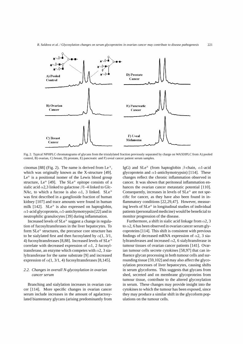
Fig. 2. Typical NPHPLC chromatograms of glycans from the trisialylated fraction previously separated by charge on WAXHPLC from A) pooledcontrol, B) ovarian, C) breast, D) prostate, E) pancreatic and F) uveal cancer patient serum samples.
cinomas [88] (Fig. 2). The name is derived from Le x,which was originally known as the X-structure [49].Lex is a positional isomer of the Lewis blood groupstructure, Lea [49]. The SLex epitope consists of asialic acid α2,3 linked to galactose β1–4 linked to Glc-NAc, to which a fucose is also α1, 3 linked. SLex
was first described in a ganglioside fraction of humankidney [107] and trace amounts were found in humanmilk [142]. SLex is also expressed on haptoglobin,α1-acid glycoprotein, α1-antichymotrypsin [22] and inneutrophilic granulocytes [39] during inflammation.
Increased levels of SLex suggest a change in regula-tion of fucosyltransferases in the liver hepatocytes. Toform SLex structures, the precursor core structure hasto be sialylated first and then fucosylated by α(1, 3/1,4) fucosyltransferases [8,88]. Increased levels of SLex
correlate with decreased expression of α1, 2 fucosyl-transferase, an enzyme which competes with α2, 3 sia-lyltransferase for the same substrate [9] and increasedexpression of α(1, 3/1, 4) fucosyltransferases [8,145].
2.2. Changes in overall N-glycosylation in ovariancancer serum
Branching and sialylation increases in ovarian can-cer [114]. More specific changes in ovarian cancerserum include increases in the amount of agalactosy-lated biantennary glycans (arising predominantly from
IgG) and SLex (from haptoglobin β-chain, α1-acidglycoprotein and α1-antichymotrypsin) [114]. Thesechanges reflect the chronic inflammation observed incancer. It was shown that peritoneal inflammation en-hances the ovarian cancer metastatic potential [110].Consequently, increases in levels of SLex are not spe-cific for cancer, as they have also been found in in-flammatory conditions [22,29,47]. However, measur-ing levels of SLex in longitudinal studies of individualpatients (personalized medicine) would be beneficial tomonitor progression of the disease.
Furthermore, a shift in sialic acid linkage from α2, 3to α2, 6 has been observed in ovarian cancer serum gly-coproteins [114]. This shift is consistent with previousfindings of decreased mRNA expression of α2, 3 sia-lyltransferases and increased α2, 6 sialyltransferase intumour tissues of ovarian cancer patients [141]. Ovar-ian tumour cells secrete cytokines [58,97] that can in-fluence glycan processing in both tumour cells and sur-rounding tissue [59,102] and may also affect the glyco-sylation processes of liver hepatocytes, causing shiftsin serum glycoforms. This suggests that glycans fromshed, secreted and on membrane glycoproteins fromtumour tissue, contribute to the altered glycosylationin serum. These changes may provide insight into thecytokines to which the tumour has been exposed, sincethey may produce a similar shift in the glycoform pop-ulations on the tumour cells.

222 R. Saldova et al. / Glycosylation changes on serum glycoproteins in ovarian cancer may contribute to disease pathogenesis
2.3. N-glycosylation on CA125 does not contribute tothe major changes in glycan levels in ovariancancer patient serum
CA125 is a mucin, first detected by Bast et al. us-ing the monoclonal antibody OC125 [14]. CA125 maybe an antigen that can elicit antibody-dependent, cell-mediated cytotoxicity against ovarian tumour cells. Itmay play a key physiological role that promotes tu-mour development in patients with ovarian cancer [71].Wong et al. described CA125 major N -glycans onOVCAR3 cell line, which are 20% high mannose typeand 80% complex type structures [71]. They are most-ly mono-fucosylated bi-antennary, tri-antennary andtetra-antennary-bisected structures, with no more thenone sialic acid [71]. CA125 glycans do not contributeto the major changes in glycan levels in ovarian cancerpatient serum [114].
3. Inflammation
Inflammation is a complex defence mechanism, bywhich leucocytes migrate into damaged tissues to de-stroy the agents that can potentially cause tissue in-jury [40]. Acute inflammation is a limited shortterm response, particularly during infectious challenge,whereas, chronic inflammation is a persistent phe-nomenon that can lead to tissue damage [40]. In acuteinflammation, initially the leukocyte infiltrate is mostlyneutrophilic but after 1 to 2 days, monocytic cells pre-dominate [40]. Chronic inflammation is associated withthe presence of mononuclear cells such as macrophagesand lymphocytes [40]. Cytokines play an importantrole in the response to inflammation [40]. Interleukin6 (IL-6) has a dual effect; at some levels it acts as adefence mechanism but in chronic inflammation it is aninflammatory agent [40].
4. The acute phase response leads to substantialchanges in the plasma concentration of acutephase proteins
The acute phase response, which occurs during in-fection, trauma, surgery, burns and inflammatory con-ditions, leads to substantial increases in the plasma con-centration of acute-phase proteins such as C-reactiveprotein, serum amyloid A, haptoglobin, α1-acid glyco-protein, α1-antitrypsin, α1-antichymotrypsin and fib-rinogen (positive acute phase proteins) or decreases in
levels of albumin and transferrin (negative acute phaseproteins). Two weeks following the inflammatory stim-ulus, the plasma concentrations of these proteins returnto normal, with the exception of haptoglobin and fib-rinogen, which can take three weeks to return to nor-mal levels [41]. The chronic inflammation associatedwith cancer can induce an acute phase response withthe same changes in the serum concentration of theseproteins but these persist longer.
Cytokines are major stimulators of acute phase pro-tein production [41]. They are produced during in-flammatory processes by a variety of cells. The mostimportant sources are macrophages and monocytes atinflammatory sites [41].
Altered glycosylation on haptoglobin, α1-acid gly-coprotein, α1-antichymotrypsin and α1-antitrypsin inadvanced ovarian cancer patient sera has been identi-fied [114].
4.1. Positive acute-phase proteins with alteredN-glycosylation
Positive acute-phase proteins increase in concentra-tion during the acute immune response [41]. Theseare proteins related to the complement, coagulationand fibrinolytic systems (e.g. fibrinogen), antiproteases(e.g. α1-antitrypsin, α1-antichymotrypsin), transportproteins (e.g. haptoglobin, hemopexin), participants ininflammatory responses (e.g. secreted phospholipaseA2) and others (C-reactive protein, serum amyloid A,α1-acid glycoprotein, ferritin) [41].
4.1.1. HaptoglobinHaptoglobin (Hp) is a α2-sialoglycoprotein with
haemoglobin-binding capacity, that is secreted intoplasma by the liver [2,20]. The hepatic synthesis of Hpis induced by pro-inflammatory cytokines such as IL-6,IL-1 and tumour necrosis factor (TNF) [20].
Hp forms a complex with haemoglobin (Hb) dur-ing haemolysis. After destruction of erythrocytes,free haemoglobin in the circulation passes through theglomerulus of the kidney. Renal damage is preventedby the binding of haemoglobin by haptoglobin. There-fore, this prevents both iron loss and kidney dam-age during intravascular haemolysis [45]. This acute-phase protein also has bacteriostatic properties, inhibitsprostaglandin synthesis and angiogenesis [75] and pro-tects against free radicals [138].
Haptoglobin is important in iron homeostasis [138].The identification of functional differences in hap-toglobin molecules results from relatively common

R. Saldova et al. / Glycosylation changes on serum glycoproteins in ovarian cancer may contribute to disease pathogenesis 223
polymorphisms [138]. There are three major pheno-types: Hp 1-1, Hp 2-1 and Hp 2-2 [2,20,45].
Human haptoglobin is a tetrameric protein, com-posed of two α and two β chains [72]. The β-chain (49kDa) is heavier than the α-chain and is identical in allHp types. The β chain of Hp contains four Asn-linkedglycosylation sites, all of which may be occupied [17,72]. Glycosylation accounts for, approximately, 19%of β-haptoglobin mass [17]. Hp contains biantennarycomplex glycans in neutral,monosialylated and disialy-lated forms and triantennary complex glycans in disia-lylated and trisialylated forms [52].
Changes in haptoglobin glycan composition are as-sociated with disease. Expression levels of haptoglobinβ-chain increase in ovarian cancer, decrease withchemotherapy and correlate with levels of CA125 [3].Increase in fucosylation, sialylation or branching beenfound in breast [46], ovarian [114,126,127], lung [53],pancreatic [98], and prostate cancers [38]. Fu-cose content on haptoglobin increases with tumoursize [127]. Highly-sialylated Hp was found in Crohn’sdisease [46].
4.1.2. α1-acid glycoproteinα 1-acid glycoprotein (also known as orosomucoid,
AGP, Fig. 3) has a molecular weight of 41–43 kDaand is heavily glycosylated (45%) [117]. AGP is anacute phase protein, which is synthesized mainly byhepatocytes but extrahepatic synthesis has also beenreported [37]. The serum concentration of AGP risestwo to five times during an acute phase response. Basedon concavalin A (Con A) reactivity (ConA is mainlyselective for mannosylated N -glycans [21]), AGP canbe fractionated to ConA non-reactive, weakly reactiveand strongly reactive forms.
Protein synthesis and glycosylation of AGP are in-dependently regulated [135,136], both by cytokines(mainly IL-1 and IL-6) and glucocorticoids [11,85–87].AGP has the ability to bind and transport several basicand neutral drugs of endogenous and exogenous ori-gin [60,70]. AGP has also been classified as a mem-ber of the immunocalin family, a lipocalin subfami-ly that modulates immune and inflammatory respons-es [81]. AGP stimulates cytokine secretion and thus,contributes to the inflammatory response. This effectcan be enforced by the local production of AGP bymonocytes in response to some of these cytokines [54].AGP has a beneficial role in wound healing, protectingagainst tissue damage and is involved in the inductionof non-specific resistance to infection [54].
Five N -glycans are attached to the human pro-tein [147]. Each of the N -glycosylation sites ofAGP can express any of the glycans, which can havedi-, tri- or tetra-antennary structures [37]. AGP is alsonegatively charged (pI is 2.7–3.2) due to the presenceof sialic acids (12% of total glycans) [54]. There arechanges in glycosylation in inflammation such as anincrease in SLex. There is a relative increase of AGPglycoforms with biantennary glycans in acute inflam-mation and a relative decrease of AGP glycoforms withbiantennary glycans in chronic inflammation, pregnan-cy, estrogen administration and liver damage [29,84].
Changes in glycosylation can affect the biologicalproperties of AGP. For instance, immunomodulato-ry activity of AGP depends on its glycosylation [37].AGP, containing branched glycans, is more effectivein the inhibition of lymphocyte proliferation [106],and desialylated AGP enhances inhibition of plateletsaggregation [27]. Inflammation-induced increases inSLex-substituted glycans on AGP might represent amechanism for feedback inhibition of granulocyte ex-travasation into inflamed tissues [37].
4.1.3. α1-antichymotrypsinHuman α1-antichymotrypsin is a plasma glycopro-
tein with a relative molecular mass of approximately58 kDa and carbohydrate content of 24% [74]. It is anacute phase protein, secreted by the liver which belongsto the superfamily of serpins [130]. Its concentrationincreases more than four-fold within a few hours inresponse to an inflammatory stimulus [7] and it is al-so elevated in cancer [64]. The physiological functionof α1-antichymotrypsin has not yet been determined,however α1-antichymotrypsin inhibits chymotrypsin-like proteases [129], regulates cathepsin G activity [15],modulates the cellular functions of neutrophils [67] andlymphocytes [57] and inhibits platelet-activating-factorsynthesis [25].
α1-antichymotrypsin has six potential glycosylationsites [112], in which disialyl bi-antennary, trisialyltri-antennary and disialyl tri-antennary, tri- and tetra-antennary glycans have been identified [73]. SLexα1-antichymotrypsin is increased in ovarian cancer [114].
4.1.4. Increase of positive acute phase proteins inplasma correlates with altered glycosylation
An increase in SLex on the haptoglobinβ-chain, α1-acid glycoprotein and α1-antichymotrypsin has beenobserved [114]. SLex is also present during inflamma-tion on all these proteins [22].

224 R. Saldova et al. / Glycosylation changes on serum glycoproteins in ovarian cancer may contribute to disease pathogenesis
Fig. 3. Molecular model of α1-acid glycoprotein. The peptide fold (green) is a homology model of human alpha-1-acid glycoprotein (Pieperet al., personal communication) based on the crystal structure of chicken plasma retinol-binding protein [148]. Glycans were added to thethree accessible glycosylation sites in the model (Swissprot numbering: P02763). N -linked glycan structures (yellow), chosen on the basis ofsequencing results [147], were generated using the database of glycosidic linkage conformations [143] and in vacuo energy minimisation to relieveunfavourable steric interactions. The Asn-GlcNAc linkage conformations (Asn – orange) were based on the observed range of crystallographicvalues [104], the torsion angles around the Asn Cα-Cβ and Cβ-Cγ bonds then being adjusted to eliminate unfavourable steric interactionsbetween the glycans and the protein surface.
Terminal sialic acid and fucose on SLex inhibits theamount of free galactose accessible to the asialoglyco-protein and Kupffer cell receptors [26] in the liver andcan therefore prolong their clearance from the circula-tion, resulting in higher plasma concentrations [122].The presence of SLex glycans from haptoglobin β-chain, α1-antichymotrypsin and α1-acid glycoproteinin cancer and also in inflammation suggests that there isregulation of these acute phase protein concentrations.The biological significance of these increases is to in-crease their anti-apoptotic [28] and anti-inflammatoryproperties [35]. These anti-apoptotic properties may
aid cancer metastasis. Glycosylation of these serumproteins derives from the glycosylation process duringtheir biosynthesis in liver parenchymal cells. Inflam-matory cytokines, corticosteroids and growth factorsare involved in regulation of these changes [137].
4.2. Negative acute-phase proteins with alteredN-glycosylation
Negative acute-phase proteins decrease in concentra-tion during the acute immune response. These are albu-min, transferrin, transthyretin and α-fetoprotein [41].
R. Saldova et al. / Glycosylation changes on serum glycoproteins in ovarian cancer may contribute to disease pathogenesis 225
4.2.1. TransferrinA family of proteins, known as transferrins, control
iron levels in the body [4,51]. Transferrin is present inthe blood (serum transferrin), in other bodily secretions(lactoferrin), in avian egg white (ovotransferrin) and inmelanotransferrin (broad range of tissue types) [139].
Transferrin is critical to protect the body from freeiron in the aerobic environment of the blood and bodilyfluids. Damage occurs when ferrous iron is convertedto ferric iron and forms harmful free radicals. Transfer-rin is also involved in protection from insoluble ferriciron [140].
Additionally, serum transferrin transports iron inthe blood by chelating free ferric iron from degradedhaemoglobin and delivering it to cells in a receptor-mediated endocytotic process. While the transferrin-receptor complex is internalised, iron is released in theendosome, and the complex is recycled to the cell sur-face where the transferrin is released [51].
The regulation of transferrin receptor expression invarious tissues is related to specific cellular iron re-quirements. For most non-erythroid cells, iron can reg-ulate the transferrin receptor expression in a recipro-cal manner, through modulating the stability of the re-ceptor mRNA. Whereas, in haemoglobin-synthesizingcells, the transferrin receptor expression is independentof the cellular iron loading [82].
Serum transferrin and transferrin receptors play animportant role in iron homeostasis – iron regulationin the human body. Human serum transferrin is syn-thesized in the liver. It is a polypeptide chain of 679amino acid residues [12]. There are two homologousdomains; the N -terminal and the C-terminal domains(with glycans attached to the C-terminal domain - Forreview see [19,30]). Transferrin has two glycan chains(mostly complex biantennary types, which terminate insialic acids. The protein is fully glycosylated with theseglycans present on two major Asn-linked glycosylationsites Asn 432 (Asn-Lys-Ser) and Asn 630 (Asn-Val-Thr) and one minor site Asn 491 (Asn-His-Cys) [116].The glycosylation is site specific, especially, the corefucosylation, which occurs only at Asn 630 site [115].Changes in glycosylation on transferrin can occur dur-ing disease; an increase in highly branched fucosylatedglycans was reported in hepatocellular carcinoma pa-tients [144]. However, non-glycosylated recombinanttransferrin was found to be functionally indistinguish-able from glycosylated serum transferrin [90,91].
4.2.2. Altered glycosylation of the negative acutephase protein transferrin plays an importantrole in iron homeostasis
The level of transferrin decreases in ovarian cancerpatients’ serum [3,128], in other gynaecological can-cers [128] and in inflammation [108]. After chemother-apy, levels of transferrin increase or remain constantin ovarian cancer patients [3]. Glycosylated transferrinand its glycans have anti-apoptotic properties, as shownby deglycosylation, which abrogated this effect. Theyplay an important role in regulation of the programmedcell death via alterations in cytokine expression [77].There are many transferrin receptors present on cancercells and this could play a role in anaemia, which isfound in more than 30% of patients [42]. The trans-ferrin receptor has three fully glycosylated sites; Asn251, Asn-Gly-Ser; 317, Asn-His-Thr and 727, Asn-Glu-Thr [24]. Glycosylation of the transferrin receptoris critical for its folding, stability and/or secretion [24,100]. Altered glycosylation of the N -linked glycans ofthe transferrin receptor from diabetic patients leads toreduced binding affinity for transferrin [43]. Glycosy-lation of transferrin does not have influence on its bind-ing capacity, but transferrin and iron uptake is reducedafter transferrin deglycosylation [55].
4.2.3. Iron homeostasisFormanowicz et al. [36] developed a Petri net-based
model of body iron homeostasis, in which they simu-lated iron homeostasis, describing the homeostasis asfollows:
Iron is taken from the diet. There are two formsof iron; heme iron (ferrous, Fe2+) and non-heme iron(ferric, Fe3+). Iron Fe3+ is reduced to Fe2+ in thestomach in low pH by reducing agents (e.g. ascorbicacid) and transported into small intestine mucous mem-brane. If the iron is supplemented in the Fe2+ form itis transported directly. Most of the metal in the labileiron pool in the cell is metabolically drawn into Fe-dependent enzymes, transported into mitochondria forheme synthesis or incorporated into ferritin for storageor detoxification. The red blood cells exist in the humanbody for about 120 days and after mono-nuclear cellsphagocytose them. These cells are responsible for therecirculation of iron derived from red blood cells. Ironthen enters the circulation, binds to transferrin and istransported to the bone marrow for red cell production.
During inflammation, iron metabolism is changing.In one of these changes, iron is not released from bodyiron stores (ferritin) because it would enable the devel-opment of micro-organisms, which need iron for theirgrowth. Infection and inflammation thus results in hy-poferremia. If persistent, it can lead to anaemia.

226 R. Saldova et al. / Glycosylation changes on serum glycoproteins in ovarian cancer may contribute to disease pathogenesis
5. Glycosylation of molecules involved in theimmune system
Almost all of the key molecules involved in the in-nate and adaptive immune response are glycoproteins.In the cellular immune system, specific glycoforms areinvolved in the folding, quality control and assemblyof peptide-loaded major histocompatibility complex(MHC) antigens and the T cell receptor complex [113].In the humoral immune system, all of the immunoglob-ulins and most of the complement components are gly-cosylated [113].
Immunoglobulins are glycoproteins and the majorsecretory products of the adaptive immune system [6,79]. They provide long-term defence against foreignantigens [6]. There are five classes identified in hu-mans; IgG, IgM, IgA, IgE and IgD; they share sim-ilar structures and are composed of Ig domains [5].The immunoglobulins differ in the location and num-ber of N -linked glycosylation sites, which are situatedon the Fc and Fab region and the glycans attached tothe immunoglobulins are large, approximately 2 kDaeach [6].
5.1. Decreased galactosylation on immunoglobulin Ghas impact on its function
Human serum IgG consists of four subclasses,which differ in their γ-chain sequences and disulphidebridges. The most abundant serum Ig is IgG1, whichcirculates at concentrations of 10–15 mg/ml [6]. AllIgG molecules contain two N -linked glycosylationsites, which can be differently glycosylated [6]. Theglycan helps to maintain the quaternary structure andthe stability of the Fc region [94,95]. Aberrant glyco-sylation of immunoglobulins is linked to disease andpathogenesis, for example, increase in levels of agalac-tosylated glycoforms of IgG in rheumatoid arthritis [6].
A significant decrease in the level of galactosylationand sialylation in ovarian cancer patients serum hasbeen observed [114]. Increase of agalactosylated IgGoligosaccharides is caused by decreased Gal-T activityin plasma cells [10] or increased production of specif-ic subsets of plasma cells with low expression levelsof galactosyltransferases [99]. Different glycoformsmay differ in their affinity for ligands [61,89,94,120,133]. The IgG-G0 glycoform is elevated in rheumatoidarthritis serum [101] and terminal GlcNAc of this gly-coform on the Fc region of the IgG molecule clusteredon target cells can be recognized by collagenous lectinmannose-binding protein (MBL) resulting in comple-
ment activation [89]. Kaneko et al. [61] have shownthat sialylation on IgG reduces its cytotoxicity to naturalkiller cells, exhibiting an anti-inflammatory effect. De-creased galactosylation and sialylation on IgG in ovar-ian cancer patients then may increase the cytotoxicityand complement activation via MBL.
Increase of agalactosylated IgG glycoforms has pre-dominantly been identified with tumour progressionand metastasis of gastric and lung cancer [63], as wellas in chronic inflammatory diseases such as rheuma-toid arthritis, tuberculosis or inflammatory bowel dis-ease [10,101] and vasculitis [56]. IgGs from systemiclupus erythematosus patients with Sjogren’s syndrome,also have decreased galactosylation on IgG [18].
Decrease in sialylation on IgG glycans has also beenfound in rheumatoid arthritis [92]. Therefore, this in-crease of agalactosylated glycans on IgG of ovariancancer sera may be indicative of an inflammatory state.
5.2. Function of the immune response
The human immune system recognises cancer as aninflammatory process and responds accordingly. It ac-tively produces acute phase proteins, which have alsoanti-apoptotic properties. In inflammation, it helps toreconstitute the damaged tissue but it protects and pro-motes cancer cells, considering them as its own. If thishypothesis is correct, anti-inflammatory drugs shouldhave a powerful effect in cancer treatment. Indeed,non-steroidal anti-inflammatory drugs (NSAID) have apositive effect both in prevention [150] and in protec-tion against cancer development and progression [121].
6. Apoptosis
Apoptosis is a physiological form of cell death [76].The term describes a specific morphology during thatprocess and is associated with a distinct set of biochem-ical and physical changes involving the cytoplasm, nu-cleus and plasma membrane [76]. Early in apoptosis,cells round up, losing contact with their neighbours andshrink [76]. Under physiological conditions, certainmodifications in the plasma membrane occur, whichenable the recognition of apoptotic bodies by phago-cytic cells [76]. Apoptotic bodies are surrounded by anintact plasma membrane, so, apoptosis usually occurswithout leakage of cell content and without inflamma-tion [76]. Apoptosis occurs during embryonic devel-opment, tissue remodelling, immune regulation and tu-mour regression [118]. It is induced by death recep-

R. Saldova et al. / Glycosylation changes on serum glycoproteins in ovarian cancer may contribute to disease pathogenesis 227
tors, which have been identified as a subgroup of theTNF-receptor superfamily [118].
For cell homeostasis to be maintained, a balancebetween the increase (differentiation from precursorsand proliferation) and decrease (further differentiationand cell death) has to be achieved [76].
6.1. Apoptosis in cancer
Apoptosis and the genes that control it have an im-portant effect on the malignant phenotype [83]. Someoncogenic mutations, which disrupt apoptosis, leadto tumour initiation, progression or metastasis [83].While other oncogenic changes promote apoptosisand over-ride apoptosis during multistage carcinogen-esis [83]. The majority of cytotoxic anti-cancer agentsinduce apoptosis but the possibility of defects in apop-totic programs contribute to treatment failure [83]. Thesame mutations that suppress apoptosis during tumourdevelopment also reduce treatment sensitivity [83].Therefore, apoptosis provides a link between cancergenetics and cancer therapy [83]. There is intensive re-search on the mechanisms regulating apoptosis, the re-sults of which will provide new strategies for improvingtherapeutic outcome [83].
6.2. Apoptosis and glycosylation
Glycosylation has an important role in apoptosis.Resistance to apoptosis is a critical feature of cancercells. Increase of endogenous sialylation, may be oneanti-apoptotic mechanism that converts tumour cells toa more malignant phenotype [66]. This supports thefinding that sialidase expression is inversely associatedwith metastatic potential and tumour growth in cancercells, probably through a regulation mechanism thatsuppresses cell growth and promotes apoptosis [65].
Galectin-1 is a mammalian lectin that induces celldeath in leukemia, lymphoma, breast and prostate can-cer. T cells express specific glycoprotein receptors thatbear the specific glycans recognized by galectin-1 sus-ceptible to galectin-1 mediated T-cell apoptosis [109].A characteristic “glycotype” with sialylated core 1 O-glycans promote galectin-1 resistance [109]. Core 2 N -acetylglucosaminyltransferase is required for galectin-1 susceptibility of T lymphoma and is down-regulatedin galectin-1-resistant cells. This indicates that sim-ilar O-glycan ligands on different polypeptide back-bones are common death trigger receptors, recognisedby galectin-1 on different types of cancer cells [134].Loss of galectin-1 susceptibility and synthesis of en-
dogenous galectin-1 has been proposed to promote tu-mor evasion of immune attack [134].
The Golgi enzyme, GlcNAc-TV, is upregulatedin cancer and produces N -glycans with poly N -acetyllactosamine, which is the preferred ligand forgalectin-3 [31,102]. GlcNAc-TV glycans have positiveeffect on tumour growth, metastasis and resistance toapoptosis [31,32].
7. Conclusion
Changes in glycosylation are very important in ovar-ian cancer, providing both potential markers and insightinto cancer pathogenesis. Changes in glycosyltrans-ferase levels and/or glycan nucleotide donors, lead tomodifications in both N - and O- linked glycans. Themost significant N -glycosylation changes in ovariancancer serum glycoproteins are increases in agalactosy-lated biantennary glycans and SLex. N -glycosylationon CA125 does not contribute to these major changes.An increase of SLex has been found on haptoglobin β-chain, α1-acid glycoprotein and α1-antichymotrypsin.The concentration of these positive acute phase pro-teins is increased in the acute phase response and thiscould be affected by the altered glycosylation. A de-crease in galactosylation and sialylation on IgG modu-lates its function. Glycosylation also has an importantrole in apoptosis, which is deregulated in cancer. Tak-en together, these data suggest that the progression ofovarian cancer involves pathways which promote cellgrowth and metastasis, in part bymimicking inflamma-tion processes. Therefore, tumours that survive the hostresponse are these which make changes contributing toits chances for survival.
Acknowledgements
We acknowledge the Oxford Glycobiology Insti-tute’s endowment fund and EUROCarbDB (http://www.eurocarbdb.org) RIDS Contract No. 011952, forsupporting much of the work discussed in this review.We would like to thank Dr. Dereck Chatterton for hiscontributions to this paper.
Abbreviations
AGP: α1-acid glycoprotein (also called orosomu-coid)

228 R. Saldova et al. / Glycosylation changes on serum glycoproteins in ovarian cancer may contribute to disease pathogenesis
GlcNAc-TV: N -acetylglucosaminyltransferase VHb: haemoglobinHp: haptoglobinIL: interleukinLea: Lewis aMBL: mannose-binding lectinM-CSF: macrophage-colony-stimulating factorMHC: major histocompatibility complexNSAID: non-steroidal anti-inflammatory drugsPSA: polysialic acidSLex: sialyl Lewis xTNF: tumour necrosis factorTPS: tissue polypeptide specific antigen
References
[1] Cancer Research UK, [cited 2007 25.4.]; http://info.cancerresearchuk.org/cancerstats/mortality/cancerdeaths/.
[2] Nature and Metabolism of Extracellular Proteins, MolecularBiology of Human Proteins, H.E. Schultze and J.F. Heremans,eds., Amsterdam: Elsevier, 1 (1966), 384–402.
[3] N. Ahmed, K.T. Oliva, G. Barker, P. Hoffmann, S. Reeve,I.A. Smith, M.A. Quinn and G.E. Rice, Proteomic trackingof serum protein isoforms as screening biomarkers of ovariancancer, Proteomics 5(17) (2005), 4625–4636.
[4] P. Aisen, A. Leibman and J. Zweier, Stoichiometric and sitecharacteristics of the binding of iron to human transferrin, JBiol Chem 253(6) (1978), 1930–1937.
[5] L.M. Amzel and R.J. Poljak, Three-dimensional structure ofimmunoglobulins, Annu Rev Biochem 48 (1979), 961–997.
[6] J.N. Arnold, M.R. Wormald, R.B. Sim, P.M. Rudd and R.A.Dwek, The Impact of Glycosylation on the Biological Func-tion and Structure of Human Immunoglobulins, Annu RevImmunol 25 (2007), 21–50.
[7] K.F. Aronsen, G. Ekelund, C.O. Kindmark and C.B. Laurell,Sequential changes of plasma proteins after surgical trauma,Scand J Clin Lab Invest Suppl 124 (1972), 127–136.
[8] M. Aubert, L. Panicot-Dubois, C. Crotte, V. Sbarra, D. Lom-bardo, M.O. Sadoulet and E. Mas, Peritoneal colonizationby human pancreatic cancer cells is inhibited by antisenseFUT3 sequence, Int J Cancer 88(4) (2000), 558–565.
[9] M. Aubert, L. Panicot, C. Crotte, P. Gibier, D. Lombar-do, M.O. Sadoulet, and E. Mas, Restoration of alpha(1,2)fucosyltransferase activity decreases adhesive and metastat-ic properties of human pancreatic cancer cells, Cancer Res60(5) (2000), 1449–1456.
[10] J.S. Axford, N. Sumar, A. Alavi, D.A. Isenberg, A. Young,K.B. Bodman, and I.M. Roitt, Changes in normal glycosy-lation mechanisms in autoimmune rheumatic disease, J ClinInvest 89(3) (1992), 1021–1031.
[11] Y. Azuma, M. Murata and K. Matsumoto, Alteration of sug-ar chains on alpha(1)-acid glycoprotein secreted followingcytokine stimulation of HuH-7 cells in vitro, Clin Chim Acta294(1–2) (2000), 93–103.
[12] V.S. Baranov, A.L. Schwartzman, V.N. Gorbunova, V.S. Gait-skhoki, N.B. Rubtsov, N.A. Timchenko and S.A. Neifakh,Chromosomal localization of ceruloplasmin and transferringenes in laboratory rats, mice and in man by hybridizationwith specific DNA probes, Chromosoma 96(1) (1987), 60–66.
[13] R.C. Bast, Jr. D. Badgwell, Z. Lu, R. Marquez, D. Rosen,J. Liu, K.A. Baggerly, E.N. Atkinson, S. Skates, Z. Zhang,A. Lokshin, U. Menon, I. Jacobs and K. Lu, New tumormarkers: CA125 and beyond, Int J Gynecol Cancer 15(3)(2005), 274–281.
[14] R.C. Bast, Jr., M. Feeney, H. Lazarus, L.M. Nadler, R.B.Colvin and R.C. Knapp, Reactivity of a monoclonal antibodywith human ovarian carcinoma, J Clin Invest 68(5) (1981),1331–1337.
[15] K. Beatty, J. Bieth and J. Travis, Kinetics of association of ser-ine proteinases with native and oxidized alpha-1-proteinaseinhibitor and alpha-1-antichymotrypsin, J Biol Chem 255(9)(1980), 3931–3934.
[16] S. Bengtsson, M. Krogh, C.A. Szigyarto, M. Uhlen, K.Schedvins, C. Silfversward, S. Linder, G. Auer, A. Alaiya andP. James, Large-scale proteomics analysis of human ovariancancer for biomarkers, J Proteome Res 6(4) (2007), 1440–1450.
[17] J.A. Black, G.F. Chan, C.L. Hew and G.H. Dixon, Geneaction in the human haptoglobins. 3. Isolation of the alpha-chains as single gene products. Isolation, molecular weight,and amino acid composition of alpha and beta chains, Can JBiochem 48(1) (1970), 123–132.
[18] A. Bond, A. Alavi, J.S. Axford, B.E. Bourke, F.E. Bruckn-er, M.A. Kerr, J.D. Maxwell, K.J. Tweed, M.J. Weldon, P.Youinou and F.C. Hay, A detailed lectin analysis of IgG gly-cosylation, demonstrating disease specific changes in termi-nal galactose and N-acetylglucosamine, J Autoimmun 10(1)(1997), 77–85.
[19] B.H. Bowman, F.M. Yang and G.S. Adrian, Transferrin: evo-lution and genetic regulation of expression, Adv Genet 25(1988), 1–38.
[20] B.H. Bowman, Haptoglobin, in: Hepatic Plasma Proteins,B.H. Bowman, Ed., Academic Press: San Diego, 1993,pp. 159–167.
[21] C.F. Brewer and L. Bhattacharyya, Specificity of con-canavalin A binding to asparagine-linked glycopeptides. Anuclear magnetic relaxation dispersion study, J Biol Chem261(16) (1986), 7306–7310.
[22] E.C. Brinkman-Van der Linden, P.F. de Haan, E.C. Havenaarand W. Van Dijk, Inflammation-induced expression of sialylLewisX is not restricted to alpha1-acid glycoprotein but alsooccurs to a lesser extent on alpha1-antichymotrypsin andhaptoglobin, Glycoconj J 15(2) (1998), 177–182.
[23] S.A. Brooks, M.W. Dwek and U. Schumacher, Functional &Molecular Glycobiology P. Dines, Oxford: BIOS ScientificPublishers Limited, 2002.
[24] S.L. Byrne, R. Leverence, J.S. Klein, A.M. Giannetti, V.C.Smith, R.T. MacGillivray, I.A. Kaltashov and A.B. Mason,Effect of glycosylation on the function of a soluble, recom-binant form of the transferrin receptor, Biochemistry 45(21)(2006), 6663–6673.
[25] G. Camussi, C. Tetta, F. Bussolino and C. Baglioni, Synthesisand release of platelet-activating factor is inhibited by plasmaalpha 1-proteinase inhibitor or alpha 1-antichymotrypsin andis stimulated by proteinases, J Exp Med 168(4) (1988), 1293–1306.
[26] P.J. Coombs, M.E. Taylor and K. Drickamer, Two categoriesof mammalian galactose-binding receptors distinguished byglycan array profiling, Glycobiology 16(8) (2006), 1C–7C.
[27] M. Costello, B.A. Fiedel and H. Gewurz, Inhibition of plateletaggregation by native and desialised alpha-1 acid glycopro-tein, Nature 281(5733) (1979), 677–678.

R. Saldova et al. / Glycosylation changes on serum glycoproteins in ovarian cancer may contribute to disease pathogenesis 229
[28] M.A. Daemen, V.H. Heemskerk, C. van’t Veer, G. Denecker,T.G. Wolfs, P. Vandenabeele and W.A. Buurman, Functionalprotection by acute phase proteins alpha(1)-acid glycopro-tein and alpha(1)-antitrypsin against ischemia/reperfusion in-jury by preventing apoptosis and inflammation, Circulation102(12) (2000), 1420–1426.
[29] T.W. De Graaf, M.E. Van der Stelt, M.G. Anbergen and W.van Dijk, Inflammation-induced expression of sialyl LewisX-containing glycan structures on alpha 1-acid glycoprotein(orosomucoid) in human sera, J Exp Med 177(3) (1993),657–666.
[30] G. de Jong, J.P. van Dijk and H.G. van Eijk, The biology oftransferrin, Clin Chim Acta 190(1–2) (1990), 1–46.
[31] J.W. Dennis, J. Pawling, P. Cheung, E. Partridgeand M. Demetriou, UDP-N-acetylglucosamine:alpha-6-D-mannoside beta1, 6 N-acetylglucosaminyltransferase V(Mgat5) deficient mice, Biochim Biophys Acta 1573(3)(2002), 414–422.
[32] J.W. Dennis, S. Laferte, C. Waghorne, M.L. Breitman andR.S. Kerbel, Beta 1-6 branching of Asn-linked oligosac-charides is directly associated with metastasis, Science236(4801) (1987), 582–585.
[33] D.H. Dube and C.R. Bertozzi, Glycans in cancer andinflammation–potential for therapeutics and diagnostics, NatRev Drug Discov 4(6) (2005), 477–488.
[34] M.J. Duffy, J.M. Bonfrer, J. Kulpa, G.J. Rustin, G. Soletor-mos, G.C. Torre, M.K. Tuxen and M. Zwirner, CA125 inovarian cancer: European Group on Tumor Markers guide-lines for clinical use, Int J Gynecol Cancer 15(5) (2005),679–691.
[35] T. Emoto, K. Nakamura, Y. Nagasaka, F. Numa, Y. Sum-inami and H. Kato, Alpha 1-antichymotrypsin inhibitschymotrypsin-induced apoptosis in rat hepatoma cells, Apop-tosis 3(3) (1998), 155–160.
[36] D. Formanowicz, A. Sackmann, P. Formanowicz and J.Blazewicz, Petri net based model of the body iron homeosta-sis, J Biomed Inform, 2006.
[37] T. Fournier, N.N. Medjoubi and D. Porquet, Alpha-1-acidglycoprotein, Biochim Biophys Acta 1482(1–2) (2000), 157–171.
[38] T. Fujimura, Y. Shinohara, B. Tissot, P.C. Pang, M. Kuro-gochi, S. Saito, Y. Arai, M. Sadilek, K. Murayama, A. Dell,S. Nishimura and S.I. Hakomori, Glycosylation status ofhaptoglobin in sera of patients with prostate cancer vs. be-nign prostate disease or normal subjects, Int J Cancer 122(1)(2008), 39–49.
[39] M. Fukuda, E. Spooncer, J.E. Oates, A. Dell and J.C. Klock,Structure of sialylated fucosyl lactosaminoglycan isolatedfrom human granulocytes, J Biol Chem 259(17) (1984),10925–10935.
[40] C. Gabay, Interleukin-6 and chronic inflammation, ArthritisRes Ther 8(2) (2006), S3.
[41] C. Gabay and I. Kushner, Acute-phase proteins and othersystemic responses to inflammation, N Engl J Med 340(6)(1999), 448–454.
[42] K.C. Gatter, G. Brown, I.S. Trowbridge, R.E. Woolston andD.Y. Mason, Transferrin receptors in human tissues: theirdistribution and possible clinical relevance, J Clin Pathol36(5) (1983), 539–545.
[43] M.K. Georgieff, C.D. Petry, M.M. Mills, H. McKay and J.D.Wobken, Increased N-glycosylation and reduced transferrin-binding capacity of transferrin receptor isolated from placen-tae of diabetic women, Placenta 18(7) (1997), 563–568.
[44] C. Gercel-Taylor, L.B. Bazzett and D.D. Taylor, Presence ofaberrant tumor-reactive immunoglobulins in the circulationof patients with ovarian cancer, Gynecol Oncol 81(1) (2001),71–76.
[45] E.R. Giblett, The haptoglobin system., Series Haematologica1 (1968), 3–20.
[46] M.T. Goodarzi and G.A. Turner, Reproducible and sensitivedetermination of charged oligosaccharides from haptoglobinby PNGase F digestion and HPAEC/PAD analysis: glycancomposition varies with disease, Glycoconj J 15(5) (1998),469–475.
[47] O. Gornik, L. Royle, D.J. Harvey, C.M. Radcliffe, R. Saldova,R.A. Dwek, P. Rudd and G. Lauc, Changes of serum glycansduring sepsis and acute pancreatitis, Glycobiology 17(12)(2007), 1321–1332.
[48] S. Hakomori and Y. Zhang, Glycosphingolipid antigens andcancer therapy, Chem Biol 4(2) (1997), 97–104.
[49] S. Hakomori and A. Kobata, in: The Antigens, M. Sela, Ed.,Academic Press: New York, 1974, pp. 79–140.
[50] J. Hamori, P. Arkosy, A. Lenkey and P. Sapy, The role ofdifferent tumor markers in the early diagnosis and prognosisof pancreatic carcinoma and chronic pancreatitis, Acta ChirHung 36(1–4) (1997), 125–127.
[51] D.C. Harris and P. Aisen, Iron Carriers and Iron Proteins,New York: VCH Publishers, 1989.
[52] Z. He, L.P. Aristoteli, L. Kritharides and B. Garner, HPLCanalysis of discrete haptoglobin isoform N-linked oligosac-charides following 2D-PAGE isolation, Biochem Biophys ResCommun 343(2) (2006), 496–503.
[53] L.F.t. Hoagland, M.J. Campa, E.B. Gottlin, J.E. Herndon, 2ndand E.F. Patz, Jr., Haptoglobin and posttranslational glycan-modified derivatives as serum biomarkers for the diagnosisof nonsmall cell lung cancer, Cancer 110(10) (2007), 2260–2268.
[54] T. Hochepied, F.G. Berger, H. Baumann and C. Libert,Alpha(1)-acid glycoprotein: an acute phase protein withinflammatory and immunomodulating properties, CytokineGrowth Factor Rev 14(1) (2003), 25–34.
[55] P. Hoefkens, M.I. Huijskes-Heins, C.M. de Jeu-Jaspars, W.L.van Noort, and H.G. van Eijk, Influence of transferrin glycanson receptor binding and iron-donation, Glycoconj J 14(2)(1997), 289–295.
[56] M. Holland, K. Takada, T. Okumoto, N. Takahashi, K. Ka-to, D. Adu, A. Ben-Smith, L. Harper, C.O. Savage and R.Jefferis, Hypogalactosylation of serum IgG in patients withANCA-associated systemic vasculitis, Clin Exp Immunol129(1) (2002), 183–190.
[57] D. Hudig, T. Haverty, C. Fulcher, D. Redelman and J.Mendelsohn, Inhibition of human natural cytotoxicity bymacromolecular antiproteases, J Immunol 126(4) (1981),1569–1574.
[58] M. Huleihel, E. Maymon, B. Piura, I. Prinsloo, D. Benhar-roch, I. Yanai-Inbar and M. Glezerman, Distinct patterns ofexpression of interleukin-1 alpha and beta by normal and can-cerous human ovarian tissues, Eur Cytokine Netw 8(2)(1997),179–187.
[59] Y. Ishibashi, Y. Inouye, T. Okano and A. Taniguchi, Regula-tion of sialyl-Lewis x epitope expression by TNF-alpha andEGF in an airway carcinoma cell line, Glycoconj J 22(1–2)(2005), 53–62.
[60] Z.H. Israili and P.G. Dayton, Human alpha-1-glycoproteinand its interactions with drugs, Drug Metab Rev 33(2) (2001),161–235.

230 R. Saldova et al. / Glycosylation changes on serum glycoproteins in ovarian cancer may contribute to disease pathogenesis
[61] Y. Kaneko, F. Nimmerjahn and J.V. Ravetch, Anti-inflammatory activity of immunoglobulin G resulting fromFc sialylation, Science 313(5787) (2006), 670–673.
[62] D. Kanikowska, R. Madry, J. Drozdz-Gorska, M. Sobieska,J. Markowska, and K. Wiktorowicz, [Microheterogeneity oftwo acute phase proteins in patients with ovarian carcinoma],Ginekol Pol 72(1) (2001), 17–21.
[63] Y. Kanoh, T. Mashiko, M. Danbara, Y. Takayama, S. Ohtani,T. Imasaki, T. Abe and T. Akahoshi, Analysis of the oligosac-charide chain of human serum immunoglobulin g in patientswith localized or metastatic cancer, Oncology 66(5) (2004),365–370.
[64] A.I. Karseladze, I.E. Rytin and V.B. Matveev, [Study ofalpha1-antichymotrypsin (ACT) in prostatic carcinoma],Arkh Patol 67(1) (2005), 30–33.
[65] T. Kato, Y. Wang, K. Yamaguchi, C.M. Milner, R. Shineha,S. Satomi and T. Miyagi, Overexpression of lysosomal-typesialidase leads to suppression of metastasis associated withreversion of malignant phenotype in murine B16 melanomacells, Int J Cancer 92(6) (2001), 797–804.
[66] O.T. Keppler, M.E. Peter, S. Hinderlich, G. Moldenhauer,P. Stehling, I. Schmitz, R. Schwartz-Albiez, W. Reutter andM. Pawlita, Differential sialylation of cell surface glycocon-jugates in a human B lymphoma cell line regulates suscep-tibility for CD95 (APO-1/Fas)-mediated apoptosis and forinfection by a lymphotropic virus, Glycobiology 9(6) (1999),557–569.
[67] L. Kilpatrick, J.L. Johnson, E.B. Nickbarg, Z.M. Wang, T.F.Clifford, M. Banach, B.S. Cooperman, S.D. Douglas and H.Rubin, Inhibition of human neutrophil superoxide genera-tion by alpha 1-antichymotrypsin, J Immunol 146(7) (1991),2388–2393.
[68] Y.J. Kim and A. Varki, Perspectives on the significance ofaltered glycosylation of glycoproteins in cancer, Glycoconj J14(5) (1997), 569–576.
[69] K.R. Kozak, F. Su, J.P. Whitelegge, K. Faull, S. Reddy andR. Farias-Eisner, Characterization of serum biomarkers fordetection of early stage ovarian cancer, Proteomics 5(17)(2005), 4589–4596.
[70] J.M. Kremer, J. Wilting and L.H. Janssen, Drug bindingto human alpha-1-acid glycoprotein in health and disease,Pharmacol Rev 40(1) (1988), 1–47.
[71] N. Kui Wong, R.L. Easton, M. Panico, M. Sutton-Smith, J.C.Morrison, F.A. Lattanzio, H.R. Morris, G.F. Clark, A. Delland M.S. Patankar, Characterization of the oligosaccharidesassociated with the human ovarian tumor marker CA125, JBiol Chem 278(31) (2003), 28619–28634.
[72] A. Kurosky, D.R. Barnett, T.H. Lee, B. Touchstone, R.E.Hay, M.S. Arnott, B.H. Bowman and W.M. Fitch, Covalentstructure of human haptoglobin: a serine protease homolog,Proc Natl Acad Sci USA 77(6) (1980), 3388–3392.
[73] A. Laine, E. Hachulla, G. Strecker, J.C. Michalski andJ.M. Wieruszeski, Structure determination of the glycansof human-serum alpha 1-antichymotrypsin using 1H-NMRspectroscopy and deglycosylation by N-glycanase, Eur JBiochem 197(1) (1991), 209–215.
[74] A. Laine and A. Hayem, Purification and characterizationof alpha 1-antichymotrypsin from human pleural fluid andhuman serum, Biochim Biophys Acta 668(3) (1981), 429–438.
[75] M.R. Langlois and J.R. Delanghe, Biological and clinicalsignificance of haptoglobin polymorphism in humans, ClinChem 42(10) (1996), 1589–1600.
[76] A. Lawen, Apoptosis-an introduction, Bioessays 25(9)(2003), 888–896.
[77] V. Lesnikov, M. Lesnikova and H.J. Deeg, Pro-apoptotic andanti-apoptotic effects of transferrin and transferrin-derivedglycans on hematopoietic cells and lymphocytes, Exp Hema-tol 29(4) (2001), 477–489.
[78] A. Liede, B.Y. Karlan, R.L. Baldwin, L.D. Platt, G. Kuper-stein and S.A. Narod, Cancer incidence in a population ofJewish women at risk of ovarian cancer, J Clin Oncol 20(6)(2002), 1570–1577.
[79] G.W. Litman, M.K. Anderson and J.P. Rast, Evolution ofantigen binding receptors, Annu Rev Immunol 17 (1999),109–147.
[80] J. Llorca, M.J. Lopez-Diaz, C. Gonzalez-Juanatey, W.E. Ol-lier, J. Martin and M.A. Gonzalez-Gay, Persistent ChronicInflammation Contributes to the Development of Cancer inPatients with Rheumatoid Arthritis from a Defined Popula-tion of Northwestern Spain, Semin Arthritis Rheum, 2007.
[81] L. Logdberg and L. Wester, Immunocalins: a lipocalin sub-family that modulates immune and inflammatory responses,Biochim Biophys Acta 1482(1–2) (2000), 284–297.
[82] C.N. Lok and T.T. Loh, Regulation of transferrin functionand expression: review and update, Biol Signals Recept 7(3)(1998), 157–178.
[83] S.W. Lowe and A.W. Lin, Apoptosis in cancer, Carcinogen-esis 21(3) (2000), 485–495.
[84] A. Mackiewicz and K. Mackiewicz, Glycoforms of serumalpha 1-acid glycoprotein as markers of inflammation andcancer, Glycoconj J 12(3) (1995), 241–247.
[85] A. Mackiewicz, M. Laciak, A. Gorny and H. Baumann,Leukemia inhibitory factor, interferon gamma and dexam-ethasone regulate N-glycosylation of alpha 1-protease in-hibitor in human hepatoma cells, Eur J Cell Biol 60(2) (1993),331–336.
[86] A. Mackiewicz, M.K. Ganapathi, D. Schultz and I. Kushner,Monokines regulate glycosylation of acute-phase proteins, JExp Med 166(1) (1987), 253–258.
[87] A. Mackiewicz and I. Kushner, Interferon beta 2/B-cell stim-ulating factor 2/interleukin 6 affects glycosylation of acutephase proteins in human hepatoma cell lines, Scand J Im-munol 29(3) (1989), 265–271.
[88] J.L. Magnani, The discovery, biology and drug developmentof sialyl Lea and sialyl Lex, Arch Biochem Biophys 426(2)(2004), 122–131.
[89] R. Malhotra, M.R. Wormald, P.M. Rudd, P.B. Fischer, R.A.Dwek and R.B. Sim, Glycosylation changes of IgG associat-ed with rheumatoid arthritis can activate complement via themannose-binding protein, Nat Med 1(3) (1995), 237–243.
[90] A.B. Mason, Q.Y. He, P.J. Halbrooks, S.J. Everse, D.R.Gumerov, I.A. Kaltashov, V.C. Smith, J. Hewitt and R.T.MacGillivray, Differential effect of a his tag at the N- and C-termini: functional studies with recombinant human serumtransferrin, Biochemistry 41(30) (2002), 9448–9454.
[91] A.B. Mason, M.K. Miller, W.D. Funk, D.K. Banfield, K.J.Savage, R.W. Oliver, B.N. Green, R.T. MacGillivray and R.C.Woodworth, Expression of glycosylated and nonglycosylat-ed human transferrin in mammalian cells. Characterizationof the recombinant proteins with comparison to three com-mercially available transferrins, Biochemistry 32(20) (1993),5472–5479.
[92] A. Matsumoto, K. Shikata, F. Takeuchi, N. Kojima and T.Mizuochi, Autoantibody activity of IgG rheumatoid factorincreases with decreasing levels of galactosylation and sia-lylation, J Biochem (Tokyo) 128(4) (2000), 621–628.

R. Saldova et al. / Glycosylation changes on serum glycoproteins in ovarian cancer may contribute to disease pathogenesis 231
[93] R.P. McEver, Selectin-carbohydrate interactions during in-flammation and metastasis, Glycoconj J 14(5) (1997), 585–591.
[94] Y. Mimura, S. Church, R. Ghirlando, P.R. Ashton, S. Dong,M. Goodall, J. Lund and R. Jefferis, The influence of glyco-sylation on the thermal stability and effector function expres-sion of human IgG1-Fc: properties of a series of truncatedglycoforms, Mol Immunol 37(12–13) (2000), 697–706.
[95] Y. Mimura, P. Sondermann, R. Ghirlando, J. Lund, S.P.Young, M. Goodall and R. Jefferis, Role of oligosaccharideresidues of IgG1-Fc in Fc gamma RIIb binding, J Biol Chem276(49) (2001), 45539–45547.
[96] R.B. Ness and C. Cottreau, Possible role of ovarian epithelialinflammation in ovarian cancer, J Natl Cancer Inst 91(17)(1999), 1459–1467.
[97] M.B. Nilsson, R.R. Langley and I.J. Fidler, Interleukin-6, se-creted by human ovarian carcinoma cells, is a potent proan-giogenic cytokine, Cancer Res 65(23) (2005), 10794–10800.
[98] N. Okuyama, Y. Ide, M. Nakano, T. Nakagawa, K. Yamana-ka, K. Moriwaki, K. Murata, H. Ohigashi, S. Yokoyama,H. Eguchi, O. Ishikawa, T. Ito, M. Kato, A. Kasahara, S.Kawano, J. Gu, N. Taniguchi and E. Miyoshi, Fucosylatedhaptoglobin is a novel marker for pancreatic cancer: a de-tailed analysis of the oligosaccharide structure and a possiblemechanism for fucosylation, Int J Cancer 118(11) (2006),2803–2808.
[99] L.A. Omtvedt, L. Royle, G. Husby, K. Sletten, C.M. Rad-cliffe, D.J. Harvey, R.A. Dwek and P.M. Rudd, Glycan analy-sis of monoclonal antibodies secreted in deposition disordersindicates that subsets of plasma cells differentially processIgG glycans, Arthritis Rheum 54(11) (2006), 3433–3440.
[100] G. Orberger, H. Fuchs, R. Geyer, R. Gessner, E. Kottgenand R. Tauber, Structural and functional stability of the ma-ture transferrin receptor from human placenta, Arch BiochemBiophys 386(1) (2001), 79–88.
[101] R.B. Parekh, R.A. Dwek, B.J. Sutton, D.L. Fernandes, A.Leung, D. Stanworth, T.W. Rademacher, T. Mizuochi, T.Taniguchi, K. Matsuta et al., Association of rheumatoidarthritis and primary osteoarthritis with changes in the glyco-sylation pattern of total serum IgG, Nature 316(6027) (1985),452–457.
[102] E.A. Partridge, C. Le Roy, G.M. Di Guglielmo, J. Pawling,P. Cheung, M. Granovsky, I.R. Nabi, J.L. Wrana and J.W.Dennis, Regulation of cytokine receptors by Golgi N-glycanprocessing and endocytosis, Science 306(5693) (2004), 120–124.
[103] J. Petaja, S. Pitkanen, K. Vettenranta, A. Fasth and M. Heik-inheimo, Serum tumor marker CA 125 is an early and sen-sitive indicator of veno-occlusive disease in children under-going bone marrow transplantation, Clin Cancer Res 6(2)(2000), 531–535.
[104] A.J. Petrescu, A.L. Milac, S.M. Petrescu, R.A. Dwek andM.R. Wormald, Statistical analysis of the protein environ-ment of N-glycosylation sites: implications for occupancy,structure and folding, Glycobiology 14(2) (2004), 103–114.
[105] M.S. Piver, T.R. Baker, M. Piedmonte and A.M. Sandecki,Epidemiology and etiology of ovarian cancer, Semin Oncol.18(3) (1991), 177–185.
[106] O. Pos, R.A. Oostendorp, M.E. van der Stelt, R.J. Scheperand W. Van Dijk, Con A-nonreactive human alpha 1-acid gly-coprotein (AGP) is more effective in modulation of lympho-cyte proliferation than Con A-reactive AGP serum variants,Inflammation 14(2) (1990), 133–141.
[107] H. Rauvala, Gangliosides of human kidney, J Biol Chem251(23) (1976), 7517–7520.
[108] R.F. Ritchie, G.E. Palomaki, L.M. Neveux, O. Navolotska-ia, T.B. Ledue, and W.Y. Craig, Reference distributions forthe negative acute-phase serum proteins, albumin, transferrinand transthyretin: a practical, simple and clinically relevantapproach in a large cohort, J Clin Lab Anal 13(6) (1999),273–279.
[109] A.A. Roberts, M. Amano, C. Felten, M. Galvan, G. Sulur,L. Pinter-Brown, U. Dobbeling, G. Burg, J. Said and L.G.Baum, Galectin-1-mediated apoptosis in mycosis fungoides:the roles of CD7 and cell surface glycosylation, Mod Pathol16(6) (2003), 543–551.
[110] T.M. Robinson-Smith, I. Isaacsohn, C.A. Mercer, M.Zhou, N. Van Rooijen, N. Husseinzadeh, M.M. McFarland-Mancini and A.F. Drew, Macrophages mediate inflammation-enhanced metastasis of ovarian tumors in mice, Cancer Res67(12) (2007), 5708–5716.
[111] C. Rodriguez, A.V. Patel, E.E. Calle, E.J. Jacob and M.J.Thun, Estrogen replacement therapy and ovarian cancer mor-tality in a large prospective study of US women, Jama285(11) (2001), 1460–1465.
[112] H. Rubin, Z.M. Wang, E.B. Nickbarg, S. McLarney, N.Naidoo, O.L. Schoenberger, J.L. Johnson and B.S. Coop-erman, Cloning, expression, purification and biological ac-tivity of recombinant native and variant human alpha 1-antichymotrypsins, J Biol Chem 265(2) (1990), 1199–1207.
[113] P.M. Rudd, T. Elliott, P. Cresswell, I.A. Wilson and R.A.Dwek, Glycosylation and the immune system, Science291(5512) (2001), 2370–2376.
[114] R. Saldova, L. Royle, C.M. Radcliffe, U.M. Abd Hamid, R.Evans, J.N. Arnold, R.E. Banks, R. Hutson, D.J. Harvey, R.Antrobus, S.M. Petrescu, R.A. Dwek and P.M. Rudd, OvarianCancer is Associated with Changes in Glycosylation in BothAcute-Phase Proteins and IgG, Glycobiology 17(12) (2007),1344–1356.
[115] Y. Satomi, Y. Shimonishi, T. Hase and T. Takao, Site-specificcarbohydrate profiling of human transferrin by nano-flow liq-uid chromatography/electrospray ionization mass spectrom-etry, Rapid Commun Mass Spectrom 18(24) (2004), 2983–2988.
[116] Y. Satomi, Y. Shimonishi and T. Takao, N-glycosylation atAsn(491) in the Asn-Xaa-Cys motif of human transferrin,FEBS Lett 576(1–2) (2004), 51–56.
[117] K. Schmid, R.B. Nimerg, A. Kimura, H. Yamaguchi and J.P.Binette, The carbohydrate units of human plasma alpha1-acidglycoprotein, Biochim Biophys Acta 492(2) (1977), 291–302.
[118] K. Schulze-Osthoff, D. Ferrari, M. Los, S. Wesselborg andM.E. Peter, Apoptosis signaling by death receptors, Eur JBiochem 254(3) (1998), 439–459.
[119] S. Sell, Cancer-associated carbohydrates identified by mon-oclonal antibodies, Hum Pathol 21(10) (1990), 1003–1019.
[120] R.L. Shields, J. Lai, R. Keck, L.Y. O’Connell, K. Hong,Y.G. Meng, S.H. Weikert and L.G. Presta, Lack of fucoseon human IgG1 N-linked oligosaccharide improves bindingto human Fcgamma RIII and antibody-dependent cellulartoxicity, J Biol Chem 277(30) (2002), 26733–26740.
[121] S.S. Smyth and C. Patterson, Tiny dancers: the integrin-growth factor nexus in angiogenic signaling, J Cell Biol158(1) (2002), 17–21.
[122] R.J. Stockert, The asialoglycoprotein receptor: relationshipsbetween structure, function and expression, Physiol Rev75(3) (1995), 591–609.

232 R. Saldova et al. / Glycosylation changes on serum glycoproteins in ovarian cancer may contribute to disease pathogenesis
[123] G. Tabares, C.M. Radcliffe, S. Barrabes, M. Ramirez, R.N.Aleixandre, W. Hoesel, R.A. Dwek, P.M. Rudd, R. Peracaulaand R. de Llorens, Different glycan structures in prostate-specific antigen from prostate cancer sera in relation to sem-inal plasma PSA, Glycobiology 16(2) (2006), 132–145.
[124] D.G. Tang and K.V. Honn, Adhesion molecules and tumormetastasis: an update, Invasion Metastasis 14(1–6) (1994),109–122.
[125] J. Taylor-Papadimitriou and A.A. Epenetos, Exploiting al-tered glycosylation patterns in cancer: progress and chal-lenges in diagnosis and therapy, Trends Biotechnol 12(6)(1994), 227–233.
[126] S. Thompson, E. Dargan and G.A. Turner, Increased fuco-sylation and other carbohydrate changes in haptoglobin inovarian cancer, Cancer Lett 66(1) (1992), 43–48.
[127] S. Thompson and G.A. Turner, Elevated levels of abnormally-fucosylated haptoglobins in cancer sera, Br J Cancer 56(5)(1987), 605–610.
[128] J. Tosner, J. Krejsek and B. Louda, Serum prealbumin, trans-ferrin and alpha-1-acid glycoprotein in patients with gyneco-logical carcinomas, Neoplasma 35(4) (1988), 403–411.
[129] J. Travis, J. Bowen and R. Baugh, Human alpha-1-antichymotrypsin: interaction with chymotrypsin-like pro-teinases, Biochemistry 17(26) (1978), 5651–5656.
[130] J. Travis and G.S. Salvesen, Human plasma proteinase in-hibitors, Annu Rev Biochem 52 (1983), 655–709.
[131] G.A. Turner, M.T. Goodarzi and S. Thompson, Glycosylationof alpha-1-proteinase inhibitor and haptoglobin in ovariancancer: evidence for two different mechanisms, Glycoconj J12(3) (1995), 211–218.
[132] M. Ugorski and A. Laskowska, Sialyl Lewis(a): a tumor-associated carbohydrate antigen involved in adhesion andmetastatic potential of cancer cells, Acta Biochim Pol 49(2)(2002), 303–311.
[133] P. Umana, J. Jean-Mairet, R. Moudry, H. Amstutz and J.E.Bailey, Engineered glycoforms of an antineuroblastoma IgG1with optimized antibody-dependent cellular cytotoxic activ-ity, Nat Biotechnol 17(2) (1999), 176–180.
[134] H.F. Valenzuela, K.E. Pace, P.V. Cabrera, R. White, K. Por-vari, H. Kaija, P. Vihko and L.G. Baum, O-glycosylation reg-ulates LNCaP prostate cancer cell susceptibility to apopto-sis induced by galectin-1, Cancer Res 67(13) (2007), 6155–6162.
[135] W. van Dijk, E.C. Havenaar and E.C. Brinkman-van der Lin-den, Alpha 1-acid glycoprotein (orosomucoid): pathophysi-ological changes in glycosylation in relation to its function,Glycoconj J 12(3) (1995), 227–233.
[136] W. van Dijk, G.A. Turner and A. Mackiewicz, Changes inglycosylation of acute phase proteins in health and disease:occurence, regulation and function, Glycosyl Dis 1 (1994),5–14.
[137] W. Van Dijk and A. Mackiewicz, Interleukin-6-typecytokine-induced changes in acute phase protein glycosyla-tion, Ann NY Acad Sci 762 (1995), 319–330.
[138] H. Van Vlierberghe, M. Langlois and J. Delanghe, Hap-toglobin polymorphisms and iron homeostasis in health andin disease, Clin Chim Acta 345(1–2) (2004), 35–42.
[139] J. Wally and S.K. Buchanan, A structural comparison ofhuman serum transferrin and human lactoferrin, Biometals,2007.
[140] C. Wandersman and P. Delepelaire, Bacterial iron sources:from siderophores to hemophores, Annu Rev Microbiol 58(2004), 611–647.
[141] P.H. Wang, W.L. Lee, C.M. Juang, Y.H. Yang, W.H. Lo, C.R.Lai, S.L. Hsieh and C.C. Yuan, Altered mRNA expressionsof sialyltransferases in ovarian cancers, Gynecol Oncol 99(3)(2005), 631–639.
[142] W.T. Wang, T. Lundgren, F. Lindh, B. Nilsson, G. Gron-berg, J.P. Brown, H. Mentzer-Dibert and D. Zopf, Isola-tion of two novel sialyl-Lewis X-active oligosaccharides byhigh-performance liquid affinity chromatography using mon-oclonal antibody Onc-M26, Arch Biochem Biophys 292(2)(1992), 433–441.
[143] M.R. Wormald, A.J. Petrescu, Y.L. Pao, A. Glithero, T. El-liott and R.A. Dwek, Conformational studies of oligosaccha-rides and glycopeptides: complementarity of NMR, X-raycrystallography and molecular modelling, Chem Rev 102(2)(2002), 371–386.
[144] K. Yamashita, N. Koide, T. Endo, Y. Iwaki and A. Kobata,Altered glycosylation of serum transferrin of patients withhepatocellular carcinoma, J Biol Chem 264(5) (1989), 2415–2423.
[145] S. Yazawa, R. Madiyalakan, M.S. Piver and K.L. Matta, El-evated activities of blood group Le gene dependent alpha(1–3)-L-fucosyltransferase in human saliva of Lewis negativepatients with epithelial ovarian cancer, Cancer Lett 32(2)(1986), 165–169.
[146] B. Ye, S. Skates, S.C. Mok, N.K. Horick, H.F. Rosenberg,A. Vitonis, D. Edwards, P. Sluss, W.K. Han, R.S. Berkowitzand D.W. Cramer, Proteomic-based discovery and charac-terization of glycosylated eosinophil-derived neurotoxin andCOOH-terminal osteopontin fragments for ovarian cancer inurine, Clin Cancer Res 12(2) (2006), 432–441.
[147] H. Yoshima, A. Matsumoto, T. Mizuochi, T. Kawasaki andA. Kobata, Comparative study of the carbohydrate moietiesof rat and human plasma alpha 1-acid glycoproteins, J BiolChem 256(16) (1981), 8476–8484.
[148] G. Zanotti, V. Calderone, M. Beda, G. Malpeli, C. Folli andR. Berni, Structure of chicken plasma retinol-binding protein,Biochim Biophys Acta 1550(1) (2001), 64–69.
[149] S. Zhang, H.S. Zhang, V.E. Reuter, S.F. Slovin, H.I. Scher andP.O. Livingston, Expression of potential target antigens forimmunotherapy on primary and metastatic prostate cancers,Clin Cancer Res 4(2) (1998), 295–302.
[150] Y. Zhang, P.F. Coogan, J.R. Palmer, B.L. Strom and L. Rosen-berg, Use of nonsteroidal antiinflammatory drugs and risk ofbreast cancer: the Case-Control Surveillance Study revisited,Am J Epidemiol 162(2) (2005), 165–170.
Copyright © 2022 FDOKUMEN