Immunodominant HIV-1 Cd4+ T Cell Epitopes in Chronic Untreated Clade C HIV-1 Infection
10
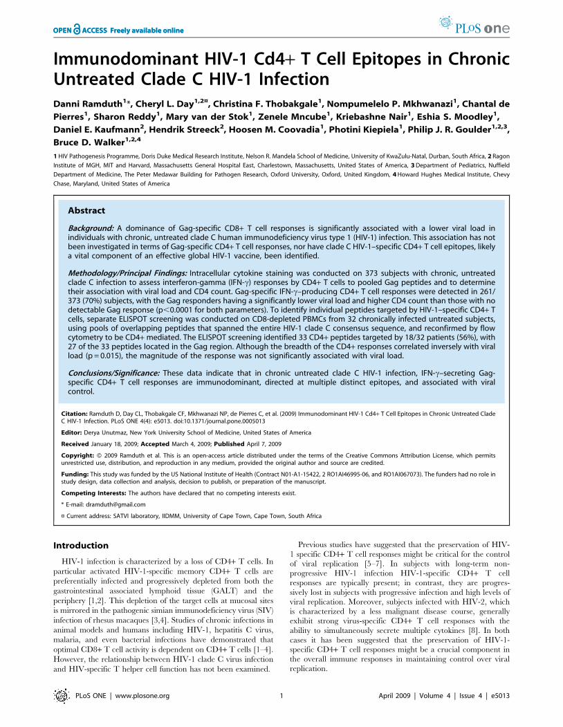
Immunodominant HIV-1 Cd4+ T Cell Epitopes in Chronic Untreated Clade C HIV-1 Infection Danni Ramduth 1 *, Cheryl L. Day 1,2¤ , Christina F. Thobakgale 1 , Nompumelelo P. Mkhwanazi 1 , Chantal de Pierres 1 , Sharon Reddy 1 , Mary van der Stok 1 , Zenele Mncube 1 , Kriebashne Nair 1 , Eshia S. Moodley 1 , Daniel E. Kaufmann 2 , Hendrik Streeck 2 , Hoosen M. Coovadia 1 , Photini Kiepiela 1 , Philip J. R. Goulder 1,2,3 , Bruce D. Walker 1,2,4 1 HIV Pathogenesis Programme, Doris Duke Medical Research Institute, Nelson R. Mandela School of Medicine, University of KwaZulu-Natal, Durban, South Africa, 2 Ragon Institute of MGH, MIT and Harvard, Massachusetts General Hospital East, Charlestown, Massachusetts, United States of America, 3 Department of Pediatrics, Nuffield Department of Medicine, The Peter Medawar Building for Pathogen Research, Oxford University, Oxford, United Kingdom, 4 Howard Hughes Medical Institute, Chevy Chase, Maryland, United States of America Abstract Background: A dominance of Gag-specific CD8+ T cell responses is significantly associated with a lower viral load in individuals with chronic, untreated clade C human immunodeficiency virus type 1 (HIV-1) infection. This association has not been investigated in terms of Gag-specific CD4+ T cell responses, nor have clade C HIV-1–specific CD4+ T cell epitopes, likely a vital component of an effective global HIV-1 vaccine, been identified. Methodology/Principal Findings: Intracellular cytokine staining was conducted on 373 subjects with chronic, untreated clade C infection to assess interferon-gamma (IFN-c) responses by CD4+ T cells to pooled Gag peptides and to determine their association with viral load and CD4 count. Gag-specific IFN-c–producing CD4+ T cell responses were detected in 261/ 373 (70%) subjects, with the Gag responders having a significantly lower viral load and higher CD4 count than those with no detectable Gag response (p,0.0001 for both parameters). To identify individual peptides targeted by HIV-1–specific CD4+ T cells, separate ELISPOT screening was conducted on CD8-depleted PBMCs from 32 chronically infected untreated subjects, using pools of overlapping peptides that spanned the entire HIV-1 clade C consensus sequence, and reconfirmed by flow cytometry to be CD4+ mediated. The ELISPOT screening identified 33 CD4+ peptides targeted by 18/32 patients (56%), with 27 of the 33 peptides located in the Gag region. Although the breadth of the CD4+ responses correlated inversely with viral load (p = 0.015), the magnitude of the response was not significantly associated with viral load. Conclusions/Significance: These data indicate that in chronic untreated clade C HIV-1 infection, IFN-c–secreting Gag- specific CD4+ T cell responses are immunodominant, directed at multiple distinct epitopes, and associated with viral control. Citation: Ramduth D, Day CL, Thobakgale CF, Mkhwanazi NP, de Pierres C, et al. (2009) Immunodominant HIV-1 Cd4+ T Cell Epitopes in Chronic Untreated Clade C HIV-1 Infection. PLoS ONE 4(4): e5013. doi:10.1371/journal.pone.0005013 Editor: Derya Unutmaz, New York University School of Medicine, United States of America Received January 18, 2009; Accepted March 4, 2009; Published April 7, 2009 Copyright: ß 2009 Ramduth et al. This is an open-access article distributed under the terms of the Creative Commons Attribution License, which permits unrestricted use, distribution, and reproduction in any medium, provided the original author and source are credited. Funding: This study was funded by the US National Institute of Health (Contract N01-A1-15422, 2 RO1AI46995-06, and RO1AI067073). The funders had no role in study design, data collection and analysis, decision to publish, or preparation of the manuscript. Competing Interests: The authors have declared that no competing interests exist. * E-mail: [email protected] ¤ Current address: SATVI laboratory, IIDMM, University of Cape Town, Cape Town, South Africa Introduction HIV-1 infection is characterized by a loss of CD4+ T cells. In particular activated HIV-1-specific memory CD4+ T cells are preferentially infected and progressively depleted from both the gastrointestinal associated lymphoid tissue (GALT) and the periphery [1,2]. This depletion of the target cells at mucosal sites is mirrored in the pathogenic simian immunodeficiency virus (SIV) infection of rhesus macaques [3,4]. Studies of chronic infections in animal models and humans including HIV-1, hepatitis C virus, malaria, and even bacterial infections have demonstrated that optimal CD8+ T cell activity is dependent on CD4+ T cells [1–4]. However, the relationship between HIV-1 clade C virus infection and HIV-specific T helper cell function has not been examined. Previous studies have suggested that the preservation of HIV- 1 specific CD4+ T cell responses might be critical for the control of viral replication [5–7]. In subjects with long-term non- progressive HIV-1 infection HIV-1-specific CD4+ T cell responses are typically present; in contrast, they are progres- sively lost in subjects with progressive infection and high levels of viral replication. Moreover, subjects infected with HIV-2, which is characterized by a less malignant disease course, generally exhibit strong virus-specific CD4+ T cell responses with the ability to simultaneously secrete multiple cytokines [8]. In both cases it has been suggested that the preservation of HIV-1- specific CD4+ T cell responses might be a crucial component in the overall immune responses in maintaining control over viral replication. PLoS ONE | www.plosone.org 1 April 2009 | Volume 4 | Issue 4 | e5013
-
Upload
independent -
Category
Documents
-
view
1 -
download
0
Transcript of Immunodominant HIV-1 Cd4+ T Cell Epitopes in Chronic Untreated Clade C HIV-1 Infection
Immunodominant HIV-1 Cd4+ T Cell Epitopes in ChronicUntreated Clade C HIV-1 InfectionDanni Ramduth1*, Cheryl L. Day1,2¤, Christina F. Thobakgale1, Nompumelelo P. Mkhwanazi1, Chantal de
Pierres1, Sharon Reddy1, Mary van der Stok1, Zenele Mncube1, Kriebashne Nair1, Eshia S. Moodley1,
Daniel E. Kaufmann2, Hendrik Streeck2, Hoosen M. Coovadia1, Photini Kiepiela1, Philip J. R. Goulder1,2,3,
Bruce D. Walker1,2,4
1 HIV Pathogenesis Programme, Doris Duke Medical Research Institute, Nelson R. Mandela School of Medicine, University of KwaZulu-Natal, Durban, South Africa, 2 Ragon
Institute of MGH, MIT and Harvard, Massachusetts General Hospital East, Charlestown, Massachusetts, United States of America, 3 Department of Pediatrics, Nuffield
Department of Medicine, The Peter Medawar Building for Pathogen Research, Oxford University, Oxford, United Kingdom, 4 Howard Hughes Medical Institute, Chevy
Chase, Maryland, United States of America
Abstract
Background: A dominance of Gag-specific CD8+ T cell responses is significantly associated with a lower viral load inindividuals with chronic, untreated clade C human immunodeficiency virus type 1 (HIV-1) infection. This association has notbeen investigated in terms of Gag-specific CD4+ T cell responses, nor have clade C HIV-1–specific CD4+ T cell epitopes, likelya vital component of an effective global HIV-1 vaccine, been identified.
Methodology/Principal Findings: Intracellular cytokine staining was conducted on 373 subjects with chronic, untreatedclade C infection to assess interferon-gamma (IFN-c) responses by CD4+ T cells to pooled Gag peptides and to determinetheir association with viral load and CD4 count. Gag-specific IFN-c–producing CD4+ T cell responses were detected in 261/373 (70%) subjects, with the Gag responders having a significantly lower viral load and higher CD4 count than those with nodetectable Gag response (p,0.0001 for both parameters). To identify individual peptides targeted by HIV-1–specific CD4+ Tcells, separate ELISPOT screening was conducted on CD8-depleted PBMCs from 32 chronically infected untreated subjects,using pools of overlapping peptides that spanned the entire HIV-1 clade C consensus sequence, and reconfirmed by flowcytometry to be CD4+ mediated. The ELISPOT screening identified 33 CD4+ peptides targeted by 18/32 patients (56%), with27 of the 33 peptides located in the Gag region. Although the breadth of the CD4+ responses correlated inversely with viralload (p = 0.015), the magnitude of the response was not significantly associated with viral load.
Conclusions/Significance: These data indicate that in chronic untreated clade C HIV-1 infection, IFN-c–secreting Gag-specific CD4+ T cell responses are immunodominant, directed at multiple distinct epitopes, and associated with viralcontrol.
Citation: Ramduth D, Day CL, Thobakgale CF, Mkhwanazi NP, de Pierres C, et al. (2009) Immunodominant HIV-1 Cd4+ T Cell Epitopes in Chronic Untreated CladeC HIV-1 Infection. PLoS ONE 4(4): e5013. doi:10.1371/journal.pone.0005013
Editor: Derya Unutmaz, New York University School of Medicine, United States of America
Received January 18, 2009; Accepted March 4, 2009; Published April 7, 2009
Copyright: � 2009 Ramduth et al. This is an open-access article distributed under the terms of the Creative Commons Attribution License, which permitsunrestricted use, distribution, and reproduction in any medium, provided the original author and source are credited.
Funding: This study was funded by the US National Institute of Health (Contract N01-A1-15422, 2 RO1AI46995-06, and RO1AI067073). The funders had no role instudy design, data collection and analysis, decision to publish, or preparation of the manuscript.
Competing Interests: The authors have declared that no competing interests exist.
* E-mail: [email protected]
¤ Current address: SATVI laboratory, IIDMM, University of Cape Town, Cape Town, South Africa
Introduction
HIV-1 infection is characterized by a loss of CD4+ T cells. In
particular activated HIV-1-specific memory CD4+ T cells are
preferentially infected and progressively depleted from both the
gastrointestinal associated lymphoid tissue (GALT) and the
periphery [1,2]. This depletion of the target cells at mucosal sites
is mirrored in the pathogenic simian immunodeficiency virus (SIV)
infection of rhesus macaques [3,4]. Studies of chronic infections in
animal models and humans including HIV-1, hepatitis C virus,
malaria, and even bacterial infections have demonstrated that
optimal CD8+ T cell activity is dependent on CD4+ T cells [1–4].
However, the relationship between HIV-1 clade C virus infection
and HIV-specific T helper cell function has not been examined.
Previous studies have suggested that the preservation of HIV-
1 specific CD4+ T cell responses might be critical for the control
of viral replication [5–7]. In subjects with long-term non-
progressive HIV-1 infection HIV-1-specific CD4+ T cell
responses are typically present; in contrast, they are progres-
sively lost in subjects with progressive infection and high levels of
viral replication. Moreover, subjects infected with HIV-2, which
is characterized by a less malignant disease course, generally
exhibit strong virus-specific CD4+ T cell responses with the
ability to simultaneously secrete multiple cytokines [8]. In both
cases it has been suggested that the preservation of HIV-1-
specific CD4+ T cell responses might be a crucial component in
the overall immune responses in maintaining control over viral
replication.
PLoS ONE | www.plosone.org 1 April 2009 | Volume 4 | Issue 4 | e5013

Apart from the decline in CD4+ T cell numbers, HIV-1
infection also impairs the functional and phenotypic heterogeneity
of HIV-1 specific CD4+ T cells. In untreated HIV-1 clade B
infection, characterized by antigen persistence and high antigen
load, HIV-1-specific CD4+ T cell responses tend to be weak or
absent. Detectable virus-specific CD4+ T cell cytokine responses in
HIV-1 infection consist mainly of IFN-c, whereas in subjects able
to control viral replication CD4+ T cells that secrete IL-2 are also
detectable, and may be a key component of an effective immune
response. Central memory T cells produce mainly IL-2 while
effector memory cells produce both IFN-c and IL-2 [9]. Persistent
exposure to antigen, such as occurs in HIV-1 infection, is believed
to generate short-lived IFN-c producing effector memory CD4+ T
cells which are impaired in their ability to develop into IL-2
producing central memory cells [10]. This phenomenon has been
observed at all stages of the infection and is postulated to disrupt
the IL-2 producing capacity of CD4+ T cells directed against
HIV-1 [11].
This skewing of cytokine secretion is in turn believed to
diminish the ability of these cells to proliferate in response to
HIV-1 antigens [12]. Very early studies of HIV-1 specific CD4+T cell responses found strong proliferative responses against the
p24 Gag antigen in individuals who were able to control HIV
replication without therapy [6]. However many studies have
shown that chronic progressive infection is associated with no or
minimal proliferative responses while IFN-c producing HIV-1
specific CD4+ T cells are retained [5,13–16]. Taken in the
context of IL-2 producing and memory T cells, the resultant
concept is that persistent HIV-1 antigenemia skews the
production of functional IL-2 producing central memory
CD4+ T cells to a less functional IFN-c producing phenotype
that reduces the proliferative capacity of HIV-1 specific CD4+ T
cells [10].
The vast majority of studies that have contributed to our
current understanding of CD4+ T cell immunology in HIV-1
infection have been conducted in the context of clade B
infection, which is the predominant clade in North America and
Europe. The brunt of the AIDS epidemic is however borne by
Sub-Saharan Africa where clade C virus predominates. In 2007
alone, 32% of all new HIV infections globally occurred in
Southern Africa [17]. Understanding the immunology of HIV-1
specific T cell responses in Southern Africa is therefore
imperative in designing a vaccine for this particular region but
there have been limited analyses of HIV-1 specific CD4+ T cell
responses in the context of clade C HIV infection [18,19]. The
identification of HIV-1 CD4+ T cell epitopes and an
understanding of the relationship between these responses and
viral control is crucial in designing an HIV-1 vaccine that elicits
protective CD4+ T cell responses. Neither the elucidation of
these epitopes, nor the pattern of cytokine production by HIV-1
specific CD4+ T cells has been determined.
Here we evaluate the hypothesis that HIV-1-speccific CD4+ T
cell responses to clade C virus infection are associated with control
of viremia, by analyzing a large cohort of persons with chronic
untreated clade C virus infection. We demonstrate that the HIV
Gag protein is the major target for HIV-1-specific CD4 T cell
responses in clade C virus infection, and show that Gag-specific
CD4+ T cell responses in chronically infected clade C subjects are
significantly associated with lower viral load and higher CD4
counts. In addition, we identify HIV-1 clade C CD4+ T cell
epitopes and find that the majority of these epitopes are located in
the Gag region. These data demonstrate an immunodominance of
Gag-specific CD4+ T cell responses related to control of viremia in
clade C virus infection.
Results
IFN-c–producing, Gag-specific CD4+ T cell responses areassociated with lower viremia and higher CD4 counts
Previous studies in clade B virus infection have suggested that
robust HIV-1-specific CD4+ responses are mounted during
primary HIV-1 infection, but subsequently lost during disease
progression, whereas the preservation of detectable Gag-specific
CD4+ T cell responses has been associated with a slower disease
progression [20]. We were therefore interested to assess, in a large
untreated cohort, whether the presence of Gag-specific CD4
responses is associated with the level of viremia. PBMCs from 373
patients with chronic, untreated clade C infection were stimulated
with a pool of HIV-1 clade C Gag peptides and screened for IFN-
c-producing CD4+ T cell responses by intracellular cytokine
staining (ICS) assay. Among the 373 subjects tested for Gag-
specific CD4+ T cell 70% (261/273) showed detectable IFN-csecretion upon antigenic stimulation. Interestingly, the subjects
with the highest IFN-c CD4+ T cell response appeared to also
have the lowest viral loads. We next compared the subjects with a
detectable Gag-specific CD4+ response with subjects who lacked
significant IFN-c secretion upon Gag stimulation. The 261
subjects with a detectable CD4+ T cell response had a median
viral load of 29,900 RNA copies/ml plasma [range 49 to 8.56106]
which was significantly lower than the median viral load of
subjects without a detectable CD4 response (median 91,900 RNA
copies/ml plasma) [range 49 to 6.56106] (p,0.0001, Mann
Whitney, Figure 1A), although there was considerable overlap.
The 112 subjects without a detectable CD4+ T cell response also
had a significantly lower CD4+ T cell count (p,0.0001, Mann
Whitney, Figure 1B, median CD4 count 274 vs 394 of Gag
responders). As it has been demonstrated that HIV-1 preferentially
infects HIV-1-specific CD4+ T cells [21], it is plausible that the
lack of CD4+ T cell responses might also reflect the stage of
disease progression. The distribution of the data showed that some
patients with a positive CD4+ Gag-specific response had viral
loads greater than 100,000 RNA copies/ml, while 4 patients with
no CD4+ Gag specific response had undetectable viral loads (,50
copies/ml plasma). Thus, although the presence of a CD4+ Gag
specific response is significantly associated with a low viral load
and higher CD4+ T cell count, it does not always predict a state of
viral control.
The HIV-1 Gag protein has the highest CD4+ T cellepitope density
Previous studies in clade B infection as well as limited studies in
clade C infection have shown the HIV-1 Gag protein to be the
dominant target of virus-specific CD4+ T cell responses [18,20].
However, little is known regarding the exact regions of the HIV
proteome that are preferentially targeted by HIV-1-specific CD4+T cell responses in clade C infection. In order to define the target
antigens and the relative immunodominance of the overall HIV-1-
specific CD4 T cell response in clade C virus infection, we used an
IFN-c Elispot assay to screen for HIV-1-specific CD4+ responses
using an overlapping peptide set spanning the entire HIV-1 clade
C proteome in 32 randomly selected subjects. CD8-depleted
PBMC were used in the Elispot assay to identify specific peptides
eliciting a CD4+ T cell response. A total of 33 peptides containing
CD4+ T cell epitopes were identified, with 27 peptides located in
the Gag region, 13 of which were targeted by two or more persons
(Figure 2A). The most commonly targeted CD4+ peptides were a
p17 Gag peptide (ERFALNPGLLETSEGGK) and a Nef peptide
(FKGAFDLSFFLKEKGGL), each targeted in 8 of the 32
subjects, followed by a p24 Gag peptide detected in 7 of the
CD4 Clade C HIV Infection
PLoS ONE | www.plosone.org 2 April 2009 | Volume 4 | Issue 4 | e5013

subjects (YVDRFFKTLRAEQATQDV, Table 1). There was a
paucity of peptides containing epitopes in HIV-1 proteins other
than Gag: 3 in the Polymerase region (protease; reverse
transcriptase and integrase), 2 in Nef, 1 in the Vpr region and
none in the Vif, Vpu, Rev, Tat or Envelope proteins. Each peptide
identified was confirmed to be CD4+ restricted by flow cytometric
analysis of PBMCs (representative data Figure 2B).
Eighteen of the 32 screened subjects (56%) had detectable HIV-
1-specific CD4+ T cell responses, with fifteen of these subjects
targeting the Gag region (Figure 3). When Gag was targeted it
always comprised at least 50% of the total HIV-1-specific CD4 T
cell response, indicating a relative immunodominance of this
antigen. Unlike clade B infection, in which Gag and Nef CD4+ T
cell responses dominated, we noted a restriction of CD4+ T cell
responses to the Gag protein. This difference in the detection of
Nef responses could be due to the different treatment status of our
subjects compared to those of the clade B study [20].
The breadth of CD4+ responses correlates inversely withviral load
As it has been recently suggested that the breadth of HIV-1-
specific CD8+ T cell responses is significantly associated with
disease progression in HIV-1 clade C infection [22], we were
interested in whether this is also the case for HIV-1-specific CD4+T cell responses. The total breadth of the CD4+ T cell response
was obtained by summing the number of peptides identified by
ELISPOT assay. While the total magnitude of the response was
not significantly associated with the viral load level (p = 0.088,
R = 20.306, Spearman Rank correlation, Figure 4B), we found a
significant inverse correlation between the breadth of response and
viral load (p = 0.013, R = 20.436, Spearman Rank, Figure 4A).
When looking more specifically at the breadth and magnitude of
Gag-specific CD4+ T cell responses, we found an inverse
correlation between each of these parameters and viral load
(p = 0.02, R = 20.41 for breadth and p = 0.01, R = 20.45,
Spearman Rank correlation; Figure 4C and 4D). These correla-
tions continued to be significant even when the single patient with
the highest number and magnitude of Gag-specific responses was
excluded (p = 0.01 for breadth and magnitude vs VL, Figure 4E
and 4F). These data suggest that the number of peptides targeted
may be particularly important in viral control. Similar analyses in
larger cohorts is required to investigate if the induction of a broad
range of CD4+ T cell responses induced by HIV-1 peptides are
protective, and to what extent the breadth of responses to proteins
other than Gag may contribute to control.
IFN-c producing HIV-1–specific CD4+ T cells dominateover IFN-c/IL-2 HIV-1–specific CD4+ T cells
Previous studies found an inverse correlation between HIV-1-
specific CD4+ T cells producing IL-2 or IFN-c/IL-2 and viremia
[23,24]. In order to further characterize the HIV-1-specific CD4+ T
cell responses in chronic clade C infection; we stimulated PBMCs
from 10 chronically infected patients with pooled HIV-1 synthetic
peptides spanning all HIV proteins and assayed for the simulta-
neous production of IFN-c and IL-2 by CD4+ T cells by ICS and
flow cytometry. Representative data are provided in Figure 5A,
where distinct populations of IFN-c-single positive, IL-2-single-
positive, and IFN-c/IL-2-double positive CD4+ T cells are seen.
The responses to all HIV-1 protein pools were then added together
to represent the total HIV-1 cytokine specific response. We found
that the percentage of HIV-1-specific CD4+ T cells producing IFN-
c only was significantly higher than the percentage of HIV-1 specific
CD4+ T cells simultaneously producing both IFN-c and IL-2
(p = 0.0099, Kruskal-Wallis analysis and corrected for multiple
comparisons with Dunns Multiple Comparison Test, Figure 5B).
These data indicate that, similar to clade B infected subjects, there is
a lack of IL-2 production by HIV-1-specific CD4+ T cell responses
associated with chronic infection and chronic exposure to antigen
[11,14,25]. Unlike previous publications, we found no correlation
between Gag-specific IL-2 and total IL-2 production and viral load
(p = 0.13, R = 20.52, Spearman Rank, data not shown). This could
be due to the small number of subjects evaluated for multiple
cytokine secretion, and further studies are clearly warranted.
Figure 1. CD4+ T cell responses targeting HIV Gag are associated with immunological control. Persons with a CD4+ T cell responseagainst a pool of Gag peptides have significantly lower viral loads and higher CD4 counts than those with no response against the Gag peptide pool.A total of 373 subjects were screened and statistics were calculated using the Mann Whitney test.doi:10.1371/journal.pone.0005013.g001
CD4 Clade C HIV Infection
PLoS ONE | www.plosone.org 3 April 2009 | Volume 4 | Issue 4 | e5013

Figure 2. The majority of the HIV-1 C clade CD4+ epitopes cluster in the Gag region. ELISPOT screening was conducted on 32 randomlyselected subjects. (A) 33 CD4+ restricted epitopes were identified, with 27 epitopes located in the Gag region, 3 in the Polymerase region, 1 in the Vprregion and 2 in the Nef region. There were no CD4+ epitopes present in the Vif, Vpu, Rev, Tat, or Envelope proteins. (B) The peptides identified by theELISPOT assay utilizing CD8+ depleted PBMCs were confirmed by flow cytometry using whole PBMCs to be CD4+ restricted. The dot plot aboverepresents data from the negative control on the left and a CD4+ IFN-c response to the p24 Gag peptide YVDRFFKTLRAEQATADV.doi:10.1371/journal.pone.0005013.g002
Table 1. Most frequently recognized clade C CD4+ T cell epitopes.
Protein SequenceNumber of subjectswith response
% Of Subjectswith response
Range of response (SFCs/millionCD8 depleted cells)
p17 ERFALNPGLLETSEGCK 8 25 70–870
p17 GKKHYMLKHLVWASREL 3 9 120–320
p17 ASRELERFALNPGLL 4 12.5 70–700
p24 QMVHQAISPRTLNAWVKV 2 6 70–120
p24 YVDRFFKTLRAEQATQDV 7 22 70–700
p24 AFSPEVIPMFTALSEGA 2 6 170–470
p24 GGHQAAMQMLKDTINEEA 2 6 110–240
p24 LHPVHAGPIAPGQMREPR 2 6 90–840
p24 PVGDIYKRWIILGLNKIV 4 12.5 60–650
p24 WIILGLNKIVRMYSPVSI 2 6 75–680
p24 DVKNWMTDTLLVQNA 2 6 65–220
p24 MTDTLLVQNANPDCKTIL 2 6 150–540
P15 GKIWPSHKGRPGNFLQSR 2 6 800–940
Protease FIKVRQYDQIPIEICGKK 2 6 60–110
Vpr SRIGILRQRRARNGASRS 2 6 90–210
Nef FKGAFDLSFFLKEKGGL 8 25 70–850
doi:10.1371/journal.pone.0005013.t001
CD4 Clade C HIV Infection
PLoS ONE | www.plosone.org 4 April 2009 | Volume 4 | Issue 4 | e5013

High degree of cross clade recognition for the mostcommonly targeted p24 Gag peptide
Our data above indicate a preference for CD4+ T cells to
target epitopes within the Gag HIV-1 protein. We next sought to
determine whether the most immunodominant peptides in clade
C and clade B infection are cross-recognized. The 8 most
commonly targeted CD4+ T cell epitopes in B clade infection
[20] were tested for cross recognition in twenty eight randomly
selected patients. Only 3/8 B clade CD4+ T cell epitopes were
recognized by just 9/28 individuals of this clade C infected
cohort. These peptides were all located in the Gag region:
ERFAVNPGLLETSEGCR in p17 Gag, YVDRFYKTLRAE-
QASQEV in p24 Gag and RQANFLGKIWPSHKGR in p15
Gag. One patient responded to the p15 peptide only, one to the
p17 peptide only; five to the p24 peptide and 2 patients
recognized both the p17 and p24 peptides.
We next compared the magnitudes of the clade B and C peptide
responses of these 3 peptides to further define the degree of cross-
clade recognition of these targeted CD4+ peptides. The clade B
version of the p15 peptide, RQANFLGKIWPSHKGR, elicited a
response of 70 SFCs while the clade C peptide had a response of
160 SFCs in the same patient (data not shown). The clade B
sequence ERFAVNPGLLETSEGCR was recognized by only 3 of
the clade C virus infected subjects, all of whom had lower
magnitude responses (data not shown). In contrast, all 7 subjects
who responded to YVDRFFKTLRAEQATQDV, also responded
with similar magnitude to the B clade sequence of the peptide
(YVDRFYKTLRAEQASQEV, p = 0.9375, Wilcoxon matched
pairs test, Figure 6). These data suggest that cross-clade recognition
by HIV-1-specific CD4+ T epitopes is confined to a single p24
Gag peptide, and even minor sequence changes between clade B
and clade C might be sufficient to account for lack of cross
recognition.
Discussion
Evidence from both Hepatitis C virus (HCV), HIV-1 and SIV
infection suggests that virus-specific CD4+ T cells are crucial in the
effective immune control of chronic viral infections [7,26,27]. There is
a paucity of data on clade C HIV-1-specific CD4+ T cell responses,
despite the fact that clade C infection accounts for the majority of
infections globally [17,18]. In this study, we comprehensively assessed
HIV-1 specific CD4+ T cell activity in an African cohort of untreated
subjects, defining the dominant targeted epitopes, the ability to
produce IFN-c and IL-2, and cross-clade epitope recognition. Our
results indicate that 70% of clade C infected persons target the HIV-1
Gag protein which contains the majority of CD4+ T cell epitopes, that
targeting of Gag is associated with lower viral loads, and that there is
an inverse correlation between the breadth of the HIV-1-specific
CD4+ T cell response and viral load.
In an earlier study of 65 chronically infected subjects using
clade C peptide pools in an intracellular cytokine staining assay,
we found Gag to be the dominant target of HIV-1 specific CD4+T cells, but could not detect a relationship between the detection
of Gag-specific responses and viral load [18]. Here we do find a
significant albeit weak association between Gag-specific CD4+ T
cell responses and viral load. However, despite the dominance of
Gag-specific CD4+ T cell responses and their correlation with
lower viral loads, the vast majority of untreated chronically
infected patients do progress to AIDS. A possible reason is that
these are immunodominant responses captured at a particular
timepoint and which wane with continuing viral replication. A
longitudinal study that tracks these responses will be able to
address this particular point. Proliferative capacity and IL-2
production by HIV-1 specific CD4+ T cells appear to be clearer
markers of CD4+ T cell activity than IFN-c as there is a distinct
decline of these parameters in patients exposed to increasing
levels of viremia [28]. Although IFN-c production may also be
affected, the decline may be more subtle which could explain why
a cross sectional analysis is unable to establish a strong
association.
It has been recently demonstrated that the upregulation of
inhibitory immunoregulatory molecules, CTLA-4 and PD-1, on
HIV-1 specific CD4+ T cells is associated with disease progression
[29,30]. Although not investigated in this study, the PD-1
expression on CD4+ T cells has been shown to be associated
with viral load in clade B virus infection [29]. Although it is likely
that patients with strong Gag-specific CD4+ T cell responses
Figure 3. The total HIV-1–specific CD4+ T cell response is dominated by Gag. Using IFN-c ELISPOT assay to identify HIV-1 specific CD4+ Tcell epitopes in 32 subjects, we found that irrespective of the viral load, the Gag protein is the most commonly targeted and therefore makes thegreatest contribution to the overall CD4+ T cell response. Subjects 1–16 had viral loads below the median, while subjects 18–32 had viremia greaterthan the median (36 550 copies/ml plasma). Acc (accessory) denotes the Vpr, Vpu and Vif proteins pooled together. Subjects 24 and 25 both targeteda single Vpr protein.doi:10.1371/journal.pone.0005013.g003
CD4 Clade C HIV Infection
PLoS ONE | www.plosone.org 5 April 2009 | Volume 4 | Issue 4 | e5013

express fewer CTLA-4 and PD-1 molecules than patients with no
Gag-specific CD4+ T cell responses, an additional study will be
required to investigate this hypothesis.
Since CD4+ T cells play such a central role in coordinating
functions of the immune system, a comprehensive analysis of
cytotoxic CD8+ T cell function, neutralizing antibody activity and
Figure 4. Correlation of total breadth and magnitude of CD4+ T cell response to viral load. (A) The total breadth of the HIV-1-specificCD4+ T cell response was obtained by summing the number of peptides identified by the ELISPOT assay and was found to correlate inversely withviral load. (B) The total magnitude of the response was calculated by adding the number of spot forming cells for each peptide response. Here wefound no association between the magnitude of the response and viral load. More specifically the breadth (C) and magnitude (D) of Gag-specificCD4+ T cell responses, correlated inversely with viral load. Excluding the outlier with 17 peptide responses, these correlations continued to besignificant for both breadth (E) and magnitude (F).doi:10.1371/journal.pone.0005013.g004
CD4 Clade C HIV Infection
PLoS ONE | www.plosone.org 6 April 2009 | Volume 4 | Issue 4 | e5013

CD4+ T cell receptor signaling pathways will shed more light on
mechanisms of viral control.
The results from our current study using the ELISPOT assay
add to our earlier data by demonstrating that the Gag protein
contains the highest CD4+ T cell epitope density. Although we
only detected 2 epitopes in Nef, one was among the two most
highly targeted of all epitopes. Gag and Nef, were also found to be
the most frequently targeted proteins by CD4+ T cells in clade B
HIV-1 infection and in the less pathogenic HIV-2 infection
[20,31]. The frequent targeting of Gag and the contrasting lack of
responses to Vif, Vpu, Rev, Tat and envelope also correlate with
observations from clade B CD4+ T cell epitope mapping [20].
The reason for the frequent targeting of Gag by CD4+ T cells
and the link with control of viremia, could be associated with the
high number of Gag molecules (1500 molecules of Gag and 100 of
the precursor Gag-Pol molecules in a single immature virus
particle), coupled with a relatively high degree of conservancy of
this protein [32,33]. Among the Gag epitopes targeted, particu-
larly noticeable is the dominant targeting of the p24 Gag peptide
YVDRFFKTLRAEQATQDV. This was found to be frequently
targeted in clade B and clade A/G HIV-1 infection [20,31], and
displayed a high degree of cross recognition between HIV-1 and
HIV-2 infected individuals [31]. Our data have also confirmed
that at least for some epitopes, clade C infected individuals can
mount an IFN-c CD4+ T cell response of equal magnitude against
both the clade B and C sequences. This peptide overlaps partially
with the Major Homology Region (MHR), located at the C
terminal dimerization domain of p24 (the capsid antigen) and
displays significant homology amongst the various genre of
retroviruses [32]. Mutations to the MHR result in viral particles
that are defective in viral assembly, maturation and infectivity
[32]. The peptide, YVDRFFKTLRAEQATQDV, therefore
presents as a possible valuable component in an HIV vaccine
and deserves further characterization with regards to the immune
response it elicits.
The relative lack of responses in proteins other than Gag may
also be due to the use of the consensus clade C sequence which
could potentially limit our detection of antigen specific CD4+ T
cell epitopes to proteins that are more variable. There was a
significant augmentation in the detection of CD8+ T cell epitopes
in clade B infection using autologous HIV-1 peptides in
comparison to the consensus sequence [34]. This may also be
one of the reasons why we detected fewer Nef responses in contrast
to the clade B CD4+ epitope screening study [20]. Another reason
is that our study focused on chronically infected untreated patients,
compromising both controllers and non-controllers of HIV-1
infection, while the clade B study consisted of patients undergoing
structured treatment interruption and acutely infected patients.
These differing disease states and levels of virus exposure could
also account for the differences observed with regard to the Nef
responses.
In this study we were able to identify HIV-1 specific epitopes
producing IFN-c only. Since IL-2 and IFN-c/IL-2 CD4+ T cell
responses are associated with control of viremia [12,23,24], it will
be important to focus future studies on identification the breadth
and specificities of clade C IL-2 and IFN-c/IL-2 producing CD4+epitopes. Here we observed comparatively sparse IL-2 and IFN-c/
IL-2 HIV-1 specific CD4+ T cells by ICS in our cohort. This
could be explained by our cohort being chronically infected, with
an immune response already skewed to a dominant IFN-c only
Figure 5. Dual cytokine staining to pools of overlappingpeptides. Dual IFN-c/IL-2 ICS was conducted on 10 subjects. (A) Thedot plot on the left shows the unstimulated control while the one onthe right shows the simultaneous detection of IFN-c and IL-2 inresponse to stimulation with a pool of Gag peptides. The cells weregated on CD4+ T cells. (B) The cytokine response to all HIV-1 proteinpools were added to obtain the total HIV-1 cytokine specific response.The percentage of HIV-1-specific CD4+ T cells producing only IFN-c wassignificantly higher than the percentage of HIV-1-specific CD4+ T cellsproducing both IFN-c and IL-2.doi:10.1371/journal.pone.0005013.g005
Figure 6. Cross clade recognition of the p24 Gag peptideYVDRFFKTLRAEQATQDV. In a subset of 28 subjects, we found thatthe C and B clade versions of peptide YVDRFFKTLRAEQATQDV inducedthe same magnitude of IFN-c response in 7 patients, as detected by theELISPOT assay.doi:10.1371/journal.pone.0005013.g006
CD4 Clade C HIV Infection
PLoS ONE | www.plosone.org 7 April 2009 | Volume 4 | Issue 4 | e5013
producing effector memory phenotype [10]. A limitation of this
study is that because of constraints on the number of parameters
available for flow cytometric analysis, we were not able to
characterize the polyfunctional capacity and memory phenotype
of epitope specific CD4+ T cells. This aspect, along with
evaluating CD4+ T cell activity in acutely infected patients, also
requires further investigation.
Although the breadth of the CD4+ T cell responses in our
cohort correlated inversely with viral load, the magnitude had no
significant association with viremia. Research on clade B HIV-1
specific CD4+ T cell responses has revealed that although CD4+T cell responses can be readily detected, they are generally of low
magnitudes [15,20]. The reason for this observation is unclear but
it could be associated with the level of sensitivity of current assays,
or to the inability to test with peptides representing autologous
viruses. The impact of host genetic factors on HIV-1 specific
CD4+ T cell responses in clade C HIV-1 infection also warrants
investigation as different HLA Class I molecules have shown to
significantly influence CD8+ T cell responses [35]. Analysis of
clade B CD4+ T cell epitopes binding to HLA Class II molecules
has revealed a high degree of promiscuity amongst the epitopes
[20,36]. This property may also be beneficial in vaccine design as
these epitopes can induce responses amongst a wide array of HLA
Class II types [36].
In conclusion, dominant HIV-1-specific CD4+ responses are
detectable against a limited number of epitopes in chronic HIV-1
clade C infection. Most CD4+ T cell responses are directed against
epitopes in Gag and the presence of Gag-specific CD4+ T cells is
significantly associated with lower viral load and higher CD4+ T
cell counts compared to subjects with no detectable Gag-specific
CD4+ T cell responses. Further studies are required to determine
whether HIV-1-specific CD4+ T cell activity contributes directly
to the control of viremia or is a consequence of viral control. The
further identification and characterization of HIV-1 CD4+epitopes will be important in characterizing clade C immunoge-
nicity and will likely play an important role for clade specific
vaccine development.
Materials and Methods
Ethics statementEthical approval was granted by the Institutional Review Board
of Massachusetts General Hospital and the Ethics Committee of
the University of KwaZulu-Natal. All participants enrolled in the
study signed a written consent form agreeing to participate in the
study.
Study populationThree hundred seventy three subjects, recruited from the
Sinikithemba Care Center at McCord Hospital in Durban South
Africa, were screened for Gag-specific IFN-c CD4+ T cell
responses by intracellular cytokine staining (ICS) using freshly
isolated PBMCs. All subjects were chronically infected with clade
C HIV-1 and had not received any antiretroviral medication
before or at the time of analysis [median viral load (VL) – 44,300,
range 49 to 8.56106 RNA copies/ml plasma; and median CD4
count 357, range 11 to 1414 cells//ml whole blood]. Thirty two
were randomly selected (to exclude the possibility of introducing a
bias in our analysis) and screened by an IFN-c ELISPOT assay
(detailed below) using individual HIV peptides to identify CD4+epitopes. The median viral load of this group was 36,550 RNA
copies/ml plasma and median CD4 count 342 cells/ml whole
blood. From this group, 10 were randomly selected to assess HIV-
1-specific intracellular IFN-c and IL-2 production by flow
cytometry. Twenty eight patients were tested for cross clade
recognition of the most common CD4+ epitopes in clade B
infection using the IFN-c ELISPOT assay.
Ten HIV-1 negative subjects (as determined by a negative
ELISA and viral load measurement) served as controls.
CD4+ counts and viral loadAbsolute CD4 counts were measured by using the MultiTest
TruCount kit and the MultiSet software on the FACS Calibur
(Becton Dickinson). Plasma HIV-1 viral loads were measured
using the Roche Amplicor kit version 1.5 on the Roche Cobas
Amplicor instrument. The sensitivity of the test was 50 RNA
copies/ml plasma.
Intracellular cytokine stainingThe intracellular cytokine assay was conducted using peripheral
blood mononuclear cells (PBMC) isolated from whole blood within
4 hours of phlebotomy, as described previously [41]. This protocol
was followed to assess Gag-specific responses in 373 patients.
Changes to the protocol to evaluate IFN-c and IL-2 cytokine levels
included a 12 hour incubation period with pooled and individual
peptides; 2 tubes of negative/unstimulated controls per patient
(with the average background being subtracted from the
experimental tubes). The following antibodies were used: anti-
human CD8 Peridinin-chlorophyll-protein Complex (PerCP),
anti-human CD4 Allophycocyanin (APC), anti-human IFN-cFluorescein isothiocyanate (FITC) and anti-human IL-2 Phycoer-
ythrin (PE).
Ten HIV-1 unexposed, RT-PCR and ELISA negative adults
served as controls, where the range of response to any pool of
HIV-1 peptides was 0.00–0.04% of CD4+ T cells. A response was
therefore considered positive for HIV-1-specific CD4+ T cells if
after subtraction of the average background, the response was still
greater than 0.04%.
CD8+ T cell depletion and ELISPOTWhole blood obtained within 4 hours of phlebotomy was
depleted of CD8+ T cells by incubation with anti-CD8 RosetteSep
antibody (Stem Cell Technologies) prior to PBMC separation by
ficoll-histopaque density centrifugation. The purity of the isolated
cells was confirmed by flow cytometry and contained on average
98% CD4+ cells. These cells were used for the ELISPOT assay as
described previously [37].
Ninety six-well polyvinylidene difluoride-backed Elispot plates
(MAIP S45, Millipore) were coated overnight at 4uC with 100 ml
anti-IFN-c antibody (1-D1k, 0.5 mg/ml, MabTech, Sweden). The
plates were then washed six times with blocking buffer (1% fetal
calf serum (FCS) in PBS). 50 ml of R10 (RPMI 1640 medium
supplemented with 10% FCS, 1% L-glutamine, and 1%
penicillin/streptomycin) were added to the empty wells. Ten
microliters of pooled overlapping peptides (6 peptides/pool)
spanning the entire clade C consensus sequence were added into
the wells so that the final concentration of each peptide in a pool
was 2 mg/ml in the well. In addition, cells were added to 6 wells
containing medium only as a negative control, and 2 wells
containing phytohemagglutinin (PHA) (1 mg/ml in the well) as a
positive control. Cells were plated at 100 000 cells/well and
incubated for 16 hrs at 37uC and 5% CO2. The plates were then
washed 6 times with PBS and 0.5 mg/ml of biotinylated anti-IFN-
c antibody (7-B6-1, MabTech) was added to the plates which were
incubated at room temperature in the dark. The plates were
washed once again with PBS and 0.5 mg/ml of streptavidin-
alkaline phosphatase conjugated antibody (Mabtech) was added
and left in the dark for 45 minutes at room temperature. After a
CD4 Clade C HIV Infection
PLoS ONE | www.plosone.org 8 April 2009 | Volume 4 | Issue 4 | e5013

final wash, IFN-c producing cells were identified by direct
visualization of spots produced by the addition of alkaline
phosphatase colour reagents (Bio-Rad).
The number of IFN-c producing cells was quantified by
counting the number of spots using an automated ELISPOT plate
reader (AID ELISPOT Reader System, Auoimmun Diagnstika,
Germany). Results are expressed as the number of Spot Forming
Cells (SFCs)/million CD8 depleted PBMCs. A response was
considered positive if it exceeded 100 SFCs/million CD8 depleted
PBMCs (the highest response observed in HIV-1 negative control
patients) and was $3 standard deviations above the mean of the 4
negative control wells. Individual peptides identified using the
above matrix screenings were reconfirmed by ELISPOT and then
by intracellular cytokine staining of PBMCs to be CD4+ restricted.
Eight dominant CD4+ B clade epitopes [20] were individually
screened by ELISPOT for cross clade recognition at the same
concentration and using the same number of cells as described
above. Positive responses were confirmed by ICS assay. The
sequences of the 8 peptides screened were: p17 amino acid
position 37–51 ASRELERFAVNPGLL; p17 amino acid position
42–58 ERFAVNPGLLETSEGCR; p17 amino acid position 77–
94 SLYNTVATLYCVHQRIEV; p15 amino acid position 66–81
RQANFLGKIWPSHKGR; p24 amino acid position 133–150
WIILGLNKIVRMYSPTSI; p24 amino acid position 164–181
YVDRFYKTLRAEQASQEV; Nef PEKEVLVWKFDSR-
LAFHH amino acid position 176–196; Nef KFDSRLAFHH-
MARELH amino acid 184–199.
Statistical analysisAll statistical analyses and graphs were plotted using
GraphPad Prism software (version 4). The data were assessed
for Gaussian distributions and the relevant statistical test was
conducted by the software. A p value of ,0.05 was considered
significant.
Acknowledgments
We thank the subjects and staff, in particular Kesia Mgwenya and
Nokuthula at The Sinikithemba Support Group at McCords Hospital in
Durban.
Author Contributions
Conceived and designed the experiments: DR CLD PG BDW. Performed
the experiments: DR CFT NM CdP SR MvdS ZM KN ESM. Analyzed
the data: DR CLD CdP PK PG BDW. Contributed reagents/materials/
analysis tools: DEK. Wrote the paper: DR CLD CdP HS HMC BDW.
References
1. Douek DC, Betts MR, Brenchley JM, Hill BJ, Ambrozak DR, et al. (2002) A
novel approach to the analysis of specificity, clonality, and frequency of HIV-
specific T cell responses reveals a potential mechanism for control of viral
escape. J Immunol 168(6): 3099–104.
2. Brenchley JM, Price DA, Douek DC (2006) HIV disease: fallout from a mucosal
catastrophe? Nat Immunol 7(3): 235–9.
3. Li Q, Duan L, Estes JD, Ma ZM, Rourke T, et al. (2005) Peak SIV replication in
resting memory CD4+ T cells depletes gut lamina propria CD4+ T cells. Nature
434(7037): 1148–52.
4. Mattapallil JJ, Hill B, Douek DC, Roederer M (2006) Systemic vaccination
prevents the total destruction of mucosal CD4 T cells during acute SIV
challenge. J Med Primatol 35(4–5): 217–24.
5. Pitcher CJ, Quittner C, Peterson DM, Connors M, Koup RA, et al. (1999) HIV-
1-specific CD4+ T cells are detectable in most individuals with active HIV-1
infection, but decline with prolonged viral suppression. Nat Med 5(5): 518–25.
6. Rosenberg ES, Billingsley JM, Caliendo AM, Boswell SL, Sax PE, et al. (1997)
Vigorous HIV-1-specific CD4+ T cell responses associated with control of
viremia. Science 278(5342): 1447–50.
7. Zajac AJ, Blattman JN, Murali-Krishna K, Sourdive DJ, Suresh M, et al. (1998)
Viral immune evasion due to persistence of activated T cells without effector
function. J Exp Med 188(12): 2205–13.
8. Duvall MG, Jaye A, Dong T, Brenchley JM, Alabi AS, et al. (2006) Maintenance
of HIV-specific CD4+ T cell help distinguishes HIV-2 from HIV-1 infection.
J Immunol 176(11): 6973–81.
9. Sallusto F, Lenig D, Forster R, Lipp M, Lanzavecchia A (1999) Two subsets of
memory T lymphocytes with distinct homing potentials and effector functions.
Nature 401(6754): 708–12.
10. Day CL, Walker BD (2003) Progress in defining CD4 helper cell responses in
chronic viral infections. J Exp Med 198(12): 1773–7.
11. Yue FY, Kovacs CM, Dimayuga RC, Parks P, Ostrowski MA (2004) HIV-1-
specific memory CD4+ T cells are phenotypically less mature than cytomeg-
alovirus-specific memory CD4+ T cells. J Immunol 172(4): 2476–86.
12. Younes SA, Yassine-Diab B, Dumont AR, Boulassel MR, Grossman Z, et al.
(2003) HIV-1 viremia prevents the establishment of interleukin 2-producing
HIV-specific memory CD4+ T cells endowed with proliferative capacity. J Exp
Med 198(12): 1909–22.
13. McNeil AC, Shupert WL, Iyasere CA, Hallahan CW, Mican JA, et al. (2001)
High-level HIV-1 viremia suppresses viral antigen-specific CD4(+) T cell
proliferation. Proc Natl Acad Sci U S A 98(24): 13878–83.
14. Palmer BE, Boritz E, Blyveis N, Wilson CC (2002) Discordance between
frequency of human immunodeficiency virus type 1 (HIV-1)-specific gamma
interferon-producing CD4(+) T cells and HIV-1-specific lymphoproliferation in
HIV-1-infected subjects with active viral replication. J Virol 76(12): 5925–36.
15. Betts MR, Ambrozak DR, Douek DC, Bonhoeffer S, Brenchley JM, et al. (2001)
Analysis of total human immunodeficiency virus (HIV)-specific CD4(+) and
CD8(+) T-cell responses: relationship to viral load in untreated HIV infection.
J Virol 75(24): 11983–91.
16. Harari A, Rizzardi GP, Ellefsen K, Ciuffreda D, Champagne P, et al. (2002)Analysis of HIV-1- and CMV-specific memory CD4 T-cell responses during
primary and chronic infection. Blood 100(4): 1381–7.
17. UNAIDS (2007) AIDS Epidemic Update 07.
1. Ramduth D, Chetty P, Mngquandaniso NC, Nene N, Harlow JD, et al. (2005)
Differential immunogenicity of HIV-1 clade C proteins in eliciting CD8+ and
CD4+ cell responses. J Infect Dis 192(9): 1588–96.
2. Ngumbela KC, Day CL, Mncube Z, Nair K, Ramduth D, et al. (2008)
Targeting of a CD8 T cell env epitope presented by HLA-B*5802 is associated
with markers of HIV disease progression and lack of selection pressure. AIDS
Res Hum Retroviruses 24(1): 72–82.
3. Kaufmann DE, Bailey PM, Sidney J, Wagner B, Norris PJ, et al. (2004)
Comprehensive analysis of human immunodeficiency virus type 1-specific
CD4 responses reveals marked immunodominance of gag and nef and
the presence of broadly recognized peptides. J Virol 78(9):
4463–77.
4. Douek DC, Brenchley JM, Betts MR, Ambrozak DR, Hill BJ, et al. (2002)
HIV preferentially infects HIV-specific CD4+ T cells. Nature 417(6884):
95–8.
5. Geldmacher C, Currier JR, Gerhardt M, Haule A, Maboko L, et al. (2007) In a
mixed subtype epidemic, the HIV-1 Gag-specific T-cell response is biased
towards the infecting subtype. Aids 21(2): 135–43.
6. Boaz MJ, Waters A, Murad S, Easterbrook PJ, Vyakarnam A (2002) Presence of
HIV-1 Gag-specific IFN-gamma+IL-2+ and CD28+IL-2+ CD4 T cell responses
is associated with nonprogression in HIV-1 infection. J Immunol 169(11):
6376–85.
7. Harari A, Vallelian F, Meylan PR, Pantaleo G (2005) Functional heterogeneity
of memory CD4 T cell responses in different conditions of antigen exposure and
persistence. J Immunol 174(2): 1037–45.
8. Harari A, Petitpierre S, Vallelian F, Pantaleo G (2004) Skewed representation of
functionally distinct populations of virus-specific CD4 T cells in HIV-1-infected
subjects with progressive disease: changes after antiretroviral therapy. Blood
103(3): 966–72.
9. Veazey RS, Tham IC, Mansfield KG, DeMaria M, Forand AE, et al. (2000)
Identifying the target cell in primary simian immunodeficiency virus (SIV)
infection: highly activated memory CD4(+) T cells are rapidly eliminated in early
SIV infection in vivo. J Virol 74(1): 57–64.
10. Grakoui A, Shoukry NH, Woollard DJ, Han JH, Hanson HL, et al. (2003) HCV
persistence and immune evasion in the absence of memory T cell help. Science
302(5645): 659–62.
11. Iyasere C, Tilton JC, Johnson AJ, Younes S, Yassine-Diab B, et al. (2003)
Diminished proliferation of human immunodeficiency virus-specific CD4+ T
cells is associated with diminished interleukin-2 (IL-2) production and is
recovered by exogenous IL-2. J Virol 77(20): 10900–9.
12. Day CL, Kaufmann DE, Kiepiela P, Brown JA, Moodley ES, et al. (2006) PD-1
expression on HIV-specific T cells is associated with T-cell exhaustion and
disease progression. Nature 443(7109): 350–4.
13. Kaufmann DE, Kavanagh DG, Pereyra F, Zaunders JJ, Mackey EW, et al.
(2007) Upregulation of CTLA-4 by HIV-specific CD4+ T cells correlates with
disease progression and defines a reversible immune dysfunction. Nat Immunol
8(11): 1246–54.
14. Ondondo BO, Rowland-Jones SL, Dorrell L, Peterson K, Cotten M, et al.
(2008) Comprehensive analysis of HIV Gag-specific IFN-gamma response in
CD4 Clade C HIV Infection
PLoS ONE | www.plosone.org 9 April 2009 | Volume 4 | Issue 4 | e5013

HIV-1- and HIV-2-infected asymptomatic patients from a clinical cohort in The
Gambia. Eur J Immunol 38(12): 3549–60.15. Freed EO (1998) HIV-1 gag proteins: diverse functions in the virus life cycle.
Virology 251(1): 1–15.
16. Kurle S, Tripathy S, Jadhav S, Agnihotri K, Paranjape R (2004) Full-length gagsequences of HIV type 1 subtype C recent seroconverters from Pune, India.
AIDS Res Hum Retroviruses 20(10): 1113–8.17. Altfeld M, Addo MM, Shankarappa R, Lee PK, Allen TM, et al. (2003)
Enhanced detection of human immunodeficiency virus type 1-specific T-cell
responses to highly variable regions by using peptides based on autologous virussequences. J Virol 77(13): 7330–40.
18. Kiepiela P, Leslie AJ, Honeyborne I, Ramduth D, Thobakgale C, et al. (2004)
Dominant influence of HLA-B in mediating the potential co-evolution of HIVand HLA. Nature 432(7018): 769–75.
19. Malhotra U, Holte S, Dutta S, Berrey MM, Delpit E, et al. (2001) Role for HLA
class II molecules in HIV-1 suppression and cellular immunity followingantiretroviral treatment. J Clin Invest 107(4): 505–17.
20. Addo MM, Yu XG, Rathod A, Cohen D, Eldridge RL, et al. (2003)Comprehensive epitope analysis of human immunodeficiency virus type 1
(HIV-1)-specific T-cell responses directed against the entire expressed HIV-1
genome demonstrate broadly directed responses, but no correlation to viral load.J Virol 77(3): 2081–92.
CD4 Clade C HIV Infection
PLoS ONE | www.plosone.org 10 April 2009 | Volume 4 | Issue 4 | e5013