Molecular and metabolic retinoid pathways in human amniotic membranes
Upload
independentCategory
view
1download
0
PROTEOMIC ANALYSIS OF CERVICAL-VAGINAL FLUID:IDENTIFICATION OF NOVEL BIOMARKERS FOR DETECTION OFINTRA-AMNIOTIC INFECTION
Michael G. Gravett1,2, Archana Thomas3, Kimberly A. Schneider3, Ashok P. Reddy3,Surendra Dasari4, Thomas Jacob3, Xinfang Lu4, Matthew Rodland4, Leonardo Pereira2,Drew W. Sadowsky1, Charles T. Roberts Jr.4, Miles J. Novy1,2, and Srinivasa R. Nagalla3,4
1Division of Reproductive Sciences, Oregon National Primate Research Center, Beaverton, OR
2Departments of Obstetrics & Gynecology, Oregon Health and Science University, Portland, OR
4Pediatrics, Oregon Health and Science University, Portland, OR
3ProteoGenix Inc., Portland, OR
AbstractIntra-amniotic infection (IAI) is associated with preterm birth and perinatal mortality. To identifypotential biomarkers, we performed a comprehensive survey of cervical-vaginal fluid (CVF)proteome from a primate IAI model utilizing Multidimensional protein identification technology(LC/LC-MS/MS) and MALDI-TOF-MS analyses. Analyses of CVF proteome identified 205 uniqueproteins and differential expression of 27 proteins in controls and IAI samples. Protein expressionsignatures and immunodetection of specific biomarkers identified can be employed for non-invasivedetection of IAI.
INTRODUCTIONIntrauterine infections, including intra-amniotic infections and chorioamnionitis, are animportant and potentially preventable cause of maternal and perinatal mortality and morbidity1. Intra-amniotic infection (IAI), in particular, accounts for up to 40% of cases of febrilemorbidity in the peri-partum period, and is associated with at least one-third of early neonatalsepsis and pneumonia 2. More recently, IAI has been implicated as a major cause of pretermbirth. Despite improvements in prenatal care, preterm birth still occurs in 12.3% of births inthe United States and remains the major obstetrical problem in developed countries 3. Intra-amniotic infections are associated with more than 50% of the very-low-birth-weight neonatesthat account for the highest number of neonatal deaths, the most serious complications,including neurologic handicap, and a disproportionate share of perinatal health care costs 1.Accurate and early diagnosis of IAI would facilitate timelier and more appropriateinterventions, as well as enhance the design of therapeutic trials. Early diagnosis of IAI isproblematic, however, because clinical signs and symptoms tend to be late manifestations ofthis condition. Furthermore, the available noninvasive tests, e.g., maternal white blood cell
Correspondence: Michael G. Gravett, M.D., Department of Obstetrics & Gynecology, University of Washington, 1959 NE Pacific St,Box 356460, Seattle, WA 98195, Email: [email protected] DisclosureOregon Health & Science University and Drs. Gravett, Roberts, and Nagalla all have a significant financial interest in ProteoGenix, Inc.,a company that may have a commercial interest in the results of this research and technology. This potential conflict of interest has beenreviewed and a management plan approved by the OHSU Conflict of Interest in Research Committee.
NIH Public AccessAuthor ManuscriptJ Proteome Res. Author manuscript; available in PMC 2008 September 9.
Published in final edited form as:J Proteome Res. 2007 January ; 6(1): 89–96. doi:10.1021/pr060149v.
NIH
-PA Author Manuscript
NIH
-PA Author Manuscript
NIH
-PA Author Manuscript
count or C-reactive protein, have limited predictive value, or, in the case of more predictivetests of amniotic fluid, e.g., interleukin-6, polymerase chain reaction, or microbial culture, theresults are often delayed and amniocentesis is required 4, 5.
We have previously demonstrated, in a non-human primate model, the causal relationshipsamong experimental IAI with Group B streptococcus, Ureaplasma parvum, or Mycoplasmahominis, and preterm birth 6, 7. We have also identified a distinct proteomic profile in amnioticfluid (AF) of both rhesus monkeys with experimental IAI and from a cohort of women withpreterm labor 8. Our objective in the current study was to extend these studies to a proteomicanalysis of cervical-vaginal fluid (CVF). Our interest in CVF was based upon the non-invasivenature of obtaining specimens from a readily available site and previous observations thatdetermination of CVF pro-inflammatory cytokines and fetal fibronectin have been utilized toidentify women at risk of preterm delivery or IAI 9–12. These currently available tests havelimited predictive value for IAI, however, because they may also be influenced by theinflammatory microenvironment of the vaginal milieu. We sought to determine if proteomicevaluation of biomarkers previously identified in AF might also be reflected in CVF in thesetting of experimental IAI in a non-human primate model.
In this study, we utilized multidimensional liquid chromotography coupled to tandem massspectrometry (Multi-dimensional Protein Identification Technology; MudPIT) and spectralcounting to characterize the proteins present in CVF and to determine the relative abundanceof these proteins to detect the early appearance of sensitive and specific protein markers forIAI in CVF in non-human primates with experimental IAI caused by Ureaplasma parvum. Inaddition, we utilized matrix-assisted laser desorption/ionization time-of-flight massspectrometry (MALDI-TOF-MS) to examine IAI-specific protein signatures in CVF and tocompare them to signatures that we have previously described in AF.
METHODSExperimental IAI in non-human primates
This protocol was approved by the Institutional Animal Care and Utilization Committee ofOregon Health & Science University. Four pregnant rhesus monkeys (Macaca mulatta) withtimed gestations were chronically catheterized at 120 days gestation (term is 167 days) aspreviously described 6. Experimental IAI was established by intra-amniotic inoculation of107 colony-forming units of a clinical low-passage Ureaplamsa parvum, serovar 1, grown in10B culture media 7. Each animal served as its own control. Before and after inoculation, AFand CVF samples were serially collected for quantitative bacterial cultures, white blood cellanalysis, and cytokine and prostaglandin concentrations, as previously reported 6, 7, and forproteomic analysis. CVF was collected from the posterior vaginal fornix with sterile Dacronswabs (Solon, catalogue #36816), which were then placed into phosphate-buffered salinecontaining a protease inhibitor cocktail (Roche Diagnostics, catalogue #11836), to prevent non-specific proteolysis from naturally occurring proteases within the vagina). Following proteinextraction, samples were centrifuged to remove cellular debris and the supernatant stored at−70°C until assayed. For these assays, pooled CVF samples were utilized from samplesobtained prior to infection and from 24–72 hours after infection. Uterine contractility wasrecorded as the area under the amniotic fluid pressure curve and expressed as the hourlycontraction area (HCA; mmHg times second/per). Fetal, decidual, placental, and bacterialcultures were obtained after delivery, by Cesarean, from infected animals to confirm infection,and histopathologic studies were performed to confirm histologic chorioamnionitis.
Gravett et al. Page 2
J Proteome Res. Author manuscript; available in PMC 2008 September 9.
NIH
-PA Author Manuscript
NIH
-PA Author Manuscript
NIH
-PA Author Manuscript
MALDI-TOF-MS profiling of CVF and AFA total of 0.5–3.0 μg of unfractionated protein from CVF and AF was analyzed on a MALDI-TOF-TOF mass spectrometer (AutoFLEX II TOF/TOF, Bruker Daltonics, Billerica, MA)equipped with a pulsed-ion extraction source. Briefly, 1 μl of sample was diluted with 4 μl of50% acetonitrile (ACN)/0.1% trifluoroacetic acid (TFA) and 5 μl of matrix solution (saturatedsinapinic acid in 50% ACN/0.5% TFA). Samples were spotted (2 μl) in quadruplicate, onto a382-well ground steel Scout target (Bruker Daltonics, Billerica, MA). The Autoflex was usedin linear mode with an accelerating voltage of +20 kV. The pulsed-ion extraction drop voltagewas 1500 V with a delay time of 350 ns. Matrix ions were suppressed up to 3000 Da using themaximum ion gating setting. The sampling rate was 2.0 GHz, and each profile spectrumrepresents a sum of 500 laser shots fired at 10 different positions. A nitrogen laser (λ= 337 nm)operating at 50 Hz was used to irradiate samples. The output energy of the laser was ~ 110μJ attenuated with an offset of 62% and a range of 36%. Samples were irradiated at a laserpower of 30% and standards at 20%. Spectra were manually collected from m/z 3000 to 20000at a fixed laser power. Spectra were calibrated by external calibration using Protein calibrationstandard I mixture (Bruker Daltonics, Billerica, MA) containing the following: insulin (m/z5734.6), ubiquitin (m/z 8565.9), cytochrome c (m/z 12361.9), and myoglobin (m/z 16952.6),and analyzed using ClinProt software version 2.0 (Bruker Daltonics, Billerica, MA).
One-dimensional PAGE coupled to LC-MS/MS analysisOne hundred μg of CVF protein from control and infected samples was reduced withiodoacetamide and resolved on a Tris-tricine, 10–20% gradient SDS-PAGE gel. The gel wasstained with Coomassie blue R-250 and distinct bands from each lane were cut from the gel,destained, and digested in-gel with trypsin for 24 hours at 37°C using the method of Courchesneand Patterson 13. Peptides were then extracted with 0.1% TFA and purified using Zip-Tip c18pipette tips from Millipore. After in-gel digestion, samples were analyzed on a Q-Tof-2 massspectrometer (Micromass UK Ltd, United kingdom) coupled to a CapLC (Waters, Inc.,Milford, MA). Masses from 400 to 1500 Da were scanned for the MS survey, and masses of50 to 1900 Da were scanned for MS/MS. Data analysis for protein identification was done asdescribed below in MudPIT analysis.
MudPIT protein identification and spectral countingFor CVF MudPIT analysis, 100 μg from each of 4 control and infected samples were pooledto create a 0.4 mg sample from each condition. Protein was dissolved in 100 μl of digestionbuffer containing 8 M urea, 1 M Tris base, 80 mM methylamine, and 8 mM CaCl2 (pH 8.5).For reduction and alkylation of cysteine residues, samples were first incubated at 50°C in 12.5μl of 0.9 M DTT for 15 min. and then in 25 μl of 1.0 M iodoacetamide in the dark at roomtemperature for another 15 min. Before adding 40 μl of mass spectrometry-grade trypsin (1μg/μl; Promega, Madison WI), an additional 12.5 μl of 0.9 M DTT along with 210 μl of waterand 1 N NaOH to adjust the solution to pH 8.5 was added. Samples were then thoroughly mixedand incubated overnight at 37°C. Digestion was halted by the addition of 40 μl of formic acid.Digests were desalted prior to MudPIT analysis using C18 Sep-Pak cartridges (Waters, Inc.,Milford, MA).
Desalted digests (1 ml) were injected onto a polysulfoethyl strong cation exchange column(2.1 mm ID × 100 mm, 5 μm particles and 300-μ pore size (Nest Group, Southborough, MA),and fractionated using an HPLC equipped with a UV detector and a fraction collector. SolventA was 5.6 mM potassium phosphate (pH 3) with 25% acetonitrile (ACN), and Solvent B was5.6 mM potassium phosphate (pH 3) and 350 mM KCl with 25% ACN. A 95-min. gradient ata flow rate of 200 μl/min was employed for fractionation of peptides: 100 %A for 10 min.,ramp to 50 %B over 45 min., ramp to 100 %B over 15 min., and ramp back to 100 %A in0.1min., hold at 100 %A for 20 min. A total of 80 fractions were collected and stored at −20°
Gravett et al. Page 3
J Proteome Res. Author manuscript; available in PMC 2008 September 9.
NIH
-PA Author Manuscript
NIH
-PA Author Manuscript
NIH
-PA Author Manuscript
C. The fractions were evaporated and resuspended in 100 μl of 0.1% TFA for desalting usinga 96-well spin column (Vydac C18 silica: Nest Group, Southborough, MA). After elution in80% ACN/0.1% formic acid (FA), fractions were consolidated into 43 fractions, evaporated,and resuspended in 25 μl of 5% FA.
SCX fractions (5 μl each) were analyzed on a Q-Tof-2 mass spectrometer connected to a CapLC(Waters Inc., Milford, MA). The Q-Tof-2 was equipped with a nanospray source. Each SCXfraction was separated using a Nanoease C18 75 μm ID × 15 cm fused silica capillary column(Waters Inc., Milford, MA) and a 95-min water/ACN gradient. The mass spectrometer wascalibrated using Glu1Fibrinopeptide B. An MS/MSMS survey method was used to acquirespectra. Masses from m/z 400 to 1500 were scanned for MS survey and masses from m/z 50to 1900 for MSMS. MS/MS spectra were processed with ProteinLynx Global Server v.2.1software (Waters Inc., Milford, MA).
A total of 3,120 MS/MS spectra from control samples and 2,800, MS/MS spectra from IAIsamples were searched against a combined database containing known contaminants andforward and reverse entries of the Swiss-Prot human database (version 46.6) using threeindependent search engines: OpenSea 14, 15, TurboSequest (ThermoFinnigan, Waltham,MA), and X! Tandem 16. PEAKS software (Bioinformatics Solutions, Ontario, CA) was usedto generate de novo sequences for the OpenSea search engine. Protein identifications fromindividual search engine results were combined using probabilistic protein identificationalgorithms implemented in Scaffold software (Proteome Software, Portland, OR). 52% of thespectra from the control sample and 50% of the spectra from the IAI sample were assigned toproteins with at least one confident peptide (probability ≥ 0.8) identification. Proteinidentifications having at least two independent peptide identifications (probability ≥ 0.8) wereconsidered likely to be present in the sample.
Polyclonal antibodies and western immunoblottingImmunogenic peptides and/or recombinant proteins were used to generate rabbit and goatpolyclonal antibodies (DSL Laboratories, Webster, TX). Affinity-purified antibodies were thenused for western blots. One hundred μg of CVF protein was resolved on 4–20% SDS-PAGEand transferred to PVDF membranes. The membranes were blocked with 5% fat-free milk inPBST for 45 min at room temperature and incubated with 1 μg/ml primary antibody (IGFBP-1,Azurocidin, Calgranulin-A, Calgranulin-B, Anexin II, Lipocalin, Profilin) overnight at 4°C.After three washes with TBST, the membrane was incubated with IgG-HRP secondaryantibody (Sigma-Aldrich Co.) and visualized with enhanced chemiluminescence (Pierce).
Statistical analysisSpectral counting was used to determine the proteins that were differentially expressed betweencontrol and infected MudPIT samples. All proteins with more than two confident peptideidentifications were considered for protein quantification using spectral counting. Identifiedprotein lists were further curated by collapsing spectral counts for similar proteins (e.g.,immunoglobulins, α-1-acid-glycoproteins 1 and 2, and pregnancy-specific glycoproteins) intoa single entry. Spectral counts of identical peptides between dissimilar proteins were splitbetween the proteins in equal ratios. Curated protein lists for both samples were merged andan independent 2×2 χ2 test on the spectral counts for each protein between the samples wasused to find proteins that were differentially present between them. In order to reduce the false-positive rate of differentially abundant proteins, only proteins with a p-value≤0.1 and with atleast two independent peptides matched to at least four MS/MS spectra (probability ≥ 0.8) inat least one of the samples were considered as statistically significant. Fold changes of proteinspassing the above criteria were determined using a published formula for calculating spectralcount ratios 17.
Gravett et al. Page 4
J Proteome Res. Author manuscript; available in PMC 2008 September 9.
NIH
-PA Author Manuscript
NIH
-PA Author Manuscript
NIH
-PA Author Manuscript
RESULTSExperimental IAI following Ureaplasma parvum infection
Following intra-amniotic inoculation, infection was rapidly established in all animals.Increases in uterine contractility from basal levels of 100 HCA to levels in excess of 3,000–6,000 HCA occurred an average of 54 (range 34–72) hours after inoculation with Ureaplasmaparvum, and led to progressive cervical changes, as measured by the Bishop score. Increasesin uterine contractility were preceded by significant elevations in the pro-inflammatorycytokines TNF-α, IL-1β, IL-6, and IL-8, and prostaglandins E2 and F2α as previously reported6, 7. No animal had other clinical signs of IAI at the time of initial increases in uterinecontractility. Following delivery, histopathologic examination confirmed chorioamnionitis inall cases.
Global analysis of the CVF proteome in a non-human primate experimental IAI model usingmultidimensional protein identification technology (MudPIT)
Increasing confidence in mass spectrometry-based peptide identification and quantificationmethods has launched the development of extensive and varied multidimensional peptideseparations coupled with MS/MS. Such “shotgun” peptide sequencing endeavors producereliable protein identifications, as well as relative quantitative information for comparing setsof samples analyzed in parallel.
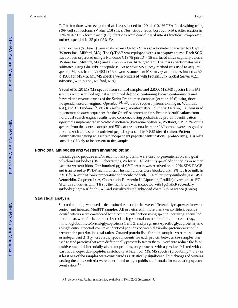
A total of 205 unique proteins (supplementary table 1) were identified in CVF using MudPITand gel-based fractionation (1D PAGE coupled to LC-Tandem mass spectrometry) analyses.Functional annotation of the CVF proteome using GeneOntology terms (DAVID V 2.1)showed (Figure 1A) a majority of them to be associated with metabolism (25%) and immuneresponse (23%). Analysis of the predicted sub-cellular location of the proteins identified fromCVF (Figure 1B) showed that the annotated proteins are from cytoplasmic (24%), secretory(18%), cytoskeletal (14%), and nuclear (14%) categories. No information was availableregarding the cellular location of 13% of the proteins identified.
For the analysis of differential protein levels in the setting of infection, CVF samples obtainedbefore and after experimental IAI were digested with trypsin and subjected to MudPIT analysis.MS/MS spectra derived from the MudPIT analysis led to the high-confidence identification (2or more peptides/protein) of 149 and 151 proteins in the control and infected samples,respectively. To decrease false-positive protein identification rates, MS/MS spectra weresearched against a database containing known contaminants (i.e., trypsin, keratin, and serumalbumin) and both forward and reverse peptide sequence entries from the Swiss-Prot humanand primate databases using three independent search engines. A probability-based algorithm,Scaffold (Proteome Software Inc., Portland, OR), was used to combine results from the threesearch engines. The use of multiple searching algorithms increases the confidence in reportedidentifications by decreasing peptide identifications occurring by chance. Protein identificationnumbers reported above had two or more unique peptide identifications.
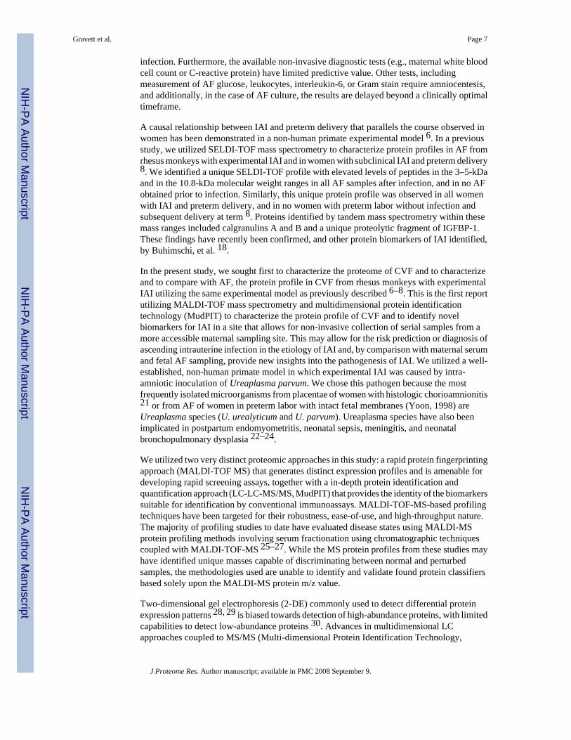
For quantitative comparison of control and IAI samples, a spectral counting method wasimplemented. Spectral counting permits rapid detection of abundance differences between twosample pools without resorting to complicated differential labeling experimentation (Zybailov2005). Curated protein lists from control and IAI were compiled, and independent χ2 tests onthe spectral counts of each protein were performed. Proteins with calculated χ2 values over2.706 (90% confidence interval) are reported in Table 1. Included in the table are the spectralcounts and the number of MS/MS peptide spectra matching to the given protein, for the controland IAI samples. The fold change between control and IAI for each of the significant proteinswas also calculated 17. Of the 27 proteins found to be differentially present between control
Gravett et al. Page 5
J Proteome Res. Author manuscript; available in PMC 2008 September 9.
NIH
-PA Author Manuscript
NIH
-PA Author Manuscript
NIH
-PA Author Manuscript
and IAI by spectral counting, 19 proteins had χ2 values in the 99% confidence interval, and 8proteins had χ2 values in the 95% confidence interval. When compared to quantitativeproteomic studies performed using protein separations (1D PAGE LC-MS/MS), 15 proteinsfound by spectral counting corresponded to differential trends seen in 1-D gel-basedexperiments. The identification of potential lower-abundance serum protein markers is one ofthe benefits of MudPIT analysis. The multi-dimensional front-end peptide separations (SCXand RP-LC) permit the interrogation of a wider dynamic range of concentration over gel-basedproteomic analyses as well as MALDI profiling technologies.
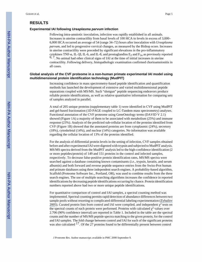
The potential biomarkers for detection of IAI in CVF, summarized in Table 1, werepredominantly immunoregulatory proteins. Several of these, including calgranulins A and B,azurocidin, and IGFBP-1, which were differentially present in IAI AF 8, were also found tobe up-regulated in IAI CVF. The differential abundance of total IGFBP-1 (Table 1) reflectedboth the intact 30-kDa protein and a proteolytic fragment, identified by Western blot in Figure2. However, the majority of IGFBP-1 present in the setting of IAI was comprised of theproteolytic fragment (Figure 2). Defensins, previously identified by others as markers for intra-amniotic or lower genital tract infections 18–20, were also identified in the 3–5-kDa peak.However, their differential presence in control and IAI CVF did not achieve statisticalsignificance by spectral analysis. Of interest, the basal levels of some of the immunoregulatorypeptides were higher in CVF compared to AF, consistent with a more chronically inflammatorymilieu in the microbial rich lower genital tract than in the normally sterile amniotic cavity.
Identification of IAI protein profiles by MALDI-TOF MSMALDI-TOF MS analyses of CVF and AF protein extracts revealed several peak intensitydifferences in 3–5-kDa and 11–12-kDa regions between infected and non-infected primate andhuman CVF and AF (Figure 2), similar to the previously reported protein signature profile inAF obtained by SELDI-TOF 8.
A 10.8-kDa cluster was consistently up-regulated in infected CVF and amniotic fluid in allcases. Of interest, the relative intensity of this peak was greater among CVF samples thanamong AF samples following infection, consistent with the hypothesis that the basal state ofthe lower genital tract milieu is pro-inflammatory. The increased expression of the 3–5-kDacluster in response to IAI is more robust in AF compared to CVF. The proteins with masses3432 and 4128 Da were commonly over-expressed in AF and CVF in the presence of IAI.These masses may represent defensins, as reported earlier 18. Longitudinal sampling followingUreaplasma parvum infection revealed that the 10.8-kDa cluster intensity was increased asearly as 24 hours after inoculation, and preceded increases in HCA in infected animals in bothCVF and AF samples (data not shown).
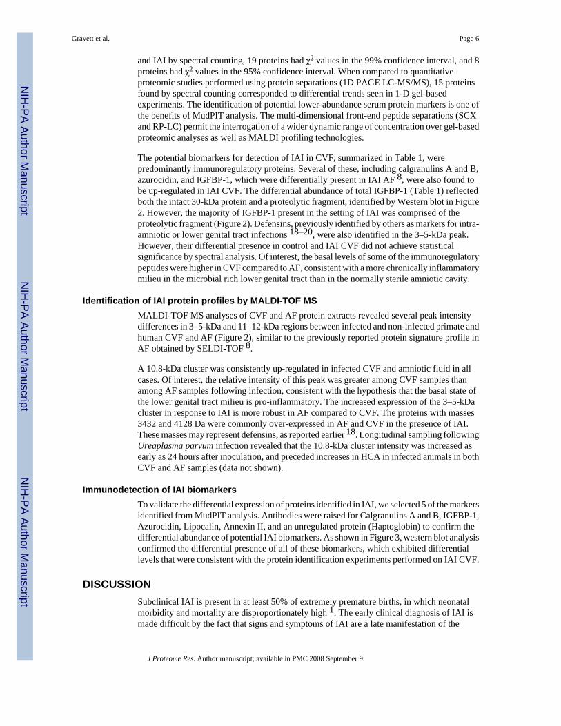
Immunodetection of IAI biomarkersTo validate the differential expression of proteins identified in IAI, we selected 5 of the markersidentified from MudPIT analysis. Antibodies were raised for Calgranulins A and B, IGFBP-1,Azurocidin, Lipocalin, Annexin II, and an unregulated protein (Haptoglobin) to confirm thedifferential abundance of potential IAI biomarkers. As shown in Figure 3, western blot analysisconfirmed the differential presence of all of these biomarkers, which exhibited differentiallevels that were consistent with the protein identification experiments performed on IAI CVF.
DISCUSSIONSubclinical IAI is present in at least 50% of extremely premature births, in which neonatalmorbidity and mortality are disproportionately high 1. The early clinical diagnosis of IAI ismade difficult by the fact that signs and symptoms of IAI are a late manifestation of the
Gravett et al. Page 6
J Proteome Res. Author manuscript; available in PMC 2008 September 9.
NIH
-PA Author Manuscript
NIH
-PA Author Manuscript
NIH
-PA Author Manuscript
infection. Furthermore, the available non-invasive diagnostic tests (e.g., maternal white bloodcell count or C-reactive protein) have limited predictive value. Other tests, includingmeasurement of AF glucose, leukocytes, interleukin-6, or Gram stain require amniocentesis,and additionally, in the case of AF culture, the results are delayed beyond a clinically optimaltimeframe.
A causal relationship between IAI and preterm delivery that parallels the course observed inwomen has been demonstrated in a non-human primate experimental model 6. In a previousstudy, we utilized SELDI-TOF mass spectrometry to characterize protein profiles in AF fromrhesus monkeys with experimental IAI and in women with subclinical IAI and preterm delivery8. We identified a unique SELDI-TOF profile with elevated levels of peptides in the 3–5-kDaand in the 10.8-kDa molecular weight ranges in all AF samples after infection, and in no AFobtained prior to infection. Similarly, this unique protein profile was observed in all womenwith IAI and preterm delivery, and in no women with preterm labor without infection andsubsequent delivery at term 8. Proteins identified by tandem mass spectrometry within thesemass ranges included calgranulins A and B and a unique proteolytic fragment of IGFBP-1.These findings have recently been confirmed, and other protein biomarkers of IAI identified,by Buhimschi, et al. 18.
In the present study, we sought first to characterize the proteome of CVF and to characterizeand to compare with AF, the protein profile in CVF from rhesus monkeys with experimentalIAI utilizing the same experimental model as previously described 6–8. This is the first reportutilizing MALDI-TOF mass spectrometry and multidimensional protein identificationtechnology (MudPIT) to characterize the protein profile of CVF and to identify novelbiomarkers for IAI in a site that allows for non-invasive collection of serial samples from amore accessible maternal sampling site. This may allow for the risk prediction or diagnosis ofascending intrauterine infection in the etiology of IAI and, by comparison with maternal serumand fetal AF sampling, provide new insights into the pathogenesis of IAI. We utilized a well-established, non-human primate model in which experimental IAI was caused by intra-amniotic inoculation of Ureaplasma parvum. We chose this pathogen because the mostfrequently isolated microorganisms from placentae of women with histologic chorioamnionitis21 or from AF of women in preterm labor with intact fetal membranes (Yoon, 1998) areUreaplasma species (U. urealyticum and U. parvum). Ureaplasma species have also beenimplicated in postpartum endomyometritis, neonatal sepsis, meningitis, and neonatalbronchopulmonary dysplasia 22–24.
We utilized two very distinct proteomic approaches in this study: a rapid protein fingerprintingapproach (MALDI-TOF MS) that generates distinct expression profiles and is amenable fordeveloping rapid screening assays, together with a in-depth protein identification andquantification approach (LC-LC-MS/MS, MudPIT) that provides the identity of the biomarkerssuitable for identification by conventional immunoassays. MALDI-TOF-MS-based profilingtechniques have been targeted for their robustness, ease-of-use, and high-throughput nature.The majority of profiling studies to date have evaluated disease states using MALDI-MSprotein profiling methods involving serum fractionation using chromatographic techniquescoupled with MALDI-TOF-MS 25–27. While the MS protein profiles from these studies mayhave identified unique masses capable of discriminating between normal and perturbedsamples, the methodologies used are unable to identify and validate found protein classifiersbased solely upon the MALDI-MS protein m/z value.
Two-dimensional gel electrophoresis (2-DE) commonly used to detect differential proteinexpression patterns 28, 29 is biased towards detection of high-abundance proteins, with limitedcapabilities to detect low-abundance proteins 30. Advances in multidimensional LCapproaches coupled to MS/MS (Multi-dimensional Protein Identification Technology,
Gravett et al. Page 7
J Proteome Res. Author manuscript; available in PMC 2008 September 9.
NIH
-PA Author Manuscript
NIH
-PA Author Manuscript
NIH
-PA Author Manuscript
MudPIT) have enabled better sample enrichment, separation, and in-depth peptide coverageto study global protein expression changes from tryptic digests of complex mixtures 31–35.Recently, MS/MS spectral sampling from complex peptide mixtures has been identified as asource of relative quantitative information. Using spectral counting, the total number of peptideidentifications in a complex peptide mixture analyzed by MudPIT was found to correlatelinearly with protein abundance over a 100-fold concentration range and to be morereproducible with a wider dynamic range over mass spectrometry-derived ion chromatograms17, 36, 37.
Characterization of proteins expressed in CVF in control and IAI using MudPIT analysesrevealed a significant number of immune response/defense-related proteins that were up-regulated in IAI. There is a considerable degree of overlap between the differentially abundantproteins in AF and CVF during IAI. In our study, calgranulins, azurocidin, lipocalin, L-plastin,and others, which were previously identified as potential biomarkers for IAI in amniotic fluid,were also differentially present in CVF. In addition to the above immunomodulators, thedetection of the antibacterial protein azurocidin in CVF in response to infection provides newinsights into the intrauterine immune response. Azurocidin (CAP37) is a cationic antimicrobialprotein isolated from human neutrophils that has potentially important actions in host defenseand inflammation 38. Another antimicrobial protein with elevated expression in IAI is cathelin,which has a C-terminal 37-residue alpha-helical peptide active against bacterial infection.39. The increased levels of annexins in infected CVF may relate to CVF-specific IAI responses.Annexins are a group of Ca2+-binding proteins that are associated with inflammatory anddefense responses. Annexin A2 is up-regulated in viral-transformed cell lines and in humantumors 40. Annexin 1 modulates the anti-inflammatory actions of the steroid hormones 41.Matrix metalloproteinases (MMPs) are a family of zinc-dependent endopeptidases that areexpressed in many inflammatory conditions and contribute to connective tissue breakdown. Ithas been proposed that bacterial products and/or the proinflammatory cytokines IL-1beta andTNF-alpha, as paracrine or autocrine signals, may trigger amniochorion cells to induce MMPexpression 42, 43.
In the second approach, we utilized MALDI-TOF MS and detected a significantly over-expressed 10.8-kDa cluster in CVF in the setting of experimental primate IAI. This is similarto the AF proteome profile observed in our previous studies 8, and confirms the specificity ofthis signature profile for the detection of IAI in CVF. This over-expressed cluster couldrepresent the basic intrauterine immune response to infection, as one set of proteins identifiedin this unique cluster, i.e., the calgranulins, are members of the S-100 calcium binding proteinfamily that is expressed by macrophages and by epithelial cells in acutely inflamed tissues.The second candidate from this cluster, a proteolytic fragment of IGFBP-1, indicates a potentialprotease-related mechanism in response to infection. Intact IGFBP-1 is the major IGFBP foundin AF, and is synthesized by both fetal membranes and maternal decidua. Notably, however,this signature is present, albeit in lower relative concentrations, in CVF samples, but absent inAF samples prior to infection. The higher basal levels of these immunoregulatory peptides mayreflect the basal inflammatory characteristics of the vaginal milieu compared to that of amnioticfluid. Amniotic fluid is normally sterile, with minimal concentrations of inflammatory markers.In contrast, the vagina is characterized by a pro-inflammatory, microbe-rich environment.Thus, while CVF samples may have the advantage of ease of non-invasive sampling, the resultsmay be confounded by local inflammatory conditions such as bacterial vaginosis.
Characterization of the CVF proteome and identification of a significant number of proteinsdifferentially expressed in IAI complements the sensitive proteomic approaches used toidentify biomarkers and their potential value in development of non-invasive testing for IAI.Much can be learned about the pathogenesis of IAI by analysis of temporal and quantitativesamples from CVF, AF, and maternal serum. Analogous issues have been raised by surveys
Gravett et al. Page 8
J Proteome Res. Author manuscript; available in PMC 2008 September 9.
NIH
-PA Author Manuscript
NIH
-PA Author Manuscript
NIH
-PA Author Manuscript
of other cervical-vaginal inflammatory biomarkers such as pro-inflammatory cytokines andfetal fibronectin 9–12. These observations, and ours, are consistent with the hypothesis that,during infection-associated preterm birth, there is a disruption of the extracellular matrix at thechoriodecidual interface, and that inflammatory mediators produced at this interface reach thevaginal pool, possibly in association with a breakdown in cervical barriers.
In summary, we utilized two complimentary proteomic approaches to characterize the globalexpression of cervical-vaginal proteins and to identify potential biomarkers of IAI in cervicalvaginal fluid. Distinct immunoregulatory peptides were identified that were differentiallyexpressed in CVF following experimental IAI. The differential expression of these peptideswas confirmed with immunoassay, and provides an opportunity for the development of non-invasive reliable tests for the diagnosis of IAI.
Supplementary MaterialRefer to Web version on PubMed Central for supplementary material.
AcknowledgementsSupported in part by NIH Grants AI42490 and HD06159 and by ProteoGenix, Inc.
References1. Goldenberg RL, Hauth JC, Andrews WW. Intrauterine infection and preterm delivery. N Engl J Med
2000;342(20):1500–7. [PubMed: 10816189]2. Newton ER. Chorioamnionitis and intraamniotic infection. Clin Obstet Gynecol 1993;36(4):795–808.
[PubMed: 8293582]3. Martin JA, Hamilton BE, Sutton PD, Ventura SJ, Menacker F, Munson ML. Births: final data for 2003.
Natl Vital Stat Rep 2005;54(2):1–116.4. Hitti J, Riley DE, Krohn MA, Hillier SL, Agnew KJ, Krieger JN, Eschenbach DA. Broad-spectrum
bacterial rDNA polymerase chain reaction assay for detecting amniotic fluid infection among womenin premature labor. Clin Infect Dis 1997;24(6):1228–32. [PubMed: 9195088]
5. Watts DH, Krohn MA, Hillier SL, Eschenbach DA. The association of occult amniotic fluid infectionwith gestational age and neonatal outcome among women in preterm labor. Obstet Gynecol 1992;79(3):351–7. [PubMed: 1738513]
6. Gravett MG, Witkin SS, Haluska GJ, Edwards JL, Cook MJ, Novy MJ. An experimental model forintraamniotic infection and preterm labor in rhesus monkeys. Am J Obstet Gynecol 1994;171(6):1660–7. [PubMed: 7802084]
7. Novy, MJ.; Duffy, L.; Axthelm, MK.; Cook, MJ.; Haluska, GJ.; Witkin, SS.; Gerber, S.; Gravett, MG.;Sadowsky, DW.; Cassell, GH. Experimental primate model for Ureaplasma chorioamnionitis andpreterm labor. Society for Gynecologic Investigation, 2001; Toronto, Canada. March 14–17, 2001;
8. Gravett MG, Novy MJ, Rosenfeld RG, Reddy AP, Jacob T, Turner M, McCormack A, Lapidus JA,Hitti J, Eschenbach DA, Roberts CT Jr, Nagalla SR. Diagnosis of intra-amniotic infection by proteomicprofiling and identification of novel biomarkers. Jama 2004;292(4):462–9. [PubMed: 15280344]
9. Rizzo G, Capponi A, Rinaldo D, Tedeschi D, Arduini D, Romanini C. Interleukin-6 concentrations incervical secretions identify microbial invasion of the amniotic cavity in patients with preterm laborand intact membranes. Am J Obstet Gynecol 1996;175(4 Pt 1):812–7. [PubMed: 8885727]
10. Holst RM, Mattsby-Baltzer I, Wennerholm UB, Hagberg H, Jacobsson B. Interleukin-6 andinterleukin-8 in cervical fluid in a population of Swedish women in preterm labor: relationship tomicrobial invasion of the amniotic fluid, intra-amniotic inflammation, and preterm delivery. ActaObstet Gynecol Scand 2005;84(6):551–7. [PubMed: 15901266]
11. Di Naro E, Ghezzi F, Raio L, Romano F, Mueller MD, McDougall J, Cicinelli E. C-reactive proteinin vaginal fluid of patients with preterm premature rupture of membranes. Acta Obstet Gynecol Scand2003;82(12):1072–9. [PubMed: 14616249]
Gravett et al. Page 9
J Proteome Res. Author manuscript; available in PMC 2008 September 9.
NIH
-PA Author Manuscript
NIH
-PA Author Manuscript
NIH
-PA Author Manuscript

12. Yoon BH, Romero R, Moon JB, Oh SY, Han SY, Kim JC, Shim SS. The frequency and clinicalsignificance of intra-amniotic inflammation in patients with a positive cervical fetal fibronectin. AmJ Obstet Gynecol 2001;185(5):1137–42. [PubMed: 11717647]
13. Courchesne PL, Patterson SD. Identification of proteins by matrix-assisted laser desorption/ionizationmass spectrometry using peptide and fragment ion masses. Methods Mol Biol 1999;112:487–511.[PubMed: 10027273]
14. Searle BC, Dasari S, Turner M, Reddy AP, Choi D, Wilmarth PA, McCormack AL, David LL, NagallaSR. High-throughput identification of proteins and unanticipated sequence modifications using amass-based alignment algorithm for MS/MS de novo sequencing results. Anal Chem 2004;76(8):2220–30. [PubMed: 15080731]
15. Searle BC, Dasari S, Wilmarth PA, Turner M, Reddy AP, David LL, Nagalla SR. Identification ofprotein modifications using MS/MS de novo sequencing and the OpenSea alignment algorithm. JProteome Res 2005;4(2):546–54. [PubMed: 15822933]
16. Craig R, Cortens JP, Beavis RC. Open source system for analyzing, validating, and storing proteinidentification data. J Proteome Res 2004;3(6):1234–42. [PubMed: 15595733]
17. Old WM, Meyer-Arendt K, Aveline-Wolf L, Pierce KG, Mendoza A, Sevinsky JR, Resing KA, AhnNG. Comparison of label-free methods for quantifying human proteins by shotgun proteomics. MolCell Proteomics 2005;4(10):1487–502. [PubMed: 15979981]
18. Buhimschi IA, Christner R, Buhimschi CS. Proteomic biomarker analysis of amniotic fluid foridentification of intra-amniotic inflammation. Bjog 2005;112(2):173–81. [PubMed: 15663581]
19. Valore EV, Park CH, Quayle AJ, Wiles KR, McCray PB Jr, Ganz T. Human beta-defensin-1: anantimicrobial peptide of urogenital tissues. J Clin Invest 1998;101(8):1633–42. [PubMed: 9541493]
20. Balu RB, Savitz DA, Ananth CV, Hartmann KE, Miller WC, Thorp JM, Heine RP. Bacterial vaginosisand vaginal fluid defensins during pregnancy. Am J Obstet Gynecol 2002;187(5):1267–71. [PubMed:12439518]
21. Hillier SL, Martius J, Krohn M, Kiviat N, Holmes KK, Eschenbach DA. A case-control study ofchorioamnionic infection and histologic chorioamnionitis in prematurity. N Engl J Med 1988;319(15):972–8. [PubMed: 3262199]
22. Chaim W, Horowitz S, David JB, Ingel F, Evinson B, Mazor M. Ureaplasma urealyticum in thedevelopment of postpartum endometritis. Eur J Obstet Gynecol Reprod Biol 2003;109(2):145–8.[PubMed: 12860331]
23. Viscardi RM, Manimtim WM, Sun CC, Duffy L, Cassell GH. Lung pathology in premature infantswith Ureaplasma urealyticum infection. Pediatr Dev Pathol 2002;5(2):141–50. [PubMed: 11910508]
24. Yoon BH, Romero R, Kim M, Kim EC, Kim T, Park JS, Jun JK. Clinical implications of detectionof Ureaplasma urealyticum in the amniotic cavity with the polymerase chain reaction. Am J ObstetGynecol 2000;183(5):1130–7. [PubMed: 11084554]
25. Tang N, Tornatore P, Weinberger SR. Current developments in SELDI affinity technology. MassSpectrom Rev 2004;23(1):34–44. [PubMed: 14625891]
26. Issaq HJ, Conrads TP, Prieto DA, Tirumalai R, Veenstra TD. SELDI-TOF MS for diagnosticproteomics. Anal Chem 2003;75(7):148A–155A.
27. Petricoin EF, Ardekani AM, Hitt BA, Levine PJ, Fusaro VA, Steinberg SM, Mills GB, Simone C,Fishman DA, Kohn EC, Liotta LA. Use of proteomic patterns in serum to identify ovarian cancer.Lancet 2002;359(9306):572–7. [PubMed: 11867112]
28. Tsangaris G, Weitzdorfer R, Pollak D, Lubec G, Fountoulakis M. The amniotic fluid cell proteome.Electrophoresis 2005;26(6):1168–73. [PubMed: 15706572]
29. Pieper R, Gatlin CL, Makusky AJ, Russo PS, Schatz CR, Miller SS, Su Q, McGrath AM, Estock MA,Parmar PP, Zhao M, Huang ST, Zhou J, Wang F, Esquer-Blasco R, Anderson NL, Taylor J, SteinerS. The human serum proteome: display of nearly 3700 chromatographically separated protein spotson two-dimensional electrophoresis gels and identification of 325 distinct proteins. Proteomics2003;3(7):1345–64. [PubMed: 12872236]
30. Gorg A, Weiss W, Dunn MJ. Current two-dimensional electrophoresis technology for proteomics.Proteomics 2004;4(12):3665–85. [PubMed: 15543535]
Gravett et al. Page 10
J Proteome Res. Author manuscript; available in PMC 2008 September 9.
NIH
-PA Author Manuscript
NIH
-PA Author Manuscript
NIH
-PA Author Manuscript
31. Washburn MP, Wolters D, Yates JR 3rd. Large-scale analysis of the yeast proteome bymultidimensional protein identification technology. Nat Biotechnol 2001;19(3):242–7. [PubMed:11231557]
32. Schirmer EC, Florens L, Guan T, Yates JR 3rd, Gerace L. Nuclear membrane proteins with potentialdisease links found by subtractive proteomics. Science 2003;301(5638):1380–2. [PubMed:12958361]
33. Le Roch KG, Johnson JR, Florens L, Zhou Y, Santrosyan A, Grainger M, Yan SF, Williamson KC,Holder AA, Carucci DJ, Yates JR 3rd, Winzeler EA. Global analysis of transcript and protein levelsacross the Plasmodium falciparum life cycle. Genome Res 2004;14(11):2308–18. [PubMed:15520293]
34. Peng J, Schwartz D, Elias JE, Thoreen CC, Cheng D, Marsischky G, Roelofs J, Finley D, Gygi SP.A proteomics approach to understanding protein ubiquitination. Nat Biotechnol 2003;21(8):921–6.[PubMed: 12872131]
35. Ideker T, Thorsson V, Ranish JA, Christmas R, Buhler J, Eng JK, Bumgarner R, Goodlett DR,Aebersold R, Hood L. Integrated genomic and proteomic analyses of a systematically perturbedmetabolic network. Science 2001;292(5518):929–34. [PubMed: 11340206]
36. Liu H, Sadygov RG, Yates JR 3rd. A model for random sampling and estimation of relative proteinabundance in shotgun proteomics. Anal Chem 2004;76(14):4193–201. [PubMed: 15253663]
37. Zybailov B, Coleman MK, Florens L, Washburn MP. Correlation of relative abundance ratios derivedfrom peptide ion chromatograms and spectrum counting for quantitative proteomic analysis usingstable isotope labeling. Anal Chem 2005;77(19):6218–24. [PubMed: 16194081]
38. Gabay JE, Scott RW, Campanelli D, Griffith J, Wilde C, Marra MN, Seeger M, Nathan CF. Antibioticproteins of human polymorphonuclear leukocytes. Proc Natl Acad Sci U S A 1989;86(14):5610–4.[PubMed: 2501794]
39. Zhao C, Nguyen T, Boo LM, Hong T, Espiritu C, Orlov D, Wang W, Waring A, Lehrer RI. RL-37,an alpha-helical antimicrobial peptide of the rhesus monkey. Antimicrob Agents Chemother 2001;45(10):2695–702. [PubMed: 11557457]
40. Filipenko NR, MacLeod TJ, Yoon CS, Waisman DM. Annexin A2 is a novel RNA-binding protein.J Biol Chem 2004;279(10):8723–31. [PubMed: 14672933]
41. Castro-Caldas M, Duarte CB, Carvalho AR, Lopes MC. 17beta-estradiol promotes the synthesis andthe secretion of annexin I in the CCRF-CEM human cell line. Mediators Inflamm 2001;10(5):245–51. [PubMed: 11759108]
42. Vadillo-Ortega F, Sadowsky DW, Haluska GJ, Hernandez-Guerrero C, Guevara-Silva R, GravettMG, Novy MJ. Identification of matrix metalloproteinase-9 in amniotic fluid and amniochorion inspontaneous labor and after experimental intrauterine infection or interleukin-1 beta infusion inpregnant rhesus monkeys. Am J Obstet Gynecol 2002;186(1):128–38. [PubMed: 11810098]
43. Vadillo-Ortega F, Estrada-Gutierrez G. Role of matrix metalloproteinases in preterm labour. Bjog2005;112(Suppl 1):19–22. [PubMed: 15715589]
Gravett et al. Page 11
J Proteome Res. Author manuscript; available in PMC 2008 September 9.
NIH
-PA Author Manuscript
NIH
-PA Author Manuscript
NIH
-PA Author Manuscript
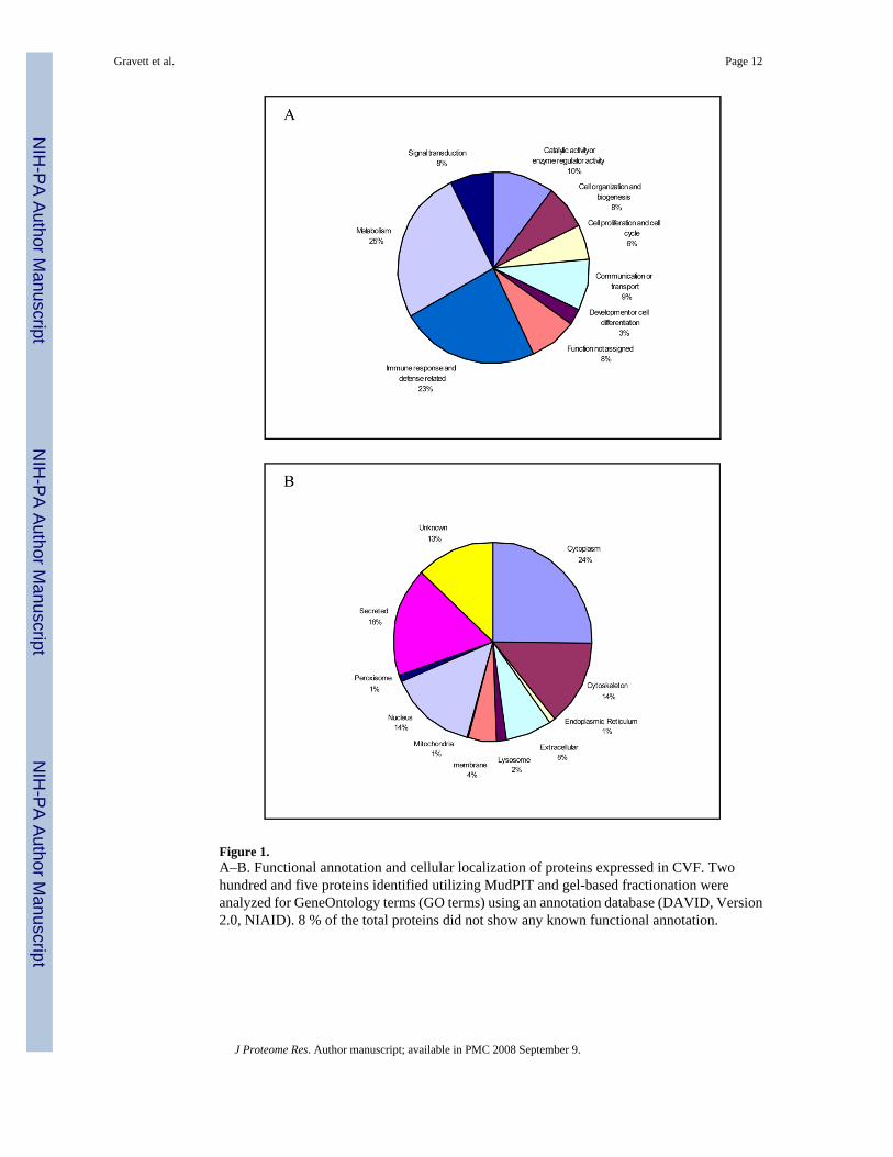
Figure 1.A–B. Functional annotation and cellular localization of proteins expressed in CVF. Twohundred and five proteins identified utilizing MudPIT and gel-based fractionation wereanalyzed for GeneOntology terms (GO terms) using an annotation database (DAVID, Version2.0, NIAID). 8 % of the total proteins did not show any known functional annotation.
Gravett et al. Page 12
J Proteome Res. Author manuscript; available in PMC 2008 September 9.
NIH
-PA Author Manuscript
NIH
-PA Author Manuscript
NIH
-PA Author Manuscript
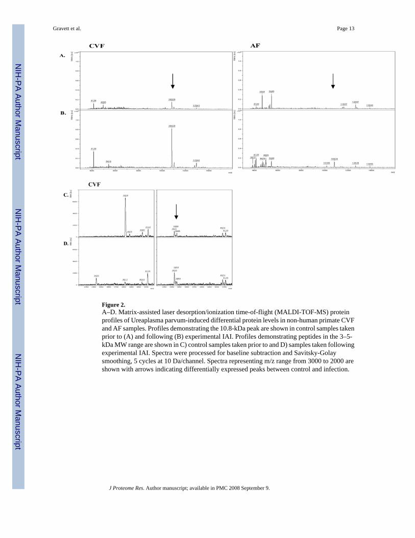
Figure 2.A–D. Matrix-assisted laser desorption/ionization time-of-flight (MALDI-TOF-MS) proteinprofiles of Ureaplasma parvum-induced differential protein levels in non-human primate CVFand AF samples. Profiles demonstrating the 10.8-kDa peak are shown in control samples takenprior to (A) and following (B) experimental IAI. Profiles demonstrating peptides in the 3–5-kDa MW range are shown in C) control samples taken prior to and D) samples taken followingexperimental IAI. Spectra were processed for baseline subtraction and Savitsky-Golaysmoothing, 5 cycles at 10 Da/channel. Spectra representing m/z range from 3000 to 2000 areshown with arrows indicating differentially expressed peaks between control and infection.
Gravett et al. Page 13
J Proteome Res. Author manuscript; available in PMC 2008 September 9.
NIH
-PA Author Manuscript
NIH
-PA Author Manuscript
NIH
-PA Author Manuscript

Figure 3.Immunodetection of potential CVF biomarkers for IAI. Haptoglobin, unregulated controlmarker. IGFBP-1 bands represent the intact protein (~30kDa) and proteolytic fragment (~19kDa).
Gravett et al. Page 14
J Proteome Res. Author manuscript; available in PMC 2008 September 9.
NIH
-PA Author Manuscript
NIH
-PA Author Manuscript
NIH
-PA Author Manuscript

NIH
-PA Author Manuscript
NIH
-PA Author Manuscript
NIH
-PA Author Manuscript
Gravett et al. Page 15Ta
ble
1D
iffer
entia
lly ex
pres
sed
prot
eins
/pep
tides
det
ecte
d us
ing
Mud
PIT
and
de n
ovo
sequ
enci
ng an
alys
is o
f inf
ecte
d no
n-hu
man
prim
ate C
VF.
Prot
ein
IDD
escr
iptio
nC
ontr
ol S
pect
ral C
ount
Infe
cted
Spe
ctra
l Cou
ntχ2
Fold
Cha
nge
(P04
083)
Ann
exin
A1
2253
23.5
43.
1(P
0510
9)C
algr
anul
in A
(Mig
ratio
n in
hibi
tory
fact
or-r
elat
edpr
otei
n 8)
8311
518
.73
1.9
(P08
833)
Insu
lin-li
ke g
row
th fa
ctor
bin
ding
pro
tein
10
1417
.56
16.2
(P31
949)
Cal
gizz
arin
(S10
0 ca
lciu
m-b
indi
ng p
rote
in A
11)
116
17.2
310
.2(Q
9UB
C9)
Smal
l pro
line-
rich
prot
ein
3 (C
orni
fin b
eta)
116
17.2
310
.2(P
6070
9)A
ctin
, cyt
opla
smic
123
4716
.72.
7(P
0735
5)A
nnex
in A
214
3516
.17
3.2
(Q01
469)
Fatty
aci
d-bi
ndin
g pr
otei
n, e
pide
rmal
(E-F
AB
P)12
3013
.85
3.1
(P06
702)
Cal
gran
ulin
B (M
igra
tion
inhi
bito
ry fa
ctor
-rel
ated
prot
ein
14)
170
187
13.3
21.
5
(P35
322)
Cor
nifin
(Sm
all p
rolin
e-ric
h pr
otei
n I)
(SPR
-I)
418
12.8
44.
9(Q
862Z
5)C
ysta
tin B
1024
10.5
43
(P14
780)
Mat
rix m
etal
lopr
otei
nase
-9 p
recu
rsor
(MM
P-9)
2540
9.12
2.1
(P04
217)
Alp
ha-1
B-g
lyco
prot
ein
prec
urso
r13
08.
91−8
.7(P
0419
6)H
istid
ine-
rich
glyc
opro
tein
pre
curs
or13
08.
91−8
.7(Q
4R5C
0)C
ofili
n-1
(Cof
ilin,
non
-mus
cle
isof
orm
).0
67.
017.
7(P
1379
6)L-
plas
tin (L
ymph
ocyt
e cy
toso
lic p
rote
in 1
)26
365.
731.
8(P
0813
3)A
nnex
in A
68
165.
462.
5(P
0397
3)A
ntile
ukop
rote
inas
e 1
prec
urso
r26
85.
33−2
.2(Q
2851
4)G
luta
thio
ne S
-tran
sfer
ase
P9
175.
292.
4(Q
9GLV
5)C
athe
lin.
2432
4.55
1.7
(P29
034)
S100
cal
cium
-bin
ding
pro
tein
A2
05
4.49
6.6
(Q09
666)
Neu
robl
ast d
iffer
entia
tion
asso
ciat
ed p
rote
in A
HN
AK
04
3.22
5.6
(P08
246)
Leuk
ocyt
e el
asta
se p
recu
rsor
04
3.22
5.6
(P16
401)
His
tone
H1.
5 (H
isto
ne H
1a)
04
3.22
5.6
(P02
545)
Lam
in A
/C (7
0 kD
aa la
min
)0
43.
225.
6(P
2581
5)S-
100P
pro
tein
04
3.22
5.6
J Proteome Res. Author manuscript; available in PMC 2008 September 9.
Copyright © 2022 FDOKUMEN