Interleukin-18, From Neuroinflammation to Alzheimers Disease
Upload
independentCategory
view
1download
0
Amniotic Fluid Interleukin 6 in Preterm LaborAssociation with Infection
Roberto Romero, Cecilia Avila, Uma Santhanam,* and Pravinkumar B. Sehgal*Department ofObstetrics and Gynecology, Yale University School ofMedicine, New Haven, Connecticut 06510;and *The Rockefeller University, New York 10021
Abstract
To evaluate whether IL-6 participates in the host response tointrauterine infection, we studied IL-6 bioactivity and isoformsin amniotic fluid (AF). Two different assays for IL-6 were used:the hepatocyte stimulating factor assay (in Hep3B2 cells) andthe SDS-PAGE/immunoblot assay. IL-6 determinations wereperformed in 205 AF samples. Samples were obtained frompatients in the midtrimester of pregnancy (n = 25), at termwith no labor (n = 31), at term in active labor (n = 40), andfrom patients in preterm labor (n = 109). Higher AF IL-6levels were observed in women in preterm labor with intraam-niotic infection than in women in preterm labor without in-traamniotic infection (median = 375 ng/ml, range = 30-5000ng/ml vs. median = 1.5 ng/ml, range = 0-500, respectively, P< 0.0001). The 23-25- and 28-30-kD IL-6 species could bereadily detected in SDS-PAGE immunoblots performed di-rectly on 10-,ul aliquots ofAF from patients with intraamnioticinfection. Among women in preterm labor with culture-nega-tive AF, those who failed to respond to subsequent tocolytictreatment had higher AF IL-6 concentrations than those whoresponded to therapy (median = 50 ng/ml vs. median = 1.2ng/ml, respectively, P < 0.05). Only low levels of IL-6 weredetected in AF obtained from normal women in the midtrimes-ter and third trimester of pregnancy. Decidual tissue explantsobtained from the placentas of women undergoing elective ce-sarean section at term without labor (n = 11) produced IL-6 inresponse to bacterial endotoxin. In a pilot study, AF IL-6 wasdetermined in 56 consecutive women admitted with pretermlabor. All patients (n = 10) with elevated AF IL-6 (cutoff = 46ng/ml) delivered a premature neonate. 4 of these 10 patientshad positive AF cultures for microorganisms. These studiesimplicate IL-6 in the host response to intrauterine infectionand suggest that evaluation of AF IL-6 levels may have diag-nostic and prognostic value in the management of women inpreterm labor. (J. Clin. Invest. 1990. 85:1392-1400.) prema-turity - parturition- decidua * cytokines - bacterial endotoxins
Introduction
Prematurity is the leading cause of perinatal mortality andmorbidity worldwide (1). A growing body of evidence suggestsan association between subclinical intrauterine infection and
Address correspondence to Dr. P. B. Sehgal, The Rockefeller Univer-sity, 1230 York Avenue, New York, NY 10021.
Receivedfor publication 29 June 1989 and in revisedform 7 De-cember 1989.
preterm labor (PTL)' (reviewed in reference 2). Recently, wehave estimated that at least one of every five premature neo-nates is born to a mother with an intraamniotic infection (3,4). The majority ofthese infections are subclinical and difficultto diagnose (3, 4). The participation ofcytokines in the inflam-matory process associated with intraamniotic infection is sup-ported by the detection of IL- 1 and tumor necrosis factor(TNF) in the amniotic fluid (AF) ofwomen with this condition(5, 6). The purpose of this study was to evaluate the participa-tion of IL-6 in the pathophysiology of premature labor.
IL-6 has been implicated as a major mediator of the hostresponse to infection and tissue damage (7, 8). This cytokineconsists of a group of differentially modified phosphoglyco-proteins ranging in size from 23 to 30 kD and additional com-plexes of 43-45 kD (8-13) derived from a single gene locatedat 7p2l in the human genome (14-16). IL-6 gene expression isinduced by several inflammation-associated cytokines (in-cluding IL- 1, TNF, and interferons), bacterial products, RNA-and DNA-containing viruses, and second messenger agonists(diacylglycerol, cAMP, and Ca2+) that activate any ofthe threemajor signal transduction pathways (17-26). Cell types capa-ble of secreting IL-6 in response to stimulation include fibro-blasts, monocytes/macrophages, endothelial cells, keratino-cytes, and endometrial stromal cells (7-27).
IL-6 has a broad range of biological effects (reviewed inreference 25). It elicits major changes in the biochemical, phys-iological, and immunological status ofthe host (e.g., the "acutephase" plasma protein response, activation ofT and NK cells,and stimulation of proliferation and immunoglobulin produc-tion by B cells). The alterations in plasma protein compositionmediated by IL-6 are thought to seal the site of tissue injuryand reduce the systemic effects of infection and tissue damage(7, 25). For example, increased concentrations of fibrinogenand other coagulation factors may promote thrombus forma-tion.
The acute phase plasma protein response may be impor-tant in the context of intraamniotic infection. Clinical studieshave indicated that elevation of maternal serum C-reactiveprotein (CRP) often precedes the development of clinicalchorioamnionitis and the onset of premature labor in womenwith preterm premature rupture of membranes (28-30). Fur-thermore, patients in PTL with elevated levels of CRP aremore likely to be unresponsive to tocolytic therapy than thosewith nondetectable CRP (30-32). As IL-6 plays a critical rolein the induction of CRP synthesis (33, 34), it was consideredlikely that this cytokine participated in the host response tointrauterine infection.
1. Abbreviations used in this paper: ACT, a1-antichymotrypsin; AF,amniotic fluid; CRP, C-reactive protein; HSF, hepatocyte stimulatingfactor; PTL, preterm labor; r, recombinant; TL, term labor; TNF,tumor necrosis factor.
1392 R. Romero, C. Avila, U. Santhanam, and P. B. Sehgal
J. Clin. Invest.© The American Society for Clinical Investigation, Inc.0021-9738/90/05/1392/09 $2.00Volume 85, May 1990, 1392-1400

IL-6 bioactivity was measured and IL-6 isoforms werecharacterized in the AF of women with and without intraam-niotic infection. Highly elevated AF IL-6 levels were observedin women with PTL and intraamniotic infection who pro-gressed to delivery. AF samples from this group of patientscontained the highest levels of IL-6 observed by us in anyhuman body fluid. Indeed, the levels were so high (median= 375 ng/ml; range = 30-5,000 ng/ml) that the 23-25- and28-30-kD IL-6 species could be readily detected in immuno-blots performed directly on 10-jd aliquots ofthe fluid. Womenwith PTL unresponsive to tocolysis had a higher median AFconcentration of IL-6 than women who responded to tocol-ysis. These observations were confirmed in a pilot study of 56consecutive women admitted with PTL. All patients with ele-vated AF IL-6 (cutoff = 46 ng/ml) subsequently delivered apremature neonate. Furthermore, all patients with culture-positive AF had elevated IL-6 in their AF as defined in thisassay. These data suggest that elevated AF IL-6 levels may beof value in the detection of intraamniotic infection and mayalso provide prognostic information regarding the effective-ness of tocolysis in impeding preterm delivery.
Methods
Patient population and collection ofAF. AF was collected from womenin four different populations: group 1, women admitted with PTL andintact membranes on whom amniocenteses were performed for micro-biological assessment ofthe amniotic cavity and for fetal lung maturitystudies (n = 109); group 2, women in the midtrimester of pregnancy(gestational age: 16-18 wk) who underwent amniocenteses for geneticindications (maternal age > 35 yr) (n = 25); group 3, women in thethird trimester (gestational age: 37-40 wk) who had amniocenteses forthe assessment of fetal lung maturity before elective cesarean section (n= 31); and group 4, women in spontaneous active labor (cervicaldilatation of at least 6 cm) at 38-41 wk (term labor [TL]) from whomAF was obtained by transvaginal amniotomy (n = 40).
To study the relationship between intraamniotic infection, PTL,and AF IL-6 concentrations, a cross-sectional study of patients ingroup 1 (PTL) was constructed. Patients with PTL were divided intothree subgroups according to their response to tocolysis and the resultsof their AF culture: subgroup la, women with PTL and negative AFcultures who were responsive to tocolysis; subgroup lb, women withPTL and negative AF cultures who were unresponsive to tocolysis anddelivered a preterm neonate; and subgroup 1 c, women with PTL andintraamniotic infection who delivered a preterm neonate. In addition,we also conducted a pilot study to explore the potential clinical valueofAF IL-6 determinations. This cytokine was assayed in the AF of 56consecutive women admitted with the diagnosis of PTL.
An intraamniotic infection was considered to be present ifmicrobi-ological studies ofAF yielded a positive result (2). PTL was defined asthe presence of regular uterine contractions with a frequency of at leasttwo every 10 min for at least 60 min. A 132-adrenergic drug (ritodrine inmost cases) was administered intravenously as the tocolytic agent ac-cording to a protocol described elsewhere (35). Amniocentesis wasperformed before the initiation of therapy. Failure of tocolysis wasdiagnosed when cervical dilatation progressed beyond 5 cm and/ordelivery occurred.
After defining the criteria for entry into the study, AF samples wereobtained from the AF bank in the Department ofObstetrics and Gyne-cology, Yale University School of Medicine (New Haven, CT). Thisbank consists of aliquots of AF that were not used for clinical testing.AF samples were centrifuged at 400 g for 10 min at 4°C immediatelyafter collection, and the fluid was separated from the cell pellet andstored frozen at -70°C. AF samples analyzed for IL-6 in this studywere from consecutive patients admitted to the hospital who fulfilled
the criteria ofeach ofthe study groups. Informed consent was obtainedfrom all patients following the guidelines of the Yale UniversityHuman Investigations Committee.
Microbiological studies. AF in a capped plastic syringe was trans-ported to the microbiology laboratory immediately after collection andplated within 30 min of amniocentesis. AF was cultured for aerobicand anaerobic bacteria as well as for Mycoplasma hominis and Urea-plasma urealyticum, as described previously (36). AF samples fromwomen in groups 2 and 3 were sterile. AF cultures were not performedon samples obtained by transvaginal amniotomy (group 4). Previousexperience has indicated that the results of these cultures do not reflectthe microbiological status of the amniotic cavity because contamina-tion with cervical or vaginal flora cannot always be avoided duringtransvaginal amniotomy (2).
Cultures of decidual tissue explants. Decidual explants were ob-tained from 11 pregnant women at term who had experienced un-complicated pregnancies and were scheduled to have elective repeatcesarean sections. None had spontaneous onset oflabor before surgery.Care was taken to avoid contact of the placenta with the skin or otherpotential sources of endotoxin. Immediately after delivery of the pla-centa, the decidua adherent to the chorion was mechanically strippedwith sterile forceps. The explants were placed in ice-cold, pyrogen-freeKrebs-Ringer's solution. They were incubated in DME (Gibco Labora-tories, Grand Island, NY) with 10% FCS, 100 U/ml of penicillin,streptomycin (final concentration 100 gg/ml), and 4 AM L-glutamine.Explants were -1 cm X 1 cm in size and were incubated in 2 ml ofmedia in 35-mm plastic tissue culture dishes (Coming Glass Works,Corning, NY). Explants were incubated at 37"C in a humidified incu-bator with 95% 02 and 5% CO2 for 20 h. The medium was centrifugedat 200 g for 10 min. Supernatants were removed and frozen immedi-ately at -20'C until the time of the assay.
Hepatocyte stimulatingfactor (HSF) assayforIL-6 biological activ-ity. The levels of biologically active IL-6 in AF samples were estimatedusing the HSF assay as previously described in detail (9, 10, 12, 13).Briefly, the human hepatoma cell line Hep3B clone 2, obtained fromthe American Type Culture Collection (Cat. No. HB8064; Rockville,MD), was grown to near-confluence in 24-well tissue culture plates(Falcon Plastics, Cockeysville, MD). Appropriately diluted AF samples(diluted 1:5-1:250) were added to Hep3B2 cultures (0.5 ml/well) in thepresence of excess insulin (8 ,ug/ml) and dexamethasone (1 AM). Afterincubation for 20-24 h at 37°C, the cultures were washed with PBSand incubated for an additional 24 h in methionine-free medium con-taining [35S]methionine (100 ACi/ml; DuPont-New England Nuclear,Boston, MA). The culture medium from each well was then collected,and the amount of labeled a1-antichymotrypsin (ACT) secreted wasquantitated by immunoprecipitation (rabbit antiserum to human ACTwas purchased from Calbiochem-Behring Corp., San Diego, CA),SDS-PAGE, autoradiography, and densitometry (Ultroscan XL laserdensitometer; LKB Instruments, Inc., Gaithersburg, MD) using pro-cedures described earlier (10, 12, 13). A purified preparation of naturalhuman IL-6 (produced by IL-la-induced fibroblasts) was used as aninternal laboratory standard, and appropriate dilutions ofthis prepara-tion were also assayed in duplicate in every experiment. The concen-tration of IL-6 in the laboratory standard (2 Ag/ml) was determined bysilver-staining of the IL-6 proteins after SDS-PAGE, and the samevalue (2 Ag/ml) was also arrived at independently by comparing thismixture with electrophoretically homogeneous Coomassie blue-stained Escherichia coli-derived recombinant (r) IL-6 (8) in immuno-blots using rabbit anti-rIL-6 (8). Concentrations of biologically activeIL-6 in AF are expressed in nanograms/milliliter by comparison to thislaboratory standard.
The biological activity in AF samples observed in this HSF assaywas verified to be due to IL-6, based on the ability of a rabbit polyclo-nal antiserum (1:100 dilution) prepared against purified E. coli-de-rived human IL-6 (8) to completely neutralize the activity. The proper-ties and characteristics of this anti-rIL-6 antiserum have been exten-sively described previously (8-13, 37, 38). This antiserum specificallyblocks IL-6 activity not only in hepatocyte stimulation assays but also
Amniotic Fluid Interleukin 6 1393

in B cell growth and differentiation assays, hybridoma growth assays,and T cell activation assays. We have verified that IL- 1 a at concentra-tions up to 50 ng/ml, IL- 1# at 2 ng/ml, TNF at concentrations up to 1ug/ml, and bacterial LPS up to 5 Ag/ml do not affect ACT synthesis inthe Hep3B2 cells used (8, 10, and data not shown). Nevertheless, AFsamples in group 1 (PTL) were also assayed in the presence of excessneutralizing antibodies to both IL- 1 a and IL-I,8 (2 ug/ml each), be-cause some of these AF samples were previously found to containimmunoreactive IL- 1 fI (up to a maximum of 2-4 ng/ml; Romero, R.,unpublished observations). The murine neutralizing monoclonal anti-bodies to human IL-I a or IL-1,8 were kindly provided by Dr. A. C.Allison (Syntex Research, Palo Alto, CA). At 1 gg/ml, both antibodiesblock 1 ng/ml of IL- la or IL- I # in the thymocyte comitogenic assayand the fibroblast proliferation assay (39). An anti-TNF monoclonalantibody was obtained from the Suntory Institute for Biomedical Re-search (Osaka, Japan); I ug/ml of this antibody neutralizes 2 ng/ml ofrTNF activity in the L929 cytotoxicity assay.
In summary, the HSF assay for biologically active IL-6 consists oftwo parts: (a) estimation of the enhancement of ACT synthesis inHep3B2 cells, and (b) inhibition of this enhancement by anti-rIL-6. Amajor advantage ofthis assay is that it is not affected by the presence ofbacterial products in the test samples.
Immunoblot assayfor IL-6 proteins. AF samples from women withPTL and intact membranes (group I) were also assayed for IL-6 con-tent using an immunoblot procedure (8-10, 12, 13). 10-Ad aliquots ofAF were electrophoresed through SDS-PAGE (17.5%) under reducingand denaturing conditions, electroblotted onto nitrocellulose paper,and probed using rabbit anti-rIL-6 (8-13) and the ABC Elite Vecta-stain kit (Vector Laboratories, Inc., Burlingame, CA). Each blot alsocontained appropriate dilutions of the laboratory standard for naturalIL-6. As controls, additional blots were probed using rabbit preim-mune serum, serum from an unrelated rabbit, or the first antibodyomitted from the reaction.
Statistical analysis. Comparisons of IL-6 estimates in AF samplesfrom different groups were conducted using a Kruskal-Wallis one-wayanalysis of variance. The Dunn test was used for post-hoc multiplecomparisons among groups (True Epistat; Epistat Services, Richard-son, TX).
Results
We have evaluated IL-6 bioactivity in AF from women atmidtrimester, at term not in labor, at term in active spontane-ous labor, and from patients in PTL with and without in-traamniotic infection. We shall focus on the results ofAF IL-6from women with PTL because these patients presented withintact membranes, and, therefore, a positive AF culture is areliable indicator of microbial invasion of the amniotic cavity.
IL-6 in AF in PTL. Fig. 1 summarizes a representativeIL-6/HSF assay carried out on AF samples from women inPTL. A 1:5 dilution ofAF from six patients in each ofthe threesubgroups I a, lb, and I c (as defined in Methods), is illustrated.As can be seen, the IL-6/HSF activity of AF samples fromwomen in PTL who were responsive to tocolysis and who wereAF culture-negative (subgroup 1 a) is the lowest. That of AFsamples of women in PTL who were not responsive to tocol-ysis and who were AF culture-positive (subgroup Ic) is thehighest (also see Figs. 3 and 4). It is striking that all of the AFsamples in subgroup lc (Figs. 1 C, 3, and 4) were stronglypositive for IL-6/HSF activity.
That the biological activity observed in this IL-6/HSFassay in these and other AF samples was due specifically toIL-6 was verified by a separate set of experiments similar tothose summarized in Fig. 2. Appropriate dilutions ofAF (threeeach from Fig. 1, B and Cand three from a separate group with
Figure 1. IL-6/HSF bioactivityin the AF ofwomen with PTL.AF diluted 1:5 (500 Ad final
1 2 3 4 5 6 volume) from women in PTLwas assayed for its ability toenhance synthesis and secre-
tion of [35S]methionine-labeledACT in Hep3B2 cultures. Thefigure is a composite of data inone autoradiogram and showsthe stimulation of ACT syn-thesis (arrowheads) by AFsamples from six different pa-tients in each of the followingthree subgroups: A, AF cul-ture-negative and PTL respon-
C sive to tocolysis (subgroup la);B, AF culture-negative andPTL not responsive to tocol-ysis (subgroup I b); and C, AFculture-positive and PTL notresponsive to tocolysis (sub-
1 2 3 4 group Ic). For comparison, Dillustrates ACT stimulation bythe natural IL-6 standard prep-aration at 10, 5, 2, 1, and 0
> ~ ~ - If ng/ml in duplicate assays insets 1, 2, 3, 4, and 5, respec-tively.
PTL and intraamniotic infection) were mixed with a 1:100dilution of the anti-rIL-6 antibody (lanes a), and the residualHSF activity was assayed. The data shown in Fig. 2 clearlydemonstrate that this anti-rIL-6 antibody was able to stronglyinhibit the HSF activity observed. Additionally, we have veri-
A bI 1 2 3 4
.. .a" MO
a a a a
B bI 5 6 7 8 9
a a a a a
a a a a a
Figure 2. Neutralizationof AF IL-6/HSF bioac-tivity by anti-rIL-6 anti-serum. The figure illus-trates composite autora-
diograms from twodifferent experiments (Aand B) in which theability of appropriatelydiluted (1:20-1:100) AFsamples (500 IAd finalvolume) to stimulateACT synthesis (arrow-heads) in Hep3B2 cul-
tures was assayed after incubation with rabbit anti-rIL-6 antiserum at1:100 dilution (lanes a). Three AF samples from each of the follow-ing three groups were evaluated: A2, B5, B6, AF culture-negative,and PTL not responsive to tocolysis (subgroup 1 b; these three sam-
ples are from among the six illustrated in Fig. I B); Al, A3, B7, AFculture-positive, and PTL not responsive to tocolysis (subgroup 1c;these three samples are from among the six illustrated in Fig. 1 C);A4, B8, B9 correspond to a separate set of samples from women withPTL and infection. In additional experiments, control unrelated rab-bit serum at 1:100 dilution had little effect on the stimulation ofACT synthesis (not shown). Residual HSF activity seen in some ofthe lanes (particularly 7a), even in the presence of anti-rIL-6 at 1:100dilution, is consistent with the very high concentrations of IL-6 inthese samples (200-5,000 ng/ml as subsequently verified by immun-oblot assays; see Fig. 3). bl, ACT synthesis in unstimulated controlHep3B2 cultures.
1394 R. Romero, C. Avila, U. Santhanam, and P. B. Sehgal

fied in similar neutralization experiments that the followingreagents have little or no effect on the HSF activity observed inAF samples: (a) preimmune or unrelated rabbit serum, (b)neutralizing anti-TNF antibody, and (c) a combination of theneutralizing anti-IL-la and anti-IL-l1B antibodies. We thusconclude that the observed HSF biological activity in AF sam-ples can be ascribed to IL-6.
To substantiate this conclusion further, we have evaluatedthe IL-6 content of AF samples from women in PTL by im-munoblot analysis. This procedure allows characterization ofthe IL-6 species present in AF. Fig. 3 illustrates immunoblotdata obtained from 10 samples each from subgroups la andlc. It is clear that AF samples in subgroup lc (PTL, not re-sponsive to tocolysis and AF culture-positive) contain IL-6 atsuch high concentrations that the 23-25- and 28-30-kD spe-cies of this cytokine can be readily detected using only a 10-p4laliquot in this assay. The data in Fig. 3 are consistent withthose in Fig. 1 in that the lowest IL-6 concentrations are seenin AF samples in subgroup la, and the highest levels in sub-group lc. Immunoblot assays on the AF samples in group 1were consistent with data obtained using the HSF bioassay(data not shown). Strikingly, all of the samples in subgroup 1 care strongly positive for IL-6 in both the HSF and immunoblotassays (also see below).
The immunoblot data in Fig. 3 provide the first descriptionof IL-6 species present in human AF. These data show thatdifferentially modified IL-6 species (the 23-25-kD species areO-glycosylated; the 28-30-kD species are 0- and N-glycosy-lated [8, 10, 25]) are indeed present in human body fluids.These immunoblot assays reveal that IL-6 isoforms are presentin AF at concentrations as high as 1-5 gg/ml.
The concentrations of IL-6 in all the AF samples fromwomen in PTL (group 1) were estimated using the HSF assay.AF samples were diluted as appropriate (up to 1:250) to obtaina stimulation in ACT synthesis that would be within the log-linear range of the assay. Furthermore, strongly positive sam-ples (in groups lb and 1 c) were reassayed in the presence ofexcess neutralizing antibody to both IL- 1 a and IL- 1f. Fig. 4 asummarizes the estimates of IL-6 content in all of the AFsamples in the group with PTL. Patients with intraamnioticinfection had significantly greater concentrations of AF IL-6than women without intraamniotic infection regardless oftheir response to tocolysis (median = 375 ng/ml, range= 30-5,000 ng/ml vs. median = 1.5 ng/ml, range = 0-500,respectively, P < 0.0001). Patients without intraamniotic in-fection but refractory to tocolytic therapy had higher medianconcentrations of AF IL-6 than those women without in-
traamniotic infection who responded to tocolysis (P < 0.05)(Fig. 4 b).
The clinical picture of several outliers is noteworthy. Onepatient with PTL responsive to tocolysis had an AF IL-6 levelof 125 ng/ml. This patient presented with PTL and a tempera-ture of 100°F. Her white blood cell count was 12,200 with adifferential count of 81% segmented neutrophils and 5%monocytes. Although AF and urine cultures were negative, itis possible that this patient had a self-limiting inflammatoryreaction that could not be diagnosed. She delivered at term 16d later.
Two women in the group of patients with PTL who wereunresponsive to tocolysis (group lb) had AF IL-6 concentra-tions of 500 ng/ml. AF from one of these patients stainedpositive for gram-positive cocci and gram-negative rods, butthe culture grew only Corynebacterium species. Isolation ofthis microorganism is generally interpreted as consistent with askin contaminant. This interpretation may have been errone-ous. The presence of elevated IL-6 suggests that there was anongoing inflammatory reaction in the amniotic cavity. Thesecond patient was clinically suspected to have chorioamnion-itis because of the association of fever and PTL. Although thegram stain ofAF was negative for bacteria, and the culture wasnegative, histopathologic examination of the placenta showedintense chorioamnionitis. Therefore, this patient may havehad an intraamniotic infection that escaped detection usingcurrent microbiologic techniques, an infection limited to theextraamniotic membranes, or an inflammatory reaction of anoninfectious etiology. In any case, the high AF IL-6 levelsappear to be indicative of an ongoing inflammatory process.
Elevated AF IL-6 levels are associated with intraamnioticinfection by a variety of microorganisms including gram-posi-tive and gram-negative species and Ureaplasma urealyticum(see Table I). These data strongly suggest that elevated AF IL-6levels are indicative of an inflammatory reaction associatedwith infection regardless of the specific microorganism in-volved.
IL-6 in the AF from spontaneous TL. We extended ourstudies of AF IL-6 levels during parturition to a group of pa-tients in spontaneous TL (group 4). As controls, we studied AFIL-6 levels in patients in midtrimester (group 2) and in womenat term but not in labor (group 3). Fig. 5 summarizes the dataobtained. AF from women in the midtrimester and third tri-mester (at term) of pregnancy contained detectable but lowlevels of IL-6 (median 10 ng/ml in group 2 and 13 ng/ml ingroup 3). However, AF IL-6 levels were higher in women inspontaneous labor at term (group 4) than in women who were
a b c 1 2 3 4 5 6 7 8 9 10 Figure 3. Multiple forms of IL-6 proteins in AF inkD PTL. This composite figure illustrates SDS-PAGE/
-31 immunoblot assays for IL-6 carried out using l0-tlAF aliquots (undiluted final volume) from 10 pa-
21 tients in each of the following two groups: A, AF cul-I ture-negative and PTL responsive to tocolysis (sub-
I I group la, two of the samples are from among the six.. .. j..31 illustrated in Fig. 1 A); B, AF culture-positive and
I L-6 PTL not responsive to tocolysis (subgroup Ic, four of21 the samples are from among the six illustrated in Fig.
1 C). Lanes a, b, and c in each panel illustrate im-munoblot assays using the natural IL-6 standard preparation at 4, 2, and 1 ng of antigen per lane in A and at 10, 2, and 0.4 ng per lane in B. Inadditional immunoblot analyses of AF samples, unrelated rabbit serum or the second anti-rabbit antibody by itself did not react with the23-25- and 28-30-kD anti-rlL-6-immunoreactive proteins (data not shown).
Amniotic Fluid Interleukin 6 1395
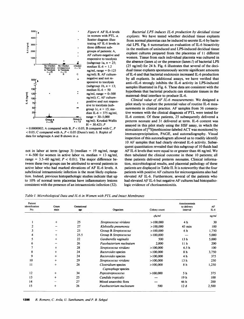
5000 a Figure 4. AF IL-6 levelsin women with PTL. a,
4000 * Scatter diagram illus-
E 3000 - trating AF IL-6 levels inthree different sub-
2000 - groups of patients: A,AF culture-negative and
1000 responsive to tocolysis_ (subgroup la, n = 25;
No Infection No Infection Infection median IL-6 = 1.2Responsive Unresponsive n = 14to Tocolysis toTocoyss ng/ml, range = 0-125n=25 n=13ngmrae=-2
A B C ng/ml); B, AF culture-
500 negative and not re-Jb sponsive to tocolysis
(subgroup I b, n = 13;150. median IL-6 = 50
E 125- ng/ml, range = 0-500= 100 ng/ml); C, AF culture->75 . positive and not respon-
sive to tocolysis (sub-50- ~ ~group Ic, n = 15; me-25 .dian IL-6 = 375 ng/ml,0 S range = 30-5,000
Gropa Group-13 ng/ml). Kruskal-WallisH = 30.425; P
=0.0000002; A compared with B, P < 0.05; B compared with C, P< 0.05; C compared with A, P < 0.05 (Dunn's test). b, Replot ofdata for subgroups A and B shown in a.
not in labor at term (group 3) (median = 19 ng/ml, range= 4-500 for women in active labor vs. median = 13 ng/ml,range = 3.5-60 ng/ml; P < 0.01). The major difference be-tween these two groups can be attributed to several patients inactive labor who had marked elevations of AF IL-6 levels. Asubclinical intraamniotic infection is the most likely explana-tion. Indeed, previous histopathologic studies indicate that upto 10% of normal term placentas have inflammatory lesionsconsistent with the presence ofan intraamniotic infection (32).
Bacterial LPS induces IL-6 production by decidual tissueexplants. We have tested whether decidual tissue explantsfrom normal placentas can be induced to secrete IL-6 by bacte-rial LPS. Fig. 6 summarizes an evaluation of IL-6 bioactivityin the medium of uninduced and LPS-induced decidual tissueexplant cultures prepared from the placentas of 11 differentwomen. Tissue from each individual placenta was cultured inthe absence (lanes u) or the presence (lanes 1) of bacterial LPS(25 ng/ml) for 24 h. Fig. 6 illustrates that several of the deci-dual tissue explants spontaneously secrete significant amountsof IL-6 and that bacterial endotoxin increased IL-6 productionby all explants. In additional assays, we have verified thatanti-rIL-6 strongly inhibits the IL-6 activity in LPS-inducedsamples illustrated in Fig. 6. These data are consistent with thehypothesis that bacterial products can stimulate tissues in thematernal-fetal interface to produce IL-6.
Clinical value ofAF IL-6 measurements. We designed apilot study to explore the potential value of routine IL-6 mea-surements in clinical practice. AF samples from 56 consecu-tive women with the clinical diagnosis of PTL were tested forIL-6 content. Of these patients, 25 subsequently delivered apreterm neonate and 31 delivered at term. IL-6 content wasassayed in this pilot study using the HSF assay, in which thestimulation of [35S]methionine-labeled ACT was monitored byimmunoprecipitation, PAGE, and autoradiography. Visualinspection of this autoradiogram allowed us to readily identify10 AF samples that had clearly elevated IL-6 activity. Subse-quent quantitation revealed that this subgroup of 10 fluids hadAF IL-6 levels that were equal to or greater than 46 ng/ml. Wethen tabulated the clinical outcome in these 10 patients; allthese patients delivered preterm neonates. Clinical informa-tion, microbiological results, and placental pathology of thesepatients are displayed in Table II. It is noteworthy that the fourpatients with positive AF cultures for microorganisms also hadelevated AF IL-6. Furthermore, several of the patients whohad elevated AF IL-6 but negative AF cultures had histopatho-logic evidence of chorioamnionitis.
Table I. Microbiological Data and IL-6 in Women with PTL and Intact Membranes
Patient Amniocentesisidentification Gram Gestational to delivery AF
No. stain age Organism Colony count interval IL-6
cfu/ml ng/ml
1 + 25 Streptococcus viridans >100,000 4 h 302 - 27 Klebsiella pneumonia >100,000 45 min 1003 - 25 Group B Streptococcus >100,000 6 h 1,7504 + 25.5 Group B Streptococcus >100,000 5,0005 + 22 Gardnerella vaginalis 500 13 h 5,0006 - 26 Fusobacterium nucleatum 2,000 11 h 2007 + 28 Streptococcus viridans >100,000 6.5 h 1008 + 24 Bacteroides species >100,000 8 h 3,7509 + 24 Bacteroides species >100,000 4 h 37510 + 29 Streptococcus viridans >100,000 13 h 25011 + 26 Clostridium species >100,000 8 h 1,250
Capnophage species12 + 34 Peptostreptococcus >100,000 5 h 37513 + 25 Candida tropicalis - 19 h 10014 - 27 Mixed anaerobic flora 46 h 20015 + 26 Fusobacterium nucleatum 500 12 d 2,500
1396 R. Romero, C. Avila, U. Santhanam, and P. B. Sehgal

500 * Figure 5. AF IL-6 levelsin women in midtri-mester and at term.
0300 Scatter diagram illus-trating AF IL-6 levels in
co 20 three different groups ofpatients: A, midtrimes-
100 ter (group 2, n = 25;_mm~.- *., 4, smedian IL-6 = 10
& Midtrimester Term Term ng/ml, range = 2-27n -25 No Labor Labor
n = 31 n = 40 ng/ml); B, at term butnot in labor (group 3, n
= 31; median IL-6 = 13, range = 0-60 ng/ml); and C, at term and inspontaneous labor (group 4, n = 40; median IL-6 = 19.5 ng/ml,range = 4-500 ng/ml). Kruskal-Wallis H = 23.35; P = 0.0000085; Acompared with B, P > 0.05; B compared with C, P < 0.05; C com-pared with A, P < 0.05 (Dunn's test).
Discussion
This is the first report to describe the presence of IL-6 in AF.We describe a dramatic increase in the AF IL-6 levels inwomen with intrauterine infection and PTL. The data ob-tained suggest that AF IL-6 levels may be of diagnostic andprognostic value in the management of PTL.
Biologically active IL-6 was detected in AF using the HSFassay in Hep3B2 cells; this bioactivity was confirmed to be dueto IL-6 per se by its neutralization with an anti-rIL-6 anti-serum. Although the HSF assay is not as sensitive as otherbioassays available for IL-6 (e.g., the hybridoma growth factorassay) (25), it is robust and insensitive to the presence ofbacte-rial products in the body fluid tested. Immunoblot analysesconfirmed the presence of the 23-25- and 28-30-kD IL-6 spe-cies in AF. This extends previous descriptions of the heteroge-neity of IL-6 species in human serum/plasma, cerebrospinalfluid, and synovial fluid (10, 12, 13, 25) to the AF. IL-6 hetero-geneity is, thus, a very general feature of this cytokine inhuman body fluids and is due to N- and O-glycosylation and
differential phosphorylation of the 23-30-kD IL-6 species (8,9, 25). The 23-25-kD IL-6 species are O-glycosylated, whereasthe 28-30-kD IL-6 species are both 0- and N-glycosylated.Additionally, higher molecular mass immunoreactive com-plexes (43-45-kD) have been reported in serum/plasma and insynovial fluid (10-13). In cell culture, induced human fibro-blasts, monocytes, endothelial cells, keratinocytes, and endo-metrial stromal cells have all been shown to secrete multipledifferentially modified IL-6 species of molecular mass 23-30kD in cell culture (8, 9, 11, 25). The biological consequences ofthis heterogeneity are unclear.
A major finding in the present study is that the AF ofwomen with PTL and intraamniotic infection contained veryhigh levels of IL-6. All of the AF samples from' women withPTL and infection were strongly positive for IL-6. This indi-cates that IL-6 is a participant in the host response to intraam-niotic infection. Microbiologic data from these patients dem-onstrate that elevated AF IL-6 levels are observed in womenwith intraamniotic infections due to a wide variety of organ-isms including gram-negative and gram-positive bacterial spe-cies.
It should be stressed that PTL leading to preterm deliveryin the absence of detectable infection was also associated withelevations in AF IL-6 levels, although of a lesser magnitudethan that observed in cases of intraamniotic infection. Severalpossible explanations for this observation may be considered.First, this subgroup of women may have had an intrauterineinflammatory reaction unrelated to intraamniotic infection(i.e., an extraamniotic infection or a noninfectious inflamma-tory process). Second, an elevation of IL-6 may be associatedwith the physiologic process of parturition. Third, an intraam-niotic infection may have escaped detection using standardmicrobiological techniques. This latter possibility is a likelyexplanation for two specific cases with elevated AF IL-6 con-centrations (500 ng/ml). The first case had a positive gramstain of AF for bacteria, but the AF grew Corynebacteriumspecies, which are considered a skin contaminant. The secondcase had clinical and histopathologic evidence of chorioam-nionitis, but no bacteria could be recovered. Further studies
5 6 7 8 9 10 11
B 1 2 3 4 5
Figure 6. Production of IL-6 by decidual tissue explants in responseto LPS. Decidual tissue explants from 11 placentas from women un-dergoing elective cesarean sections without spontaneous labor werecultured overnight in the absence (u) or presence of LPS (25 ng/ml)(1), and the accumulation of IL-6 bioactivity in the culture mediumwas monitored (A). B illustrates the stimulation of ACT synthesis induplicate cultures by the natural IL-6 standard preparation used at10, 2.5, 1, 0.4, and 0 ng/ml in this experiment (sets 1-5).
are required to clarify these issues.A perennial clinical problem is the interpretation ofmicro-
biological cultures of biological fluids in which the initial gramstain and culture yield conflicting results. A potential role forIL-6 in clinical practice could be to help identify between sam-ple contamination or culture failure. For example, a positivegram stain ofAF with elevated IL-6 levels is more likely to bedue to a true intraamniotic infection even with a negative AFculture. Additionally, in one of the cases studied by us, it isunclear whether the Corynebacterium isolated was a skin con-taminant introduced into the sample during the amniocentesisprocedure or a true pathogen. The identification of microor-ganisms in gram stains coupled with a high level of IL-6 in AFsuggests a pathogenic role for microorganisms. This conceptbroadens the potential clinical utility of cytokine analysis ofbiological fluids.
In the context ofPTL, it is ofconsiderable importance thatwomen who went on to deliver a premature neonate had ele-vated levels ofAF IL-6, regardless ofthe presence or absence ofdocumented infection. This observation may have importantclinical applications in obstetrics. It would seem that a high AFIL-6 level may identify a group of patients who would notbenefit from tocolysis. Tocolytic therapy is associated with
Amniotic Fluid Interleukin 6 1397
A 1 2 3 4

Table II. Clinical Data of Women with ElevatedAF IL-6 (from a Pilot Study of56 Consecutive Patients Admitted with PTL)
AmniocentesisGestational Gram Preterm Placental to delivery
Serial age stain AF culture IL-6 delivery chorioamnionitis interval Comment
1 26 + Streptococcus agalactae + + + 8 h2 23 - Mycoplasma hominis + + + 1 wk3 25 + Mixed flora + + + I d4 33 - Ureaplasma urealyticum + + NA* 5 d AIDS5 26 - Negative + + NA 1 d Patient had been previously treated
with ampicillin.6 35 - Negative + + NA 24 h7 23 - Negative + + NA 12 wk8 31 - Negative + + + 3 d9 25 - Negative + + + 34 h Patient had been previously treated.
The neonate died of group BStreptococcus sepsis.
10 30 - Negative + + + 3 wk
* NA = not available.
significant side effects for both mother and fetus (32, 40). Ourfindings (also see below) justify additional larger prospectivestudies to determine whether evaluation ofAF IL-6 levels canhelp identify patients in PTL who will progress to deliverydespite tocolytic treatment.
The association between AF IL-6 levels and parturitionwas also evaluated at term. We observed that the median con-centration of AF IL-6 was higher in women in spontaneouslabor at term than in women at term who were not in labor.These observations are similar to those reported by us earlierfor AF IL-1 bioactivity (5). Inspection of the IL-1 and IL-6data sets reveals that this difference is due to a subset of pa-tients in active labor who had elevations ofboth IL- I and IL-6in their AF samples. It is possible that this subset consists ofpatients with subclinical intraamniotic infection or chorioam-nionitis. Because the present study did not include histopatho-logic examination of placentas from women delivering atterm, we cannot directly address this question. Nevertheless,our observations provide a basis for constructing a prospectivestudy to explore this question. A limitation ofour study is thatthe method used to collect AF in women in active labor atterm was different from that used to retrieve fluid from womenat term not in labor (transvaginal amniotomy vs. transabdom-inal amniocentesis).
After obtaining data that strongly linked elevated AF IL-6levels to, first, intraamniotic infection and, second, parturi-tion, we designed a pilot study to evaluate the clinical value ofAF IL-6 measurements in women with PTL. The most strikingobservation was that all patients with elevated AF IL-6 wenton to deliver a premature neonate. Four of these patients hadpositive AF cultures, implicating an intraamniotic infection asthe etiologic factor responsible for preterm delivery and alsofor an elevation ofAF IL-6. On the other hand, a demonstra-ble intraamniotic infection (defined as a positive AF culturefor microorganisms) was absent in the remaining six patientswith detectable AF IL-6. However, histological signs of cho-rioamnionitis were demonstrated in three of the three cases inwhich placentas were available for examination. This suggests
that an intrauterine infection was present but may have eludeddetection with microbiological techniques. This is probablythe case in two patients who had been treated with antibioticsbefore amniocentesis. Antibiotic treatment may have ham-pered our ability to recover microorganisms but not our abilityto detect IL-6 as an index of the host response to infection.
IL-6 in the AF is likely to be of both maternal and fetalorigin. We have recently shown that freshly explanted endo-metrial stromal cells, a normal component of the maternaldecidua, are capable of producing IL-6 (27). Similarly, kera-tinocytes, a normal component of AF due to fetal desquama-tion, are also capable of producing IL-6 (26). It is likely thatinflamed tissues within the uterus, containing a variety of dif-ferent cell types (macrophages, endothelial cells, and fibro-blasts) produce large amounts of IL-6. The observation thatdecidual tissue explants, which contain many of these celltypes, can be induced by LPS to secrete IL-6 is consistent withthis possibility. In situ nucleic acid hybridization analyses forcells containing IL-6 mRNA and immunohistochemical local-ization of IL-6 protein (41) in tissue sections are proceduresnow available to directly address questions concerning thesources ofAF IL-6.
The production of IL-6 is a general feature of processes inwhich the integrity ofa tissue is challenged. Normal pregnancyending in spontaneous labor calls forth major changes in hostbiochemistry; intraamniotic infection greatly adds to thisstress. Parturition itselfmay be viewed as part ofa repertoire ofhost defense mechanisms elicited by intraamniotic infection.The production of abundant IL-6 would clearly contribute tothe production of protective acute-phase plasma proteins andthe activation of immune mechanisms that would help limittissue damage. IL-6 production at the maternal-fetal interfacemay restrict tissue damage to the intrauterine compartmentand protect the mother from the systemic consequences ofdisseminated infection. Likewise, the fetus may produce IL-6in response to localized bacterial invasion (i.e., after aspirationof infected AF), leading to a protective acute phase plasmaprotein response in the fetus. This interpretation is compatible
1398 R. Romero, C. Avila, U. Santhanam, and P. B. Sehgal

with the observation that infected neonates with elevated CRPhave higher survival rates than those with nondetectable serumCRP (42, 43).
The present study identifies AF IL-6 as a marker ofPTL. Alarger prospective study will be necessary to further substan-tiate the value of AF IL-6 as a marker cytokine in clinicalpractice. From a biological point of view, we propose thatvarious cytokines may contribute to different aspects of thehost response to intrauterine infection. IL-1 and TNF, whichstrongly stimulate prostaglandin biosynthesis by intrauterinetissues (6, 44), may signal the onset of parturition and alsostrongly upregulate IL-6 production. IL-6, in turn, may or-chestrate biochemical, immunological, and physiologicalchanges that contribute to maternal and fetal survival.
Acknowledgments
We thank Mr. Ralph Zinner for excellent technical assistance.This research was supported by grants from the Walter Scott
Foundation for Medical Research, the National Institutes of Health(AI-16262), a Physician-Scientist Award from the National InstitutesofHealth (to Dr. Romero), and a contract from the National Founda-tion for Cancer Research.
References
1. van den Berg, B. J., and F. W. Oechsli. 1984. Prematurity. InPerinatal Epidemiology. M. Bracken, editor. Oxford University Press,London, UK. 69-85.
2. Romero, R., and M. Mazor. 1988. Infection and preterm labor.Clin. Obstet. Gynecol. 31:553-584.
3. Romero, R., R. Quintero, E. Oyarzun, Y. K. Wu, M. Mazor, V.Sabo, and J. C. Hobbins. 1988. Intraamniotic infection and the onsetof labor in preterm rupture of membranes. Am. J. Obstet. Gynecol.159:661-666.
4. Romero, R., M. Sirtori, E. Oyarzun, C. Avila, M. Mazor, R.Callahan, V. Sabo, A. Athanassiadis, and J. C. Hobbins. 1989. Preva-lence, microbiology and clinical significance ofintraamniotic infectionin women with preterm labor and intact membranes. Am. J. Obstet.Gynecol. 161:817-824.
5. Romero, R., D. T. Brody, Y. K. Wu, M. Mazor, E. Oyarzun,J. C. Hobbins, and S. Durum. 1989. Infection and labor. III. Interleu-kin- 1: a signal for the initiation of parturition. Am. J. Obstet. Gynecol.160:1117-1123.
6. Romero, R., K. R. Manouge, M. D. Mitchell, Y. K. Wu, E.Oyarzun, J. C. Hobbins, and A. Cerami. 1989. Infection and labor. IV.Cachectin-tumor necrosis factor in the amniotic fluid of women withintraamniotic infection and preterm labor. Am. J. Obstet. Gynecol.161:336-341.
7. Gauldie, J., C. Richards, D. Harnish, P. Lansdorp, and H. Bau-mann. 1987. Interferon J2/B-cell stimulatory factor type 2 sharesidentity with monocyte-derived hepatocyte stimulating factor and reg-ulates the major acute phase protein response in liver cells. Proc. Nati.Acad. Sci. USA. 84:7251-7255.
8. May, L. T., J. Ghrayeb, U. Santhanam, S. B. Tatter, Z. Sthoeger,D. C. Helfgott, N. Chiorazzi, G. Grieninger, and P. B. Sehgal. 1988.Synthesis and secretion of multiple forms of (32-interferon/B cell dif-ferentiation factor-2-hepatocyte stimulating factor by human fibro-blasts and monocytes. J. Biol. Chem. 263:7760-7766.
9. May, L. T., U. Santhanam, S. B. Tatter, N. Bhardwaj, J.Ghrayeb, and P. B. Sehgal. 1988. Phosphorylation ofsecreted forms ofhuman j32-interferon/hepatocyte stimulating factor/interleukin-6.Biochem. Biophys. Res. Commun. 152:1144-1150.
10. Helfgott, D. C., S. B. Tatter, U. Santhanam, R. H. Clarick, N.Bhardwaj, L. T. May, and P. B. Sehgal. 1989. Multiple forms of
IFN-l2/IL-6 in serum and body fluids during acute bacterial infection.J. Immunol. 143:948-953.
11. May, L. T., G. Torcia, F. Cozzolino, A. Ray, S. B. Tatter, U.Santhanam, P. B. Sehgal, and D. Stern. 1989. Interleukin-6 gene ex-pression in human endothelial cells: RNA start sites, multiple IL-6proteins and inhibition of proliferation. Biochem. Biophys. Res. Com-mun. 159:991-998.
12. Jablons, D. M., J. J. Mule, J. K. McIntosh, P. B. Sehgal, L. T.May, C. M. Huang, S. A. Rosenberg, and M. T. Lotze. 1989. Interleu-kin-6/interferon-(32 as a circulating hormone: induction by cytokineadministration in man. J. Immunol. 142:1542-1547.
13. Fong, Y., L. L. Moldawer, M. Marano, H. Wei, S. B. Tatter,R. M. Clarick, U. Santhanam, D. Sherris, L. T. May, P. B. Sehgal, andS. F. Lowry. 1989. Endotoxemia elicits increased circulating,62-IFN/IL-6 in man. J. Immunol. 142:2321-2324.
14. Sehgal, P. B., A. Zilberstein, M. R. Ruggieri, L. T. May, A. C.Fergusson-Smith, D. L. Slate, and F. H. Ruddle. 1986. Human chro-mosome 7 carries the fl2-interferon gene. Proc. NatL. Acad. Sci. USA.83:5219-5222.
15. Ferguson-Smith, A. C., Y. F. Chen, M. S. Newman, L. T. May,P. B. Sehgal, and F. H. Ruddle. 1988. Regional localization of theinterferon-#2/B cell stimulatory factor 2/hepatocyte stimulating factorgene to human chromosome 7pl 5-p2 1. Genomics. 2:203-208.
16. Bowcock, A., J. R. Kidd, M. Lathrop, L. Daneshvar, L. T. May,A. Ray, P. B. Sehgal, K. K. Kidd, and L. L. Cavalli-Sforza. 1988. Thehuman "interferon-,62/B cell stimulating factor/interleukin-6" gene:DNA polymorphism studies and localization to chromosome 7p21.Genomics. 3:8-16.
17. Kohase, M., D. Henriksen-DiStefano, L. T. May, J. Vilcek, andP. B. Sehgal. 1986. Induction of (2-interferon by tumor necrosis factor:a heomeostatic mechanism in the control of cell proliferation. Cell.45:659-666.
18. Kohase, M., L. T. May, I. Tamm, J. Vilek, and P. B. Sehgal.1987. A cytokine network in human diploid fibroblasts: interactions of,3 interferons, tumor necrosis factor, platelet-derived growth factor andinterleukin-l. Mol. Cell. Bio. 7:273-280.
19. Sehgal, P. B., Z. Walther, and L. T. May. 1987. Rapid enhance-ment of (32-interferon/B cell differentiation factor BSF-2 gene expres-sion in human fibroblasts by diacylglycerols and the calcium iono-phore A23187. Proc. Natl. Acad. Sci. USA. 84:3663-3667.
20. Helfgott, D. C., L. T. May, Z. Sthoeger, I. Tamm, and P. B.Sehgal. 1987. Bacterial lipopolysaccharide (endotoxin) enhances ex-pression and secretion of (2-interferon by human fibroblasts. J. Exp.Med. 166:1300-1307.
21. Walther, Z., L. T. May, and P. B. Sehgal. 1988. Transcriptionalregulation of the interferon-f2/B cell differentiation factor BSF-2/he-patocyte stimulating factor HSF gene in human fibroblasts by othercytokines. J. Immunol. 140:974-977.
22. Sehgal, P. B., D. C. Helfgott, U. Santhanam, S. B. Tatter, R. H.Clarick, J. Ghrayeb, and L. T. May. 1988. Regulation of the acutephase and immune responses in viral disease. Enhanced expression ofthe "132-interferon/hepatocyte stimulating factor/interleukin-6" genein virus-infected human fibroblasts. J. Exp. Med. 167:1951-1956.
23. Zhang, Y., Y. Lin, and J. Vilcek. 1988. Synthesis of interleu-kin-6 (interferon-f2l1B cell stimulatory factor 2) in human fibroblasts istriggered by an increase in intracellular cyclic AMP. J. Bid. Chem.263:6177-6182.
24. Ray, A., S. B. Tatter, L. T. May, and P. B. Sehgal. 1988.Activation of the human "(2-interferon/hepatocyte stimulating fac-tor/interleukin-6" promoter by cytokines, viruses and second messen-gers. Proc. Natl. Acad. Sci. USA. 85:6701-6705.
25. Sehgal, P. B., G. Grieninger, and G. Tosato. 1989. Regulationof the acute phase and immune responses: interleukin-6. Ann. NYAcad. Sci. 557:1-583.
26. Kupper, T., K. Min, P. B. Sehgal, H. Mizutani, N. Birchall, A.Ray, and L. May. 1989. Production of IL-6 by keratinocytes: implica-tions for epidermal inflammation and immunity. Ann. NYAcad. Sci.557:454-465.
Amniotic Fluid Interleukin 6 1399

27. Tabibzadeh, S. S., U. Santhanam, P. B. Sehgal, and L. T. May.1989. Cytokine-induced production of interferon-#2/interleukin-6 byfreshly-explanted human endometrial stromal cells: modulation byestradiol- 17#. J. Immunol. 142:3134-3139.
28. Evans, M. I., S. N. Hajj, L. D. Devoe, N. S. Angerman, andA. H. Moawad. 1980. C-reactive protein as a predictor of infectiousmorbidity with premature rupture of membranes. Am. J. Obstet.Gynecol. 138:628-652.
29. Hawrylyshyn, P., P. Bernstein, J. E. Milligan, S. Soldin, A.Pilard, B. Chir, and F. R. Papsin. 1983. Premature rupture of mem-branes: the role of C-reactive protein in the prediction of chorioam-nionitis. Am. J. Obstet. Gynecol. 147:240-246.
30. Potkul, R. K., A. H. Moawad, and K. L. Ponto. 1985. Theassociation of subclinical infection with preterm labor: the role ofC-reactive protein. Am. J. Obstet. Gynecol. 153:642-645.
31. Dodds, W. G., and J. D. lams. 1987. Maternal C-reactive pro-tein and preterm labor. J. Reprod. Med. 32:527-530.
32. Guzick, D. S., and K. Winn. 1985. The association of chorio-amnionitis with preterm delivery. Obstet. Gynecol. 65:11-16.
33. Moshage, H. J., H. M. J. Roelofs, J. F. van Pelt, B. P. C.Hazenberg, M. A. van Leeuwen, P. C. Limburg, L. A. Aarden, andS. H. Yap. 1988. The effect of interleukin-l, interleukin-6 and itsrelationship on the synthesis of serum amyloid A and C-reactive pro-tein in primary cultures of adult human hepatocytes. Biochem.Biophys. Res. Commun. 155:112-117.
34. Ganapathi, M. K., L. T. May, D. Schultz, A. Brabenec, J.Weinstein, P. B. Sehgal, and I. Kushner. 1988. Role of interleukin-6 inregulating synthesis of C-reactive protein and serum amyloid A inhuman hepatoma cell lines. Biochem. Biophys. Res. Commun.157:271-277.
35. Caritis, S. N. 1988. A pharmacologic approach to the infusionof ritodrine. Am. J. Obstet. Gynecol. 158:380-384.
36. Romero, R., K. Scharf, M. Mazor, M. Emamian, J. Ryan, andJ. C. Hobbins. 1988. The clinical value of gas liquid chromatographyin the detection ofintra-amniotic microbial invasion. Obstet. Gynecol.72:44-50.
37. Tosato, G., K. B. Seamon, N. D. Goldman, P. B. Sehgal, L. T.May, G. C. Washington, K. D. Jones, and S. E. Pike. 1988. Mono-cyte-derived human cell growth factor as interferon-02 (BSF-2, IL-6).Science (Wash. DC). 239:502-504.
38. Tosato, G., and S. E. Pike. 1988. Interferon-,62/interleukin-6 isa costimulant for human T lymphocytes. J. Immunol. 141:1556-1562.
39. Kenney, J. S., M. P. Masada, E. M. Eugui, B. M. Delustro,M. A. Mulkins, and A. C. Allison. 1987. Monoclonal antibodies tohuman recombinant interleukin- l (IL-l)#t: quantitation of IL- I 3t andinhibition of biological activity. J. Immunol. 138:4236-4242.
40. Benedetti, T. J. 1983. Maternal complications of parenteral,B-sympathomimetic therapy for premature labor. Am. J. Obstet. Gy-necol. 145:1-6.
41. Grossman, R. M., J. Krueger, D. Yourish, A. Granelli-Piperno,D. P. Murphy, L. T. May, T. S. Kupper, P. B. Sehgal, and A. B.Gottlieb. 1989. Interleukin-6 (IL-6) is expressed in high levels in psori-atic skin and stimulates proliferation ofcultured human keratinocytes.Proc. Natl. Acad. Sci. USA. 86:6367-6371.
42. Philip, A. G. S. 1985. Response ofC-reactive protein in neona-tal group B streptococcal infection. Pediatr. Infect. Dis. J. 4:145-148.
43. Philip, A. G. S. 1979. The protective effect of acute phaseresponse reactants in neonatal sepsis. Acta Paediatr. Scand. 68:481 -483.
44. Romero, R., S. Durum, C. A. Dinarello, E. Oyarzun, J. C.Hobbins, and M. D. Mitchell. 1989. Interleukin-l stimulates prosta-glandin biosynthesis by human amnion. Prostaglandins. 37:13-22.
1400 R. Romero, C. Avila, U. Santhanam, and P. B. Sehgal
Copyright © 2022 FDOKUMEN