Proteolytic Degradation of SCOP in the Hippocampus Contributes to Activation of MAP Kinase and...
23
Proteolytic Degradation of SCOP in the Hippocampus Contributes to Activation of MAP Kinase and Memory Kimiko Shimizu 1 , Trongha Phan 1 , Isabelle Mansuy 2 , and Daniel R. Storm 1,* 1 Department of Pharmacology, The University of Washington, Seattle, Washington 98195, USA 2 Swiss Federal Institute of Technology, Zurich, Switzerland Summary Because activation of Erk1/2 MAP kinase (MAPK) is critical for hippocampus-dependent memory, there is considerable interest in mechanisms for regulation of MAPK during memory formation. Here we report that MAPK and CREB-mediated transcription are negatively regulated by SCOP (SCN Circadian Oscillatory Protein) and that SCOP is proteolyzed by calpain when hippocampal neurons are stimulated by BDNF, KCl depolarization, or NMDA. Moreover, training for novel object memory decreases SCOP in the hippocampus. To determine if hippocampus-dependent memory is influenced by SCOP in vivo, we generated a transgenic mouse strain for the inducible overexpression of SCOP in the forebrain. Overexpression of SCOP completely blocked memory for novel objects. We conclude that degradation of SCOP by calpain contributes to activation of MAPK during memory formation. Introduction MAPK plays a critical role in several forms of neuroplasticity including memory formation (for reviews see Impey et al., 1999;Mazzucchelli and Brambilla, 2000; Frankland et al., 2000; Sweatt, 2004) and neuronal survival (for reviews see Hetman and Xia, 2000; Heerssen and Segal, 2002). Activation of MAPK may be required for memory consolidation because it mediates activity-dependent, Ca 2+ stimulation of cAMP Response Element (CRE)-mediated transcription (Xing et al., 1996; Impey 1998a; Athos et al., 2002; Arthur et al., 2004) and dendritic translation (Kelleher et al., 2004). For example, MAPK is activated during training for hippocampus-dependent memory (Athos et al., 2002;Atkins et al., 1998;Blum et al., 1999; Sindreu et al.) and by stimulus paradigms that evoke long-term potentiation (LTP) (English and Sweatt, 1996;Impey et al., 1998). Inhibitors of MAPK signaling block memory formation (Athos et al., 2002;Atkins et al., 1998;Blum et al., 1999) and increases in CRE- mediated transcription which are required for contextual memory (Athos et al., 2002;Atkins et al., 1998;Blum et al., 1999). Gene disruption studies also support the hypothesis that MAPK activity is critical for synaptic plasticity and memory. Disruption of the gene for RasGRF, an activator of Ras, attenuates long-term memory (LTM) for cued fear conditioning and LTP in the basolateral amygdala (Brambilla et al., 1997). In addition, disruption of the Erk1 gene has implicated MAPK-dependent signaling in behavioral plasticity (Mazzucchelli et al., 2002). Consequently, it is important to understand how MAPK is activated in CNS neurons. Activity- induced increases in intracellular free Ca 2+ can stimulate MAPK by several mechanisms *To whom correspondence should be addressed (email: [email protected]). Publisher's Disclaimer: This is a PDF file of an unedited manuscript that has been accepted for publication. As a service to our customers we are providing this early version of the manuscript. The manuscript will undergo copyediting, typesetting, and review of the resulting proof before it is published in its final citable form. Please note that during the production process errors may be discovered which could affect the content, and all legal disclaimers that apply to the journal pertain. NIH Public Access Author Manuscript Cell. Author manuscript; available in PMC 2008 March 23. Published in final edited form as: Cell. 2007 March 23; 128(6): 1219–1229. NIH-PA Author Manuscript NIH-PA Author Manuscript NIH-PA Author Manuscript
Transcript of Proteolytic Degradation of SCOP in the Hippocampus Contributes to Activation of MAP Kinase and...

Proteolytic Degradation of SCOP in the Hippocampus Contributesto Activation of MAP Kinase and Memory
Kimiko Shimizu1, Trongha Phan1, Isabelle Mansuy2, and Daniel R. Storm1,*
1Department of Pharmacology, The University of Washington, Seattle, Washington 98195, USA 2SwissFederal Institute of Technology, Zurich, Switzerland
SummaryBecause activation of Erk1/2 MAP kinase (MAPK) is critical for hippocampus-dependent memory,there is considerable interest in mechanisms for regulation of MAPK during memory formation. Herewe report that MAPK and CREB-mediated transcription are negatively regulated by SCOP (SCNCircadian Oscillatory Protein) and that SCOP is proteolyzed by calpain when hippocampal neuronsare stimulated by BDNF, KCl depolarization, or NMDA. Moreover, training for novel object memorydecreases SCOP in the hippocampus. To determine if hippocampus-dependent memory is influencedby SCOP in vivo, we generated a transgenic mouse strain for the inducible overexpression of SCOPin the forebrain. Overexpression of SCOP completely blocked memory for novel objects. Weconclude that degradation of SCOP by calpain contributes to activation of MAPK during memoryformation.
IntroductionMAPK plays a critical role in several forms of neuroplasticity including memory formation(for reviews see Impey et al., 1999;Mazzucchelli and Brambilla, 2000; Frankland et al., 2000;Sweatt, 2004) and neuronal survival (for reviews see Hetman and Xia, 2000; Heerssen andSegal, 2002). Activation of MAPK may be required for memory consolidation because itmediates activity-dependent, Ca2+ stimulation of cAMP Response Element (CRE)-mediatedtranscription (Xing et al., 1996; Impey 1998a; Athos et al., 2002; Arthur et al., 2004) anddendritic translation (Kelleher et al., 2004). For example, MAPK is activated during trainingfor hippocampus-dependent memory (Athos et al., 2002;Atkins et al., 1998;Blum et al.,1999; Sindreu et al.) and by stimulus paradigms that evoke long-term potentiation (LTP)(English and Sweatt, 1996;Impey et al., 1998). Inhibitors of MAPK signaling block memoryformation (Athos et al., 2002;Atkins et al., 1998;Blum et al., 1999) and increases in CRE-mediated transcription which are required for contextual memory (Athos et al., 2002;Atkinset al., 1998;Blum et al., 1999). Gene disruption studies also support the hypothesis that MAPKactivity is critical for synaptic plasticity and memory. Disruption of the gene for RasGRF, anactivator of Ras, attenuates long-term memory (LTM) for cued fear conditioning and LTP inthe basolateral amygdala (Brambilla et al., 1997). In addition, disruption of the Erk1 gene hasimplicated MAPK-dependent signaling in behavioral plasticity (Mazzucchelli et al., 2002).Consequently, it is important to understand how MAPK is activated in CNS neurons. Activity-induced increases in intracellular free Ca2+ can stimulate MAPK by several mechanisms
*To whom correspondence should be addressed (email: [email protected]).Publisher's Disclaimer: This is a PDF file of an unedited manuscript that has been accepted for publication. As a service to our customerswe are providing this early version of the manuscript. The manuscript will undergo copyediting, typesetting, and review of the resultingproof before it is published in its final citable form. Please note that during the production process errors may be discovered which couldaffect the content, and all legal disclaimers that apply to the journal pertain.
NIH Public AccessAuthor ManuscriptCell. Author manuscript; available in PMC 2008 March 23.
Published in final edited form as:Cell. 2007 March 23; 128(6): 1219–1229.
NIH
-PA Author Manuscript
NIH
-PA Author Manuscript
NIH
-PA Author Manuscript
including calmodulin stimulation of RasGRF (Farnsworth et al., 1995), or indirectly throughstimulation of adenylyl cyclase (Wang and Storm, 2003), subsequent activation of PKA, andstimulation of Rap1 (de Rooij et al., 1998;Vossler et al., 1997).
Negative regulation of MAPK signaling may be just as important for memory as stimulatorymechanisms. Mice deficient in neurofibromatosis type 1, a RAS GTPase-activating protein,have defects in hippocampus-dependent spatial memory, suggesting that a balance betweenRas stimulation and inhibition is critical for memory (Silva et al., 1997). Recently, it wasdiscovered that suprachiasmatic nucleus circadian oscillatory protein (SCOP) (Shimizu et al.,1999) is a negative regulator of K-Ras in PC12 cells (Shimizu et al., 2003). SCOP interactsdirectly with K-Ras through its leucine-rich repeat and inhibits K-Ras function by associatingwith the nucleotide-free form of K-Ras. This prevents binding of GTP to K-Ras, inhibits itsactivity, and prevents neurotrophin stimulation of MAPK. In PC12 cells, SCOP overexpressioninhibits depolarization stimulation of MAPK by blocking K-Ras activity (Shimizu et al.,2003). SCOP inhibition of K-Ras activity is especially interesting because the K-Ras/MEK/MAPK pathway is critical for synaptic and behavioral plasticity (Ohno et al., 2001).
Although SCOP attenuation of MAPK signaling has the potential to play an important role inneuroplasticity, mechanisms for regulation of SCOP have not been reported. Furthermore, ithas not been established that SCOP regulates MAPK/CRE-mediated transcription in CNSneurons, a transcriptional pathway important for long-term memory (Athos et al., 2002;Atkinset al., 1998;Blum et al., 1999;Bourtchuladze et al., 1994;Pittenger et al., 2002) and neuronalsurvival (Hetman et al., 1999;Mabuchi et al., 2001;Watt et al., 2004). Here, we report thatSCOP negatively regulates CRE-mediated transcription in hippocampal neurons and is rapidlydegraded by calpain when neurons are stimulated with BDNF, NMDA or KCl-induceddepolarization. Interestingly, SCOP is also rapidly degraded during training for hippocampus-dependent memory, and SCOP-overexpressing transgenic mice have no long-term memory fornovel objects. These data suggest that SCOP constrains MAPK activity and that its degradationcontributes to the transient activation of MAPK required for some forms of hippocampus-dependent memory.
ResultsSCOP Expression in the Hippocampus
SCOP was originally discovered as a protein that undergoes a circadian oscillation in the SCNbut not in other regions of brain (Shimizu et al., 1999). To explore the role of SCOP in regulationof MAPK signaling in the hippocampus, we examined its distribution in brain byimmunohistochemistry (Figure 1). SCOP immunoreactivity was present predominantly in thesomata and processes extending from somata in area CA1 of the rat and mouse hippocampus.SCOP is also expressed in area CA3, but not the dentate gyrus. The SCOP immunostaining inthe hippocampus was completely blocked when the SCOP antibody was pre-absorbed withSCOP protein. Because of the importance of the hippocampus for memory formation, thisstudy focused on the regulatory role of SCOP in hippocampal neurons in vitro and in vivo.
SCOP Inhibits CRE-Mediated Transcription in Cultured Hippocampal NeuronsOne of the major MAPK-activated transcriptional pathways implicated in hippocampus-dependent memory is CRE-mediated transcription (Athos et al., 2002;Bourtchuladze et al.,1994;Impey et al., 1998;Xing et al., 1996). To determine if SCOP inhibits stimulation of thistranscriptional pathway in hippocampal neurons, neurons were co-transfected with a reporterfor CRE-mediated transcription with varying amounts of a SCOP expression vector (Figure2A). BDNF stimulation of CRE-mediated transcription in hippocampal neurons wasprogressively inhibited by increasing amounts of SCOP, with almost complete inhibition at
Shimizu et al. Page 2
Cell. Author manuscript; available in PMC 2008 March 23.
NIH
-PA Author Manuscript
NIH
-PA Author Manuscript
NIH
-PA Author Manuscript

the highest levels of SCOP. This indicates that SCOP can function as a negative regulator ofCRE-mediated transcription in hippocampal neurons.
If endogenous SCOP inhibits MAPK and CRE-mediated transcription, inhibition of SCOPexpression should increase CRE-mediated transcription. To test this, hippocampal neuronswere cotransfected with SCOP siRNA, or a scrambled RNA sequence of the same composition,and a reporter for CRE-mediated transcription. In unstimulated cells, SCOP siRNA increasedCRE-mediated transcription four fold, a stimulation twice that obtained with BDNF alone(Figure 2B). These data indicate that endogenous SCOP constrains CRE-mediatedtranscription in hippocampal neurons. We confirmed that SCOP siRNA attenuates MAPKactivation using a lenti-virus construct to express the SCOP siRNA. Lenti-virus expression ofthe SCOP siRNA depressed SCOP protein and activated pERK in unstimulated and BDNF-treated hippocampal neurons (Supplemental Fig. 1).
Treatment of Hippocampal Neurons with BDNF, NMDA or KCl Decreases SCOP ProteinBecause SCOP inhibits MAPK signaling and blocks stimulation of CRE-mediatedtranscription, it was important to determine how SCOP itself is regulated in hippocampalneurons. Since we were unable to detect phosphorylation of SCOP (data not shown), weconsidered the possibility that SCOP may be degraded when neurons are stimulated with BDNFor other activators of MAPK. We monitored SCOP protein levels by Western analysis afterhippocampal neurons were treated with BDNF. Within 5 min of BDNF treatment, SCOPprotein was reduced by 50 % compared to controls and returned to unstimulated levels by 60min (Figure 3A). We also found that depolarization of cultured hippocampal neurons with KClor treatment with NMDA, reduced SCOP levels 52% and 44%, respectively (Figure 3B).Similar results were obtained when cultured cortical neurons were treated with BDNF; SCOPprotein was rapidly degraded and returned to baseline within 3 hr (data not shown). The factthat SCOP protein was never totally degraded suggests that SCOP levels may be maintainedby a balance between protein synthesis and proteolysis.
SCOP is Proteolyzed by CalpainThe rapid reduction in SCOP protein when neurons are treated with BDNF, KCl, or NMDAsuggests that it may be degraded by a protease. The fact that SCOP levels return to baselinerelatively quickly suggest that transient activation of a protease stimulates SCOP degradation.We focused on Ca2+-stimulated proteases because activation of NMDA receptors (Dingledine,1983), BDNF (Berninger et al., 1993;Li et al., 1998) and KCl depolarization (Di Virgilio etal., 1987) all cause transient increases in intracellular free Ca2+.
In cell-free, hippocampal extracts, SCOP was rapidly degraded in the presence of free Ca2+
but not in it absence, suggesting that SCOP is susceptible to degradation by a Ca2+-activatedprotease (Figure 4A). To identify the protease responsible for degradation of SCOP weexamined inhibitors of several proteases including caspase-3 (Chan and Mattson, 1999;Dashet al., 2000) proteasome (Cline, 2003;Hegde, 2004;Lopez-Salon et al., 2001) and calpain (Chanand Mattson 1999). These proteases are present at synapses and have been implicated in neuralplasticity and memory formation (For reviews see Cline, 2003;Chan and Mattson, 1999).Neither the caspase-3 inhibitor nor lactacystin, a proteasome inhibitor, blocked Ca2+-inducedSCOP degradation (Figure 4B). Only calpeptin inhibited SCOP degradation, indicating thatSCOP may be degraded by Ca2+-stimulated calpain. We confirmed that calpain is activatedwhen hippocampal neurons are treated with BDNF (data not shown).
There are two major calpain isoforms, calpain I (μ-form) and calpain II (m-form), which areactivated at different Ca2+concentrations. Within the brain, calpain I is predominantlyneuronal, existing at higher levels in the dendrites and cell bodies, whereas relatively higher
Shimizu et al. Page 3
Cell. Author manuscript; available in PMC 2008 March 23.
NIH
-PA Author Manuscript
NIH
-PA Author Manuscript
NIH
-PA Author Manuscript
levels of calpain II have been observed in axons and glia (Nixon, 1986;Onizuka et al., 1995).To determine if SCOP is selectively proteolyzed by calpain I or II, we incubated purifiedcalpain I and II with immunoprecipitated SCOP. Both calpains degraded SCOP protein invitro (Figure 4C).
To ascertain if calpain proteolyzes SCOP in hippocampal neurons, we treated neurons withcalpain inhibitor III and monitored SCOP degradation when BDNF was applied (Figure 5A).The decrease in SCOP levels caused by BDNF was completely blocked by calpain inhibitorIII and furthermore, there was a significant increase in SCOP levels after treatment with theprotease inhibitor. This suggests that SCOP levels are controlled by a balance betweensynthesis of the protein and degradation by calpain. This was confirmed by examining SCOPlevels when hippocampal neurons were treated with calpain inhibitor III, without BDNF(Figure 5B). Endogenous SCOP protein increases approximately two-fold by 90 min aftertreatment with the protease inhibitor. If SCOP negatively regulates MAPK signaling andcalpain degrades SCOP, proteolytic degradation of SCOP may contribute to MAPK activation.Indeed, stimulation of MAPK activity in hippocampal neurons by BDNF was decreased atleast two-fold when calpain activity was blocked (Figure 5C).
SCOP Degradation Following Training for Novel Object MemorySince activation of MAPK is required for object recognition memory (Kelly et al., 2003) , itwas of considerable interest to determine if the levels of SCOP protein decrease during trainingfor object recognition memory. We measured SCOP 5 min after training for novel objectrecognition (Figure 6). In this paradigm, a mouse is allowed to briefly examine two novelobjects, A and B. When the mouse is tested at a later time, it is presented with one of the originalobjects, A, and a new object C. If the mouse remembers object A, it will spend more timeexamining object C because of natural curiosity for new objects. Memory for novel objects isreflected by greater preference for the new object C during testing (Figure 6A). We examinedSCOP levels following training for novel objects because this is a milder form of training thatdoes not depend on shocking the animal. Untrained controls were handled and placed in thetraining cage without novel objects. Although the decrease in SCOP seen at 5 min after trainingfor novel objects was only 16%, it was significant (p<.01), and was paralleled by an increasein MAPK activity (p<.05) (Figure 6B). SCOP protein also decreased when animals were trainedfor context (Supplemental Fig. 4). Presumably changes in SCOP and MAPK activity are muchgreater in neurons directly affected during training than that measured by Western analysis ofextracts from the entire hippocampus.
Overexpression of SCOP in the Hippocampus Blocks Memory for Novel ObjectsTo determine if hippocampus-dependent memory is sensitive to the amount of SCOP expressedin the hippocampus in vivo, we made a tetracycline-inducible, SCOP overexpressing mousestrain using the CamKIIα promoter to limit overexpression to the forebrain. The integration oftransgene was verified by PCR based genotyping using specific primers for tetracyclineresponse element- SCOP (TRE-SCOP) and CamKII-rtTA2 transgene, respectively (Figure7A). Doxycycline treated TRE-SCOP x CamKll-rtTA2 mice showed 1.5 to 2.5-fold higherSCOP in the hippocampus than those not treated with doxycyline (Figure 7A, right panel ofwestern blot and graph); doxycycline treated TRE-SCOP, or CamKll-rtTA2 mice showed noincrease in SCOP compared to littermate controls. In control experiments, we verified thatdoxycycline treatment did not affect the memory of wild-type, TRE-SCOP, or CamKll-rtTA2mice for novel objects (data not shown). Overexpression of SCOP did not alter open fieldactivity or contextual memory indicating that general mobility and vision were unaffected (datanot shown). Doxycyline treatment of TRE-SCOP x CamKll-rtTA2 mice, which increasesSCOP in the hippocampus, had no effect on memory for novel objects measured 8 min aftertraining (Figure 7B) but completely blocked memory for novel objects measured 24 hr after
Shimizu et al. Page 4
Cell. Author manuscript; available in PMC 2008 March 23.
NIH
-PA Author Manuscript
NIH
-PA Author Manuscript
NIH
-PA Author Manuscript
training (Figure 7C). These data are consistent with other experiments showing that MAPKactivity is required for memory consolidation, but not acquisition or short-term memory (Blumet al., 1999;Kelleher et al., 2004). We confirmed that overexpression of SCOP in the transgenicmouse strain blocks decreases in SCOP protein and the increase in pERK associated withtraining for novel objects (Supplemental Fig. 2). When Doxycyline-treated mice were allowedto recover for 3 months, they exhibited normal 24 hr memory for novel objects (Figure 7D).Overexpression of SCOP did not completely block the increases in pERK observed duringtraining for context (Supplemental Fig. 5) and did not block contextual memory. We concludethat memory for novel objects is sensitive to the concentration of SCOP in the hippocampus.
The data described above indicates that memory for novel objects may depend on the levelsof SCOP protein in the hippocampus. If calpain degradation of SCOP contributes to memoryfor novel objects, inhibition of calpain, in vivo, should block memory. Indeed, when a mixtureof calpain inhibitors was bilaterally administered into area CA1 of the hippocampus, memoryfor novel objects measured 24 hr after training was completely blocked with no effect on short-term memory measured 8 min after training (Figure 8). This indicates that calpain activity isrequired for novel object long-term memory, possibly because calpain degrades SCOP invivo. We confirmed that the decrease in SCOP and increase in pERK observed 5 min aftertraining for novel objects is blocked by infusion of calpain inhibitors into are CA1 of thehippocampus (Supplemental Fig. 3).
DiscussionSCOP was originally identified as a protein whose expression is regulated in a circadian mannerwithin the SCN (Shimizu et al., 1999). The discovery that SCOP inhibits MAPK activity(Shimizu et al., 2003) provides an explanation for the circadian oscillation of MAPK activityand CRE-mediated transcription in the SCN (Obrietan et al., 1998). The presence of SCOP inthe hippocampus raised the interesting possibility that it may inhibit MAPK in the hippocampusand play an important role in MAPK signaling during memory formation and other MAPK-regulated events. The objectives of this study were to determine if SCOP regulates CRE-mediated transcription in hippocampal neurons, and to identify mechanisms for signalingthrough SCOP. Furthermore, it was important to determine if SCOP is regulated during trainingfor hippocampus-dependent memory in vivo and if varying the levels of SCOP in vivo affectsmemory formation.
We discovered that SCOP inhibits CRE-mediated transcription in hippocampal neurons andthat treatment with BDNF, KCl, or NMDA causes rapid degradation of SCOP protein. Thedecrease in SCOP is dependent upon increased intracellular Ca2+ and blocked by calpaininhibitors, suggesting that activity-dependent increases in Ca2+ stimulate calpain whichdegrades SCOP. When cultured neurons are treated with calpain inhibitors there is a constantrise in SCOP protein suggesting that the levels of SCOP reflect a balance between newlytranslated protein and proteolysis. Furthermore, BDNF stimulation of MAPK activity inhippocampus neurons is greatly reduced by calpain inhibitors supporting the hypothesis thatcalpain-stimulated degradation of SCOP is necessary for full activation of MAPK in neurons.
We believe that SCOP may regulate MAPK signaling in vivo because SCOP decreased andMAPK activity increased during training for novel objects or contextual memory. The decreasein SCOP protein, stimulation of MAPK, and memory for novel objects were completelyblocked by overexpressing SCOP in the hippocampus. When SCOP was over expressed,training for contextual fear still gave a measurable decrease in SCOP protein and an increasein MAPK activity. Since the MAPK activation is only partially blocked by SCOPoverexpression during contextual training, the mice still exhibit memory for context. Thissuggests that the training stimulus during contextual training is more robust and leads to greater
Shimizu et al. Page 5
Cell. Author manuscript; available in PMC 2008 March 23.
NIH
-PA Author Manuscript
NIH
-PA Author Manuscript
NIH
-PA Author Manuscript
reduction in SCOP levels. We hypothesize that proteolytic degradation of SCOP releases aconstraint on K-Ras activity and contributes to MAPK activation (Figure 9). This hypothesisis supported by data showing that memory for novel objects depends on calpain activity (Figure8). Our data does not exclude other mechanisms by which calpain may contribute to activationof MAPK. Although calpain-1 knockout mice show no apparent defects in LTP or fearconditioning (Grammer et al., 2005), SCOP is degraded by both calpain-1 and calpain-2.Consequently, definition of the role of calpains in memory formation through gene disruptionmay require conditional double-knockouts of calpain-1 and calpain2.
Considering the general importance of MAPK for neuroplasticity, one would expect that itsactivity in CNS neurons would be tightly regulated (for a review see Agell et al., 2002). Thereare several mechanisms for activation of MAPK by Ca2+ including direct activation throughcalcium-stimulated Ras GEFs (Farnsworth et al., 1995) and indirect mechanisms mediatedthrough calcium-stimulated adenylyl cyclases and PKA. We propose that in unstimulatedneurons, SCOP restrains MAPK activity and that transient decreases in SCOP caused byCa2+ stimulation of calpain is required for activation of MAPK caused by neurotrophins andCa2+ signals. Thus, activation of MAPK activation in neurons may depend upon a dynamicbalance between SCOP degradation and MAPK-activating mechanisms.
Although this study focused on regulation of SCOP in hippocampal neurons and its relationshipto memory formation, the data have broad implications because of the central role played byMAPK and CRE-mediated transcription in neuroplasticity (Frankland et al., 2003;Impey et al.,1999;Mazzucchelli and Brambilla, 2000;Sweatt, 2004). It seems likely that other MAPKregulated events in CNS neurons including synaptic plasticity (Coogan et al., 1999;Englishand Sweatt, 1996), protein synthesis (Kelleher et al., 2004), circadian rhythm (Obrietan et al.,1999;Obrietan et al., 1998), modulation of K+ion channels (Yuan et al., 2002), and neuronalsurvival (Hetman et al., 1999), may depend upon SCOP protein and its regulated degradation.For example, the circadian oscillation of SCOP protein in the SCN correlates with the changesin MAPK activity during the circadian cycle (Shimizu et al., 1999). The extent to whichregulation of SCOP levels contribute to these processes remains to be established.
In summary, maximal activation of MAPK activity in hippocampal neurons depends ondegradation of SCOP by calpain. Training for novel object memory lowers SCOP in brain, andmemory for novel objects was blocked when SCOP was increased two-fold in thehippocampus. We hypothesize that calpain-catalyzed degradation of SCOP in response toactivity-dependent Ca2+ increases is required for activation of MAPK during formation ofhippocampus-dependent memory.
Experimental ProceduresCell Culture
Neurons were cultured as previously described with some modifications (Chan et al., 1998).Primary hippocampal neurons were cultured from newborn C57BL6 pups and maintained inNeurobasal-A (GIBCO) with minimal supplements. Pups were sacrificed by decapitation andthe hippocampi were digested in 5 ml of 10 units/ml papain at 37°C for 30 min. After tworinses, the tissue was triturated three times in dissociation medium (27 mM K2SO4, 15 mMMgCl2, 74 mM Na2SO4, 18 mM glucose, 225 μM CaCl2, 0.0012% phenol red and 2 mMHEPES, pH 7.4) using a 5 ml disposable plastic pipette. Cells were plated into 24-well platesor 3 cm dishes coated with poly-D-lysine (50 μg/ml) at 5X10⁴ cells/cm2 in Neurobasal-Amedium containing B-27 supplement with 10 units/ml penicillin, 10 μg/ml streptomycin, 0.5μg/ml glutamine and 1% Fetal Bovine Serum. The medium was changed to Neurobasal-A withB-27 containing 0.5 μg/ml glutamine and 2 μM AraC 24 hr after plating. Neurons were
Shimizu et al. Page 6
Cell. Author manuscript; available in PMC 2008 March 23.
NIH
-PA Author Manuscript
NIH
-PA Author Manuscript
NIH
-PA Author Manuscript
maintained in Neurobasal-A with B-27 and 0.5 μg/ml glutamine and half of the media waschanged every two days. Plates were incubated in a CO2 incubator at 37°C.
Preparation of siRNAThe siRNA targeted against SCOP mRNA was designed with a two-base 3’-overhang. Thetarget site was nuceotides 292–312 from the start codon (AACTCGCTGCTGCTGAGGAGA).The single strand siRNA nucleotides were chemically synthesized with dTdT 3’-overhangs(CUCGCUGCUGCUGAGGAGAtt, UCUCCUCAGCAGCAGCGAGtt )(Ambion). Ascrambled sequence of the same nucleotide composition as the SCOP siRNA with nosignificant sequence homology to the genome was used as a negative control(UAUGCCGGCUGGAGUACCGtt, CGGUACUCCAGCCGGCAUAtt). Complementarypairs of siRNA oligonucleotides were annealed in annealing buffer (6 mM HEPES, pH 7.4, 20mM potassium acetate, 0.4 mM magnesium acetate). The annealed double stranded RNAs wereconfirmed by acrylamide electrophoresis. For transfection of neurons, 16 nM of siRNA wasused.
Expression PlasmidsThe full-length SCOP overexpression plasmid and CRE (α168)-luciferase have been describedpreviously (Matthews et al., 1994;Shimizu et al., 2003).
Reporter Assays for CRE-Mediated TranscriptionCultured hippocampal neurons in 24-well plates at day 7 in vitro were transfected with acomplex of 0.15 μg of CRE-luciferase, 0.15 μg of the internal control plasmid EF1α-lacZ, 0.3μg of the described plasmid (in each well of a 24-well plate) and Lipofectamine 2000(Invitrogen) according to the manufacture’s protocol. Twenty-four hours after transfection, 5ng/ml BDNF in 0.2% BSA or vehicle (0.2% BSA) was applied. Reporter assays were conducted6 hr post-BDNF stimulation. Luciferase activity was measured as previously described(Matthews et al., 1994) and normalized to β-galactosidase activity (Impey et al., 1996).
ImmunohistochemistryAnimals were deeply anesthetized with Ketamine/Xylazine cocktail and intracardiallyperfused with saline containing 0.1% NaNO2 and 2.0 mM EDTA. They were then continuouslyperfused with ice-cold 2% paraformaldehyde in PBS. Brains were removed, post-fixed for 3hrs and dehydrated in 30% sucrose in PBS overnight. The dehydrated brains were frozen indry ice and sliced into 25 μm thick sections using a microtome. Sections were washed threetimes with 0.5% tritonX-100 in PBS and incubated in 0.5% tritonX-100, 0.5% BSA and 1%normal goat serum for 30 min at 37°C. They were then incubated with anti-SCOP antibody(αEC) (1:50) overnight at 4°C (Shimizu et al., 1999). Immunoreactivity was detected by theconventional avidin-biotin complex method (Vectastain ABC Kit; Vector Lab) using 3-3’diaminobenzidine as chromogen. Brain sections were mounted using DPX mountant (Fluka).
Immunoprecipitation of SCOPWhole brains from adult male mice were homogenized in 10 fold excess (v/v) of buffer A (50mM tris-HCl, pH.7.4, 50 mM NaCl, 1 mM EDTA). The resulting homogenate was centrifugedat 10,000 X g for 15 min. The supernatant was used for immunoprecipitation and pre-clearedon protein A agarose beads (Oncogene) at 4 °C for 2 hr incubation on a rotating wheel. Thepre-cleared cell lysate was incubated overnight at 4°C with a C-terminal SCOP antibody(Shimizu et al., 1999). New protein agarose beads were added and incubated for 2 h at 4 °C.The beads were washed four times with buffer A.
Shimizu et al. Page 7
Cell. Author manuscript; available in PMC 2008 March 23.
NIH
-PA Author Manuscript
NIH
-PA Author Manuscript
NIH
-PA Author Manuscript
Western AnalysisMice were sacrificed by cervical dislocation and the hippocampi were rapidly dissected andimmediately frozen in dry ice. Two hippocampi were sonicated for 10 sec in the sonicationbuffer (20 mM Tris-HCl, 150 mM NaCl, 1% NP-40, 2 mM EDTA, 5 mM EGTA, 5 mM β-mercaptoethanol, 50 mM NaF, 1 mM Na3VO4, 1 mM phenylmethanesulfonyl fluoride, pH7.4)with a protease inhibitor cocktail (complete mini, EDTA-free, Roche) and phosphataseinhibitor cocktail I and II (Sigma). Neurons from three cm culture dishes were treated with 150μl of 2X SDS-sample buffer and cells were scraped from the dish. The resulting homogenateswere treated with SDS-sample buffer, boiled for 3 min and subjected to SDS-PAGE andWestern analysis.
Housing and Handling of miceMale C57BL6 mice at 8–11weeks of age were used. Food and water were provided adlibitum, and animals were kept on a 12 hr light/dark cycle. To reduce handling-induced stress,mice were housed individually and handled daily for one week prior to experiments.
Behavioral AssaysContext-trained mice were placed into a conditioning chamber (Solid State Shocker/Distributor; Coulbourn Instruments, Allentown, Pennsylvania) and allowed to explore. After2 min, they were shocked 1 times (0.7 mA, two sec). Mice were then allowed to recover forone min and returned to home cages. Unpaired controls were shocked once for 2 secimmediately after being placed in context. Novel object-trained mice were first habituated ina training rat cage for 3 to 4 hr before training. After which two different objects A and B werepresented for 5 min after which they were returned to their home cage. They were tested in thetraining cage with a new object C and one of the original objects A or B. During training andtesting the time spent examining each object was recorded for 5 min. Memory for familiarobjects is manifested as a preference for the new object C during testing. Control mice wereplaced into the training cage with no novel objects.
Cannulation and Calpain inhibitor infusion—Mice (8 weeks old) were cannulated asdescribed previously (Athos et al., 2002). Briefly, mice were anesthetized with anintraperitonial injection (20 μl/g body weight) of a mixture of ketamine (7.0 mg/ml) andxylazine (0.44 mg/ml) dissolved in bacteriostatic saline. Anesthetized mice were mounted ona stereotaxic frame (10 micron model, Cartesian Research), and 30-gauge dummy cannulaewere implanted just above the pyramidal cell layer of CA1 region of dorsal hippocampus.Cannulae resided 1.5 mm lateral, 1.25 mm ventral and 1.5 mm posterior from bregma. Thecannulae were affixed with dental acrylic and fitted with 24-gauge guide cannulae to maintaincannula position. Mice were housed individually and allowed at least one week of postoperativerecovery before handling. Calpain inhibitor I, calpain inhibitor III and calpeptin were dissolvedin DMSO and further diluted in 10% Cremophor EL (SIGMA) in saline so that the finalconcentrations of inhibitors were 2 mM, each. Drug and vehicle solutions had a finalconcentration of 18% DMSO and 8.2% Cremophor EL. One-half microliter of drug or vehiclewas infused bilaterally into the cannulae through a 30-gauge injection needle 45 min beforeobject recognition training. The final concentrations of the inhibitors in the hippocampus were20 μM. The infusions were carried out at a rate of 0.25 μl/min using a motorized syringe pump(kdScientific).
Generation of tet-on SCOP over-expression mouse—Complementary DNAencoding rat SCOP was inserted into the multicloning site of pTRE2 (BD Biosciences) so thatthe expression of SCOP was placed under the control of Tet-responsive promoter (TRE). ATRE-SCOP-poly A cassette from the subcloned plasmid was injected (pronuclear) into
Shimizu et al. Page 8
Cell. Author manuscript; available in PMC 2008 March 23.
NIH
-PA Author Manuscript
NIH
-PA Author Manuscript
NIH
-PA Author Manuscript
C57BL6/C3H X C57BL6 embryos. The genotype was determined by PCR using transgene-specific primers. Transgenic mice were bred into the C57BL6 background for at least 6generations. Manipulations were approved and in accordance with the animal care committee’sguidelines at University of Washington.
SCOP overexpression in the forebrain—CaMKIIα promoter-rtTA2 (CaM-rtTA2)mouse was generously provided by Dr. Isabelle Mansuy’s laboratory at the Swiss FederalInstitute of Technology. CaM-rtTA2 mice were mated with TRE-SCOP mice to obtain doubletransgenic mice, which allowed SCOP overexpression in the forebrain to be mice controlledby tetracycline. Genotyping was determined by PCR. Control mice were littermates carryingno transgene or either one of the transgenes. Before testing, mice were fed doxycycline(SIGMA) at 6 mg per g food, as indicated. For all experiments, we used male mice 2–5 monthsold for behavioral studies.
Supplementary MaterialRefer to Web version on PubMed Central for supplementary material.
Acknowledgements
We thank, Dr. Katsuya Nagai at Y.M.P. International Corporation, Dr. Masato Okada at the Research Institute forMicrobial Diseases of Osaka University and Dr. Yoshitaka Fukada at University of Tokyo for laying the groundworkfor SCOP protein. And we thank members of the Storm lab for critical reading of the manuscript. This research wassupported by NIH grant NS 20498.
ReferencesAgell N, Bachs O, Rocamora N, Villalonga P. Modulation of the Ras/Raf/MEK/ERK pathway by Ca(2
+), and calmodulin. Cell Signal 2002;14:649–654. [PubMed: 12020764]Athos J, Impey S, Pineda VV, Chen X, Storm DR. Hippocampal CRE-mediated gene expression is
required for contextual memory formation. Nat Neurosci 2002;5:1119–1120. [PubMed: 12368807]Atkins CM, Selcher JC, Petraitis JJ, Trzaskos JM, Sweatt JD. The MAPK cascade is required for
mammalian associative learning. Nat Neurosci 1998;1:602–609. [PubMed: 10196568]Berninger B, Garcia DE, Inagaki N, Hahnel C, Lindholm D. BDNF and NT-3 induce intracellular Ca2+
elevation in hippocampal neurones. Neuroreport 1993;4:1303–1306. [PubMed: 7505114]Blum S, Moore AN, Adams F, Dash PK. A mitogen-activated protein kinase cascade in the CA1/CA2
subfield of the dorsal hippocampus is essential for long-term spatial memory. J Neurosci1999;19:3535–3544. [PubMed: 10212313]
Bourtchuladze R, Frenguelli B, Blendy J, Cioffi D, Schutz G, Silva AJ. Deficient long-term memory inmice with a targeted mutation of the cAMP-responsive element-binding protein. Cell 1994;79:59–68.[PubMed: 7923378]
Brambilla R, Gnesutta N, Minichiello L, White G, Roylance AJ, Herron CE, Ramsey M, Wolfer DP,Cestari V, RossiArnaud C, et al. A role for the Ras signalling pathway in synaptic transmission andlong-term memory. Nature 1997;390:281–286. [PubMed: 9384379]
Chan GC, Hinds TR, Impey S, Storm DR. Hippocampal neurotoxicity of Delta9-tetrahydrocannabinol.J Neurosci 1998;18:5322–5332. [PubMed: 9651215]
Chan SL, Mattson MP. Caspase and calpain substrates: roles in synaptic plasticity and cell death. JNeurosci Res 1999;58:167–190. [PubMed: 10491581]
Cline H. Synaptic plasticity: importance of proteasome-mediated protein turnover. Curr Biol2003;13:R514–516. [PubMed: 12842027]
Coogan AN, O'Leary DM, O'Connor JJ. P42/44 MAP kinase inhibitor PD98059 attenuates multiple formsof synaptic plasticity in rat dentate gyrus in vitro. J Neurophysiol 1999;81:103–110. [PubMed:9914271]
Dash PK, Blum S, Moore AN. Caspase activity plays an essential role in long-term memory. Neuroreport2000;11:2811–2816. [PubMed: 10976968]
Shimizu et al. Page 9
Cell. Author manuscript; available in PMC 2008 March 23.
NIH
-PA Author Manuscript
NIH
-PA Author Manuscript
NIH
-PA Author Manuscript
de Rooij J, Zwartkruis FJT, Verheijen MHG, Cool RH, Hijman SMB, Wittinghofer A, Bos JL. Epac isa Rap1 guanine-nucleotide exchange factor directly activated by cAMP. Nature 1998;396:474–477.[PubMed: 9853756]
Di Virgilio F, Milani D, Leon A, Meldolesi J, Pozzan T. Voltage-dependent activation and inactivationof calcium channels in PC12 cells. Correlation with neurotransmitter release. J Biol Chem1987;262:9189–9195. [PubMed: 2439506]
Dingledine R. Excitatory amino acids: modes of action on hippocampal pyramidal cells. Fed Proc1983;42:2881–2885. [PubMed: 6136421]
English JD, Sweatt DJ. Activation of p42 MAP kinase in hippocampal long term potentiation. J BiolChem 1996;271:24329–24332. [PubMed: 8798683]
Farnsworth CL, Freshney NW, Rosen LB, Ghosh A, Greenberg ME, Feig LA. Calcium activation of Rasmediated by neuronal exchange factor Ras-GRF. Nature 1995;376:524–527. [PubMed: 7637786]
Frankland PW, Ohno M, Takahashi E, Chen AR, Costa RM, Kushner SA, Silva AJ. Pharmacologicallyregulated induction of silent mutations (PRISM): combined pharmacological and genetic approachesfor learning and memory. Neuroscientist 2003;9:104–109. [PubMed: 12708614]
Grammer M, Kuchay S, Baudry M. Lack of phenotype for LTP and fear conditioning learning in calpain1 knock-out mice. Neurobiol Learning and Memory 2005;84:222–227.
Hegde AN. Ubiquitin-proteasome-mediated local protein degradation and synaptic plasticity. ProgNeurobiol 2004;73:311–357. [PubMed: 15312912]
Hetman M, Kanning K, Cavanaugh JE, Xia Z. Neuroprotection by brain-derived neurotrophic factor ismediated by extracellular signal-regulated kinase and phosphatidylinositol 3-kinase. J Biol Chem1999;274:22569–22580. [PubMed: 10428835]
Impey S, Mark M, Villacres EC, Poser S, Chavkin C, Storm DR. Induction of CRE-mediated geneexpression by stimuli that generate long-lasting LTP in area CA1 of the hippocampus. Neuron1996;16:973–982. [PubMed: 8630255]
Impey S, Obrietan K, Storm DR. Making new connections: role of ERK/MAP kinase signaling in neuronalplasticity. Neuron 1999;23:11–14. [PubMed: 10402188]
Impey S, Obrietan K, Wong ST, Poser S, Yano S, Wayman G, Deloulme JC, Chan G, Storm DR. Crosstalk between ERK and PKA is required for Ca2+ stimulation of CREB-dependent transcription andERK nuclear translocation. Neuron 1998;21:869–883. [PubMed: 9808472]
Kelleher RJ 3rd, Govindarajan A, Jung HY, Kang H, Tonegawa S. Translational control by MAPKsignaling in long-term synaptic plasticity and memory. Cell 2004;116:467–479. [PubMed:15016380]
Kelly A, Laroche S, Davis S. Activation of mitogen-activated protein kinase/extracellular signal-regulated kinase in hippocampal circuitry is required for consolidation and reconsolidation ofrecognition memory. J Neurosci 2003;23:5354–5360. [PubMed: 12832561]
Li YX, Zhang Y, Lester HA, Schuman EM, Davidson N. Enhancement of neurotransmitter releaseinduced by brain-derived neurotrophic factor in cultured hippocampal neurons. J Neurosci1998;18:10231–10240. [PubMed: 9852560]
Lopez-Salon M, Alonso M, Vianna MR, Viola H, Mello e Souza T, Izquierdo I, Pasquini JM, MedinaJH. The ubiquitin-proteasome cascade is required for mammalian long-term memory formation. EurJ Neurosci 2001;14:1820–1826. [PubMed: 11860477]
Mabuchi T, Kitagawa K, Kuwabara K, Takasawa K, Ohtsuki T, Xia Z, Storm D, Yanagihara T, Hori M,Matsumoto M. Phosphorylation of cAMP response element-binding protein in hippocampal neuronsas a protective response after exposure to glutamate in vitro and ischemia in vivo. J Neurosci2001;21:9204–9213. [PubMed: 11717354]
Matthews RP, Guthrie CR, Wailes LM, Zhao X, Means AR, McKnight GS. Calcium/calmodulin-dependent protein kinase types II and IV differentially regulate CREB-dependent gene expression.Mol Cell Biol 1994;14:6107–6116. [PubMed: 8065343]
Mazzucchelli C, Brambilla R. Ras-related and MAPK signalling in neuronal plasticity and memoryformation. Cell Mol Life Sci 2000;57:604–611. [PubMed: 11130460]
Mazzucchelli C, Vantaggiato C, Ciamei A, Fasano S, Pakhotin P, Krezel W, Welzl H, Wolfer DP, PagesG, Valverde O, et al. Knockout of ERK1 MAP kinase enhances synaptic plasticity in the striatum
Shimizu et al. Page 10
Cell. Author manuscript; available in PMC 2008 March 23.
NIH
-PA Author Manuscript
NIH
-PA Author Manuscript
NIH
-PA Author Manuscript
and facilitates striatal-mediated learning and memory. Neuron 2002;34:807–820. [PubMed:12062026]
Nixon RA. Fodrin degradation by calcium-activated neutral proteinase (CANP) in retinal ganglion cellneurons and optic glia: preferential localization of CANP activities in neurons. J Neurosci1986;6:1264–1271. [PubMed: 3012012]
Obrietan K, Impey S, Smith D, Athos J, Storm DR. Circadian regulation of cAMP response element-mediated gene expression in the suprachiasmatic nuclei. J Biol Chem 1999;274:17748–17756.[PubMed: 10364217]
Obrietan K, Impey S, Storm DR. Light and circadian rhythmicity regulate MAP kinase activation in thesuprachiasmatic nuclei. Nat Neurosci 1998;1:693–700. [PubMed: 10196585]
Ohno M, Frankland PW, Chen AP, Costa RM, Silva AJ. Inducible, pharmacogenetic approaches to thestudy of learning and memory. Nat Neurosci 2001;4:1238–1243. [PubMed: 11713472]
Onizuka K, Kunimatsu M, Ozaki Y, Muramatsu K, Sasaki M, Nishino H. Distribution of mu-calpainproenzyme in the brain and other neural tissues in the rat. Brain Res 1995;697:179–186. [PubMed:8593575]
Pittenger C, Huang YY, Paletzki RF, Bourtchouladze R, Scanlin H, Vronskaya S, Kandel ER. Reversibleinhibition of CREB/ATF transcription factors in region CA1 of the dorsal hippocampus disruptshippocampus-dependent spatial memory. Neuron 2002;34:447–462. [PubMed: 11988175]
Shimizu K, Okada M, Nagai K, Fukada Y. Suprachiasmatic nucleus circadian oscillatory protein, a novelbinding partner of K-Ras in the membrane rafts, negatively regulates MAPK pathway. J Biol Chem2003;278:14920–14925. [PubMed: 12594205]
Shimizu K, Okada M, Takano A, Nagai K. SCOP a novel gene product expressed in a circadian mannerin rat suprachiasmatic nucleus. FEBS Lett 1999;458:363–369. [PubMed: 10570941]
Silva AJ, Frankland PW, Marowitz Z, Friedman E, Lazlo G, Cioffi D, Jacks T, Bourtchuladze R. A mousemodel for the learning and memory deficits associated with neurofibromatosis type I. Nat Genet1997;15:281–284. [PubMed: 9054942]
Sindreu CB, Schiner ZS, Storm DR. Ca2+-stimulated adenylyl cyclases regulate ERK-dependentactivation of MSK1 during fear conditioning. Neuron. (in Press)
Sweatt JD. Mitogen-activated protein kinases in synaptic plasticity and memory. Curr Opin Neurobiol2004;14:311–317. [PubMed: 15194111]
Vossler MR, Yao H, York RD, Pan MG, Rim CS, Stork PJ. cAMP activates MAP kinase and Elk-1through a B-Raf- and Rap1-dependent pathway. Cell 1997;89:73–82. [PubMed: 9094716]
Wang H, Ferguson GD, Pineda VV, Storm DR. A Genetic increase in type I adenylyl cyclase in mousebrain enhances LTP and recognition memory. Nature Neuroscience Nat Neurosci 2004;7:635–642.
Wang H, Storm DR. Calmodulin-regulated adenylyl cyclases: cross-talk and plasticity in the centralnervous system. Mol Pharmacol 2003;63:463–468. [PubMed: 12606751]
Watt W, Sakano H, Lee Z, Reusch J, Trinh K, Storm D. Odorant stimulation enhances survival of olfactorysensory neurons via MAPK and CREB. Neuron 2004;41:955–967. [PubMed: 15046727]
Xing J, Ginty DD, Greenberg ME. Coupling of the RAS-MAPK pathway to gene activation by RSK2, agrowth factor-regulated CREB kinase. Science 1996;273:959–963. [PubMed: 8688081]
Shimizu et al. Page 11
Cell. Author manuscript; available in PMC 2008 March 23.
NIH
-PA Author Manuscript
NIH
-PA Author Manuscript
NIH
-PA Author Manuscript
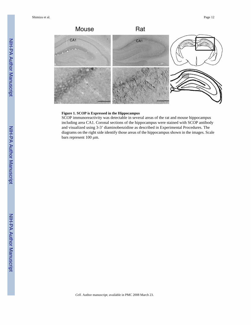
Figure 1. SCOP is Expressed in the HippocampusSCOP immunoreactivity was detectable in several areas of the rat and mouse hippocampusincluding area CA1. Coronal sections of the hippocampus were stained with SCOP antibodyand visualized using 3-3’ diaminobenzidine as described in Experimental Procedures. Thediagrams on the right side identify those areas of the hippocampus shown in the images. Scalebars represent 100 μm.
Shimizu et al. Page 12
Cell. Author manuscript; available in PMC 2008 March 23.
NIH
-PA Author Manuscript
NIH
-PA Author Manuscript
NIH
-PA Author Manuscript
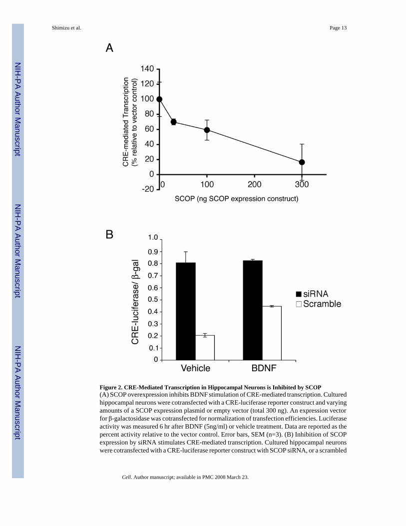
Figure 2. CRE-Mediated Transcription in Hippocampal Neurons is Inhibited by SCOP(A) SCOP overexpression inhibits BDNF stimulation of CRE-mediated transcription. Culturedhippocampal neurons were cotransfected with a CRE-luciferase reporter construct and varyingamounts of a SCOP expression plasmid or empty vector (total 300 ng). An expression vectorfor β-galactosidase was cotransfected for normalization of transfection efficiencies. Luciferaseactivity was measured 6 hr after BDNF (5ng/ml) or vehicle treatment. Data are reported as thepercent activity relative to the vector control. Error bars, SEM (n=3). (B) Inhibition of SCOPexpression by siRNA stimulates CRE-mediated transcription. Cultured hippocampal neuronswere cotransfected with a CRE-luciferase reporter construct with SCOP siRNA, or a scrambled
Shimizu et al. Page 13
Cell. Author manuscript; available in PMC 2008 March 23.
NIH
-PA Author Manuscript
NIH
-PA Author Manuscript
NIH
-PA Author Manuscript
RNA of the same composition. Luciferase activity was measured 24 hr after transfection andwere normalized to β-galactosidase activity. Error bars, SEM (n=3).
Shimizu et al. Page 14
Cell. Author manuscript; available in PMC 2008 March 23.
NIH
-PA Author Manuscript
NIH
-PA Author Manuscript
NIH
-PA Author Manuscript
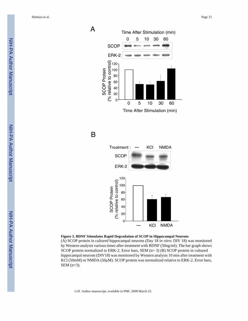
Figure 3. BDNF Stimulates Rapid Degradation of SCOP in Hippocampal Neurons(A) SCOP protein in cultured hippocampal neurons (Day 18 in vitro: DIV 18) was monitoredby Western analysis various times after treatment with BDNF (50ng/ml). The bar graph showsSCOP protein normalized to ERK-2. Error bars, SEM (n= 3) (B) SCOP protein in culturedhippocampal neurons (DIV18) was monitored by Western analysis 10 min after treatment withKCl (50mM) or NMDA (50μM). SCOP protein was normalized relative to ERK-2. Error bars,SEM (n=3).
Shimizu et al. Page 15
Cell. Author manuscript; available in PMC 2008 March 23.
NIH
-PA Author Manuscript
NIH
-PA Author Manuscript
NIH
-PA Author Manuscript

Figure 4. Calpain Catalyzes the Proteolysis of SCOP(A) SCOP protein was monitored by Western analysis after adding Mg2+ (10 mM) and/orCa2+ (4 mM) to mouse brain lysates and incubating for three hours at 4°C. (B) Calpeptininhibited Ca2+-stimulated proteolysis of SCOP. The mouse brain extract was pretreated witha caspase-3 inhibitor (10 μM), lactacystin (5 μM) or calpeptin (10 μM) 30 min before addingCa2+. SCOP protein was monitored by Western analysis. (C) Calpain I and II both catalyzedthe proteolysis of SCOP in vitro. Calpain I or calpain II was incubated with immunoprecipitatedSCOP at 37 °C for one hr. SCOP protein was monitored by Western analysis.
Shimizu et al. Page 16
Cell. Author manuscript; available in PMC 2008 March 23.
NIH
-PA Author Manuscript
NIH
-PA Author Manuscript
NIH
-PA Author Manuscript
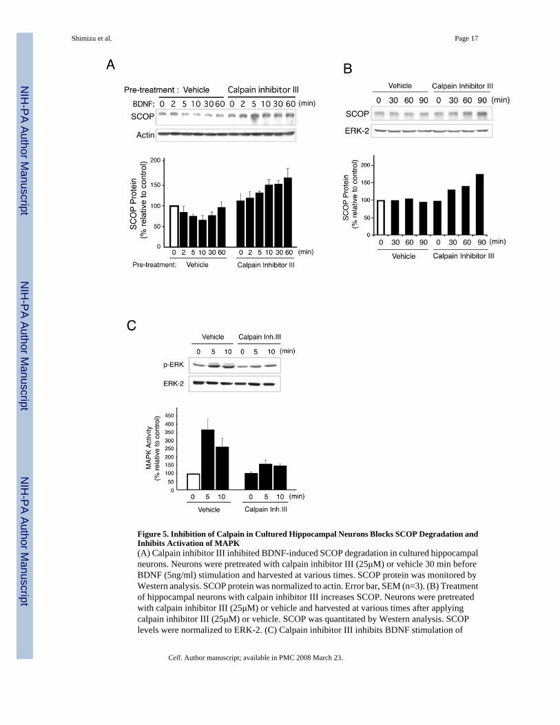
Figure 5. Inhibition of Calpain in Cultured Hippocampal Neurons Blocks SCOP Degradation andInhibits Activation of MAPK(A) Calpain inhibitor III inhibited BDNF-induced SCOP degradation in cultured hippocampalneurons. Neurons were pretreated with calpain inhibitor III (25μM) or vehicle 30 min beforeBDNF (5ng/ml) stimulation and harvested at various times. SCOP protein was monitored byWestern analysis. SCOP protein was normalized to actin. Error bar, SEM (n=3). (B) Treatmentof hippocampal neurons with calpain inhibitor III increases SCOP. Neurons were pretreatedwith calpain inhibitor III (25μM) or vehicle and harvested at various times after applyingcalpain inhibitor III (25μM) or vehicle. SCOP was quantitated by Western analysis. SCOPlevels were normalized to ERK-2. (C) Calpain inhibitor III inhibits BDNF stimulation of
Shimizu et al. Page 17
Cell. Author manuscript; available in PMC 2008 March 23.
NIH
-PA Author Manuscript
NIH
-PA Author Manuscript
NIH
-PA Author Manuscript
MAPK activity measured as pERK in cultured hippocampal neurons. Total ERK-2 measuredon the same Western blot was used as loading controls. The percentage change in pERK relativeto the zero time point is reported. Error bars, SEM (n=3).
Shimizu et al. Page 18
Cell. Author manuscript; available in PMC 2008 March 23.
NIH
-PA Author Manuscript
NIH
-PA Author Manuscript
NIH
-PA Author Manuscript
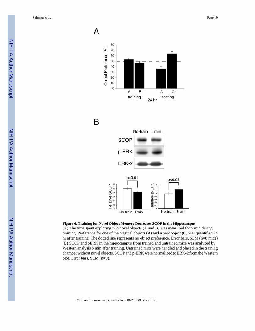
Figure 6. Training for Novel Object Memory Decreases SCOP in the Hippocampus(A) The time spent exploring two novel objects (A and B) was measured for 5 min duringtraining. Preference for one of the original objects (A) and a new object (C) was quantified 24hr after training. The dotted line represents no object preference. Error bars, SEM (n=8 mice)(B) SCOP and pERK in the hippocampus from trained and untrained mice was analyzed byWestern analysis 5 min after training. Untrained mice were handled and placed in the trainingchamber without novel objects. SCOP and p-ERK were normalized to ERK-2 from the Westernblot. Error bars, SEM (n=9).
Shimizu et al. Page 19
Cell. Author manuscript; available in PMC 2008 March 23.
NIH
-PA Author Manuscript
NIH
-PA Author Manuscript
NIH
-PA Author Manuscript
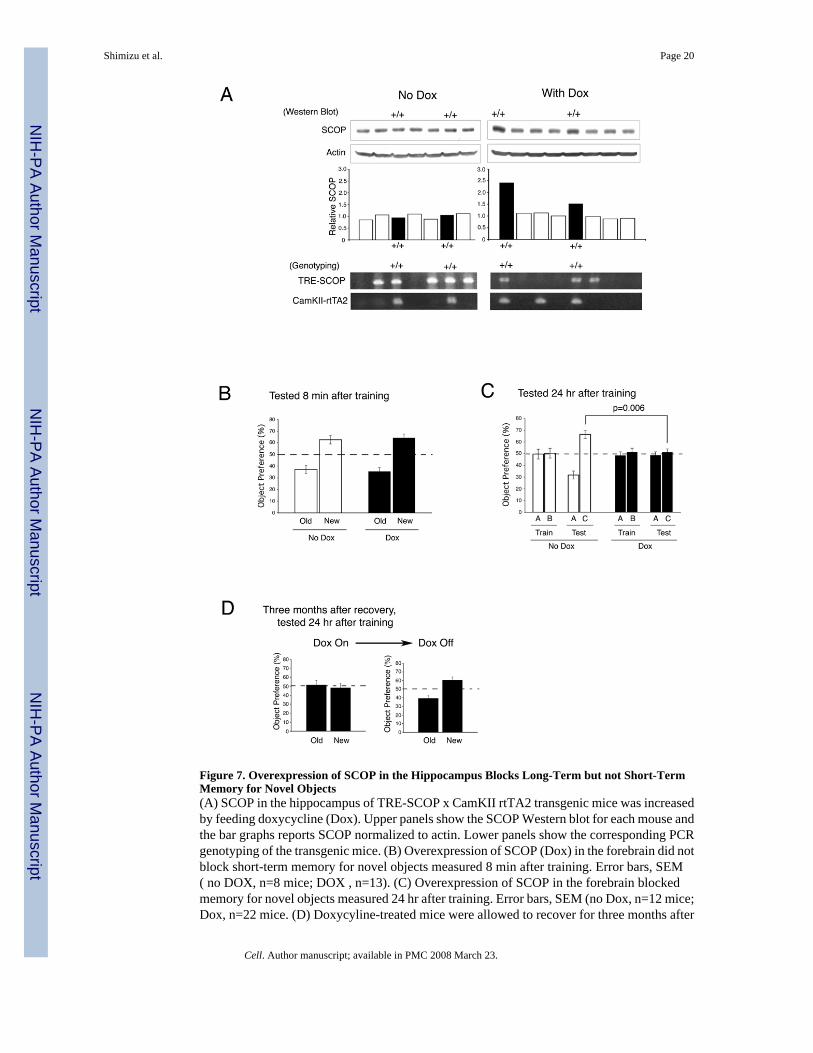
Figure 7. Overexpression of SCOP in the Hippocampus Blocks Long-Term but not Short-TermMemory for Novel Objects(A) SCOP in the hippocampus of TRE-SCOP x CamKII rtTA2 transgenic mice was increasedby feeding doxycycline (Dox). Upper panels show the SCOP Western blot for each mouse andthe bar graphs reports SCOP normalized to actin. Lower panels show the corresponding PCRgenotyping of the transgenic mice. (B) Overexpression of SCOP (Dox) in the forebrain did notblock short-term memory for novel objects measured 8 min after training. Error bars, SEM( no DOX, n=8 mice; DOX , n=13). (C) Overexpression of SCOP in the forebrain blockedmemory for novel objects measured 24 hr after training. Error bars, SEM (no Dox, n=12 mice;Dox, n=22 mice. (D) Doxycyline-treated mice were allowed to recover for three months after
Shimizu et al. Page 20
Cell. Author manuscript; available in PMC 2008 March 23.
NIH
-PA Author Manuscript
NIH
-PA Author Manuscript
NIH
-PA Author Manuscript
removal of doxycyline, trained for novel objects, and tested 24 hr after training. Error bars,SEM (n=8 mice).
Shimizu et al. Page 21
Cell. Author manuscript; available in PMC 2008 March 23.
NIH
-PA Author Manuscript
NIH
-PA Author Manuscript
NIH
-PA Author Manuscript
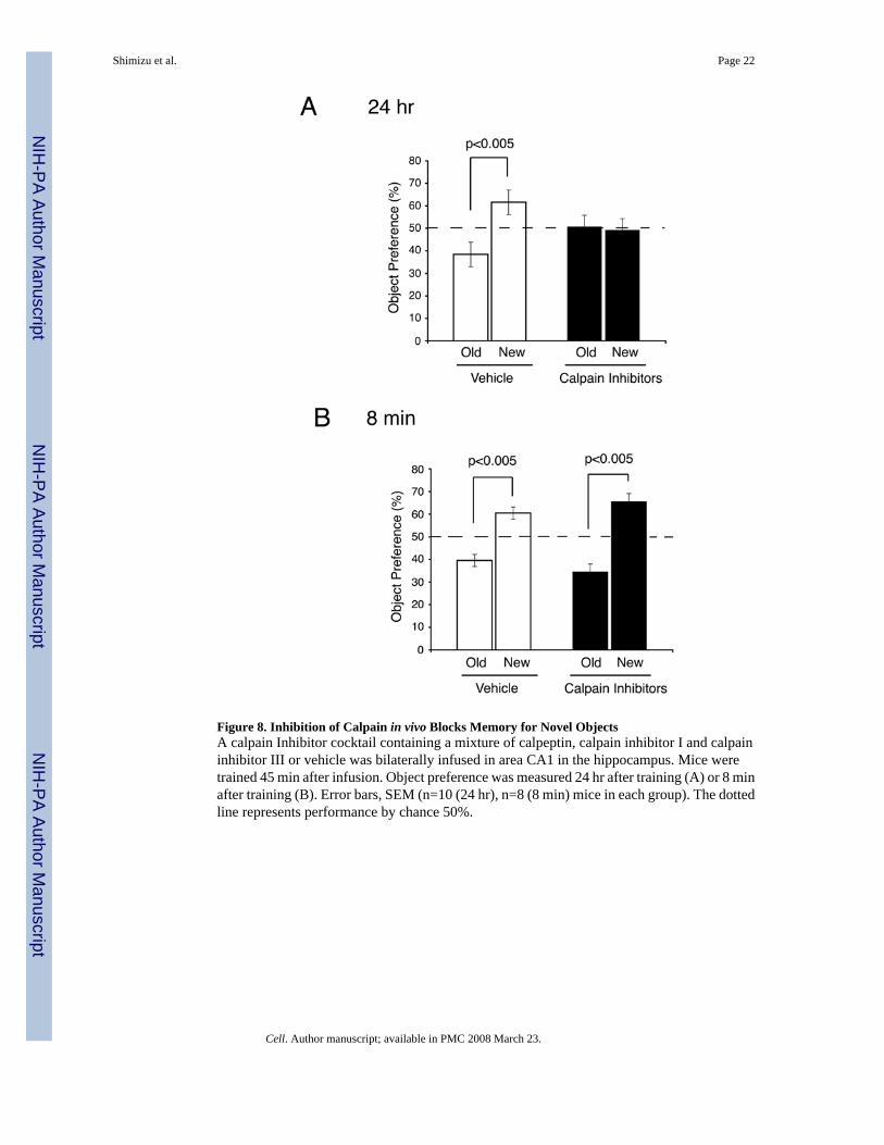
Figure 8. Inhibition of Calpain in vivo Blocks Memory for Novel ObjectsA calpain Inhibitor cocktail containing a mixture of calpeptin, calpain inhibitor I and calpaininhibitor III or vehicle was bilaterally infused in area CA1 in the hippocampus. Mice weretrained 45 min after infusion. Object preference was measured 24 hr after training (A) or 8 minafter training (B). Error bars, SEM (n=10 (24 hr), n=8 (8 min) mice in each group). The dottedline represents performance by chance 50%.
Shimizu et al. Page 22
Cell. Author manuscript; available in PMC 2008 March 23.
NIH
-PA Author Manuscript
NIH
-PA Author Manuscript
NIH
-PA Author Manuscript

Figure 9. Model for Role of SCOP in MAPK ActivationIt is hypothesized that SCOP attenuates MAPK activity in hippocampal neurons by binding tothe nucleotide-free form of K-Ras, thereby inhibiting stimulation of the MAPK pathway.Calapin may contribute to ERK activation by degrading SCOP.
Shimizu et al. Page 23
Cell. Author manuscript; available in PMC 2008 March 23.
NIH
-PA Author Manuscript
NIH
-PA Author Manuscript
NIH
-PA Author Manuscript