Proteolytic Cleavage of Protein Tyrosine Phosphatase Regulates Glioblastoma Cell Migration
Upload
independentCategory
view
3download
0
Brain Pathology (2003) in press
+
Biomarkers Of Proteolytic Damage Following Traumatic Brain Injury
Jose A. Pineda1,3*, Kevin K.W. Wang1,2,4, Ronald Hayes1,2
Center for Traumatic Brain Injury Studies, Evelyn F. and William L. McKnight Brain Institute of The University of Florida, Gainesville, FL 1; Dept. of Neuroscience, 2 Dept. of Pediatrics 3, Dept. Dept. of Psychiatry4.
*To whom all correspondence should be addressed. PICU PO Box 100296 Gainesville, Florida 32610-0296 [email protected]
Key words: biomarkers, surrogate markers, protease, caspase, calpain, traumatic brain injury

Brain Pathology (2003) in press
Brain Pathology (2003) in press
Introduction
Brain injury resulting from traumatic, ischemic and/or chemical etiology is a significant
international health concern, representing a potentially catastrophic debilitating medical
emergency with poor prognosis for long-term disability. It represents a major problem to military
care, accounting for 25% of all combat casualties and is the leading cause of death (approaching
50% incidence) among wounded soldiers reaching Echelon I medical treatment [8]. In civilian
life, the incidence of brain injury and resultant long-term disabilities caused by traumatic insults
(automobile accidents, gunshots, sports, etc.) and ischemic events (strokes, cerebral hemorrhage,
cardiac arrest, etc.) are several orders of magnitude greater. There are more than 1 million
traumatic brain injury (TBI) cases that are treated and released from an emergency department
annually in the United States resulting in more than 230,000 hospitalizations, 50,000 deaths and
80,000 disabilities. Among all age groups, the top three causes of TBI are motor vehicle
accidents, falls and violence [1] . Despite modern automobile design and injury prevention
campaigns, important causes of TBI in children such as ejections from cars during traffic
accidents, have increased in recent years [111]. The current estimation is 5.3 million Americans
live with TBI-related disability. TBI is the greatest cause of death and disability in young people
less than 24 years old [12].
With the exception of supportive measures, there are currently no approved drug
treatments for TBI [77]. There have been a large number of clinical trials studying potential
therapies for traumatic brain injury (TBI) that have resulted in negative findings with a cost of
over $200 million [15, 30]. Many investigators have pointed out that the absence of biochemical
markers of injury could have contributed to these failures [77, 104]. Unlike other organ-based
diseases where rapid diagnosis employing biomarkers (usually involving blood tests) prove
invaluable to guide treatment of the disease, no such rapid, definitive diagnostic tests exist for
Brain Pathology (2003) in press
TBI to provide physicians with quantifiable neurochemical markers to help determine the
seriousness of the injury, the anatomical and cellular pathology of the injury, and to guide
implementation of appropriate triage and medical management.
Criteria For Biochemical/Surrogate Markers
In the course of research on biomarkers, our laboratories have developed criteria for
biomarker development. Useful biomarkers should employ readily accessible biological material
such as CSF or blood (CSF is routinely accessible in severely injured TBI patients), predict the
magnitude of injury and resulting functional deficits and possess high sensitivity and specificity,
have a rapid appearance in blood and be released in a time-locked sequence after injury. Ideally,
biomarkers should employ biological substrates unique to the CNS and provide information on
injury mechanisms, a criterion often used to distinguish biochemical markers from surrogate
markers of injury, which usually do not provide information on injury mechanisms. Potential
gender and age related differences on biomarker profiles are also important and should be taken
into account when developing useful biochemical markers [43].
Uses Of Biomarkers
Biomarkers would have important applications in diagnosis, prognosis and clinical
research of brain injuries. Simple, rapid diagnostic tools will immensely facilitate allocation of
the major medical resources required to treat TBI and other brain injuries. Accurate diagnosis in
acute care environments can significantly enhance decisions about patient management including
decisions whether to admit or discharge or administer other time consuming and expensive tests
including computer tomography (CT) and magnetic resonance imaging (MRI) scans. Biomarkers
Brain Pathology (2003) in press
could have important prognostic functions especially in patients suffering mild TBI, which make
up an estimated 80% of the 2.5 to 6.5 million individuals who suffer from lifelong impairment as
a result of TBI [2, 83]. Accurate identification of these patients could facilitate development of
guidelines for return to duty, work or sports activities and also provide opportunities for
counseling of patients suffering from these deficits. Biomarkers could provide major
opportunities for the conduct of clinical research including confirmation of injury mechanism(s)
and drug target identification. The temporal profile of changes in biomarkers could guide timing
of treatment and assist in monitoring the response to therapy and intervention. Finally,
biomarkers could provide a clinical trial outcome measure obtainable much more cheaply and
readily than conventional neurological assessments, thereby significantly reducing the risks and
costs of human clinical trials. Relevant, easily available biomarkers are needed in order to
maximize chances of success in developing long awaited effective drugs for traumatic brain
injury [77].
Current Status of Research on Markers of Traumatic Brain Injury
Analysis of specific biochemical markers has provided useful information on the
mechanism and diagnosis specific organ dysfunction in humans [112]. However, although
analysis of cerebrospinal fluid, cerebral microdialysis samples, and brain tissue specimens has
provided insight into the mechanisms of brain injury [61, 63], there are no biomarkers of proven
clinical utility for TBI.
TBI is difficult to assess and clinical examinations are of restricted value during the first
hours and days after injury. Conventional diagnoses of TBI are based on neuroimaging
techniques such as CT scanning, MRI and single-photon emission CT scanning [46, 58, 72]. CT
scanning has low sensitivity to diffuse brain damage, and the availability of MRI is limited [60,
Brain Pathology (2003) in press
64]. Single-photon emission CT scanning detects regional blood-flow abnormalities not
necessarily related to structural damage.
A recent review of biomarkers of TBI highlighted the need for biomarker development
[43]. The most studied potential biochemical markers for TBI include creatine kinase (CK), glial
fibrillary acidic protein (GFAP), lactate dehydrogenase (LDH), myelin basic protein (MBP),
neuron-specific enolase (NSE) and S-100 proteins. The bulk of research in TBI has focused on
NSE and S-100β. The specificity of NSE for brain is high [49] , sex- and age-related variability
is low [28, 51, 74, 86, 98, 120, 121, 133], and NSE is rapidly detectible in serum after TBI [129].
However, studies relating NSE serum levels to admission GCS in patients with severe TBI show
conflicting results. Similar data have been reported concerning relationships with CT scan
findings, ICP and long-term outcomes. In mild TBI, NSE failed to separate patients from
controls [43, 44, 108, 131]. Thus, NSE is predominantly used as a marker for tumors [22]. NSE
is also released in the blood by hemolysis, which could be a major source of error [22].
The S-100 protein family now consists of 19 members, of which S-100B is the one
viewed as a marker of brain damage [49, 65], although it is present in other tissues such as
adipocytes and chondrocytes [40]. Investigators have reported S-100β serum levels correlate to
both GCS scores, neuroradiologic findings at admission and long-term outcomes [100, 99, 128].
However, investigators have recently raised questions about the utility of S-100β reporting that
high serum levels of S-100β are detectible in trauma patients not having head injuries, a factor
not adequately controlled for in earlier studies [3]. In addition, serum levels of S-100β following
mild TBI do not show strong correlations with neuropsychological outcome [107]. Research in
this area continues and recent reports have indicated the potential utility of measures of blood
GFAP [71], spinal fluid Interleukin-6 [115] and cleaved tau protein in serum [45, 114] and spinal
fluid [135] following brain injury.
Brain Pathology (2003) in press
Investigators have also generally recognized the need for more objective assessments of
outcome following stroke, including biochemical markers [27, 68]. The approval of tPA as a
treatment for acute stroke has additionally highlighted the potential utility of biochemical
markers. Use of tPA may be hindered by diagnostic concerns because neurological deficits
accompanying stroke can mimic those seen during transient ischemic attacks, complex migraine,
space-occupying lesions and post-ictal paralysis. A reliable biochemical marker might give
assurance to physicians considering administering thrombolitic agents for acute stroke [41, 50].
Previously reported biomarkers of cerebral ischemia include NSE, brain specific creatine
kinase enzyme (CPK-BB), S-100β and inflammatory cytokines such as IL-6 [76, 89]. NSE and
S-100β have been the most studied. After cardiac arrest, NSE elevations in serum and CSF have
been correlated with neurological recovery [26, 67, 106]. Serum and CSF NSE values were
reported to be elevated in rodent models of focal ischemia in proportion to the eventual infarct
volume [24, 25, 42]. In clinical trials, peak serum NSE values also predicted infarct volumes as
shown by CT. Correlating serum NSE values with functional outcome was less successful [24,
25, 70], possibly because functional neurological deficit is influenced as much by location of
brain injury as by infarct size [70]. S-100β protein has been studied most extensively for
characterization of ischemic injuries after cardiac surgery, and several reports have documented
post-operative serum elevations [29, 113, 127]. However, many of these reports do not include
careful studies of neurological outcome, and several investigators have recently criticized the
diagnostic utility of S-100β during cardiac surgery. [3].
Proteolytic Damage and the Pathobiology of Traumatic Brain Injury
After TBI, brain cells can deteriorate following more than one pathway, and many genes
and proteins may be involved. Programmed cell death is an evolutionarily conserved form of cell
Brain Pathology (2003) in press
suicide that occurs widely throughout development [13]. This type of cell death often has the
morphological appearance of apoptosis [119]. Apoptosis occurs following TBI in animals [20,
59, 130] and humans [16, 17]. Studies of apoptosis pose special challenges since there are
multiple apoptotic pathways, and apoptosis is extremely sensitive to a number of variables
including injury type and magnitude [10, 14, 101], cell type [38, 57] and stimulation/antagonism
of specific receptors [14, 19, 23, 38, 39, 48].
The molecular events occurring after TBI are just beginning to be understood. Elevated
neuronal calcium levels activate a number of calcium-dependent enzymes such as
phospholipases [84], kinases [132], phosphatases [75], and proteases [5, 81], all of which can
modulate post-TBI cytoskeletal protein loss. Caspase-3 is a member of the caspase family of
cysteine proteases. Activated caspase-3 has many cellular targets that, when severed and/or
activated, produce the morphologic features of apoptosis [18]. Calpains are calcium-activated,
neutral cysteine proteases with relative selectivity for proteolysis of a subset of cellular proteins.
Calpain activation has been implicated in different models of apoptosis and in different cell
types, including neurons [93]. Understanding of the contributions of calpains and caspases to cell
injury/death following TBI may have important diagnostic and therapeutic implications.
Contributions of Caspase-3 and Calpain to Cell Death Following Traumatic Brain Injury
Numerous studies from our own [6, 91, 93] and other laboratories [31, 35, 73] have
provided evidence that the caspase family of cysteine proteases is an important intracellular
effector of apoptosis in various cell lines and apoptotic models. Caspase 3-like proteases have
been shown to cleave a variety of cytoplasmic, nuclear and cytoskeletal proteins during apoptosis
including αII-spectrin [69, 78, 79, 122], poly(ADP-ribose) polymerase (PARP: 51) and others
(52-57). In vitro studies in our laboratories using a model of stretch injury have demonstrated
Brain Pathology (2003) in press
caspase-3 processing of α-spectrin to the apoptotic-linked 120-kDa fragment 24 hours after
moderate, but not mild or severe injury [6]. In vivo studies have provided evidence of caspase-3
activation following TBI. First, Clark et al. demonstrated cleavage of capsase-3 to its p18 and
p12 subunits in humans [17]. Yakovlev et al. reported that TBI increased caspase-3, but not
caspase-1, activity [130]. Caspase-3 inhibition reduced DNA fragmentation and TUNEL staining
and improved behavioral outcome. We have also concurrently examined caspase-3 and calpain
activation after TBI. Distinct regional and temporal patterns of calpain/caspase-3 processing of
αII-spectrin in brain regions ipsilateral to the site of injury after TBI have been observed.
Caspase-3-mediated break down products (BDP’s) to αII-spectrin were absent in the cortex but
showed significant increases in hippocampus and striatum early (hours) after TBI [92].
Immunohistological examinations revealed increased expression of the proteolitically active
subunit of caspase-3, p18, in neurons, astrocytes, and oligodendrocytes from 6 to 72 hours
following controlled cortical impact injury. Moreover, concurrent assessment of nuclear
histopathology using hematoxylin identified p18-immunopositive cells exhibiting apoptotic-like
morphological profiles in the cortex ipsilateral to the injury site [6].
Calpains are Ca2+ activated cysteine proteases that have been implicated in a variety of
neuropathological conditions [55, 126]. Intracellular substrates of activated calpain include
cytoskeletal proteins, calmodulin-binding proteins, enzymes involved in signal transduction,
membrane proteins and transcription factors [110, 118, 125]. While calpain activation has
historically been associated with necrotic cell death [82], calpain activation has also been
implicated in different models of apoptosis and in different cell types, including neurons [7, 52,
78, 117, 123]. Research in our own and other laboratories have documented calpain activation
following TBI in vivo [55]. TBI results in altered Ca2+ homeostasis [134] and activates several
Ca2+ -dependent enzymes including the calpains. Overactivation of calpains occurs in many
neurodegenerative diseases and injuries to the CNS [4, 55, 126]. Increased calpain activity
Brain Pathology (2003) in press
following TBI has been inferred by a variety of techniques [54, 81, 94, 97], including protection
by calpain inhibitors [95, 109].
Pathological calpain activation is believed to occur when intracellular free calcium levels
surpass a certain threshold. Importantly, increases in free calcium via voltage and receptor gated
calcium channels have been reported in CNS trauma in vivo [19, 48, 56]. Calpains are located
throughout the neuron, in somatodendritic regions and in axons [57]. Calpain may also be a
constituent of myelin [116]. Therefore, pathological calpain activity and subsequent substrate
proteolysis can have profound effects on neuronal structure and function.
Cytoskeletal alterations after experimental brain injury have pointed to the likelihood of
calpain mediated proteolysis. Preferred substrates for calpains include the cytoskeletal protein
spectrin [47, 66], microtubule associated protein-2 (MAP-2)[33], and neurofilament proteins [20,
21, 85, 105]. Increased degradation of MAP2 [32, 130], the neurofilament triplet proteins [53,
87] and spectrin [80] have been reported in cerebral ischemia. In addition, loss of MAP-2 [62],
neurofilament 68 (NF 68) and neurofilament 200 (NF 200)[94-96] have been reported following
traumatic brain injury (TBI) in vivo. Additional evidence that calpain is activated in neurons
following experimental brain injury has been provided by the use of antibodies which bind
specifically to calpain mediated BDP’s of cytoskeletal proteins in models of TBI [81].
αII-Spectrin Degradation-A Prototype Biomarker
Our research program to develop biomarkers for TBI has focused on α-spectrin
degradation as a prototypical biochemical marker [34, 103]. αII-spectrin (280 kDa) is the major
structural component of the cortical membrane cytoskeleton and is particularly abundant in
axons and presynaptic terminals [36, 37]. Importantly, αII-spectrin is a major substrate for both
calpain and caspase-3 cysteine proteases [124]. Our laboratory has provided considerable
Brain Pathology (2003) in press
evidence that αII-spectrin is processed by calpains and/or caspase-3 to signature cleavage
products in vivo after TBI [6, 11, 81, 92] and in vitro models of mechanical stretch injury [91].
Calpain produces two major αII-spectrin breakdown products of 150 kDa and 145 kDa
(SBDP150 and SBDP145) in a sequential manner (Wang TINS 2002). On the other hand,
caspase-3 initially produces a 150 kDa SBDP that is further cleaved into a 120 kDa fragment
(SBDP120) (Wang TINS 2000). Immunoblots of αII-spectrin degradation thus provide
concurrent information on the activation of calpain and caspase-3, potentially important
regulators of cell death following TBI. The calcium sensitivity and low basal levels of calpain
optimize its utility as a marker of cell injury. Although not found in erythrocytes and thus robust
to confounding by blood contamination, αII-spectrin is not specific to the CNS [37]. Following
injury, native αII-spectrin protein was decreased in brain tissue and increased in CSF from 24
hrs to 72 hrs after injury. Calpain-specific breakdown products increased in both brain and CSF
after injury. Caspase-3-specific breakdown products increased in some animals, but to a lesser
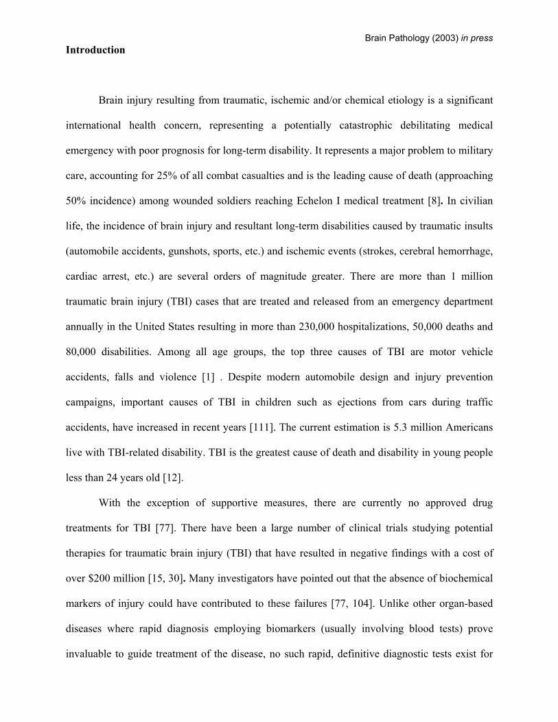
degree [90]. Considerable laboratory data exists on the potential utility of αII-spectrin
degradation as a biomarker for TBI, including our ability to detect differences in severity of
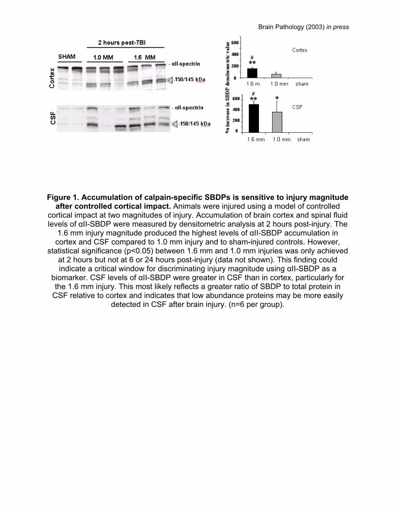
injury based on αII-spectrin detection in spinal fluid of rodents after TBI (Figure 1). Preliminary
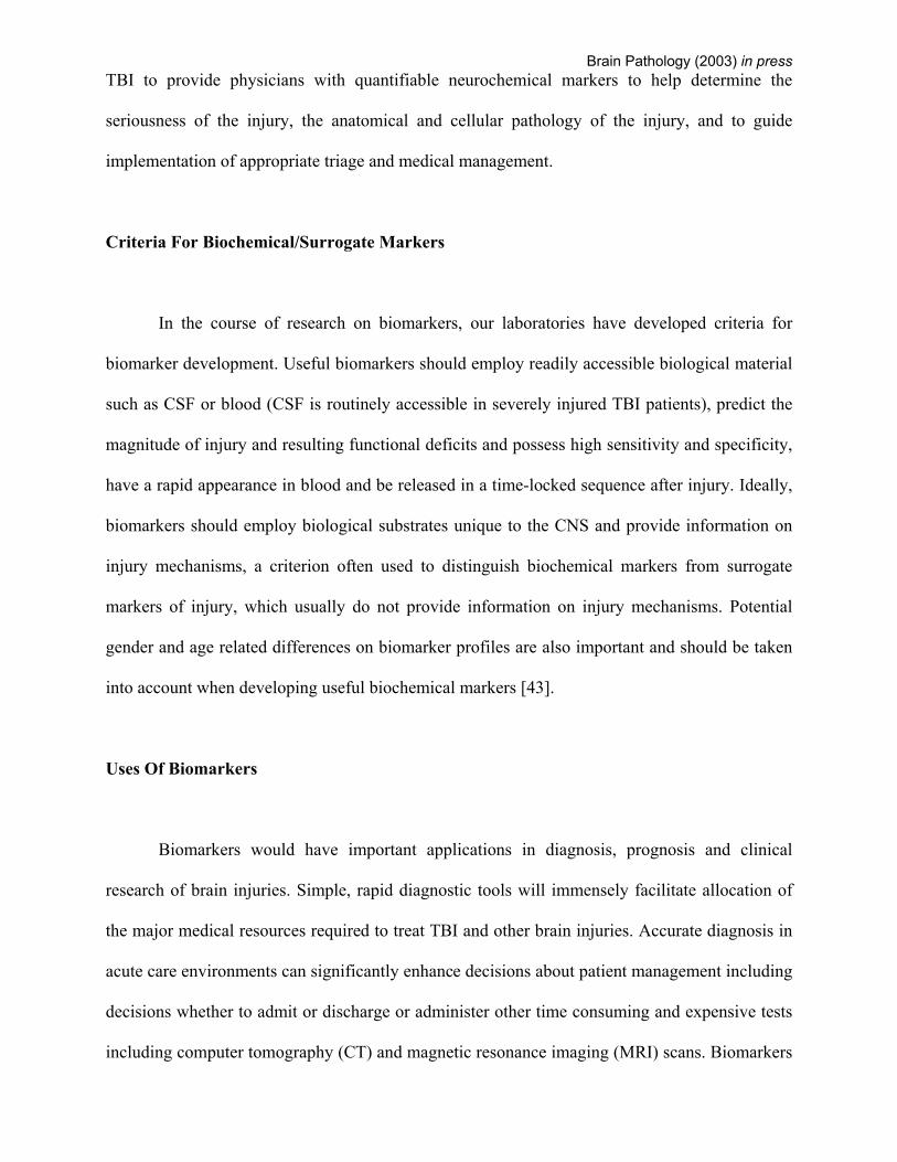
human data are also promising (Figure 2). However, what is not known is whether BDP’s and
other biomarkers that discriminate injury magnitudes at the biochemical level (i.e., magnitude
differences in CSF/serum levels of a biomarker) will be useful as predictors of clinically relevant
measures of outcome. That is, two different injury magnitudes may produce significantly
different biochemical responses in the brain that are discernable in CSF or serum, but these same
injury magnitudes may not result in functionally different behavioral, pathological, or other
clinical outcome measures. Thus, biomarkers that do not correlate with clinically relevant
outcome measures will not be useful for assessment of functional ability, functional recovery, or
for gauging effects of therapy on outcome. In addition, preliminary results from our laboratories
Brain Pathology (2003) in press
suggest that there is a spontaneous susceptibility of cytoskeletal protein degradation by calpain in
aging rats [9]. These findings emphasize the importance of accounting for multiple clinical
variables including but not limited to age when evaluating the clinical utility of biomarkers of
brain injury.
Additional Cytoskeletal Proteins with Potential Utility as Biomarkers
Initial research focused on proteolytic processing of cytoskeletal proteins such as lower
molecular weight neurofilament 68 protein (NF-68) highlights their potential to provide useful
information on activity of specific proteases such as µ-calpain and m-calpain. Importantly, 2-D
gel electrophoresis studies suggested dephosphorylation of NF-68 may be associated with NF
protein loss following TBI, a post translational modification that could have significance for
biomarker development [81, 97]. This important biomarker could provide important information
on the pathophysiology of both dendritic and axonal damage after TBI [96]. Importantly, NF-68
has been used to quantify axonal injury in closed head injury models [102]. Since diffuse axonal
injury (DAI) is presently considered one of the most common types of primary lesions in patients
with severe closed head injury [88], a biomarker that provides information on axonal injury
could potentially have clinical utility.
Future Directions
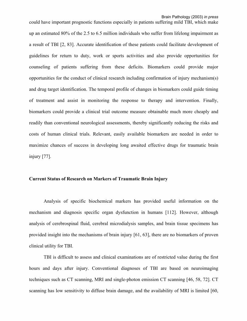
The pathology of TBI is extremely complex. As our understanding of the numerous
biochemical cascades involved continues to evolve, sophisticated diagnostic tools such as
biomarkers will be developed (Figure 3). Ideal biomarkers will provide information on the
pathobiology of TBI and facilitate better stratification of patients by their severity of injury,
Brain Pathology (2003) in press
better monitoring of the progression of secondary damage, response to treatment/intervention,
and prediction of outcome. Although the initial characterization of biomarkers will be mainly
based on spinal fluid analysis (Figure 3), methods for measurement of such biomarkers in blood
(plasma or serum) will be developed. The development of accessible and reliable biomarkers is
likely to change the way clinical studies of head injury are conducted, resulting in more
mechanism driven, optimally timed therapies.
Acknowledgments
The authors like to acknowledge the original contributions of Dr. Brian Pike, and the support
from NIH R01 # NS #38105-01 and NS #39091-02.
BIBLIOGRAPHY
1. Consensus conference. Rehabilitation of persons with traumatic brain injury. NIH
Consensus Development Panel on Rehabilitation of Persons With Traumatic Brain
Injury. Jama, 1999. 282(10): p. 974-83.
2. Alexander, M.P., Mild traumatic brain injury: pathophysiology, natural history, and
clinical management. Neurology, 1995. 45(7): p. 1253-60.
3. Anderson RE, H.L., Nilsson O, Dijlai-Merzoug R, Settergen G., High serum S100B levels
for trauma patients without head injuries. Neurosurgery, 2001. 49(5): p. 1272-3.
4. Bartus, R.T., P.J. Elliott, N.J. Hayward, R.L. Dean, S. Harbeson, J.A. Straub, Z. Li, and
J.C. Powers, Calpain as a novel target for treating acute neurodegenerative disorders.
Neurol Res, 1995. 17(4): p. 249-58.
5. Bartus, R.T., N.J. Hayward, P.J. Elliott, S.D. Sawyer, K.L. Baker, R.L. Dean, A.
Akiyama, J.A. Straub, S.L. Harbeson, Z. Li, and et al., Calpain inhibitor AK295 protects
Brain Pathology (2003) in press
neurons from focal brain ischemia. Effects of postocclusion intra-arterial
administration.PG - 2265-70. Stroke, 1994. 25(11).
6. Beer, R., G. Franz, A. Srinivasan, R.L. Hayes, B.R. Pike, J.K. Newcomb, X. Zhao, E.
Schmutzhard, W. Poewe, and A. Kampfl, Temporal profile and cell subtype distribution
of activated caspase-3 following experimental traumatic brain injury. J Neurochem,
2000. 75(3): p. 1264-73.
7. Behrens, M.M., J.L. Martinez, C. Moratilla, and J. Renart, Apoptosis induced by protein
kinase C inhibition in a neuroblastoma cell line. Cell Growth Differ, 1995. 6(11): p.
1375-80.
8. Bellamy, R., The causes of death in conventional land warfares: implications for combat
casualty research. Military Medicine, 1984. 149:: p. 55-62.
9. Bernath, E.e.a., Spontaneous Cytoskeletal Protein Proteolysis in Aged Wistar Rat Brains.
In Preparation.
10. Bonfoco, E., D. Krainc, M. Ankarcrona, P. Nicotera, and S.A. Lipton, Apoptosis and
necrosis: two distinct events induced, respectively, by mild and intense insults with N-
methyl-D-aspartate or nitric oxide/superoxide in cortical cell cultures. Proc Natl Acad
Sci U S A, 1995. 92(16): p. 7162-6.
11. Buki, A., D.O. Okonkwo, K.K. Wang, and J.T. Povlishock, Cytochrome c release and
caspase activation in traumatic axonal injury. J Neurosci, 2000. 20(8): p. 2825-34.
12. CDC, Fact Book. 2000.
13. Chang, L.K., G.V. Putcha, M. Deshmukh, and E.M. Johnson, Mitochondrial involvement
in the point of no return in neuronal apoptosis. Biochimie, 2002. 84(2-3): p. 223-31.
14. Choi, D.W., Ischemia-induced neuronal apoptosis. Curr Opin Neurobiol, 1996. 6(5): p.
667-72.
Brain Pathology (2003) in press
15. Choi SC, B.R., Design and statistical issues in multicenter trials of severe head injury.
Neurological Research, 2001. Mar-Apr(23(2-3)): p. 190-2.
16. Clark, R.S., P.M. Kochanek, P.D. Adelson, M.J. Bell, J.A. Carcillo, M. Chen, S.R.
Wisniewski, K. Janesko, M.J. Whalen, and S.H. Graham, Increases in bcl-2 protein in
cerebrospinal fluid and evidence for programmed cell death in infants and children after
severe traumatic brain injury. J Pediatr, 2000. 137(2): p. 197-204.
17. Clark, R.S., P.M. Kochanek, M. Chen, S.C. Watkins, D.W. Marion, J. Chen, R.L.
Hamilton, J.E. Loeffert, and S.H. Graham, Increases in Bcl-2 and cleavage of caspase-1
and caspase-3 in human brain after head injury. Faseb J, 1999. 13(8): p. 813-21.
18. Clark, R.S., P.M. Kochanek, S.C. Watkins, M. Chen, C.E. Dixon, N.A. Seidberg, J.
Melick, J.E. Loeffert, P.D. Nathaniel, K.L. Jin, and S.H. Graham, Caspase-3 mediated
neuronal death after traumatic brain injury in rats. J Neurochem, 2000. 74(2): p. 740-53.
19. Cohen, G.M., Caspases: the executioners of apoptosis. Biochem J, 1997. 326 ( Pt 1): p.
1-16.
20. Colicos, M.A. and P.K. Dash, Apoptotic morphology of dentate gyrus granule cells
following experimental cortical impact injury in rats: possible role in spatial memory
deficits. Brain Res, 1996. 739(1-2): p. 120-31.
21. Conti, A.C., R. Raghupathi, J.Q. Trojanowski, and T.K. McIntosh, Experimental brain
injury induces regionally distinct apoptosis during the acute and delayed post-traumatic
period. J Neurosci, 1998. 18(15): p. 5663-72.
22. Cooper, E., Neuron-specific enolase. Int J Biol Markers, 1994(4): p. 205-210.
23. Copin, J.C., Y. Li, L. Reola, and P.H. Chan, Trolox and 6,7-dinitroquinoxaline-2,3-dione
prevent necrosis but not apoptosis in cultured neurons subjected to oxygen deprivation.
Brain Res, 1998. 784(1-2): p. 25-36.
Brain Pathology (2003) in press
24. Cunningham, R.T., M. Watt, J. Winder, S. McKinstry, J.T. Lawson, C.F. Johnston, S.A.
Hawkins, and K.D. Buchanan, Serum neurone-specific enolase as an indicator of stroke
volume. Eur J Clin Invest, 1996. 26(4): p. 298-303.
25. Cunningham, R.T., I.S. Young, J. Winder, M.J. O'Kane, S. McKinstry, C.F. Johnston,
O.M. Dolan, S.A. Hawkins, and K.D. Buchanan, Serum neurone specific enolase (NSE)
levels as an indicator of neuronal damage in patients with cerebral infarction. Eur J Clin
Invest, 1991. 21(5): p. 497-500.
26. Dauberschmidt, e.a., Changes of neuron-specific enolase concentration in plasma after
cardiac arrest and resuscitation. Mol Chem Neuropathol, 1991. 14: p. 237-245.
27. Dirnagl U, C.I., and Moskowitz MA, Pathology of ischaemic stroke: an integrated view.
TINS, 1999. 22(9): p. 391-397.
28. Donato, R., Functional roles of S100 proteins, calcium-binding proteins of the EF-hand
type. Biochim Biophys Acta., 1999. 1450: p. 191-231.
29. Eberle, e.a., Rapid prearrest cooling is associated with elevated S100 levels immediately
after deep hypothermic circulatory arrest in adults. Anesth Analg, 1997. 84(4S(SCA12).
30. Egon M.R. Doppenberg, S.C.C., and Ross Bullock, Clinical trials in TBI, what can we
learn from previous studies. Annals of the New York Academy of Sciences, 1997. 825: p.
305-322.
31. Eldadah, B.A., A.G. Yakovlev, and A.I. Faden, The role of CED-3-related cysteine
proteases in apoptosis of cerebellar granule cells.PG - 6105-13. J Neurosci, 1997.
17(16).
32. Ellis, E.F., J.S. McKinney, K.A. Willoughby, S. Liang, and J.T. Povlishock, A new model
for rapid stretch-induced injury of cells in culture: characterization of the model using
astrocytes. J Neurotrauma, 1995. 12(3): p. 325-39.
Brain Pathology (2003) in press
33. Fischer, I., G. Romano-Clarke, and F. Grynspan, Calpain-mediated proteolysis of
microtubule associated proteins MAP1B and MAP2 in developing brain. Neurochem
Res, 1991. 16(8): p. 891-8.
34. Flint, J.P., BR; Hayes,RL ; Moffett, J.R. Dave, JR; X.-C.M. Lu, and F.C. and Tortella.,
Accumulation of calpain and caspase-3 cleaved alpha-II spectrin breakdown products in
CSF after middle cerebral artery occulsion in rats. J. Neurotrauma. (in press). 2002 (In
Press).
35. Fraser, A. and G. Evan, A license to kill. Cell, 1996. 85(6): p. 781-4.
36. Friederer BM, Z.I., Goodman SR, Brain spectrin (240/235) and brain spectrin
(240/235E): two distinct spectrin subtypes with different locations within mammalian
neural cells. J Cell Biol, 1986. Jun(102(6)): p. 2088-97.
37. Goodman SR, Z.W., Clark MB, Zagon IS, Barker JE, and Bloom ML, Brain spectrin: of
mice and men. Brain Res Bull, 1995. 36: p. 593-606.
38. Gottron, F.J., H.S. Ying, and D.W. Choi, Caspase inhibition selectively reduces the
apoptotic component of oxygen-glucose deprivation-induced cortical neuronal cell death.
Mol Cell Neurosci, 1997. 9(3): p. 159-69.
39. Gwag, B.J., D. Lobner, J.Y. Koh, M.B. Wie, and D.W. Choi, Blockade of glutamate
receptors unmasks neuronal apoptosis after oxygen-glucose deprivation in vitro.
Neuroscience, 1995. 68(3): p. 615-9.
40. Haimoto, H., S. Hosoda, and K. Kato, Differential distribution of immunoreactive S100-
alpha and S100-beta proteins in normal nonnervous human tissues. Lab Invest, 1987.
57(5): p. 489-98.
41. Hill, M.D., G. Jackowski, N. Bayer, M. Lawrence, and R. Jaeschke, Biochemical markers
in acute ischemic stroke. Cmaj, 2000. 162(8): p. 1139-40.
Brain Pathology (2003) in press
42. Horn, M., F. Seger, and W. Schlote, Neuron-specific enolase in gerbil brain and serum
after transient cerebral ischemia. Stroke, 1995. 26(2): p. 290-6; discussion 296-7.
43. Ingebrigtsen T, R.B., Biochemical Serum Markers of TBI. The Journal of Trauma Injury,
Infection, and Critical Care, 2002. 52: p. 798-808.
44. Ingebrigtsen T, R.B., Trumpy JH., Management of minor head injury: the value of early
CT and serum protein S-100 measurements. Journal of Clinical Neuroscience, 1997. 4: p.
29-33.
45. Irazuzta, J.E., G. de Courten-Myers, F.P. Zemlan, M.Y. Bekkedal, and J. Rossi, 3rd,
Serum cleaved Tau protein and neurobehavioral battery of tests as markers of brain
injury in experimental bacterial meningitis. Brain Res, 2001. 913(1): p. 95-105.
46. Jacobs A, P.E., Ingels M, Put T, Bossuyt A, One-year follow-up of technetium-99m-
HMPAO SPECT in mild head injury. J Nucl Med, 1996. 37: p. 1605-1609.
47. Jenkins, L.W., K. Moszynski, B.G. Lyeth, W. Lewelt, D.S. DeWitt, A. Allen, C.E.
Dixon, J.T. Povlishock, T.J. Majewski, G.L. Clifton, and et al., Increased vulnerability of
the mildly traumatized rat brain to cerebral ischemia: the use of controlled secondary
ischemia as a research tool to identify common or different mechanisms contributing to
mechanical and ischemic brain injury. Brain Res, 1989. 477(1-2): p. 211-24.
48. Johnson, E.M., Jr., L.J. Greenlund, P.T. Akins, and C.Y. Hsu, Neuronal apoptosis:
current understanding of molecular mechanisms and potential role in ischemic brain
injury. J Neurotrauma, 1995. 12(5): p. 843-52.
49. Johnsson, P., Markers of cerebral ischemia after cardiac surgery. J Cardiothorac Vasc
Anesth, 1996. 10(1): p. 120-6.
50. Jonas, S., V. Aiyagari, D. Vieira, and M. Figueroa, The failure of neuronal protective
agents versus the success of thrombolysis in the treatment of ischemic stroke. The
predictive value of animal models. Ann N Y Acad Sci, 2001. 939: p. 257-67.
Brain Pathology (2003) in press
51. Jonsson H, J.P., Hoglund P, Alling C, Blomquist S., The elimination of S-100b and renal
function after cardiac surgery. J Cardiothorac Vasc Aneth, 2000. 14: p. 698-701.
52. Jordan, J., M.F. Galindo, and R.J. Miller, Role of calpain- and interleukin-1 beta
converting enzyme-like proteases in the beta-amyloid-induced death of rat hippocampal
neurons in culture. J Neurochem, 1997. 68(4): p. 1612-21.
53. Kaku, Y., Y. Yonekawa, T. Tsukahara, N. Ogata, T. Kimura, and T. Taniguchi,
Alterations of a 200 kDa neurofilament in the rat hippocampus after forebrain ischemia.
J Cereb Blood Flow Metab, 1993. 13(3): p. 402-8.
54. Kampfl, A., R. Posmantur, R. Nixon, F. Grynspan, X. Zhao, S.J. Liu, J.K. Newcomb,
G.L. Clifton, and R.L. Hayes, mu-calpain activation and calpain-mediated cytoskeletal
proteolysis following traumatic brain injury. J Neurochem, 1996. 67(4): p. 1575-83.
55. Kampfl, A., R.M. Posmantur, X. Zhao, E. Schmutzhard, G.L. Clifton, and R.L. Hayes,
Mechanisms of calpain proteolysis following traumatic brain injury: implications for
pathology and therapy: implications for pathology and therapy: a review and update. J
Neurotrauma, 1997. 14(3): p. 121-34.
56. Kampfl, A., J.S. Whitson, X. Zhao, R. Posmantur, G.L. Clifton, and R.L. Hayes, Calpain
inhibitors reduce depolarization induced loss of tau protein in primary septo-
hippocampal cultures. Neurosci Lett, 1995. 194(3): p. 149-52.
57. Kampfl, A., X. Zhao, J.S. Whitson, R. Posmantur, C.E. Dixon, K. Yang, G.L. Clifton,
and R.L. Hayes, Calpain inhibitors protect against depolarization-induced neurofilament
protein loss of septo-hippocampal neurons in culture. Eur J Neurosci, 1996. 8(2): p. 344-
52.
58. Kant R, S.-S.L., Isaac G, Duffy J., Tc-HMPAO SPECT in persistent post-concussion
syndrome after mild head injury: comparison with MRI/CT. Brain Injury, 1997(Feb;
11(2):): p. 115-24.
Brain Pathology (2003) in press
59. Kaya, S.S., A. Mahmood, Y. Li, E. Yavuz, M. Goksel, and M. Chopp, Apoptosis and
expression of p53 response proteins and cyclin D1 after cortical impact in rat brain.
Brain Res, 1999. 818(1): p. 23-33.
60. Kesler, S.R., H.F. Adams, and E.D. Bigler, SPECT, MR and quantitative MR imaging:
correlates with neuropsychological and psychological outcome in traumatic brain injury.
Brain Inj, 2000. 14(10): p. 851-7.
61. Kochanek, P.M., R.S. Clark, R.A. Ruppel, and C.E. Dixon, Cerebral resuscitation after
traumatic brain injury and cardiopulmonary arrest in infants and children in the new
millennium. Pediatr Clin North Am, 2001. 48(3): p. 661-81.
62. Lammie, G.A., I.R. Piper, D. Thomson, and F. Brannan, Neuropathologic
characterization of a rodent model of closed head injury--addition of clinically relevant
secondary insults does not significantly potentiate brain damage. J Neurotrauma, 1999.
16(7): p. 603-15.
63. Laskowitz, e.a., Serum Markers of Cerebral Ischemia. Journal of Stroke and
Cerebrovascular Diseases, 1998. 7(4 (July-August)): p. 234-241.
64. Levi, L., J.N. Guilburd, A. Lemberger, J.F. Soustiel, and M. Feinsod, Diffuse axonal
injury: analysis of 100 patients with radiological signs. Neurosurgery, 1990. 27(3): p.
429-32.
65. Leviton, A. and O. Dammann, Brain damage markers in children. Neurobiological and
clinical aspects. Acta Paediatr, 2002. 91(1): p. 9-13.
66. Lyeth, B.G., L.W. Jenkins, R.J. Hamm, C.E. Dixon, L.L. Phillips, G.L. Clifton, H.F.
Young, and R.L. Hayes, Prolonged memory impairment in the absence of hippocampal
cell death following traumatic brain injury in the rat. Brain Res, 1990. 526(2): p. 249-58.
67. Martens, P., Serum neuron-specific enolase as a prognostic marker for irreversible brain
damage in comatose cardiac arrest surviviors. Acad Emerg Med, 1996. 3: p. 126-131.
Brain Pathology (2003) in press
68. Martens, P., A. Raabe, and P. Johnsson, Serum S-100 and neuron-specific enolase for
prediction of regaining consciousness after global cerebral ischemia. Stroke, 1998.
29(11): p. 2363-6.
69. Martin, S.J., G.A. O'Brien, W.K. Nishioka, A.J. McGahon, A. Mahboubi, T.C. Saido, and
D.R. Green, Proteolysis of fodrin (non-erythroid spectrin) during apoptosis. J Biol Chem,
1995. 270(12): p. 6425-8.
70. Missler, U., M. Wiesmann, C. Friedrich, and M. Kaps, S-100 protein and neuron-specific
enolase concentrations in blood as indicators of infarction volume and prognosis in acute
ischemic stroke. Stroke, 1997. 28(10): p. 1956-60.
71. Missler U, W.M., Wittmann G, Magerkurth O, Hagenstrom H., Measurement of glial
fibrillary acidic protein in human blood: analytical method and preliminary clinical
results. Clin Chem, 1999. 45: p. 138-141.
72. Mitchener, A., D.J. Wyper, J. Patterson, D.M. Hadley, J.T. Wilson, L.C. Scott, M. Jones,
and G.M. Teasdale, SPECT, CT, and MRI in head injury: acute abnormalities followed
up at six months. J Neurol Neurosurg Psychiatry, 1997. 62(6): p. 633-6.
73. Miura, M., H. Zhu, R. Rotello, E.A. Hartwieg, and J. Yuan, Induction of apoptosis in
fibroblasts by IL-1 beta-converting enzyme, a mammalian homolog of the C. elegans cell
death gene ced-3.PG - 653-60. Cell, 1993. 75(4).
74. Moore, B., A soluble protein characteristic of the nervous system. Biochem Biophys Res
Commun, 1965. 19: p. 739-744.
75. Morioka, M., K. Fukunaga, S. Yasugawa, S. Nagahiro, Y. Ushio, and E. Miyamoto,
Regional and temporal alterations in Ca2+/calmodulin-dependent protein kinase II and
calcineurin in the hippocampus of rat brain after transient forebrain ischemia.PG -
1798-809. J Neurochem, 1992. 58(5).
Brain Pathology (2003) in press
76. Nagy, Z., L. Simon, and Z. Bori, [Regulatory mechanisms in focal cerebral ischemia.
New possibilities in neuroprotective therapy]. Ideggyogy Sz, 2002. 55(3-4): p. 73-85.
77. Narayan, R.K., M.E. Michel, B. Ansell, A. Baethmann, A. Biegon, M.B. Bracken, M.R.
Bullock, S.C. Choi, G.L. Clifton, C.F. Contant, W.M. Coplin, W.D. Dietrich, J. Ghajar,
S.M. Grady, R.G. Grossman, E.D. Hall, W. Heetderks, D.A. Hovda, J. Jallo, R.L. Katz,
N. Knoller, P.M. Kochanek, A.I. Maas, J. Majde, D.W. Marion, A. Marmarou, L.F.
Marshall, T.K. McIntosh, E. Miller, N. Mohberg, J.P. Muizelaar, L.H. Pitts, P. Quinn, G.
Riesenfeld, C.S. Robertson, K.I. Strauss, G. Teasdale, N. Temkin, R. Tuma, C. Wade,
M.D. Walker, M. Weinrich, J. Whyte, J. Wilberger, A.B. Young, and L. Yurkewicz,
Clinical trials in head injury. J Neurotrauma, 2002. 19(5): p. 503-57.
78. Nath, R., K.J. Raser, K. McGinnis, R. Nadimpalli, D. Stafford, and K.K. Wang, Effects of
ICE-like protease and calpain inhibitors on neuronal apoptosis. Neuroreport, 1996. 8(1):
p. 249-55.
79. Nath, R., K.J. Raser, D. Stafford, I. Hajimohammadreza, A. Posner, H. Allen, R.V.
Talanian, P. Yuen, R.B. Gilbertsen, and K.K. Wang, Non-erythroid alpha-spectrin
breakdown by calpain and interleukin 1 beta-converting-enzyme-like protease(s) in
apoptotic cells: contributory roles of both protease families in neuronal apoptosis.
Biochem J, 1996. 319 ( Pt 3): p. 683-90.
80. Neumar, R.W., F.H. Meng, A.M. Mills, Y.A. Xu, C. Zhang, F.A. Welsh, and R. Siman,
Calpain activity in the rat brain after transient forebrain ischemia. Exp Neurol, 2001.
170(1): p. 27-35.
81. Newcomb, J.K., A. Kampfl, R.M. Posmantur, X. Zhao, B.R. Pike, S.J. Liu, G.L. Clifton,
and R.L. Hayes, Immunohistochemical study of calpain-mediated breakdown products to
alpha-spectrin following controlled cortical impact injury in the rat. J Neurotrauma,
1997. 14(6): p. 369-83.
Brain Pathology (2003) in press
82. Newcomb, J.K., X. Zhao, B.R. Pike, and R.L. Hayes, Temporal profile of apoptotic-like
changes in neurons and astrocytes following controlled cortical impact injury in the rat.
Exp Neurol, 1999. 158(1): p. 76-88.
83. NIH, Rehabilitation of persons with traumatic brain injury. NIH Consens Statement,
1998. 16(1): p. 1-41.
84. Nishida, A., K. Emoto, M. Shimizu, T. Uozumi, and S. Yamawaki, Brain ischemia
decreases phosphatidylcholine-phospholipase D but not phosphatidylinositol-
phospholipase C in rats.PG - 1247-51. Stroke, 1994. 25(6).
85. Nitatori, T., N. Sato, S. Waguri, Y. Karasawa, H. Araki, K. Shibanai, E. Kominami, and
Y. Uchiyama, Delayed neuronal death in the CA1 pyramidal cell layer of the gerbil
hippocampus following transient ischemia is apoptosis. J Neurosci, 1995. 15(2): p. 1001-
11.
86. Nygaard O. Langbakk B, R.B., Neuro-specific enolase concentrations in serum and
cerebrospinal fluid in patients with no previous history of neurological disorder. Scand J
Clin Lab Invest., 1998. 58: p. 183-186.
87. Ogata, N., Y. Yonekawa, W. Taki, R. Kannagi, T. Murachi, T. Hamakubo, and H.
Kikuchi, Degradation of neurofilament protein in cerebral ischemia. J Neurosurg, 1989.
70(1): p. 103-7.
88. Paterakis, K., A.H. Karantanas, A. Komnos, and Z. Volikas, Outcome of patients with
diffuse axonal injury: the significance and prognostic value of MRI in the acute phase. J
Trauma, 2000. 49(6): p. 1071-5.
89. Perini, F., M. Morra, M. Alecci, E. Galloni, M. Marchi, and V. Toso, Temporal profile of
serum anti-inflammatory and pro-inflammatory interleukins in acute ischemic stroke
patients. Neurol Sci, 2001. 22(4): p. 289-96.
Brain Pathology (2003) in press
90. Pike, B.R., J. Flint, S. Dutta, E. Johnson, K.K. Wang, and R.L. Hayes, Accumulation of
non-erythroid alpha II-spectrin and calpain-cleaved alpha II-spectrin breakdown
products in cerebrospinal fluid after traumatic brain injury in rats. J Neurochem, 2001.
78(6): p. 1297-306.
91. Pike, B.R., X. Zhao, J.K. Newcomb, C.C. Glenn, D.K. Anderson, and R.L. Hayes,
Stretch injury causes calpain and caspase-3 activation and necrotic and apoptotic cell
death in septo-hippocampal cell cultures. J Neurotrauma, 2000. 17(4): p. 283-98.
92. Pike, B.R., X. Zhao, J.K. Newcomb, R.M. Posmantur, K.K. Wang, and R.L. Hayes,
Regional calpain and caspase-3 proteolysis of alpha-spectrin after traumatic brain
injury. Neuroreport, 1998. 9(11): p. 2437-42.
93. Pike, B.R., X. Zhao, J.K. Newcomb, K.K. Wang, R.M. Posmantur, and R.L. Hayes,
Temporal relationships between de novo protein synthesis, calpain and caspase 3-like
protease activation, and DNA fragmentation during apoptosis in septo-hippocampal
cultures.PG - 505-20. J Neurosci Res, 1998. 52(5).
94. Posmantur, R., R.L. Hayes, C.E. Dixon, and W.C. Taft, Neurofilament 68 and
neurofilament 200 protein levels decrease after traumatic brain injury. J Neurotrauma,
1994. 11(5): p. 533-45.
95. Posmantur, R., A. Kampfl, R. Siman, J. Liu, X. Zhao, G.L. Clifton, and R.L. Hayes, A
calpain inhibitor attenuates cortical cytoskeletal protein loss after experimental
traumatic brain injury in the rat. Neuroscience, 1997. 77(3): p. 875-88.
96. Posmantur, R.M., J.K. Newcomb, A. Kampfl, and R.L. Hayes, Light and confocal
microscopic studies of evolutionary changes in neurofilament proteins following cortical
impact injury in the rat. Exp Neurol, 2000. 161(1): p. 15-26.
97. Posmantur, R.M., X. Zhao, A. Kampfl, G.L. Clifton, and R.L. Hayes, Immunoblot
analyses of the relative contributions of cysteine and aspartic proteases to neurofilament
Brain Pathology (2003) in press
breakdown products following experimental brain injury in rats. Neurochem Res, 1998.
23(10): p. 1265-76.
98. Raabe A, G.C., Keller M, Dohnert J, Sorge O, Seifert V., Correlation of computed
tomography findings and serum brain damage markers following severe head injury.
Acta Neurochir (Wein), 1998. 140: p. 787-792.
99. Raabe A, G.C., Seifert V., Serum markers of brain damage and outcome prediction in
patients after severe head injury. BR J Neurosurg, 1999. 13: p. 56-59.
100. Raabe, A., D.K. Menon, S. Gupta, M. Czosnyka, and J.D. Pickard, Jugular venous and
arterial concentrations of serum S-100B protein in patients with severe head injury: a
pilot study. J Neurol Neurosurg Psychiatry, 1998. 65(6): p. 930-2.
101. Raghupathi, R., A.C. Conti, D.I. Graham, S. Krajewski, J.C. Reed, M.S. Grady, J.Q.
Trojanowski, and T.K. McIntosh, Mild traumatic brain injury induces apoptotic cell
death in the cortex that is preceded by decreases in cellular Bcl-2 immunoreactivity.
Neuroscience, 2002. 110(4): p. 605-16.
102. Raghupathi, R. and S.S. Margulies, Traumatic axonal injury after closed head injury in
the neonatal pig. J Neurotrauma, 2002. 19(7): p. 843-53.
103. Ringger NC, e.a., CSF Calpain-induced spectrin breakdown products predict injury
magnitude and chronic lesion sized in a rat model of TBI. In Preparation.
104. Ringleb, P.A., P.D. Schellinger, C. Schranz, and W. Hacke, Thrombolytic therapy within
3 to 6 hours after onset of ischemic stroke: useful or harmful? Stroke, 2002. 33(5): p.
1437-41.
105. Rink, A., K.M. Fung, J.Q. Trojanowski, V.M. Lee, E. Neugebauer, and T.K. McIntosh,
Evidence of apoptotic cell death after experimental traumatic brain injury in the rat. Am
J Pathol, 1995. 147(6): p. 1575-83.
Brain Pathology (2003) in press
106. Roine, e.a., Neurological outcome after out-of-hospital cardiac arrest. Prediction by
cerebrospinal fluid enzyme analysis. Arch Neurol, 1989. 46: p. 753-756.
107. Romner B, I.T., Kongstad P, Borgesen SE, Traumatic brain injury: serum S-100
measurements related to neuroradiological findings. J Neurotrauma, 2000. 17: p. 641-
647.
108. Ross SA, C.R., Johnston CF, Rowlands BJ, Neuron-specific enolase as an aid to outcome
prediction in head injury. BR J Neurosurg, 1996. 10: p. 471-476.
109. Saatman, K.E., D. Bozyczko-Coyne, V. Marcy, R. Siman, and T.K. McIntosh, Prolonged
calpain-mediated spectrin breakdown occurs regionally following experimental brain
injury in the rat. J Neuropathol Exp Neurol, 1996. 55(7): p. 850-60.
110. Saido, T.C., H. Sorimachi, and K. Suzuki, Calpain: new perspectives in molecular
diversity and physiological-pathological involvement. Faseb J, 1994. 8(11): p. 814-22.
111. Scheidler, M.G., B.L. Shultz, L. Schall, and H.R. Ford, Risk factors and predictors of
mortality in children after ejection from motor vehicle crashes. J Trauma, 2000. 49(5): p.
864-8.
112. Schwartz, S.M., J.Y. Duffy, J.M. Pearl, and D.P. Nelson, Cellular and molecular aspects
of myocardial dysfunction. Crit Care Med, 2001. 29(10 Suppl): p. S214-9.
113. Sellman, M., T. Ivert, G. Ronquist, K. Caesarini, L. Persson, and B.K. Semb, Central
nervous system damage during cardiac surgery assessed by 3 different biochemical
markers in cerebrospinal fluid. Scand J Thorac Cardiovasc Surg, 1992. 26(1): p. 39-45.
114. Shaw GJ, J.E., Zemlan FP, Serum cleaved tau protein levels and clinical outcome in
adult patients with closed head injury. Ann Emerg Med, 2002. Mar(39): p. 254-7.
115. Singhal, A., A.J. Baker, G.M. Hare, F.X. Reinders, L.C. Schlichter, and R.J. Moulton,
Association between Cerebrospinal Fluid Interleukin-6 Concentrations and Outcome
after Severe Human Traumatic Brain Injury. J Neurotrauma, 2002. 19(8): p. 929-37.
Brain Pathology (2003) in press
116. Sloviter, R.S., Hippocampal pathology and pathophysiology in temporal lobe epilepsy.
Neurologia, 1996. 11 Suppl 4: p. 29-32.
117. Squier, M.K., A.C. Miller, A.M. Malkinson, and J.J. Cohen, Calpain activation in
apoptosis. J Cell Physiol, 1994. 159(2): p. 229-37.
118. Suzuki, K., H. Sorimachi, T. Yoshizawa, K. Kinbara, and S. Ishiura, Calpain: novel
family members, activation, and physiologic function. Biol Chem Hoppe Seyler, 1995.
376(9): p. 523-9.
119. Thomaidou, D., M.C. Mione, J.F. Cavanagh, and J.G. Parnavelas, Apoptosis and its
relation to the cell cycle in the developing cerebral cortex. J Neurosci, 1997. 17(3): p.
1075-85.
120. Usui A, K.K., Abe T, Murase M, Tanaka M, Takeuchi E, S-100ao protein in blood and
urine during open-heart surgery. Clin Chem, 1989. 35: p. 1942-1944.
121. van Engelen BG, L.K., Gabreels FJM, Wevers RA, van Geel WJA, Borm GF, Age-
related changes of neuron-specific enolase, S-100 protein, and myelin basic protein
concentrations in cerebrospinal fluid. Clin Chem, 1992. 38: p. 813-816.
122. Vanags, D.M., M.I. Porn-Ares, S. Coppola, D.H. Burgess, and S. Orrenius, Protease
involvement in fodrin cleavage and phosphatidylserine exposure in apoptosis. J Biol
Chem, 1996. 271(49): p. 31075-85.
123. Villa, P.G., W.J. Henzel, M. Sensenbrenner, C.E. Henderson, and B. Pettmann, Calpain
inhibitors, but not caspase inhibitors, prevent actin proteolysis and DNA fragmentation
during apoptosis. J Cell Sci, 1998. 111 ( Pt 6): p. 713-22.
124. Wang, K.K., R. Posmantur, R. Nath, K. McGinnis, M. Whitton, R.V. Talanian, S.B.
Glantz, and J.S. Morrow, Simultaneous degradation of alphaII- and betaII-spectrin by
caspase 3 (CPP32) in apoptotic cells. J Biol Chem, 1998. 273(35): p. 22490-7.
Brain Pathology (2003) in press
125. Wang, K.K., A. Villalobo, and B.D. Roufogalis, Calmodulin-binding proteins as calpain
substrates. Biochem J, 1989. 262(3): p. 693-706.
125a. Wang, K.K.W. (2000) Calpain and Caspase: Can You Tell the Difference. Trends
Neurosci. 23, 20-26.
126. Wang, K.K. and P.W. Yuen, Calpain inhibition: an overview of its therapeutic potential.
Trends Pharmacol Sci, 1994. 15(11): p. 412-9.
127. Westaby, S., P. Johnsson, A.J. Parry, S. Blomqvist, J.O. Solem, C. Alling, R. Pillai, D.P.
Taggart, C. Grebenik, and E. Stahl, Serum S100 protein: a potential marker for cerebral
events during cardiopulmonary bypass. Ann Thorac Surg, 1996. 61(1): p. 88-92.
128. Woertgen C, R.R., Holzschuh M, Metz C, Brawanski A, Comparison of clinical,
radiologic, and serum marker as prognostic factors after severe head injury. J Trauma,
1999. 47: p. 1126-1130.
129. Woertgen C, R.R., Holzschuh M, Metz C, Brawanski A, Comparison of serial S-100 and
NSSE serum measurement after severe head injury. Acta Neurochir (Wein). 71: p. 1161-
1165.
130. Yakovlev, A.G., S.M. Knoblach, L. Fan, G.B. Fox, R. Goodnight, and A.I. Faden,
Activation of CPP32-like caspases contributes to neuronal apoptosis and neurological
dysfunction after traumatic brain injury. J Neurosci, 1997. 17(19): p. 7415-24.
131. Yamazaki Y, Y.K., Morii S, Kitahara T, Ohwada T., Diagnositic significance of serum
neuron-specific enolase and myelin basic protein assay in patients with acute head
injury. Surg Neuorol, 1997. 43: p. 267-271.
132. Yang, K., W.C. Taft, C.E. Dixon, R.K. Yu, and R.L. Hayes, Endogenous
phosphorylation of a 61,000 dalton hippocampal protein increases following traumatic
brain injury.PG - 523-32. J Neurotrauma, 1994. 11(5).

Brain Pathology (2003) in press
133. Ytrebo LM, N.G., Korvald C, et al, Renal elimination of protein S-100beta in picgs with
acute encephalopathy. Scand J Clin Lab Invest., 2001. 61: p. 217-225.
134. Zauner, A. and R. Bullock, The role of excitatory amino acids in severe brain trauma:
opportunities for therapy: a review. J Neurotrauma, 1995. 12(4): p. 547-54.
135. Zemlan, F.P., E.C. Jauch, J.J. Mulchahey, S.P. Gabbita, W.S. Rosenberg, S.G. Speciale,
and M. Zuccarello, C-tau biomarker of neuronal damage in severe brain injured patients:
association with elevated intracranial pressure and clinical outcome. Brain Res, 2002.
947(1): p. 131-9.
Brain Pathology (2003) in press
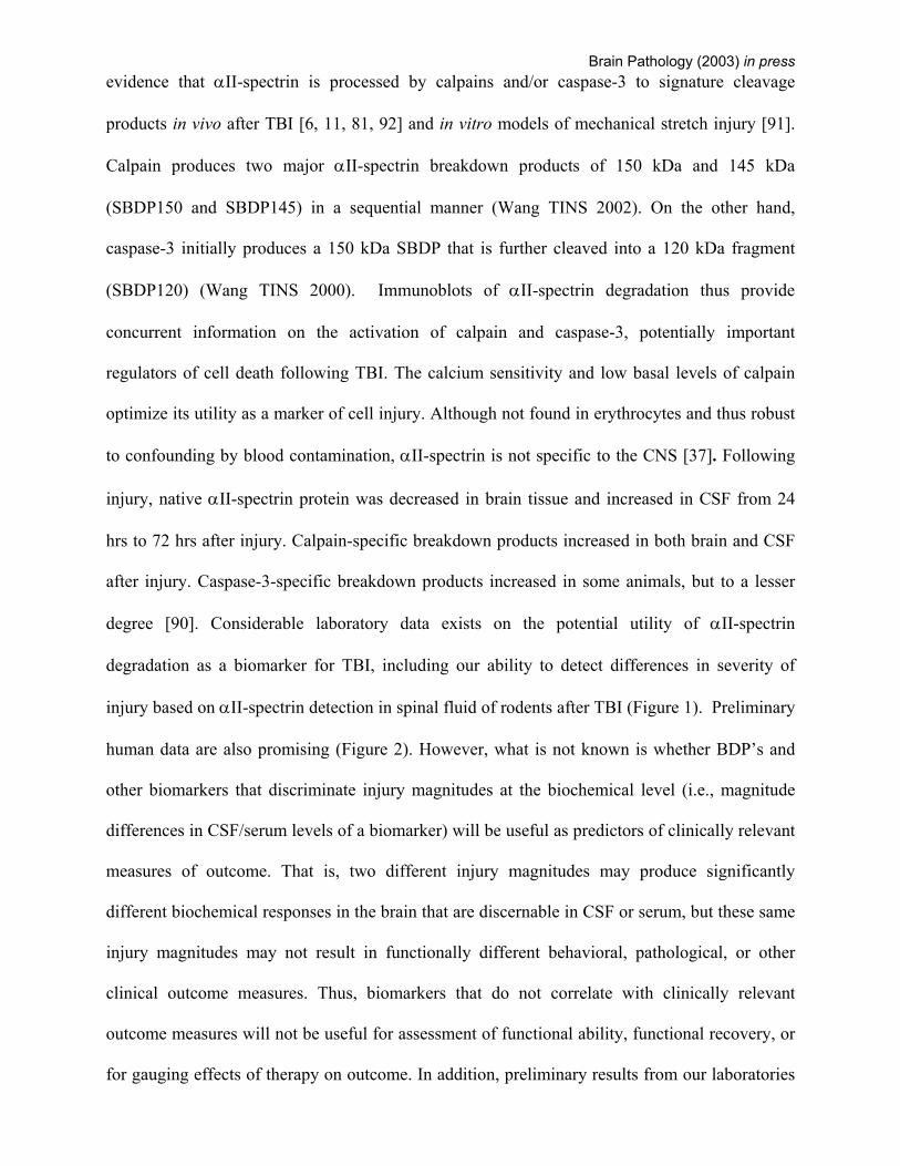
Figure 1. Accumulation of calpain-specific SBDPs is sensitive to injury magnitude
after controlled cortical impact. Animals were injured using a model of controlled cortical impact at two magnitudes of injury. Accumulation of brain cortex and spinal fluid levels of αII-SBDP were measured by densitometric analysis at 2 hours post-injury. The
1.6 mm injury magnitude produced the highest levels of αII-SBDP accumulation in cortex and CSF compared to 1.0 mm injury and to sham-injured controls. However,
statistical significance (p<0.05) between 1.6 mm and 1.0 mm injuries was only achieved at 2 hours but not at 6 or 24 hours post-injury (data not shown). This finding could indicate a critical window for discriminating injury magnitude using αII-SBDP as a
biomarker. CSF levels of αII-SBDP were greater in CSF than in cortex, particularly for the 1.6 mm injury. This most likely reflects a greater ratio of SBDP to total protein in
CSF relative to cortex and indicates that low abundance proteins may be more easily detected in CSF after brain injury. (n=6 per group).
Brain Pathology (2003) in press
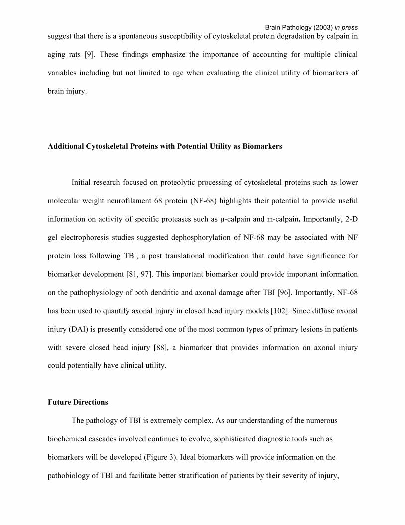
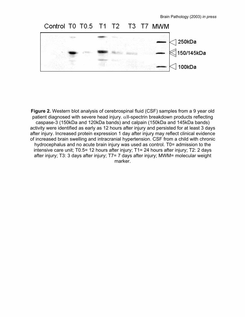
Figure 2. Western blot analysis of cerebrospinal fluid (CSF) samples from a 9 year old patient diagnosed with severe head injury. αII-spectrin breakdown products reflecting
caspase-3 (150kDa and 120kDa bands) and calpain (150kDa and 145kDa bands) activity were identified as early as 12 hours after injury and persisted for at least 3 days after injury. Increased protein expression 1 day after injury may reflect clinical evidence of increased brain swelling and intracranial hypertension. CSF from a child with chronic
hydrocephalus and no acute brain injury was used as control. T0= admission to the intensive care unit; T0.5= 12 hours after injury; T1= 24 hours after injury; T2: 2 days after injury; T3: 3 days after injury; T7= 7 days after injury; MWM= molecular weight
marker.
Brain Pathology (2003) in press
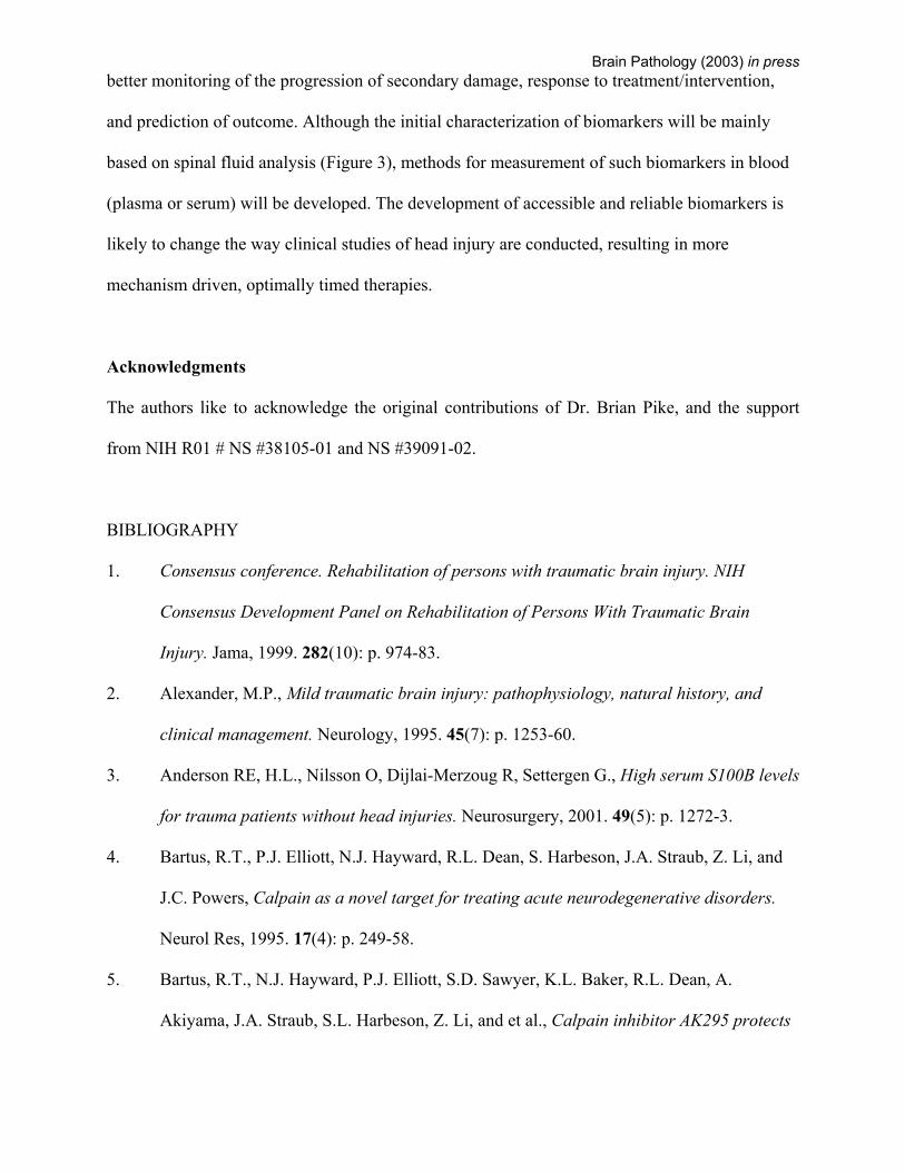
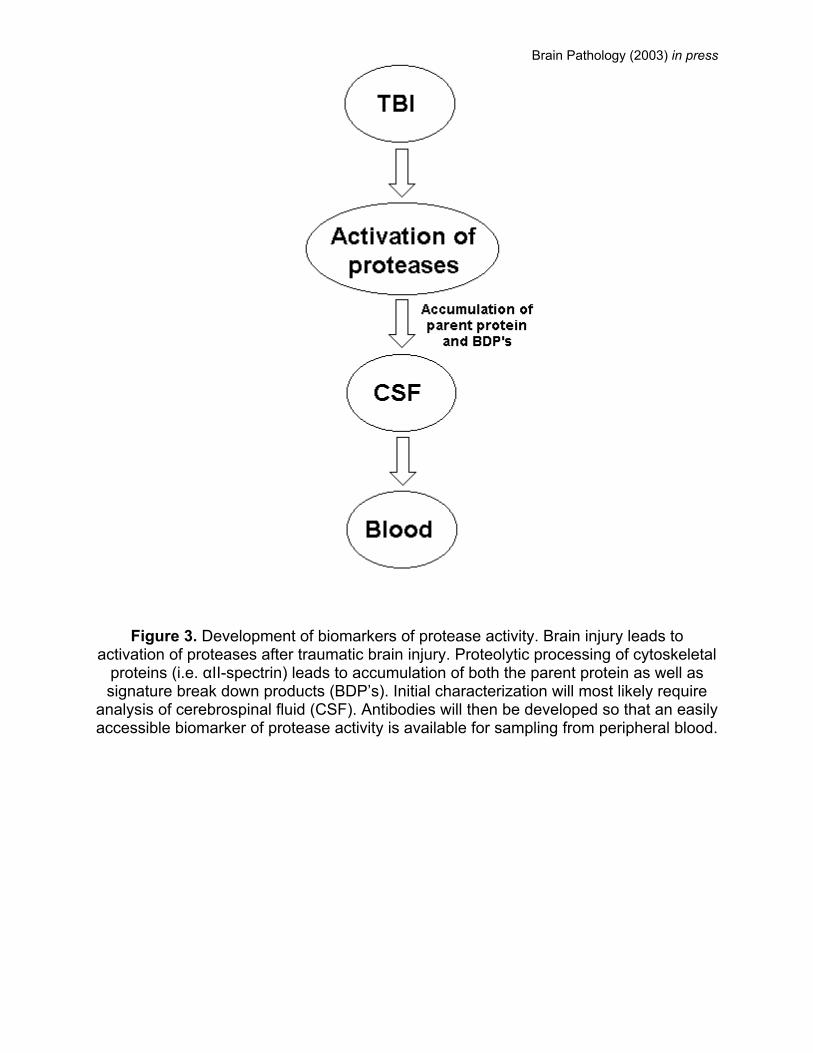
Figure 3. Development of biomarkers of protease activity. Brain injury leads to activation of proteases after traumatic brain injury. Proteolytic processing of cytoskeletal
proteins (i.e. αII-spectrin) leads to accumulation of both the parent protein as well as signature break down products (BDP’s). Initial characterization will most likely require
analysis of cerebrospinal fluid (CSF). Antibodies will then be developed so that an easily accessible biomarker of protease activity is available for sampling from peripheral blood.
Copyright © 2022 FDOKUMEN