Autoantibodies as diagnostic biomarkers for lung cancer
15
Yang et al. Cell Death Discovery (2019)5:126 https://doi.org/10.1038/s41420-019-0207-1 Cell Death Discovery REVIEW ARTICLE Open Access Autoantibodies as diagnostic biomarkers for lung cancer: A systematic review Bin Yang 1 , Xiaoyan Li 1 , Tianyi Ren 2 and Yiyu Yin 1 Abstract Lung cancer (LC) accounts for the largest number of tumor-related deaths worldwide. As the overall 5-year survival rate of LC is associated with its stages at detection, development of a cost-effective and noninvasive cancer screening method is necessary. We conducted a systematic review to evaluate the diagnostic values of single and panel tumor-associated autoantibodies (TAAbs) in patients with LC. This review included 52 articles with 64 single TAAbs and 19 with 20 panels of TAAbs. Enzyme-linked immunosorbent assays (ELISA) were the most common detection method. The sensitivities of single TAAbs for all stages of LC ranged from 3.1% to 92.9% (mean: 45.2%, median: 37.1%), specificities from 60.6% to 100% (mean: 88.1%, median: 94.9%), and AUCs from 0.416 to 0.990 (mean: 0.764, median: 0.785). The single TAAb with the most significant diagnostic value was the autoantibody against human epididymis secretory protein (HE4) with the maximum sensitivity 91% for NSCLC. The sensitivities of the panel of TAAbs ranged from 30% to 94.8% (mean: 76.7%, median: 82%), specificities from 73% to 100% (mean: 86.8%, median: 89.0%), and AUCs from 0.630 to 0.982 (mean: 0.821, median: 0.820), and the most significant AUC value in a panel (M13 Phage 908, 3148, 1011, 3052, 1000) was 0.982. The single TAAb with the most significant diagnostic calue for early stage LC, was the autoantibody against Wilms tumor protein 1 (WT1) with the maximum sensitivity of 90.3% for NSCLC and its sensitivity and specificity in a panel (T7 Phage 72, 91, 96, 252, 286, 290) were both above 90.0%. Single or TAAbs panels may be useful biomarkers for detecting LC patients at all stages or an early-stage in high-risk populations or health people, but the TAAbs panels showed higher detection performance than single TAAbs. The diagnostic value of the panel of six TAAbs, which is higher than the panel of seven TAAbs, may be used as potential biomarkers for the early detection of LC and can probably be used in combination with low-dose CT in the clinic. Facts ● LC is one of the most common types of cancer and accounts for the majority of tumor-related deaths globally. ● Patients diagnosed with LC at an early-stage have a higher 5-year survival rate. ● Low-dose spiral computed tomography (CT) is the most widely used diagnostic method in clinical practice, but its the high false positive rates and cost may prevent it from becoming a routine screening method. ● Current research and studies aim to identify the possibility of the molecular makers in body fluids, like TAAbs, for the early detection of LC. Open questions ● Currently some TAAbs have been studied. How are they related to diagnosis and how can the appropriate TAAbs for detecting early-stage LC be selected? ● It is still worth investigating whether the different distributions of TAAbs in the body are long lasting and have high concentration in blood. ● TAAb detection combined with CT can probably be used in clinic for detection of LC in the future. © The Author(s) 2019 Open Access This article is licensed under a Creative Commons Attribution 4.0 International License, which permits use, sharing, adaptation, distribution and reproduction in any medium or format, as long as you give appropriate credit to the original author(s) and the source, provide a link to the Creative Commons license, and indicate if changes were made. The images or other third party material in this article are included in the article’ s Creative Commons license, unless indicated otherwise in a credit line to the material. If material is not included in the article’s Creative Commons license and your intended use is not permitted by statutory regulation or exceeds the permitted use, you will need to obtain permission directly from the copyright holder. To view a copy of this license, visit http://creativecommons.org/licenses/by/4.0/. Correspondence: Yiyu Yin ([email protected]) 1 China–Japan Union Hospital of Jilin University, Changchun, China 2 National Institutes of Health (NIH)), Bethesda, USA Edited by N. Barlev Official journal of the Cell Death Differentiation Association 1234567890():,; 1234567890():,; 1234567890():,; 1234567890():,;
-
Upload
khangminh22 -
Category
Documents
-
view
1 -
download
0
Transcript of Autoantibodies as diagnostic biomarkers for lung cancer

Yang et al. Cell Death Discovery (2019) 5:126
https://doi.org/10.1038/s41420-019-0207-1 Cell Death Discovery
REV I EW ART ICLE Open Ac ce s s
Autoantibodies as diagnostic biomarkersfor lung cancer: A systematic reviewBin Yang1, Xiaoyan Li1, Tianyi Ren2 and Yiyu Yin 1
AbstractLung cancer (LC) accounts for the largest number of tumor-related deaths worldwide. As the overall 5-year survival rate ofLC is associated with its stages at detection, development of a cost-effective and noninvasive cancer screening method isnecessary. We conducted a systematic review to evaluate the diagnostic values of single and panel tumor-associatedautoantibodies (TAAbs) in patients with LC. This review included 52 articles with 64 single TAAbs and 19 with 20 panels ofTAAbs. Enzyme-linked immunosorbent assays (ELISA) were the most common detection method. The sensitivities of singleTAAbs for all stages of LC ranged from 3.1% to 92.9% (mean: 45.2%, median: 37.1%), specificities from 60.6% to 100% (mean:88.1%, median: 94.9%), and AUCs from 0.416 to 0.990 (mean: 0.764, median: 0.785). The single TAAb with the mostsignificant diagnostic value was the autoantibody against human epididymis secretory protein (HE4) with the maximumsensitivity 91% for NSCLC. The sensitivities of the panel of TAAbs ranged from 30% to 94.8% (mean: 76.7%, median: 82%),specificities from 73% to 100% (mean: 86.8%, median: 89.0%), and AUCs from 0.630 to 0.982 (mean: 0.821, median: 0.820),and the most significant AUC value in a panel (M13 Phage 908, 3148, 1011, 3052, 1000) was 0.982. The single TAAb with themost significant diagnostic calue for early stage LC, was the autoantibody against Wilms tumor protein 1 (WT1) with themaximum sensitivity of 90.3% for NSCLC and its sensitivity and specificity in a panel (T7 Phage 72, 91, 96, 252, 286, 290) wereboth above 90.0%. Single or TAAbs panels may be useful biomarkers for detecting LC patients at all stages or an early-stagein high-risk populations or health people, but the TAAbs panels showed higher detection performance than single TAAbs.The diagnostic value of the panel of six TAAbs, which is higher than the panel of seven TAAbs, may be used as potentialbiomarkers for the early detection of LC and can probably be used in combination with low-dose CT in the clinic.
Facts
● LC is one of the most common types of cancer andaccounts for the majority of tumor-related deathsglobally.
● Patients diagnosed with LC at an early-stage have ahigher 5-year survival rate.
● Low-dose spiral computed tomography (CT) is themost widely used diagnostic method in clinicalpractice, but its the high false positive rates and costmay prevent it from becoming a routine screeningmethod.
● Current research and studies aim to identify thepossibility of the molecular makers in body fluids,like TAAbs, for the early detection of LC.
Open questions
● Currently some TAAbs have been studied. How arethey related to diagnosis and how can theappropriate TAAbs for detecting early-stage LC beselected?
● It is still worth investigating whether the differentdistributions of TAAbs in the body are long lastingand have high concentration in blood.
● TAAb detection combined with CT can probably beused in clinic for detection of LC in the future.
© The Author(s) 2019OpenAccessThis article is licensedunder aCreativeCommonsAttribution 4.0 International License,whichpermits use, sharing, adaptation, distribution and reproductionin any medium or format, as long as you give appropriate credit to the original author(s) and the source, provide a link to the Creative Commons license, and indicate if
changesweremade. The images or other third partymaterial in this article are included in the article’s Creative Commons license, unless indicated otherwise in a credit line to thematerial. Ifmaterial is not included in the article’s Creative Commons license and your intended use is not permitted by statutory regulation or exceeds the permitted use, you will need to obtainpermission directly from the copyright holder. To view a copy of this license, visit http://creativecommons.org/licenses/by/4.0/.
Correspondence: Yiyu Yin ([email protected])1China–Japan Union Hospital of Jilin University, Changchun, China2National Institutes of Health (NIH)), Bethesda, USAEdited by N. Barlev
Official journal of the Cell Death Differentiation Association
1234
5678
90():,;
1234
5678
90():,;
1234567890():,;
1234
5678
90():,;
● TAAbs combined with other biomarkers like miRNAswill probably have improved diagnostic performance.
IntroductionLung cancer (LC) is one of the most common types of
cancer and accounts for the largest number of tumor-related deaths globally. There are an estimated 705,000cases and 569,000 deaths due to LC in China, and 214,000cases and 168,000 deaths in US in 20121,2. The overall 5-year survival rate of LC is associated with its stages atdoagnosis, which is <20% as the majority of cases arediagnosed at late stages, In contrast, tumors diagnosed atstage IA have a 5-year survival rate of ~70%3. Therefore,early detection and immediate treatment can reduce themortality of LC significantly. However, the detection anddiagnosis of early stage LC is still a challenge, because ofthe lack of effective screening methods. It has been proventhat sputum exfoliative cytologic examination cannoteffectively reduce LC mortality4. In contrast, low-dosespiral computed tomography (CT) is highly sensitive atthe early detection of small lung nodules and has led to a20% reduction in LC mortality5, but its high false positiverates and cost may prevent it from becoming a routinescreening method4,6.Thus, it is necessary to develop more cost-effective and
noninvasive cancer screening methods. Current researchand studies aim to identify molecular makers, that couldbe detected in body fluids for the early detection of LC.Current diagnostic methods have concentrated on tumor-associated antigens (TAAs) markers, such as the carbo-hydrate antigen (CA) 125, CA19-9, carcino-embryonicantigen (CEA) and alpha fetal protein (AFP), which areeffective at diagnosing LC at advanced stages7, but have alow sensitivity and specificity for early stage LC. However,detection of tumor-associated autoantibodies (TAAbs),which are produced by cancer cells against TAAs inblood, may become a potential cancer screening method8.TAAbs are more stable in peripheral blood than TAAs,and have better sensitivity and specificity. Clinical trialsevaluating the diagnostic value of TAAbs have shownthem to be potential diagnostic method as detective bio-markers for LC, and a series of candidates and multiplexTAAbs have been identified and analyzed.Hence, we provided a systematic and comprehensive
review and summary of the published articles that inves-tigated TAAbs for LC detection. We reported on researchresults and indicators for assessing the diagnostic per-formance of TAAbs in the patients’ blood, and also putforward new research problems and new possibilities forfuture studies9–12.
Search strategyOur review was conducted according to a predefined
protocol in accordance with the PRISMA statement13. A
systematic literature search was performed to identifystudies that assessed TAAbs in relation to LC. We sear-ched Pubmed and ISI Web of Science for articles thatwere published from 1 January 1990 to 31 December2018. The following combinations of search keywordswere used to retrieve articles: ((lung OR pulmonary) AND(cancer OR carcinoma OR neoplasm OR tumor ORadenocarcinoma OR squamous carcinoma OR malig-nancy) AND (autoantibody OR antibody) AND (detectionOR diagnosis OR biomarker OR marker) AND (serumOR blood OR plasma))in all fields. Duplicated articleswere removed.
Eligibility criteriaWe initially read the titles and abstracts to screen the
potential eligible articles, with the following exclusioncriteria (Fig. 1): (1) non-English articles, (2) non-originalarticles (reviews, meta-analyses, or proceedings), (3)non-LC studies, (4) nonhuman studies, (5) not related toTAABs, (6) not based on serum or plasma samples, and(7) non-full-text articles. The second round of the pre-liminary screening involved reading the full-text of thearticles, and studies with the following were excluded:(1) diseased controls used, (2) not reporting critical dataor no sensitivity, specificity, or area under the curve(AUC).
Data extraction and statistical analysisTwo reviewers (Yiyu Yin and Xiaoyan Li) indepen-
dently read and extracted all the eligible articles above.Any disagreements and arguments were discussed andresolved among the authors. We extracted the firstauthor, publication year, country, TAAs associated withthe autoantibodies, study method, basic populationcharacteristics (including size, age, sex, histological type,and tumor stage), specimen type, targeted TAAbsmarkers, and evaluation indicators (sensitivity, specifi-city, AUC, and p-value). Individual TAAbs with a p-value > 0.5 were eliminated. We use Statistical R (ver-sion 3.5.1) to calculate the mean or median ages if thesestatistics were not presented but the raw data wereavailable.
Quality assessmentThe quality of each eligible article was assessed by two
independent researchers according to quality assessmentof diagnostic accuracy studies (QUADAS-2, www.bris.ac.uk/quadas), using Review Manager (version 5.3).QUADAS-2 contains four domains on bias and applic-ability of the the research question: (1) patient selection,(2) index test(s), (3) reference standard, and (4) flow andtiming, and each item was assessed as “yes” or “no” or“unclear”. Applicability concerns were assessed using thefirst three domains as well.
Yang et al. Cell Death Discovery (2019) 5:126 Page 2 of 15
Official journal of the Cell Death Differentiation Association

Study identification and literature searchA flow process diagram of the study search process is
shown in Fig. 1. A total of 8424 potentially relevantpublications were identified by the initial independentsearch using the search terms mentioned above, 5498from PubMed and 2926 from Web of Science (Fig. 1).1251 duplicate articles were removed. The titles andabstracts of 7173 articles were screened and a total of7079 were excluded based on the exclusion criteriadescribed above. Of the remaining 94 full-text articles, 10were excluded because a disease control was used14–23, 9were excluded because they did not have satisfied out-comes24–32, and 8 were excluded because of their smallsample size (n < 10)33–40, Ultimately, 67 articles were
included in this systematic review evaluating the diag-nostic performance of TAAbs in serum or plasma for LCdetection (Tables 1 and 2).
Study quality and characteristicsStudy quality was evaluated by two reviewers (Yiyu Yin
and Xiaoyan Li) independently. Any academic con-troversy was resolved by the following discussion amongthe researchers. All the studies in our research were ofhigh quality with no risk of bias or the concern regardingtheir applicability, however, there were still unclear risksof bias and unclear applicability in patient selection andindex tests in several studies. The statistics of theQUADAS-2 results of the 67 studies are shown in Table 3.
Incl
usi
on
Non-full-text articles:17
Ide
nti
�ica
tio
nS
cre
en
ing
Eli
gib
ilit
y
Total:8424Pubmed:5498
Web of Science:2926
Duplicates:1251
Titles and Abstracts reviewed:7173
Non-English articles:29
Non-original articles:343
Non-LC studies:4014
Non-human studies:1036
Non-relative toTAABs:1620
Not based on serum orplasma samples:20
Full-text reviewed:94
Total:67
No sensitivity, specificity or AUC value reported:9
Small size studies:8
Diseased controls used:10
Fig. 1 Flow process diagram showing the overview of the literature (From January 1st 1990 to December 31st 2018)
Yang et al. Cell Death Discovery (2019) 5:126 Page 3 of 15
Official journal of the Cell Death Differentiation Association
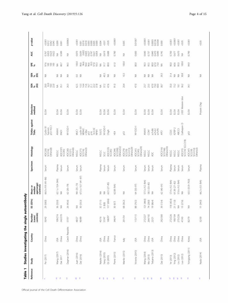
Table
1Stud
iesinve
stigatingthesingle
autoan
tibod
y
Referenc
eStud
yCou
ntry
Num
ber
(cases/
controls)
ES(ES%
)Mea
nor
med
ian
age(rang
e)(con
trols)
Specim
enHistology
TAAbsag
ainst
TAAs
Detection
metho
dSE
N% (AS)
SEN
% (ES)
SPE
%AUC
p-value
9Pei(2017)
China
50/42
29(58.0)
66.0±9.9(45–86)
Serum
ADC(26)
SCC(16)
Others(8)
Cyclin
B1Survivin
p53HCC1
ELISA
20.0
32.0
18.0
22.0
NA
97.6
100
100
100
0.767
0.653
0.623
0.622
<0.001
0.012
0.042
0.045
45Wang(2017)
USA
109/216
36(33.0)
62.1±10.4(NA)
Plasma
NSC
LCANXA
1ELISA
NA
NA
NA
NA
0.009
79Dai2017
China
242/270
NA
NA
Serum
ADC(197)
SCC(45)
ENO1
ELISA
35.1
NA
80.7
0.589
0.001
63Dagmar
(2016)
Czech
Repu
blic
57/57
26(45.6)
62(30–79)
Serum
ADC(30)
SCC(21)
Others(6)
NY-ESO-1
ELISA
26.3
NA
96.5
NA
0.00063
80Juan
(2016)
China
48/27
NA
NA(35–73)
Serum
NSC
LCMUC1
ELISA
62.5
0.0
100.0
0.870
<0.001
10Dai(2016)
China
90/89
30(33.3)
67.5±10.7(41–87)
Serum
ADC(71)
SCC(16)
Others(3)
cyclin
B1MDM2
c-Myc
p53
p16
14-3-3ζ
NPM
1
ELISA
13.3
14.4
15.6
16.7
21.1
22.2
37.8
NA
96.6
96.6
94.4
96.6
95.5
97.8
93.3
0.639
0.602
0.603
0.627
0.747
0.621
0.854
0.001
0.018
0.018
0.003
0.000
0.005
0.000
46Natalie
(2016)
USA
45/16
32(71.1)
NA
Plasma
NSC
LCSU
LF2
ELISA
NA
NA
NA
NA
0.004
64Yang
(2015)
China
57/47
5(8.8)
NA
Serum
SCLC
NY-ESO-1
ELISA
37.2
45.5
91.7
0.619
<0.01
81Qi(2015)
China
168/97
117(69.6)
62.5(27–85)
Serum
ADC(123)
SCC(45)
Chg
AELISA
47.6
NA
80.0
0.688
<0.05
82Pierre
(2015)
France
346/41
30(8.7)
62.08(NA)
Serum
ADC(94)
SCC(200)
Others(52)
HE4
ELISA
91.0
NA
61.0
0.780
<0.0001
54Manlio
(2015)
Italy
201/54
68(36.2)
NA
Serum
ADC(79)
SCC(70)
SCLC
(13)
Others(39
p53
ELISA
20.4
10.3
100.0
NA
0.005
65Victoria(2015)
USA
115/115
88(76.5)
64(50–97)
Serum
ADC(41)
SCC(45)
Others(29)
NY-ESO-1
ELISA
47.0
NA
80.0
0.600
0.01007
83Wang(2014)
China
272/227
121(44.6)
57.5±9.2(NA)
Serum
NSC
LCANXA
1ELISA
23.7
NA
90.3
0.640
<0.0001
84Ma(2013)
China
264/192
74(28.0)
58.5(33–85)
Serum
NSC
LCCCNY
ELISA
23.5
NA
95.5
0.737
<0.001
85Tetyana(2013)
USA
22/21
19(86.4)
NA
Serum
ADC
scFvB6
scFvG1
scFvP6
ELISA
67.0
73.0
60.0
NA
80.0
67.0
73.0
0.840
0.470
0.690
0.0003
0.0136
0.0304
86Dai(2013)
China
292/300
39(13.4)
62(40–91)
Serum
ADC(116)
SCC(166)
Others(10)
APE1
ELISA
38.7
39.3
NA
0.745
0.000
47Ye
(2013)
China
272/226
118(43.4)
57.4±9.2(NA)
Plasma
NSC
LCCD25
ELISA
35.0
31.4
90.0
0.700
<0.001
69Ying
(2012)
China
190/104
21(11.0)
61.38(27–82)
Serum
NSC
LCIGFBP-2
ELISA
73.2
NA
60.6
0.677
<0.0001
87Liu(2012)
China
275/226
NA
57.6±9.2(NA)
Serum
NSC
LCABC
C3ELISA
18.1
NA
95.0
0.670
<0.001
50Luo(2012)
China
47/43
13(27.6)
NA
Serum
ADC(15)
SCC(14)
SCLC
(18)
Cathe
psin
D2-DEWestern
blot
36.2
30.8
100.0
NA
<0.05
55Yo
ngjung
(2011)
Korea
82/79
NA
63.5(55.9–70.0)
Serum
ADC(47)
SCC(18)
SCLC
(14)
Others(3)
p53
ELISA
34.1
NA
94.9
0.790
<0.001
41Nada(2014)
USA
32/30
11(34.0)
66.2±0.5(NA)
Plasma
ProteinChip
NA
<0.05
Yang et al. Cell Death Discovery (2019) 5:126 Page 4 of 15
Official journal of the Cell Death Differentiation Association
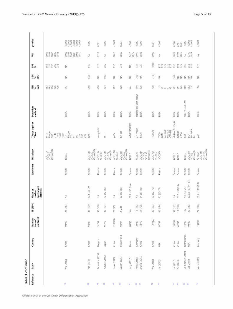
Table
1continue
d
Referenc
eStud
yCou
ntry
Num
ber
(cases/
controls)
ES(ES%
)Mea
nor
med
ian
age(rang
e)(con
trols)
Specim
enHistology
TAAbsag
ainst
TAAs
Detection
metho
dSE
N% (AS)
SEN
% (ES)
SPE
%AUC
p-value
ADC(10)
SCC(11)
Others(11)
M13
Phage
908
3148
1011
3052
1000
84.3
84.3
90.6
90.6
90.6
66.6
73.3
63.3
70.0
73.3
0.945
0.893
0.866
0.849
0.848
42Wu(2010)
China
90/90
21(23.3)
NA
Serum
NSC
LCT7 Ph
age
72 91 96 252
286
290
ELISA
NA
NA
NA
0.905
0.897
0.908
0.887
0.908
0.810
<0.001
<0.001
<0.001
<0.001
<0.001
<0.001
88Yao(2010)
China
93/87
38(40.9)
60.3(33–79)
Serum
ADC(53)
SCC(23)
Others(17)
DKK1
ELISA
62.0
65.8
84.0
NA
<0.05
48Rado
stina(2010)
Bulgaria
51/52
30(58.8)
NA
Plasma
ADC(15)
SCC(36)
α-crystallin
ELISA
62.0
NA
72.0
0.712
0.001
68Yu
suke
(2009)
Japan
91/70
45(49.4)
70(42–85)
Serum
ADC(54)
SCC(29)
Others(8)
WT1
ELISA
26.4
90.3
90.2
NA
<0.05
49Huan(2018)
China
211/200
20(9.5)
NA
Plasma
ADC(124)
SCC(87)
p16a
ELISA
32.7
NA
95.0
0.818
<0.001
89Maxim
(2017)
Switzerland
93/94
2(2.1)
59(19–86)
Serum
ADC(42)
SCC(22)
Others(29)
BARD
1ELISA
80.0
NA
77.5
0.860
0.003
90Jung
(2017)
Korea
80/80
NA
68.5±9.3(NA)
Serum
NSC
LCAIMP2-DXIMP2
ELISA
NA
NA
NA
0.416
0.579
<0.05
66Petra(2008)
Germany
39/40
18(46.2)
NA
Serum
SCC(39)
27Ph
age
serologicalspo
tassays
92.9
79.0
93.1
0.978
<0.05
91Zh
ang(2017)
China
72/70
51(70.8)
64(37–82)
Serum
ADC(38)
SCC(24)
SCLC
(4)
Others(6)
ENO1
ELISA
80.6
NA
72.7
0.806
<0.05
52Wu(2018)
China
127/127
39(30.7)
57(32–76)
Serum
ADC(70)
SCC(57)
TOPO
48ELISA
76.0
71.8
100.0
0.990
0.001
43Jie
(2015)
USA
97/87
46(47.4)
70(62–77)
Plasma
ADC(97)
TTC14
BRAF
ACTL6B
MORC
2CTA
G1B
ELISA
11.3
5.2
3.1
4.1
9.3
NA
97.7
97.7
97.7
97.7
97.7
NA
<0.05
92Lei(2017)
China
206/99
32(15.5)
NA
Serum
NSC
LCdickkopf-1
PepB
ELISA
58.1
76.9
85.3
0.821
0.008
93Pei(2016)
China
62/43
13(21.0)
66.0±9.9(NA)
Serum
NSC
LCMDM2
c-Myc
ELISA
37.1
35.5
NA
NA
97.7
97.7
0.777
0.815
0.001
0.001
53Dom
inique
(2014)
Nethe
rland
s44/49
NA
NA(50–75)
Serum
NSC
LCFab
SDS-PA
GE,LC
MS
84.0
NA
90.0
NA
<0.05
94Dai(2017)
USA
90/89
30(33.3)
67.5±10.7(41-87)
Serum
ADC(81)
SCC(6)
Others(3)
ECH1
HNRN
PAB1
ELISA
62.2
72.2
NA
NA
95.5
95.5
0.799
0.874
<0.001
<0.001
56Mack(2000)
Germany
134/46
29(21.6)
61.9±10.9(NA)
Serum
SCC(44)
ADC(44)
SCLC
(35)
Others(11)
p53
ELISA
12.6
NA
97.8
NA
<0.001
Yang et al. Cell Death Discovery (2019) 5:126 Page 5 of 15
Official journal of the Cell Death Differentiation Association
Table
1continue
d
Referenc
eStud
yCou
ntry
Num
ber
(cases/
controls)
ES(ES%
)Mea
nor
med
ian
age(rang
e)(con
trols)
Specim
enHistology
TAAbsag
ainst
TAAs
Detection
metho
dSE
N% (AS)
SEN
% (ES)
SPE
%AUC
p-value
57Jerzy(1998)
Poland
84/20
37(44.0)
NA
Serum
SCC(43)
ADC(27)
LCC(14)
p53
IHC
22.6
40.4
NA
NA
0/002
58Toshihiko(1998)
Japan
62/41
33(53.2)
65.7(48–85)
Serum
ADC(33)
SCC(21)
LCC(8)
p53
ELISA
40.3
48.5
NA
NA
0.0025
99Mikio
(2001)
Japan
50/130
NA
NA
Serum
ADC(32)
SCC(47)
SCLC
(4)
LCC(6)
Others(27)
HSP40
ELISA
NA
NA
NA
NA
<0.001
59Jassem
(2001)
Poland
96/41
60(62.5)
58(35–86)
Serum
SCLC
p53
ELISA
27.0
25.0
97.5
NA
<0.001
60Cioffi
(2001)
Italy
109/80
21(19.3)
NA
Serum
NSC
LC(57)
SCLC
(52)
p53
ELISA
32.1
38.1
100.0
NA
NA
61Mon
ica(2002)
Italy
78/106
2(3.6)
62.4±9.3(NA)
Serum
ADC(18)
SCC(19)
SCLC
(3)
Others(8)
p53
ELISA
12.8
098.1
NA
0.01
62Suleep
orn(2003)
Thailand
133/200
30(22.6)
NA
Serum
ADC(59)
SCC(29)
LCC(4)
SCLC
(13)
p53
ELISA
18.8
6.7
97.5
NA
<0.001
100
Tsuji(1997)
Japan
67/60
NA
NA
Serum
ADC(51)
SCC(9)
SCLC
(5)
LCC(2)
TRD-L1
ELISA
55.2
NA
97.7
NA
<0.001
102
Den
nis(2003)
USA
49/40
0(0)
NA
Serum
ADC(14)
SCC(17)
LCC(1)
Others(17)
HSP70
ELISA
74.7
NA
73.0
0.731
<0.001
103
Zhon
g(2004)
USA
49/40
12(30.0)
NA
Serum
ADC(12)
SCC(19)
Others(18)
HSP70
ELISA
74.0
NA
73.0
0.731
0.0009
104
Zhon
g(2006)
USA
23/23
23(100)
65.1(51–79)
Serum
ADC(7)
SCC(8)Others(8)
L1919
L1896
G2004
G1954
G1689
ELISA
82.6
87.0
82.6
82.6
82.6
82.6
87.0
82.6
82.6
82.6
78.3
87.0
65.2
87.0
65.2
0.850
0.950
0.800
0.740
0.820
<0.001<0.001
<0.001
<0.001
<0.001
105
Daniel(2008)
USA
105/102
88(83.0)
66.4(43–85)
Serum
ADC
AZG
P1ELISA
40.0
NA
NA
NA
<0.05
51Myrna
(1997)
Germany
170/50
70(41.0)
61.4(NA)
Serum
SCLC
p53
Western
blot
16.0
NA
100
NA
<0.05
ASall-stage
,ESearly
-stage
(stage
Iand
IIinclud
ed),Co
ntrolsbe
nign
diseases
andno
rmal
healthydo
nors,A
UCarea
unde
rthecurve,SENsensitivity,SPE
specificity,ELISA
enzyme-lin
kedim
mun
oassay,W
Bwestern
blottin
g,ADCad
enocarcino
ma,
SCCsqua
mou
scarcinom
a,SC
LCsm
allcelllung
cancer,N
SCLC
non-sm
allcelllung
cancer,N
Ano
tavailable
Yang et al. Cell Death Discovery (2019) 5:126 Page 6 of 15
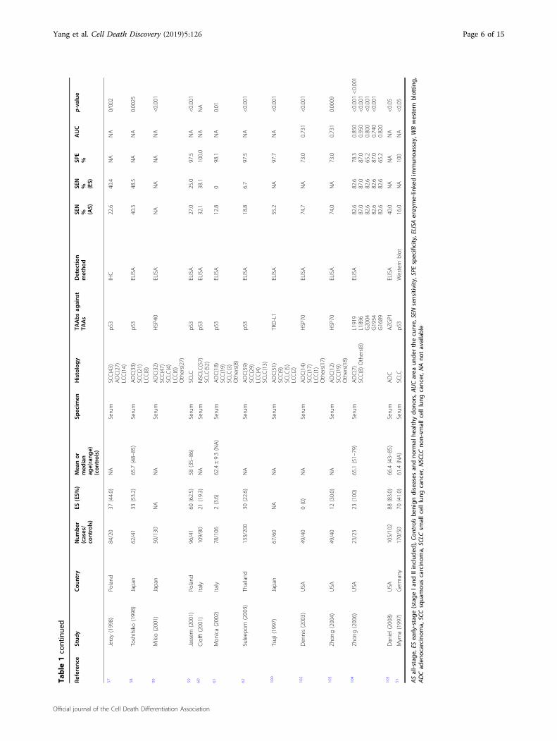
Official journal of the Cell Death Differentiation Association

Table
2Stud
iesinve
stigatingthepan
elau
toan
tibod
ies
Referenc
eStud
yCou
ntry
Num
ber
(cases/
controls)
ES(ES%
)Mea
nor
med
ian
age(rang
e)(con
trols)
Specim
enHistology
TAAbs
againstTA
As
Detection
metho
dSE
N% (AS)
SEN
% (ES)
SPE%
AUC
p-value
9Pei(2017)
China
60/31
NA
NA
Serum
NA
Pane
l1ELISA
65.0
NA
100
0.908
<0.001
89Maxim
(2017)
Switzerland
93/94
2(2.1)
65(28–86)
Serum
ADC(42)
SCC(22)
Others(29)
Pane
l2ELISA
80.0
NA
78.0
0.961
NA
10Dai(2016)
China
90/89
30(33.3)
67.5±10.7(41–87)
Serum
ADC(71)
SCC(16)
Others(3)
Pane
l3ELISA
68.9
73.3
79.5
0.863
<0.05
85Tetyana(2013)
USA
22/21
19(86.4)
NA
Serum
ADC
Pane
l4ELISA
80.0
NA
87.0
0.880
NA
71Erin
(2010)
USA
(10/10)
9(90.0)
72(65–86)
Serum
ADC
Pane
l5ELISA
94.8
NA
91.1
0.964
<0.05
65Victoria(2015)
USA
(75/75)
23(31.0)
68.5(50–99)
Serum
ADC
SCC
Others
Pane
l6Luminex
MAP
77.0
71.2
80.0
0.810
<0.0001
72Bo
yle(2010)
UK
(145/146)
81(55.9)
66.0(41–87)
Serum
ADC(29)
SCC(21)
SCLC
(22)
Others(73)
Pane
l7ELISA
36.0
NA
91.0
0.710
NA
72Bo
yle(2010)
UK
(241/88)
0(0)
63.0(28–87)
Serum
ADC(56)
SCC(42)
SCLC
(70)
Others(73)
Pane
l8ELISA
39.0
0.0
89.0
0.630
NA
72Bo
yle(2010)
UK
(269/NA)
86(32.0)
65.0(38–87)
Serum
ADC(67)
SCC(88)
SCLC
(73)
Others(27)
Pane
l8ELISA
37.0
NA
90.0
0.640
NA
41Nada(2010)
USA
(32/30)
11(34.0)
66.2±10.5(NA)
Plasma
ADC(10)
SCC(11)
Others(11)
Pane
l9ProteinChip
90.0
NA
73.0
0.982
<0.05
42Wu(2010)
China
(90/90)
21(23.0)
NA
Serum
NSC
LCPane
l10
ELISA
92.2
92.2
92.2
0.956
<0.001
43Wang(2015)
USA
(97/87)
46(47.4)
70.0(62–77)
Plasma
ADC(97)
Pane
l13
ELISA
30.0
NA
88.0
NA
<0.05
70Ren(2018)
China
(818/1190)
213(26.0)
54.0
(18–91)
Serum
ADC(429)
SCC(277)
SCLC
(91)
Others(21)
Pane
l11
ELISA
61.0
62.0
90.0
0.781
<0.05
95Jia
(2014)
China
(48/50)
NA
59.7±8.7(39–79)
Serum
NCSLC
Pane
l12
Luminex
MAP
NA
NA
NA
0.820
<0.05
96Qiang
(2018)
China
(352/129)
133(37.8)
60.51±
9.41
(NA)
Serum
ADC(243)
SCC(42)
SCLC
(47)
Pane
l14
ELISA
56.5
56.4
91.6
NA
<0.001
97Caroline(2010)
UK
(243/247)
90(37%
)66
±9.6(33–87)
Serum
SCLC
(243)
Pane
l15
ELISA
55.0
53.0
90.0
0.761
<0.001
98Qiu
(2008)
USA
(85/85)
NA
NA
Serum
NSC
LCPane
l16
proteinmicroarrays
51.0
NA
82.0
0.730
<0.05
101
Mitche
ll(1990)
USA
(52/52)
25.0%
64.7±9(NA)
Serum
ADC(12)
SCC(22)
SCLC
(7)
Others(11)
Pane
l17
ELISA
73.0
NA
NA
NA
<0.06
Yang et al. Cell Death Discovery (2019) 5:126 Page 7 of 15
Official journal of the Cell Death Differentiation Association

Table
2continue
d
Referenc
eStud
yCou
ntry
Num
ber
(cases/
controls)
ES(ES%
)Mea
nor
med
ian
age(rang
e)(con
trols)
Specim
enHistology
TAAbs
againstTA
As
Detection
metho
dSE
N% (AS)
SEN
% (ES)
SPE%
AUC
p-value
103
Zhon
g(2004)
USA
49/40
12(30.0)
NA
Serum
ADC(12)
SCC(19)
Others(18)
Pane
l18
ELISA
82.0
NA
83.0
0.837
0.0002
44Chapm
an(2007)
Germany
82/50
9(11.0)
63(36–83)
Plasma
ADC(35)
SCC(25)
Others(22)
Pane
l19
ELISA
76.0
NA
92.0
NA
<0.05
98Qiu
(2008)
USA
85/85
NA
NA
Serum
NSC
LCPane
l20
ELISA
51.0
NA
82.0
0.730
0.017
ASall-stage
,ESearly
-stage
(stage
Iand
IIinclud
ed),Co
ntrolsbe
nign
diseases
andno
rmal
healthydo
nors,A
UCarea
unde
rthecurve,SENsensitivity,SPE
specificity,ELISA
enzyme-lin
kedim
mun
oassay,W
Bwestern
blottin
g,ADCad
enocarcino
ma,
SCCsqua
mou
scarcinom
a,SC
LCsm
allcelllung
cancer,N
SCLC
non-sm
allcelllung
cancer,N
Ano
tavailable
Pane
l1(cyclin
B1,S
urvivin,
p53,
HCCI)
Pane
l2(p37
,p13
,p10
,p17
,p12
,p14
,p15
,p16
,p22
andp1
)Pa
nel3(cyclin
B1,M
DM2,
c-Myc,p
53,p
16,1
4-3-3ζ,N
PM1)
Pane
l4(scFVB
6,3E,G
1,J4,P
6,J1)
Pane
l5(IM
PDH,p
hospho
glyceratemutase,
ubiquilin
,Ann
exin
I,Ann
exin
II,HSP
70-9B)
Pane
l6(CEA
,CA-125
,and
CYF
RA21
–1an
tigen
s,an
ti-NY-ESO-1)
Pane
l7(p53
,NY-ESO-1,C
AGE,
GBU
4-5)
Pane
l8(p53
,NY-ESO-1,C
AGE,
GBU
4-5,
Ann
exin
1,SO
X2)
Pane
l9(M
13Ph
age90
8,31
48,1
011,
3052
,100
0)Pa
nel10
(Pha
gepe
ptide72
,91,
96,2
52,2
86,2
90)
Pane
l11
(p53
,GAGE7,P
GP9
.5,C
AGE,
MAGEA
1,SO
X2,G
BU4-5)
Pane
l12
(p62
,BIRC,L
ivin-1,p
53,P
RDX,
NYE
SO-1,u
biqu
ilin)
Pane
l13
(TTC
14,B
RAF,
ACTL6B
,MORC
2,CTA
G1B
)Pa
nel14
(p53
,PGP9
.5,S
OX2
,GAGE7,G
BU4-5,
CAGE,
MAGEA
1)Pa
nel15
(p53
,CAGE,
NY-ESO-1,G
BU4-5,
Ann
exin
I,SO
X2,H
u-D)
Pane
l16
(Ann
exin
I,14
-3-3
Theta,
LAMR1
)Pa
nel17
(MAb5E8,
IF10
,and
5C7)
Pane
l18
(BMI-1
,p13
0,GAGE,
HSP
70,a
ndHSP
90)
Pane
l19
(p53
,c-m
yc,H
ER2an
dCAGE)
Pane
l20
(ann
exin
I,14
-3-3
theta,
andLA
MR1
)
Yang et al. Cell Death Discovery (2019) 5:126 Page 8 of 15
Official journal of the Cell Death Differentiation Association

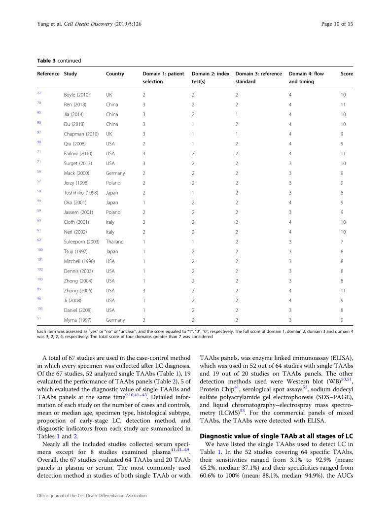
Table 3 Quality assessment of QUADAS-2
Reference Study Country Domain 1: patient
selection
Domain 2: index
test(s)
Domain 3: reference
standard
Domain 4: flow
and timing
Score
9 Li (2017) China 2 2 1 4 9
10 Dai (2016) China 3 2 2 4 11
44 Chapman (2007) Germany 3 2 2 3 10
45 Wang (2017) USA 2 2 1 3 8
79 Dai (2017) China 2 2 1 4 9
63 Mysikova (2016) Czech Republic 3 1 2 3 9
80 Wang (2016) China 3 1 1 4 9
46 Lui (2016) USA 2 2 1 4 9
64 Yang (2015) China 2 2 2 4 10
81 Qi (2015) China 3 2 1 3 9
82 Lamy (2015) France 3 1 1 3 8
54 Mattioni (2015) Italy 2 2 1 4 9
65 Doseeva (2015) USA 2 2 2 4 10
83 Wang (2014) China 2 1 2 3 8
84 Ma (2013) China 3 2 1 4 10
85 Pedchenko (2013) USA 2 2 2 3 9
86 Dai (2013) China 3 2 2 4 11
47 Ye (2013) China 3 2 2 4 11
69 Zhang (2012) China 3 2 1 4 10
87 Liu (2012) China 3 1 2 4 10
50 Luo (2012) China 2 2 1 4 9
55 Park (2011) Korea 3 2 2 4 11
41 Khattar (2010) USA 3 2 2 3 10
42 Wu (2010) China 2 2 2 4 10
88 Yao (2010) China 3 1 2 4 10
48 Cherneva (2010) Bulgaria 2 1 2 4 10
68 Oji (2009) Japan 2 1 1 4 8
49 Zhao (2018) China 2 1 1 3 7
89 Pilyugin (2017) Switzerland 3 1 2 3 9
90 Jung (2017) Korea 2 2 1 4 9
66 Leidinger (2008) Germany 2 2 2 4 10
91 Zhang (2017) China 3 1 2 3 9
52 Wu (2018) China 2 2 2 4 10
43 Wang (2015) USA 3 2 2 3 10
92 Shen (2017) China 2 2 2 4 10
93 Li (2016) China 3 1 2 4 10
53 Costa (2014) Netherlands 2 1 2 4 9
94 Dai (2017) USA 3 1 1 4 9
Yang et al. Cell Death Discovery (2019) 5:126 Page 9 of 15
Official journal of the Cell Death Differentiation Association
A total of 67 studies are used in the case-control methodin which every specimen was collected after LC diagnosis.Of the 67 studies, 52 analyzed single TAAbs (Table 1), 19evaluated the performance of TAAbs panels (Table 2), 5 ofwhich evaluated the diagnostic value of single TAABs andTAAbs panels at the same time9,10,41–43. Detailed infor-mation of each study on the number of cases and controls,mean or median age, specimen type, histological subtype,proportion of early-stage LC, detection method, anddiagnostic indicators from each study are summarized inTables 1 and 2.Nearly all the included studies collected serum speci-
mens except for 8 studies examined plasma41,43–49.Overall, the 67 studies evaluated 64 TAAbs and 20 TAAbpanels in plasma or serum. The most commonly useddetection method in studies of both single TAAb or with
TAAbs panels, was enzyme linked immunoassay (ELISA),which was used in 52 out of 64 studies with single TAAbsand 19 out of 20 studies on TAAbs panels. The otherdetection methods used were Western blot (WB)50,51,Protein Chip41, serological spot assays52, sodium dodecylsulfate polyacrylamide gel electrophoresis (SDS–PAGE),and liquid chromatography–electrospray mass spectro-metry (LCMS)53. For the commercial panels of mixedTAAbs, the TAAbs were detected with ELISA.
Diagnostic value of single TAAb at all stages of LCWe have listed the single TAAbs used to detect LC in
Table 1. In the 52 studies covering 64 specific TAAbs,their sensitivities ranged from 3.1% to 92.9% (mean:45.2%, median: 37.1%) and their specificities ranged from60.6% to 100% (mean: 88.1%, median: 94.9%), the AUCs
Table 3 continued
Reference Study Country Domain 1: patient
selection
Domain 2: index
test(s)
Domain 3: reference
standard
Domain 4: flow
and timing
Score
72 Boyle (2010) UK 2 2 2 4 10
70 Ren (2018) China 3 2 2 4 11
95 Jia (2014) China 3 2 1 4 10
96 Du (2018) China 3 1 2 4 10
97 Chapman (2010) UK 3 1 1 4 9
98 Qiu (2008) USA 2 1 2 4 9
71 Farlow (2010) USA 3 2 2 4 11
71 Surget (2013) USA 3 2 2 3 10
56 Mack (2000) Germany 2 2 2 3 9
57 Jerzy (1998) Poland 2 2 2 3 9
58 Toshihiko (1998) Japan 2 1 2 3 8
99 Oka (2001) Japan 1 2 2 4 9
59 Jassem (2001) Poland 2 2 2 3 9
60 Cioffi (2001) Italy 2 2 2 4 10
61 Neri (2002) Italy 2 2 2 4 10
62 Suleeporn (2003) Thailand 1 1 2 3 7
100 Tsuji (1997) Japan 1 2 2 3 8
101 Mitchell (1990) USA 1 2 2 3 8
102 Dennis (2003) USA 1 2 2 3 8
103 Zhong (2004) USA 1 2 2 3 8
84 Zhong (2006) USA 3 2 2 4 11
98 Ji (2008) USA 1 2 2 4 9
105 Daniel (2008) USA 1 2 2 3 8
51 Myrna (1997) Germany 2 2 2 3 9
Each item was assessed as “yes” or “no” or “unclear”, and the score equaled to “1”, “0”, “0”, respectively. The full score of domain 1, domain 2, domain 3 and domain 4was 3, 2, 2, 4, respectively. The total score of four domains greater than 7 was considered
Yang et al. Cell Death Discovery (2019) 5:126 Page 10 of 15
Official journal of the Cell Death Differentiation Association

ranged from 0.416 to 0.990 (mean: 0.764, median: 0.785).However, the sensitivity of individual autoantibodies in27 studies (51.9%) was lower than 50%. Twelve articlesreported on the autoantibody against p539,10,51,54–62, andfound sensitivities ranging from 12.6% to 40.3% andspecificities ranging from 94.9% to 100%. Three articlesreported on the autoantibody against New York esopha-geal squamous cell carcinoma-1 (NY-ESO-1), and repor-ted sensitivities from 26.3% to 47%, and specificities from80.0% to 96.5%63–65. Two articles reported on the auto-antibody against cyclin B1, with the sensitivities of 13.3%and 20%, and specificities of 96.6% and 97.6%9,10. Thesingle TAAb with the most significant diagnostic value isthe autoantibody against 27 Phage with the maximumsensitivity of 92.9% for SCC66.
Diagnostic value of panels of TAAbs at all stagesof LCThe diagnostic values of the 20 panels of TAAbs from
19 articles for all LC stages are listed in Table 2. Theirsensitivities ranged from 30% to 94.8% (mean: 76.7%,median: 82%), their specificities ranged from 73% to 100%(mean: 86.8%, median: 89.0%), and their AUCs rangedfrom 0.630 to 0.982 (mean: 0.821, median: 0.820). In twoarticles, both of the sensitivity and specificity of TAAbspanels were >90.0%. These included panel 5 (IMPDH,phosphoglycerate mutase, ubiquitin, Annexin I, AnnexinII, and HSP70-9B)67, and panel 10 (Phage 72, 91, 96, 252,286, 290)42. The most significant AUC in panel 9 (M13Phage 908, 3148, 1011, 3052, and 1000) was 0.98242.
Diagnostic value of single TAAbs or panels ofTAAbs for early-stage LCThe 11 specific TAAbs (including MUC1, NY-ESO-1,
p53, APE1, CD25, CathepsinD, DKK1, WT1, 27Phage,TOPO48, and dickkopf-1 PepB) from 16 studies listed inTable 1. Their sensitivities ranged from 0% to 90.3%(mean: 41.2%, median: 39.3%), and their specificitiesranged from 0% to 100% (mean: 91.8%, median: 95.3%).The TAAb with the most significant diagnostic value fordetecting early stage LC is the autoantibody againstWilms tumor protein 1 (WT1) with a maximum sensi-tivity of 90.3% for NSCLC68.The seven studies examining panels of TAAbs for
detecting early stage LC were listed in Table 2. They showsensitivities ranging from 0% to 92.2% (mean: 58.3%,median: 62.0%), and specificities ranging from 79.5% to92.2% (mean: 87.5%, median: 90.0%). Both the sensitivityand specificity in panel 10 (T7 Phage 72, 91, 96, 252, 286,290) were above 90.0%42.
Prospect of TAAbs as diagnostic biomarkers for LCWe performed a systematic review and identified
67 studies to evaluate the diagnostic performance of
serum or plasma single TAAbs or TAAb panels for LCdetection. From our results, we proposed that single ormultiplex TAAbs may have diagnostic potential for bothearly stage or any stage of LC. Our results showed thatalthough the great majority of individual TAAbs had lowdiagnositc sensitivities (Table 1), the TAAb panels sup-plied relatively high sensitivities, and some panels evenhad promising sensitivities and specificities (both>90%)42,65. In this present systematic review, our resultscomfirmed that the panel of 6 and 7 TAAbs had moderatediagnostic accuracy with mean AUCs of 0.850 and 0.806,respectively, at all LC stages, indicating that the diagnosticperformance of the panel of six TAAbs at detecting LCwas higher than that of the panel of seven TAAbs,However, the studies on the panel of six TAABs did notshow any diagnostic values for the patients with early-stage LC except for only one study, which report a greatsensitivity of 92.2%42.Veronesi et al.8 reviewed the advances in LC-related
markers, and found that the TAABs and miRNAs(MicroRNA) had great development potential for clinicaldetection and diagnosis of LC. However, they did notanalyze the concrete diagnostic value of different singleTAAbs or TAAb panels. Our systematic review foundthat different single and combinations of multiple TAAbshad different diagnostic performance for all stages of LC,and that more than half of the single TAAbs had lowsatisfactory diagnostic value with sensitivities lower than50%. However,the panels of different TAAbs showedhigher diagnostic performance with sensitivities rangingfrom 30.0% to 94.8% (mean: 76.7%, median: 82%), speci-ficities ranging from 73.0% to 100.0% (mean: 86.8%,median: 89.0%), and AUCs ranging from 0.630 to 0.982(mean: 0.821, median: 0.820). Doseeva et al.65 confirmedthe value of using a mixed panel of tumor antigens andautoantibodies in the early detection of NSCLC in high-risk individuals. Their research showed that the use ofNY-ESO-1 autoantibodies substantially increased theoverall sensitivity of NSCLC detection. With the threetumor markers showing 77% sensitivity, 80% specificity,and a 0.850 AUC, while NY-ESO-1 alone only had 47%sensitivity, 80% specificity, and a 0.600 AUC. This wascomfirmed by two studies by Zhang et al. and Parket al.55,69, which indicated that single TAAbs combinedwith other conventional markers (tumor antigens) werehelpful at increasing the sensitivity and specificity fordetecting LC. Therefore, while single TAAbs were barelycapable of detecting LC at any stag with a high specificityand sensitivity, nevertheless their combinations with othermarkers could significantly improve theirdiagnostic value.In our study, we summarized the studies on three
panels42,67,70 containing six different TAAbs, two of whichshowed good sensitivities of 94.8% and 92.2% and
Yang et al. Cell Death Discovery (2019) 5:126 Page 11 of 15
Official journal of the Cell Death Differentiation Association

specificities of 91.1% and 92.2%. Farlow et al.71 studied thepanel of six TAAbs, which included inosine-5-monophosphate dehydrogenase (IMPDH), phosphogly-cerate mutase, ubiquillin, Annexin I, Annexin II, and heatshock protein 70-9B (HSP70-9B), and found that itssensitivity for detecting LC was 94.8%. However, the studyhad a number of limitations, the first of which was thatthe sample size was too small, with only 10 cases in theexperimental group, secondly, the adenocarcinoma wasthe only pathological subtype included. Therefore, theactual diagnostic value of this panel needs to be furtherverified. Wu et al.42 included 90 patients with NSCLC,and used an antigen panel of six TAAbs (phage peptide72, 91, 96, 252, 286, 2906). Compared with the controlgroup, the sensitivity was 92.2% and the specificity was92.2%. In addition, they tested the serum of 21 early-stageNSCLC patients, and found that the sensitivity was asloabove 90%. They established a six phage peptides detectorthat could be used to diagnose early-stage NSCLC anddiscriminate between patients with NSCLC and patientswith chronic obstructive pulmonary diseases (COPD). Inorder to make sure that the six phage peptide clones hadhigh sensitivities and specificities for NSCLC, theresearchers concentrated the NSCLC-specific phagepeptide clones using biopannings. The 22 clones that hadhigh reactivity with NSCLC but low reactivity with heal-thy control were selected for identification of the peptidetargets, and the six highest immunoreactive phage cloneswere selected using individual serum samples of another30 NSCLC patients. Hence, we indicated that panel of sixTAAbs could probably be used to detect LC, especially atthe early-stage in the near future. Another study by Boyleet al.72 did not report satisfactory results, with a sensitivityof only 37.0%. The antigens of the panel of six TAAbsthey used were p53, NY-ESO-1, CAGE, GBU4-5, AnnexinI, and SOX2, p53 is a tumor suppressor gene, which is themost frequently mutated gene in cancer (in addition toLC, it still can be found in breast cancer etc.72), indicatingthat it plays a crucial role in preventing cancer forma-tion73. However, it can also be detected in some patientswith chronic obstructive pulmonary disease (COPD)7.Therefore, TAAbs for p53 are nonspecific for LC detec-tion. NY-ESO-1 is a cancer testis antigen, NY-ESO-1appears to be expressed in 20–25% of NSCLC in most USstudies, and SCC is more common in Japan while ADC isdominant in the United States and Europe74, stressingthat different pathological subtypes may be involved andgive clues to the basis of NY-ESO-1 expression in LC.CAGE is a cancer-associated gene, which expressed in avariety of cancers but not in normal tissues except thetestis75, so it could be a target for antitumor immu-notherapy. GBU4-5 is also a protein described as inducingautoantibodies in LC76. Annexin I, a phospholipid-binding protein has also been described as including
autoantibodies, SOX2 was reported to induce autoanti-body responses in SCLC77,78, indicating that auto-antibodies to SOX2 could serve as good markers forSCLC, but are not appropriate for NSCLC. Most of thearticles had high QUADAS-2 scores, showing that theoverall methodological quality of most of the studieswere good.Low-dose CT screenings have the potential to detect
early-stage LC and have demonstrated 20% lower LCmortality compared to chest X-ray screenings78. However,it is still difficult to detect LC in high-risk populationsusing only radiography. So identifying potential bio-markers, like TAAbs, that can be used to detect early-stage LC in a high-risk populations is urgently required, asthey could have a distinctly beneficial and clinically sig-nificant impact on patient survival12. In our systematicreview, several studies were included that reported onsingle or combinations of multiple TAAbs for detection ofearly-stage LC. For single TAAbs, the sensitivity for early-stage LC ranged from 0% to 90.3% (mean: 41.2%, median:39.3%), and the specificities ranged from 0% to 100%(mean: 91.8%, median: 95.3%). One study reported thatthe autoantibody against Wilms tumor protein 1 (WT1)had the maximum sensitivity of 90.3% for NSCLC68. Thesensitivities of TAAb panels at detecting early-stage LCpatients ranged from 0% to 92.2% (mean: 58.3%,median:62.0%), and their specificities ranged from 79.5% to 92.2%(mean: 87.5%, median: 90.0%). Although the sensitivitiesin most of the included studies were below 50.0%, in astudy conducted by Wu et al.42, six cancer-associatedproteins (Phage peptide 72, 91, 96, 252, 286, and 290)were used as markers of LC with a maximum sensitivity of92.2% and specificity of 92.2% in 21 patients with stageI–II NSCLC. However, the sensitivity of a seven TAAbspanel (cyclin B1, MDM2, c-Myc, p53, p16, 14-3-3ζ, andNPM1), was 73.3% and its specificity was 79.5%, the panelof CEA, CA-125, and CYFRA21-1 antigens, and NY-ESO-1 antibody, had a sensitivity of 71.2%, in addition, theseven TAAb panels (p53, GAGE7, PGP9.5, CAGE,MAGEA1, SOX2, and GBU4-5), (p53, PGP9.5, SOX2,GAGE7, GBU4-5, CAGE, and MAGEA1), (p53, CAGE,NY-ESO-1, GBU4-5, Annexin I, SOX2, and Hu-D) hadsensitivities of 62.0%, 56.4%, and 53.0%, respectively. Inconclusion, the diagnostic value of the panel of six TAAbsseems to be higher than the panels of seven TAAbs.Our study has some deficiencies. First, we just searched
Pubmed and ISI Web of Science for articles publishedfrom 1 January 1990 to 31 December 2018, which may notcover the all relevant studies. Second, we defined stage ILC as early-stage, and a few studies included did notreport the exact number of the patients with stage I LC,but stage I–II instead, which may cause some publicationbias. Third, the studies included used different methods,which may influence our results. Although some studies
Yang et al. Cell Death Discovery (2019) 5:126 Page 12 of 15
Official journal of the Cell Death Differentiation Association
did find great diagnostic value for LC, the diagnosticTAABs still cannot be used alone in a clinical setting, asthey must be integrated with low-dose CT scan imaging inthe screening procedure.
ConclusionOur study indicated that single TAAbs or TAAb panels
may be useful biomarkers for detecting LC patients at allstages or specifically early-stage LC in high-risk popula-tions or healthy people, but the TAAb panels showed ahigher diagnostic performance than single TAAbs. Thediagnostic value of the panel of six TAAbs is higher thanthe panels of seven TAAbs, and may be used as potentialbiomarkers for the early detection of LC and in combi-nation with low-dose CT can probably be used in clinicalsettings79–105.
Conflict of interestThe authors declare that they have no conflict of interest.
Publisher’s noteSpringer Nature remains neutral with regard to jurisdictional claims inpublished maps and institutional affiliations.
Received: 13 May 2019 Revised: 5 July 2019 Accepted: 12 July 2019
References1. Torre, L. A. et al. Global cancer statistics, 2012. CA Cancer J. Clin. 65, 87–108
(2015).2. Chen, W. Q. et al. Report of cancer incidence and mortality in China 2012.
China. Cancer 1, 1–8 (2016).3. Field, J. K. & Raji, O. Y. The potential for using risk models in future lung
cancer screening trials. F1000 Med. Rep. 2 (2010).4. Manser, R. et al. Screening for lung cancer. Cochrane Database Syst. Rev. 6,
CD001991 (2013).5. National Lung Screening Trial Research Team. Reduced lung-cancer mor-
tality with low-dose computed tomographic screening. N. Engl. J. Med. 5,395–409 (2011).
6. Jennifer, M. C., Stuart, G. B., Pamela, M. M., Jonathan, D. C. & Kramer, B. S.Cumulative incidence of false-positive test results in lung cancer screening.Ann. Intern. Med. 152, 505–512 (2012).
7. Tarro, G., Perna, A. & Esposito, C. Early diagnosis of lung cancer by detectionof tumor liberated protein. J. Cell. Physiol. 1, 1–5 (2015).
8. Giulia, V., Fabrizio, B., Maurizio, I. & Marco, A. The challenge of small lungnodules identified in CT screening: can biomarkers assist diagnosis? Biomark.Med. 1 (2016).
9. Pei, L. et al. Evaluation of serum autoantibodies against tumor-associatedantigens as biomarkers in lung cancer. Tumor Biol. 10, 1–10 (2017).
10. Dai, L. P. et al. Serological proteome analysis approach-based identification ofENO1 as a tumor-associated antigen and its autoantibody could enhancethe sensitivity of CEA and CYFRA 21-1 in the detection of non-small cell lungcancer. Oncotarget 22, 36664–36673 (2017).
11. Muren, H. H. et al. A novel antibody-drug conjugate, HcHAb18-DM1, haspotent anti-tumor activity against human non-small cell lung cancer. Bio-chem. Biophys. Res. Commun. 513 (2019).
12. Okano, T. et al. Identification of haptoglobin peptide as a novel serumbiomarker for lung squamous cell carcinoma by serum proteome andpeptidome profiling. Int. J. Oncol. 3, 945–952 (2016).
13. David, M., Alessandro, L., Jennifer, T. & Douglas, G. A., The PRISMA Group.Preferred reporting items for systematic reviews and meta-analyses: ThePRISMA Statement. PLoS Med. 7, 1–6 (2016).
14. Yanagita, K. et al. Serum anti-Gal-3 autoantibody is a predictive marker of theefficacy of platinum-based chemotherapy against pulmonary adenocarci-noma. Asian Pac. J. Cancer P. 17, 7959–7965 (2015).
15. Mendell, J. et al. Clinical translation and validation of a predictive biomarkerfor patritumab, an anti-human epidermal growth factor receptor 3 (HER3)monoclonal antibody, in patients with advanced non-small cell lung cancer.EBioMedicine 3, 264–271 (2015).
16. Ohue, Y. et al. Prolongation of overall survival in advanced lung adeno-carcinoma patients with the XAGE1 (GAGED2a) antibody. Clin. Cancer Res. 19,5052–5063 (2014).
17. Li, H., Zhang, A., Hao, Y., Guan, H. & Lv, Z. Coexistence of Lambert-Eatonmyasthenic syndrome and autoimmune encephalitis with anti-CRMP5/CV2and anti-GABAB receptor antibodies in small cell lung cancer: a case report.Med. (Baltim.). 19, e0696 (2018).
18. Titulaer, M. J. et al. SOX antibodies in small-cell lung cancer and Lambert-Eaton myasthenic syndrome: frequency and relation with survival. J. Clin.Oncol. 26, 4260–4267 (2009).
19. Matsumoto, T. et al. Anti-HuC and -HuD autoantibodies are differential sero-diagnostic markers for small cell carcinoma from large cell neuroendocrinecarcinoma of the lung. Int. J. Oncol. 6, 1957–1962 (2012).
20. Tetsuya, M. et al. Clinical implications of p53 autoantibodies in the sera ofpatients with non-small-cell lung cancer. J. Natl. Cancer Inst. 90, 1563–1568(1998).
21. Murray, P. V. et al. Serum p53 antibodies: predictors of survival in small-celllung cancer? Br. J. Cancer 83, 1418–1424 (2000).
22. Fumihiro, T. et al. Evaluation of angiogenesis in non-small cell lung cancer:comparison between anti-CD34 antibody and anti-CD105 antibody. Clin.Cancer Res. 7, 3410–3415 (2001).
23. Jennifer, S. et al. Lack of association between serum antibodies of Chlamydiapneumoniae infection and the risk of lung cancer. Int. J. Cancer 123,2469–2471 (2008).
24. Liu, C. Y., Xie, W. G., Wu, S., Tian, J. W. & Li, J. A comparative study oninflammatory factors and immune functions of lung cancer and pul-monary ground-glass attenuation. Eur. R. Med. Pharmacol. Sci. 21,4098–4103 (2017).
25. Campa, M. J., Gottlin, E. B., Herndon, J. E. & Patz, E. F. Rethinking autoantibodysignature panels for cancer diagnosis. J. Thorac. Oncol. 6, 1011–1014 (2017).
26. Broodman, I. et al. Survivin autoantibodies are not elevated in lung cancerwhen assayed controlling for specificity and smoking status. Cancer Immunol.Res. 2, 165–172 (2016).
27. Graham, F. H. et al. Signal stratification of autoantibody levels in serumsamples and its application to the early detection of lung cancer. J. Thorac.Dis. 5, 618–625 (2013).
28. Schneider, J. et al. p53 protein, EGF receptor, and anti-p53 antibodies inserum from patients with occupationally derived lung cancer. Br. J. Cancer80, 1987–1994 (1999).
29. Michael, B. et al. The role of circulating anti-p53 antibodies in patients withadvanced non-small cell lung cancer and their correlation to clinical para-meters and survival. BMC Cancer 4, 1–6 (2004).
30. Ann, M. E., Joel, W., Stephanie, R. L. & Olivera, J. F. Evaluation of anticyclinB1 serum antibody as a diagnostic and prognostic biomarker for lungcancer. Ann. N. Y. Acad. Sci. 1062, 29–40 (2005).
31. Sissel, E. M., Lars, D., Geir, O. S., Jan, H. A. & Christian, A. V. CRMP5 antibodies inpatients with small-cell lung cancer or thymoma. Cancer Immunol. Immun-other. 57, 227–232 (2008).
32. Ashraf, A. et al. A highly sensitive particle agglutination assay for thedetection of p53 autoantibodies in patients with lung cancer. Cancer 110,2502–2506 (2007).
33. Nakajima, M. et al. CV2/CRMP5-antibody-related paraneoplastic optic neu-ropathy associated with small-cell lung cancer. Intern. Med. 11, 1645–1649(2018).
34. Zeng, Y. et al. A sandwich-type electrochemical immunoassay for ultra-sensitive detection of non-small cell lung cancer biomarker CYFRA21-1.Bioelectrochemistry 120, 183–189 (2018).
35. Geevasinga, N., Burrell, J. R., Hibbert, M., Vucic, S. & Ng, K. C9ORF72 familialmotor neuron disease − frontotemporal dementia associated with lungadenocarcinoma and anti-Ma2/Ta antibodies: a chance association? Eur. J.Neurol. 4, e31–e33 (2014).
36. Nagashio, R. et al. Detection of tumor-specific autoantibodies in sera ofpatients with lung cancer. Lung Cancer 3, 364–373 (2008).
Yang et al. Cell Death Discovery (2019) 5:126 Page 13 of 15
Official journal of the Cell Death Differentiation Association

37. Kazuhiro, T., Hiroaki, I., Naomi, K., Toyokazu, S. & Hisayuki, K. Anti-Hu antibodyin a patient with Lambert-Eaton Myasthenic Syndrome and early detectionof small cell lung cancer. Intern. Med. 34, 1082–1085 (1995).
38. Richard, L. et al. Serum p53 antibodies as early markers of lung cancer. Nat.Med. 1, 701–702 (1995).
39. Marina, S. S. et al. Antirecoverin autoantibodies in the patient with non-smallcell lung cancer but without cancer-associated retinopathy. Lung Cancer 41,363–367 (2003).
40. Ryo, N. et al. Detection of tumor-specific autoantibodies in sera of patientswith lung cancer. Lung Cancer 62, 364–373 (2008).
41. Khattar, N. H., Coe-Atkinson, S. P., Stromberg, A. J., Jett, J. R. & Hirschowitz, E. A.Lung cancer-associated auto-antibodies measured using seven amino acidpeptides in a diagnostic blood test for lung cancer. Cancer Biol. Ther. 3,267–272 (2014).
42. Wu, L. et al. Development of autoantibody signatures as novel diagnosticbiomarkers of non-small cell lung cancer. Clin. Cancer Res. (2010).
43. Wang, J. et al. Comparative study of autoantibody responses between lungadenocarcinoma and benign pulmonary nodules. J. Thorac. Oncol. 11,334–345 (2016).
44. Chapman, C. J. et al. Autoantibodies in lung cancer: possibilities for earlydetection and subsequent cure. Thorax 3, 228–233 (2008).
45. Wang, W. L., Zhong, W., Chen, C., Meng, Q. & Wei, J. Circulating antibodies tolinear peptide antigens derived from ANXA1 and FOXP3 in lung cancer.Anticancer Res. 6, 3151–3155 (2017).
46. Lui, N. S. et al. SULF2 expression is a potential diagnostic and prognosticmarker in lung cancer. PLoS One 2, e0148911 (2016).
47. Ye, L. et al. A study of circulating anti-CD25 antibodies in non-small cell lungcancer. Clin. Transl. Oncol. 8, 633–637 (2013).
48. Cherneva, R., Petrov, D., Georgiev, O. & Trifonova, N. Clinical usefulness ofalpha-crystallin antibodies in non-small cell lung cancer patients. Interact.Cardiovasc. Thorac. Surg. 10, 14–17 (2010).
49. Zhao, H., Zhang, X., Han, Z. & Wang, Y. Circulating anti-p16a IgG auto-antibodies as a potential prognostic biomarker for non-small cell lung can-cer. FEBS Open Bio 8, 1875–1881 (2018).
50. Luo, X. et al. Comparative autoantibody profiling before and after appear-ance of malignance: identification of anti-cathepsin D autoantibody as apromising diagnostic marker for lung cancer. Biochem. Biophys. Res. Commun.4, 704–709 (2012).
51. Myrna, R. R. et al. Serum anti-p53 antibodies and prognosis of patients withsmall-cell lung cancer. J. Natl. Cancer Inst. 89, 381–385 (1997).
52. Wu, W. B. et al. An autoantibody against human DNA-topoisomerase I is anovel biomarker for non-small cell lung cancer. Ann. Thorac. Surg. 105,1664–1670 (2018).
53. de Costa, D. et al. Peptides from the variable region of specific antibodies areshared among lung cancer patients. PLoS One 9, e96029 (2014).
54. Mattioni, M. et al. Prognostic role of serum p53 antibodies in lung cancer.BMC Cancer 1 (2015).
55. Park, Y., Kim, Y., Lee, J. H., Lee, E. Y. & Kim, H. S. Usefulness of serum anti-p53antibody assay for lung cancer diagnosis. Arch. Pathol. Lab. Med. 12,1570–1575 (2011).
56. Mack, U., Ukena, D., Montenarh, M. & Sybrecht, G. W. Serum anti-p53 anti-bodies in patients with lung cancer. Oncol. Rep. 7, 669–674 (2000).
57. Jerzy, L. et al. Prognostic value of serum p53 antibodies in patients withresected non-small cell lung cancer. Lung Cancer 22, 191–200 (1998).
58. Toshihiko, I., Takehiko, F., Yukio, S., Kenzo, H. & Hidemi, O. Serum anti-p53autoantibodies in primary resected non-small-cell lung carcinoma. CancerImmunol. Immunother. 46, 345–349 (1998).
59. Ewa, J. et al. Serum p53 antibodies in small cell lung cancer: the lack ofprognostic relevance. Lung Cancer 31, 17–23 (2001).
60. Cioffi, M. et al. Serum anti-p53 antibodies in lung cancer: comparison withestablished tumor markers. Lung Cancer 33, 163–169 (2001).
61. Monica, N. et al. Serum anti-p53 autoantibodies in pleural malignantmesothelioma, lung cancer and non-neoplastic lung diseases. Lung Cancer39, 165–172 (2003).
62. Suleeporn, S., Adisak, S., Gun, C. & Thierry, S. Serum p53 antibodies in patientswith lung cancer: correlation with clinicopathologic features and smoking.Lung Cancer 39, 297–301 (2003).
63. Mysikova, D. et al. Case-control study: smoking history affects the productionof tumor antigen-specific antibodies NY-ESO-1 in patients with lung cancerin comparison with cancer disease-free group. J. Thorac. Oncol. 2, 249–257(2017).
64. Yang, J. H., Jiao, S. C., Kang, J. B., Rong, L. & Zhang, G. Z. Application of serumNY-ESO-1 antibody assay for early SCLC diagnosis. Int. J. Clin. Exp. Pathol. 11,14959–14964 (2015).
65. Doseeva, V., Colpitts, T., Gao, G., Woodcock, J. & Knezevic, V. Performance of amultiplexed dual analyte immunoassay for the early detection of non-smallcell lung cancer. J. Transl. Med. 13, 55 (2015).
66. Leidinger, P. et al. Toward an early diagnosis of lung cancer: an autoantibodysignature for squamous cell lung carcinoma. Int. J. Cancer 123, 1631–1636(2008).
67. Surget, S., Khoury, M. P. & Bourdon, J. C. Uncovering the role of p53 splicevariants in human malignancy: a clinical perspective. OncoTargets Ther. 7,57–68 (2013).
68. Oji, Y. et al. WT1 IgG antibody for early detection of nonsmall cell lung cancerand as its prognostic factor. Int. J. Cancer 125, 381–387 (2009).
69. Zhang, Y. et al. Autoantibodies against insulin-like growth factorbindingprotein-2 as a serological biomarker in the diagnosis of lung cancer. Int. J.Oncol. 1, 93–100 (2013).
70. Ren, S. et al. Early detection of lung cancer by using an autoantibody panelin Chinese population. Oncoimmunology 7, e1384108 (2018).
71. Farlow, E. C. et al. Development of a multiplexed tumor-associated auto-antibody-based blood test for the detection of non-small cell lung cancer.Clin. Cancer Res. 16, 3452–3462 (2010).
72. Boyle, P. et al. Clinical validation of an autoantibody test for lung cancer. Ann.Oncol. 22, 383–389 (2011).
73. Gnjatic, S. et al. NY‐ESO‐1: Review of an immunogenic tumor antigen. CancerRes. 1–30 (2016).
74. Cho, B. et al. Identification and characterization of a novel cancer/testisantigen gene CAGE. Biochem. Biophys. Res. Commun. 292, 715–726(2012).
75. Krause, P. et al. SeroGRID: an improved method for the rapid selection ofantigens with disease related immunogenicity. J. Immunol. Methods 283,261–267 (2003).
76. Brichory, F. M. et al. An immune response manifested by the commonoccurrence of annexins I and II autoantibodies and high circulating levels ofIL-6 in lung cancer. Proc. Natl. Acad. Sci. USA 98, 9824–9829 (2010).
77. Vural, B. et al. Frequency of SOX Group B (SOX1, 2, 3) and ZIC2 antibodies inTurkish patients with small cell lung carcinoma and their correlation withclinical parameters. Cancer 103, 2575–2583 (2005).
78. Kovalchik, S. A. et al. Targeting of low-dose CT screening according to the riskof lung-cancer death. N. Engl. J. Med. 369, 245–254 (2013).
79. Dai, L. et al. Identification of autoantibodies to ECH1 and HNRNPA2B1 aspotential biomarkers in the early detection of lung cancer. Oncoimmunology5, e1310359 (2017).
80. Wang, J. et al. Development and application of a double-antibody sandwichELISA kit for the detection of serum MUC1 in lung cancer patients. CancerBiomark. 4, 369–376 (2016).
81. Qi, S. et al. Autoantibodies to chromogranin A are potential diagnosticbiomarkers for non-small cell lung cancer. Tumour Biol. 12, 9979–9985 (2015).
82. de Mello, R. A., Lamy, P. J., Plassot, C. & Pujol, J. L. Serum HE4: an independentprognostic factor in non-small cell lung cancer. PLoS ONE 6 (2015).
83. Wang, W. et al. Detection of circulating antibodies to linear peptide antigensderived from ANXA1 and DDX53 in lung cancer. Tumour Biol. 5, 4901–4905(2014).
84. Ma, L. et al. Serum anti-CCNY autoantibody is an independent prognosisindicator for postoperative patients with early-stage nonsmall-cell lung car-cinoma. Dis. Markers 35, 317–325 (2013).
85. Gangopadhyay, N. et al. Early detection of NSCLC with scFv selected againstIgM autoantibody. PLoS ONE 4 (2013).
86. Dai, N. et al. Serum APE1 autoantibodies: a novel potential tumor marker andpredictor of chemotherapeutic efficacy in non-small cell lung cancer. PLoSOne 3, e58001 (2013).
87. Liu, L. et al. Are circulating autoantibodies to ABCC3 transporter a potentialbiomarker for lung cancer? J. Cancer Res. Clin. Oncol. 10, 1737–1742 (2013).
88. Yao, X. et al. Dickkopf-1 autoantibody is a novel serological biomarker fornon-small cell lung cancer. Biomarkers 2, 128–134 (2010).
89. Pilyugin, M. et al. BARD1 serum autoantibodies for the detection of lungcancer. PLoS One 12, e0182356 (2017).
90. Jung, J. Y. et al. Ratio of autoantibodies of tumor suppressor AIMP2 and itsoncogenic variant is associated with clinical outcome in lung cancer. J.Cancer 8, 1347–1354 (2017).
Yang et al. Cell Death Discovery (2019) 5:126 Page 14 of 15
Official journal of the Cell Death Differentiation Association

91. Zhang, L., Wang, H. & Dong, X. Diagnostic value of alpha-enolase expressionand serum alpha-enolase autoantibody levels in lung cancer. J. Bras. Pneu-mol. 44, 18–23 (2018).
92. Shen, L. et al. Combined detection of dickkopf-1 subtype classificationautoantibodies as biomarkers for the diagnosis and prognosis of non-smallcell lung cancer. OncoTargets Ther. 10, 3545–3556 (2017).
93. Li, P. et al. Serum anti-MDM2 and anti-c-Myc autoantibodies as biomarkers inthe early detection of lung cancer. Oncoimmunology 5, e1138200 (2016).
94. Dai, L. et al. Autoantibodies against tumor-associated antigens in the earlydetection of lung cancer. Lung Cancer 99, 172–179 (2016).
95. Jia, J. et al. Development of a multiplex autoantibody test for detection oflung cancer. PLoS One 9, e95444 (2014).
96. Du, Q. et al. Significance of tumor-associated autoantibodies in the earlydiagnosis of lung cancer. Clin. Respir. J. 12, 2020–2028 (2018).
97. Chapman, C. J. et al. Immunobiomarkers in small cell lung cancer: potentialearly cancer signals. Clin. Cancer Res. 17, 1474–1480 (2011).
98. Qiu, J. et al. Occurrence of autoantibodies to annexin I, 14-3-3 theta andLAMR1 in prediagnostic lung cancer sera. J. Clin. Oncol. 26, 5060–5066 (2008).
99. Mikio, O. et al. Autoantibody to heat shock protein Hsp40 in sera of lungcancer patients. Jpn. J. Cancer Res. 92, 316–320 (2001).
100. Tsuji, K. et al. Detection of the circulating lung cancer marker LeAP with anew monoclonal antibody TRD-L1. Int. J. Biol. Markers 12, 49–54 (1997).
101. Mitchell, L. M., Bhavna, D., Edward, G. & Brain, S. S. Frequency and clinicalimplications of monoclonal antibody detection of tumor-associated antigensin serum of patients with lung cancer. Lung Cancer Diagn. 142, 1059–1062(1990).
102. Li, Z. et al. Antibodies to HSP70 and HSP90 in serum in non-small cell lungcancer patients. Cancer Detect. Prev. 27, 285–290 (2003).
103. Li, Z. et al. Identification of circulating antibodies to tumor-associated pro-teins for combined use as markers of non-small cell lung cancer. Proteomics4, 1216–1225 (2004).
104. Li, Z. et al. Profiling tumor-associated antibodies for early detection of non-small cell lung cancer. J. Thorac. Oncol. 1, 513–519 (2006).
105. Daniel, L. A. et al. AZGP1 autoantibody predicts survival and histone dea-cetylase inhibitors increase expression in lung adenocarcinoma. J. Thorac.Oncol. 3, 1236–1244 (2008).
Yang et al. Cell Death Discovery (2019) 5:126 Page 15 of 15
Official journal of the Cell Death Differentiation Association