Autism-specific maternal autoantibodies recognize critical proteins in developing brain
9
Autism-specific maternal autoantibodies recognize critical proteins in developing brain D Braunschweig 1,2,3 , P Krakowiak 4 , P Duncanson 1,2,3 , R Boyce 1,2,3 , RL Hansen 2,3,5 , P Ashwood 2,3,6 , I Hertz-Picciotto 2,3,4 , IN Pessah 2,3,7 and J Van de Water 1,2,3 Autism spectrum disorders (ASDs) are neurodevelopmental in origin, affecting an estimated 1 in 88 children in the United States. We previously described ASD-specific maternal autoantibodies that recognize fetal brain antigens. Herein, we demonstrate that lactate dehydrogenase A and B (LDH), cypin, stress-induced phosphoprotein 1 (STIP1), collapsin response mediator proteins 1 and 2 (CRMP1, CRMP2) and Y-box-binding protein to comprise the seven primary antigens of maternal autoantibody-related (MAR) autism. Exclusive reactivity to specific antigen combinations was noted in 23% of mothers of ASD children and only 1% of controls. ASD children from mothers with specific reactivity to LDH, STIP1 and CRMP1 and/or cypin (7% vs 0% in controls; Po0.0002; odds ratios of 24.2 (95% confidence interval: 1.45–405)) had elevated stereotypical behaviors compared with ASD children from mothers lacking these antibodies. We describe the first panel of clinically significant biomarkers with over 99% specificity for autism risk thereby advancing our understanding of the etiologic mechanisms and therapeutic possibilities for MAR autism. Translational Psychiatry (2013) 3, e277; doi:10.1038/tp.2013.50; published online 9 July 2013 Introduction Autism spectrum disorders (ASDs) are a group of etiologically and phenotypically heterogeneous neurodevelopmental dis- orders manifesting in early childhood, currently estimated at a prevalence of 1 in 88 children. 1 ASD is defined by core deficits in communication and reciprocal social interaction, and by the presence of repetitive or stereotypical behaviors. 2 Findings of dysregulated immune function, 3,4 neuroinflammation, 5 as well as the presence of maternal autoantibodies directed against rodent, 6 human 7 and non-human primate 8 fetal brain tissue, strongly support an etiological role for the immune system in some forms of ASD. We first observed immuno- reactivity to proteins at approximately 37 and 73 kDa exclusively in the mothers of children with an ASD, 7 and subsequently confirmed these findings in an expanded cohort, while demonstrating that maternal reactivity to the 37 and 73 kDa antigens were associated with increased severity of language deficits in the offspring. 8 Furthermore, we noted reactivity to an additional pair of bands in the region of 39 and 73 kDa that associated significantly with increased irritability and self-injurious behavior in the children of positive mothers. We have also demonstrated that the functional MET promoter variant rs1858830 C allele is associated with the presence of these specific maternal autoantibodies. 9 An etiological role for maternal antibodies in ASD is plausible because of the gestational transfer of maternal IgG during pregnancy where maternal IgG is detected in fetal circulation as early as 13 weeks of gestation in humans. By 30 weeks of gestation, levels in the fetal compartment reach approximately 50% of circulating levels in the mother, 10 with levels at birth exceeding maternal IgG levels. 11 The develop- ing blood–brain barrier is actively changing during fetal neurodevelopment and is permissive to IgG molecules during this period. 12 Moreover, studies in rodents 13–15 and non- human primates 16 have identified ASD-like behavioral impair- ments in offspring born to dams exposed during pregnancy to passively transferred human IgG from mothers with brain- reactive antibodies that were not observed in animals exposed to control IgG. Recognizing that identification of the target antigens for MAR autism is the next critical step toward advancing this area of research, we employed a proteomic approach to attain this goal. The identity of each of the candidate antigens was successfully determined by tandem mass spectrometry peptide sequencing, and subsequently confirmed with western blotting experiments using purified target proteins. Further, we characterized behavioral outcomes in the children of MAR-positive mothers that associated with the presence of the most common MAR pattern. Materials and methods Study subjects. Consenting mothers were enrolled through the Center for Children’s Environmental Health as part of the 1 Department of Internal Medicine, University of California at Davis, Davis, CA, USA; 2 Davis M.I.N.D. Institute, University of California, Davis, CA, USA; 3 Children’s Center for Environmental Health, University of California at Davis, Davis, CA, USA; 4 Division of Epidemiology, Department of Public Health Sciences, University of California at Davis, Davis, CA, USA; 5 Department of Pediatrics, University of California at Davis, Davis, CA, USA; 6 Department of Medical Microbiology, University of California at Davis, Davis, CA, USA and 7 Department of Molecular Biosciences, School of Veterinary Medicine, University of California at Davis, Davis, CA, USA Correspondence: Professsor J Van de Water, Division of Rheumatology, Allergy and Clinical Immunology, Department of Internal Medicine, University of California at Davis, 451 E Health Sciences Drive, Suite 6510; Davis, CA 95616, USA. Email: [email protected] Received 2 January 2013; revised 22 April 2013; accepted 23 April 2013 Keywords: autism; autoantibodies; fetal brain; neurodevelopment Citation: Transl Psychiatry (2013) 3, e277; doi:10.1038/tp.2013.50 & 2013 Macmillan Publishers Limited All rights reserved 2158-3188/13 www.nature.com/tp
Transcript of Autism-specific maternal autoantibodies recognize critical proteins in developing brain

Autism-specific maternal autoantibodies recognizecritical proteins in developing brain
D Braunschweig1,2,3, P Krakowiak4, P Duncanson1,2,3, R Boyce1,2,3, RL Hansen2,3,5, P Ashwood2,3,6, I Hertz-Picciotto2,3,4,
IN Pessah2,3,7 and J Van de Water1,2,3
Autism spectrum disorders (ASDs) are neurodevelopmental in origin, affecting an estimated 1 in 88 children in the United States.
We previously described ASD-specific maternal autoantibodies that recognize fetal brain antigens. Herein, we demonstrate that
lactate dehydrogenase A and B (LDH), cypin, stress-induced phosphoprotein 1 (STIP1), collapsin response mediator proteins
1 and 2 (CRMP1, CRMP2) and Y-box-binding protein to comprise the seven primary antigens of maternal autoantibody-related
(MAR) autism. Exclusive reactivity to specific antigen combinations was noted in 23% of mothers of ASD children and only 1% of
controls. ASD children from mothers with specific reactivity to LDH, STIP1 and CRMP1 and/or cypin (7% vs 0% in controls;
Po0.0002; odds ratios of 24.2 (95% confidence interval: 1.45–405)) had elevated stereotypical behaviors compared with ASD
children from mothers lacking these antibodies. We describe the first panel of clinically significant biomarkers with over 99%
specificity for autism risk thereby advancing our understanding of the etiologic mechanisms and therapeutic possibilities for
MAR autism.
Translational Psychiatry (2013) 3, e277; doi:10.1038/tp.2013.50; published online 9 July 2013
Introduction
Autism spectrum disorders (ASDs) are a group of etiologically
and phenotypically heterogeneous neurodevelopmental dis-
orders manifesting in early childhood, currently estimated at a
prevalence of 1 in 88 children.1 ASD is defined by core deficits
in communication and reciprocal social interaction, and by the
presence of repetitive or stereotypical behaviors.2 Findings of
dysregulated immune function,3,4 neuroinflammation,5 as
well as the presence of maternal autoantibodies directed
against rodent,6 human7 and non-human primate8 fetal brain
tissue, strongly support an etiological role for the immune
system in some forms of ASD. We first observed immuno-
reactivity to proteins at approximately 37 and 73 kDa
exclusively in the mothers of children with an ASD,7 and
subsequently confirmed these findings in an expanded cohort,
while demonstrating that maternal reactivity to the 37 and
73 kDa antigens were associated with increased severity of
language deficits in the offspring.8 Furthermore, we noted
reactivity to an additional pair of bands in the region of 39 and
73 kDa that associated significantly with increased irritability
and self-injurious behavior in the children of positive mothers.
We have also demonstrated that the functionalMET promoter
variant rs1858830 C allele is associated with the presence of
these specific maternal autoantibodies.9
An etiological role for maternal antibodies in ASD is
plausible because of the gestational transfer of maternal IgG
during pregnancy where maternal IgG is detected in fetal
circulation as early as 13 weeks of gestation in humans. By 30
weeks of gestation, levels in the fetal compartment reach
approximately 50% of circulating levels in the mother,10 with
levels at birth exceeding maternal IgG levels.11 The develop-
ing blood–brain barrier is actively changing during fetal
neurodevelopment and is permissive to IgG molecules during
this period.12 Moreover, studies in rodents13–15 and non-
human primates16 have identified ASD-like behavioral impair-
ments in offspring born to dams exposed during pregnancy to
passively transferred human IgG from mothers with brain-
reactive antibodies that were not observed in animals
exposed to control IgG.
Recognizing that identification of the target antigens for
MAR autism is the next critical step toward advancing this
area of research, we employed a proteomic approach to attain
this goal. The identity of each of the candidate antigens was
successfully determined by tandem mass spectrometry
peptide sequencing, and subsequently confirmed with
western blotting experiments using purified target proteins.
Further, we characterized behavioral outcomes in the children
of MAR-positive mothers that associated with the presence of
the most common MAR pattern.
Materials and methods
Study subjects. Consenting mothers were enrolled through
the Center for Children’s Environmental Health as part of the
1Department of Internal Medicine, University of California at Davis, Davis, CA, USA; 2Davis M.I.N.D. Institute, University of California, Davis, CA, USA; 3Children’s Centerfor Environmental Health, University of California at Davis, Davis, CA, USA; 4Division of Epidemiology, Department of Public Health Sciences, University of California atDavis, Davis, CA, USA; 5Department of Pediatrics, University of California at Davis, Davis, CA, USA; 6Department of Medical Microbiology, University of California atDavis, Davis, CA, USA and 7Department of Molecular Biosciences, School of Veterinary Medicine, University of California at Davis, Davis, CA, USACorrespondence: Professsor J Van de Water, Division of Rheumatology, Allergy and Clinical Immunology, Department of Internal Medicine, University of California atDavis, 451 E Health Sciences Drive, Suite 6510; Davis, CA 95616, USA.Email: [email protected]
Received 2 January 2013; revised 22 April 2013; accepted 23 April 2013
Keywords: autism; autoantibodies; fetal brain; neurodevelopment
Citation: Transl Psychiatry (2013) 3, e277; doi:10.1038/tp.2013.50
& 2013 Macmillan Publishers Limited All rights reserved 2158-3188/13
www.nature.com/tp

continuing CHARGE (CHildhood Autism Risks from Genetics
and Environment) Study at the M.I.N.D. Institute at the
University of California at Davis as described previously.17
This study protocol followed the ethical guidelines of the most
recent Declaration of Helsinki, and was approved by the
institutional review boards at the University of California,
Davis, the State of California Department of Developmental
Services and the University of Southern California. Informed
consent was obtained before participation.
Recruitment, eligibility and psychometric assessment pro-
tocols have been previously described.17,18 The CHARGE
study participants in this study included children diagnosed
with autism or ASD (n¼ 246), and children selected from
the general population (typically developing (TD); n¼ 149).
Diagnosis of all enrolled children was confirmed at the UC
Davis M.I.N.D. Institute. The diagnosis of ASD was based on
the Autism Diagnostic Observation Schedule19 and the Autism
Diagnostic Interview—Revised.20 All other children were
screened on the Social Communication Questionnaire, and
those scoring at or above the cutoff were then assessed using
the ADI-R and Autism Diagnostic Observation Schedule. A
diagnosis of typical development was assigned to general
population controls based on the Social Communication
Questionnaire and composite scores of 70 or higher for the
Mullen Scales of Early Learning21 and the Vineland Adaptive
Behavioral Scales.22 In addition, all mothers of children
enrolled in this study completed the Aberrant Behavior
Checklist (ABC),23 which consists of 58 questions designed
to measure the severity of several deviant behaviors, yielding
subscores in the domains of irritability, lethargy, stereotypy,
hyperactivity and inappropriate speech. Higher scores indi-
cate more severe aberration.
Sample collection. Maternal blood was collected in acid
citrate dextrose tubes (BD Diagnostic, Franklin Lakes, NJ,
USA). Plasma was separated from cells, coded and aliquoted
to minimize freeze/thaw cycles then stored at � 80 1C
until use.
Fetal brain antigen preparation. Banked fetal Rhesus
macaque brain (FRB) of 152 days gestation from the California
National Primate Research Center and used to prepare a
protein extract as described previously.8 Briefly, tissue was
homogenized in a detergent buffer containing a phosphatase
and protease inhibitor cocktail (Roche Complete, Roche,
Mannheim, Germany) and sonicated. Insoluble material was
removed by centrifugation and a buffer exchange performed
against 50mM Tris-HCl containing 1% lithium dodecyl sulfate.
Protein concentration was determined using the bicinchoninic
acid reaction (Pierce, Rockford, IL, USA) and adjusted to
4.5mgml� 1.
Western blot. For initial screening of maternal plasma
samples, 300 mg of prepared FRB was separated under
reducing conditions in a prep well 4–12% gradient SDS-
PAGE mini-gel (Invitrogen, Carlsbad, CA, USA) and trans-
ferred to nitrocellulose. The nitrocellulose strips were probed
with maternal plasma diluted 1:400, washed and incubated
with 1:20 000 diluted horseradish peroxidase conjugated
goat anti-human IgG (Invitrogen). The strips were then
washed, incubated with SuperSignal West Chemillumines-
cent Substrate (Pierce), and imaged using a FluorChem
8900 imager using AlphaEaseFC software (Protein Simple,
Santa Clara, CA, USA). Mothers found to react against FRB
were subsequently used for proteomic antigen identification.
Prep cell fractionation. To enrich for target proteins at the
observed molecular weights, and to provide material for
subsequent two-dimensional (2D) gel analysis, 100mg of FRB
was first fractionated using a Prep Cell apparatus (Bio-Rad,
Hercules, CA, USA). Briefly, FRB was electrophoresed
through a 28-mm cylindrical 10% poly-acrylamide gel for
17 h at 12W. A total of 110 fractions were collected at 5min
intervals at a flow rate of 0.75mlmin� 1. Fractions were
concentrated to 5mgml� 1 using Amicon Ultra-4 with
Ultracel-10 k membranes (Millipore, Cork, Ireland) and
assayed by western blot to determine molecular weight and
73 kDa
44 kDa37 kDa
30-40kDa
39-50kDa
60-85kDa
Spot GelWestern
39 kDa
Figure 1 Prep cell protein fractionation and two-dimensional (2D) gel. (a) Ponceau stained nitrocellulose membrane containing samples from every sixth fraction collectedfrom Prep Cell separation of Rhesus fetal brain protein. (b) Western blots of the membranes in a probed with maternal plasma reactive against the target bands. (c) Duplicate2D gels were run with target region 30–40 kDa, 39–50 kDa and 60–85 kDa optimized Prep Cell fractions. The left column shows chemiluminescent images of 2D western blotprobed with diluted plasma from mothers reactive against each of the antigens. Circles on the gels in the right column represent spots of reactivity between maternal antibodiesand cognate antigens on the 2D blots on the left that were used to guide spot picking.
Autism-specific maternal autoantibodies
D Braunschweig et al
2
Translational Psychiatry
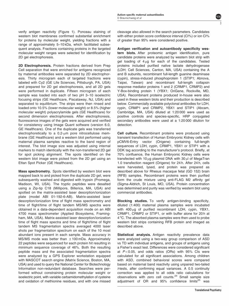
verify antigen reactivity (Figure 1). Ponceau staining of
western blot membranes confirmed substantial enrichment
for proteins by molecular weight, yielding fractions with a
range of approximately 5–10 kDa, which facilitated subse-
quent analysis. Fractions containing proteins in the targeted
molecular weight ranges were selected for identification by
2D gel electrophoresis.
2D Electrophoresis. Protein fractions derived from Prep
Cell separation that were enriched for antigens recognized
by maternal antibodies were separated by 2D electrophor-
esis. Thirty microgram each of targeted fractions were
labeled with Cy2 (GE Life Sciences, Pittsburgh, PA, USA)
and prepared for 2D gel electrophoresis, and all 2D gels
were performed in duplicate. Fifteen microgram of each
sample was loaded into each of two pH 3–10 isoelectric
focusing strips (GE Healthcare, Piscataway, NJ, USA) and
separated to equilibrium. The strips were then rinsed and
loaded onto 10.5% (lower molecular weight) or 8.5% (higher
molecular weight) polyacrylamide gels (GE Healthcare) for
second dimension electrophoresis. After electrophoresis,
fluorescence images of the gels were acquired and verified
for consistency using Image Quant software (version 6.0,
GE Healthcare). One of the duplicate gels was transferred
electrophoretically to a 0.2-mm pore nitrocellulose mem-
brane (GE Healthcare) and a western blot performed using
maternal plasma samples reactive to the band region of
interest. The blot image size was adjusted using internal
markers to match identically with the non-transferred 2D gel
for spot picking alignment. The spots identified on the
western blot image were picked from the 2D gel using an
Ettan Spot Picker (GE Healthcare).
Mass spectrometry. Spots identified by western blot were
mapped back to and picked from the duplicate 2D gel, were
subsequently washed and digested with trypsin (Promega,
Madison, WI, USA). The tryptic peptides were desalted
using a Zip-tip C18 (Millipore, Billerica, MA, USA) and
spotted on the matrix-assisted laser desorption/ionization
plate (model ABI 01-192-6-AB). Matrix-assisted laser
desorption/ionization time of flight mass spectrometry and
time of flight/time of flight tandem MS/MS spectra were
obtained in a data-dependent acquisition mode on an ABI
4700 mass spectrometer (Applied Biosystems, Framing-
ham, MA, USA). Matrix-assisted laser desorption/ionization
time of flight mass spectra and time of flight/time of flight
tandem MS fragmentation spectra averaged 4000 laser
shots per fragmentation spectrum on each of the 10 most
abundant ions present in each sample. Mass accuracy in
MS/MS mode was better than ±100mDa. Approximately
22 peptides were sequenced for each protein hit resulting in
minimum sequence coverage of 46%. Both the resulting
peptide mass and the associated fragmentation spectra
were analyzed by a GPS Explorer workstation equipped
with MASCOT search engine (Matrix Science, Boston, MA,
USA) and used to query the National Center for Biotechnology
Information non-redundant database. Searches were per-
formed without constraining protein molecular weight or
isoelectric point, with variable carbamidomethylation of cysteine
and oxidation of methionine residues, and with one missed
cleavage also allowed in the search parameters. Candidates
with either protein score confidence interval (CI%) or ion CI%
of greater than 95% were considered positive.
Antigen verification and autoantibody specificity wes-
tern blots. After proteomic antigen identification, pure
candidate proteins were analyzed by western blot strip with
gel loading of 4 mg for each of the candidates. Tested
proteins included purified native lactate dehydrogenase
(LDH; Cell Sciences, Canton, MA, USA) containing the A
and B subunits, recombinant full-length guanine deaminase
(cypin), stress-induced phosphoprotein 1 (STIP1; Abnova,
Taipei, Taiwan) and recombinant full-length collapsin
response mediator proteins 1 and 2 (CRMP1, CRMP2) and
Y-Box-binding protein 1 (YBX1; OriGene, Rockville, MD,
USA). Recombinant proteins produced in-house were also
used in these western blots and their production is described
below. Commercially available polyclonal antibodies for LDH,
cypin, CRMP1 and CRMP2, YBX1 and STIP1 (Abcam,
Cambridge, MA, USA) diluted at 1:20 000 were used as
positive controls and species-specific, HRP conjugated
secondary antibodies were used at a 1:20 000 dilution for
detection.
Cell culture. Recombinant proteins were produced using
transient transfection of Human Embryonic Kidney cells with
pCMV6-Entry vector (Origene) containing full-length
sequences of LDH, cypin, CRMP1, YBX1 or STIP1 with a
DDK tag according to the manufacturer’s protocol. Briefly, at
70% confluence, the Human Embryonic Kidney cells were
transfected with 10 mg plasmid DNA with 30 ml of MegaTran
1.0 transfection reagent (Origene) for 24 h. After 24 h, cells
were harvested, lysed, and protein was prepared as
described above for Rhesus macaque fetal (GD 152) brain
(RFB) samples. Recombinant proteins were then purified
form the crude mixture using anti-FLAG M2 affinity gel
(Sigma-Aldrich, St Louis, MO, USA). Protein concentration
was determined and purity was verified by western blot using
commercial antibodies.
Blocking studies. To verify antigen-binding specificity,
diluted (1:400) maternal plasma samples were incubated
with 400 mg of purified recombinant LDH, cypin, YBX1,
CRMP1, CRMP2 or STIP1, or with buffer alone for 20 h at
4 oC. The absorbed plasma samples were then used to probe
western blot strips containing RFB protein and imaged as
described above.
Statistical analysis. Antigen reactivity prevalence data
were analyzed using a two-way group comparison of ASD
vs TD with individual antigens, and groups of antigens using
a Fisher’s exact test. Differences were considered significant
at Po0.05, and odds ratios (ORs) with 95% CIs were
calculated for all significant associations. Among children
with ASD, combined behavioral scores were compared
based on maternal band reactivity using unpaired two-tailed
t-tests, after confirming equal variances. A 0.5 continuity
correction was applied to all odds ratio calculations for
observations with zero cell counts.24 Small sample size
adjustment of OR and 95% confidence limits25 was
Autism-specific maternal autoantibodies
D Braunschweig et al
3
Translational Psychiatry

implemented using R code (http://rss.acs.unt.edu/Rdoc/
library/epitools/html/oddsratio.html). Behavioral associations
with autoantibody pattern were analyzed by analysis of
covariance adjusted for child’s age; very little difference in
coefficients or standard errors was noted when age was
taken into account. All statistical analysis was carried out
using SAS software (SAS Institute, Cary, NC, USA).
Results
Antigen identification. To provide an enriched fraction of
RFB homogenate for mass spectrometric analysis, the RFB
homogenate was separated into 110 fractions by molecular
mass using a size fractionation column. Fractions containing
proteins in the target regions of 30–40 kDa, 39–50 kDa and
60–85 kDa were each analyzed individually on separate pairs
of 2D gels for a total of six gels (Figure 1a). One 2-D gel from
each pair was transferred to nitrocellulose and probed with
diluted maternal plasma from a mother of a child with ASD
who displayed reactivity to the bands in the region of interest.
Positive spots were observed on each of the 2D western
blots, and all identified spots were selected for mass
spectrometric analysis. The protein with the highest con-
fidence (98–100%) from each spot was selected for addi-
tional verification by western blot (Figure 1b).
Antigen verification. Commercially available purified or
full-length recombinant human proteins were used to verify
maternal antibody reactivity to the identified antigens by
western blot analysis (Figure 2a). Among the antigens
tested in an initial screen of our candidate proteins,
maternal IgG reactivity to LDH (37 kDa band), YBX1
(39 kDa band), STIP1 (upper 73 kDa band) and CRMP1
(lower 70 kDa band) or cypin, (a 44-kDa protein not noted in
our initial studies) was observed in more than 75% of
mothers of children with ASD (n¼ 50) tested for reactivity,
based upon their original fetal monkey brain autoantibody
profile. These candidate proteins were thus used for
screening of the larger cohort. Detection of subunit
specificity for LDH-reactive maternal antibodies was carried
out using purified human LDH and full-length recombinant
human LDHA and LDHB subunits by western blot
(Figure 2b). The LDHA and LDHB subunits share approxi-
mately 90% sequence homology and although some
mothers had reactivity individually to LDHA or LDHB, most
maternal antibodies that bound to LDH recognized both
subunits. Screening of all maternal plasma samples was
thus carried out with purified LDH containing both subunits.
Binding inhibition of maternal antibodies. To further
verify that the candidate autoantigens corresponded to the
targeted fetal brain protein bands, we performed binding
inhibition studies. For the target antigen corresponding to the
37 kDa band, diluted maternal plasma samples were
incubated overnight with or without purified human LDH,
and then used to probe western blots containing RFB protein
(Figure 3). Preincubation with LDH abolished any reactivity to
fetal brain protein in maternal plasma samples previously
shown to be positive for the 37-kDa band, while reactivity to
other bands was unaffected. Blocking studies involving
YBX1, cypin, CRMP1, CRMP2 and STIP1 yielded similar
LDH
AU TD
AU TD
CYPIN
AU TD
CRMP1
AU TD
STIP1
P
P
P
Pa
b
TD
CRMP2
AUPAUP TD
YBX1
Figure 2 Western blots of candidate antigens. (a) Purified lactate dehydro-genase (LDH, containing A and B subunits) or recombinant full-length Cypin, Y-box-binding protein (YBX1), collapsin response mediator protein (CRMP) 1, CRMP2 andstress-induced phosphoprotein 1 (STIP1) proteins were probed with plasma frommothers of children with autism (AU) or mothers of typically developing (TD) controlsdiluted to 1:400, P indicates polyclonal antibody positive controls. Arrows indicatelocation of band(s) of interest. Note the different patterns of reactivity to LDH A andB subunits in lanes 1–3 of the LDH panel. This reactivity is further characterized inpanel b where the blot of recombinant human glutathion-S-transferase (GST)-tagged LDHA (A), recombinant human GST-tagged LDHB (B) or purified nativehuman LDH (Enz) was probed with maternal plasma diluted to 1:400, demonstratingvariable reactivity to the LDH subunits. Also of note, as demonstrated in Table 1,some mothers of typically developing children have antibodies to the individualproteins and it is reactivity to the specific antigen combinations that confersspecificity for MAR autoantibodies.
Figure 3 Blocking western blot. Maternal plasma samples incubated withoutany target antigen (U–unblocked) or blocked with lactate dehydrogenase (LDH),collapsin response mediator protein (CRMP) 1, stress-induced phosphoprotein 1(STIP1; mother with reactivity to all three proteins), CRMP2 (mother who isCRMP1� and CRMP2 þ ), Y-box-binding protein (YBX1) and Cypin (mother withreactivity to Cypin, STIP1 and CRMP1) were used to probe a western blotcontaining Rhesus macaque fetal brain. The pure recombinant proteins wereincubated with the plasma sample diluted 1:400 overnight, centrifuged and appliedto the membrane blot containing fetal monkey brain. Arrowheads indicate location ofthe target antigen removed by overnight pre-incubation. Note that only the specificband(s) is/are removed following pre-incubation with each individual target proteinindicating no cross-reactivity between the candidate antigens.
Autism-specific maternal autoantibodies
D Braunschweig et al
4
Translational Psychiatry
results (Figure 3). Thus, it was verified that the bands
targeted by our proteomic search were accurately identified.
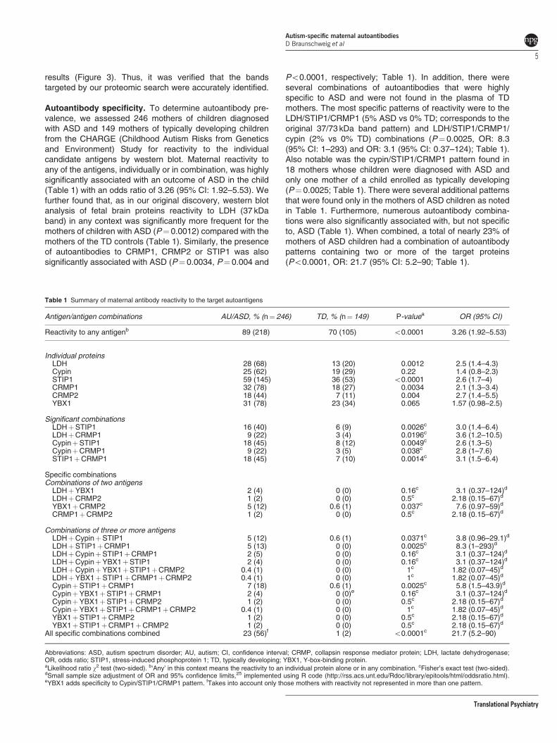
Autoantibody specificity. To determine autoantibody pre-
valence, we assessed 246 mothers of children diagnosed
with ASD and 149 mothers of typically developing children
from the CHARGE (Childhood Autism Risks from Genetics
and Environment) Study for reactivity to the individual
candidate antigens by western blot. Maternal reactivity to
any of the antigens, individually or in combination, was highly
significantly associated with an outcome of ASD in the child
(Table 1) with an odds ratio of 3.26 (95% CI: 1.92–5.53). We
further found that, as in our original discovery, western blot
analysis of fetal brain proteins reactivity to LDH (37 kDa
band) in any context was significantly more frequent for the
mothers of children with ASD (P¼ 0.0012) compared with the
mothers of the TD controls (Table 1). Similarly, the presence
of autoantibodies to CRMP1, CRMP2 or STIP1 was also
significantly associated with ASD (P¼ 0.0034, P¼ 0.004 and
Po0.0001, respectively; Table 1). In addition, there were
several combinations of autoantibodies that were highly
specific to ASD and were not found in the plasma of TD
mothers. The most specific patterns of reactivity were to the
LDH/STIP1/CRMP1 (5% ASD vs 0% TD; corresponds to the
original 37/73 kDa band pattern) and LDH/STIP1/CRMP1/
cypin (2% vs 0% TD) combinations (P¼ 0.0025, OR: 8.3
(95% CI: 1–293) and OR: 3.1 (95% CI: 0.37–124); Table 1).
Also notable was the cypin/STIP1/CRMP1 pattern found in
18 mothers whose children were diagnosed with ASD and
only one mother of a child enrolled as typically developing
(P¼ 0.0025; Table 1). There were several additional patterns
that were found only in the mothers of ASD children as noted
in Table 1. Furthermore, numerous autoantibody combina-
tions were also significantly associated with, but not specific
to, ASD (Table 1). When combined, a total of nearly 23% of
mothers of ASD children had a combination of autoantibody
patterns containing two or more of the target proteins
(Po0.0001, OR: 21.7 (95% CI: 5.2–90; Table 1).
Table 1 Summary of maternal antibody reactivity to the target autoantigens
Antigen/antigen combinations AU/ASD, % (n¼ 246) TD, % (n¼ 149) P-valuea OR (95% CI)
Reactivity to any antigenb 89 (218) 70 (105) o0.0001 3.26 (1.92–5.53)
Individual proteinsLDH 28 (68) 13 (20) 0.0012 2.5 (1.4–4.3)Cypin 25 (62) 19 (29) 0.22 1.4 (0.8–2.3)STIP1 59 (145) 36 (53) o0.0001 2.6 (1.7–4)CRMP1 32 (78) 18 (27) 0.0034 2.1 (1.3–3.4)CRMP2 18 (44) 7 (11) 0.004 2.7 (1.4–5.5)YBX1 31 (78) 23 (34) 0.065 1.57 (0.98–2.5)
Significant combinationsLDHþSTIP1 16 (40) 6 (9) 0.0026c 3.0 (1.4–6.4)LDHþCRMP1 9 (22) 3 (4) 0.0196c 3.6 (1.2–10.5)CypinþSTIP1 18 (45) 8 (12) 0.0049c 2.6 (1.3–5)CypinþCRMP1 9 (22) 3 (5) 0.038c 2.8 (1–7.6)STIP1þCRMP1 18 (45) 7 (10) 0.0014c 3.1 (1.5–6.4)
Specific combinationsCombinations of two antigensLDHþYBX1 2 (4) 0 (0) 0.16c 3.1 (0.37–124)d
LDHþCRMP2 1 (2) 0 (0) 0.5c 2.18 (0.15–67)d
YBX1þCRMP2 5 (12) 0.6 (1) 0.037c 7.6 (0.97–59)d
CRMP1þCRMP2 1 (2) 0 (0) 0.5c 2.18 (0.15–67)d
Combinations of three or more antigensLDHþCypinþSTIP1 5 (12) 0.6 (1) 0.0371c 3.8 (0.96–29.1)d
LDHþSTIP1þCRMP1 5 (13) 0 (0) 0.0025c 8.3 (1–293)d
LDHþCypinþSTIP1þCRMP1 2 (5) 0 (0) 0.16c 3.1 (0.37–124)d
LDHþCypinþYBX1þSTIP1 2 (4) 0 (0) 0.16c 3.1 (0.37–124)d
LDHþCypinþYBX1þSTIP1þCRMP2 0.4 (1) 0 (0) 1c 1.82 (0.07–45)d
LDHþYBX1þSTIP1þCRMP1þCRMP2 0.4 (1) 0 (0) 1c 1.82 (0.07–45)d
CypinþSTIP1þCRMP1 7 (18) 0.6 (1) 0.0025c 5.8 (1.5–43.9)d
CypinþYBX1þSTIP1þCRMP1 2 (4) 0 (0)e 0.16c 3.1 (0.37–124)d
CypinþYBX1þSTIP1þCRMP2 1 (2) 0 (0) 0.5c 2.18 (0.15–67)d
CypinþYBX1þSTIP1þCRMP1þCRMP2 0.4 (1) 0 (0) 1c 1.82 (0.07–45)d
YBX1þSTIP1þCRMP2 1 (2) 0 (0) 0.5c 2.18 (0.15–67)d
YBX1þSTIP1þCRMP1þCRMP2 1 (2) 0 (0) 0.5c 2.18 (0.15–67)d
All specific combinations combined 23 (56)f 1 (2) o0.0001c 21.7 (5.2–90)
Abbreviations: ASD, autism spectrum disorder; AU, autism; CI, confidence interval; CRMP, collapsin response mediator protein; LDH, lactate dehydrogenase;OR, odds ratio; STIP1, stress-induced phosphoprotein 1; TD, typically developing; YBX1, Y-box-binding protein.aLikelihood ratio w
2 test (two-sided). b‘Any’ in this context means the reactivity to an individual protein alone or in any combination. cFisher’s exact test (two-sided).dSmall sample size adjustment of OR and 95% confidence limits,25 implemented using R code (http://rss.acs.unt.edu/Rdoc/library/epitools/html/oddsratio.html).eYBX1 adds specificity to Cypin/STIP1/CRMP1 pattern. fTakes into account only those mothers with reactivity not represented in more than one pattern.
Autism-specific maternal autoantibodies
D Braunschweig et al
5
Translational Psychiatry

Behavioral correlations. To characterize the potential
association between maternal antibody status and child
behavior, scores on the ABC, Mullen Scales of Early
Learning and Vineland Adaptive Behavior Scales were
compared between offspring of MAR-positive mothers and
those possessing no antibody reactivity; both the ASD and
TD subjects were assessed with these instruments. We also
analyzed the Autism Diagnostic Observation Schedule and
Social Communication Questionnaire behavior scales in the
ASD group, but there were no significant associations found
with any of the band patterns. In addition, although no
significant differences in subscale or composite scores were
noted for the Mullen Scales of Early Learning or the Vineland
Adaptive Behavioral Scales, increased impairments in the
stereotypical behavior subscale of the ABC were observed
for children of mothers possessing antibodies reactive to
LDH in any context (P¼ 0.024), CRMP1 (trending toward
significance; P¼ 0.055), as well as combined reactivity
to LDH and STIP1 (P¼ 0.015), or LDH/STIP1/CRMP1
(P¼ 0.007) in comparison to children with an ASD whose
mothers lack IgG reactivity to these antigens. In addition,
increased overall impairment was reflected in the composite
ABC score for children of mothers reactive against LDH and
CRMP1 (P¼ 0.046) as well as LDH/STIP1/CRMP1 (trending
toward significance; P¼ 0.06).
Discussion
Several studies have implicated maternal immune dysregula-
tion during pregnancy in association with ASD. Prominent
among them are reports of maternal antibodies that react
against fetal brain proteins.6–8,13–16,26–28 The hypothesis is
that gestational exposure to maternal antibodies directed
against proteins abundantly expressed in the fetal brain could
lead to alterations in the neurodevelopment characteristic of
ASD is further supported by the identification of specific IgG
reactivity to LDH, YBX1, cypin, STIP1, CRMP1 and CRMP2,
six proteins highly expressed in developing brain. Thus, MAR
autoantibodies could represent one mechanism underlying
the development of one or more of the ASD core features in a
subgroup of cases. Moreover, with their exceptionally high
specificity, several of the MAR autoantibody profiles could
serve as the first true biomarkers of ASD risk.
Facilitated passage of IgG antibodies during human
gestation is a well-characterized phenomenon that provides
passive protection for the newborn child,29 which persists in
the child up to 6 months post-natally.30 However, autoanti-
bodies that react to fetal ‘self’ proteins can also cross the
placenta.31 In addition, the blood–brain barrier is not fully
mature during early brain development, and maternal IgG has
been found within mouse32 and human fetal brains.7 Dalton
et al.13 first observed that when plasma from a mother of
multiple children with ASD possessing reactivity to rodent
Purkinje cells was administered to mid-gestation mice,
behavioral deficits were present in the offspring. In another
study,6mothers and their ASD-affected children were found to
have consistent patterns of antibody reactivity against rat pre-
natal (day 18) brain proteins in contrast to control mothers.We
recently employed a passive transfer rodent model using a
single gestational exposure to an intravenous dose of purified,
brain-reactive IgG antibodies from individual mothers of ASD
children or mothers with typically developing children. Growth
and behavioral outcomes in offspring revealed alterations in
early growth trajectories, significantly impaired motor and
sensory development, and increased anxiety.18 Thus, there is
compelling evidence that maternally derived antibodies could
lead to neurodevelopmental alterations in offspring.
Consistent with our early studies on fetal brain homoge-
nates, each of the identified antigens is expressed at
significant levels in the human fetal brain33 and has an
established role in neurodevelopment. For example, cypin
is an enzyme with guanine deaminase activity that has
an important role in dendritic branching of hippocampal
neurons.34 It was also recently reported that the postsynaptic
protein PSD-95 and cypin regulate dendrite patterning in
hippocampal neurons, where cypin promotes microtubule
assembly and PSD-95 disrupts microtubule organization.
The authors further demonstrated that cypin regulates the
density of dendritic spines/filopodia through overexpression/
knockdown experiments. The number of protrusions per
dendrite decreased with an overexpression of cypin, whereas
a cypin knockdown resulted in an increase in spines/filopodia
on the dendrite.35 In another study, exposure to brain-derived
neurotrophic factor, an important mediator of dendrite
arborization and a factor previously implicated in ASD,
increases cypin in cultured rat hippocampal neurons. Thus,
one can speculate that interfering with cypin through autoanti-
body exposure during neurodevelopment could affect den-
drite formation, potentially increasing the number of dendritic
spines in certain regions of the brain. This is consistent with
recent data describing spine dysmorphogenesis in ASD
where analysis of post-mortem ASD human brain tissue
demonstrated an increase in spine density on apical dendrites
of pyramidal neurons in a subset of individuals.36
STIP1, which is first detected in the developing nervous
system,37 is a major ligand of the cellular prion protein
(PrP(C)), and together these two proteins mediate neurito-
genesis in cultured hippocampal neurons.38 Further, protein
synthesis in neurons is enhanced via the binding of PrP(C)
with STIP, and the neuroprotection and neuritogenesis
mediated by PrP(C)-STI1 engagement are dependent upon
this protein increase.39 Inhibition of this interaction leads to
impaired memory formation in rodents.40 A recent report also
describes the presence of autoantibodies to STIP1 in the
blood and CSF of patients with neuro-Behcet’s disease,41
a chronic recurrent inflammatory disorder with central nervous
system involvement that occurs in 5–10% of Behcet’s disease
patients.41
The group of CRMPs 1–5 are thought to contribute to
semaphorin-induced growth cone collapse.42 One of the
MAR-specific proteins in this group, CRMP1, is highly
expressed in the developing brain and is required for proper
cell migration and growth cone collapse.42,43 It has also been
recently demonstrated that CRMP1 and CRMP2 synergisti-
cally control dendritic projection,44 whereas knockdown of
CRMP1 in spinal cord cultures leads to a dramatic reduction in
cell survival.45 CRMP2 was originally identified as a signaling
molecule required for growth cone collapse of dorsal root
ganglion neurons in response to a repulsive guidance cue by
semaphorin-3A. CRMP2 was also reported to have a positive
Autism-specific maternal autoantibodies
D Braunschweig et al
6
Translational Psychiatry

effect on axonal extension and to have a critical role in axon-
dendrite specification and axon regeneration.42 Further,
blocking experiments suggest that inhibition of CRMP2 does
not change the number of neurites, but rather increases the
number of cells bearing neurites as well as neurite length.42
These findings are of interest when one considers the
magnetic resonance imaging studies noting changes in brain
growth in children with autism46–48 and further studies are
currently underway to determine the relationship between
MAR autoantibodies and brain growth in ASD. Interestingly,
the CRMP1 gene has been associated with the anhedonia
endophenotype of schizophrenia in a DISC1 (Disrupted-in-
schizophrenia 1) gene-dependent manner, and a CRMP1
epitope found in brains from schizophrenia patients is thought
to potentially discriminate schizophrenia-derived peripheral
blood lymphoblasts from those of normal controls.49 CRMP2
has also been associated with several neuropathologic or
psychiatric conditions, including Alzheimer’s disease and
schizophrenia, through genetic polymorphisms, changes in
protein expression, post-translational modifications or
through protein/protein interactions.50
YBX1 is involved in a number of cellular processes,
including proliferation, differentiation and the cellular stress
response (reviewed in Eliseeva et al.51). YBX1 functions both
in the cytoplasm and in the cell nucleus and it can also be
secreted from cells and interact with cell surface receptors to
activate intracellular signaling. With respect to fetal brain,
YBX1 has an important role in late embryogenesis through in
the enhanced cell neuronal motility and migration required for
successful neural tube formation. YBX1 mRNA is highly
expressed in testicular and fetal brain cells, as well as early
precursors of erythroid and lymphoid cells. Interestingly,
YBX1 has been found to interact with the Rett Syndrome
gene, MeCP2, where it is thought to have RNA splicing
activity. YBX1 has also been shown to interact with FMPR, a
regulator of mRNA transport and translation, that when absent
or defective, gives rise to Fragile-X Syndrome (reviewed in
Eliseeva et al.51).
The enzyme LDH is found in the rodent fetal brain52where it
functions in cellular metabolism. Although autoantibodies
targeting LDH have not yet been shown to directly have a role
in altering neurodevelopment, such autoantibodies have been
identified in sera from individuals exposed to the industrial
solvent trichloroethylene.53 This observation is of interest with
respect to the ontogeny of the maternal autoantibodies
identified in this report. Circulating LDH levels are often used
as a measure of necrotic cell damage following toxic
exposures54 and viral infections,55 and in this inflammatory
context could give rise to an antibody-producing immune
response in the mother. For example, a mouse model of
maternal immune activation where maternal inflammation
during gestation results in behavioral changes in the offspring,
demonstrated an increase in LDH-B in the prefrontal cortex.56
The relatively high prevalence (28% overall amongmothers of
children with ASD) indicates a critical need to define the role
for LDH in the developing brain.
Our finding of an increased incidence of stereotypic
behavior in children whose mothers have autoantibodies
targeting LDH, CRMP1 and STIP1 suggests a role for these
proteins in a core behavioral feature of ASD. Interestingly, this
result is consistent with behavior observations from our earlier
non-human primate gestational IgG passive transfer study16
using some of the same antibody sources as those discussed
herein. The stereotypic behavior in Rhesus monkey offspring
noted after gestational exposure, coupled with the increased
stereotypical behavior among children of mothers analyzed in
this study, suggests that a subset of MAR autoantibodies may
be pathologically significant, eliciting changes in all aspects of
ASD, but with a particularly strong effect in the behavioral
domain. Other behavioral differences have been character-
ized between ASD cases based on maternal autoantibody
status, such as increased irritability and impaired use of
expressive language8 using a similar study population. The
inconsistencies between our two studies regarding behavior–
autoantibody associations may stem from the increased
detection sensitivity afforded by the use of pure antigens
rather than the crude fetal brain protein preparations
employed in previous studies. Another possibility is the
presence of additional autoantigens that we have not yet
discovered, and that could be related to impaired use of
expressive language. We are currently exploring these
additional candidates.
Each of the target autoantigens defined herein is known to
have a critical role in the developing brain and interference
with the level or function of more than one of them could act
synergistically to change the trajectory of brain development.
Figure 4 illustrates the known functions for each of the
antigenic proteins during neuron migration and development.
The effect of MAR autoantibodies could occur through a
direct antigen–antibody interaction, thereby either decreasing
the abundance of or causing functional interference of
the target proteins. Alternatively, the presence of these
maternal antibodies may merely serve as a biomarker of cell
destruction. One of the limitations of the current study is that
all of the clinical samples used for analysis of reactivity to the
YBX-1-Neural tube
formation,
cell division
AxonGrowth
cone
Neurodevelopment
Dendrites
LDH-Metabolism
STIP1-Neuritogenesis
CRMP1-Growth
cone collapse
Cypin (GDA)-Dendritic
branching
CRMP1-Cell migration
Embryonic
Cross-section
CRMP2-Axon outgrowth
Growth cone collapse
Basal dendrite
patterning
YBX-1-Transcription
regulation
Figure 4 Schematic model depicting the neurodevelopmental roles for each ofthe maternal autoantibody target proteins. Disruption of protein function or quantitymay negatively alter neurodevelopment, potentially leading to the core featuresassociated with autism. There is likely an additive effect as illustrated by the autismspectrum disorder specificity of the various autoantibody combinations. CRMP,collapsin response mediator protein; LDH, lactate dehydrogenase; STIP1, stress-induced phosphoprotein 1; YBX1, Y-box-binding protein.
Autism-specific maternal autoantibodies
D Braunschweig et al
7
Translational Psychiatry

target autoantigens were obtained from mothers of children
at the time of the child’s diagnosis (retrospective sample).
Studies are currently underway to assess the predictive value
of the MAR autoantibodies in a prospective study sample.
Addressing the basis for the association between maternal
autoantibodies and autism risk in the child is also of urgent
concern. Animal studies involving targeted autoantibody
exposure throughout gestation are currently underway to
address the hypothesis that pre-natal exposure to these
specific antibodies leads to behavioral alterations in offspring.
The clinical significance of these findings may be twofold: (1)
early diagnosis of a MAR-ASD child would allow for early
behavioral intervention and (2) if pathologically significant,
medical interventions that would limit fetal exposure to these
antibodies might prove helpful in reducing risk of ASD
symptom development in the children of affected mothers.
Conflict of interest
The corresponding (Van de Water) and first (Braunschweig)
authors have a published patent on the proteins described in
this manuscript. Van de Water is a consultant for Pediatric
Bioscience, a company that has licensed this technology from
UC Davis. The remaining authors declare no conflict of
interest.
Acknowledgements. This work was supported by NIEHS 1 P01ES11269-01, the US Environmental Protection Agency (USEPA) through theScience to Achieve Results (STAR) program (Grant R829388), NIEHS 1 R01-ES015359, the UC Davis M.I.N.D. Institute and Autism Speaks graduate fellowship(DB) and US patent 8,383,360 B2. We thank Elizabeth Fox for her help in readingand editing this manuscript.
1. Investigators CAaDDMNSYP. Prevalence of autism spectrum disorders—autism and
developmental disabilities monitoring network, 14 Sites, United States, 2008. MMWR
Surveill Summ 2012; 61: 1–19.
2. APA Diagnostic and Statistical Manual of Mental Disorders: DSM-IV Text Revision.
American Psychiatric Association: Washington, DC.
3. Goines P, Van de Water J. The immune system’s role in the biology of autism. Curr Opin
Neurol 2010; 23: 111–117.
4. Heuer L, Ashwood P, Schauer J, Goines P, Krakowiak P, Hertz-Picciotto I et al. Reduced
levels of immunoglobulin in children with autism correlates with behavioral symptoms.
Autism Res 2008; 1: 275–283.
5. Vargas DL, Nascimbene C, Krishnan C, Zimmerman AW, Pardo CA. Neuroglial activation
and neuroinflammation in the brain of patients with autism. Ann Neurol 2005; 57: 67–81.
6. Zimmerman AW, Connors SL, Matteson KJ, Lee LC, Singer HS, Castaneda JA et al.
Maternal antibrain antibodies in autism. Brain Behav Immun 2007; 21: 351–357.
7. Braunschweig D, Ashwood P, Krakowiak P, Hertz-Picciotto I, Hansen R, Croen LA et al.
Autism: maternally derived antibodies specific for fetal brain proteins. Neurotoxicology
2008; 29: 226–231.
8. Braunschweig D, Duncanson P, Boyce R, Hansen R, Ashwood P, Pessah IN et al.
Behavioral correlates of maternal antibody status among children with autism. J Autism
Dev Disord 2011; 42: 1435–1445.
9. Heuer L, Braunschweig D, Ashwood P, Van de Water J, Campbell DB. Association of a
MET genetic variant with autism-associated maternal autoantibodies to fetal brain proteins
and cytokine expression. Translational Psychiatry 2011; 1: e48.
10. Simister NE. Placental transport of immunoglobulin G. Vaccine 2003; 21: 3365–3369.
11. Malek A, Sager R, Kuhn P, Nicolaides KH, Schneider H. Evolution of maternofetal transport
of immunoglobulins during human pregnancy. Am J Reprod Immunol 1996; 36: 248–255.
12. Bake S, Friedman JA, Sohrabji F. Reproductive age-related changes in the blood brain
barrier: expression of IgG and tight junction proteins. Microvasc Res 2009; 78: 413–424.
13. Dalton P, Deacon R, Blamire A, Pike M, McKinlay I, Stein J et al. Maternal
neuronal antibodies associated with autism and a language disorder. Ann Neurol 2003;
53: 533–537.
14. Singer HS, Morris C, Gause C, Pollard M, Zimmerman AW, Pletnikov M. Prenatal exposure
to antibodies from mothers of children with autism produces neurobehavioral alterations: a
pregnant dam mouse model. J Neuroimmunol 2009; 211: 39–48.
15. Braunschweig D, Golub MS, Koenig CM, Qi L, Pessah IN, Van de Water J et al. Maternal
autism-associated IgG antibodies delay development and produce anxiety in a mouse
gestational transfer model. J Neuroimmunol 2012; 252: 56–65.
16. Martin LA, Ashwood P, Braunschweig D, Cabanlit M, Van de Water J, Amaral DG.
Stereotypies and hyperactivity in rhesus monkeys exposed to IgG from mothers of children
with autism. Brain Behav Immun 2008; 22: 806–816.
17. Hertz-Picciotto I, Croen LA, Hansen R, Jones CR, van de Water J, Pessah IN. The
CHARGE study: an epidemiologic investigation of genetic and environmental factors
contributing to autism. Environ Health Perspect 2006; 114: 1119–1125.
18. Braunschweig D, Duncanson P, Boyce R, Hansen R, Ashwood P, Pessah IN et al.
Behavioral correlates of maternal antibody status among children with autism. J Autism
Dev Disord 2012; 42: 1435–1445.
19. Lord C, Rutter M, diLavore P, Risi S. Autism Diagnostic Observation Schedule Manual.
Western Psychological Services: Los Angeles, CA, 2003.
20. Le Couteur A, Lord C, Rutter M. The Autism Diagnostic Interview, Revised (ADI-R).
Western Psychological Services: Los Angeles, CA, 1989.
21. Mullen E. Mullen Scale of Early Learning. American Guidance Service: Circle Pines,
MN, 1995.
22. Sparrow SB, Cicchetti DA, Vineland DV. Adaptive Behavior Scales Survey Form Manual.
American Guidance Service: Circle Pines, MN, 1985.
23. Aman M, Singh N. Abberant Behavioral Checklist-Community Supplementary Manual
Slosson Educational Publications: East Aurora, NY, 1986.
24. Subbiah M, Srinivasan MR. Classification of 2 � 2 sparse data sets with zero cells. Statist
Probab Lett 2008; 78: 3212–3215.
25. Jewell NP, Kalbfleisch JD. Maximum likelihood estimation of ordered multinomial
parameters. Biostatistics 2004; 5: 291–306.
26. Croen LA, Braunschweig D, Haapanen L, Yoshida CK, Fireman B, Grether JK et al.
Maternal mid-pregnancy autoantibodies to fetal brain protein: the early markers for autism
study. Biol Psychiatry 2008; 64: 583–588.
27. Singer HS, Morris CM, Gause CD, Gillin PK, Crawford S, Zimmerman AW. Antibodies
against fetal brain in sera of mothers with autistic children. J Neuroimmunol 2008; 194:
165–172.
28. Warren RP, Cole P, Odell JD, Pingree CB, Warren WL, White E et al. Detection of
maternal antibodies in infantile autism. J Am Acad Child Adolesc Psychiatry 1990; 29:
873–877.
29. Garty BZ, Ludomirsky A, Danon YL, Peter JB, Douglas SD. Placental transfer of
immunoglobulin G subclasses. Clin Diagn Lab Immunol 1994; 1: 667–669.
30. Heininger U, Desgrandchamps D, Schaad UB. Seroprevalence of Varicella-Zoster
virus IgG antibodies in Swiss children during the first 16 months of age. Vaccine 2006; 24:
3258–3260.
31. Tincani A, Rebaioli CB, Taglietti M, Shoenfeld Y. Heart involvement in systemic lupus
erythematosus, anti-phospholipid syndrome and neonatal lupus. Rheumatology (Oxford)
2006; 45(Suppl 4): iv8–13.
32. Lee JY, Huerta PT, Zhang J, Kowal C, Bertini E, Volpe BT et al. Neurotoxic autoantibodies
mediate congenital cortical impairment of offspring in maternal lupus. Nat Med 2009; 15:
91–96.
33. Wheeler DL, Church DM, Federhen S, Lash AE, Madden TL, Pontius JU et al.
Database resources of the National Center for Biotechnology. Nucleic Acids Res 2003;
31: 28–33.
34. Akum BF, Chen M, Gunderson SI, Riefler GM, Scerri-Hansen MM, Firestein BL. Cypin
regulates dendrite patterning in hippocampal neurons by promoting microtubule assembly.
Nat Neurosci 2004; 7: 145–152.
35. Tseng CY, Firestein BL. The role of PSD-95 and cypin in morphological changes in
dendrites following sublethal NMDA exposure. J Neurosci 2011; 31: 15468–15480.
36. Hutsler JJ, Zhang H. Increased dendritic spine densities on cortical projection neurons in
autism spectrum disorders. Brain Res 2010; 1309: 83–94.
37. Hajj GN, Santos TG, Cook ZS, Martins VR. Developmental expression of prion protein and
its ligands stress-inducible protein 1 and vitronectin. J Comp Neurol 2009; 517: 371–384.
38. Lopes MH, Hajj GN, Muras AG, Mancini GL, Castro RM, Ribeiro KC et al. Interaction of
cellular prion and stress-inducible protein 1 promotes neuritogenesis and neuroprotection
by distinct signaling pathways. J Neurosci 2005; 25: 11330–11339.
39. Roffe M, Beraldo FH, Bester R, Nunziante M, Bach C, Mancini G et al. Prion protein
interaction with stress-inducible protein 1 enhances neuronal protein synthesis via mTOR.
Proc Natl Acad Sci USA 2010; 107: 13147–13152.
40. Coitinho AS, Lopes MH, Hajj GN, Rossato JI, Freitas AR, Castro CC et al. Short-term
memory formation and long-term memory consolidation are enhanced by cellular prion
association to stress-inducible protein 1. Neurobiol Dis 2007; 26: 282–290.
41. Vural B, Ugurel E, Tuzun E, Kurtuncu M, Zuliani L, Cavus F et al. Anti-neuronal and stress-
induced-phosphoprotein 1 antibodies in neuro-Behcet’s disease. J Neuroimmunol 2011;
239: 91–97.
42. Quach TT, Duchemin AM, Rogemond V, Aguera M, Honnorat J, Belin MF et al.
Involvement of collapsin response mediator proteins in the neurite extension
induced by neurotrophins in dorsal root ganglion neurons. Mol Cell Neurosci 2004; 25:
433–443.
43. Charrier E, Reibel S, Rogemond V, Aguera M, Thomasset N, Honnorat J. Collapsin
response mediator proteins (CRMPs): involvement in nervous system development and
adult neurodegenerative disorders. Mol Neurobiol 2003; 28: 51–64.
Autism-specific maternal autoantibodies
D Braunschweig et al
8
Translational Psychiatry

44. Yamashita N, Ohshima T, Nakamura F, Kolattukudy P, Honnorat J, Mikoshiba K et al.
Phosphorylation of CRMP2 (collapsin response mediator protein 2) is involved in proper
dendritic field organization. J Neurosci 2012; 32: 1360–1365.
45. Kurnellas MP, Li H, Jain MR, Giraud SN, Nicot AB, Ratnayake A et al. Reduced expression
of plasma membrane calcium ATPase 2 and collapsin response mediator protein 1
promotes death of spinal cord neurons. Cell Death Differ 2010; 17: 1501–1510.
46. Nordahl CW, Lange N, Li DD, Barnett LA, Lee A, Buonocore MH et al. Brain enlargement is
associated with regression in preschool-age boys with autism spectrum disorders. Proc
Natl Acad Sci USA 2011; 108: 20195–20200.
47. Nordahl CW, Scholz R, Yang X, Buonocore MH, Simon T, Rogers S et al. Increased rate of
amygdala growth in children aged 2 to 4 years with autism spectrum disorders: a
longitudinal study. Arch Gen Psychiatry 2012; 69: 53–61.
48. Aoki Y, Kasai K, Yamasue H. Age-related change in brain metabolite abnormalities in
autism: a meta-analysis of proton magnetic resonance spectroscopy studies. Transl
Psychiatry 2012; 2: e69.
49. Bader V, Tomppo L, Trossbach SV, Bradshaw NJ, Prikulis I, Leliveld SR et al. Proteomic,
genomic and translational approaches identify CRMP1 for a role in schizophrenia and its
underlying traits. Hum Mol Genet 2012; 21: 4406–4418.
50. Hensley K, Venkova K, Christov A, Gunning W, Park J. Collapsin response mediator
protein-2: an emerging pathologic feature and therapeutic target for neurodisease
indications. Mol Neurobiol 2011; 43: 180–191.
51. Eliseeva IA, Kim ER, Guryanov SG, Ovchinnikov LP, Lyabin DN. Y-box-binding protein 1
(YB-1) and its functions. Biochemistry (Mosc) 2011; 76: 1402–1433.
52. Hashimoto T, Hussien R, Cho HS, Kaufer D, Brooks GA. Evidence for the mitochondrial
lactate oxidation complex in rat neurons: demonstration of an essential component of brain
lactate shuttles. PLoS One 2008; 3: e2915.
53. Liu J, Xing X, Huang H, Jiang Y, He H, Xu X et al. Identification of antigenic proteins
associated with trichloroethylene-induced autoimmune disease by serological proteome
analysis. Toxicol Appl Pharmacol 2009; 240: 393–400.
54. Silva RF, Falcao AS, Fernandes A, Gordo AC, Brito MA, Brites D. Dissociated primary
nerve cell cultures as models for assessment of neurotoxicity. Toxicol Lett 2006; 163:
1–9.
55. Shin SY, Kim JH, Kim HS, Kang YA, Lee HG, Kim JS et al. Clinical characteristics of
Korean pediatric patients critically ill with influenza A (H1N1) virus. Pediatr Pulmonol 2010;
45: 1014–1020.
56. Deng MY, Lam S, Meyer U, Feldon J, Li Q, Wei R et al. Frontal-subcortical protein
expression following prenatal exposure to maternal inflammation. PLoS One 2011; 6:
e16638.
Translational Psychiatry is an open-access journal
published by Nature Publishing Group. This work is
licensed under a Creative Commons Attribution-NonCommercial-
NoDerivs 3.0 Unported License. To view a copy of this license, visit
http://creativecommons.org/licenses/by-nc-nd/3.0/
Autism-specific maternal autoantibodies
D Braunschweig et al
9
Translational Psychiatry