Brain Insulin Controls Adipose Tissue Lipolysis and Lipogenesis
Upload
independentCategory
view
1download
0
Amable et al. Stem Cell Research & Therapy 2014, 5:53http://stemcellres.com/content/5/2/53
RESEARCH Open Access
Protein synthesis and secretion in humanmesenchymal cells derived from bone marrow,adipose tissue and Wharton’s jellyPaola Romina Amable1*†, Marcus Vinicius Telles Teixeira1,2†, Rosana Bizon Vieira Carias1,2,José Mauro Granjeiro2 and Radovan Borojevic1,2
Abstract
Introduction: Different mesenchymal stromal cells (MSC) have been successfully isolated and expanded in vitro andnowadays they are tested in clinical trials for a wide variety of diseases. Whether all MSC express the same cellsurface markers or have a similar secretion profile is still controversial, making it difficult to decide which stromalcell may be better for a particular application.
Methods: We isolated human mesenchymal stromal cells from bone marrow (BM), adipose tissue (AT) andWharton’s jelly (WJ) and cultured them in fetal bovine serum supplemented media. We evaluated proliferation,in vitro differentiation (osteogenic, adipogenic and chondrogenic potential), expression of cell surface markers andprotein secretion using Luminex and ELISA assays.
Results: Cell proliferation was higher for WJ-MSC, followed by AT-MSC. Differences in surface expression markerswere observed only for CD54 and CD146. WJ-MSC secreted higher concentrations of chemokines, pro-inflammatoryproteins and growth factors. AT-MSC showed a better pro-angiogenic profile and secreted higher amounts ofextracellular matrix components and metalloproteinases.
Conclusions: Mesenchymal stromal cells purified from different tissues have different angiogenic, inflammatory andmatrix remodeling potential properties. These abilities should be further characterized in order to choose the bestprotocols for their therapeutic use.
IntroductionMesenchymal stromal cells (MSC) are a small popula-tion of multipotent progenitor cells present in nearly allhuman tissues, being mostly found in the perivascularniche [1]. MSC were first isolated from bone marrow[2], but they have been obtained subsequently from awide variety of fetal and adult tissues: adipose tissue [3],placenta [4], lung [5], umbilical cord [6], synovial mem-brane [7] and dental pulp [8] among many others.Regenerative medicine makes use of MSC and of their
multipotent properties to promote tissue regeneration.MSC are able to migrate into injured tissues, engraft anddifferentiate into many cell types, participating thus directly
* Correspondence: [email protected]†Equal contributors1Excellion Biomedical Services S.A, Rua Afrânio de Mello Franco 333,25651000 PetrópolisRio de Janeiro, BrazilFull list of author information is available at the end of the article
© 2014 Amable et al.; licensee BioMed CentraCommons Attribution License (http://creativecreproduction in any medium, provided the orDedication waiver (http://creativecommons.orunless otherwise stated.
in tissue repair and regeneration [9]. They also secrete para-crine mediators, reducing inflammation and acceleratingtissue regeneration by activation of resident stem cells andmobilization of circulating systemic stem cells throughchemotactic signaling [10,11]. Clinical trials have alreadyconfirmed that use of MSC is safe and effective [12]. Evenwhen MSC express major histocompatibility complexclass I, they proved to be safe in allogeneic transplants, alsobetween HLA-incompatible individuals, since they do notelicit alloreactive lymphocyte proliferative responsesin vitro, they produce locally immunomodulatory proteinsand their expression levels of major histocompatibility com-plex class II are negligible [13,14].Studies comparing MSC derived from different tissues
are limited to in vitro and pre-clinical studies. Clinicaltrials are generally focused on safety and efficiency of atherapy using a specific type of MSC, without demon-strating which MSC is the best for each therapy, or even
l Ltd. This is an Open Access article distributed under the terms of the Creativeommons.org/licenses/by/2.0), which permits unrestricted use, distribution, andiginal work is properly credited. The Creative Commons Public Domaing/publicdomain/zero/1.0/) applies to the data made available in this article,

Amable et al. Stem Cell Research & Therapy 2014, 5:53 Page 2 of 13http://stemcellres.com/content/5/2/53
justifying why a specific cell type was believed to be thebest option. Basic MSC comparative studies are requiredto better understand MSC properties and abilities, indi-cating the most appropriate MSC type for a particularclinical application.In vitro studies have already shown that MSC from
different origins varied when considering differentiationpotential: some cells are better for differentiation intoosteoblast-like cells [15], while synovium-derived MSCand umbilical cord-derived MSC are better differentiatedinto chondrocytes than the bone marrow-derived MSC[16,17]. MSC isolated from fetal tissues are superior re-garding cardiomyocyte and endothelial cell differenti-ation when compared to adult tissue-derived MSC [18].Besides differentiation potential, recent studies have
approached other MSC attributes that allowed a deeperunderstanding of tissue-derived properties. Hsieh andcolleagues compared MSC derived from Wharton’s Jellyand bone marrow regarding their ability to regenerateinfarcted myocardia; they described secretome differ-ences that make Wharton’s Jelly-derived MSC a more an-giogenic, neuroprotective and neurogenic option [19].Naftali-Shani and coworkers carried out a pre-clinical trialof myocardial infarction in rats comparing the effects ofhuman stromal cells obtained from four locations (epicar-dial fat, pericardial fat, subcutaneous fat and the rightatrium) and they showed that hMSCs from epicardialfat and the right atrium secreted the highest amounts oftrophic and inflammatory cytokines in vitro, expressedhigher amounts of inflammation- and fibrosis-related genesin vitro and impaired heart recovery in vivo [20]. Researchstudies like these mentioned here are essential for deter-mining the best tissue-derived mesenchymal stromal cellfor a particular regenerative therapy strategy.In the present study, we compared three different cell
types: MSC derived from bone marrow, adipose tissue andWharton’s Jelly with the purpose to better understandtheir differences and similarities. We monitored prolifera-tion, differentiation into mesodermal cell types and cellsurface marker expression. We also quantified productionand secretion of cytokines, chemokines, growth factorsand extracellular matrix components into the cell culturesupernatants.
MethodsEthics statementsAll the experimental procedures were done after EthicsResearch Committees approval and all donors signedan informed consent. Adipose tissue-derived MSC (AT-MSC) were obtained from abdominal liposuction duringplastic surgery (age: 53.8 ± 5.4 years; sex: 50/50% male/female; CEP No. 55219/12, Research Ethics Committeeof Pro-Cardíaco Hospital, Rio de Janeiro, Brazil). Wharton’sJelly-derived MSC (WJ-MSC) were obtained from umbilical
cords of full-term cesarean births (38 to 39 weeks; CEPNo. 336/10, Research Ethics Committee of Pro-CardíacoHospital, Rio de Janeiro, Brazil). Bone marrow-derivedMSC (BM-MSC) were purified from bone marrow har-vested from posterior iliac crests during orthopedic surgery(age: 55.5 ± 13.4 years; sex: 50/50% male/female; CEPNo. 473/12, Research Ethics Committee of Pro-CardíacoHospital, Rio de Janeiro, Brazil).
Cell isolation and cultureStromal cells were isolated as previously described [21].Briefly, nucleated cells were separated from humanbone marrow using Ficoll-Paque™ PLUS (GE Healthcare,Uppsala, Uppsala, Sweden, #17-1440-02) by density gradi-ent centrifugation at 700 g during a 15-minute period.After washing cells with phosphate-buffered saline (PBS -LGC, Cotia, São Paulo, Brazil, #13-30259-05), they wereplated in T25 flasks in alpha-Minimum Essential Medium(α-MEM - LGC, BR30007-05) supplemented with 10%fetal bovine serum (FBS - LGC, #10-BIO-500). Humanadipose tissue was washed three times with PBS and wastreated with 1.76 mg collagenase type I/gram tissue(Sigma-Aldrich, St. Louis, Missouri, USA, C9891) for 30minutes at 4°C and 30 minutes at 37°C with agitation.After proteolytic activity inhibition and centrifugation (700g, seven minutes), pelleted cells were plated in T25 flasks.Human umbilical cords were washed with PBS and bloodvessels were removed. Wharton’s jelly was cut into smallpieces and digested with 0.9 mg collagenase type II/gramtissue (Sigma, C6885) at 37°C for one hour. Washed cellswere centrifuged at 700 g for seven minutes and plated inT25 flasks. All cells were expanded up to passage number3 in order to obtain a higher number of cells and were thencryopreserved.For all the experiments, cells obtained from four dif-
ferent donors in the same passage number were thawedand mixed in order to prepare cell pools, which wereimmediately plated for proliferation experiments.
Proliferation curvesCells of each cell pool were seeded in 24-well plates at aconcentration of 6,000 cells/mL in α-MEM supplementedwith 10% FBS. Cells grown in different wells were trypsi-nized and counted in a Neubauer hemocytometer at threedifferent times: 96, 144 and 192 hours.
Flow cytometryAfter cell detachment using a 0.125% trypsin solution, cellswere washed with PBS and resuspended in PBS containing2% FBS. Cell concentration and viability were monitoredusing Trypan blue in a Neubauer hemocytometer.The following monoclonal antibodies were used asindicated by the manufacturers (BD Pharmingen®(BD, Franklin Lakes, New Jersey, USA)): CD90-PE

Amable et al. Stem Cell Research & Therapy 2014, 5:53 Page 3 of 13http://stemcellres.com/content/5/2/53
(BD, #555596), CD73-FITC (BD, #561254), CD105-FITC(BD, #561443), CD45-FITC (BD, #347463), CD14-PE (BD,#555398), CD34-PEcy5 (BD, #561819), CD31-PE(BD, #555446), IgG-FITC (BD, #555786), HLA-DR-FITC(BD, #555558), CD166-PE (BD, #560903), CD44-PE (BD,#555479), CD54-PEcy5 (BD, #555512), CD146-PE (BD,#559263). Isotype controls were used for determiningnonspecific binding and defining cut-off values. A mini-mum of 20,000 events were acquired on a BD FACSCalibur® flow cytometer and results were analyzed usingCellQuest™ software.
Differentiation assaysPre-cultured cells were seeded into 24-well plates (1 mL/well)at 13,157 cells/cm2 (adipogenic differentiation), 5,263cells/cm2 (osteogenic differentiation) or cultured as pelletscontaining 1 × 10⁵ cells (chondrogenic differentiation).Differentiation medium was replaced twice a week.
Adipogenic mediumLG-DMEM supplemented with 10% FBS, 1 μM dexametha-sone, 0.5 mM 3-Isobutyl-1-methylxanthine, 10 μM humaninsulin (Humulin-N®, Eli Lilly and Company, Indianapolis,Indiana, USA), 0.2 mM indomethacin and a penicillin/streptomycin solution at 100 U/mL and 100 μg/mL,respectively.
Osteogenic mediumLG-DMEM supplemented with 10% FBS, 10 nM dexa-methasone, 10 mM β-glycerophosphate, 50 μM L-ascorbicacid 2-phosphate and penicillin/streptomycin (all reagentsobtained from Sigma-Aldrich, St. Louis, Missouri, USA) at100 U/mL and 100 μg/mL, respectively.
Chondrogenic mediumLG-DMEM supplemented with 1% FBS, 50 μg/mL L-ascorbic acid 2-phosphate, 10 ng/mL transforming growthfactor-β3, 0.169 UI/mL human insulin and 6.25 μg/mLhuman transferrin.After 17 to 21 days, cell cultures were fixed in formalin
buffer and washed with PBS. Intracellular accumulated lipidswere stained with 0.5% Oil Red O solution. Calcium depositswere stained with 1% Alizarin Red S solution, pH 4.2. Glycos-aminoglycans were stained with 1% toluidine blue solution.
Cytokine, growth factor and extracellular matrixquantificationWe quantified cell supernatant concentration of 49 differentcytokines, growth factors and extracellular matrix-relatedproteins: pro-inflammatory cytokines (GM-CSF, IL-1β, IL-6,IL-8, TNF-α, IFN-γ, IL-2, IL-2R, IL-7, IL-12p40/p70, IL-15and IL-17), anti-inflammatory cytokines (IL-1RA, IL-4, IL-5,IL-10, IL-13 and IFN-α,), chemokines (eotaxin, IP-10,MCP-1, MIG, MIP-1α, MIP-1β and RANTES),
angiogenic growth factors (VEGF, VEGF-D, endostatin,aFGF, thrombospondin-2, angiopoietin-1, angiogenin andPLGF), matrix metalloproteinases (MMP-1, -3, -7, -8 and-13) and growth factors (EGF, HGF, bFGF, G-CSF, TGF-β1,TGF-β2, TGF-β3, PDGF-AA, PDGF-AB, PDGF-BB andIGF-1). Commercial Luminex kits were used: HumanCytokine 30-plex Assay (Invitrogen, Carlsbad, California,USA), Fluorokine MAP TGF-β Multiplex Kit (R&D,Minneapolis, Minnesota, USA), Human AngiogenesisFluorokine Multi Analyte Profiling Kit (R&D, USA), Fluor-okine MAP Human MMP kit (R&D, USA) and MilliplexMAP Human IGF-1 Single Plex Kit (Millipore, Billerica,Massachusetts, USA). Only PDGF-AB was quantifiedusing an ELISA kit: Quantikine hPDGF-AB ELISA (R&D,USA). Procedures were performed according to the manu-facturers’ instructions.Extracellular matrix proteins were also quantified in su-
pernatants using commercial ELISA kits and following themanufacturers’ instructions: heparan sulphate (E0623h,EIAab, Wuhan, Hubei, China), aggrecan (E91908Hu,USCN, Wuhan, Hubei, China), decorin (E92127Hu,USCN, USA), elastin (E91337Hu, USCN, USA), laminin(E90082Hu, USCN, USA), perlecan (E82748Hu, USCN,USA), fibronectin (E90037Hu, USCN, USA) and collagens I(E90571Hu, USCN, USA), II (E90572Hu, USCN, USA), III(E90176Hu, USCN, USA), IV (E90180Hu, USCN, USA).We analyzed cell supernatants obtained from prolifera-
tion experiments at Day 8 (end of the exponential growthphase). Quantified supernatants contained FBS, so FBS-supplemented media were also quantified. In order tomake results comparable, concentrations were normalizedby cell concentration and culture time, therefore express-ing them in pg/106 cells/day.
Statistical analysisResults were expressed as mean ± standard deviation for atleast three replicates. Statistical significance was assessed byone-way non-parametric analysis of variance followedby the Bonferroni test (to compare the three groups).P-values <0.05 were considered statistically significant. Stat-istical analyses were performed using the Prism 5.00 Soft-ware (GraphPad Software Inc., San Diego, California, USA).
ResultsCell proliferationProliferation curves are shown in Figure 1 and cumulativeproliferation times are shown in Table 1. WJ-MSC showeda higher proliferation potential, achieving a final cell con-centration four times higher than AT- and BM-MSC aftereight days. A slight decrease in cell proliferation was ob-served for all cell types after nine days in culture, as ex-pected since the number of cell doublings is increasingand cell culture media nutrients are slowly depleted.

Figure 1 Proliferation curves of human mesenchymal stromalcells in α-MEM supplemented with 10% fetal bovine serum.BM-MSC: Bone marrow-derived mesenchymal stromal cells, AT-MSC:adipose tissue-derived mesenchymal stromal cells; WJ-MSC:Wharton’s Jelly-derived mesenchymal stromal cells, α-MEM,alpha-Minimum Essential Medium.
Amable et al. Stem Cell Research & Therapy 2014, 5:53 Page 4 of 13http://stemcellres.com/content/5/2/53
It is well known that single “strong” clones can pre-dominate in a long term culture of cell pools. Consider-ing the duplication time of human MSC, an excessiveproliferation of a single “strong” clone would take longerthan the extension of our experiment, and we thereforeassume that the pool is homogeneous during our eight-day proliferation curve.
ImmunophenotypingCell cultures in 10% FBS-supplemented medium wereanalyzed regarding cell surface markers expression byflow cytometry. Results are shown in Table 2. Cell sur-face marker expressions were similar for all cell types,except CD54 and CD146. CD54 was expressed in 40.0 ±11.5% BM-MSC, 4.6 ± 3.7% AT-MSC and 97.2 ± 1.7%WJ-MSC. CD146 expression was higher for WJ-MSC
Table 1 Cumulative population doubling times formesenchymal stromal cells grown in α-MEMsupplemented with 10% FBS
Time (hours) BM-MSC AT-MSC WJ-MSC
96 79.5 ± 16.1 59.8 ± 3.3 28.4 ± 3.6
144 79.7 ± 2.1 51.5 ± 3.4 34.0 ± 1.8
192 88.4 ± 8.1 63.5 ± 6.6 38.4 ± 1.7
Cell pools were plated in 24-well plates at a concentration of 6,000 cells/mLand were trypsinized and counted after 96, 144 and 192 hours.FBS: fetal bovine serum, α-MEM: alpha-Minimum Essential Medium, AT-MSC:adipose tissue-derived mesenchymal stromal cells, BM-MSC: bonemarrow-derived mesenchymal stromal cells; WJ-MSC: Wharton’s Jelly-derivedmesenchymal stromal cells (WJ-MSC).
(90.0 ± 6.0%) and BM-MSC (81.7 ± 4.4%) and lower inAT-MSC (24.9 ± 3.4%).
Cell differentiationAll three cell types were differentiated into adipogenic,osteogenic and chondrogenic cell phenotypes (Figure 2).AT- and BM-MSC showed a higher adipogenic poten-tial when compared to WJ-MSC. Regarding osteogenicpotential, AT-MSC showed the highest calcium depos-ition, judging by Alizarin Red S staining. BM-MSC re-vealed a higher staining for glycosaminglycan withtoluidine blue solution but pellet size was smaller thanAT- and WJ-MSC.
Cytokine, growth factor and extracellular matrixquantificationResults from chemokine quantification in cell super-natant of MSC cultures are shown in Figure 3. Amongthe quantified chemokines, only MIG and MIP-1α werenot detected in any supernatant. RANTES was secretedonly by AT- and WJ-MSC, with WJ-MSC secretion be-ing 5.4 times higher than AT-MSC. AT- and BM-MSCdid not secrete MIP-1β in detectable concentrations;only WJ-MSC secreted this chemokine, but in a lowconcentration (1.8 ± 1.3 pg/106 cells/day). MCP-1 secre-tion was similar for BM- and AT-MSC and was signifi-cantly higher for WJ-MSC (WJ-MSC secretion was 17.2and 9.6 times higher than BM- and AT-MSC, respect-ively). IP-10 secretion was also higher in WJ-MSC, butAT-MSC secreted the highest eotaxin concentration.Comparing all three cell lines, WJ-MSC secreted higherchemokine concentrations. BM-MSC secreted the lowestamount of all chemokines, suggesting a low capacity ofcell mobilization into the tissue.Considering anti-inflammatory cytokines, only IL-1RA
was detected in all three cell types, with WJ-MSC secret-ing the highest concentration (35.2 and 3.7 times higherthan BM- and AT-MSC, respectively; Figure 4). IFN-α wasnot secreted by AT- and BM-MSC in FBS-supplementedmedia in detectable concentrations; only WJ-MSC se-creted 22.20 ± 0.01 pg IFN-α/106 cells/day. Therefore, WJ-MSC showed the stronger anti-inflammatory profile.A total of 12 pro-inflammatory cytokines were quanti-
fied in all three cell culture supernatants. GM-CSF,TNF-α, IFN-γ, IL-1β, IL-2, IL-2R, IL-15 and IL-17 werenot detected in any of them. WJ-MSC secreted the high-est concentrations of IL-6 and IL-8 and AT-MSC thehighest amounts of IL-7 and IL-12; BM-MSC secretedall four cytokines, but in a lower concentration. Theseresults again confirmed the pro-inflammatory profile ofWJ-MSC and the anti-inflammatory behavior of BM-MSC. Results are shown in Figure 5.The growth factors EGF, bFGF, TGF-β3, PDGF-AB
and IGF-1 were not detected in any supernatant,

Table 2 Quantification of cell surface markers by flow cytometry
Marker BM-MSC AT-MSC WJ-MSC
CD73 99.0 ± 0.9 (65.3 ± 8.6) 92.3 ± 7.0 (153.3 ± 95.4) 96.4 ± 3.3 (283.7 ± 259.6)
CD90 98.3 ± 0.8 (125.9 ± 63.6) 97.0 ± 3.1 (203.6 ± 94.7) 99.5 ± 0.4 (295.4 ± 79.2)
CD105 94.3 ± 7.3 (66.0 ± 44.3) 94.3 ± 5.7 (142.4 ± 78.8) 87.4 ± 11.7 (151.3 ± 196.0)
CD45 0.8 ± 0.9 (3.5 ± 1.6) 0.6 ± 0.7 (11.3 ± 11.0) 1.1 ± 1.1 (7.9 ± 3.8)
CD14 1.5 ± 0.8 (2.6 ± 0.8) 0.8 ± 1.0 (13.6 ± 0.1) 0.9 ± 0.7 (8.3 ± 4.4)
CD34 0.7 ± 0.6 (2.8 ± 1.6) 0.7 ± 0.9 (2.6 ± 2.1) 2.6 ± 2.7 (8.7 ± 4.7)
CD31 0.9 ± 1.1 (10.1 ± 7.9) 0.3 ± 0.5 (3.0 ± 1.0) 1.3 ± 2.0 (5.9 ± 5.5)
CD44 99.6 ± 0.2 (233.2 ± 58.1) 99.3 ± 0.1 (1,036.0 ± 1,306.4) 95.6 ± 5.4 (231.7 ± 118.3)
CD54 40.0 ± 11.5 (6.0 ± 1.9) 4.6 ± 3.7 (5.7 ± 4.8) 97.2 ± 1.7 (81.3 ± 94.7)
CD146 81.7 ± 4.4 (26.5 ± 24.4) 24.9 ± 3.4 (38.9 ± 10.6) 90.9 ± 6.0 (110.4 ± 48.4)
CD166 94.7 ± 7.8 (71.2 ± 17.8) 72.4 ± 29.9 (143.2 ± 59.1) 91.0 ± 3.6 (118.9 ± 8.8)
HLA-DR 1.8 ± 2.4 (6.0 ± 6.3) 1.1 ± 1.2 (29.1 ± 17.9) 1.1 ± 1.1 (8.0 ± 3.2)
IgG 0.7 (8.24) 0.6 ± 0.0 (7.3 ± 5.1) 0.6 (22.8)
Results are expressed in mean percentage ± standard deviation, with MPI values in brackets. AT-MSC: adipose tissue-derived mesenchymal stromal cells, BM-MSC:bone marrow-derived mesenchymal stromal cells, WJ-MSC: Wharton’s Jelly-derived mesenchymal stromal cells.
Amable et al. Stem Cell Research & Therapy 2014, 5:53 Page 5 of 13http://stemcellres.com/content/5/2/53
meaning that their concentrations were lower than thedetection limit of the corresponding assays. PDGF-AAwas secreted by all cells under FBS-supplemented media(Figure 6). PDGF-BB was only secreted by BM-MSC, butin a low concentration (1.6 ± 0.8 pg/106 cells/day). High-est concentrations of HGF, TGF-β2 and PDGF-AA weresecreted by WJ-MSC. Best producers of TGF-β1 wereAT-MSC, but the difference with BM- and WJ-MSC
Figure 2 Cell differentiation assays of human mesenchymal stromal cAfter 21 days, intracellular accumulated lipids were stained with Oil Red O,glycosaminoglycans were stained with toluidine blue. BM-MSC: bone marromesenchymal stromal cells,WJ-MSC: Wharton’s Jelly-derived mesenchymal
secretion was not significant. G-CSF was secreted onlyby WJ-MSC at a rate of 28.8 ± 9.1 pg/106 cells/day. Con-sidering the whole growth factor family here quantified,WJ-MSC showed the highest mitogenic profile.Results regarding concentration of angiogenic factors in
cell supernatants are shown in Figure 7. All the studied an-giogenic factors were found in supernatants of the three celltypes, except VEGF-D that was only detected in BM-MSC
ells to the adipogenic, osteogenic and chondrogenic phenotypes.calcium deposits were stained with 1% Alizarin Red S andw-derived mesenchymal stromal cells, AT-MSC: adipose tissue-derivedstromal cells.

Figure 3 Concentration of chemokines (RANTES, MCP-1, eotaxin and IP-10) in human mesenchymal stromal cell supernatants. Valuesare expressed as average ± standard deviation of three replicates. Statistical significance difference was determined by analysis of variancefollowed by the Bonferroni post-hoc test (n = 3, α = 0.05). BM-MSC: bone marrow-derived mesenchymal stromal cells, AT-MSC adiposetissue-derived mesenchymal stromal cells, WJ-MSC: Wharton’s Jelly-derived mesenchymal stromal cells.
Figure 4 IL-1ra concentration in human mesenchymal stromalcell supernatants. Values are expressed as average ± standarddeviation of three replicates. Statistical significance difference wasdetermined by analysis of variance followed by the Bonferronipost-hoc test (n = 3, α = 0.05). BM-MSC: bone marrow-derivedmesenchymal stromal cells, AT-MSC adipose tissue-derivedmesenchymal stromal cells, WJ-MSC: Wharton’s Jelly-derivedmesenchymal stromal cells.
Amable et al. Stem Cell Research & Therapy 2014, 5:53 Page 6 of 13http://stemcellres.com/content/5/2/53
supernatants (23.8 ± 8.4 pg/106 cells/day). WJ-MSC se-creted very low concentrations of VEGF, 4,070 and 4,614times lower than BM- and AT-MSC, respectively. All threecell types showed similar angiopoietin-1 concentrations intheir supernatants. AT-MSC secreted the highest amountsof angiogenin, PLGF and aFGF. Thrombospondin-2 secre-tion by WJ-MSC was 3.2 and 6.8 times higher than AT-and BM-MSC, respectively, and endostatin concentrationin WJ-MSC was also higher than AT- and BM-MSC (1.8and 7.2 times, respectively). Considering these results wecan conclude that BM-MSC have a lower angiogenic poten-tial, and that AT- and WJ-MSC are more angiogenic buttheir effects might be different since they secreted a differ-ent panel of angiogenic factors.All the studied extracellular matrix proteins were de-
tected (Figure 8). AT-MSC secreted higher concentra-tions of collagen I, II and III. AT-MSC were also theonly cell types able to secrete collagen IV at detectableconcentrations (190.1 ± 48.8 pg/106 cells/day). Fibronectinwas not detected in any supernatant. BM-MSC secretedsignificantly higher amounts of heparan sulfate. Elastin andaggrecan concentrations in BM-MSC supernatants werealso higher than in AT- and WJ-MSC, but these differenceswere not statistically significant. WJ-MSC did not secretedecorin, laminin, fibronectin, heparan sulfate, collagen II

Figure 5 Concentration of secreted pro-inflamatory cytokines (IL-6, IL-7, IL-8 and IL-12) in mesenchymal stromal cell supernatants.Values are expressed as average ± standard deviation of three replicates. Statistical significance difference was determined by analysis of variancefollowed by the Bonferroni post-hoc test (n = 3, α = 0.05). BM-MSC: bone marrow-derived mesenchymal stromal cells, AT-MSC adiposetissue-derived mesenchymal stromal cells, WJ-MSC: Wharton’s Jelly-derived mesenchymal stromal cells.
Amable et al. Stem Cell Research & Therapy 2014, 5:53 Page 7 of 13http://stemcellres.com/content/5/2/53
and IV and aggrecan in FBS-supplemented medium. Thus,AT-MSC are an excellent collagen-secreting cell type andWJ-MSC secrete a limited amount of extracellular matrixcomponents.Metalloproteinase concentrations are shown in Figure 9.
AT-MSC secreted the highest concentrations of MMP1(collagenase 1) and MMP3 (stromelysin 1). None of thethree cell types secreted MMP7 (matrilysin). MMP8 (col-lagenase 2) was secreted by BM-MSC only (50.3 ± 16.8pg/106 cells/day). MMP13 (collagenase 3) secretion wasonly quantified in AT-MSC supernatants (25.6 ± 0.1 pg/106
cells/day).Results from all proteins quantified are also shown in
Additional file 1: Table S1.
DiscussionThe present study was done to compare biological pa-rameters of the three MSC isolated from the most fre-quently used tissue sources: bone marrow, adipose tissueand Wharton’s Jelly of the umbilical cord, under thebasal conditions of in vitro cell culture using syntheticmedia and fetal bovine serum. This first study is beingpursued by in vitro analyses under conditions more closelyrepresentative of the in vivo tissue environments found in
cell therapies using MSC. We describe the character-ization of a representative range of secreted proteins bythree mesenchymal stromal cells, isolated from differenthuman tissues. The proposal is the selection of the mostappropriate type of MSC for a specific therapy, since keycharacteristics are needed for the cells to effectivelyparticipate in tissue regeneration for a particular disease.For example, for cartilage-regenerating therapies, a cell se-creting high amounts of cartilage-specific components ofthe extracellular matrix would be desired; ischemic dis-eases should be ideally treated with a cell showing anti-inflammatory and pro-angiogenic potential, in order tocontrol the inflammatory environment and to promote there-vascularization of the affected area. Our results con-firmed that mesenchymal cells derived from different ori-gins have different properties in terms of pro-regenerative,pro-angiogenic or anti-inflammatory activities. Analyses ofthe behavior of the three different MSC studied here mayindicate the good choice of the type of the MSC to be usedin each therapy.Nowadays, MSC are used in regenerative medicine in
two contexts: autologous and allogeneic. In the formerone, integration of MSC into the regenerating tissues issought, and the capacity of proliferation and production

Figure 6 Growth factor concentration (PDGF-AA, HGF, TGF-β1 and TGF-β2) in human mesenchymal stromal cell supernatants. Valuesare expressed as average ± standard deviation of three replicates. Statistical significance difference was determined by analysis of variancefollowed by the Bonferroni post-hoc test (n = 3, α = 0.05). BM-MSC: bone marrow-derived mesenchymal stromal cells, AT-MSC adiposetissue-derived mesenchymal stromal cells, WJ-MSC: Wharton’s Jelly-derived mesenchymal stromal cells.
Amable et al. Stem Cell Research & Therapy 2014, 5:53 Page 8 of 13http://stemcellres.com/content/5/2/53
of extracellular matrix are relevant. In the latter one, along term integration of MSC is not expected, even ifMSC express only low levels of the histocompatibilitymarkers. Their anti-inflammatory activity and theircapacity to activate resident stem cells and mobilize thecirculating ones into the regenerating tissues are thusrelevant.We evaluated MSC proliferation and we confirmed that
WJ-MSC have a higher proliferation rate, results that werealready reported by other authors [17,22,23]. This is ex-pected since WJ-MSC are of fetal origin, and the cell pro-liferation decreases sharply with maturation of the donor.With a rare exception of the cryopreserved cells of theWharton’s Jelly for future use in which autologous cellsmay be available, their use is at present essentially dedi-cated to the allogeneic use. Nevertheless, their availabilityat the perinatal period may be considered in therapy ofpreterm birth, repair of inborn defects and in perinatalaccidents, and, in view of their high protrophic and angio-genic capacity described in the present study, they mayoffer promising solutions [24,25].The relatively high expression of CD54 and CD146 in
WJ-MSC may be consistent with the proposal of their highpotential in regenerative medicine. The CD54 (ICAM-1)is an adhesion molecule that upon stimulation with pro-
inflammatory mediators promotes mobilization and trans-endothelial migration of circulating cells into injured tissues[26]. CD146 (MCAM, MUC18) is a cell adhesion molecule,whose high expression on MSC is associated with greaterdifferentiation potential [27].When considering an inflammatory profile, WJ-MSC se-
creted the highest concentrations of RANTES, MCP-1and IP-10, chemokines able to attract a wide range ofinflammatory cells (monocytes, macrophages, dendriticcells, T lymphocytes). IL-6 was secreted at higher concen-trations by WJ-MSC, but all three cells secreted high IL-6concentrations: while plasmatic concentration in healthypatients varied from 0.02 up to 10.1 pg/mL [28], IL-6 con-centration in cell supernatants was much higher: 697.6 ±232.8 pg/mL for BM-MSC, 620.7 ± 49.4 for AT-MSC and4,001.0 ± 1,484.1 for WJ-MSC. IL-6 was recently associ-ated with MSC pluripotency and immunoprivilege: BM-MSC differentiation induced loss of immune privilegeand down-regulation of IL-6 secretion [29,30]. Nasir andcolleagues demonstrated that a combination of BM-MSCand IL-6 is much more effective in attenuating liver fibro-sis than BM-MSC alone [31], and this result makes ussuggest that maybe WJ-MSC alone would be a more ap-propriate candidate for this therapy, since these cellssecrete higher IL-6 concentrations.
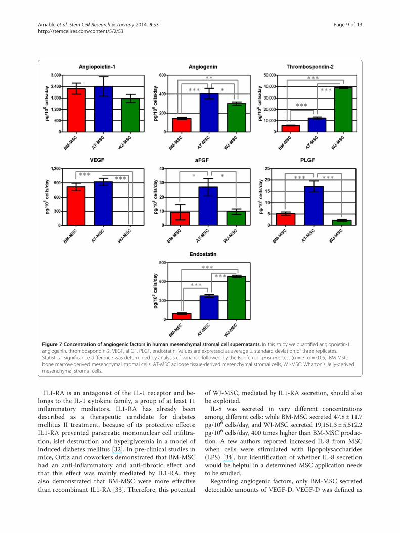
Figure 7 Concentration of angiogenic factors in human mesenchymal stromal cell supernatants. In this study we quantified angiopoietin-1,angiogenin, thrombospondin-2, VEGF, aFGF, PLGF, endostatin. Values are expressed as average ± standard deviation of three replicates.Statistical significance difference was determined by analysis of variance followed by the Bonferroni post-hoc test (n = 3, α = 0.05). BM-MSC:bone marrow-derived mesenchymal stromal cells, AT-MSC adipose tissue-derived mesenchymal stromal cells, WJ-MSC: Wharton’s Jelly-derivedmesenchymal stromal cells.
Amable et al. Stem Cell Research & Therapy 2014, 5:53 Page 9 of 13http://stemcellres.com/content/5/2/53
IL1-RA is an antagonist of the IL-1 receptor and be-longs to the IL-1 cytokine family, a group of at least 11inflammatory mediators. IL1-RA has already beendescribed as a therapeutic candidate for diabetesmellitus II treatment, because of its protective effects:IL1-RA prevented pancreatic mononuclear cell infiltra-tion, islet destruction and hyperglycemia in a model ofinduced diabetes mellitus [32]. In pre-clinical studies inmice, Ortiz and coworkers demonstrated that BM-MSChad an anti-inflammatory and anti-fibrotic effect andthat this effect was mainly mediated by IL1-RA; theyalso demonstrated that BM-MSC were more effectivethan recombinant IL1-RA [33]. Therefore, this potential
of WJ-MSC, mediated by IL1-RA secretion, should alsobe exploited.IL-8 was secreted in very different concentrations
among different cells: while BM-MSC secreted 47.8 ± 11.7pg/106 cells/day, and WJ-MSC secreted 19,151.3 ± 5,512.2pg/106 cells/day, 400 times higher than BM-MSC produc-tion. A few authors reported increased IL-8 from MSCwhen cells were stimulated with lipopolysaccharides(LPS) [34], but identification of whether IL-8 secretionwould be helpful in a determined MSC application needsto be studied.Regarding angiogenic factors, only BM-MSC secreted
detectable amounts of VEGF-D. VEGF-D was defined as

Figure 8 Secreted amounts of extracellular matrix components in human mesenchymal stromal cell supernatants. Here we reportresults obtained for collagen I, II and III, elastin, heparan sulfate, decorin, aggrecan and laminin. Values are expressed as average ± standarddeviation of three replicates. Statistical significance difference was determined by analysis of variance followed by the Bonferroni post-hoctest (n = 3, α = 0.05). BM-MSC: bone marrow-derived mesenchymal stromal cells, AT-MSC adipose tissue-derived mesenchymal stromal cells,WJ-MSC: Wharton’s Jelly-derived mesenchymal stromal cells.
Amable et al. Stem Cell Research & Therapy 2014, 5:53 Page 10 of 13http://stemcellres.com/content/5/2/53
the strongest angiogenic and lymphangiogenic VEGFisoform [35] and it has been tested in phase I clinical tri-als for myocardial infarction in a gene therapy approach(ClinicalTrials.gov; Identifier: NCT01002430).WJ-MSC produced almost undetectable amounts of
VEGF; on the other hand, AT- and BM-MSC were thehighest VEGF producers. Research articles like thosepublished by Deuse’s and Augustin’s groups showedthat MSC genetically modified to overexpress VEGF aremore appropriate than MSC alone for treating acute myo-cardial infarction, as VEGF extended MSC survival and pro-tected them against apoptosis and improved heart functionrecovery [36,37]. This information suggests that BM- and
AT-MSC would be better candidates for infarction therapythan WJ-MSC.Thrombospondin-2 was another angiogenic factor that
was found in high concentrations in all cell culture super-natants and WJ-MSC secreted the highest amounts. Jeongand colleagues have shown that WJ-MSC exerteda regenerative effect on cartilage and they suggestedthat this effect was mediated by thrombospondin-2, sincethis molecule alone could exert a similar effect andthrombospondin-2 knock-down in WJ-MSC using siRNAabolished its chondro-regenerative potential [38]. Con-sidering that AT-MSC also secreted high concentrationsof thrombospondin-2, they would be a good potential

Figure 9 Concentration of secreted matrix metalloproteinases (MMP1 and MMP3) in human mesenchymal stromal cell supernatants.Values are expressed as average ± standard deviation of three replicates. Statistical significance difference was determined by analysis of variancefollowed by the Bonferroni post-hoc test (n = 3, α = 0.05). BM-MSC: bone marrow-derived mesenchymal stromal cells, AT-MSC adiposetissue-derived mesenchymal stromal cells, WJ-MSC: Wharton’s Jelly-derived mesenchymal stromal cells.
Amable et al. Stem Cell Research & Therapy 2014, 5:53 Page 11 of 13http://stemcellres.com/content/5/2/53
candidate for cartilage regeneration, especially takinginto account autologous adipose tissue availability incases when the patients did not cryopreserve their ownumbilical cord cells.Angiopoietin-1 is an important pro-angiogenic factor
involved in vascular maturation and neovasculogenesis.Similar to what was described for VEGF, geneticallymodified MSC expressing angiopoietin-1 were moreeffective in regenerating myocardial tissue after in-farction than MSC alone [39]. Liu and coworkers alsodemonstrated that MSC expressed functional receptorsfor angiopoietin-1 and that angiopoietin-1 protectedMSC against apoptosis induced by hypoxia and serumdeprivation [40]. Angiopoietin-1 secretion was not sta-tistically different for all three MSC, but the secretionrate of WJ-MSC was lower than those of AT- andBM-MSC.PLGF displays a high homology with VEGF, binds to the
VEGF receptor and is able to augment the action of VEGFboth in vivo and in vitro. Liu and colleagues showed thatPLGF exerted a neuroprotective and angiogenic effect incerebral ischemia: BM-MSC injected intravenously re-duced lesion volume and induced angiogenesis while im-proving functional recovery, but the effect was greaterwhen BM-MSC were genetically modified to over-expressPLGF [41]. Considering our results, AT-MSC secretedhigher concentrations of PLGF: 3.3 and 7.8 times higherthan BM- and WJ-MSC. This origin-related differential ofAT-MSC would make them more appropriate for tissueregeneration after cerebral ischemia.When analyzing growth factor secretion, WJ-MSC se-
creted higher amounts of TGF-β2, HGF and PDGF-AAand was the only cell type secreting G-CSF in detectablelevels. HGF promotes migration, proliferation and survivalof a wide range of cell types and was shown to induce ex-pression of cardiac-specific markers in MSC [42]. G-CSF
is a potent chemotactic factor for MSC and has beensuccessfully used in different applications, such as acutemyocardial infarction [43] and injured brain [44].Regarding extracellular matrix components, AT-MSC se-
creted the highest amounts of collagen I, II, III and IV andWJ-MSC did not secrete any detectable amounts of colla-gen II and IV. WJ-MSC did not produce fibronectin, hepa-ran sulphate, decorin, laminin and aggrecan. Collagen II isthe most abundant protein in articular and hyaline cartil-age; therefore, WJ-MSC would not be appropriate for car-tilage regeneration since it would generate a fibrous tissuedue to its higher collagen III secretion. Collagen I and elas-tin are the main components of tendons, making AT-MSCthe most appropriate cell type for tendon regeneration.MSC are also key players in skin regeneration and agingprevention: they are attracted by PDGF-BB secreted byendothelial cells and they increase collagen IV secretion, acomponent of basement membranes [45]. AT-MSC werethe only cell type that secreted PDGF-BB in vitro and theyproduced the highest amounts of collagen IV, suggestingthey are the most appropriate cell type for skin regener-ation, since neither BM- nor WJ-MSC secreted PDGF-BBand collagen IV.MMP1, MMP3 and MMP13 are important enzymes in-
volved in the degradation of bone, cartilage and tendon.Serum and synovial levels of MMP1, MMP3 and MMP13are increased in rheumatoid arthritis and osteoarthritis[46,47]. Analyzing our results, AT-MSC produced thehighest concentrations of MMP1 and MMP3, this being anegative point for using AT-MSC in bone, cartilage andtendon applications. AT- and WJ-MSC secreted highamounts of MMP1 and MMP3, but low levels or noMMP13. A good candidate for bone, cartilage and tendonregeneration, when considering matrix metalloproteinases,would be BM-MSC, which secreted the lowest concentra-tions of MMP1 and MMP3 and no MMP13.

Amable et al. Stem Cell Research & Therapy 2014, 5:53 Page 12 of 13http://stemcellres.com/content/5/2/53
In summary, WJ-MSC showed a higher pro-inflammatoryand mitogenic profile, while AT-MSC secreted higherconcentrations of pro-angiogenic proteins, extracellularmatrix components and matrix metalloproteinases. Acquir-ing a deeper molecular knowledge about MSC from dif-ferent origins will help us make a more rational andeffective selection of the MSC for different applicationsin regenerative medicine.
ConclusionsOur data showed that MSC from different tissues havedifferent properties in vitro and this can be the reasonfor their different behavior in different clinical applica-tions. In conclusion, cell type for regenerative therapiesshould be carefully chosen. The present results need tobe compared with in vivo pre-clinical studies in order tosuggest the best protocols for mesenchymal stromal celluse in each clinical therapy.
Additional file
Additional file 1: Table S1. Protein concentration in humanmesenchymal stromal cell supernatants. Results are expressed inexpressed in mean pg/106 cells/day ± standard deviation of threereplicates. BM-MSC: bone marrow-derived mesenchymal stromal cells, AT-MSC adipose tissue-derived mesenchymal stromal cells, WJ-MSC:Wharton’s Jelly-derived mesenchymal stromal cells.
AbbreviationsaFGF: Acidic fibroblast growth factor; AT-MSC: Adipose tissue-derived MSC;bFGF: Basic fibroblast growth factor; BM-MSC: Bone marrow-derived MSC;DMEM: Dulbecco’s modified Eagle media; EGF: Endothelial growth factor;FBS: Fetal bovine serum; FCS: Forward scatter; G-CSF: Granulocyte colony-stimulating factor; GM-CSF: Granulocyte macrophage colony-stimulatingfactor; HGF: Hepatocyte growth factor; IFN: Interferon; IGF-1: Insulin growthfactor 1; IL: Interleukin; IP-10: Interferon gamma-induced protein 10;LG-DMEM: Low-glucose DMEM; MCP-1: Monocyte chemoattractant protein-1;MEM: Minimum essential media; MIG: Monokine induced by IFN-gamma;MIP-1: Macrophage inflammatory protein 1; MMP: Matrix metalloproteinase;MSC: Mesenchymal stromal cells; PBS: Phosphate-buffered saline;PDGF: Platelet-derived growth factor; PLGF: Placental growth factor;RANTES: Regulated on activation, normal T cell expressed and secreted;SSC: Side scatter; TGF: Transforming growth factor; TNF-α: Tumor necrosisfactor alpha; VEGF: Vascular endothelial growth factor; WJ-MSC: Wharton’sjelly-derived MSC.
Competing interestsPRA, MVTT, RBVC and RB are employees at Excellion Biomedical Services S.A.Results reported here have no connections or influence on the company’sproducts. The authors declared that no competing financial and/or privateinterests exist.
Authors’ contributionsPRA, MVTT, RBVC, RB and JMG participated in the conception and design ofthe study. PRA, RBVC and MVTT performed the experiments, collected andanalyzed the data. PRA performed the statistical analysis and wrote themanuscript. MTVV and RBVC revised the manuscript. JMG and RB revised andapproved the final manuscript. All authors read and approved the finalmanuscript.
AcknowledgementsThe authors thank professor Gutemberg Alves from the Clinical ResearchUnit, Antônio Pedro Hospital, Fluminense Federal University (Niterói, Rio deJaneiro, Brazil) for the support with the Luminex assays.
Author details1Excellion Biomedical Services S.A, Rua Afrânio de Mello Franco 333,25651000 PetrópolisRio de Janeiro, Brazil. 2National Institute of Metrology,Quality and Technology (Inmetro), Xerém, RJ, Brazil.
Received: 13 December 2013 Revised: 7 March 2014Accepted: 1 April 2014 Published: 16 April 2014
References1. Crisan M, Yap S, Casteilla L, Chen CW, Corselli M, Park TS, Andriolo G, Sun B,
Zheng B, Zhang L, Norotte C, Teng PN, Traas J, Schugar R, Deasy BM,Badylak S, Buhring HJ, Giacobino JP, Lazzari L, Huard J, Péault B:A perivascular origin for mesenchymal stem cells in multiple humanorgans. Cell Stem Cell 2008, 13:301–313.
2. Friedenstein J, Chailakhjan RK, Lalykina KS: The development of fibroblastcolonies in monolayer cultures of guinea-pig bone marrow and spleencells. Cell Tissue Kinet 1970, 3:393–403.
3. Zuk PA, Zhu M, Mizuno H, Huang J, Futrell JW, Katz AJ, Benhaim P, Lorenz HP,Hedrick MH: Multilineage cells from human adipose tissue: implications forcell-based therapies. Tissue Eng 2001, 7:211–228.
4. Miao Z, Jin J, Chen L, Zhu J, Huang W, Zhao J, Qian H, Zhang X: Isolation ofmesenchymal stem cells from human placenta: comparison with humanbone marrow mesenchymal stem cells. Cell Biol Int 2006, 30:681–687.
5. Griffiths MJ, Bonnet D, Janes SM: Stem cells of the alveolar epithelium.Lancet 2005, 366:249–260.
6. Wang HS, Hung SC, Peng ST, Huang CC, Wei HM, Guo YJ, Fu YS, Lai MC,Chen CC: Mesenchymal stem cells in the Wharton's jelly of the humanumbilical cord. Stem Cells 2004, 22:1330–1337.
7. De Bari C, Dell'Accio F, Tylzanowski P, Luyten FP: Multipotentmesenchymal stem cells from adult human synovial membrane. ArthritisRheum 1928–1942, 2001:44.
8. Gronthos S, Mankani M, Brahim J, Robey PG, Shi S: Postnatal human dentalpulp stem cells (DPSCs) in vitro and in vivo. Proc Natl Acad Sci U S A 2000,97:13625–13630.
9. Kholodenko IV, Konieva AA, Kholodenko RV, Yarygin KN: Molecularmechanisms of migration and homing of intravenously transplantedmesenchymal stem cells. J Regen Med Tissue Eng 2013, 2:4–11.
10. Galindo LT, Filippo TR, Semedo P, Ariza CB, Moreira CM, Camara NO,Porcionatto MA: Mesenchymal stem cell therapy modulates theinflammatory response in experimental traumatic brain injury. Neurol ResInt 2011, 2011:564089.
11. Nivet E, Vignes M, Girard SD, Pierrisnard C, Baril N, Devèze A, Magnan J,Lanté F, Khrestchatisky M, Féron F, Roman FS: Engraftment of human nasalolfactory stem cells restores neuroplasticity in mice with hippocampallesions. J Clin Invest 2011, 121:2808–2820.
12. Lee JS, Hong JM, Moon GJ, Lee PH, Ahn YH: Bang OY; STARTINGcollaborators: A long-term follow-up study of intravenous autologousmesenchymal stem cell transplantation in patients with ischemic stroke.Stem Cells 2012, 28:1099–1106.
13. Le Blanc K, Tammik C, Rosendahl K, Zetterberg E, Ringdén O: HLAexpression and immunologic properties of differentiated andundifferentiated mesenchymal stem cells. Exp Hematol 2003, 31:890–896.
14. Ryan JM, Barry FP, Murphy JM, Mahon BP: Mesenchymal stem cells avoidallogeneic rejection. J Inflamm (Lond) 2005, 2:8.
15. Al-Nbaheen M, Vishnubalaji R, Ali D, Bouslimi A, Al-Jassir F, Megges M,Prigione A, Adjaye J, Kassem M, Aldahmash A: Human stromal(mesenchymal) stem cells from bone marrow, adipose tissue and skinexhibit differences in molecular phenotype and differentiation potential.Stem Cell Rev 2013, 9:32–43.
16. Sakaguchi Y, Sekiya I, Yagishita K, Muneta T: Comparison of human stemcells derived from various mesenchymal tissues: superiority of synoviumas a cell source. Arthritis Rheum 2005, 52:2521–2529.
17. Wang L, Tran I, Seshareddy K, Weiss ML, Detamore MS: A comparison ofhuman bone marrow-derived mesenchymal stem cells and humanumbilical cord-derived mesenchymal stromal cells for cartilage tissueengineering. Tissue Eng Part A 2009, 15:2259–2266.
18. Ramkisoensing AA, Pijnappels DA, Askar SFA, Passier R, Swildens J, GoumansMJ, Schutte CI, de Vries AA, Scherjon S, Mummery CL, Schalij MJ, Atsma D:Human embryonic and fetal mesenchymal stem cells differentiatetoward three different cardiac lineages in contrast to their adultcounterparts. PLoS ONE 2011, 6:e24164.

Amable et al. Stem Cell Research & Therapy 2014, 5:53 Page 13 of 13http://stemcellres.com/content/5/2/53
19. Hsieh JY, Wang HW, Chang SJ, Liao KH, Lee IH, Li WS, Wu CH, Lin WY,Cheng SM: Mesenchymal stem cells from human umbilical cord expresspreferentially secreted factors related to neuroprotection, neurogenesis,and angiogenesis. PLoS ONE 2013, 8:e72604.
20. Naftali-Shani N, Itzhaki-Alfia A, Landa-Rouben N, Kain D, Holbova R,Adutler-Lieber S, Molotski N, Asher E, Grupper A, Millet E, Tessone A,Winkler E, Kastrup J, Feinberg MS, Zipori D, Pevsner-Fischer M, Raanani E,Leor J: The origin of human mesenchymal stromal cells dictates theirreparative properties. J Am Heart Assoc 2013, 2:e000253.
21. Amable PR, Teixeira MV, Carias RB, Granjeiro JM, Borojevic R: Identificationof appropriate reference genes for human mesenchymal cells duringexpansion and differentiation. PLoS One 2013, 8:e73792.
22. Hsieh JY, Fu YS, Chang SJ, Tsuang YH, Wang HW: Functional moduleanalysis reveals differential osteogenic and stemness potentials inhuman mesenchymal stem cells from bone marrow and Wharton's jellyof umbilical cord. Stem Cells Dev 1895–1910, 2010:19.
23. Deuse T, Stubbendorff M, Tang-Quan K, Phillips N, Kay MA, Eiermann T, PhanTT, Volk HD, Reichenspurner H, Robbins RC, Schrepfer S: Immunogenicity andimmunomodulatory properties of umbilical cord lining mesenchymal stemcells. Cell Transplant 2011, 20:655–667.
24. Borghesi A, Cova C, Gazzolo D, Stronati M: Stem cell therapy for neonataldiseases associated with preterm birth. J Clin Neonatol 2013, 2:1–7.
25. Messerli M, Wagner A, Sager R, Mueller M, Baumann M, Surbek DV,Schoeberlein A: Stem cells from umbilical cord Wharton's jelly frompreterm birth have neuroglial differentiation potential. Reprod Sci 2013,20:1455–1464.
26. Yang L, Froio RM, Sciuto TE, Dvorak AM, Alon R, Luscinskas FW: ICAM-1regulates neutrophil adhesion and transcellular migration of TNF-alpha-activated vascular endothelium under flow. Blood 2005, 106:584–592.
27. Russell KC, Phinney DG, Lacey MR, Barrilleaux BL, Meyertholen KE, O'ConnorKC: In vitro high-capacity assay to quantify the clonal heterogeneity intrilineage potential of mesenchymal stem cells reveals a complexhierarchy of lineage commitment. Stem Cells 2010, 28:788–798.
28. Ridker PM, Rifai N, Stampfer MJ, Hennekens CH: Plasma concentrationof interleukin-6 and the risk of future myocardial infarction amongapparently healthy men. Circulation 2000, 101:1767–1772.
29. Li P, Li SH, Wu J, Zang WF, Dhingra S, Sun L, Weisel RD, Li RK: Interleukin-6downregulation with mesenchymal stem cell differentiation results inloss of immunoprivilege. J Cell Mol Med 2013, 17:1136–1145.
30. Pricola KL, Kuhn NZ, Haleem-Smith H, Song Y, Tuan RS: Interleukin-6maintains bone marrow-derived mesenchymal stem cell stemness byan ERK1/2-dependent mechanism. J Cell Biochem 2009, 108:577–588.
31. Nasir GA, Mohsin S, Khan M, Shams S, Ali G, Khan SN, Riazuddin S:Mesenchymal stem cells and interleukin-6 attenuate liver fibrosis inmice. J Transl Med 2013, 11:78.
32. Sandberg JO, Andersson A, Eizirik DL, Sandler S: Interleukin-1 receptorantagonist prevents low dose streptozotocin induced diabetes in mice.Biochem Biophys Res Commun 1994, 202:543–548.
33. Ortiz LA, Dutreil M, Fattman C, Pandey AC, Torres G, Go K, Phinney DG:Interleukin 1 receptor antagonist mediates the antiinflammatory andantifibrotic effect of mesenchymal stem cells during lung injury.Proc Natl Acad Sci U S A 2007, 104:11002–11007.
34. Kilroy GE, Foster SJ, Wu X, Ruiz J, Sherwood S, Heifetz A, Ludlow JW, StrickerDM, Potiny S, Green P, Halvorsen YD, Cheatham B, Storms RW, Gimble JM:Cytokine profile of human adipose-derived stem cells: expression ofangiogenic, hematopoietic, and pro-inflammatory factors. J Cell Physiol2007, 212:702–709.
35. Rissanen TT, Markkanen JE, Gruchala M, Heikura T, Puranen A, Kettunen MI,Kholová I, Kauppinen RA, Achen MG, Stacker SA, Alitalo K, Ylä-Herttuala S:VEGF-D is the strongest angiogenic and lymphangiogenic effectoramong VEGFs delivered into skeletal muscle via adenoviruses. Circ Res2003, 92:1098–1106.
36. Deuse T, Peter C, Fedak PW, Doyle T, Reichenspurner H, Zimmermann WH,Eschenhagen T, Stein W, Wu JC, Robbins RC, Schrepfer S: Hepatocytegrowth factor or vascular endothelial growth factor gene transfermaximizes mesenchymal stem cell-based myocardial salvage afteracute myocardial infarction. Circulation 2009, 120:S247–S254.
37. Augustin M, Mahar MA, Lakkisto P, Tikkanen I, Vento A, Pätilä T, Harjula A:VEGF overexpression improves mesenchymal stem cell sheettransplantation therapy for acute myocardial infarction. J Tissue EngRegen Med 2013, 7:742–750.
38. Jeong SY, Kim DH, Ha J, Jin HJ, Kwon SJ, Chang JW, Choi SJ, Oh W, Yang YS,Kim G, Kim JS, Yoon JR, Cho DH, Jeon HB: Thrombospondin-2 secreted byhuman umbilical cord blood-derived mesenchymal stem cells promoteschondrogenic differentiation. Stem Cells 2013, 31:2136–2148.
39. Paul A, Nayan M, Khan AA, Shum-Tim D, Prakash S: Angiopoietin-1-expressingadipose stem cells genetically modified with baculovirus nanocomplex:investigation in rat heart with acute infarction. Int J Nanomedicine 2012,7:663–682.
40. Liu XB, Jiang J, Gui C, Hu XY, Xiang MX, Wang JA: Angiopoietin-1 protectsmesenchymal stem cells against serum deprivation and hypoxia-inducedapoptosis through the PI3K/Akt pathway. Acta Pharmacol Sin 2008,29:815–822.
41. Liu H, Honmou O, Harada K, Nakamura K, Houkin K, Hamada H, Kocsis JD:Neuroprotection by PlGF gene-modified human mesenchymal stem cellsafter cerebral ischaemia. Brain 2006, 129:2734–2745.
42. Forte G, Minieri M, Cossa P, Antenucci D, Sala M, Gnocchi V, Fiaccavento R,Carotenuto F, De Vito P, Baldini PM, Prat M, Di Nardo P: Hepatocyte growthfactor effects on mesenchymal stem cells: proliferation, migration, anddifferentiation. Stem Cells 2006, 24:23–33.
43. Kawada H, Fujita J, Kinjo K, Matsuzaki Y, Tsuma M, Miyatake H, Muguruma Y,Tsuboi K, Itabashi Y, Ikeda Y, Ogawa S, Okano H, Hotta T, Ando K, Fukuda K:Nonhematopoietic mesenchymal stem cells can be mobilized anddifferentiate into cardiomyocytes after myocardial infarction. Blood 2004,104:3581–3587.
44. Deng J, Zou ZM, Zhou TL, Su YP, Ai GP, Wang JP, Xu H, Dong SW: Bonemarrow mesenchymal stem cells can be mobilized into peripheral bloodby G-CSF in vivo and integrate into traumatically injured cerebral tissue.Neurol Sci 2011, 32:641–651.
45. Soma T, Yamanishi H, Ishimatsu-Tsuji Y, Fujiwara S: Role of PDGF-BB in thebehavior of mesenchymal stem cells in human skin. J Invest Dermatol2012, 132:S135–S148.
46. Lindy O, Konttinen YT, Sorsa T, Ding Y, Santavirta S, Ceponis A, López-OtínC: Matrix metalloproteinase 13 (collagenase 3) in human rheumatoidsynovium. Arthritis Rheum 1997, 40:1391–1399.
47. Mahmoud RK, El-Ansary AK, El-Eishi HH, Kamal HM, El-Saeed NH: Matrixmetalloproteinases MMP-3 and MMP-1 levels in sera and synovial fluidsin patients with rheumatoid arthritis and osteoarthritis. Ital J Biochem2005, 54:248–257.
doi:10.1186/scrt442Cite this article as: Amable et al.: Protein synthesis and secretion inhuman mesenchymal cells derived from bone marrow, adipose tissueand Wharton’s jelly. Stem Cell Research & Therapy 2014 5:53.
Submit your next manuscript to BioMed Centraland take full advantage of:
• Convenient online submission
• Thorough peer review
• No space constraints or color figure charges
• Immediate publication on acceptance
• Inclusion in PubMed, CAS, Scopus and Google Scholar
• Research which is freely available for redistribution
Submit your manuscript at www.biomedcentral.com/submit
Copyright © 2022 FDOKUMEN