Regional Differences in Adipose Tissue of the Sinclair Minipig
Upload
independentCategory
view
7download
0

123
Advances in Biochemical
Engineering/Biotechnology
Series Editor: T. Scheper
Editorial Board:
S. Belkin l I. Endo l S.-O. Enfors l W.-S. Hu l
B. Mattiasson l J. Nielsen l G. Stephanopoulos l G. T. Tsao
R. Ulber l A.-P. Zeng l J.-J. Zhong l W. Zhou

Advances in Biochemical Engineering/Biotechnology
Series Editor: T. Scheper
Recently Published and Forthcoming Volumes
Bioreactor Systems for TissueEngineering IIStrategies for the Expansion and DirectedDifferentiation of Stem CellsVolume Editors: Cornelia Kasper,Martijn van Griensven, Ralf PortnerVol. 123, 2010
Biotechnology in China IIChemicals, Energy and EnvironmentVolume Editors: Tsao, G.T., Ouyang, P.,Chen, J.Vol. 122, 2010
Biosystems Engineering IILinking Cellular Networks and BioprocessesVolume Editors: Wittmann, C., Krull, R.Vol. 121, 2010
Biosystems Engineering ICreating Superior BiocatalystsVolume Editors: Wittmann, C., Krull, R.Vol. 120, 2010
Whole Cell Sensing Systems IIVolume Editors: Belkin, S., Gu, M.B.Vol. 118, 2010
Whole Cell Sensing Systems IVolume Editors: Belkin, S., Gu, M.B.Vol. 117, 2010
Optical Sensor Systems in BiotechnologyVolume Editor: Rao, G.Vol. 116, 2009
Disposable BioreactorsVolume Editor: Eibl, R., Eibl, D.Vol. 115, 2009
Engineering of Stem CellsVolume Editor: Martin, U.Vol. 114, 2009
Biotechnology in China IFrom Bioreaction to Bioseparation andBioremediationVolume Editors: Zhong, J.J., Bai, F.-W.,Zhang, W.Vol. 113, 2009
Bioreactor Systems for TissueEngineeringVolume Editors: Kasper, C.,van Griensven, M., Poertner, R.Vol. 112, 2008
Food BiotechnologyVolume Editors: Stahl, U.,Donalies, U. E. B., Nevoigt, E.Vol. 111, 2008
Protein – Protein InteractionVolume Editors: Seitz, H., Werther, M.Vol. 110, 2008
Biosensing for the 21st CenturyVolume Editors: Renneberg, R., Lisdat, F.Vol. 109, 2007
BiofuelsVolume Editor: Olsson, L.Vol. 108, 2007
Green Gene TechnologyResearch in an Area of Social ConflictVolume Editors: Fiechter, A., Sautter, C.Vol. 107, 2007
White BiotechnologyVolume Editors: Ulber, R., Sell, D.Vol. 105, 2007
Analytics of Protein-DNA InteractionsVolume Editor: Seitz, H.Vol. 104, 2007
Tissue Engineering IIBasics of Tissue Engineering and TissueApplicationsVolume Editors: Lee, K., Kaplan, D.Vol. 103, 2007

Bioreactor Systems forTissue Engineering II
Strategies for the Expansion and DirectedDifferentiation of Stem Cells
Volume Editors:Cornelia Kasper � Martijn van Griensven � Ralf Portner
With contributions by
P. Adamietz �W. Brehm � J. Burk � R. Cancedda � T. Cantz �Y.-H. Choi � P. Czermak � U. Delling � E. Dohle �R. M. El Backly � M. A. Esteban � D. Freimark � S. Fuchs �C.Geißler � P. Geigle �C.Gittel �C.Goepfert �M.Griensven �R. Grillari-Voglauer � R. Hass � T. Hatlapatka � H. Julke �C. Kasper � C. J. Kirkpatrick � K. Klose � M. Kolbe �J. W. Kuhbier � A. Lavrentieva � I. Majore � D. Marten �U. Martin � P. Moretti � D. Pei � A. Peterbauer-Scherb �P. Pino-Grace �R. Poertner � S. Pohl �R. Portner K. Reimers �I. Ribitsch � A. F. Schilling � A. Slobodianski � C. Stamm �H.-F. Tse � P. M. Vogt � C. Wallrapp � C. Weber �B. Weyand � S. Wolbank � J. Xu � Q. Zhuang

EditorsPD Dr. Cornelia KasperUniversitat HannoverInst. Technische ChemieCallinstr. 330167 [email protected]
Prof. Dr. Ralf PortnerTU Hamburg-HarburgInst.Biotechnologie u.VerfahrenstechnikDenickestr. 1521073 [email protected]
Prof. Martijn van GriensvenLudwig Boltzmann Institut furKlinische und ExperimentelleTraumatologieDonaueschingenstr. 131200 [email protected]
ISSN 0724-6145 e-ISSN 1616-8542ISBN 978-3-642-16050-9 e-ISBN 978-3-642-16051-6DOI 10.1007/978-3-642-16051-6Springer Heidelberg Dordrecht London New York
Library of Congress Control Number: 2008939436
# Springer-Verlag Berlin Heidelberg 2010This work is subject to copyright. All rights are reserved, whether the whole or part of the material isconcerned, specifically the rights of translation, reprinting, reuse of illustrations, recitation, broadcasting,reproduction on microfilm or in any other way, and storage in data banks. Duplication of this publicationor parts thereof is permitted only under the provisions of the German Copyright Law of September 9,1965, in its current version, and permission for use must always be obtained from Springer. Violationsare liable to prosecution under the German Copyright Law.The use of general descriptive names, registered names, trademarks, etc. in this publication does notimply, even in the absence of a specific statement, that such names are exempt from the relevantprotective laws and regulations and therefore free for general use.
Cover design: WMXDesign GmbH, Heidelberg, Germany
Printed on acid-free paper
Springer is part of Springer Science+Business Media (www.springer.com)

Series Editor
Prof. Dr. T. Scheper
Institute of Technical Chemistry
University of Hannover
Callinstraße 3
30167 Hannover, Germany
Volume Editors
PD Dr. Cornelia Kasper
Universitat Hannover
Inst. Technische Chemie
Callinstr. 3
30167 Hannover
Germany
Prof. Dr. Ralf Portner
TU Hamburg-Harburg
Inst.Biotechnologie u.
Verfahrenstechnik
Denickestr. 15
21073 Hamburg
Germany
Prof. Martijn van Griensven
Ludwig Boltzmann Institut fur
Klinische und Experimentelle
Traumatologie
Donaueschingenstr. 13
1200 Wien
Austria
Editorial Board
Prof. Dr. S. Belkin
Interfaculty Biotechnology Program
Institute of Life Sciences
The Hebrew University of Jerusalem
Jerusalem 91904, Israel
Prof. Dr. I. Endo
Saitama Industrial Technology Center
3-12-18, Kamiaoki Kawaguchi-shi
Saitama, 333-0844, Japan
Prof. Dr. W.-S. Hu
Chemical Engineering
and Materials Science
University of Minnesota
421Washington Avenue SE
Minneapolis, MN 55455-0132, USA
Prof. Dr. B. Mattiasson
Department of Biotechnology
Chemical Center, Lund University
P.O. Box 124, 221 00 Lund, Sweden

Prof. Dr. S.-O. Enfors
Department of Biochemistry
and Biotechnology
Royal Institute of Technology
Teknikringen 34,
100 44 Stockholm, Sweden
Prof. Dr. G. Stephanopoulos
Department of Chemical Engineering
Massachusetts Institute of Technology
Cambridge, MA 02139-4307, USA
Prof. Dr. G. T. Tsao
Professor Emeritus
Purdue University
West Lafayette, IN 47907, USA
[email protected]@yahoo.com
Prof. Dr. Roland Ulber
FB Maschinenbau und Verfahrenstechnik
Technische Universitat Kaiserslautern
Gottlieb-Daimler-Straße
67663 Kaiserslautern, Germany
Prof. Dr. A.-P. Zeng
Technische Universitat Hamburg-Harburg
Institut fur Bioprozess- und Biosystem-
technik
Denickestrasse 1
21073 Hamburg, Germany
Honorary Editors
Prof. Dr. A. Fiechter
Institute of Biotechnology
Eidgenossische Technische Hochschule
ETH-Honggerberg
8093 Zurich, Switzerland
Prof. Dr. J. Nielsen
Chalmers University of Technology
Department of Chemical and Biological
Engineering
Systems Biology
Kemivagen 10
41296 Goteborg
Sweden
Prof. Dr. J.-J. Zhong
Bio-Building #3-311
College of Life Science & Biotechnology
Key Laboratory of Microbial Metabolism,
Ministry of Education
Shanghai Jiao Tong University
800 Dong-Chuan Road
Minhang, Shanghai 200240, China
Dr. W. Zhou
Sr. Director, BioProcess Engineering
Technology Development
Genzyme Corporation
45 New York Avenue
Framingham, MA 01701-9322, USA
Prof. Dr. K. Schugerl
Institute of Technical Chemistry
University of Hannover, Callinstraße 3
30167 Hannover, Germany
vi Editorial Board

Advances in Biochemical Engineering/
Biotechnology Also Available Electronically
Advances in Biochemical Engineering/Biotechnology is included in Springer’s
eBook package Chemistry and Materials Science. If a library does not opt for the
whole package the book series may be bought on a subscription basis. Also, all back
volumes are available electronically.
For all customers who have a standing order to the print version of Advances inBiochemical Engineering/Biotechnology, we offer free access to the electronic
volumes of the Series published in the current year via SpringerLink.
If you do not have access, you can still view the table of contents of each volume
and the abstract of each article by going to the SpringerLink homepage, clicking
on “Chemistry and Materials Science,” under Subject Collection, then “Book
Series,” under Content Type and finally by selecting Advances in BiochemicalBioengineering/Biotechnology
You will find information about the
– Editorial Board
– Aims and Scope
– Instructions for Authors
– Sample Contribution
at springer.com using the search function by typing in Advances in BiochemicalEngineering/Biotechnology.
Color figures are published in full color in the electronic version on SpringerLink.
vii

Aims and Scope
Advances in Biochemical Engineering/Biotechnology reviews actual trends in
modern biotechnology.
Its aim is to cover all aspects of this interdisciplinary technologywhere knowledge,
methods and expertise are required for chemistry, biochemistry, microbiology,
genetics, chemical engineering and computer science.
Special volumes are dedicated to selected topics which focus on new biotechno-
logical products and new processes for their synthesis and purification. They give the
state-of-the-art of a topic in a comprehensive way thus being a valuable source for the
next 3-5 years. It also discusses new discoveries and applications.
In general, special volumes are edited by well known guest editors. The series
editor and publisher will however always be pleased to receive suggestions and
supplementary information. Manuscripts are accepted in English.
In references Advances in Biochemical Engineering/Biotechnology is abbreviatedas Adv. Biochem. Engin./Biotechnol. and is cited as a journal.
Special volumes are edited bywell known guest editors who invite reputed authors for
the review articles in their volumes.
Impact Factor in 2009: 4.165; Section “Biotechnology and Applied Microbiology”:
Rank 23 of 150
viii Advances in Biochemical Engineering/Biotechnology

Attention all Users
of the “Springer Handbook of Enzymes”
Information on this handbook can be found on the internet at springeronline.com
A complete list of all enzyme entries either as an alphabetical Name Index or as the
EC-Number Index is available at the above mentioned URL. You can download
and print them free of charge.
A complete list of all synonyms (more than 57,000 entries) used for the enzymes is
available in print form (ISBN 978-3-642-14015-0) and electronic form (ISBN 978-
3-642-14016-7).
Save 15%We recommend a standing order for the series to ensure you automatically receive
all volumes and all supplements and save 15% on the list price.
ix

.

Preface
First of all, the editors of this special volume would like to thank all the authors for
their excellent contributions. We would also like to thank Prof. Dr. Thomas Scheper
as well as Dr.Marion Hertel and Ingrid Samide from Springer for providing the oppor-
tunity to compose this volume and Springer for organizational and technical support.
Tissue engineering represents one of the major emerging fields in modern bio-
technology. Tissue engineering combines different disciplines ranging from bio-
logical and material sciences to engineering and clinical disciplines. The aim of
tissue engineering is the development of therapeutic approaches to substitute
diseased organs or tissues or improve their function. Stem cells are early progeni-
tors that may substitute diseased tissues or provide cues for endogenous healing.
Stem cells are present in virtually all tissues. The first chapters describe different
sources of stem cells including isolation and expansion. The use of fetal tissues
and umbilical cord is discussed as they come from immunoprivileged sites and
are considered to be early stem cells. The use of adipose-derived stem cells is dis-
cussed as a readily available autologous source. Subsequently, newer techniques for
“manufacturing” stem cells from somatic cells using “induced pluripotent stem cell”
technology are discussed and described in two chapters. The following chapter deals
with bioreactor cultivation of stem cells. Specific tissues such as cartilage and
endothelial precursors built the bridge to the last chapters. In those chapters, clinical
applications are the focus of interest. It covers a wide range of clinical applications
from veterinary orthopedics and human bone diseases until cardiologic applications.
This small overview indicates that we have tried to cover the area of stem cells
from isolation, expansion up to clinical applications. The road has been walked
already for a substantial distance. However, we are still at the beginning of this
exciting new technology.
We hope that this state-of-the-art book is helpful to your research. Please enjoy
reading it, as much as we enjoyed preparing it.
Summer 2010 Cornelia Kasper
Ralf Portner
Martijn van Griensven
xi

.

Contents
Alternative Sources of Adult Stem Cells: Human Amniotic Membrane . . . . 1
Susanne Wolbank, Martijn van Griensven, Regina Grillari-Voglauer,
and Anja Peterbauer-Scherb
Mesenchymal Stromal Cells Derived from Human Umbilical Cord
Tissues: Primitive Cells with Potential for Clinical and Tissue
Engineering Applications . . . . . . . . . . . . . . . . . . . . . . . . . . . . . . . . . . . . . . . . . . . . . . . . . . . . . 29
Pierre Moretti, Tim Hatlapatka, Dana Marten, Antonina Lavrentieva,
Ingrida Majore, Ralf Hass, and Cornelia Kasper
Isolation, Characterization, Differentiation, and Application
of Adipose-Derived Stem Cells . . . . . . . . . . . . . . . . . . . . . . . . . . . . . . . . . . . . . . . . . . . . . . . 55
Jorn W. Kuhbier, Birgit Weyand, Christine Radtke, Peter M. Vogt,
Cornelia Kasper, and Kerstin Reimers
Induced Pluripotent Stem Cells: Characteristics and Perspectives . . . . . . 107
Tobias Cantz and Ulrich Martin
Induced Pluripotent Stem Cell Technology in Regenerative
Medicine and Biology . . . . . . . . . . . . . . . . . . . . . . . . . . . . . . . . . . . . . . . . . . . . . . . . . . . . . . . . 127
Duanqing Pei, Jianyong Xu, Qiang Zhuang, Hung-Fat Tse,
and Miguel A. Esteban
Production Process for Stem Cell Based Therapeutic Implants:
Expansion of the Production Cell Line and Cultivation
of Encapsulated Cells . . . . . . . . . . . . . . . . . . . . . . . . . . . . . . . . . . . . . . . . . . . . . . . . . . . . . . . . 143
C. Weber, S. Pohl, R. Poertner, Pablo Pino-Grace, D. Freimark,
C. Wallrapp, P. Geigle, and P. Czermak
Cartilage Engineering from Mesenchymal Stem Cells . . . . . . . . . . . . . . . . . . . . 163
C. Goepfert, A. Slobodianski, A.F. Schilling, P. Adamietz, and R. Portner
xiii

Outgrowth Endothelial Cells: Sources, Characteristics and Potential
Applications in Tissue Engineering and Regenerative Medicine . . . . . . . . . 201
Sabine Fuchs, Eva Dohle, Marlen Kolbe, and Charles James Kirkpatrick
Basic Science and Clinical Application of Stem Cells
in Veterinary Medicine . . . . . . . . . . . . . . . . . . . . . . . . . . . . . . . . . . . . . . . . . . . . . . . . . . . . . . 219
I. Ribitsch, J. Burk, U. Delling, C. Geißler, C. Gittel, H. Julke, and W. Brehm
Bone Marrow Stem Cells in Clinical Application: Harnessing
Paracrine Roles and Niche Mechanisms . . . . . . . . . . . . . . . . . . . . . . . . . . . . . . . . . . . 265
Rania M. El Backly and Ranieri Cancedda
Clinical Application of Stem Cells in the Cardiovascular System . . . . . . . 293
Christof Stamm, Kristin Klose, and Yeong-Hoon Choi
Index . . . . . . . . . . . . . . . . . . . . . . . . . . . . . . . . . . . . . . . . . . . . . . . . . . . . . . . . . . . . . . . . . . . . . . . . . . 319
xiv Contents

Adv Biochem Engin/Biotechnol (2010) 123: 1–27DOI: 10.1007/10_2010_71# Springer-Verlag Berlin Heidelberg 2010Published online: 17 March 2010
Alternative Sources of Adult Stem Cells:
Human Amniotic Membrane
Susanne Wolbank, Martijn van Griensven, Regina Grillari-Voglauer,
and Anja Peterbauer-Scherb
Abstract Human amniotic membrane is a highly promising cell source for tissue
engineering. The cells thereof, human amniotic epithelial cells (hAEC) and human
amniotic mesenchymal stromal cells (hAMSC), may be immunoprivileged, they
represent an early developmental status, and their application is ethically uncontro-
versial. Cell banking strategies may use freshly isolated cells or involve in vitroexpansion to increase cell numbers. Therefore, we have thoroughly characterized
the effect of in vitro cultivation on both phenotype and differentiation potential of
hAEC. Moreover, we present different strategies to improve expansion including
replacement of animal-derived supplements by human platelet products or the
introduction of the catalytic subunit of human telomerase to extend the in vitrolifespan of amniotic cells. Characterization of the resulting cultures includes phe-
notype, growth characteristics, and differentiation potential, as well as immuno-
genic and immunomodulatory properties.
Keywords Adipogenesis, Expansion, hTERT, Human amniotic cells, Immor-
talization, Immunomodulation, Immunophenotype, Mesenchymal markers,
Osteogenesis, Platelet lysate, Stem cell markers, Telomerase
S. Wolbank, M. van Griensven (*)
Ludwig Boltzmann Institute for Experimental and Clinical Traumatology, Donaueschingenstraße
13, 1200 Vienna, Austria
Austrian Cluster for Tissue Regeneration, Vienna, Austria
e-mail: [email protected]
A. Peterbauer-Scherb
Red Cross Blood Transfusion Service of Upper Austria, Krankenhausstrasse 7, 4020 Linz, Austria
R. Grillari-Voglauer
Department of Biotechnology, Institute of Applied Microbiology, University of Natural Resources
and Applied Life Sciences, Muthgasse 18, 1190 Vienna, Austria

Contents
1 Introduction . . . . . . . . . . . . . . . . . . . . . . . . . . . . . . . . . . . . . . . . . . . . . . . . . . . . . . . . . . . . . . . . . . . . . . . . . . . . . . . . . 3
1.1 Stem Cell Characteristics of Amnion-Derived Cells . . . . . . . . . . . . . . . . . . . . . . . . . . . . . . . . . . 4
1.2 Expansion and Cryoconservation of Amnion-Derived Cell: Towards Cell Banking . . 4
2 Stem Cell Characteristics and Immunomodulatory Potential of Human
Amnion-Derived Stem Cells . . . . . . . . . . . . . . . . . . . . . . . . . . . . . . . . . . . . . . . . . . . . . . . . . . . . . . . . . . . . . . . . 7
2.1 Isolation of Separate Populations of hAEC and hAMSC . . . . . . . . . . . . . . . . . . . . . . . . . . . . . 7
2.2 Stem Cell Characteristics and Immunomodulation of hAEC and hAMSC . . . . . . . . . . . 8
3 Phenotypic Shift and Reduced Osteogenesis During In Vitro Expansion
of Human Amnion Epithelial Cells . . . . . . . . . . . . . . . . . . . . . . . . . . . . . . . . . . . . . . . . . . . . . . . . . . . . . . . 10
3.1 Shift in Surface Antigen Expression During Cultivation of hAEC . . . . . . . . . . . . . . . . . 10
3.2 Osteogenic Differentiation Potential of hAEC Decreases
upon In Vitro Cultivation . . . . . . . . . . . . . . . . . . . . . . . . . . . . . . . . . . . . . . . . . . . . . . . . . . . . . 12
4 Platelet Lysate for FCS-Free Expansion and Cryoconservation of Amnion-Derived Cells . . . 12
5 hTERT Induced Extension of In Vitro Life Span of Amnion-Derived Stem Cells . . . . . . . 14
6 Discussion . . . . . . . . . . . . . . . . . . . . . . . . . . . . . . . . . . . . . . . . . . . . . . . . . . . . . . . . . . . . . . . . . . . . . . . . . . . . . . . . 19
6.1 Stem Cell Characteristics and Immunomodulatory Potential of Human
Amnion-Derived Stem Cells . . . . . . . . . . . . . . . . . . . . . . . . . . . . . . . . . . . . . . . . . . . . . . . . . . . . . . . . 19
6.2 Phenotypic Shift and Reduced Osteogenesis During In Vitro Expansion of
Human Amnion Epithelial Cells . . . . . . . . . . . . . . . . . . . . . . . . . . . . . . . . . . . . . . . . . . . . . . . . . . . . 20
6.3 Platelet Lysate for FCS-Free Expansion and Cryoconservation of
Amnion-Derived Cells . . . . . . . . . . . . . . . . . . . . . . . . . . . . . . . . . . . . . . . . . . . . . . . . . . . . . . . . . 22
6.4 hTERT Induced Extension of In Vitro Life Span of Amnion-Derived Stem Cells . . . 22
References . . . . . . . . . . . . . . . . . . . . . . . . . . . . . . . . . . . . . . . . . . . . . . . . . . . . . . . . . . . . . . . . . . . . . . . . . . . . . . . . . . . . . 23
Abbreviations
7-AAD 7-Amino-actinomycin D
ALPL Alkaline phosphatase gene
AP Alkaline phosphatase
AR Alizarin red
ASC Adipose-derived stem cells
BGLAP Bone gamma-carboxyglutamate protein
BMPR1B Bone morphogenetic protein receptor 1B
BMPR2 Bone morphogenetic protein receptor 2
BMSC Bone marrow mesenchymal stem cells
BrdU 5-Bromo-2-deoxy uridine
CBFA1 Core binding factor alpha
DMSO Dimethylsulfoxide
EDTA Ethylenediaminetetraacetic acid
ELISA Enzyme-linked immunosorbent asssay
FCS Fetal calf serum
FOI Fold of induction
hAEC Human amniotic epithelial cells
hAMSC Human amniotic mesenchymal stromal cells
2 S. Wolbank et al.

HPRT Hypoxanthine-guanine phosphoribosyltransferase
hTERT Human telomerase reverse transcriptase
Lep Leptin
MLR Mixed lymphocyte reaction
MSC Mesenchymal stem cells
O-Kit Mesenchymal stem cell osteogenic stimulatory kit
OO Oil red O
PBMC Peripheral blood mononuclear cells
PBS Phosphate buffered saline
PCR Polymerase chain reaction
PD Population doubling
PDpT Population doubling post transduction
PHA Phytohemagglutinin
PL Platelet lysate
PPARg Peroxisome proliferator-activated receptor gamma
PRP Platelet-rich plasma
pT Post transduction
RT-PCR Reverse transcriptase polymerase chain reaction
SA-b-gal Senescence associated b-galactosidaseSC Stem cells
TA Telomerase activity
TGF-b Transforming growth factor beta
TRAP Telomeric repeat amplification protocol
vK von Kossa
1 Introduction
Various cell sources have been proposed for regenerative medicine, each having
their advantages and drawbacks. Since mature cell types are rarely available in
sufficient quality and amounts, research has focused on undifferentiated stem cells.
Embryonic stem cells are characterized by pluripotency and an unlimited self-
renewal capacity [1]. The major drawback of these cells is their high tumorigenic
potential. Additionally, their generation is associated with major ethical concerns.
In contrast, recovery of adult stem cells is not ethically restricted, tumorigenic
conversion was observed only in sparse cases [2], and autologous application is
possible. In 1999, Pittenger found that bone marrow not only contained hemato-
poietic stem cells but also mesenchymal stem cells (MSC) [3]. However, important
limitations of bone marrow mesenchymal stem cells (BMSC) are their limited
proliferation capacities, their low frequency, and donor site morbidity. Further-
more, decreased differentiation potential with donor age has been reported [4].
During recent years, human adult MSC from various sources including adipose
tissue, muscle, connective tissue, skin, placenta, blood, cord blood, synovium,
Alternative Sources of Adult Stem Cells: Human Amniotic Membrane 3

periosteum, and perichondrium have been established as promising tools in regen-
erative medicine [5–11]. The first successful cell based therapies for diseases such
as myocardial infarction, multiple sclerosis, amyotrophic lateral sclerosis, graft-
versus-host-disease, osteogenesis imperfecta, and Crohn’s fistula have been con-
ducted [12–17].
1.1 Stem Cell Characteristics of Amnion-Derived Cells
Placenta derived cells, in particular those from amniotic membrane, have been
described to combine qualities from both embryonic and adult stem cells, with a
differentiation capacity to derivatives of all three germ layers, and a lack of
tumorigenicity [18, 19]. Amniotic membrane is the innermost of the fetal mem-
branes and consists of a single layer of epithelial cells residing on a basement
membrane, overlying a stromal layer. Human amniotic epithelial cells (hAEC) and
human amniotic mesenchymal stromal cells (hAMSC), respectively, can be released
separately from these two layers by differential enzymatic digestion [19–21]. Both
of these cell types have been described to express markers of mesenchymal
and embryonic stem cells [18, 19, 22–25]. What makes these cells especially
attractive is that large amounts can be isolated from an uncontroversial material
that is usually discarded after birth. Most importantly, immunosuppressive char-
acteristics of amniotic cells might render allogeneic application possible [19, 24,
26]. Furthermore, their fetal origin may provide amniotic cells not only with stem
cell potential but also with an immunoprivileged status [27]. Human amnion is
widely used in surgery and wound treatment for burned skin, decubitus ulcers, and
in ophthalmology [28, 29]. When transplanting amniotic membrane intracorneally
or under the kidney capsule, no rejection but only a mild cell-mediated reaction was
observed [27].
All these characteristics would make amniotic cells ideal candidates for tissue
engineering and their application in regenerative medicine. For this purpose, cells
can theoretically be used directly after isolation, or after in vitro cultivation, the
latter of which permits a gain in cell numbers, but important disadvantages are
increases in the risk of contamination with pathogens, accumulation of mutations,
and loss of differentiation potential and functionality.
1.2 Expansion and Cryoconservation of Amnion-DerivedCell: Towards Cell Banking
To clarify the effect of in vitro culture on the quality of amnion-derived cells, a
thorough characterization comparing these cells before and after cultivation has
been performed.
4 S. Wolbank et al.
Applicability of these cells for allogeneic transplantation and stem cell based
therapies could further be boosted by standardized collection, quality control, and
careful selection of functional and safe cell banking products. However, in order to
provide sufficient stem cell numbers for cell banking and cell based therapies, their
limited replicative potential has to be overcome. Regarding this aim, we followed
two strategies: (1) optimization of the expansion medium using human derived
growth supplements instead of fetal calf serum (FCS) and (2) introduction of the
catalytic subunit of human telomerase.
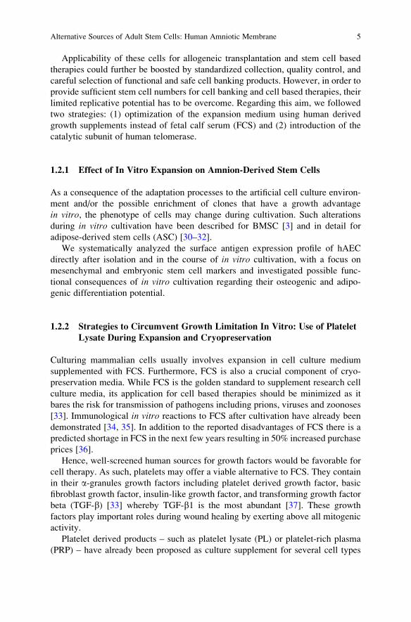
1.2.1 Effect of In Vitro Expansion on Amnion-Derived Stem Cells
As a consequence of the adaptation processes to the artificial cell culture environ-
ment and/or the possible enrichment of clones that have a growth advantage
in vitro, the phenotype of cells may change during cultivation. Such alterations
during in vitro cultivation have been described for BMSC [3] and in detail for
adipose-derived stem cells (ASC) [30–32].
We systematically analyzed the surface antigen expression profile of hAEC
directly after isolation and in the course of in vitro cultivation, with a focus on
mesenchymal and embryonic stem cell markers and investigated possible func-
tional consequences of in vitro cultivation regarding their osteogenic and adipo-
genic differentiation potential.
1.2.2 Strategies to Circumvent Growth Limitation In Vitro: Use of Platelet
Lysate During Expansion and Cryopreservation
Culturing mammalian cells usually involves expansion in cell culture medium
supplemented with FCS. Furthermore, FCS is also a crucial component of cryo-
preservation media. While FCS is the golden standard to supplement research cell
culture media, its application for cell based therapies should be minimized as it
bares the risk for transmission of pathogens including prions, viruses and zoonoses
[33]. Immunological in vitro reactions to FCS after cultivation have already been
demonstrated [34, 35]. In addition to the reported disadvantages of FCS there is a
predicted shortage in FCS in the next few years resulting in 50% increased purchase
prices [36].
Hence, well-screened human sources for growth factors would be favorable for
cell therapy. As such, platelets may offer a viable alternative to FCS. They contain
in their a-granules growth factors including platelet derived growth factor, basic
fibroblast growth factor, insulin-like growth factor, and transforming growth factor
beta (TGF-b) [33] whereby TGF-b1 is the most abundant [37]. These growth
factors play important roles during wound healing by exerting above all mitogenic
activity.
Platelet derived products – such as platelet lysate (PL) or platelet-rich plasma
(PRP) – have already been proposed as culture supplement for several cell types
Alternative Sources of Adult Stem Cells: Human Amniotic Membrane 5

including BMSC [33, 37–47], umbilical cord blood MSC [48], ASC [43, 49], and
stromal cells from dental pulp and trabecular bone [43] showing increased clono-
genic efficiency and proliferative capacity compared to standard FCS culture. PL
also stimulates proliferation and collagen production of human tenocytes and
increases the gene expression of matrix-degrading enzymes and angiogenic growth
factors [50]. Furthermore, myelomas, hybridomas, hepatocytes, fibroblasts, and
epithelial cells have already been evaluated using PL with regard to cell growth,
viability, and production efficiency [51]. Growth stimulation upon PL treatment
was also demonstrated for primary chondrocytes. However, PL failed to support a
chondrogenic phenotype [52–54] in contrast to BMSC cultures showing increased
chondrogenic marker genes in presence of PRP [55]. Primary human skeletal
muscle cells showed decreased differentiation capacity into myotubes and impaired
functionality [56]. Proliferation of primary human osteoblasts was not affected by
addition of PRP to the culture medium [57]. For human dermal and gingival
fibroblasts, contradicting results were obtained for platelet derived products as
culture supplements ranging from growth suppression [58] to growth promotion
[59, 60]. Using ASC, Davenport et al. showed that PL only initially supported cell
proliferation but led to growth arrest shortly after first subcultivation [61]. In
contrast, addition of thrombin activated PRP to the culture medium increased
ASC proliferation and retained their differentiation capacity during long-term
culture [49].
Moreover, platelet derived products and BMSC have already been used clini-
cally both to treat distraction osteogenesis of the lower extremity in patients with
achondroplasia and hypochondroplasia yielding accelerated bone regeneration [62]
and also to treat successfully a patient with severe radiation burn [63].
However, the influence of platelet derived products for the cultivation of cells
isolated from amniotic membrane has not been addressed before.
1.2.3 Strategies to Circumvent Growth Limitation In Vitro:
Introduction of hTERT
Stem cells needed at therapeutic doses, especially in adults, may require extensive
in vitro expansion. In this regard, one major drawback of these cells is their low
proliferative capacity and limited in vitro life span before reaching an irreversible
growth arrest also termed replicative senescence [19]. Additionally, long-term
cultures of human MSC may show altered or reduced responsiveness to differenti-
ation signals [64].
One strategy to circumvent these limitations is the introduction of the catalytic
subunit of human telomerase reverse transcriptase (hTERT) which has been
reported to extend the cellular life span of numerous cell types including normal
fibroblasts, endothelial or epithelial cells [65–67], in vitro propagated tumor cells
[68–70], and also of stem cells [71, 72]. It has been shown that hTERT immorta-
lized human MSC originating from sources such as bone marrow and adipose tissue
maintain their differentiation potency [72–74]. We report in this section the
6 S. Wolbank et al.

establishment and the characterization of the first hTERT immortalized hAMSC
lines including their immunomodulatory functions, a crucial factor for using these
cell lines in allogeneic cell therapies [75]. Therefore, if cell banking is intended, it is
important to monitor the cells’ ability to alloactivate peripheral blood mononuclear
cells (PBMC) as well as to modulate the proliferation of activated PBMC.
2 Stem Cell Characteristics and Immunomodulatory Potential
of Human Amnion-Derived Stem Cells
2.1 Isolation of Separate Populations of hAEC and hAMSC
Placentae were collected from Cesarean sections after obtaining informed consent
of the mothers according to the approval of the local ethical committee. Amnion
was peeled off the placenta, washed extensively with phosphate buffered saline
(PBS) at 4�C, and dissected in 2–3 cm2 pieces. Half of these were digested for
3 � 20 min with 0.05% trypsin/EDTA (PAA, Austria), the other half for 2 h with
1 mg/mL collagenase I (Biochrom, Austria) for isolation of hAEC and hAMSC,
respectively. Hematoxylin/eosin staining of paraffin embedded sections of amnion
demonstrates that digestion of amniotic membrane with trypsin and collagenase left
an essentially intact mesenchymal and epithelial layer (Fig. 1a).
After addition of ice-cold PBS, cell suspensions were filtered through a 100-mmcell strainer, centrifuged, and seeded in culture flasks at a density of 7 � 103
cells/cm2 for hAMSC and 14 � 103–21 � 103 cells/cm2 for hAEC in EGM-2
Fig. 1 Isolation of pure fractions of hAEC and hAMSC. (a) Hematoxylin and eosin stain of fresh
amnion (undigested) and after digestion with trypsin and collagenase, as performed for isolation of
hAEC and hAMSC, respectively. (b) Epithelial and mesenchymal morphology of hAEC and
hAMSC, respectively
Alternative Sources of Adult Stem Cells: Human Amniotic Membrane 7

(Lonza, Belgium). The resulting cultures are composed of pure populations with a
clearly distinguishable epithelial and mesenchymal morphology, respectively
(Fig. 1b).
2.2 Stem Cell Characteristics and Immunomodulationof hAEC and hAMSC
Both amniotic cell populations are routinely characterized by a common surface
marker expression profile including the presence of CD73, CD90, CD105, and
MHC I, and the concomitant absence or low levels of CD34, CD45, and MHC II,
analyzed by flow cytometry. Purity of amniotic subpopulations could be determined
by CD49d (a4-integrin) expression which was 2 � 2.4% in the hAEC and
96 � 3.9% in the hAMSC population.
To evaluate the reproducibility of differentiation of hAEC and hAMSC,
osteogenic differentiation was induced 24 h after seeding by changing the medium
to Mesenchymal Stem Cell Osteogenic Stimulatory Kit (O-Kit, Stemcell Tech-
nologies, Canada) and maintaining these cultures for 21 days. Adipogenic dif-
ferentiation was performed according to Portmann-Lanz et al. [23]. Osteogenic
differentiation was demonstrated by spectrophotometric assessment of Alizarin red
(AR). Typically, osteogenic differentiation of both cell types, hAEC (at P1 or P2)
and hAMSC (at P2), was successfully induced in three of four cases. Adipogenic
differentiation was evident for two of four hAMSC isolations, while, in contrast to
published data [22, 23], hAEC did not differentiate along the adipogenic lineage in
our hands (data not shown).
For investigating immunomodulation in vitro, amnion-derived cells were cocul-
tured with PBMC, isolated from whole blood as in mixed lymphocyte reactions
(MLRs). For this, 5 � 104 cells of two different allogeneic PBMC populations
were cocultured in 100 mL PBMC medium/well (RPMI1640, 9% FCS, 2 mM
L-glutamine, 100 U/mL penicillin G, and 0.1 mg/mL streptomycin) in triplicates
in 96-well flat bottom plates. Amnion-derived cells were seeded in the wells and
allowed to adhere before adding PBMC. The stem cells (SC) were added at
SC/PBMC ratios of 1:1 (5 � 104 SC), 1:2, 1:4, 1:8, and 1:16. On day 5, 10 mM5-bromo-2-deoxyuridine (BrdU) was added and BrdU ELISA (Roche) was per-
formed on day 6 according to the manufacturer’s instructions. Similarly, for
phytohemagglutinin (PHA) activation assay, 5 � 104 PBMC were activated by
5 mg/mL PHA (Sigma) on day 3 of the culture. To examine interaction between
allogeneic SC and unstimulated PBMC, SC were cocultured with unstimulated
PBMC at 1:1 (5 � 104 SC), 1:2, 1:4, 1:8, and 1:16 ratios in 100 mL. On day 4,
10 mM BrdU was added. The inhibitory effect of SC was calculated as PBMC
proliferation (%) = (ESTIM+SC � ESTIM) � 100. ESTIM+SC = mean absorption of
stimulated PBMC cocultured with allogeneic SC; ESTIM = mean absorption of
stimulated PBMC. Data were analyzed by one-way ANOVA and Tukey’s multiple
8 S. Wolbank et al.

comparison test. Data sets of cells at low vs high population doublings (PDs) were
compared by two-tailed Student’s t-test. A p-value less than 0.05 was considered assignificant.
hAMSC and hAEC inhibited proliferation of activated PBMC in a dose-depen-
dent manner as demonstrated by a decrease in proliferation with increasing stem
cell amounts. SC were most effective when added in equal cell numbers compared
to PBMC, significantly reducing PBMC proliferation in MLR experiments to a level
of 34% (range 3–73%) in the case of hAMSC (Fig. 2a) and 23% (range 0–72%) in
the case of hAEC (Fig. 2b). When PBMCwere activated by PHA, similar inhibition
was reached, in detail 33% (range 12–66%) for hAMSC (Fig. 3a), and 28% (range
0–60%) for hAEC (Fig. 3b). The lowest SC dose resulting in significant inhibition
of lymphocyte response was 25%, in single cases even 12.5%.
200a
180160140120100806040200
12.5 25
hAMSC added (%)
PB
MC
pro
lifer
atio
n (%
)
50 100
200b
180160140120100806040200
12.5 25
hAEC added (%)
PB
MC
pro
lifer
atio
n (%
)
50 100
Fig. 2 hAMSC and hAEC inhibit MLR-activated PBMC in a cell dose-dependent manner. PBMC
were cocultured with equal amounts of allogeneic PBMC and different amounts of third party (a)
hAMSC (n ¼ 12), (b) hAEC (n ¼ 9). Median � Q1 and Q3 are depicted
200a
180160140120100806040200
12.5 25
hAMSC added (%)
PB
MC
pro
lifer
atio
n (%
)
50 100
200b
180160140120100806040200
12.5 25
hAEC added (%)
PB
MC
pro
lifer
atio
n (%
)
50 100
Fig. 3 hAMSC and hAEC inhibit PHA-activated lymphocyte proliferation in a cell dose-depen-
dent manner. PBMC were cocultured with (a) hAMSC (n ¼ 12), (b) hAEC (n ¼ 8). Median �Q1 and Q3 are depicted
Alternative Sources of Adult Stem Cells: Human Amniotic Membrane 9

3 Phenotypic Shift and Reduced Osteogenesis During In Vitro
Expansion of Human Amnion Epithelial Cells
For tissue engineering purposes, cells may be applied either directly after isolation
from the tissue or after a period of in vitro expansion to obtain higher cell numbers.
In order to investigate the advantages and drawbacks of these strategies we com-
pared freshly isolated and cultivated hAEC regarding their surface antigen expres-
sion profile and their osteogenic differentiation capacity.
3.1 Shift in Surface Antigen Expression DuringCultivation of hAEC
To investigate the impact of in vitro expansion on the immunophenotype of cells
with potential for regenerative medicine, we carefully characterized the surface
antigen profile of hAEC directly after isolation and during cultivation by flow
cytometry. We focused on hAEC, as recovery of primary hAMSC is usually too
low for thorough analysis. For this purpose, freshly isolated cells and cells during
culture were immunostained for CD14, CD34, CD45, CD13, CD29, CD44, CD49c,
CD49d, CD49e, CD54, CD73, CD90, CD166, Ki67 (BD, Austria), CD105 (Abcam)
and SSEA-4, TRA-1-60, TRA-1-81 (Chemicon), by 7-AAD (BD) for dead cells and
measured by flow cytometry.
Surface antigens were clustered into four groups, according to their expression
patterns (representative histograms are depicted in Fig. 4a, summarized in Fig. 4b).
The first group comprises CD49d (integrin a4; used to differentiate hAEC from
hAMSC) and the hematopoietic markers CD14, CD34, and CD45. These antigens
are hardly detectable on freshly isolated hAEC (preP0) and remain at similar levels
during passaging.
The surface antigens of the second group are uniformly expressed at high levels,
both in primary isolates (preP0) and after further cultivation. This group comprises
the stromal cell markers CD29 (integrin b1), CD49c (integrin a3), CD73 (ecto-50-nucleotidase), and CD166 (ALCAM), and the embryonic stem cell marker SSEA-4.
Group 3 consists of the stromal cell associated markers CD13 (aminopeptidase
N), CD44 (HCAM), CD49e (integrin a5), CD54 (ICAM-1), CD90 (Thy-1), and
CD105 (endoglin), which are low (medium to undetectable) directly after isolation
(preP0) and are rapidly increased during in vitro cultivation.
Two additional embryonic stem cells markers, TRA-1-60 and TRA-1-81 (group
4), are characterized by medium expression in preP0 cells, which decreases upon
cultivation.
As in vivo, amniotic cells reside within a tissue that remains of approximately
the same size during the last weeks of pregnancy, these cells would probably be in a
quiescent state directly after isolation, but start dividing upon transfer into tissue
culture medium. Therefore, we tested the hypothesis that upregulation of group 3
antigens might be associated with re-entry of the cells into the cell cycle. Only
10 S. Wolbank et al.

2–6% of freshly isolated hAEC (preP0) were stained for the proliferation marker
Ki67, which is expressed in all phases of the cell cycle but not in G0. After a few
days in culture, Ki67 expression increased dramatically (Fig. 4a, b), concomitant
with the observed upregulation of group 3 antigens. However, expression of group 3
antigens is not dependent on proliferation of hAEC, as expression remained high
Fig. 4 Surface antigen expression of four freshly isolated (preP0) hAEC strains and during
cultivation at various passages (P0–P3) by flow cytometry. (a) Antigens were grouped according
to their expression profile during cultivation (see text) and representative histograms of one
member of each group are shown. Gray peaks represent unspecific isotype controls, solid linesrepresent the specific antibodies. (b) Summary: shown are means and corresponding standard
deviations, calculated from the data of all antigens of each group of all four hAEC isolations
(hAEC 87, �88, �90, and �91) at the indicated passages
Alternative Sources of Adult Stem Cells: Human Amniotic Membrane 11

when proliferation slowed down after several passages in vitro, concomitant with a
drastic decrease in Ki67 staining (Fig. 4a, b).
3.2 Osteogenic Differentiation Potential of hAEC Decreasesupon In Vitro Cultivation
We addressed the question as to whether the observed shift in mesenchymal and
embryonic stem cell markers during cultivation of hAEC (Fig. 4) is associated with
alterations of their functional phenotype, i.e., their capacity to differentiate along
the adipogenic and osteogenic lineages. Therefore, adipogenic and osteogenic
conditions (as described in Sect. 2.1) were applied to different hAEC isolations
seeded directly after isolation (P0) and after cultivation (P2 or P3). In addition to
mineralization, quantitative real time PCR was performed analyzing expression of
RUNX2/CBFA1 (core binding factor alpha), alkaline phosphatase (ALPL), bone
gamma-carboxyglutamate protein (BGLAP, osteocalcin), bone morphogenetic pro-
tein receptor 1B (BMPR1B), and bone morphogenetic protein receptor 2 (BMPR2)
using a light cycler TM480 (Roche) and Taqman gene expression assays (Applied
Biosystems). Expression values were normalized to the housekeeping gene hypo-
xanthine-guanine phosphoribosyltransferase (HPRT).
Similar to passaged hAEC, no adipogenic differentiation was observed using
four strains of freshly isolated hAEC (data not shown). In contrast, the same strains
showed predominantly stronger mineralization ability at P0 (in three out of four
cases) when compared to passaged cells (P2–P3). Interestingly, preliminary results
with hAMSC suggest similar mineralization and lipid accumulation after induction
of P0 vs P2 cells (data not shown). We confirmed mineralization by analysis of
mRNA levels of selected genes involved in osteogenesis. In freshly isolated hAEC
(P0) all osteogenic markers were upregulated upon cultivation in O-Kit for 14 and
21 days (Fig. 5). In cultivated hAEC (P1), RUNX2 and ALPL were also increased
under osteogenic conditions whereas virtually no alteration in transcription of
BGLAP, BMPR1B, and BMPR2 was observed.
4 Platelet Lysate for FCS-Free Expansion and
Cryoconservation of Amnion-Derived Cells
For producing PL, platelet concentrates from 36 healthy donors that could no longer
be used for patients were pooled, frozen at �80�C, and thawed quickly in a water
bath at 37�C, resulting in growth factor release from bursting platelets. Platelet
debris was removed by centrifugation at 2,000�g for 10 min while the PL was
filtered using a 0.22-mm filter, aliquoted, and stored until application at �80�C.For determining growth kinetics, cells were isolated from three donors as
described in Sect. 2.1, and 2.5 � 105 cells were seeded in T-25 flasks and cultured
12 S. Wolbank et al.

for about 100 days in PL expansion medium (DMEM-LG & Ham’s F12, 5% PL,
2 mM L-glutamine, 100 U/mL Penicillin, 0.1 mg/mL Streptomycin (PAA), 2 U/mL
Heparin (Biochrom)) at 37�C, 5% CO2, and 95% humidity. PD was calculated at
each subcultivation using the formula: SLN (cells harvested/cells seeded)/LN(2).
hAMSC from all three donors cultured in PL showed between 21 and 32 PD
without growth arrest whereas control hAMSC cultured in EGM-2 (Lonza), a
commercially available medium containing 2% FCS, showed only 5 PD before
proliferation totally ceased (Fig. 6a).
Not only during expansion but also during cryopreservation, substitution of FCS
would be favorable for establishing cell banks. In preliminary experiments, several
media containing PL (5%, 90% PL) were compared to standard FCS media (10%,
90% FCS) as well as a serum-free medium (CryoSFM, PromoCell) for cryopreser-
vation of hAMSC. Then 1 � 106 P1 cells were resuspended in the respective
medium and frozen at a freezing rate of �1�C/min. From these preliminary data,
the best conditions, namely 5% PL medium (5% PL, 10% dimethylsulfoxide
(DMSO), 85% DMEM-LG), 90% FCS medium (90% FCS, 10% DMSO), and
CryoSFM were chosen for further investigation.
After thawing, cell viability was assessed by trypanblue exclusion assay. Addi-
tionally, growth kinetic studies were performed to evaluate characteristics of
hAMSC before and after cryopreservation. Cells cryopreserved in PL showed
7RUNX2 ALPL BGLAP BMPR1B BMPR2
P 0
P 1
6
5
4
3
2
1
0d14
fold
exp
ress
ion
com
pare
d to
d0
d21 d14 d21 d14 d21 d14 d21 d14 d21
Fig. 5 Osteogenic differentiation of hAEC isolations seeded directly after isolation (P0) and after
cultivation (P1). Expression levels of selected genes implicated in osteogenesis, determined using
quantitative real-time PCR. Expression levels after cultivation in O-Kit for 14 and 21 days (d14
and d21) were normalized to the levels before induction (d0). Shown are means and standard
deviations of three measurements from two individual donors
Alternative Sources of Adult Stem Cells: Human Amniotic Membrane 13

lower cell viability after thawing when compared to those stored in FCS medium or
serum-free CryoSFM (Fig. 7). While growth kinetics after thawing seemed to be
unaffected by the cryopreservation medium applied, strong impact of the donors on
PD was evident (Fig. 6b–d). An observed phenomenon was the low attachment
capacity of hAMSC cryopreserved in 5% PL. Hence, coating experiments with
gelatine (1% in PBS) or “Coating matrix Kit” (Invitrogen) were performed but
attachment of cells to the culture vessel surface could not be substantially improved
(data not shown).
5 hTERT Induced Extension of In Vitro Life Span
of Amnion-Derived Stem Cells
For introduction of hTERT a retroviral transfection system was chosen. Therefore,
the cDNA of hTERT (kindly provided by Geron Corp.) was inserted into the
retroviral vector pLXSN (Clontech Laboratories Inc.) and retroviral particles
praecryo
donor 3 - after cryo
donor 1 - after cryo
donor 2 - after cryo
days in culture
PD
sP
Ds
PD
sP
Ds
days in culture days in culture
days in culture0 0
00
0
0 0
0
5
5 5
5
10
10 10
10
15
15 15
15
20
20 20
20
25
25 25
25
30
30 30
30
35
35 35
35
40
40 40
40
20 20
2020
40 40
4040
60 60
6060
80 80
8080
100 100
100100
120
5% PL
donor 3 - EGM-2
donor 3 - PL
donor 2 - PL
donor 1 - PL
5% PL
5% PL
5% PL praecryo 5% PL praecryo
5% PL praecryo
cryo-SFM cryo-SFM
cryo-SFM
90% FCS 90% FCS
90% FCS
a b
dc
Fig. 6 Growth characteristics of hAMSC from three donors before (a) and after cryopreservation
(b–d) cultivated in medium supplemented with either 5% PL or EGM-2 and cryopreserved in FCS,
PL containing media or serum-free Cryomedium (CryoSFM). PDs cumulative population
doublings
14 S. Wolbank et al.

were generated as described previously [70]. Gene transfer was performed at early
PD (<PD8) according to the manufacturer’s instructions (Clontech Laboratories
Inc.). Then 24 h post transduction transfectants were selected using 200 mg/mL
Geneticin Sulfate G418 and arising cell clones were grown as mass culture. PD of
transduced cell lines were calculated starting with the first passage post transduc-
tion (PDpT) using the formula stated in Sect. 4. Telomerase activity (TA) was
determined using a modification of the real-time telomeric repeat amplification
protocol (TRAP) assay as described in detail previously [70] and calculated relative
to that of HEK293 cells (positive control). For determination of senescence,
associated b-galactosidase (SA-b-gal) activity cells were fixed with 3% formalde-
hyde and stained as described in detail previously [76]. For characterizing pheno-
type, differentiation potential, and immunomodulatory properties, protocols
according to Sect. 2.2 were performed. For quantitative evaluation, AR was
measured after extraction using 20% methanol/10% acetic acid at 450 nm. For
quantification of intracellular alkaline phosphatase (AP) activity, washed cells were
frozen and thereafter incubated in 0.5% Triton X-100. After incubation with
4-nitrophenolphosphate, samples were measured at 405/620 nm. In addition to
osteogenic marker genes peroxisome proliferator-activated receptor gamma
(PPARg) and leptin (Lep) were evaluated as adipogenic marker genes by quantita-
tive RT-PCR as described in Sect. 3.2.
Immortalized stem cell lines (originating from the mesenchymal layer of the two
amniotic membrane donors hAMSC76 and hAMSC83) were established by over-
expression of hTERT. Human stem cells were isolated from amnion and propagated
in vitro until they reached replicative senescence. Representative growth curves of
hAMSC76 are shown in Fig. 8a. Senescence was evidenced by growth arrest, large
and flat cell morphology (Fig. 8b), and SA-b-gal activity (Fig. 8c). Upon ectopic
expression of hTERT stem cell populations were immortalized (so far expanded
to at least PD60 with no signs of growth retardation; Fig. 9a). Furthermore,
hTERT overexpression maintained many characteristics of the original cellular
phenotype. Figure 9b demonstrates fibroblastoid morphology of transduced cells
(hAMSC76telo-PD78pT, hAMSC83telo-PD43pT) comparable to early passage
1009080706050403020100
90%FCS
cryomedium
viab
ility
(%
)Cryo SFM
hAMSC1hAMSC2hAMSC3
5%PL
Fig. 7 Cell viability of
hAMSC from three donors
after cryopreservation in PL,
FCS containing media or
serum-free Cryomedium
(CryoSFM) and thawing as
determined by trypanblue
exclusion assay. Data are
presented in % of viability at
freezing
Alternative Sources of Adult Stem Cells: Human Amniotic Membrane 15

parental counterparts. Telomerase activity after transduction was verified by TRAP
assay (Fig. 9c). In contrast to empty vector control cells (hAMSCneo), hTERT-
transduced cell lines expressed significant telomerase activity at early as well as
higher PDpT (at least PD38pT) when compared to HEK293 control cells (49–72%
of HEK293).
Expression of selected hematopoietic (CD14, CD34, and CD45 negative) and
mesenchymal markers (CD73, CD90, and CD105 positive) on hTERT-transduced
cell lines were similar to their parental cultures, also after prolonged in vitropropagation (hAMSC76telo-PD84pT, hAMSC83telo-55pT; Table 1). Interest-
ingly, the major population of hTERT-transduced hAMSC lost expression of the
mesenchymal marker CD90. Therefore, hAMSC83telo were characterized in more
detail, i.e., at several PD and for additional antigens (Table 2). At early PD after
Fig. 8 Growth characteristics and morphology of hAMSC. (a) Cells were grown in vitro until
replicative senescence. Representative growth curve of hAMSC76 is shown. (b) Phase contrast
microscopy and (c) staining for SA-b-galactosidase activity of early and late passage cells.
Magnification in b, c: �100
Fig. 9 Growth potential and morphological characteristics of hTERT-transduced hAMSC. (a)
Growth curves of hTERT-transduced cell lines. (b) Phase contrast microscopy of hTERT-transduced
immortalized cell lines. (c) TRAP assays at two different PDs post transduction (PDpT) demonstrate
telomerase activity in cell lines. neo Vector control, telo hTERT-transduced; in b: �100
16 S. Wolbank et al.

transduction, hAMSC83telo were still homogenously positive for CD90; however
at PD55pT, only 8.7% of the cells expressed this marker. This subpopulation
remained detectable after a further 41 PDs (PD96pT). With the exception of
SSEA-4, the expression of which increased after hTERT transduction, all other
Table 1 Surface antigen expression of nontransduced (normal) and hTERT-transduced (telo)
hAMSC. hAMSC76 and hAMSC83 were analyzed at PD6 and 5, their hTERT-transduced
counterparts at population doubling 84 and 55 post transduction, respectively
Antigen hAMSC76 hAMSC83
Normal Telo Normal Telo
CD14 0.5 7.0 2.6 10.5
CD34 0.0 0.1 0.1 0.1
CD45 0.0 11.6 0.0 2.0
CD73 96.0 100.0 99.9 100.0
CD90 100.0 53.3 100.0 8.7
CD105 95.7 99.8 92.5 99.9
HLA ABC 98.9 99.8 99.4 99.7
HLA DR 0.5 2.6 0.2 2.3
Table 2 Detailed flow cytometric characterization of hTERT-transduced
hAMSC83telo during prolonged in vitro cultivation, compared to the nontrans-
duced counterpart. Additional antibodies were purchased from BD (CD13,
CD29, CD44, CD49c CD49d CD49e, CD54, CD166), Chemicon (SSEA-4,
TRA-1-60, TRA-1-81 and Oct-4), Santa Cruz Biotechnology (Vimentin)
Antigen Normal Telo
PD3 PD26pT PD55pT PD96pT
CD13 100.0 100.0 99.7 99.8
CD14 5.1 2.1 10.5 6.4
CD29 99.9 n.d. 99.6 99.8
CD34 0.4 0.1 0.1 0.2
CD44 100.0 n.d. 99.4 99.9
CD45 2.7 0.1 2.0 4.8
CD49c 100.0 n.d. 99.8 99.8
CD49d 98.2 n.d. 99.7 99.9
CD49e 100.0 n.d. 99.7 99.9
CD54 72.0 n.d. 89.0 98.7
CD73 100.0 76.6 100.0 100.0
CD90 100.0 100.0 8.7 22.8
CD105 98.5 95.8 99.9 99.8
CD166 100.0 n.d. 99.8 99.8
HLA ABC 100.0 100.0 99.7 99.9
HLA DR 7.2 0.7 2.3 5.2
SSEA-4 26.6 81.7 99.6 85.6
TRA-1-60 6.4 n.d. 3.4 0.9
TRA-1-81 6.1 n.d. 6.4 0.5
Oct-4 62.1 n.d. 72.9 89.2
Vimentin 100.0 n.d. 100.0 n.d.
n.d. Not determined; PD population doubling; pT post transduction
Alternative Sources of Adult Stem Cells: Human Amniotic Membrane 17

antigens tested showed no alteration. Analysis of the cellular karyotype revealed
that hTERT transduction did not induce abnormalities in chromosomal number or
structure since both, nontransduced stem cells and the hTERT cell lines showed a
normal karyotype. Additionally, soft agar assays showed no indication for a tumor-
igenic conversion upon hTERT transduction (data not shown).
After introduction of hTERT, amnion-derived stem cell lines showed a similar
differentiation potential towards the adipogenic and osteogenic lineage when
compared to the nontransduced counterparts. hAMSC generally show a low differ-
entiation potential towards the adipogenic lineage as demonstrated by OO staining.
Although singular hAMSC76telo cells gained the capacity for lipid accumulation,
these rare events were not quantifiable (data not shown). On the level of adipogenic
marker genes, quantitative real-time PCR revealed low levels of PPARg expressionand induction of leptin transcription in hAMSC (Fig. 10). When testing for osteo-
genic differentiation, significant mineral deposition of all hAMSC lines was
observed, as analyzed by quantification of AR staining (Fig. 11a). Also, a low
but significant increase of AP activity (Fig. 11b) as well as induction of mRNA
levels of AP at very low levels was seen (Fig. 12a). Osteocalcin mRNA was
induced in all hAMSC during differentiation (Fig. 12b). In order to test immuno-
modulation of the hTERT immortalized stem cell lines, their suppressive effect
on MLR- or PHA-activated lymphocyte proliferation was analyzed (Fig. 13).
The tested cells inhibited MLR-activated PBMC proliferation in a cell dose-
dependent manner. hTERT-transduced hAMSC inhibited significantly at a 1:8
SC/PBMC ratio (Fig. 13a), parental hAMSC even at 1:16. Similarly, when stem
cells were cocultured with PHA-activated PBMC, the inhibitory potency of
hAMSC was unaltered after hTERT overexpression, inhibiting significantly at a
ratio of 1:8 (Fig. 13b).
1000hAMSC76
hAMSC83
hAMSC83telo
hAMSC76telo100
10
1
0.1
indu
ctio
n fa
ctor
PPARγ LEP
Fig. 10 Adipogenic differentiation potential of nontransduced and hTERT-transduced hAMSC
3 weeks after induction. Relative expression of peroxisome proliferator-activated receptor gamma
(PPARg) and leptin 2 weeks after adipogenic induction of nontransduced and hTERT-transduced
hAMSC. Expression levels are normalized to HPRT and presented relative to d0 cultures (set to 1).
Means and SDs of two individual experiments are displayed
18 S. Wolbank et al.

6 Discussion
6.1 Stem Cell Characteristics and Immunomodulatory Potentialof Human Amnion-Derived Stem Cells
Taken together, our data clearly show a cell dose dependency of the immuno-
modulatory effect of amnion-derived stem cells, which corroborates published results
for MSC from adipose tissue or bone marrow [77–81]. Furthermore, we demonstrate
cell dose-dependent immunosuppression for the two distinct amniotic stem cell types
which were characterized by differential expression of a4-integrin. Interestingly,
0.2 140
120
100
80
60
40
20
0
a b
* * *
*
*
*AR AP
0.16
CM
OM
0.12
0.08OD
450
nm
0.04
0
hAMSC76 hAMSC83 hAMSC83telohAMSC76telo hAMSC76
pNP
P μ
M
hAMSC83 hAMSC83telohAMSC76telo
CM
OM
Fig. 11 Osteogenic differentiation potential of nontransduced and hTERT-transduced hAMSC
3 weeks after induction. Osteogenic differentiation demonstrated by Alizarin red (AR) quantifica-
tion (a) and alkaline phosphatase (AP) (b) of hAMSC. Differences between control cultures (CM)
and osteogenic differentiation cultures (OM) with p < 0.05 were regarded as significant
a b
*
*
AP0.20
0.15
0.10
norm
aliz
ed g
ene
expr
essi
on
0.05
0.00hAMSC76 hAMSC83 hAMSC83telohAMSC76telo
*
**
OC5.0
4.0
3.0
norm
aliz
ed g
ene
expr
essi
on
2.0
0.0
1.0
hAMSC76 hAMSC83 hAMSC83telohAMSC76telo
*CMOM
CMOM
Fig. 12 Expression levels of alkaline phosphatase (AP) (a) and osteocalcin (OC) (b) 2 weeks after
osteogenic induction of nontransduced and hTERT-transduced hAMSC. Expression levels in
osteogenic medium (OM) and control medium (CM) are presented normalized to HPRT. Means
and SDs of three individual experiments are displayed. Differences between control cultures (CM)
and osteogenic differentiation cultures (OM) with p < 0.05 were regarded as significant
Alternative Sources of Adult Stem Cells: Human Amniotic Membrane 19

although epithelial and mesenchymal fractions show distinct morphology and marker
expression, they have similar potency to modulate immunoreactions in vitro.
6.2 Phenotypic Shift and Reduced Osteogenesis During In VitroExpansion of Human Amnion Epithelial Cells
We show here that differentiation of amniotic cells strongly depends on the donor.
Hence, three of four hAEC and hAMSC strains at P2 clearly showed positive
differentiation. Adipogenic differentiation of hAMSC was less reproducible, with
two positive cases of four. Strikingly, none of the six hAEC strains tested during this
study showed evidence of adipogenic differentiation, which seems to be in contra-
diction to recent reports [8, 22, 23]. In ’t Anker et al. published adipogenic differen-
tiation of a stem cell population derived frommechanical disaggregation of the whole
amniotic membrane and selection by completely different culture conditions com-
pared to ours [8]. Portmann-Lanz et al. reported a transient growth retardation during
which morphology changed from typically epithelial, cobblestone-like to a fibroblast-
like morphology [23]. In our study, hAEC isolations with noticeable change of
morphology towards the fibroblast-like phenotype were excluded to allow separate
analysis of mesenchymal and epithelial cells. Discrepancies with published reports
may be due to the use of different cell populations with different differentiation
potential or the application of different culture conditions [8, 22, 23].
a
*
*
*
*
*
****
100
80
60
PB
MC
pro
lifer
atio
n (%
)
40
20
01:16 1:8 1:4 1:2 1:1
hAMSC
hAMSC telo
b
***
**
*
*
***
100
80
60
PB
MC
pro
lifer
atio
n (%
)
40
20
01:16 1:8 1:4 1:2 1:1
hAMSC
hAMSC telo
Fig. 13 Immunomodulation of nontransduced and hTERT-transduced hAMSC. (a) hAMSC (76
n = 4, 83 n = 3) inhibit mixed lymphocyte reaction (MLR) in a cell dose-dependent manner. (b)
hAMSC (76 n = 4, 83 n = 4) inhibit phytohemagglutinin-activated lymphocyte proliferation in a
cell dose-dependent manner. PBMC proliferation is calculated as percentage of uninhibited
proliferation. p < 0.05 was regarded as significant inhibition. Median � Q1 and Q3 are depicted.
Asterisk indicates significant inhibition of proliferation
20 S. Wolbank et al.

We further focused on a detailed characterization of the surface antigen expres-
sion profile of freshly isolated amniotic cells and its alteration during in vitrocultivation using hAEC. We demonstrate that hAEC undergo profound changes
during the first days in culture, concomitant with, but not dependent on entry into
the cell cycle. Intriguingly, several markers associated with MSC, such as CD90
and CD105, are expressed at low levels or not at all on primary isolates and are
upregulated only after cultivation. This is of major importance considering possible
immunoisolation methods of noncultured cells for stem cell enrichment, which has
been established for CD105+ bone marrow derived stem cells [8].
The observed alteration of the phenotype may be explained by several mechan-
isms. First, cells with the “altered” phenotype might be present in the primary
isolate as a minor population and overgrow the main population due to a growth
advantage in vitro; this explanation is highly implausible, as the change in surface
antigen expression occurs very rapidly and hAEC have low growth rates in vitro.Second, the immunophenotype of the entire population might shift during cultiva-
tion or, third, only a subpopulation of the isolated cells might adapt to the culture
conditions by changing its phenotype and become enriched by passaging. Directly
after isolation, the population is heterogeneous with e.g., 20–50% being CD13-
positive and the remaining 50–80% CD13-negative cells. We estimate that about
two thirds of isolated hAEC become adherent during the first 3 days in culture, after
which we usually remove the cells of the supernatant. Interestingly, preliminary
results suggest that these nonadherent cells differ from their adherent counterparts,
e.g., in reduced CD44 and increased CD54 expression (G. Stadler, unpublished
observation). However, adherence to plastic does not trigger the full spectrum of
alterations noticed after three passages, as expression of group 3 antigens is still
rather low at day 3 (d3), when cells have just adhered to the culture dish (in Fig. 1d,
CD105 and CD49e are shown as representative examples for group 3 antigen
expression). Thus, we hypothesize that, as a first selection step, only part of the
primarily isolated heterogeneous cell population adheres to plastic, concomitant
with a partial upregulation of group 3 antigens that are further upregulated during
continued in vitro cultivation to result in a homogeneous population with high
expression of mesenchymal stem cell markers.
Finally, we have addressed the crucial question regarding the consequences of
the phenotypic shift during cultivation in terms of differentiation capacity, which
has to be answered before applying cultivated stem cells for tissue engineering. We
found that osteogenic differentiation was reduced after cultivation for two passages,
at a time point at which group 3 antigens were homogeneously and highly
expressed and embryonic TRA-antigens (group 4) were reduced to undetectable
levels. Functional impairment through cultivation has also been shown for primary
murine BMSC, which lost their homing ability in vitro [82].
In conclusion, our results suggest that freshly isolated hAEC have a superior
differentiation potential compared to hAEC cultivated under standard conditions
and therefore we are currently aiming to develop culture conditions allowing
maintenance of the original phenotype and differentiation capacity.
Alternative Sources of Adult Stem Cells: Human Amniotic Membrane 21

6.3 Platelet Lysate for FCS-Free Expansion andCryoconservation of Amnion-Derived Cells
PL is an interesting alternative to FCS during expansion and cryopreservation of
cells intended for application in humans. Expansion of hAMSC in a PL containing
medium is superior to a medium containing FCS. However, cryopreservation in
PL decreases cell viability after thawing. For future cell banking attempts, a
combination of expansion in PL medium and cryopreservation in serum free
cryomedium may allow for animal free strategy for expansion, cultivation and
banking of hAMSC. However, some pitfalls including low attachment capacity of
PL-expanded hAMSC have to be overcome.
6.4 hTERT Induced Extension of In Vitro Life Spanof Amnion-Derived Stem Cells
The applied strategy was successful in creating immortalized cell lines with largely
retained characteristics of the parental cells with regard to morphology, surface
marker profile, and immunosuppressive capacity and showed similar or improved
differentiation potential. However, one of two hAMSCtelo lines resulted in a
significantly higher immunogenicity compared to the nontransduced controls,
although the surface marker profile currently regarded as most important for
human MSC characterization did not differ from the parental cells. This suggests
that yet unknown markers will have to be identified in order to predict immunoge-
nicity of the cells. In summary, the novel cell lines give proof of principle that
hTERT is a promising tool to generate sufficient material for stem cell banking and
tissue engineering, but concomitantly emphasize the need for careful and standar-
dized characterization.
Stem cell characteristics of the newly established cell lines, especially their
differentiation and immunogenicity, were variable. Concerning typical surface
marker profiles, hAMSC lost the mesenchymal marker CD90 in a subpopulation
of telomerized cells during prolonged in vitro propagation. It has recently been
published that, upon cultivation in EGM-2 (also used in our study for hAMSC), a
subpopulation of CD90 negative human BMSC evolved after prolonged culture,
probably due to angiogenic growth factors in the medium [83]. The decrease in
CD90 expression and concomitant increase of the embryonic stem cell marker
SSEA-4 found in our immortalized hAMSC lines suggests an alteration of the
phenotype during long-term culture in EGM-2.
Both parental hAMSC isolates possessed low adipogenic differentiation poten-
tial which has been described before [68, 69]. However, hTERT transduction led to
increased lipid accumulation of hAMSC76 under adipogenic conditions. This is in
contrast to the finding that mesenchymal stromal cells from chorion cotransduced
22 S. Wolbank et al.

with hTERT and Bmi-1 showed minimal adipogenic differentiation that even
decreased with time in culture [84].
We observed immunosuppression of activated PBMC by our immortalized cell
lines, the exception being hAMSC76telo. Since the cell surface marker profiles of
hAMSCtelo lines analyzed here were identical, we propose that a marker as
predictor for immunogenicity remains to be identified and included in routine
surface marker profiling.
In conclusion, the immortalized stem cell lines established in this study can be
seen as a first step to a proof of principle for their applicability in cell based therapy
approaches. Since obvious donor and cell line specific differences exist, stem cell
material for cell banks will have to be routinely tested. Specifically, their differen-
tiation potential and immunosuppressive effects are of major importance. However,
additional caveats that limit the use are controversially discussed in the literature. In
particular, the tumorigenic potential of stem cells in general and hTERT-transduced
cells specifically is a matter of debate. Most reports find that stem cells retain their
differentiation potential, contact inhibition properties, stable karyotype, and do not
show tumorigenic potential even after extensive in vitro expansion. In other studies,spontaneous transformation of human ASC after 4–5 months in culture and a high
rate of tumorigenicity evolving as a consequence of hTERT introduction in human
MSC after approximately 3 years in culture were reported [2, 85]. Hence, it can be
expected that given a high quality of starting material concerning stem cell char-
acteristics and genetic stability and by use of a reasonable culture time in vitrosuitable stem cell material can be made available for cell banking by careful
monitoring and characterization.
Acknowledgments This work was partially supported by the European STREP Project HIPPO-
CRATES (NMP3-CT-2003-505758), the Austrian Science Fund (FWF) project NRN-S093-06, the
“Herzfeldersche Familienstiftung”, and the Lorenz Boehler Fonds. Parts of this work were carried
out under the scope of the European NoE “EXPERTISSUES” (NMP3-CT-2004-500283).
References
1. Thomson JA, Itskovitz-Eldor J, Shapiro SS et al (1998) Embryonic stem cell lines derived
from human blastocysts. Science 282:1145–1147
2. Rubio D, Garcia-Castro J, Martin MC et al (2005) Spontaneous human adult stem cell
transformation. Cancer Res 65:3035–3039
3. Pittenger MF, Mackay AM, Beck SC et al (1999) Multilineage potential of adult human
mesenchymal stem cells. Science 284:143–147
4. D’Ippolito G, Schiller PC, Ricordi C et al (1999) Age-related osteogenic potential of mesen-
chymal stromal stem cells from human vertebral bone marrow. J Bone Miner Res 14:
1115–1122
5. Arai F, Ohneda O, Miyamoto T et al (2002) Mesenchymal stem cells in perichondrium
express activated leukocyte cell adhesion molecule and participate in bone marrow formation.
J Exp Med 195:1549–1563
Alternative Sources of Adult Stem Cells: Human Amniotic Membrane 23

6. De Bari C, Dell’Accio F, Tylzanowski P et al (2001) Multipotent mesenchymal stem cells
from adult human synovial membrane. Arthritis Rheum 44:1928–1942
7. Erices A, Conget P, Minguell JJ (2000) Mesenchymal progenitor cells in human umbilical
cord blood. Br J Haematol 109:235–242
8. In ’t Anker PS, Scherjon SA, Kleijburg-van der Keur C et al (2004) Isolation of mesenchymal
stem cells of fetal or maternal origin from human placenta. Stem Cells 22:1338–1345
9. Young HE, Steele TA, Bray RA et al (2001) Human reserve pluripotent mesenchymal stem
cells are present in the connective tissues of skeletal muscle and dermis derived from fetal,
adult, and geriatric donors. Anat Rec 264:51–62
10. Zuk PA, Zhu M, Mizuno H et al (2001) Multilineage cells from human adipose tissue:
implications for cell-based therapies. Tissue Eng 7:211–228
11. Zvaifler NJ, Marinova-Mutafchieva L, Adams G et al (2000) Mesenchymal precursor cells in
the blood of normal individuals. Arthritis Res 2:477–488
12. Erbs S, Linke A, Schachinger V et al (2007) Restoration of microvascular function in the
infarct-related artery by intracoronary transplantation of bone marrow progenitor cells in
patients with acute myocardial infarction: the Doppler Substudy of the reinfusion of enriched
progenitor cells and infarct remodeling in acute myocardial infarction (REPAIR-AMI) trial.
Circulation 116:366–374
13. Garcıa-Olmo D, Garcıa-Arranz M, Herreros D et al (2005) A phase I clinical trial of the
treatment of Crohn’s fistula by adipose mesenchymal stem cell transplantation. Dis Colon
Rectum 48:1416–1423
14. Horwitz EM, Gordon PL, Koo WK et al (2002) Isolated allogeneic bone marrow-derived
mesenchymal cells engraft and stimulate growth in children with osteogenesis imperfecta:
implications for cell therapy of bone. Proc Natl Acad Sci USA 99:8932–8937
15. Mazzini L, Mareschi K, Ferrero I et al (2006) Autologous mesenchymal stem cells: clinical
applications in amyotrophic lateral sclerosis. Neurol Res 28:523–526
16. Mohyeddin Bonab M, Yazdanbakhsh S, Lotfi J et al (2007) Does mesenchymal stem cell
therapy help multiple sclerosis patients? Report of a pilot study. Iran J Immunol 4:50–57
17. Ringden O, Uzunel M, Rasmusson I et al (2006) Mesenchymal stem cells for treatment of
therapy-resistant graft-versus-host disease. Transplantation 81:1390–1397
18. Miki T, Lehmann T, Cai H et al (2005) Stem cell characteristics of amniotic epithelial cells.
Stem Cells 23:1549–1559
19. Parolini O, Alviano F, Bagnara GP et al (2008) Concise review: isolation and characterization
of cells from human term placenta: outcome of the first international workshop on placenta
derived stem cells. Stem Cells 26:300–311
20. Casey ML, MacDonald PC (1996) Interstitial collagen synthesis and processing in human
amnion: a property of the mesenchymal cells. Biol Reprod 55:1253–1260
21. Sakuragawa N, Tohyama J, Yamamoto H (1995) Immunostaining of human amniotic epithe-
lial cells: possible use as a transgene carrier in gene therapy for inborn errors of metabolism.
Cell Transplant 4:343–346
22. Ilancheran S, Michalska A, Peh G et al (2007) Stem cells derived from human fetal mem-
branes display multilineage differentiation potential. Biol Reprod 77:577–588
23. Portmann-Lanz CB, Schoeberlein A, Huber A et al (2006) Placental mesenchymal stem cells
as potential autologous graft for pre- and perinatal neuroregeneration. Am J Obstet Gynecol
194:664–673
24. Wolbank S, Peterbauer A, Fahrner M et al (2007) Dose-dependent immunomodulatory effect
of human stem cells from amniotic membrane: a comparison with human mesenchymal stem
cells from adipose tissue. Tissue Eng 13:1173–1183
25. Zhao P, Ise H, Hongo M et al (2005) Human amniotic mesenchymal cells have some
characteristics of cardiomyocytes. Transplantation 79:528–535
26. Bailo M, Soncini M, Vertua E et al (2004) Engraftment potential of human amnion and
chorion cells derived from term placenta. Transplantation 78:1439–1448
24 S. Wolbank et al.

27. Kubo M, Sonoda Y, Muramatsu R et al (2001) Immunogenicity of human amniotic membrane
in experimental xenotransplantation. Invest Ophthalmol Vis Sci 42:1539–1546
28. Gajiwala K, Gajiwala AL (2004) Evaluation of lyophilised, gamma-irradiated amnion as
a biological dressing. Cell Tissue Bank 5:73–80
29. Tosi GM, Traversi C, Schuerfeld K et al (2005) Amniotic membrane graft: histopathological
findings in five cases. J Cell Physiol 202:852–857
30. Boquest AC, Shahdadfar A, Frønsdal K et al (2005) Isolation and transcription profiling of
purified uncultured human stromal stem cells: alteration of gene expression after in vitro cell
culture. Mol Biol Cell 16:1131–1141
31. Mitchell JB, McIntosh K, Zvonic S et al (2006) Immunophenotype of human adipose-derived
cells: temporal changes in stromal-associated and stem cell-associated markers. Stem Cells
24:376–385
32. Yoshimura K, Shigeura T, Matsumoto D et al (2006) Characterization of freshly isolated and
cultured cells derived from the fatty and fluid portions of liposuction aspirates. J Cell Physiol
208:64–76
33. Doucet C, Ernou I, Zhang Y et al (2005) Platelet lysates promote mesenchymal stem cell
expansion: a safety substitute for animal serum in cell-based therapy applications. J Cell
Physiol 205:228–236
34. McIntosh KR, Lopez MJ, Borneman JN et al (2009) Immunogenicity of allogeneic adipose-
derived stem cells in a rat spinal fusion model. Tissue Eng Part A (Epub ahead of print)
35. Selvaggi TA, Walker RE, Fleisher TA (1997) Development of antibodies to fetal calf serum
with arthus-like reactions in human immunodeficiency virus-infected patients given synge-
neic lymphocyte infusions. Blood 89:776–779
36. Hausl P (2008) Rinderserum wird Mangelware. Transkript 14:48
37. van den Dolder J, Mooren R, Vloon AP et al (2006) Platelet-rich plasma: quantification of
growth factor levels and the effect on growth and differentiation of rat bone marrow cells.
Tissue Eng 12:3067–3073
38. Bernardo ME, Avanzini MA, Perotti C et al (2007) Optimization of in vitro expansion of
human multipotent mesenchymal stromal cells for cell-therapy approaches: further insights in
the search for a fetal calf serum substitute. J Cell Physiol 211:121–130
39. Bieback K, Hecker A, Kocaomer A et al (2009) Human alternatives to fetal bovine serum for the
expansion of mesenchymal stromal cells from bone marrow. Stem Cells (Epub ahead of print)
40. Capelli C, Domenghini M, Borleri G et al (2007) Human platelet lysate allows expansion and
clinical grade production of mesenchymal stromal cells from small samples of bone marrow
aspirates or marrow filter washouts. Bone Marrow Transplant 40:785–791
41. Carrancio S, Lopez-Holgado N, Sanchez-Guijo FM et al (2008) Optimization of mesenchymal
stem cell expansion procedures by cell separation and culture conditions modification. Exp
Hematol 36:1014–1021
42. Lange C, Cakiroglu F, Spiess AN et al (2007) Accelerated and safe expansion of human
mesenchymal stromal cells in animal serum-free medium for transplantation and regenerative
medicine. J Cell Physiol 213:18–26
43. Mirabet V, Solves P, Minana MD et al (2008) Human platelet lysate enhances the proliferative
activity of cultured human fibroblast-like cells from different tissues. Cell Tissue Bank 9:1–10
44. Muller I, Kordowich S, Holzwarth C et al (2006) Animal serum-free culture conditions for
isolation and expansion of multipotent mesenchymal stromal cells from human BM. Cytother-
apy 8:437–444
45. Perez-Ilzarbe M, Dıez-Campelo M, Aranda P et al (2009) Comparison of ex vivo expansion
culture conditions of mesenchymal stem cells for human cell therapy. Transfusion (Epub
ahead of print)
46. Schallmoser K, Bartmann C, Rohde E et al (2007) Human platelet lysate can replace fetal
bovine serum for clinical-scale expansion of functional mesenchymal stromal cells. Transfu-
sion 47:1436–1446
Alternative Sources of Adult Stem Cells: Human Amniotic Membrane 25

47. Schallmoser K, Rohde E, Reinisch A et al (2008) Rapid large-scale expansion of functional
mesenchymal stem cells from unmanipulated bone marrow without animal serum. Tissue Eng
Part C Methods 14:185–196
48. Reinisch A, Bartmann C, Rohde E et al (2007) Humanized system to propagate cord blood-
derived multipotent mesenchymal stromal cells for clinical application. Regen Med 2:
371–382
49. Kocaoemer A, Kern S, Kluter H et al (2007) Human AB serum and thrombin-activated
platelet-rich plasma are suitable alternatives to fetal calf serum for the expansion of mesen-
chymal stem cells from adipose tissue. Stem Cells 25:1270–1278
50. de Mos M, van der Windt AE, Jahr H et al (2008) Can platelet-rich plasma enhance tendon
repair? A cell culture study. Am J Sports Med 36:1171–1178
51. Johansson L, Klinth J, Holmqvist O et al (2003) Platelet lysate: a replacement for fetal bovine
serum in animal cell culture? Cytotechnology 42:67–74
52. Choi YC, Morris GM, Sokoloff L (1980) Effect of platelet lysate on growth and sulfated
glycosaminoglycan synthesis in articular chondrocyte cultures. Arthritis Rheum 23:
220–224
53. Drengk A, Zapf A, Sturmer EK et al (2009) Influence of platelet-rich plasma on chondrogenic
differentiation and proliferation of chondrocytes and mesenchymal stem cells. Cells Tissues
Organs 189:317–326
54. Kaps C, Loch A, Haisch A et al (2002) Human platelet supernatant promotes proliferation but
not differentiation of articular chondrocytes. Med Biol Eng Comput 40:485–490
55. Mishra A, Tummala P, King A et al (2009) Buffered platelet-rich plasma enhances mesen-
chymal stem cell proliferation and chondrogenic differentiation. Tissue Eng Part C Methods
(Epub ahead of print)
56. Kramer DK, Bouzakri K, Holmqvist O et al (2005) Effect of serum replacement with plysate
on cell growth and metabolismin primary cultures of human skeletal muscle. Cytotechnology
48:89–95
57. Slapnicka J, Fassmann A, Strasak L et al (2008) Effects of activated and nonactivated platelet-
rich plasma on proliferation of human osteoblasts in vitro. J Oral Maxillofac Surg 66:297–301
58. Umeno Y, Okuda A, Kimura G (1989) Proliferative behaviour of fibroblasts in plasma-rich
culture medium. J Cell Sci 94:567–575
59. Krasna M, Domanovic D, Tomsic A et al (2007) Platelet gel stimulates proliferation of human
dermal fibroblasts in vitro. Acta Dermatovenerol Alp Panonica Adriat 16:105–110
60. Pepelassi EA, Markopoulou CE, Dereka XE et al (2009) Platelet-rich plasma effect on
periodontally affected human gingival fibroblasts: an in vitro study. J Int Acad Periodontol
11:160–168
61. Davenport M, Verrier S, Droeser R et al (2009) Platelet lysate as a serum substitute for 2D
static and 3D perfusion culture of stromal vascular fraction cells from human adipose tissue.
Tissue Eng Part A 15:869–875
62. Kitoh H, Kitakoji T, Tsuchiya H et al (2007) Distraction osteogenesis of the lower extremity
in patients with achondroplasia/hypochondroplasia treated with transplantation of culture-
expanded bone marrow cells and platelet-rich plasma. J Pediatr Orthop 27:629–634
63. Lataillade JJ, Doucet C, Bey E et al (2007) New approach to radiation burn treatment by
dosimetry-guided surgery combined with autologous mesenchymal stem cell therapy. Regen
Med 2:785–794
64. Wall ME, Bernacki SH, Loboa EG (2007) Effects of serial passaging on the adipogenic
and osteogenic differentiation potential of adipose-derived human mesenchymal stem cells.
Tissue Eng 13:1291–1298
65. Bodnar AG, Ouellette M, Frolkis M et al (1998) Extension of life-span by introduction of
telomerase into normal human cells. Science 279:349–352
66. Chang MW, Grillari J, Mayrhofer C et al (2005) Comparison of early passage, senescent and
hTERT immortalized endothelial cells. Exp Cell Res 309:121–136
26 S. Wolbank et al.

67. Wieser M, Stadler G, Jennings P et al (2008) hTERT alone immortalizes epithelial cells of
renal proximal tubules without changing their functional characteristics. Am J Physiol Renal
Physiol 295:F1365–F1375
68. Stadler G, Hennerbichler S, Lindenmair A et al (2008) Phenotypic shift of human amniotic
epithelial cells in culture is associated with reduced osteogenic differentiation in vitro.Cytotherapy 10:743–752
69. Stadler G, Wieser M, Streubel B et al (2008) Low telomerase activity: possible role in the
progression of human medullary thyroid carcinoma. Eur J Cancer 44:866–875
70. Voglauer R, Grillari J, Fortschegger K et al (2005) Establishment of human fibroma cell lines
from a MEN1 patient by introduction of either hTERT or SV40 early region. Int J Oncol
26:961–970
71. Abdallah BM, Haack-Sørensen M, Burns JS et al (2005) Maintenance of differentiation
potential of human bone marrow mesenchymal stem cells immortalized by human telomerase
reverse transcriptase gene despite extensive proliferation. Biochem Biophys Res Commun
326:527–538
72. Simonsen JL, Rosada C, Serakinci N et al (2002) Telomerase expression extends the pro-
liferative life-span and maintains the osteogenic potential of human bone marrow stromal
cells. Nat Biotechnol 20:592–596
73. Jun ES, Lee TH, Cho HH et al (2004) Expression of telomerase extends longevity and
enhances differentiation in human adipose tissue-derived stromal cells. Cell Physiol Biochem
14:261–268
74. Shi S, Gronthos S, Chen S et al (2002) Bone formation by human postnatal bone marrow
stromal stem cells is enhanced by telomerase expression. Nat Biotechnol 20:587–591
75. Wolbank S, Stadler G, Peterbauer A et al (2009) Telomerase immortalized human amnion-
and adipose-derived mesenchymal stem cells: maintenance of differentiation and immuno-
modulatory characteristics. Tissue Eng Part A 15:1843–1854
76. Dimri GP, Lee X, Basile G et al (1995) A biomarker that identifies senescent human cells in
culture and in aging skin in vivo. Proc Natl Acad Sci USA 92:9363–9367
77. Le Blanc K, Tammik L, Sundberg B et al (2003) Mesenchymal stem cells inhibit and stimulate
mixed lymphocyte cultures and mitogenic responses independently of the major histocompat-
ibility complex. Scand J Immunol 57:11–20
78. Le Blanc K, Tammik C, Rosendahl K et al (2003) HLA expression and immunologic proper-
ties of differentiated and undifferentiated mesenchymal stem cells. Exp Hematol 31:890–896
79. McIntosh K, Zvonic S, Garrett S et al (2006) The immunogenicity of human adipose-derived
cells: temporal changes in vitro. Stem Cells 24:1246–1253
80. Puissant B, Barreau C, Bourin P et al (2005) Immunomodulatory effect of human adipose
tissue-derived adult stem cells: comparison with bone marrow mesenchymal stem cells.
Br J Haematol 129:118–129
81. Rasmusson I, Ringden O, Sundberg B (2005) Mesenchymal stem cells inhibit lymphocyte
proliferation by mitogens and alloantigens by different mechanisms. Exp Cell Res 305:33–41
82. Lee JM, Dedhar S, Kalluri R et al (2006) The epithelial-mesenchymal transition: new insights
in signaling, development, and disease. J Cell Biol 172:973–981
83. Campioni D, Lanza F, Moretti S et al (2008) Loss of Thy-1 (CD90) antigen expression on
mesenchymal stromal cells from hematologic malignancies is induced by in vitro angiogenic
stimuli and is associated with peculiar functional and phenotypic characteristics. Cytotherapy
10:69–82
84. Zhang X, Soda Y, Takahashi K et al (2006) Successful immortalization of mesenchymal
progenitor cells derived from human placenta and the differentiation abilities of immortalized
cells. Biochem Biophys Res Commun 351:853–859
85. Burns JS, Abdallah BM, Guldberg P et al (2005) Tumorigenic heterogeneity in cancer stem
cells evolved from long-term cultures of telomerase-immortalized human mesenchymal stem
cells. Cancer Res 65:3126–3135
Alternative Sources of Adult Stem Cells: Human Amniotic Membrane 27

Adv Biochem Engin/Biotechnol (2010) 123: 29–54DOI: 10.1007/10_2009_15# Springer-Verlag Berlin Heidelberg 2009Published online: 12 December 2009
Mesenchymal Stromal Cells Derived from
Human Umbilical Cord Tissues: Primitive
Cells with Potential for Clinical and Tissue
Engineering Applications
Pierre Moretti*, Tim Hatlapatka*, Dana Marten, Antonina Lavrentieva,
Ingrida Majore, Ralf Hass, and Cornelia Kasper
Abstract Mesenchymal stem or stromal cells (MSCs) have a high potential for
cell-based therapies as well as for tissue engineering applications. Since Friedenstein
first isolated stem or precursor cells from the human bone marrow (BM) stroma that
were capable of osteogenesis, BM is currently the most common source for MSCs.
However, BM presents several disadvantages, namely low frequency of MSCs,
high donor-dependent variations in quality, and painful invasive intervention. Thus,
tremendous research efforts have been observed during recent years to find alterna-
tive sources for MSCs.
In this context, the human umbilical cord (UC) has gained more and more
attention. Since the UC is discarded after birth, the cells are easily accessible
without ethical concerns. This postnatal organ was found to be rich in primitive
stromal cells showing typical characteristics of bone-marrow MSCs (BMSCs), e.g.,
they grow as plastic-adherent cells with a fibroblastic morphology, express a set of
typical surface markers, and can be directly differentiated at least along mesoder-
mal lineages. Compared to BM, the UC tissue bears a higher frequency of stromal
cells with a higher in vitro expansion potential. Furthermore, immune-privileged
and immune-modulatory properties are reported for UC-derived cells, which open
highly interesting perspectives for clinical applications.
P. Moretti, T. Hatlapatka, D. Marten, A. Lavrentieva, I. Majore, and C. Kasper (*)
Institut fur Technische Chemie, Leibniz Universitat Hannover, Callinstraße, 5, 30167, Hannover,
Germany
e-mail: [email protected], [email protected], [email protected].
de, [email protected], [email protected], [email protected]
R. Hass
AG Biochemie und Tumorbiologie, Klinik fur Frauenheilkunde und Geburtshilfe, Medizinische
Hochschule Hannover, Carl-Neuberg-Str.1, 30625, Hannover, Germany
e‐mail: hass.ralf@mh‐hannover.de
*Equal Contributors

Keywords Counterflow centrifugal elutriation, Mesenchymal stem cell, Mesen-
chymal stromal cell, MSC, Umbilical cord
Contents
1 Introduction. . . . . . . . . . . . . . . . . . . . . . . . . . . . . . . . . . . . . . . . . . . . . . . . . . . . . . . . . . . . . . . . . . . . . . . . . . . . . . . . . 31
2 The Human Umbilical Cord: A Source of MSCs. . . . . . . . . . . . . . . . . . . . . . . . . . . . . . . . . . . . . . . . . . . 31
3 Isolation of MSCs from the Umbilical Cord. . . . . . . . . . . . . . . . . . . . . . . . . . . . . . . . . . . . . . . . . . . . . . . . 33
4 Characterization of UC-Derived MSCs. . . . . . . . . . . . . . . . . . . . . . . . . . . . . . . . . . . . . . . . . . . . . . . . . . . . . 35
5 In Vitro Differentiation Potential . . . . . . . . . . . . . . . . . . . . . . . . . . . . . . . . . . . . . . . . . . . . . . . . . . . . . . . . . . . 40
6 Immune Properties of MSCs and In Vivo Applications . . . . . . . . . . . . . . . . . . . . . . . . . . . . . . . . . . . . 44
7 Future Perspectives . . . . . . . . . . . . . . . . . . . . . . . . . . . . . . . . . . . . . . . . . . . . . . . . . . . . . . . . . . . . . . . . . . . . . . . . . 46
References . . . . . . . . . . . . . . . . . . . . . . . . . . . . . . . . . . . . . . . . . . . . . . . . . . . . . . . . . . . . . . . . . . . . . . . . . . . . . . . . . . . . . . . 48
Abbreviations
ALP Alkaline phosphatase
b-FGF Basic fibroblast growth factor
BM Bone marrow
BrdU 5-Bromo-2-deoxyuridine
CCE Counterflow centrifugal elutriation
CD Cluster of differentiation
CFSE Carboxyfluorescein diacetate succinimidyl ester
CFU-F Colony forming unit-fibroblast
DAPI 40,6-Diamidino-2-phenylindole
ESC Embryonic stem cell
GMP Good manufacturing practice
GvHD Graft-versus-host disease
HA Hyaluronic acid
HLA Human leukozyte antigen
HUCPVC Human umbilical cord perivascular cells
ISCT International Society for Cellular Therapy
MLC Mixed lymphocyte culture
MSC Mesenchymal stromal cell
MTT 3-(4,5-Dimethylthiazol-2-yl)-2,5-diphenyltetrazolium bromide
PBL Peripheral blood lymphocytes
PBMC Peripheral blood mononuclear cell
PBS Phosphate buffered saline
UC Umbilical cord
UCB Umbilical cord blood
UCMS Umbilical cord matrix cells
VEGF Vascular endothelial growth factor
WJ Wharton’s jelly
WJC Wharton’s jelly cell
30 P. Moretti et al.

1 Introduction
The engineering of stem cells to restore defect functions in human tissues is an
exciting challenge in the field of regenerative medicine. Embryonic stem cells
(ESC) have a great potential for this purpose due to their pluripotent differentiation
capability, but their use is limited by serious ethical considerations. Recent findings
evidencing the reprogramming of somatic cells to pluripotent stem cells (termed
IPS cells) [1] open ethically acceptable perspectives. However the concern remains
that undifferentiated IPS cells as well as ESC may form teratomas after transplan-
tation in the body.
Adult mesenchymal stem or stromal cells (MSCs) are considered a valuable
alternative to these cells. Since their discovery in bone marrow (BM) by Frieden-
stein et al. [2], BMSCs have been extensively investigated and their use in animal
studies as well as in clinical trials showed encouraging results (reviewed in [3]).
Today BM-MSCs are still considered as the “gold standard” for the use of adult
MSCs. Nevertheless BM as a source for MSCs presents several disadvantages.
Besides the invasive and painful collecting procedure, in BM-aspirates MSCs are
present at very low frequency (approximately 0.001–0.01% [4]) and their quality
varies with the age of the donor. The low frequency implies that an extensive
in vitro expansion of the cells will be required to deliver clinical doses to a patient,
which enhances the risk of epigenetic damages as well as viral and bacterial
contaminations. For these reasons, alternative sources of MSCs are needed.
In this context, the human umbilical cord (UC) gained more and more attention
during the last decade (see Fig. 1). The UC is a non-controversial and accessible
source of autologous cells, which can be easily processed after birth. It has been
demonstrated that MSCs are found both in the blood (UCB) [5] and in the tissues of
UC. However, UCB-derived MSCs may have a limited technological potential
because their frequency seems even lower than in BM (range 0.001–0.000001%
[6]) and their isolation is hardly reproducible [7, 8], whereby UCB contains larger
amounts of other tissue stem cell populations including CD133+ cells or hemato-
poietic stem cells (CD34+). In contrast, the frequency of MSCs in UC-tissues is
believed to be much higher. Thus, using robust isolation procedures, a large number
of multipotent primitive stromal cells with high proliferation capacity can be
isolated. All these features open interesting perspectives for the scalable production
and engineering of UC-derived cells for clinical applications. Here, we give an
overview of the scientific evidences collected during the recent years that human
UC may be a valuable cell source for cell-based therapies.
2 The Human Umbilical Cord: A Source of MSCs
The human UC is the lifeline between the fetus and the placenta. It is formed
during the fifth week of embryogenesis and grows to a final length of approximately
60–65 cm, weighs about 40 g, and has a mean diameter of 1.5 cm in normal
Mesenchymal Stromal Cells Derived from Human Umbilical Cord Tissues 31

pregnancies [9, 10]. UC usually comprises two arteries and a vein, which are
immersed within the so-called Wharton’s jelly (WJ) and enclosed by a simple
amniotic epithelium (see Fig. 2a). WJ is a mucoid connective tissue rich in
proteoglycans and hyaluronic acid (HA), which insulates and protects umbilical
vessels from torsion, compression, or bending and therefore ensures a constant
blood flow between fetus and placenta.
Fig. 2 Cross section of an umbilical cord. (a) UC consists of two arteries and one vein embedded
in the Wharton’s jelly and surrounded by an amniotic epithelium (modified from [95]). (b) Four
separate compartments within the umbilical cord have been shown to comprise mesenchymal
stromal cells
350
300
Cum
ulat
ive
num
ber
of p
ublic
atio
ns
250
200
150
100
50
02 2 2 2 2 3 4 6 12
24
48
82
128
191
293
349
1994 1995 1996 1997 1998 1999 2000 2001 2002 2003 2004 2005 2006 2007 2008 2009
Fig. 1 Cumulative number of publications over the last 15 years dealing with UC-derived MSCs
(entries by PubMed with the terms “mesenchymal stem cells” and “umbilical cord” till July
2009)
32 P. Moretti et al.

In recent years, several studies described at least four separate regions (see
Fig. 2b) of the UC containing MSCs. The term “MSC” has been related to several
definitions. In this chapter we use “MSC” as an acronym for mesenchymal stromal
cell (discussed in Sect. 4). MSCs could successfully be isolated fromUCB [7, 11–15],
the umbilical vein subendothelium [16–18], the intervascular region [19–29], the
perivascular region [30, 31], or from whole UC tissue [32, 33]. UC-derived MSCs
meet the basic definition of multipotent MSCs as postulated by the International
Society for Cellular Therapy (ISCT) (see Sect. 4).
Thus, this chapter will focus on MSCs derived from UC tissue, but not from
UCB (see Fig. 2b, region 2–4).
3 Isolation of MSCs from the Umbilical Cord
In recent years, several investigators published protocols for isolating MSCs from
the UC tissue. Depending on from which part of the cord the cells should be
isolated, protocols have been adopted and modified. A schematic overview of
applied isolation protocols is given in Fig. 3. Basically, the isolation procedure
starts with the removal of umbilical vessels. The cord is then cut down to smaller
segments or chopped into small pieces which are subsequently enzymatically
digested [22, 23, 29]. Alternative isolation methods without removal of vessels
[34, 35] and without enzymatic digestions [26, 34] or explant cultures [33, 36] have
also been described. To isolate cells from the perivascular tissue or the subendothe-
lium of the umbilical vein, further methods have been established [16–18, 30, 31].
We have used a protocol without enzymatic digestion and without removal of
umbilical vessels to isolate MSC-like cells from whole UC tissue in an explant
culture approach. Therefore, human UCs were obtained from patients with written
consent delivering full-term (38–40 weeks) infants by cesarean section. The use of
this material has been approved by the Institutional Review Board, project #3037 in
an extended permission on June 17, 2006.
First, blood from arteries and the vein was removed by flushing phosphate
buffered saline (PBS) through the vessels using a sterile syringe and blunt needles.
Thereafter, UC was stored in an appropriate transfer medium (PBS) enriched with
5 g L�1 glucose, 50 mg mL�1 gentamicin, 2.5 mg mL�1 amphotericin B, 100 U.mL�1
penicillin, and 100 mg mL�1 streptomycin), to minimize the risk of contaminations.
The UC was first cut into 10–15 cm long segments which were subsequently cut into
approximately 0.5 cm3 large pieces. During the isolation procedure, transfer medium
was used to keep the cord and the minced pieces moist. Finally, the small pieces were
transferred to cell culture flaks (Fig. 4a) and incubated in aMEM supplemented with
15% of allogous human serum and 50 mg m�1 gentamicin at 37�C in a humidified
atmosphere with 5% CO2. The medium was changed every second day. An outgrowth
of adherent cells from single tissue pieces could be observed after approximately
10 days (Fig. 4b). After 2 weeks the UC tissue was removed and the adherent cells
(Fig. 4c) were harvested by enzymatic treatment. The obtained cell suspension was
Mesenchymal Stromal Cells Derived from Human Umbilical Cord Tissues 33

centrifuged at 200 g for 5 min and the cells were resuspended in aMEM supplemented
with 10% human serum and 50 mg mL�1 gentamicin and subcultured at a density of
4000 cells cm�2. These culture conditions have demonstrated to support an optimal
growth of the cells.
The isolated cells exhibited a high proliferation potential. Cell population
doubling times ranged from 27.5 � 1.6 h (passage 2) to 78.9 � 6.3 h (passage 17).
Fig. 4 Isolation of mesenchymal stem cell-like cells from umbilical cord. (a) Explant culture of
minced UC tissue. (b) After approximately 10 days of culture cells start to grow out of the small
UC segments. (c) Adherent growing monolayer of fibroblast-shaped cells after 2 weeks of culture
Fig. 3 Schematic overview of applied isolation protocols. Various approaches have been used to
isolate mesenchymal stromal cells from the umbilical cord tissue. Basically, the isolation proce-
dure includes steps of removing umbilical vessels, tissue chopping and enzymatic digestion
(indicated by bold arrows), but several alterations of protocols have been described
34 P. Moretti et al.

At our culture conditions, the cells could be expanded without loss of proliferative
activity and viability at least for 20 population doublings. After approximately 50
population doublings the cells entered a phase of replicative senescence. High
proliferation potential and expansion capacity are common features for UC-derived
stromal cells, which were described by several other groups [20, 26, 29, 30, 34, 35,
37, 38]. Furthermore, UC-derived cells could be efficiently cryopreserved and
revitalized. We used a cryo-medium containing 80% human serum, 10% culture
medium, and 10 % DMSO. The cells were gradually frozen at a rate of 1�C min�1
and finally stored at �196�C. At these conditions cell survival rate after rapid
thawing at 37�C reached 75 � 12.8%.
To date, it still remains to be further investigated whether cells isolated from
different compartments or derived by different isolation procedures share the same
stem cell characteristics, e.g., proliferation and differentiation potential and immu-
nologic properties (see below).
4 Characterization of UC-Derived MSCs
The acronym “MSC” has been widely used in the literature for “mesenchymal
stromal cell” as well as for “mesenchymal stem cell” to denominate plastic-adherent
fibroblast-like cultures isolated from different adult or extra-embryonic tissues.
Because there is currently no consensus set of markers allowing the identification
of MSCs and considering the fact the definition criteria for stem cell is not
unanimously accepted [39], it appeared unwise to apply the term “stem cell” for
mesenchymal cell population. In this context, ISCT proposed to term plastic-
adherent fibroblast cultures “multipotent mesenchymal stromal cells” (MSC) [40]
and published in 2006 the minimal criteria defining these cells [41].
UC-derived stromal cells meet the basic criteria defined by the ISCT, namely the
adherence to plastic, the expression of a set of specific surface antigens (see below),
and a multipotent differentiation potential (discussed in paragraph 5 Differentiation
Potential).
Histologically, cells freshly isolated from the UC are mainly fibroblastic in
appearance (see Fig. 5a). However, some groups reported more than one phenotype
in UC-derived MSC cultures [23, 29, 31, 36] and noticed changes in the distribution
of the phenotype after several passages [23, 36]. Our group observed for instance a
broad cell size distribution and marked morphological differences in isolated UC-
MSCs cultures (see Fig. 5b). After fractionation of different populations via coun-
terflow centrifugal elutriation (CCE) according to the size of the cells, we obtained
two sub-populations with significant differences in cell size, growth properties, and
biochemical markers expression. Whereas small-sized subpopulation exhibited the
highest proliferative capacity and the most pronounced expression of MSC mar-
kers, large-sized cells were identified as senescent via b-galactosidase staining (seeFig. 6) [36]. These findings may be of importance in order to deliver high quality
cells for clinical applications.
Mesenchymal Stromal Cells Derived from Human Umbilical Cord Tissues 35

Fig. 5 Morphology of UC-derived stroma cells. (a) UC MSC show predominantly fibroblastic
morphology. The cytoplasm of cells was visualized via Calcein-AM stain (100� magnification).
(b) Cells with marked morphological differences can be rather observed in the cultures collected
from UC. The image presents large cells (a, white arrows) surrounded by small cells (b, blackarrows), which show increased nucleus-to-cytoplasm ratio. For the visualization of cell nuclei
DAPI staining was performed (200� magnification)
0
20
40
60
80
100
∗∗p< 0,01
∗∗p< 0,001
∗∗
UC-derivedprinary cells
Subpopulation ofsmall-sized cells
Subpo pulation oflarge-sized cells
Per
sent
age
of s
enes
cent
cel
ls (
%)
d
ca
b
Fig. 6 Senescence staining in sub-population of UC-derived primary cell cultures [36]. (a) UC-
derived primary cell population. (b) CCE-derived subpopulation of small-sized cells. (c) CCE-
derived subpopulation of large-sized cells. (d) Comparison of the senescence level in the
CCE-derived fractions. Cells were cultured for 6 days after elutriation. Following subculture, the
cells were seeded at a density of 6000 cells cm�2 and cultured for further 48 h in complete medium.
A relative high portion of multi-nucleated cells (arrows) were detectable in the subpopulation of
the large-sized cells. Student’s t-tests were performed for the recognition of the significant
differences (marked with asterisks) in comparison to UC-derived primary cell population
36 P. Moretti et al.

An additional feature of MSCs is their clonogenicity. A single cell is able to rise
to a fibroblastic colony in a so-called colony forming unit fibroblast (CFU-F) assay.
Historically, this characterization parameter is linked to the pioneer work of
Friedenstein et al., who first isolated stromal cells from BM according to their
capability to form fibroblastic colonies and demonstrated their osteogenic potential
in vitro [2]. The CFU-F assay gives the frequency of fibroblast-like cells within a
population liable to extensive proliferation and to rise to a colony (see Fig. 7).
This approach is commonly used to enumerate MSCs in a particular tissue [4].
For instance, Lu et al. recently evaluated the frequency at 1 CFU-F per 1609
mononuclear cells (MNCs) in whole UC tissues [35]. More specifically, 1 CFU-F
per 333 MNCs was reported in cells isolated from perivascular tissues of the UC
vein [31]. In comparison, the isolation frequency of CFU-F from BM is estimated in
a range of 1–10 CFU-F per 105 MNCs [4] and only 1 CFU-F per 108 MNCs [7] to
1–3 CFU-F per 106 MNCs are reported in UCB [42, 43]. According to these data,
the human UC is considered to harbor a higher number of MSCs than found in BM
or UCB. The results of the CFU-F assay, however, depend on different parameter
such as the isolation method, culture conditions, as well as the cell seeding density.
This leads to a high degree of variability in the results and makes the comparison of
the published data difficult. Analysis of specific molecule expression at the single
cell level via flow cytometry is strongly advisable to identify MSCs within a mixed
cell population.
In contrast to other progenitor cell populations, such as, for instance, hemato-
poeitic stem cells, there is currently no specific marker available defining human
MSCs. The expression of a set of markers combined with the demonstration of
in vitro multi-lineage differentiation potential is necessary to identify MSCs in UC-
derived cell populations. Table 1 summarizes extracellular and intracellular mole-
cules expressed by UC-MSCs reported in the literature up to July 2009.
Fig. 7 CFU-F assay of UC-derived stromal cells
Mesenchymal Stromal Cells Derived from Human Umbilical Cord Tissues 37

Table 1 Reported intra- and extra-cellular markers of UC-derived MSCs till July 2009
Marker Expression References
CD10 + [24, 29]
CD13 + [24, 29, 35, 54, 96–101]
CD14 � [23, 24, 29, 35, 97, 102, 103]
CD29 (integrin b1) + [28, 29, 35, 54, 55, 66, 100, 102–106]
CD31 (PECAM) � [29, 30, 35, 54, 66, 100, 105]
CD33 � [29, 60]
CD34 � [20, 23, 24, 28–31, 35, 36, 54, 55, 60, 66, 97,
98, 100–103, 105, 106]
CD38 � [20, 100, 106]
CD44 + [23, 24, 28, 29, 31, 35, 36, 54, 60, 66, 98, 100,
101, 103, 105, 106]
CD45 � [20, 23, 24, 28–31, 35, 36, 54, 60, 66, 98, 100,
101, 103, 105]
CD49b (integrin a2) + [29, 98]
CD49c (integrin a4) + [29]
CD49d (integrin a3) + [29]
CD49e + [29, 30]
CD51 (integrin a5) + [21, 28, 29]
CD54 (ICAM-1) �/+a [20, 98, 105]
CD56 � [29]
CD71 �/+ [103, 106]
CD73 (SH3) + [21, 23, 24, 28, 31, 35, 36, 45, 55, 66, 101–105]
CD90 (Thy-1) + [20, 24, 29–31, 35, 36, 45, 54, 55, 60, 66, 98,
101, 103, 104]
CD105 (endoglin, SH2) + [20, 21, 23, 24, 28, 29, 31, 35, 36, 45, 60, 66,
97, 98, 100, 102, 105]
CD106 (VCAM-1) �/+a [31, 35, 54, 56, 98, 107]
CD117 (c-kit) �/+a [24, 26, 30, 31, 45, 54, 60, 98, 103, 104]
CD123 (IL-3 receptor) � [31]
CD133 � [29]
CD146 + [30, 56, 108]
CD166 (ALCAM) + [35, 45, 101, 102, 104, 105, 109]
CD235a (glycophorin A) � [31]
CD271 ND
Bmi-1 + [89, 106]
Esrrb � [89]
GD2 + [47]
HLA-1 + [20, 29, 48, 106]
HLA-DR (MHC class II) � [24, 29, 31, 33, 35, 54, 55, 66, 98, 100, 101,
104, 106, 107]
HLA-DP (MHC class II) � [24, 31, 100, 103]
HLA-DQ (MHC class II) � [24, 31, 103]
HLA-A, B, C (MHC class I) + [24, 31, 35, 54, 98, 101–104]
HLA-G (MHC class I) �/+a [31, 34, 82]
Hoxb-4 � [89]
MSCA-1 n.d.
Nanog + [29, 45, 47, 48, 89, 110]
Nucleostemin + [89, 106, 110]
Oct-3/4 �/+a [34, 45, 47, 48, 89, 110, 111]
Rex-1 + [45]
Sox-2 + [29, 45, 47, 110]
(continued)
38 P. Moretti et al.

The surface antigen SH2 (CD105), SH3 (CD73), and Thy-1 (CD90) are widely
used for the identification of UC-derived stromal cells (see Table 1), as these
markers are proposed by the ISCT as positive markers for human MSCs [41].
However, these epitopes are also expressed on hematopoietic and endothelial
cells, which are two potential contaminants in UC-derived cell populations. Conse-
quently, it is necessary to carefully exclude cells from hematopoietic or endothelial
origin using surface marker such as CD45, CD34, or CD31. HA receptor CD44 is
also a commonly accepted marker, as the extracellular matrix of the UC is one of
the highest HA-containing tissue in humans [44]. Figure 8 exemplarily illustrates
the immunophenotype of a stromal cell population isolated from whole UC tissue
by our group. Additionally, like MSCs isolated from other tissues, UC-derived
stroma cells do not express the human leukocyte antigen HLA-DR but express
HLA-I. However, Sarugaser et al. reported that the expression of the latter marker
may be manipulated in vitro, which may be very promising in term of allogenic
transplantations [31].
Table 1 (continued)
Marker Expression References
SSEA-3 �/+a [45, 89]
SSEA-4 �/+a [31, 45, 47, 66, 89]
STRO-1 �/+a [30, 31, 48]
Tbx-3 � [89]
TCL-1 � [89]
Tra-1-60 �/+a [45, 89]
Tra-1-81 �/+a [45, 89]
ZFX + [89]
Zic-3 � [89]aDiscrepancy among the published results
Fig. 8 Flow cytometric analysis of UC-derived stroma cells
Mesenchymal Stromal Cells Derived from Human Umbilical Cord Tissues 39

UC-derived stroma cells were found positive for pluripotency markers usually
expressed by ESCs such as Oct-3/4, Nanog, Sox-2, or SSEA-4 (see Table 1), which
underlines their primitive nature. The primitive character of the UC-derived cells is
also illustrated by their high proliferation and expansion capacity. UC-derived
stroma cells have shorter doubling times compared to adult BM-MSCs [30, 35,
37, 38], exhibit telomerase activity [23, 26, 45], and could be expanded in vitro to a
number of population doublings ranging from 20 to 80 without evidences of
senescence or abnormal karyotype [20, 26, 29, 34]. It was first unclear whether
UC-derived stroma cells were homogenous regarding their primitiveness or if UC-
derived stroma populations rather harbor a subset of primitive MSCs [46]. For
instance, population doubling times estimated between 60 and 85 h for freshly
isolated UC-cells rapidly decrease within 2–3 passages to approximately 25 h [23,
31], which may indicate the presence of a fast growing sub-population of more
primitive cells overgrowing the initial population. This hypothesis was further
strengthened by recent works demonstrating via flow cytometry a subset of cells
expressing pluripotency markers [47, 48]. Zhang et al., for instance, reported that
approximately 20% of stroma cells isolated from perivascular tissues of the umbili-
cal arteria express Oct-3/4 and Nanog [48].
With growing evidence that MSC-like cell population isolated from UC tissues
are rather heterogeneous, at least in regard to primitive marker expression, the
identification of a universal marker defining primitive human MSCs remains
challenging. Several cell surface molecules were recently proposed for the identifi-
cation and isolation of MSCs in BM aspirates such as CD271 [49, 50], MSCA-1
[50], SSEA-4 [51], and the neural ganglioside GD2 [52, 53]. To our knowledge,
CD271 and MSCA-1 expressions have not been reported yet in UC-derived stroma
cell populations. Xu et al. recently isolated a subset of GD2+ cells exhibiting a high
clonogenicity as well as proliferation capacity but also a significantly stronger
multi-differentiation potential than GD2� cells. According to these results, GD2
may be a useful marker to isolate multipotent MSCs from UC-tissues, but further
studies are needed to verify these findings.
The most convincing biological property for the identification of MSCs remains
the capability to differentiate into mesodermal lineages. In the next section the
in vitro differentiation potential of UC-derived stromal cells will be discussed.
5 In Vitro Differentiation Potential
The differentiation repertoire of stroma cells derived from UC tissue reported in the
literature till July 2009 is summarized in Table 2.
The potential of UC stroma cells to differentiate into adipocytes, chondrocytes,
and osteocytes has been widely investigated and well established by several groups.
According to theminimal definition criteria proposed by the ISCT,UC-derived stroma
cells are considered multipotent MSCs [41]. Successful adipogenic, chondrogenic,
and osteogenic differentiation of UC-derived MSCs are presented in Fig. 9.
40 P. Moretti et al.

Adipogenic potential is usually demonstrated by the apparition of cells exhibit-
ing intracellular lipid droplets (Fig. 9a). The capacity to form chondroblasts is
evidenced by the formation of shiny cell-spheres with type II collagen expression in
the extracellular matrix in droplet cultures (Fig. 9b). Enhanced ALP expression and
mineralization assayed by von Kossa or alizarin red staining demonstrate osteo-
genic potency (Fig. 9d, e). It should also be mentioned that sub-populations of cells
spontaneously exhibiting a functional osteogenic potential with mineralized bone
nodules can be observed in UC-MSCs cultures [31]. Such bone nodules are
presented exemplary in Fig. 10.
In addition, it has been shown that UC-MSCs can successfully differentiate to
endothelial cells after addition of VEGF and b-FGF [54, 55] and can form vessel-
like structures in matrigel cultures [37, 55]. Furthermore, some UC-derived cell
populations also seem to be able to differentiate to muscle cells. For instance, WJ
cells (WJCs) could be induced to skeletal myocytes when placed in myogenic
medium [20]. Differentiation to cardiomyocytes was also reported but remains
controversial. Whang et al. demonstrated for instance that WJCs could be induced
to cells exhibiting cardiomyocyte morphology and expressing specific markers
(N-cadherin and cardiac troponin) using 5-azacytidine or cardiomyocyte-
conditioned medium [28]. Kadivar et al. observed cardiomyocyte like cells expres-
sing cardiac specific genes after 5-azacytidine induction of UC-MSCs isolated from
the endothelium/subendothelium layer of the UC vein. In contrast to these results,
Martin-Rendon et al. could not detect cardiac markers expression after in vitro
induction of MSCs isolated from the WJs and perivascular tissues [56]. Furthermore,
differentiated in vitro cultures of functional cardiomyocytes presenting beating
clusters are poorly or not demonstrated. To our knowledge, only one group reported
differentiated cells exhibiting slight spontaneous beating after 21 days of induction;
however no quantitative data are presented in this study [57].
Recent findings suggest that UC-MSCs can differentiate into endodermal
lineages. Campard et al. reported that UC-matrix cells constitutively expressed
Table 2 Differentiation potential of stroma cells derived from human umbilical cord tissue
reported in the literature till July 2009
Cell type References
Mesodermal lineage Adipocyte [16, 17, 20, 23, 28, 30, 34, 35, 37, 45, 48, 54–56, 60,
63, 67, 102, 103, 105–108, 112, 113]
Chondrocyte [17, 19, 23, 28, 30, 37, 45, 48, 55, 56, 60, 63, 67,
102, 103, 108, 112]
Osteocytea [16–18, 20, 23, 28, 30, 31, 34–37, 45, 48, 54–56, 60,
63, 66, 67, 102, 103, 105–108, 112–115]
Cardiomyocytea [28, 56, 57, 105]
Skeletal myocyte [20]
Endothelial cells [37, 54, 55]
Ectodermal lineage Neuronal cells [21–23, 25, 26, 35, 60–62]
Endodermal lineage Islet-like cells [38, 59]
Immature
hepatocyte
[58]
aDiscrepancy among the published results
Mesenchymal Stromal Cells Derived from Human Umbilical Cord Tissues 41

markers of hepatic lineage, such as albumin, alpha-fetoprotein, cytokeratin-19,
connexin-32, and dipeptidyl peptidase IV. After in vitro hepatic induction, cells
exhibiting a hepatocyte-like morphology with hepatic features such as specific
markers up-regulation and urea production were observed. However, the authors
pointed out that their cells lack important characteristics of functional liver cells
and thus conclude that UC-matrix cells can be differentiated at least to immature
hepatocytes [58]. Chao et al. were also able to induce WJCs using a four stage
Fig. 9 Adipogenic, chondrogenic and osteogenic potential of UC-derived MSCs. (a) Formation of
lipid droplets stained with oil red O in Wharton’s jelly cells after adipogenic induction, scale bar¼20 mm (modified from [23]), (b) cell sphere obtained in droplet culture of chondrogenically
induced UC-MSCs (scale bar ¼ 500 mm) with abundant type II collagen expression (in c, scale
bar ¼ 50 mm) (modified from [23]), (d) ALP expression after osteogenic differentiation of
umbilical vein derived MSCs (modified from [17]). (e) Mineralization of osteogenically induced
culture of umbilical vein derived MSCs evidenced by von Kossa staining (modified from [17])
42 P. Moretti et al.

differentiation protocol to form islet-like clusters expressing pancreatic related
genes and secreting insulin in response to glucose concentrations [59]. Recent
results from Wu et al., who successfully differentiated WJCs to pancreatic cells
and observed higher differentiation potential compared to BM-MSCs [38], further
reinforce these findings.
Finally, several groups observed the differentiation of WJCs to cells exhibiting
morphological and biochemical characteristics of neural cells, suggesting that UC-
MSCs are able to differentiate to a certain state of maturation along the neuronal
lineage [21–23, 25, 26, 35, 60–62]. Mitchell et al. were the first to observe neuronal
differentiation of WJCs after stimulation with b-FGF and other neuronal differenti-
ation reagents [26]. The differentiation was attested according to morphological
changes and expression of neuron-specific enolase, bIII-tubulin, neurofilament M
and tyrosin hydroxylase [26]. The differentiation potential was then confirmed by
several other groups [21, 23, 25, 60]. Figure 11 shows exemplary neuronal cells
obtained by Karahuseyinoglu et al. after neuronal induction of a sub-population of
WJCs [23]. Interestingly, it also seems possible to generate some sub-types of
neurones as demonstrated by Fu et al., who were able to obtain dopaminergic
neurones from WJCs [21].
Summarizing the published data, we find strong evidence to suggest that the
human UC is a source of multipotent stroma cells which are capable of differentiat-
ing into mesodermal and non-mesodermal lineages. It remains unclear whether the
differentiation potential of the UC-derived MSCs depends on their location in the
UC-tissues. For instance, Suzdal’tseva et al. reported that only a few cells isolated
from the cord vein subendothelial tissue were able to differentiate to osteoblasts
[63]. In contrast, cells isolated from perivascular tissues of the umbilical vein
showed a high osteogenic potential with spontaneous formation of bone nodules
[31], which was even evaluated higher than the potential of bone-marrow MSCs in
a comparative study [30]. Recently, two sub-populations were evidenced in cultures
of WJ-derived MSCs with regard to the expression of vimentin and pan-cytokeratin
filaments [23]. Interestingly, cells expressing cytokeratin, predominantly located
in the perivascular tissue of the cord, did not differentiate into neurones in vitro.
These findings are consistent with the results of Sarugaser et al., who showed
that perivascular UC-cells could not be induced to the neuronal lineage [31].
Fig. 10 Mineralized bone nodule in UC-MSCs culture. (a) Phase contrast microscopy picture of a
bone nodule, (b) alkaline phosphatase (violet dark cells) and alizarin red staining of a nodule
(arrow), (c) alkaline phosphatase (violet dark cells) and von Kossa staining of a nodule (arrow)
Mesenchymal Stromal Cells Derived from Human Umbilical Cord Tissues 43

The hypothesis of a location-dependent differentiation potential of UC-derived
stroma cells is also supported by the fact that a gradient of cell maturity was observed
within the UC tissues [64]. According to the cytoskeletal complexity, the most
immature cells are located in subamniotic and intervascular regions, whereas cells
of perivascular regions may represent a more differentiated state [64, 65].
Many groups most likely investigate mixed populations of UC-MSCs, particu-
larly if the cells are derived from whole UC or from the WJ. Thus, the results of
studies comparing the differentiation potential of UC-derived MSCs with other
sources (for example BM) should be carefully interpreted [17, 56, 66, 67]. More
work is needed to attest whether cells isolated from a defined compartment of the
UC is more suitable for a specific differentiation lineage. This information would be
of tremendous importance for clinical applications of UC-derived MSCs.
6 Immune Properties of MSCs and In Vivo Applications
Besides their multi-lineage differentiation potential, BM-derived MSCs have
been shown to exhibit immune-privileged and immune-modulatory properties,
which predestine them as ideal candidates for cell-based therapies. They fail to
Fig. 11 Neuronal differentiation of WJCs, modified from [23]. (a) b-III Tubulin expression, (b)
Nestin expression located in the perinuclear cytoplasm in particular (b0), (c) neurofilament-160
(NF-M), (d) neuron-specific nuclear protein expression (Neu-N) restricted to the nucleus, (e)
neuron-specific enolase (NSE), (f) microtubule-associated protein-2 (MAP2) detected as disconti-
nuities along the cells. (g) MAP2 distribution in cell–cell contact. Scale bars¼ 10 mm (b0), 20 mm(b, c), 50 mm (e), 100 mm (a, c, d)
44 P. Moretti et al.

induce proliferation of allogeneic lymphocytes in vitro and do not induce an
immune response when used in allogenic mismatched animal experimental models
[68–70]. Furthermore, they have regulatory effects on several cells of the immune
system (e.g., T, B, dendritic, and natural killer cells) [71–77], prolong skin graft
survival [78], and have been used in clinical applications to reduce acute and
chronic graft-versus-host disease (GvHD) [79, 80]. Currently, three groups have
investigated the in vitro immune properties of UC-derived MSCs and observed
similar immunologic phenotypes to that of BM-MSCs. Ennis et al. [81] used cells
isolated from the perivascular tissue of the UC [human UC perivascular cells
(HUCPVC)] in one- and two-way mixed lymphocyte cultures (MLC) with resting
or activated peripheral blood lymphocytes (PBL) to examine whether HUCPVCs
induce or modulate proliferation of immune cells. Proliferation of PBLs was
determined by measurement of 5-bromo-2-deoxyuridine (BrdU) or tritiated thymi-
dine [3H] incorporation. They could show that HUCPVCs did not induce allogenic
lymphocyte proliferation but reduced the proliferation of alloreactive PBLs in a
dose-dependent way. Weiss et al. [82] describe similar observations using WJ-
derived cells termed UC matrix stroma (UCMS) cells. In co-culture experiments
they could show that UCMS cells not only suppressed the proliferation of Con-A-
stimulated rat splenocytes [measured by live cell counting, 3-(4,5-dimethylthiazol-2-yl)-
2,5-diphenyltetrazolium bromide (MTT)-assay and carboxyfluorescein diacetate
succinimidyl ester (CFSE)-assay] and activated human peripheral blood mononu-
clear cells (PBMCs) or purified T cells (measured by tritiated thymidine
[3H] incorporation) but also did not induce any proliferation of resting immune
cells. Furthermore, flow cytometric analysis revealed the absence of the immune
response-related surface antigens CD40, CD80, and CD86. Yoo et al. [83] com-
pared the immune-suppressive effect of BM-MSCs and WJ-derived MSCs on
phytohemagglutinin-induced T cell proliferation and report that both BM-MSCs
and WJ-MSCs effectively reduced the proliferation of immune cells.
In vivo applications of UC-MSCs revealed further interesting attributes similar
to BM-MSCs. Regarding their potential for cell-based therapy applications, UC-
derived mesenchymal cells seem to support tissue repair by stimulating and mod-
ulating tissue-specific cells rather than differentiating into specialized cells. Yang
et al. [84] reported a positive modulation of microglia and reactive astrocytes
activities by UC-MSCs when transplanted into rats after complete transection of
the spinal cord. They detected an elevated production of various cytokines around
the lesion promoting spinal cord repair. Similar to these findings, Weiss et al. [29]
hypothesized a supportive function of UC-MSCs mediated by various secreted
trophic factors when used in a rodent model of Parkinson’s disease. Referring to
their preliminary work on porcine UC-derived MSCs, which were successfully
transplanted into rat brains without triggering an immune response or being rejected
[85], they then transplanted human UC-MSCs into brains of Parkinson’s disease
model rats without any immune-suppression. The transplanted cells did not produce
brain tumors or a frank host immune rejection response. Furthermore, they signifi-
cantly mitigated induced motor deficits [29]. Liao et al. [86] used UC-derived
MSCs in a rodent stroke model and observed that the cells, injected into the rat
Mesenchymal Stromal Cells Derived from Human Umbilical Cord Tissues 45

brain, survived for at least 5 weeks and reduced injury volume and neurologic
functional deficits of rats after stroke. They assume angiogenesis-promoting prop-
erties of the cells by producing angiogenic cytokines. Koh et al. [87] also applied
a rodent stroke model. They used MSCs isolated from the umbilical vein sub-
endothelium and induced differentiation of the cells into neuron-like cells, as
indicated by morphology, expression of neuronal cell markers, and secretion of
neurotrophic factors, before transplantation into rats. Since the UC-MSCs were
both morphologically differentiated into neuronal cells and able to produce neuro-
trophic factors, but had not become functionally active neuronal cells, the authors
hypothesize that the observed improvement in neurobehavioral function might be
related to the neuroprotective effects of UC-MSCs rather than to the formation of a
new network between host neurons and the implanted cells. Analogical findings
were reported by Lund et al. [24]. They suggested a supportive behavior of MSCs in
a rodent model of retinal disease when UC-MSCs were shown to contribute to
photoreceptor rescue. The cells did not transform into neurons but more likely
secreted neurotrophic factors, as indicated by higher expression levels of these
factors in vitro.
Besides their supporting properties, UC-MSCs were also shown to be easily
genetically manipulated. Friedman et al. [88] and Kermani et al. [89] both trans-
fected UC-tissue derived MSCs with a GFP-reporter gene and created a stable cell
line. Considering the immune-privileged and immune-modulatory properties, the
cytokine production and supportive functions in vivo and the ability to be easily
transfected, UC-derived mesenchymal cells are promising candidates for cell-based
therapies and clinical applications. Currently, there are first clinical trials aiming
to demonstrate if human UC-MSC have in vivo immune-suppressive effects and
can be used for GVHD treatment [“Allogeneic Mesenchymal Stem Cell forGraft-Versus-Host Disease Treatment (MSCGVHD)”; ClinicalTrials.gov Identifier:NCT00749164; www.ClinicalTrials.gov].
7 Future Perspectives
In terms of cell engineering, the human UC is a very advantageous source of MSCs.
Cells from UC are easily accessible, may be processed under GMP conditions, and
the isolation of a high number of MSCs can be rapidly achieved in a reproducible
manner. Particularly interesting features of UC-MSCs were evidenced in recent
years. Due to their youth, UC-derived MSCs exhibit a high proliferation capacity
and expansion potential. Thus, compared to other MSC-sources, for UC-derived
MSCs no extensive expansion is required to obtain clinical doses, thereby reducing
the risk of possible epigenetic damages occurring during the in vitro expansion
process. Because one of the challenges of the bioprocesses will be the generation of
clinical grade MSCs in disposable reactors, the monitoring of the cultures will be
46 P. Moretti et al.

essential to control cell quality. The development of adequate in situ sensors for the
monitoring of the cultures will be of great interest [90]. Furthermore, it has been
shown that UC-MSCs can be frozen and thawed efficiently, which makes them
suitable for their use in clinical cell banking. The therapeutic use of MSCs will
require storage prior to clinical applications. In this regard, it appears worthwhile
that UC-cells isolated at birth, may be safely stored and delivered decades later to a
patient. Nevertheless, additional studies may be necessary to attest the stability of
long term cryopreserved cells.
The clinical potential of MSCs is primary dependent on their differentiation
potential. Like BM stromal cells, UC-derived MSCs were demonstrated to be
multipotent. Interestingly, their differentiation repertoire does not seem to be
restricted to the mesodermal lineages, since the cells could be successfully induced
to neurones, liver, and pancreatic cells. A growing body of evidences suggests,
however, that UC-MSC populations are rather heterogeneous, harboring a subset of
primitive cells. The next generation of studies should focus on the identification and
characterization of these sub-populations. In particular, the question of whether the
differentiation potential of the isolated populations is dependent on their location in
the UC-tissues is of great interest for clinical application. Newly described MSCs
markers may be helpful in this regard.
Additionally, first in vitro and in vivo animal studies evidenced immune-
privileged and immune-modulatory properties of UC-derived MSCs. Low levels
of rejection were observed in all reports of in vivo transplantation experiments and
encouraging results in tissue repairs were observed. In particular, supportive func-
tion through paracrine effects seems to be involved. The next generation of studies
and first clinical trials will clarify whether the benefit of UC-derived MSCs after
transplantation experiments relies on supportive effects and/or on differentiation
in vivo.
One of the ambitious aims of regenerative medicine is the engineering of tissue
in vitro. Few but very promising applications of UC-derived MSCs have been
reported in this field. For instance, UC-MSCs are believed to have a high potential
in cardiovascular tissue engineering [91]. They grew very well on bio-degradable
polymer for the elaboration of cardiovascular constructs [33] and could be used for
the construction of human pulmonary conduits [92], for the engineering of biologi-
cally active living heart valve leaflets [27], and for the elaboration of living patches
with potential for pediatric cardiovascular tissue engineering [93]. The use of newly
developed scaffolds, mechanical strain approaches, or 3D bioreactors for tissue
generation, which were successfully applied with MSCs from other sources [94],
will also be a highly interesting issue.
Considering the very encouraging results obtained in recent years, it may only be
a question of time until UC-derived MSCs will be routinely used for clinical and
tissue engineering applications.
Acknowledgments The authors are grateful to Martina Weiss for her support with the production
of the figures and to Stefanie Boehm for the critical reading of this work.
Mesenchymal Stromal Cells Derived from Human Umbilical Cord Tissues 47

References
1. Takahashi K, Tanabe K, Ohnuki M, Narita M, Ichisaka T, Tomoda K, Yamanaka S (2007)
Induction of pluripotent stem cells from adult human fibroblasts by defined factors. Cell
131:861–872
2. Friedenstein AJ, Chailakhjan RK, Lalykina KS (1970) The development of fibroblast
colonies in monolayer cultures of guinea-pig bone marrow and spleen cells. Cell Tissue
Kinet 3:393–403
3. Bajada S, Mazakova I, Richardson JB, Ashammakhi N (2008) Updates on stem cells and
their applications in regenerative medicine. J Tissue Eng Regen Med 2:169–183
4. Castro-Malaspina H, Gay RE, Resnick G, Kapoor N, Meyers P, Chiarieri D, McKenzie S,
Broxmeyer HE, Moore MA (1980) Characterization of human bone marrow fibroblast
colony-forming cells (CFU-F) and their progeny. Blood 56:289–301
5. Kogler G, Sensken S, Airey JA, Trapp T, Muschen M, Feldhahn N, Liedtke S, Sorg RV,
Fischer J, Rosenbaum C, Greschat S, Knipper A, Bender J, Degistirici O, Gao J, Caplan AI,
Colletti EJ, Almeida-Porada G, Muller HW, Zanjani E, Wernet P (2004) A new human
somatic stem cell from placental cord blood with intrinsic pluripotent differentiation poten-
tial. J Exp Med 200:123–135
6. Sensebe L (2008) Clinical grade production of mesenchymal stem cells. Biomed Mater Eng
18:S3–S10
7. Bieback K, Kern S, Kluter H, Eichler H (2004) Critical parameters for the isolation of
mesenchymal stem cells from umbilical cord blood. Stem Cells 22:625–634
8. Wexler SA, Donaldson C, Denning-Kendall P, Rice C, Bradley B, Hows JM (2003) Adult
bone marrow is a rich source of human mesenchymal ‘stem’ cells but umbilical cord and
mobilized adult blood are not. Br J Haematol 121:368–374
9. Di Naro E, Ghezzi F, Raio L, Franchi M, D’Addario V (2001) Umbilical cord morphology
and pregnancy outcome. Eur J Obstet Gynecol Reprod Biol 96:150–157
10. Raio L, Ghezzi F, Di Naro E, Gomez R, Franchi M, Mazor M, Bruhwiler H (1999)
Sonographic measurement of the umbilical cord and fetal anthropometric parameters. Eur
J Obstet Gynecol Reprod Biol 83:131–135
11. Bieback K, Kluter H (2007) Mesenchymal stromal cells from umbilical cord blood. Curr
Stem Cell Res Ther 2:310–323
12. Gang EJ, Jeong JA, Han S, Yan Q, Jeon CJ, Kim H (2006) In vitro endothelial potential of
human UC blood-derived mesenchymal stem cells. Cytotherapy 8:215–227
13. Gang EJ, Jeong JA, Hong SH, Hwang SH, Kim SW, Yang IH, Ahn C, Han H, Kim H (2004)
Skeletal myogenic differentiation of mesenchymal stem cells isolated from human umbilical
cord blood. Stem Cells 22:617–624
14. Hou L, Cao H, Wang D, Wei G, Bai C, Zhang Y, Pei X (2003) Induction of umbilical cord
blood mesenchymal stem cells into neuron-like cells in vitro. Int J Hematol 78:256–261
15. Lee OK, Kuo TK, Chen WM, Lee KD, Hsieh SL, Chen TH (2004) Isolation of multipotent
mesenchymal stem cells from umbilical cord blood. Blood 103:1669–1675
16. Covas DT, Siufi JL, Silva AR, Orellana MD (2003) Isolation and culture of umbilical vein
mesenchymal stem cells. Braz J Med Biol Res 36:1179–1183
17. Panepucci RA, Siufi JL, Silva WA Jr, Proto-Siquiera R, Neder L, Orellana M, Rocha V,
Covas DT, Zago MA (2004) Comparison of gene expression of umbilical cord vein and bone
marrow-derived mesenchymal stem cells. Stem Cells 22:1263–1278
18. Romanov YA, Svintsitskaya VA, Smirnov VN (2003) Searching for alternative sources of
postnatal human mesenchymal stem cells: candidate MSC-like cells from umbilical cord.
Stem Cells 21:105–110
19. Bailey MM, Wang L, Bode CJ, Mitchell KE, Detamore MS (2007) A comparison of human
umbilical cord matrix stem cells and temporomandibular joint condylar chondrocytes for
tissue engineering temporomandibular joint condylar cartilage. Tissue Eng 13:2003–2010
48 P. Moretti et al.

20. Conconi MT, Burra P, Di Liddo R, Calore C, Turetta M, Bellini S, Bo P, Nussdorfer GG,
Parnigotto PP (2006) CD105(+) cells from Wharton’s jelly show in vitro and in vivo
myogenic differentiative potential. Int J Mol Med 18:1089–1096
21. Fu YS, Cheng YC, Lin MY, Cheng H, Chu PM, Chou SC, Shih YH, Ko MH, Sung MS
(2006) Conversion of human umbilical cord mesenchymal stem cells in Wharton’s jelly to
dopaminergic neurons in vitro: potential therapeutic application for Parkinsonism. Stem
Cells 24:115–124
22. Fu YS, Shih YT, Cheng YC, Min MY (2004) Transformation of human umbilical mesen-
chymal cells into neurons in vitro. J Biomed Sci 11:652–660
23. Karahuseyinoglu S, Cinar O, Kilic E, Kara F, Akay GG, Demiralp DO, Tukun A, Uckan D,
Can A (2007) Biology of stem cells in human umbilical cord stroma: in situ and in vitro
surveys. Stem Cells 25:319–331
24. Lund RD, Wang S, Lu B, Girman S, Holmes T, Sauve Y, Messina DJ, Harris IR, Kihm AJ,
Harmon AM, Chin FY, Gosiewska A, Mistry SK (2007) Cells isolated from umbilical cord
tissue rescue photoreceptors and visual functions in a rodent model of retinal disease. Stem
Cells 25:602–611
25. Ma L, Feng XY, Cui BL, Law F, Jiang XW, Yang LY, Xie QD, Huang TH (2005) Human
umbilical cord Wharton’s Jelly-derived mesenchymal stem cells differentiation into nerve-
like cells. Chin Med J (Engl) 118:1987–1993
26. Mitchell KE, Weiss ML, Mitchell BM, Martin P, Davis D, Morales L, Helwig B,
Beerenstrauch M, Abou-Easa K, Hildreth T, Troyer D, Medicetty S (2003) Matrix cells
from Wharton’s jelly form neurons and glia. Stem Cells 21:50–60
27. Schmidt D, Mol A, Odermatt B, Neuenschwander S, Breymann C, Gossi M, Genoni M,
Zund G, Hoerstrup SP (2006) Engineering of biologically active living heart valve leaflets
using human umbilical cord-derived progenitor cells. Tissue Eng 12:3223–3232
28. Wang HS, Hung SC, Peng ST, Huang CC, Wei HM, Guo YJ, Fu YS, Lai MC, Chen CC
(2004) Mesenchymal stem cells in the Wharton’s jelly of the human umbilical cord. Stem
Cells 22:1330–1337
29. Weiss ML, Medicetty S, Bledsoe AR, Rachakatla RS, Choi M, Merchav S, Luo Y, Rao MS,
Velagaleti G, Troyer D (2006) Human umbilical cord matrix stem cells: preliminary
characterization and effect of transplantation in a rodent model of Parkinson’s disease.
Stem Cells 24:781–792
30. Baksh D, Yao R, Tuan RS (2007) Comparison of proliferative and multilineage differentia-
tion potential of human mesenchymal stem cells derived from umbilical cord and bone
marrow. Stem Cells 25:1384–1392
31. Sarugaser R, Lickorish D, Baksh D, Hosseini MM, Davies JE (2005) Human umbilical cord
perivascular (HUCPV) cells: a source of mesenchymal progenitors. Stem Cells 23:220–229
32. Kadner A, Hoerstrup SP, Tracy J, Breymann C, Maurus CF, Melnitchouk S, Kadner G, Zund
G, Turina M (2002) Human umbilical cord cells: a new cell source for cardiovascular tissue
engineering. Ann Thorac Surg 74:S1422–S1428
33. Kadner A, Zund G, Maurus C, Breymann C, Yakarisik S, Kadner G, Turina M, Hoerstrup SP
(2004) Human umbilical cord cells for cardiovascular tissue engineering: a comparative
study. Eur J Cardiothorac Surg 25:635–641
34. La Rocca G, Anzalone R, Corrao S, Magno F, Loria T, Lo Iacono M, Di Stefano A,
Giannuzzi P, Marasa L, Cappello F, Zummo G, Farina F (2009) Isolation and characteriza-
tion of Oct-4+/HLA-G+ mesenchymal stem cells from human umbilical cord matrix:
differentiation potential and detection of new markers. Histochem Cell Biol 131:267–282
35. Lu LL, Liu YJ, Yang SG, Zhao QJ, Wang X, Gong W, Han ZB, Xu ZS, Lu YX, Liu D, Chen
ZZ, Han ZC (2006) Isolation and characterization of human umbilical cord mesenchymal
stem cells with hematopoiesis-supportive function and other potentials. Haematologica
91:1017–1026
36. Majore I, Moretti P, Hass R, Kasper C (2009) Identification of subpopulations in mesenchy-
mal stem cell-like cultures from human umbilical cord. Cell Commun Signal 7:6
Mesenchymal Stromal Cells Derived from Human Umbilical Cord Tissues 49

37. Chen MY, Lie PC, Li ZL, Wei X (2009) Endothelial differentiation of Wharton’s jelly-
derived mesenchymal stem cells in comparison with bone marrow-derived mesenchymal
stem cells. Exp Hematol 37:629–640
38. Wu LF, Wang NN, Liu YS, Wei X (2009) Differentiation of Wharton’s jelly primitive
stromal cells into insulin-producing cells in comparison with bone marrow mesenchymal
stem cells. Tissue Eng Part A 15:2865–2873
39. Parker GC, Anastassova-Kristeva M, Eisenberg LM, Rao MS, Williams MA, Sanberg PR,
English D (2005) Stem cells: shibboleths of development, part II: toward a functional
definition. Stem Cells Dev 14:463–469
40. Horwitz EM, Le Blanc K, Dominici M, Mueller I, Slaper-Cortenbach I, Marini FC, Deans
RJ, Krause DS, Keating A (2005) Clarification of the nomenclature for MSC: the Interna-
tional Society for Cellular Therapy position statement. Cytotherapy 7:393–395
41. Dominici M, Le Blanc K, Mueller I, Slaper-Cortenbach I, Marini F, Krause D, Deans R,
Keating A, Prockop D, Horwitz E (2006) Minimal criteria for defining multipotent mesen-
chymal stromal cells. The International Society for Cellular Therapy position statement.
Cytotherapy 8:315–317
42. Goodwin HS, Bicknese AR, Chien SN, Bogucki BD, Quinn CO, Wall DA (2001) Multi-
lineage differentiation activity by cells isolated from umbilical cord blood: expression of
bone, fat, and neural markers. Biol Blood Marrow Transplant 7:581–588
43. Wang JF, Wang LJ, Wu YF, Xiang Y, Xie CG, Jia BB, Harrington J, McNiece IK (2004)
Mesenchymal stem/progenitor cells in human umbilical cord blood as support for ex vivo
expansion of CD34(+) hematopoietic stem cells and for chondrogenic differentiation.
Haematologica 89:837–844
44. Raio L, Cromi A, Ghezzi F, Passi A, Karousou E, Viola M, Vigetti D, De Luca G, Bolis P
(2005) Hyaluronan content of Wharton’s jelly in healthy and Down syndrome fetuses.
Matrix Biol 24:166–174
45. Jo CH, Kim OS, Park EY, Kim BJ, Lee JH, Kang SB, Lee JH, Han HS, Rhee SH, Yoon KS
(2008) Fetal mesenchymal stem cells derived from human umbilical cord sustain primitive
characteristics during extensive expansion. Cell Tissue Res 334:423–433
46. Weiss ML, Troyer DL (2006) Stem cells in the umbilical cord. Stem Cell Rev 2:155–162
47. Xu J, Liao W, Gu D, Liang L, Liu M, Du W, Liu P, Zhang L, Lu S, Dong C, Zhou B, Han Z
(2009) Neural ganglioside GD2 identifies a subpopulation of mesenchymal stem cells in
umbilical cord. Cell Physiol Biochem 23:415–424
48. Zhang ZY, Teoh SH, Chong MS, Schantz JT, Fisk NM, Choolani MA, Chan J (2009)
Superior osteogenic capacity for bone tissue engineering of fetal compared with perinatal
and adult mesenchymal stem cells. Stem Cells 27:126–137
49. Buhring HJ, Battula VL, Treml S, Schewe B, Kanz L, Vogel W (2007) Novel markers for the
prospective isolation of human MSC. Ann N Y Acad Sci 1106:262–271
50. Battula VL, Treml S, Bareiss PM, Gieseke F, Roelofs H, de Zwart P, Muller I, Schewe B,
Skutella T, Fibbe WE, Kanz L, Buhring HJ (2009) Isolation of functionally distinct mesen-
chymal stem cell subsets using antibodies against CD56, CD271, and mesenchymal stem cell
antigen-1. Haematologica 94:173–184
51. Gang EJ, Bosnakovski D, Figueiredo CA, Visser JW, Perlingeiro RC (2007) SSEA-4
identifies mesenchymal stem cells from bone marrow. Blood 109:1743–1751
52. Martinez C, Hofmann TJ, Marino R, Dominici M, Horwitz EM (2007) Human bone marrow
mesenchymal stromal cells express the neural ganglioside GD2: a novel surface marker for
the identification of MSCs. Blood 109:4245–4248
53. Karahuseyinoglu S, Kocaefe C, Balci D, Erdemli E, Can A (2008) Functional structure of
adipocytes differentiated from human umbilical cord stroma-derived stem cells. Stem Cells
26:682–691
54. Wu KH, Zhou B, Lu SH, Feng B, Yang SG, DuWT, Gu DS, Han ZC, Liu YL (2007) In vitro
and in vivo differentiation of human umbilical cord derived stem cells into endothelial cells.
J Cell Biochem 100:608–616
50 P. Moretti et al.

55. Kestendjieva S, Kyurkchiev D, Tsvetkova G, Mehandjiev T, Dimitrov A, Nikolov A,
Kyurkchiev S (2008) Characterization of mesenchymal stem cells isolated from the human
umbilical cord. Cell Biol Int 32:724–732
56. Martin-Rendon E, Sweeney D, Lu F, Girdlestone J, Navarrete C, Watt SM (2008)
5-Azacytidine-treated human mesenchymal stem/progenitor cells derived from umbilical
cord, cord blood and bone marrow do not generate cardiomyocytes in vitro at high frequen-
cies. Vox Sang 95:137–148
57. Pereira WC, Khushnooma I, Madkaikar M, Ghosh K (2008) Reproducible methodology for
the isolation of mesenchymal stem cells from human umbilical cord and its potential for
cardiomyocyte generation. J Tissue Eng Regen Med 2:394–399
58. Campard D, Lysy PA, Najimi M, Sokal EM (2008) Native umbilical cord matrix stem cells
express hepatic markers and differentiate into hepatocyte-like cells. Gastroenterology
134:833–848
59. Chao KC, Chao KF, Fu YS, Liu SH (2008) Islet-like clusters derived from mesenchymal
stem cells in Wharton’s Jelly of the human umbilical cord for transplantation to control type
1 diabetes. PLoS ONE 3:e1451
60. Kadam SS, Tiwari S, Bhonde RR (2009) Simultaneous isolation of vascular endothelial cells
and mesenchymal stem cells from the human umbilical cord. In Vitro Cell Dev Biol Anim
45:23–27
61. Ma L, Cui BL, Feng XY, Law FD, Jiang XW, Yang LY, Xie QD, Huang TH (2006)
Biological characteristics of human umbilical cord-derived mesenchymal stem cells and
their differentiation into neurocyte-like cells. Zhonghua Er Ke Za Zhi 44:513–517
62. Chou SC, Ko TL, Fu YY,Wang HW, Fu YS (2008) Identification of genetic networks during
mesenchymal stem cell transformation into neurons. Chin J Physiol 51:230–246
63. Suzdal’tseva YG, Burunova VV, Vakhrushev IV, Yarygin VN, Yarygin KN (2007) Capa-
bility of human mesenchymal cells isolated from different sources to differentiation into
tissues of mesodermal origin. Bull Exp Biol Med 143:114–121
64. Nanaev AK, Kohnen G, Milovanov AP, Domogatsky SP, Kaufmann P (1997) Stromal
differentiation and architecture of the human umbilical cord. Placenta 18:53–64
65. Kobayashi K, Kubota T, Aso T (1998) Study on myofibroblast differentiation in the stromal
cells of Wharton’s jelly: expression and localization of alpha-smooth muscle actin. Early
Hum Dev 51:223–233
66. Hou T, Xu J, Wu X, Xie Z, Luo F, Zhang Z, Zeng L (2009) Umbilical cord Wharton’s jelly: a
new potential cell source of mesenchymal stromal cells for bone tissue engineering. Tissue
Eng Part A
67. Sudo K, Kanno M, Miharada K, Ogawa S, Hiroyama T, Saijo K, Nakamura Y (2007)
Mesenchymal progenitors able to differentiate into osteogenic, chondrogenic, and/or adipo-
genic cells in vitro are present in most primary fibroblast-like cell populations. Stem Cells
25:1610–1617
68. Devine SM, Cobbs C, Jennings M, Bartholomew A, Hoffman R (2003) Mesenchymal stem
cells distribute to a wide range of tissues following systemic infusion into nonhuman
primates. Blood 101:2999–3001
69. Kraitchman DL, Heldman AW, Atalar E, Amado LC, Martin BJ, Pittenger MF, Hare JM,
Bulte JW (2003) In vivo magnetic resonance imaging of mesenchymal stem cells in
myocardial infarction. Circulation 107:2290–2293
70. Pochampally RR, Neville BT, Schwarz EJ, Li MM, Prockop DJ (2004) Rat adult stem cells
(marrow stromal cells) engraft and differentiate in chick embryos without evidence of cell
fusion. Proc Natl Acad Sci USA 101:9282–9285
71. Deans RJ, Moseley AB (2000) Mesenchymal stem cells: biology and potential clinical uses.
Exp Hematol 28:875–884
72. Di Nicola M, Carlo-Stella C, Magni M, Milanesi M, Longoni PD, Matteucci P, Grisanti S,
Gianni AM (2002) Human bone marrow stromal cells suppress T-lymphocyte proliferation
induced by cellular or nonspecific mitogenic stimuli. Blood 99:3838–3843
Mesenchymal Stromal Cells Derived from Human Umbilical Cord Tissues 51

73. Le Blanc K (2003) Immunomodulatory effects of fetal and adult mesenchymal stem cells.
Cytotherapy 5:485–489
74. Le Blanc K, Ringden O (2005) Immunobiology of human mesenchymal stem cells and future
use in hematopoietic stem cell transplantation. Biol Blood Marrow Transplant 11:321–334
75. Le Blanc K, Tammik C, Rosendahl K, Zetterberg E, Ringden O (2003) HLA expression and
immunologic properties of differentiated and undifferentiated mesenchymal stem cells. Exp
Hematol 31:890–896
76. Le Blanc K, Tammik L, Sundberg B, Haynesworth SE, Ringden O (2003) Mesenchymal
stem cells inhibit and stimulate mixed lymphocyte cultures and mitogenic responses inde-
pendently of the major histocompatibility complex. Scand J Immunol 57:11–20
77. Uccelli A, Moretta L, Pistoia V (2006) Immunoregulatory function of mesenchymal stem
cells. Eur J Immunol 36:2566–2573
78. Bartholomew A, Sturgeon C, Siatskas M, Ferrer K, McIntosh K, Patil S, Hardy W, Devine S,
Ucker D, Deans R, Moseley A, Hoffman R (2002) Mesenchymal stem cells suppress lympho-
cyte proliferation in vitro and prolong skin graft survival in vivo. Exp Hematol 30:42–48
79. Le Blanc K, Rasmusson I, Sundberg B, Gotherstrom C, Hassan M, Uzunel M, Ringden O
(2004) Treatment of severe acute graft-versus-host disease with third party haploidentical
mesenchymal stem cells. Lancet 363:1439–1441
80. Ringden O, Uzunel M, Rasmusson I, Remberger M, Sundberg B, Lonnies H, Marschall HU,
Dlugosz A, Szakos A, Hassan Z, Omazic B, Aschan J, Barkholt L, Le Blanc K (2006)
Mesenchymal stem cells for treatment of therapy-resistant graft-versus-host disease. Trans-
plantation 81:1390–1397
81. Ennis J, Gotherstrom C, Le Blanc K, Davies JE (2008) In vitro immunologic properties of
human umbilical cord perivascular cells. Cytotherapy 10:174–181
82. Weiss ML, Anderson C, Medicetty S, Seshareddy KB, Weiss RJ, VanderWerff I, Troyer D,
McIntosh KR (2008) Immune properties of human umbilical cord Wharton’s jelly-derived
cells. Stem Cells 26:2865–2874
83. Yoo KH, Jang IK, Lee MW, Kim HE, Yang MS, Eom Y, Lee JE, Kim YJ, Yang SK, Jung
HL, Sung KW, Kim CW, Koo HH (2009) Comparison of immunomodulatory properties of
mesenchymal stem cells derived from adult human tissues. Cell Immunol
84. Yang CC, Shih YH, Ko MH, Hsu SY, Cheng H, Fu YS (2008) Transplantation of human
umbilical mesenchymal stem cells from Wharton’s jelly after complete transection of the rat
spinal cord. PLoS ONE 3:e3336
85. Weiss ML, Mitchell KE, Hix JE, Medicetty S, El-Zarkouny SZ, Grieger D, Troyer DL
(2003) Transplantation of porcine umbilical cord matrix cells into the rat brain. Exp Neurol
182:288–299
86. Liao W, Xie J, Zhong J, Liu Y, Du L, Zhou B, Xu J, Liu P, Yang S, Wang J, Han Z, Han ZC
(2009) Therapeutic effect of human umbilical cord multipotent mesenchymal stromal cells in
a rat model of stroke. Transplantation 87:350–359
87. Koh SH, Kim KS, Choi MR, Jung KH, Park KS, Chai YG, Roh W, Hwang SJ, Ko HJ, Huh
YM, Kim HT, Kim SH (2008) Implantation of human umbilical cord-derived mesenchymal
stem cells as a neuroprotective therapy for ischemic stroke in rats. Brain Res 1229:233–248
88. Friedman R, Betancur M, Boissel L, Tuncer H, Cetrulo C, Klingemann H (2007) Umbilical
cord mesenchymal stem cells: adjuvants for human cell transplantation. Biol Blood Marrow
Transplant 13:1477–1486
89. Kermani AJ, Fathi F, Mowla SJ (2008) Characterization and genetic manipulation of human
umbilical cord vein mesenchymal stem cells: potential application in cell-based gene
therapy. Rejuvenation Res 11:379–386
90. Glindkamp A, Riechers D, Rehbock C, Hitzmann B, Scheper T, Reardon KF (2009) Sensors
in disposable bioreactors status and trends. Adv Biochem Eng Biotechnol
91. Breymann C, Schmidt D, Hoerstrup SP (2006) Umbilical cord cells as a source of cardio-
vascular tissue engineering. Stem Cell Rev 2:87–92
52 P. Moretti et al.

92. Hoerstrup SP, Kadner A, Breymann C, Maurus CF, Guenter CI, Sodian R, Visjager JF, Zund
G, Turina MI (2002) Living, autologous pulmonary artery conduits tissue engineered from
human umbilical cord cells. Ann Thorac Surg 74:46–52 discussion 52
93. Schmidt D, Mol A, Neuenschwander S, Breymann C, Gossi M, Zund G, Turina M, Hoerstrup
SP (2005) Living patches engineered from human umbilical cord derived fibroblasts and
endothelial progenitor cells. Eur J Cardiothorac Surg 27:795–800
94. van Griensven M, Diederichs S, Roeker S, Boehm S, Peterbauer A, Wolbank S, Riechers D,
Stahl F, Kasper C (2009) Mechanical strain using 2D and 3D bioreactors induces osteogene-
sis: implications for bone tissue engineering. Adv Biochem Eng Biotechnol 112:95–123
95. www.med.uio.no/dlo/mikro/Images/img03748.jpg
96. Fan X, Liu T, Liu Y, Ma X, Cui Z (2009) Optimization of primary culture condition for
mesenchymal stem cells derived from umbilical cord blood with factorial design. Biotechnol
Prog 25:499–507
97. Lechner V, Hocht B, Ulrichs K, Thiede A, Meyer T (2007) Obtaining of mesenchymal
progenitor cells from the human umbilical cord. Zentralbl Chir 132:358–364
98. Lupatov AY, Karalkin PA, Suzdal’tseva YG, Burunova VV, Yarygin VN, Yarygin KN
(2006) Cytofluorometric analysis of phenotypes of human bone marrow and umbilical
fibroblast-like cells. Bull Exp Biol Med 142:521–526
99. Walenda T, Bork S, Horn P,Wein F, Saffrich R, Diehlmann A, Eckstein V, Ho AD,WagnerW
(2009) Co-culture with mesenchymal stromal cells increases proliferation and maintenance of
hematopoietic progenitor cells. J Cell Mol Med
100. Yan Y, XuW, Qian H, Si Y, Zhu W, Cao H, Zhou H, Mao F (2009) Mesenchymal stem cells
from human umbilical cords ameliorate mouse hepatic injury in vivo. Liver Int 29:356–365
101. Yu Y, Ren H, Yun W, Jin Y, Li K, Du L (2008) Differentiation of human umbilical cord
blood-derived mesenchymal stem cells into chondroblast and osteoblasts. Sheng Wu Yi Xue
Gong Cheng Xue Za Zhi 25:1385–1389
102. Liu XD, Liu B, Li XS, Mao N (2007) Isolation and identification of mesenchymal stem cells
from perfusion of human umbilical cord vein. Zhongguo Shi Yan Xue Ye Xue Za Zhi
15:1019–1022
103. Diao Y, Ma Q, Cui F, Zhong Y (2009) Human umbilical cord mesenchymal stem cells:
osteogenesis in vivo as seed cells for bone tissue engineering. J Biomed Mater Res A
91(1):123–131
104. Bakhshi T, Zabriskie RC, Bodie S, Kidd S, Ramin S, Paganessi LA, Gregory SA, Fung HC,
Christopherson KW 2nd (2008) Mesenchymal stem cells from the Wharton’s jelly of
umbilical cord segments provide stromal support for the maintenance of cord blood hema-
topoietic stem cells during long-term ex vivo culture. Transfusion 48:2638–2644
105. Kadivar M, Khatami S, Mortazavi Y, Shokrgozar MA, Taghikhani M, Soleimani M (2006)
In vitro cardiomyogenic potential of human umbilical vein-derived mesenchymal stem cells.
Biochem Biophys Res Commun 340:639–647
106. Qiao C, Xu W, Zhu W, Hu J, Qian H, Yin Q, Jiang R, Yan Y, Mao F, Yang H, Wang X,
Chen Y (2008) Human mesenchymal stem cells isolated from the umbilical cord. Cell Biol
Int 32:8–15
107. Lu LL, Song YP, Wei XD, Fang BJ, Zhang YL, Li YF (2008) Comparative characterization
of mesenchymal stem cells from human umbilical cord tissue and bone marrow. Zhongguo
Shi Yan Xue Ye Xue Za Zhi 16:140–146
108. Covas DT, Panepucci RA, Fontes AM, Silva WA Jr, Orellana MD, Freitas MC, Neder L,
Santos AR, Peres LC, Jamur MC, Zago MA (2008) Multipotent mesenchymal stromal cells
obtained from diverse human tissues share functional properties and gene-expression profile
with CD146+ perivascular cells and fibroblasts. Exp Hematol 36:642–654
109. Magin AS, Koerfer NR, Partenheimer H, Lange C, Zander A, Noll T (2008) Primary cells as
feeder cells for coculture expansion of human hematopoietic stem cells from umbilical cord
blood a comparative study. Stem Cells Dev
Mesenchymal Stromal Cells Derived from Human Umbilical Cord Tissues 53

110. Carlin R, Davis D, Weiss M, Schultz B, Troyer D (2006) Expression of early transcription
factors Oct-4, Sox-2 and Nanog by porcine umbilical cord (PUC) matrix cells. Reprod Biol
Endocrinol 4:8
111. Hiroyama T, Sudo K, Aoki N, Miharada K, Danjo I, Fujioka T, Nagasawa T, Nakamura Y
(2008) Human umbilical cord-derived cells can often serve as feeder cells to maintain
primate embryonic stem cells in a state capable of producing hematopoietic cells. Cell
Biol Int 32:1–7
112. Ciavarella S, Dammacco F, De Matteo M, Loverro G, Silvestris F (2009) Umbilical cord
mesenchymal stem cells: role of regulatory genes in their differentiation to osteoblasts. Stem
Cells Dev
113. Tian X, Fu RY, Chen Y, Yuan LX (2008) Isolation of multipotent mesenchymal stem cells
from the tissue of umbilical cord for osteoblasts and adipocytes differentiation. Sichuan Da
Xue Xue Bao Yi Xue Ban 39:26–29
114. Passeri S, Nocchi F, Lamanna R, Lapi S, Miragliotta V, Giannessi E, Abramo F, Stornelli
MR, Matarazzo M, Plenteda D, Urciuoli P, Scatena F, Coli A (2009) Isolation and expansion
of equine umbilical cord-derived matrix cells (EUCMCs). Cell Biol Int 33:100–105
115. Honsawek S, Dhitiseith D, Phupong V (2006) Effects of demineralized bone on proliferation
and osteogenic differentiation of mesenchymal stem cells from human umbilical cord. J Med
Assoc Thai 89(Suppl 3):S189–S195
54 P. Moretti et al.

Adv Biochem Engin/Biotechnol (2010) 123: 55–105DOI: 10.1007/10_2009_24# Springer-Verlag Berlin Heidelberg 2010Published online: 21 January 2010
Isolation, Characterization, Differentiation,
and Application of Adipose-Derived Stem Cells
Jorn W. Kuhbier, Birgit Weyand, Christine Radtke, Peter M. Vogt,
Cornelia Kasper, and Kerstin Reimers
Abstract While bone marrow-derived mesenchymal stem cells are known and
have been investigated for a long time, mesenchymal stem cells derived from the
adipose tissue were identified as such by Zuk et al. in 2001. However, as subcuta-
neous fat tissue is a rich source which is much more easily accessible than bone
marrow and thus can be reached by less invasive procedures, adipose-derived stem
cells have moved into the research spotlight over the last 8 years.
Isolation of stromal cell fractions involves centrifugation, digestion, and filtra-
tion, resulting in an adherent cell population containing mesenchymal stem cells;
these can be subdivided by cell sorting and cultured under common conditions.
They seem to have comparable properties to bone marrow-derived mesenchymal
stem cells in their differentiation abilities as well as a favorable angiogenic and
anti-inflammatory cytokine secretion profile and therefore have become widely
used in tissue engineering and clinical regenerative medicine.
Keywords Adipose-derived stem cells, ASC, Fat harvesting, Isolation protocoll,
Stem cell application
Contents
1 Introduction . . . . . . . . . . . . . . . . . . . . . . . . . . . . . . . . . . . . . . . . . . . . . . . . . . . . . . . . . . . . . . . . . . . . . . . . . . . . . . . . 56
2 Isolation of ASC . . . . . . . . . . . . . . . . . . . . . . . . . . . . . . . . . . . . . . . . . . . . . . . . . . . . . . . . . . . . . . . . . . . . . . . . . . . 57
2.1 Influence of Donor Site and Age . . . . . . . . . . . . . . . . . . . . . . . . . . . . . . . . . . . . . . . . . . . . . . . . . . . . . 58
2.2 Techniques of Harvesting Fat . . . . . . . . . . . . . . . . . . . . . . . . . . . . . . . . . . . . . . . . . . . . . . . . . . . . . . . . 60
2.3 Isolation of ASC from Fat Grafts . . . . . . . . . . . . . . . . . . . . . . . . . . . . . . . . . . . . . . . . . . . . . . . . . . . . 62
J.W. Kuhbier (*)
Department of Plastic, Hand and Reconstructive Surgery, Podbielskistrasse 380, 30659 Hannover,
Germany
e-mail: [email protected]
B. Weyand, C. Radtke, P.M. Vogt, C. Kasper, and K. Reimers
Department of Plastic, Hand and Reconstructive Surgery, Medical School Hannover, Podbielski-
strasse 380, 30659 Hannover, Germany

3 Characterization of ASC . . . . . . . . . . . . . . . . . . . . . . . . . . . . . . . . . . . . . . . . . . . . . . . . . . . . . . . . . . . . . . . . . . . 65
3.1 Characterization via Surface Marker and Gene Expression . . . . . . . . . . . . . . . . . . . . . . . . . . 65
4 Differentiation of ASC . . . . . . . . . . . . . . . . . . . . . . . . . . . . . . . . . . . . . . . . . . . . . . . . . . . . . . . . . . . . . . . . . . . . . 67
4.1 Adipogenic Differentiation . . . . . . . . . . . . . . . . . . . . . . . . . . . . . . . . . . . . . . . . . . . . . . . . . . . . . . . . . . 71
4.2 Osteogenic Differentiation . . . . . . . . . . . . . . . . . . . . . . . . . . . . . . . . . . . . . . . . . . . . . . . . . . . . . . . . . . . 72
4.3 Chondrogenic Differentiation . . . . . . . . . . . . . . . . . . . . . . . . . . . . . . . . . . . . . . . . . . . . . . . . . . . . . . . . 73
5 In Vivo Applications . . . . . . . . . . . . . . . . . . . . . . . . . . . . . . . . . . . . . . . . . . . . . . . . . . . . . . . . . . . . . . . . . . . . . . . 74
5.1 Cytokine Profile Secreted by ASC . . . . . . . . . . . . . . . . . . . . . . . . . . . . . . . . . . . . . . . . . . . . . . . . . . . 74
5.2 Angiogenesis and Functional Improvement of Ischemic Muscle Tissue . . . . . . . . . . . . . 76
5.3 Neurological and Skeletal Application of ASC . . . . . . . . . . . . . . . . . . . . . . . . . . . . . . . . . . . . . . 80
5.4 ASC for Enhancement and Acceleration of Wound Healing . . . . . . . . . . . . . . . . . . . . . . . . 83
5.5 Immunomodulatory Effect of ASC . . . . . . . . . . . . . . . . . . . . . . . . . . . . . . . . . . . . . . . . . . . . . . . . . . 86
5.6 Other Purposes in Current In Vivo Application . . . . . . . . . . . . . . . . . . . . . . . . . . . . . . . . . . . . . . 89
6 Tissue Engineering Applications . . . . . . . . . . . . . . . . . . . . . . . . . . . . . . . . . . . . . . . . . . . . . . . . . . . . . . . . . . . 91
6.1 Adipose Tissue . . . . . . . . . . . . . . . . . . . . . . . . . . . . . . . . . . . . . . . . . . . . . . . . . . . . . . . . . . . . . . . . . . . . . . . 91
6.2 Bone Tissue . . . . . . . . . . . . . . . . . . . . . . . . . . . . . . . . . . . . . . . . . . . . . . . . . . . . . . . . . . . . . . . . . . . . . . . . . . 92
6.3 Cartilage Tissue . . . . . . . . . . . . . . . . . . . . . . . . . . . . . . . . . . . . . . . . . . . . . . . . . . . . . . . . . . . . . . . . . . . . . . 93
7 Summary . . . . . . . . . . . . . . . . . . . . . . . . . . . . . . . . . . . . . . . . . . . . . . . . . . . . . . . . . . . . . . . . . . . . . . . . . . . . . . . . . . . 93
References . . . . . . . . . . . . . . . . . . . . . . . . . . . . . . . . . . . . . . . . . . . . . . . . . . . . . . . . . . . . . . . . . . . . . . . . . . . . . . . . . . . . . . 94
1 Introduction
Despite bone marrow being the primary and best studied source of stem cell
populations, multilineage progenitor cells with the potential to differentiate into
multiple cell lines have been found in the heart [1, 2], muscle [3, 4], lung [5, 6],
intestine [7], kidney [8], liver [9], pancreas [10], skin [11], and even in the brain
[12, 13]. These tissue-resident stem cells seem to posses certain repair functions in
their native tissue, but also have potential for and might contribute to tissue
regeneration processes in regions other than their origin [14, 15].
Due to their many advantages, several recent studies favor adipose-tissue
derived mesenchymal stem cells for their study purposes. A multilineage stem
cell population that was derived from the stroma of adipose tissue was first
described by Zuk et al. in 2001 [16], who used the term “processed lipoaspirate”,
as they isolated the cells from an aspirate of a liposuction. Since the lipoaspirate is
composed of several cell types, it requires processing by stepwise centrifugation in
order to isolate the stem cell fraction.
Fat tissue is composed mainly of mature adipocytes which are aligned in lobules
and surrounded by connective tissue.Within the connective tissue, a vascular network
allows nutrition and also transportation of endocrine mediators produced by adipose
tissue, especially leptin and adiponectin, an insulin-sensitizing hormone [17, 18]. In
specific anatomical locations, such as the abdominal wall, the fat layer is divided by a
thin facia, so-called Scarpa’s facia, into a superficial and a deeper layer of fat tissue.
After centrifugation of the lipoaspirate, the most voluminous fraction contains
adipocytes, which may burst and die during processing. Alongside, another
56 J.W. Kuhbier et al.

centrifugation fraction is the so-called “stromal-vascular fraction” (SVF), as the
stromal and vascular tissues show nearly identical centrifugation properties. The
SVF contains a heterogenous cell population composed of circulating blood cells,
fibroblasts, pericytes, endothelial cells, and multipotent stem cells.
Over the past few years, several nomenclatures for these multipotent cells have
been used, e.g., processed lipoaspirate, adipose tissue-derived stromal cells, or
adipose-derived mesenchymal stem cells, but in this chapter we will use the term
“adipose-derived stem cells” (ASC), based on a consensus reached by the Second
Annual Meeting of the International Fat Applied Technology Society in Pittsburgh,
PA, in 2004, and which is also favored in the literature.
In recent years, research interest in plastic and reconstructive surgery has focused
on characterization and tissue engineering approaches of ASC, since these cells are
easily and frequently harvested during lipoforming procedures [19–21]. In the
following chapter on ASC we would like to define the term “ASC” and its origin.
Furthermore, we want to cover the surgical procedures and isolation techniques for
ASC from fat tissue. In addition, we want to display the characterization of ASC via
surface markers and their differentiation capabilities into different cell lines, e.g.,
adipocytes, chondrocytes, osteoblasts, myocytes [16], endothelial cells [22], neuron-
like cells [23–26], hepatocytes [27, 28], pancreatic cells [29], and hematopoietic
supporting cells [30, 31], together with the underlying signal cascades. This will be
followed by a short overview of the application of ASC in tissue engineering and in
clinical medicine, using the angiogenic, antiapoptotic, hematopoietic, and anti-
inflammatory cytokine profile secreted by ASC [31, 32].
2 Isolation of ASC
Due to the easy accessible anatomical location and the abundant existence of
subcutaneous adipose tissue, ASC hold the advantage of a simple and above all
less invasive harvesting technique. Thus, adipose tissue might be considered as a
rich source of stem cells, especially with the increased incidence of obesity in
modern populations. In general, adipose tissue can be harvested by liposuction,
lipoplasty, or lipectomy procedures. Minimal-invasive procedures such as liposuc-
tion or lipoplasty have the advantage of a reduced patient discomfort and lower
donor site morbidity compared to lipectomy.
To avoid risks of general anesthesia, small amounts of adipose tissue (100–
200 mL) can be obtained under local anesthesia by liposuction procedures. Note-
worthy, these “small amounts” are still bigger volumes than those yielded by bone
marrow aspiration. In comparison, 1 g of adipose tissue yields approximately
5 � 103 stem cells [33], which is 500-fold greater than the number of MSCs in
1 g of bone marrow [34].
For a complete overview of the harvesting and isolation process, see Fig. 1.
Isolation, Characterization, Differentiation, and Application of Adipose-Derived Stem Cells 57

2.1 Influence of Donor Site and Age
Several studies have compared the impact of the isolation procedure via aspiration
liposuction, ultrasound-assisted liposuction, or lipectomy on the yield of ASC.
Fig. 1 Flowchart of fat harvesting and ASC isolation process. Note the different cannulas used for
the different techniques of fat harvesting
58 J.W. Kuhbier et al.

Furthermore, different donor sites such as fat derived from abdominal tissue, hips,
or thighs have been compared regarding cell yield, cell viability, and cell dif-
ferentiating capacity. Interestingly, neither the type of surgical procedure nor the
anatomical site of the adipose tissue affects the total number of viable cells that can
be obtained from the stromal-vascular cell fraction [33, 34]. However, there is
increasing evidence that both the cellular composition and the differentiation
capacity display heterogeneity according to the localization of the adipose tissue,
at least in the murine model [35].
Since different anatomical localizations of fat tissues have their own metabolic
characteristics, such as lipolytic activity, fatty acid composition, and gene expres-
sion profile, the source of subcutaneous adipose tissue grafts might influence the
long-term characteristics of the fat graft. In rabbits, the osteogenic potential of ASC
from the visceral adipose tissue is described to be more effective than those of the
subcutaneous adipose tissue [36].
In humans, data from literature are ambivalent; whilst most studies show no
difference in the proliferation rate, i.e., the culture doubling time [37–43], there is
one study that measured faster proliferation rates in preadipocytes from subcuta-
neous vs omental adipose tissue (doubling time 4 � 1 days vs 5 � 1 days) [44].
This study also found a higher number of endothelial cells in the harvested SVF
in agreement with the finding that endothelial cells from adipose tissue were
recently described to promote preadipocyte proliferation [45]. Another study
describes differences in the frequency of ASC in the adipose tissue from abdomi-
nal subcutaneous tissue and from the hip/thigh region with abdominal tissue
having superior frequencies, though the absolute cell number was the same
[46]. While few studies found attachment and proliferation ratios to be more
pronounced in ASC derived from younger donors compared with older donors
[40], others found no difference in proliferative capacity concerning the age [41,
42, 44, 45, 47, 48].
Noteworthy, in the study by Zhu et al. [43], though the authors stated ACS from
younger donors to be faster proliferating, the difference between young and old
donors was slight and not statistically significant.
In addition, the studies mentioned above also examined the differentiation
capacity of ASC. Whereas one study examined the adipogenic and osteogenic
potential [24], others focused merely on the adipogenic [8, 40, 41, 44, 49] or
osteogenic capacity [42, 47, 48, 50]. Interestingly, while some studies found no
difference in adipogenesis with regard either to the region [40, 44] or to the age
[41], other studies found the potential for adipogenic differentiation elevated in
older donors [40, 43].
In contrast, in two studies, differences related to the donor site could be found
regarding the adipogenic capacity [8, 49]. Tchkonia et al. compared the differenti-
ation into adipocytes of ASC harvested in abdominal subcutaneous, mesenteric, and
omental adipose tissue and observed the highest adipogenic capacity in abdominal
subcutaneous adipose tissue, followed by intermediate capacity in mesenteric
adipose tissue, and lowest capacity in omental adipose tissue [51]. Hauner and
Entenmann found a significantly higher metabolic activity in differentiated
Isolation, Characterization, Differentiation, and Application of Adipose-Derived Stem Cells 59

adipocytes derived from ASC, which were harvested from the abdominal subcuta-
neous adipose tissue compared to ASC obtained from the femoral adipose tissue in
obese women [52].
Concerning osteogenesis, the findings were also ambivalent; some studies found
no difference in osteogenic capacity [41, 47, 48, 50], others found higher osteogenic
differentiation rates in younger donors [43]. The finding that osteogenic capacity is
preserved during aging stands in contrast to current clinical experience, where time
for fracture healing is enhanced in the elderly or osteoporotic bones compared to the
skeleton of younger people. Hence, Khan et al. concede that the donors examined in
their study were in their later life (ranging from 57–86) and suffered from osteoar-
thritis [47].
In summary, general scientific opinion negates any age-dependant effect on ASC
proliferation, whereas the influence of age on the differentiation capacity of ASC
remains debatable.
2.2 Techniques of Harvesting Fat
Techniques of fat harvesting include direct excision such as dermolipectomy,
abdominoplasty, or removal by less or minimal-invasive procedures, such as
liposuction. Dermolipectomy comprises the surgical removal of excessive skin
and fat tissue from various body locations. The different techniques of abdomino-
plasty originated from variation in abdominal wall incision for repair of large
umbilical hernias in the early 1900s [53]. The modern abdominoplasty as a distinct
procedure with umbilical transposition and musculoaponeurotic plication was
described by Vernon in the early 1960s and was further refined by others such as
Pitanguy, Regnault, and Psillakis [54–59].
After preparation of a whole flap of skin and fat, fat lobules of approximately
0.5–1 cm3 are separated from the dermo-epidermal layer and minced by repeated
cutting with scissors until reaching a paste-like/mushy/pappy appearance. Here-
after, the processing is the same as in liposuction.
Independent of or in combination with an abdominoplasty, the body shape can
be modeled by liposuction. Technical aspects of liposuction/lipoplasty include
different cannula sizes and shapes, and the use of a wetting tumescent solution,
which can drastically reduce blood loss compared to the original dry techniques or
ultrasound-assisted devices, which can be advantageous by removing fat tissue
from fibrous or scarred areas. Besides ultra-sound-assisted liposuction, which offers
very good results concerning body sculpturing but should not be used for harvesting
fat due to poor yields of viable cells [60], there are currently four common methods
in use for liposuction/lipofilling purposes: conventional liposuction, the method
described by Coleman, [61–64], the alternative method developed by Jackson et al.
[65, 66], and the LipiVageTM syringe combination.
Whereas liposuction intends to remove fat tissue, devices for lipofilling combine
gentle removal of fat with internal processing for reinjection of a viable cell fraction
60 J.W. Kuhbier et al.

with the Coleman method being the first and probably best described technique, the
Jackson method as a mostly experimentally used technique, and the LipiVage
syringe combination as the newest technique.
Conventional liposuction, also called suction-assisted lipectomy, is performed as
follows. The patient is brought to a supine position and local, regional, epidural, or
general anesthesia is used, depending on the patient’s preference or anesthetic risk.
A small skin incision with a length of approximately 1 cm is made in the lower
abdomen to infiltrate a mixed solution with a blunt Lamis infiltrator. For local
anesthesia, the solution contains lactated Ringer’s solution with 0.5% lidocaine
with 1:200,000 of epinephrine for local hemostasis via vasoconstriction. During
epidural or general anesthesia, 1:400,000 epinephrine in Ringer’s lactate helps to
maintain hemostasis. By using epinephrine, blood loss and contamination of the
harvested fat with blood cells is minimized. The solutions are infiltrated at a ratio of
1 cc of solution per cubic centimeter of fat graft to be harvested. An aspiration
cannula, 3–4 mm in diameter and 15 or 23 cm in length with a hollow blunt tip, is
then connected to a liposuction machine with the negative pressure of the machine
being set up at a pressure no greater than 20 cm H2O (Fig. 1). Fat is harvested
through the same incision previously made for infiltration, moving the cannula
forward and backwards, disrupting adipose cells mechanically from the surround-
ing tissue. The adipose aspirate is collected in a sterile bottle; further processing is
identical to those of the other methods.
Conventional liposuction is well-tried for the aspiration of fat in the sense of not
processing or injecting it thereafter. Many adipocytes are destroyed due to rough
mechanical disruption, indicating that other cell types like stromal cells and ASC
are also damaged. Thus, the more gentle technique developed by Coleman is widely
used for fat transplantation purposes, e.g., body sculpturing or rejuvenation tech-
niques, which is well described and standardized [61–64, 67].
The anesthesia and infiltration technique is the same as described above while,
for the harvesting, a different cannula is used. Though it is also 3 mm in diameter
and 15 or 23 cm in length, the blunt tip is slightly different. The two distal openings
positioned extremely close to the end give the tip a shape reminiscent of a bucket
handle (Fig. 1). Also, the harvesting cannula is connected to a 10-cc Luer-Lok
syringe instead of using a high-pressure vacuum suction machine. Gently pulling
back the plunger of a 10-cc syringe provides a light negative pressure while the
cannula is advanced and retracted through the harvest site. Alternative devices that
lock the plunger of syringes into place should be avoided, as they can create higher
negative pressures that may damage the fragile tissue.
For the technique by Jackson et al. [65, 66], a fine needle apparatus is used
together with a 20-cc syringe. The needle for harvest is that used by veterinarians to
inject antibiotics into the udders of cows, and it is fitted to a 20-cc syringe. With a
diameter of 2 mm and a length of 7 cm, it has a wide bore with a blunt tip and
several side holes (Fig. 1). Harvesting is performed as in the other two techniques
with forward and backwards motion and intermittent aspiration.
According to recent studies, the technique by Jackson should be favored, as it
was superior to the Coleman method in the yield of viable cells in total and ASC in
Isolation, Characterization, Differentiation, and Application of Adipose-Derived Stem Cells 61

particular [65], while, in the study by Pu et al. [64], the Coleman method was
superior to conventional liposuction although only the yield of adipocytes was
measured. Declaring adipocyte viability as a marker for general cellular viability
in a fat graft, the chemical effect of epinephrine/lidocaine infiltration remains
unnoteworthy, because studies done in the 1990s showed that no chemical damage
occurred during liposuction concerning viability of adipocytes [60, 68, 69]. At least,
investigations about the influence of infiltration on ASC yield are lacking as yet.
Another promising technique is the LipiVageTM-System, quite a new fat harvest,
wash, and transfer system which also uses very slight negative pressure, though
connected to a vacuum system [49]. While first results revealed superior numbers of
viable adipocytes, no data exist for the harvest of ASC at present [49].
In summary, for fat grafting, no matter if intended either for fat transplantation
or yield of ASC, conventional liposuction should not be used due to the harsh
movements that cause damage to the cells. In contrast, one of the gentle liposuction
methods is recommended for harvesting since it has the advantage of a less invasive
procedure compared to an abdominoplasty, but regarding the yield of adipocytes,
no difference has been found so far between the described techniques [35].
2.3 Isolation of ASC from Fat Grafts
As mentioned before, the isolation processing for the yield of ASC from fat grafts is
identical for abdominoplasty- or liposuction-harvested grafts. Most publications
use the protocol by Zuk et al. [16], though actually Rodbell pioneered in centrifu-
gation and digestion of fat to identify different fractions in 1966 [70–72]. However,
the identification of the SVF was just a secondary product of his work focusing on
the metabolism of isolated fat cells, and therefore Zuk et al. are usually referred to
as the prime investigators.
After transferring the lipoaspirate or the dissected lobules into a laboratory under
sterile conditions following good manufacturing practice (GMP), they are exten-
sively washed in equal volumes of sterile phosphate-buffered saline (PBS). The
extracellular matrix (ECM) is digested at 37�C for 30 min with 0.075% collagenase
in PBS [73] under permanent shaking. Thereafter, enzyme activity is neutralized
with Dulbecco’s modified Eagle’s medium (DMEM), containing 10% fetal bovine
serum (FBS) and centrifuged at 1,200�g for 10 min to obtain a high-density pellet
of the SVF. The pellet is resuspended in 160 mM NH4Cl and incubated at
room temperature for 10 min to lyse contaminating red blood cells. The SVF was
collected by centrifugation, as detailed above, filtered through a 100-mm nylon
mesh to remove cellular debris and incubated overnight at 37�C/5% CO2 at a
density of approximately 150,000 cm� in control medium (DMEM containing
10% FBS and 1% antibiotic/antimycotic solution, for example penicillin/strepto-
mycin). Following incubation, the plates were washed extensively with PBS to
remove residual nonadherent blood cells. Further cultivation can be done for up to
15 passages before cells become senescent if cells are separated at approximately
62 J.W. Kuhbier et al.

60–70% confluence to avoid differentiation due to contact inhibition. A remarkable
finding was described by Zhu et al. who passaged ASC cultures up to 25 passages
and studied the growth kinetics and differentiation potential [74]. Interestingly,
ASC showed several logarithmic growth periods, e.g., the first between the 4th and
7th day of culture, the second between 9th and 10th day, though they allowed cells
to reach 90% confluence. While morphology and surface marker did not change
between earlier passages and the 25th, multilineage differentiation potential was
declined in the 25th passage [75].
Lee et al. observed the greatest numbers of cells obtained from cultures plated
at low density [76], so not more than 4 � 103 cells cm�2 should be plated per
passage. However, before the next passage, a phenotype characterization, for best
results with a flow cytometry cell sorter, should follow to cultivate as pure cultures
of ASC as possible. Handling these small numbers of cells while culturing, it is
noteworthy that cryopreservation is also suitable [77]. ASC appear to be fibro-
blast-like shaped, expand easily in even FBS-free culture media [78] and can be
passaged with Trypsin/ethylene-di-amine-tetra-acetic acid (EDTA) (Fig. 2). The
medium should be changed every 2 or 3 days, and passage time is usually about
1 week, depending on the density of plated cells.
Fig. 2 Isolated ASC in
monolayer culture. (a) The
cells (Passage 0) are seeded
to allow cell–cell contacts.
Magnification �100, scale bar
represents 100 mm. (b) At a
higher density the cells begin to
form characteristic structures.
Magnification �40, scale bar
represents 200 mm
Isolation, Characterization, Differentiation, and Application of Adipose-Derived Stem Cells 63

There are slight differences in the isolation process of some authors, mostly
concerning the concentration of collagenase or the buffer used [15, 65, 74, 79, 80].
Here, some authors favor longer digestion periods ranging from 30–90 min
[15], higher collagenase concentration ranging from 0.05 to 0.15% [15, 77, 79,
80], different collagenases, e.g., collagenase H (1 mg mL�1 + 2% bovine serum
albumin (BSA)) [65], a different buffer for dissolving the collagenase, e.g., Krebs–
Ringer solution (pH 7.4) buffered with 25 mM hydroxyehtyl-piperazine-ethane-
sulfonic acid (HEPES) containing 20 mg mL�1 BSA [15], or addition of BSA
(1–2%) [48, 74]. If dissected lobules instead of lipoaspirates were harvested,
Zhu et al. preferred Hank’s buffer instead of PBS for washing, while their digestion
buffer comprised 0.1% collagenase and 0.25% trypsin in Hank’s buffer [74].
Galie et al. compared different centrifugation speeds concerning the yield of
ASC and found the best results at 1,200�g [81], like in the original protocol. Kim
et al. filtered the supernatant through a 70-mm nylon mesh [82–86], while Martinez-
Lorenzo et al. preferred the use of first a 100-mm and then a 40-mm nylon mesh [87];
the group around Gonzalez used just a 40-mm nylon mesh [88, 89].
After filtering, Kim et al. used histopaque-1077 as Ficoll gradient in centrifu-
gation [78, 79, 90, 91]. Histopaque-1077 is an inexpensive polysaccharide solu-
tion mixed with a radiopaque contrast medium with a density if 1.077 g mL�1
which forms a distinct opaque layer. Thus, different fractions of a lipoaspirate can
be identified very easily. However, since most other research groups performed
isolation of ASC lacking this solution, it may not be necessary for identifying
the SVF.
Nagakami et al. used another slightly different protocol [92, 93] developed
in 1978 by Bjorntorp et al. [94], which used a digesting solution containing 0.1 M
(HEPES) buffer, 0.12 M NaCl, 0.05 M KCl, 0.001 M CaCl2, 0.005 M glucose,
and 1.5% (w/v) BSA with 0.2% (w/v) collagenase at a pH of 7.4 and a tempera-
ture of 37�C for 30 min. Filtration was with a 250-mm nylon mesh and after that
fat cells were allowed to float to the surface for 15 min. The infranatant was
aspired and filtered through a 25-mm nylon mesh and the passing cells were
cultured.
Recent studies showed that the growth kinetics of ASC can be influenced by
several exogenous supplements. Iwashima et al. displayed that addition of fibro-
blast growth factor 2 (FGF2) significantly increased proliferation speed of ASC via
the FGF-receptor 2 compared with nonsupplemented control medium [95], but
FGF-2 also increases chondrogenic differentiation [96].
Proliferation can also be stimulated by exogenous supplementation of sphingo-
sylphosphorylcholine via the activation of c-jun N-terminal kinase (JNK), platelet-
derived growth factor (PDGF) via JNK-activation, and oncostatin N via activation
of the microtubule-associated protein kinase (MEK)/extracellular signal-regulated
kinase (ERK) and the JAK3/STAT1-pathway [97–99].
We would recommend use of the original protocol and modification of
some parameters by demand to improve ASC yield. To stimulate proliferation,
it may be advisable to add FGF-2 or one of the other mentioned exogenous
supplements.
64 J.W. Kuhbier et al.

3 Characterization of ASC
Due to the fact that during the isolation process of ASC by stepwise centrifugation
and digestion only the SVF can be obtained and this fraction contains a mixture of
several cell types, there is a need for further characterization of ASC. In early
studies ASC were defined by their ability to differentiate into the adipogenic,
osteogenic, and chondrogenic pathway. Nowadays the populations of ASC can be
further characterized and sorted by their surface markers using flow cytometry,
which is an elegant way to derive pure cell populations from a cell mixture.
Thus, several surface markers have been described to characterize whether a cell
is an ASC or not, while some markers have also been found just in subpopulations
of ASC.
In the next paragraphs, first specific surface markers are displayed and also gene
expression patterns of several proteins typical for ASC are listed. Second, we are
going to explain, differentiation pathways and mechanisms of ASC as far as they
are revealed.
3.1 Characterization via Surface Marker and Gene Expression
The first publication by Zuk et al. defined the stem cell characteristics of ASC by
their ability to differentiate into several mesenchymal cell lineages, such as the
adipogenic, osterogenic, chondrogenic, and myogenic lineage [16].
With the following lineage-specific determinants and the matching histological
and immunohistochemical assays, the differentiation was proven: Adipogenic differ-
entiation was defined by lipid accumulation (monitored with Oil Red-O stain),
osteogenic differentiation by alkaline phosphatase (AP) activity (AP-stain) and calci-
fied matrix production (Von Kossa stain), chondrogenic differentiation by sulfated
proteoglycan-rich matrix (Alcian Blue (pH 1.0) stain) and Collagen II synthesis
(Collagen II-specific monoclonal antibody), and myogenic differentiation by multi-
nucleation (phase contrast microscopy) and skeletal muscle myosin heavy-chain and
MyoD1 expression (Myosin- and MyoD1-specific monoclonal antibodies (Fig. 3).
One year later, the same working group published another study in which they
characterized ASC by flow cytometry analysis and made a comparison with BSC
[21]. Both populations expressed CD13, CD29, CD44, CD71, CD90, and CD105/
SH2 and SH3, which together with SH2 is considered a marker for MSCs [100]. In
addition, both ASC and BSC expressed STRO-1, a marker for multilineage pro-
genitors from bone marrow [101]. In contrast, no expression of the hematopoietic
lineage markers CD31, CD34, and CD45 as well as absence of CD14, CD16, CD56,
CD61, CD62E, and CD104 were observed in either of the cultures. Interestingly,
there was a difference between both stem cell lines for the expression of CD49d,
which was expressed by ASC but not BSC, and CD106 for which it was vice versa.
A recent study by McIntosh et al. investigated the temporal changes of marker
expression on ASC [102]. Hence, markers seem to underlie progression or depression
Isolation, Characterization, Differentiation, and Application of Adipose-Derived Stem Cells 65

by ongoing passaging, showing a passage-dependent decrease of CD11a, CD13,
CD45, CD86, and histocompatibility locus of antigen (HLA)-DR. Instead, CD40,
CD54, and HLA-ABC increased during passaging, while CD80 showed growing
expression until passage 1 and 2, and then decreased.
Fig. 3 Histologic staining of
differentiated ASC. (a)
Adipogenic lineage stained
with Oil Red-O staining. (b)
Osteogenic lineage stained with
Von Kossa staining. (c)
Chondrogenic lineage stained
with Alcian blue staining. For
(a) magnification �200, scale
bar represents 50 mm, for (b,c)
magnification �100, scale bar
represents 100 mm
66 J.W. Kuhbier et al.

In their excellent review, Schaffler and Buchler defined positive and nega-
tive markers and genes for ASC with regard to the literature [15]. Thus, ASC are
positive for CD9, CD10, CD13, CD29, CD44, CD49d, CD49e, CD54, CD55,
CD59, CD73, CD90, CD105, CD106, CD146, CD166, HLA I, Fibronectin,
Endomucin, smooth muscle cell-specific alpha actin, Vimentin, and Collagen-I.
They are negative for CD11b, CD14, CD19, CD31, CD34, CD45, CD79a, CD80,CD117, CD133, CD144, HLA-DR, c-kit, MyD88, STRO-1, Lin, and HLA II.
Even more extensive is the study by Katz et al. [103], who analyzed the trans-
criptome of ASC using a microarray technique (Table 1). Though their results
demonstrated uniformity in some gene and surface marker expression considering
different isolation and cultivation protocols, there seem to be time-dependent
changes already seen in short-term culture with decrease of certain surface markers
as well as differences in individual gene expression profiles with a 66% consistency
in gene expression comparing samples of three persons with gene arrays and seven
persons in flow cytometrie [103].
Comparing partly contrary data from different research laboratories, it appears
difficult to determine a definitive immunophenotype. Until now, there has been no
definitive immunophenotype of ASC as surface markers do change expression
during passaging. According to results of different studies, one can agree upon a
selected surface maker expression profile as a basic prerequisite in order to define
the adipose mesenchymal stem cell: This profile should comprise positivity for
mesenchymal stem cell markers such as CD105, CD73, and CD90 as well as lack of
the hematopoietic lineage markers c-kit, CD14, CD11b, CD34, CD45, CD79a,CD19, and HLA-DR [78].
Another interesting investigation was carried out by Gonzalez et al. who studied
the inhibition of inflammatory and autoimmune responses by undifferentiated ASC
[88]. They found not only expression of surface receptors chemokine (C-C motif)
receptor 1 (CCR1), CCR2, CCR4, CCR7, CCR9, chemokine (C-X-C motif) recep-
tor 1 (CXCR1), and CXCR5, but also proof for their functionality because ASC
migrated in response to chemokine (C-C motif) ligand 5 (CCL5), CCL22, CCL19,
CCL25, chemokine (C-X-C motif) ligand 8 (CXCL8), and CXCL13 activation.
This would suggest anti-inflammatory properties as well as immunosuppressive
properties mediated by ASC, confirmed by more recent studies which are displayed
in the paragraph below regarding immunomodulation by ASC.
4 Differentiation of ASC
The perception of adipose tissue has undergone a radical change over the past
10 years. From a special type of connective tissue with the function to store excess
energy as triglycerides, new functions have been assigned to adipose tissue during
the past 10 years. Adipose tissue has been described as a real endocrinic organ
between neuroendocrine and metabolic signaling [104]. Furthermore, it has been
identified as a rich source of multipotent stem cells.
Isolation, Characterization, Differentiation, and Application of Adipose-Derived Stem Cells 67

Table 1 Transcription profile of extracellular matrix and angiogenesis-related genes in early-
passage, undifferentiated human ASC [103]
Cell adhesion moleculesIntegrinsIntegrin a1 CD49a/VLA-1
Integrin a2 CD49b
Integrin a2b CD41b
Integrin a3 CD49c
Integrin a4 CD49d
Integrin a5 CD49e/VLA-5
Integrin a6 CD49f/VLA-6
Integrin a7Integrin a8Integrin a9Integrin a11Integrin aV CD51
Integrin aX CD11c
Integrin b1 CD29
Integrin b2 CD18
Integrin b3 CD61
Integrin b4Integrin b5Integrin b8Cadherins and cateninsCadherin 1 type 1
Cadherin 5 CD144
Catenin b1Catenin d1Catenin d2Catenin a1Catenin a-like 1Other cell adhesion moleculesGPIV CD36
H-CAM CD44
CEACAM-5 CD66e
ELAM-1 CD62e
ICAM-1 CD54
PECAM-1 CD31
VCAM-1 CD106
DCC
NCAM-1 CD56
Contactin 1
NRCAM
Matrix proteinsCaveolin 1
Collagen type IV a2Collagen type 18 aCollagen a1Extracellular matrix protein 1
Fibrinogen B
Fibronectin 1
Laminin g1Osteonectin
(continued)
68 J.W. Kuhbier et al.

Table 1 (continued)
Osteopontin
Thrombospondin 1
Thrombospondin 2
Thrombospondin 3
Thrombospondin 4
Endoglin CD105
F2, Human prothrombin
Restin (RSN)
Vitronectin
Laminin b1 chain
MICA
ProteasesMatrix metalloproteinasesMetalloproteinase/METH 1
Matrix metalloproteinase 2 (MMP2)
Matrix metalloproteinase 10 (MMP10)
Membrane-type matrix metalloproteinase 1 (MMP14)
Matrix metalloproteinase 17 (MMP17)
Matrix metalloproteinase 26 (MMP26)
Human stromelysin-3 (MMP11)
Matrix metalloproteinase 9 (MMP9)
Matrix metalloproteinase 20 (MMP20)
Disintegrin-like metalloproteinase
Matrix metalloproteinase 1 (MMP1)
Matrix metalloproteinase 3 (MMP3)
Matrix metalloproteinase 7 (MMP7)
Matrix metalloproteinase 8 (MMP8)
Matrix metalloproteinase 12 (MMP12)
Matrix metalloproteinase 13 (MMP13)
Matrix metalloproteinase 15 (MMP15)
Matrix metalloproteinase 16 (MMP16)
Matrix metalloproteinase 24 (MMP24)
Other proteasesCystatin C
Cathepsin B
Cathepsin C
Heparinase
Macrophage scavenger receptor 1 (MSR 1) CD204
Plasminogen activator, urokinase
Prostaglandin-endoperoxide synthase 1 (COX1)
Prostaglandin-endoperoxide synthase 2 (COX2)
Urokinase-type plasminogen activator receptor
Transmembrane protease serine 4
Cathepsin L
Caspase 8
Meningioma expressed antigen 5 (hyaluronidase)
Plasminogen activator
Cathepsin G
Protease inhibitorsPlasminogen activator inhibitor, type I
Plasminogen activator inhibitor, type II
Protease inhibitor 5
(continued)
Isolation, Characterization, Differentiation, and Application of Adipose-Derived Stem Cells 69

Table 1 (continued)
Pigment epithelium derived factor
Tissue inhibitor of metalloproteinase 1
Tissue inhibitor of metalloproteinase 2
Tissue inhibitor of metalloproteinase 3
Growth factors and receptorsEphrin familyEphrin-A2
Ephrin-B2
Ephrin-A5
Ephrin-B5
Fibroblast growth factors and receptorsFibroblast growth factor 1 (acidic)
Fibroblast growth factor 2 (basic)
Fibroblast growth factor 4
Fibroblast growth factor 6
Fibroblast growth factor 8 (keratinocyte growth factor)
Fibroblast growth factor receptor 1
Fibroblast growth factor receptor 2
Fibroblast growth factor receptor 3
Fibroblast growth factor receptor 4
Platelet-derived growth factors and receptorsPlatelet-derived growth factor aPlatelet-derived growth factor-BB
Platelet-derived growth factor receptor a CD 140a
Platelet-derived growth factor receptor b CD140b
Platelet factor 4
Transforming growth factors and receptorsTransforming growth factor aTransforming growth factor b1Transforming growth factor b3Transforming growth factor b receptor 1
Transforming growth factor b receptor 2
Transforming growth factor b receptor 3
Transforming growth factor b2Vascular endothelial growth factors and receptorsVascular endothelial growth factor D
Placental growth factor
Vascular endothelial growth factor
Vascular endothelial growth factor B
Kinase insert domain receptor
Tyrosine kinase, endothelial
Tyrosine kinase with immunoglobulin and EGF homology domains
Vascular endothelial growth factor C
Vascular endothelial growth factor receptor
Other growth factors and receptorsAngiogenin
Angiopoietin-1
Angiopoietin-2
Angiostatin binding protein 1
Chromogranin A (parathyroid secretory protein 1, precursor for vasostatin)
Epidermal growth factor receptor
Hepatocyte growth factor
(continued)
70 J.W. Kuhbier et al.

Although there has been some theoretical discussion about the origin of stem
cells as true residents of fat tissue or as migratory mesenchymal stem cells passing
through, there is a broad consensus that fat tissue is a rich source of multipotent
cells which can be differentiated into adipogenic, osteogenic, and chondrogenic
lineages. Myogenic and neurogenic differentiation potential has also been
described [105, 106], as well as single reports about hepatic differentiation [27]
and differentiation into endothelial cells which is under current debate [107].
4.1 Adipogenic Differentiation
The multipotent stem cells residing in the vascular stroma of adipose tissue give rise
to adipocytes in a highly regulated way characterized by uniform steps starting with
Table 1 (continued)
Insulin-like growth factor 1
Melanoma growth stimulator activity aNitric oxide synthase 3
Endothelial differentiation sphingolipid G-protein-coupled receptor 1
Epidermal growth factor
Cytokines and chemokinesInterferon b1Interferon gInterleukin 8
Interleukin 10
Interleukin 12A
Midkine (neurite growth-promoting factor 2)
Neuropilin
Prolactin
Tumor Necrosis Factor aVascular endothelial cell growth inhibitor
Colony stimulating factor 3 (granulocyte)
Interferon a1Pleiotrophin (heparin binding growth factor 8/neurite growth-promoting
factor 1)
Small inducible cytokine A2
Transcription factorsDNA-binding protein inhibitor
Inhibitor of DNA binding 3 (dominant negative helix-loop-helix protein)
Mothers against decapentaplegic, Drosophila homolog 1
V-ets avian erythroblastosis virus E26 oncogene homolog 1
Hypoxia-inducible factor 1 (basic helix-loop-helix transcription factor)
V-erb-b2 avian erythroblastic leukemia viral oncogene homolog 2
H-CAM Homing-associated cell adhesion molecule, CEACAM Carcinoembryogenic antigen-
related cellular adhesion molecule, ELAM-1 Endothelial leukocyte adhesion molecule1, ICAM-1Intercellular adhesion molecule 1, PECAM-1 Platelet/endothelial cell adhesion molecule 1,
VCAM-1 Vascular cell adhesion molecule 1, DCC Deleted in colorectal carcinoma, NCAM-1Neural cell adhesion molecule 1, NRCAM Neuronal cell adhesion molecule, MICA MHC class I
chain-related protein A
Isolation, Characterization, Differentiation, and Application of Adipose-Derived Stem Cells 71

an initial commitment, in which cells are determined in the adipogenic lineage
without yet expressing markers of terminal differentiation. Although the molecular
trigger which converts the multipotent stem cells into preadipocytes has not been
identified, it was demonstrated that treatment with bone morphogenic protein 4
(BMP4) induces adipocyte commitment of the multipotent stem cell line C3H10T1/
2 via an activation of the Smad signaling pathway by phosphorylation [108]. An
identification of the transcription factors controlled by this signaling pathway might
help one to understand the factors necessary for adipogenic determination [109].
Recent studies suggest that regulation of the Wnt pathway is important in adipo-
genesis [110]; further evidence is given by the fact that expression of Dickkopf
(Dkk)1 and secreted frizzled-related protein (sFRP) is necessary for differentiation
of ASC [111]. Characteristic genes of adipogenic differentiation include peroxi-
some proliferator-activated receptor-g (PPARg), lipoprotein lipase (LPL), glycerol-3-phosphate dehydrogenase (GAPDH), and glucose transporter 4 (Glut4), whereas
adipogenic inhibitory genes like preadipocyte factor-1 (Pref-1) are repressed [112].
A high cellular density [113] and a subsequent growth arrest at the G0/G1
boundary are important prerequisites for preadipocyte differentiation [114]. FGF2
enhances adipogenic differentiation in ASC which in turn is [113] stimulated by
3-isobutyl-1-methylxanthine (IBMX), dexamethasone, insulin, and indomethacin
[115]. Thiazolidinediones like trogitazone, pioglitazone, and rosiglitazone are con-
sidered to be strong inducers of adipogenic differentiation by binding to PPARs
[116]. Hong et al. achieved an improvement of adipogenic differentiation in vitro
by supplementation with 17-b estradiol [117] consistent with the regulative influ-
ence of sexual steroid hormones on adipocyte development [118].
4.2 Osteogenic Differentiation
Since their first description by Urist [119], the osteo-inductive potential of bone
morphogenetic proteins (BMPs) has been well studied. The transcriptional activator
Runx2/Cbfa1 acts downstream of the BMP signaling pathway on the expression of
osteogenic genes including osteopontin (OPN) and Collagen type 1 subtype A1
(COL1A1) [120].
Medium formulas which have been shown to induce osteogenic phenotypes of
ASC include supplementation with dexamethasone, b-glycerolphsphate, and 1,25-
dihydroxyvitamin D3.
Mischen et al. examined the osteogenic differentiation capacity under different
growth conditions with variable supply of oxygen and nutrients [121]. They demon-
strated a dependence of osteogenic differentiation on sufficient availability of glucose
and/or oxygen with concentration ranging from physiologically normal to high. From
these results the authors concluded that therapeutic use of ASC in an hypoxic
environment, e.g., ischemic osseous defects, require supraphysiologic concentration
of glucose and glutamine to compensate for the lack of oxygen. Knippenberg et al.
combined stimulation by 1,25-dihydroxyvitamin D3 and fluid shear stress to induce
72 J.W. Kuhbier et al.
osteogenic differentiation, demonstrating that the mechanosensitivity in these cells
leads to increased expression of marker genes for bone cell development [121]. It has
been assumed that mechanical loading increases gene expression of spermidine/
spermine N1-acetyltransferase (SSAT), a regulator of polyamine catabolism, which
in turn modulates nitric oxide (NO) production and cyclooxygenase 2 (COX2) gene
expression, which indicates a bone-cell like response to mechanical stimulation [122].
Several groups induced osteogenic differentiation by genetic manipulation of
ASC. Stimulation with osteogenic protein -1 (OP-1) induced osteopontin secretion
and the production of mineralized nodules in murine ASC [123]. Dragoo et al.
demonstrated that adenoviral transmitted transfection of ASC with BMP-2 led to
bone induction comparable to cells treated with recombinant BMP-2 [79]. Rat ASC
transduced with human BMP-7 gene differentiated in vitro into osteoblasts, pro-
ducing osteocalcin and a mineralized matrix [124]. Transduction of ASC with the
osteogenic transactivator Runx2 by adenoviral gene delivery induced osteoblastic
gene expression [125] equally successfully, which means that the implementation
of effectors downstream of BMP are also suited for the induction of osteogenic
differentiation in ASC. The idea to integrate tissue engineering approaches into
single surgical procedures prompted Helder et al. to investigate the possibility of
short term stimulations to induce osteogenic differentiation. In their study, ASC
have been stimulated for 15 min with BMP-2 and BMP-7. While BMP-7 induced a
more chondrogenic phenotype, a short-term stimulation with BMP-2 resulted in an
increase of Runx-2 and osteopontin gene expression in the stimulated cultures after
4 days; gene expression, however, decreased to control values in 14-day-old
cultures [126]. So principally short inductions may be effective but other supporting
schemes have to be implemented to achieve a stable osteogenic phenotype.
4.3 Chondrogenic Differentiation
The chondrogenic potential of ASC has been described as somewhat impaired
compared to bone marrow derived stem cells which might depend on higher
expression rates of Integral membrane protein 2A (ITM2A) [127]. Culture medium
should contain tissue growth factor b1, ascorbate-2-phosphate, and dexamethasone
[128]. The regulative pathways leading to chondrogenic differentiation of ASC are
less well characterized than the pathways for adipose and bone differentiation but it
has been found that BMP-4, TGF b3, as well as the Smad 1, 2 and 6 are involved.
The growth and differentiation factor-5 (GDF5), which is an important factor in
chondrogenesis [129], is also able to promote chondrogenic differentiation in ASC
transduced with an adenovirus carrying gdf5 [130].
Chondrogenic differentiation depends on a sufficient supply of oxygen and
nutrients [131]. A high cellular density is also a prerequisite for chondrogenic
differentiation [132]. The working group of M. Longaker achieved chondrogenic
differentiation in a 3D micromass culture system [133]. Additional information was
provided by Lu et al. based on their findings that ASC culture in collagen II
Isolation, Characterization, Differentiation, and Application of Adipose-Derived Stem Cells 73

enhanced chondrogenic gene expression. The authors could correlate this with a
downregulation of Rock 2 gene expression and assume that the differences
observed result from a more rounded cell shape associated with culture in collagen
II gels under a b1 integrin-Rho A/Rock signaling pathway [134]. Controlled releaseof TGF-b1 from gelatine-chitosan microspheres resulted in enhanced expression of
the chondrogenic marker proteins collagen II and aggrecan when compared to
controls treated with gelatine microspheres [135].
Chondrogenesis was enhanced by stimulation with FGF-2 [96]. It was reported
that extended passaging (P4–P9) resulted in enhanced expression of aggrecan in
ASC stimulated with BMP-6 [136] which is a potent inducer of chondrogenic
differentiation [91].
5 In Vivo Applications
Since several studies have been carried out concerning the application of bone-
marrow derived mesenchymal stem cells (BSC), studies concerning the application
of ASC display a rapidly growing field in cell therapy. Because of their differentia-
tion potential and easy-to-obtain location, ASC display at least an equivalent, but
probably a superior, alternative. Due to the more comfortable yield and higher cell
numbers, they seem to be more practical in use than BSC [33, 34].
As previously reported, ASC secrete a favorable cytokine profile for biological
applications [31, 32, 137]. This profile is certainly one reason for the impressive
biochemical properties of those cells.
5.1 Cytokine Profile Secreted by ASC
A review of the literature regarding clinical applications of ASC reveals outstand-
ing properties of these cells, mainly caused by secretion of a very favorable mixture
of cytokines, which is not only angiogenic, but also immunosuppressive and
antioxidative [138].
Rehmann and coworkers analyzed the cytokine profile of ASC in 2004 and
found large amounts of vascular endothelial growth factor (VEGF; 1203 � 254
pg 10�6 cells), hepatocyte growth factor (HGF; 12,280 � 2944 pg 10�6 cells), and
tissue growth factor-b (TGF-b; 1247 � 346 pg 10�6 cells), but only small amounts
of granulocyte/monocyte-colony stimulating factor (GM-CSF; 84 � 15 pg 10�6
cells) or basic fibroblast growth factor (bFGF; 124 � 13 pg 10�6 cells). Strikingly,
when cultured in hypoxic medium containing just 1% O2 instead of 21% O2,
VEGF secretion increased nearly fivefold (from 1203 � 254 pg 10�6 cells to
5980 � 1066 pg 10�6 cells), which is consistent with other studies implying
hypoxia as a significant stimulus for angiogenesis at least for ASC [32].
In a study by Kilroy et al., HGF secretion could be induced by treatment of bFGF
or epidermal growth factor (EGF) [31]. Additions of 10 ng mL�1 of either bFGF or
74 J.W. Kuhbier et al.

EGF resulted in a 2- to 20-fold increase of baseline secretion of HGF, while the
presence of ascorbat-2-phosphate additionally to bFGF or EGF, respectively,
increased HGF secretion even more. Together, ascorbat-2-phosphate and bFGF or
EGF amplified HGF secretion to the 2.0- or 6.3-fold of just the growth factor alone.
Interestingly, neither one of those factors alone nor coaddition with ascorbat-2-
phosphate enhanced secretion in differentiated adipocytes. In that study, it was also
found that secretion of proinflammatory factors can be induced by treatment of
ASC with lipopolysaccharide (LPS), probably due the presence of toll-like recep-
tors in ASC [139]. Using enzyme linked immuno-sorbent assay (ELISA), the
concentration of different cytokines was analyzed for time dependence.
Thus, exposure to LPS for 24 h increased the secretion levels of proinflammatory
cytokines, though they show different temporal secretion levels. While interleukin
(IL)-6 and IL-8 exhibited theirmaximalmean level of 7845 pgmL�1 or 6506 pgmL�1
ASC-CM respectively, tumor necrosis factor a (TNF a) reached its peak (maximal
mean level of 112 pg mL�1) after 8 h of LPS exposition and declined after 24 h. The
hematopoietic, but also proinflammatory cytokines macrophage-colony stimulating
factor (M-CSF) and GM-CSF were also induced to a peak of 976 and 52 pg mL�1,
respectively, after 24 h induction. The levels of the B-cell inductive factor IL-7 and of
the proinflammatory cytokine IL-11 were low, but displayed significant induction of
LPS after 24 h, reaching maximal mean levels of 3.4 and 12.7 pg mL�1, respectively,
while neither IL-1a, IL-1b, nor IL-12were detectable in theASC conditionedmedium
(ASC-CM). While ASC were not as effective as marrow-derived stroma (MdS) in
supporting formation of clonogenicmyeloid cells (CMC) out ofCD34+CD38negLinneg
cells (64.1 � 11 CMC out of 100 CD34+CD38negLinneg cells for MdS vs 24.7 � 9
CMC for ASC), they can actually be stated as hematopoietic effective.
In a study by Bhang et al., influence of hypoxia, addition of FGF-2, and addi-
tional supplement of FGF-2 in hypoxic conditions were investigated [140].
While hypoxia and exogenous FGF-2 each elevated VEGF, endogenous FGF-2,
and hypoxia-inducible factor (HIF)-1a, but not HGF secretion, supplementation of
both together led to further increase of VEGF, endogenous FGF-2, HIF-1a, andHGF secretion.
The findings of the cytokine profile ASC were quite similar in a study by
Kim et al. which revealed the following cytokine levels in ASC-CM cultivated
for 72 h [85]: platelet-derived growth factor (PDGF; 44.41 � 2.56 pg mL�1),
placenta-derived growth factor (PlGF; 37.87 � 1.69 pg mL�1), bFGF (131.35 �30.31 pg mL�1), keratinocyte growth factor (KGF; 86.28 � 20.33 pg mL�1),
TGF-b1 and -b2 (103.33 � 1.70 and 75.42 � 95.98 pg mL�1), HGF (670.94 �86.92 pg mL�1), and VEGF (809.53 � 95.98 pg mL�1). In addition, the values
of type I collagen and fibronectin were measured, showing interestingly large
amounts, containing more than 1000-fold the values of cytokines (921.47 � 49.65
ng mL for collagen I and 1466.48 � 460.21 ng mL�1 for fibronectin).
In a study by Wei et al. that researched the application of ASC-CM in a mouse
model of hypoxic brain injury (see Sect. 4.3 for further details), contents of brain-
derived neurotrophic factor (BDNF) and insulin-like growth factor (IGF)-1 were
detected, suggesting also neuroprotective properties of ASC [141].
Isolation, Characterization, Differentiation, and Application of Adipose-Derived Stem Cells 75

Additionally, angiogenic matrix metalloproteinases (MMP), MMP-3 and MMP-
9 in particular, were detected in ASC-CM in higher amounts than BSC conditioned
medium [137]. In this study, amounts of MMP-1, MMP-2, tissue inhibitor of matrix
metalloproteinase (TIMP)-1, TIMP-2, TIMP-3, monocyte-chemoattractant protein
(MCP)-1, MCP-2, and granulocyte chemotactic peptide (GCP)-2 were also detect-
able in ASC-CM, though in low amounts, which could be slightly increased by
culturing in endothelial growth medium for microvascular cells (EGM-MV).
Proteomic analysis for antioxidants in ASC-CM revealed the presence of the
precursors of insulin-like growth factor-binding protein (IGFBP)-3, IGFBP-4,
IGFBP-5, IGFBP-6, IGFBP-7, IL-6, IL-8, latent transforming growth factor beta
binding protein (LTBP)-1, LTBP-2, pigment epithelium-derived factor, superoxide
dismutase (SOD)-2, SOD-3, and glutathione peroxidase [82].
A study by Kang et al. investigated the mRNA-expression of different immuno-
modulatory cytokines, revealing a superior expression of immunosuppressive fac-
tors [142]. Constitutive expression was measured for TGF-b, IL-6, IL-8, CCL2,CCL5, VEGF, HGF, COX2, TIMP-1, and TIMP-2, but not for IL-4, IL-10, IL-13,
IL-17A, Interferon (IFN)-g, and GM-CSF. Moreover, TNF-a production of leuko-
cytes cocultured with ASC decreased significantly, whereas TGF-b, IL-6 and IFN-gproduction significantly increased in ASC when cocultured with leukocytes. Immu-
nomodulatory factors of ASC, such as TGF-b, HGF, prostaglandin E2 (PGE2), andindoleamine-2,3-dioxygenase (IDO) increased significantly in an ASC/leukocyte
coculture.
Taken together, the mixture of constitutively or inducibly expressed angiogenic,
hematopoietic, and immunomodulatory mediators secreted by ASC suggest a major
influence of ASC on other cell types.
Several studies provided evidence for a humoral effect of ASC on other cells by
in vitro tests, in which direct cell–cell contacts were avoided by trans-well assays or
by addition of ASC-conditioned medium. Further in vitro and in vivo analysis
confirmed (see following paragraphs for further details) that no direct contact of
ASC to tissue resident cells is required to achieve the desired effect.
5.2 Angiogenesis and Functional Improvement of IschemicMuscle Tissue
The cytokine profile mentioned above is surely the main reason for the impressive
angiogenetic capacity, i.e., the induction of tissue neovascularisation. This capacity
is perhaps the most influential property of the ASC when it comes to regeneration
because sufficient nutrition and oxygenation is the basic principle for functional
tissue regeneration.
Neels et al. studied adipose tissue formation, e.g., a fat pad, from ASC in vivo
[143]. For that purpose, they injected 3T3-F442A preadipocytes subcutaneously
into nude mice. Surprisingly, they found not just mature adipocytes, but also blood
vessel formation in the derived fat pad. To investigate whether these vessels were
76 J.W. Kuhbier et al.

derived from the implanted cells or host, they prestained 3T3-F442A cells with
fluorescent green cell tracer and afterwards stained the specimen with fluorescent
endothelial cell-specific lectin. By this method they could demonstrate that the new
vascularization was derived from host cells. New vessel formation by host cells was
found in the pads explanted after 1, 2, 3, and 4 weeks, suggesting a common origin
of early and late neovascularization. Interestingly, vessel invasion occurred only at
specific points in the fat pads, probably due to the fascia surrounding the fat pad.
Thus, vascularization displayed as sprouting from larger vessels that were sur-
rounded by nerve bundles that resided just outside the fascia.
Similar findings were observed by Nakagami et al. who applied ASC in an
ischemic hindlimb model in vivo first [92].
In preliminary in vitro studies, they found an increase of endothelial cell (EC)
viability, migration and tube formation in coculture with ASC, mainly through
secretion of VEGF and HGF. The influence of both of these factors was confirmed
by antibody blocking, either of one factor alone or of both together. This also
revealed synergistic effects as blocking of one factor inhibited EC viability by 25%
(VEGF) or 23% (HGF), respectively, and EC migration by 48% (VEGF) or 26%
(HGF), respectively, while blocking of both of them returned EC viability and
migration to the baseline level.
The in vivo model of hindlimb ischemia was achieved by ligation of the distal
potions of the femoral artery and saphenous vein as wells as dissection of the side
branches. ASC were harvested from the inguinal adipose tissue of the same mice,
and the mice were divided into three groups. In one group, just PBS was injected
into the ischemic hindlimb 10 days after ischemia generation, in the second and in
the third, each 1 � 106 ASC in endothelial growth medium (EGM) either enriched
with or without growth-factors were injected. Then, 2 and 4 weeks after implanta-
tion, blood flow was evaluated by laser Doppler imaging, which showed enhanced
recovery in the ASC application group compared to the control group and further
improvement in the group with application of ASC combined with growth-factor
riched EGM.
Histological analysis of the thigh adductor muscle revealed capillary density
depending on change in the blood flow level with the capillaries being positive for
vonWillebrand factor as endothelial marker. Interestingly, the injected ACS did not
show any positive immunofluorescence staining for von Willebrand factor. Thus,
they stimulated capillary ingrowth without endothelial differentiation of themselves.
In 2006, Moon et al. studied dose and time dependency of ASC injection in the
hindlimb ischemia model by ligation of the femoral blood vessels. He assessed
blood flow by laser Doppler flowmetry and muscle necrosis by histological and
immunohistological analysis [144].
Ischemic hindlimbs in the ASC group showed normal appearance while mice
receiving just PBS injection experienced severe ischemic damage with limb con-
tracture and a 60% incidence of autoamputation after 28 days. The blood flow in the
ASC treated group was significantly higher than in the PBS-treated control group,
actually nearly comparable to the nonischemic control mice. Histological observa-
tion showed functional muscle fiber arrangement vs muscle necrosis in the control
Isolation, Characterization, Differentiation, and Application of Adipose-Derived Stem Cells 77

group, while immunostaining with von Willebrand-factor antibody also revealed
intact blood vessels vs blood vessel necrosis in the control group. These finding
were dose-dependent, as ASC injections with cell numbers ranging from 105 to 106
cells resulted in better clinical and histological outcomes in the higher cell numbers.
Surprisingly, the time-dependence of ASC injection after vessel ligation resulted in
better endpoints of blood flow in delayed application of ASC 7 days after vessel
ligation compared to application of ASC 24 h after the start of ischemia. The
authors explained these findings to be related to better cell survival by subsidence
of inflammatory responses in the hindlimb. Due to the high cell numbers required
for cell transplantation, especially when they should be yielded from small amounts
of tissue, a later injection is also favorable to expand ASC before transplantation.
The same workgroup found an essential role of MMP for vessel formation in an
additional study in 2007 [145]. Especially MMP-3 and -9 seem to have outstanding
importance for the formation as inhibition by GM6001 decreased in vitro endothe-
lial tube formation of ASC. The same effect could be observed by transfection of
ASC with silencer RNA for MMP-3 and -9 in vitro, which was confirmed in vivo in
a similar mice model. Mice transplanted with MMP-3 or -9 silencer RNA (siRNA)
oligonucleotides-transfected ASC showed lower blood flow recovery and higher
tissue injury compared to mice transplanted with ASC which were transfected with
control oligonucleotides. Comparison of injection of ASC or BSC demonstrated
improved recovery from hindlimb ischemia in the ASC group. This finding suggest
a role of MMP-dependent angiogenesis as BSC displayed lower MMP expression
rates in RT-PCR analysis compared to ASC.
Furthermore, Fang et al. found an important therapeutic role for stromal cell-
derived factor 1 (SDF-1), a factor which mobilizes endothelial progenitor cells from
the bone marrow. In this study, intraperitoneal injection of a neutralizing anti-SDF-
1 antibody in an ischemic hindlimb model resulted in smaller numbers of circulat-
ing EPC and less therapeutic efficacy of ASC [146]. A positive correlation between
ASC injection and increase of SDF-1 from the ischemic tissue was found, causing
higher numbers of circulating EPC. These results suggest SDF-1 being one of the
main factors responsible for the neovascularisation of ischemic tissue mediated by
ASC implantation. Interestingly, VEGF gene expression was not detected in ische-
mic tissue, but in ASC, which implicates a complex orchestration of growth factors
by implanted ASC.
Hypoxia has recently been shown to increase angiogenic growth factor secre-
tion, e.g., VEGF and HGF, which suggests an essential role for ASC in tissue
recovery after injury, usually leaving tissue regions with suboptimal nutrition [140].
The same effect, i.e., local increase of angiogenic growth factors can be achieved by
supplementation of FGF-2, which also supported local survival of ASC and neo-
vascularisation in an ischemic hindlimb model.
Functional improvement of ischemic muscle tissue by ASC transfer was also
observed in a myocardial infarction model by Wang et al. [147]. ASC were labeled
before implantation with superparamagnetic iron oxide (SPIO) and Lenti-GFP-
vectors. One week after ligation of the left anterior descending coronary artery
(LAD), ASC were implanted and rats were allowed to recover for 4 weeks. Left
78 J.W. Kuhbier et al.

ventricular (LV) function and thickness of the myocardial wall were monitored
with magnetic resonance (MR) imaging. ASC implanted animals showed signifi-
cantly higher LV ejection fractions than control groups as well as a thicker LV
formation and smaller ischemic size in the infarct area, which was approved by
histological analysis. Indeed, SPIO-containing GFP-labeled ASC were found in
the infarct rim and infarct core, indicating direct involvement of ASC in infarct
remodeling.
The purpose of another study by Schenke-Layland was to investigate the effect
of freshly isolated ASC on engraftment, LV-function, and remodeling of myocar-
dial tissue [148].
Myocardial infarction (MI) was created by ligation of the LAD for 45 min,
followed by a stabilizing phase of 15 min and then injection of ASC harvested from
GFP-expressing rats in the chambers of the ischemic LV of Lewis rats. Functional
assessment was revealed by echocardiography (ECG) prior to MI as well as 6 and
12 weeks after MI. ECG-evaluated ejection fractions, stroke volumes, and cardiac
outputs were compared to the preinfarction values, which displayed a significantly
better LV function of the ASC-treated group vs the saline-treated control group.
While no significant engraftment of the infracted area could be found, remodel-
ing was effectively prevented by ASC treatment, demonstrated by histological
examination.
In most recent studies, heart failure caused by myocardial infarction was also
treated with ASC injection. Recovery of heart function was monitored with MR and
ECG [147, 149]. In the study by Wang et al., improved heart function and increased
capillary density was observed though only 0.5% of the implanted cells differen-
tiated to cardiomyoblast-like cells (CLC). These findings were supported in a study
by Okura et al. in a coronary ligated mouse model in which one group of human
ASC were differentiated to CLC prior to transplantation by induction with 0.1%
dimethyl sulfoxide for 48 h, while another group of ASC remained undifferentiated.
Cell incubation at 20�C for 20 min achieved spontaneous detachment as mono-
layers, and transplantation of these monolayer-patches was performed directly into
the infracted area of the heart. CLC differentiation was confirmed by evidence of
the cardiac enzymes alpha-cardiac actin, myosin light chain, and myosin heavy
chain. Although patch-transplantation resulted in short-term improvement evalu-
ated by ECG, long-term improvement was only obtained in the CLC-transplanted
group. Histological analysis revealed engraftment of CLC to the scarred areas, but
not of ASC. CLC also differentiated into human cardiac troponin I-producing cells,
and thus resulted in recovery of cardiac function and improvement of the long-term
survival.
These findings are supported by results of a study by van der Bogt et al. in
which no long-term improvement of heart function could be found after trans-
plantation of neither ASC nor BSC. No ASC were found to be present in the heart
after 4 weeks of implantation, which was monitored by in vivo bioluminescence
measurement after injection of D-luciferin [150]. While this is in contrast to the
results of most other studies, the study mentioned beforehand could explain these
findings [149].
Isolation, Characterization, Differentiation, and Application of Adipose-Derived Stem Cells 79

In most studies, the main reason for improvement of ischemic myocardium is
mediated by neoangiogenesis, derived by growth factors secreted by ASC, while a
study by Okura et al. showed that just a very small percentage of undifferentiated
ASC stay resident in the infarction area of the heart, in contradiction to the study of
van der Bogt et al. [149, 150]. In this context, an in vivo detection of biolumines-
cence of predifferentiated CLC implanted to the infracted region of the heart could
lead to further understanding of repair mechanisms by ASC.
ASC also display homing properties, i.e., trafficking to ischemic tissues if
injected intravenously, for which a study by Bailey et al. offers an explanation
[151]. In an agent-based computer-simulation, based on 150 rules formulated after
an extensive literature review, the adhesion molecule selectin and its cell receptor
CD24 was identified as responsible for leukocyte-like “rolling”-properties of ASC,
meaning endothelial adhesion-mediated slowing of circulating cells in the blood
flow. In this manner, adhesion and extravasation of ASC were also mediated, which
could be confirmed in an in vitro model, showing that only a subpopulation of ASC
that expressed CD24 slowed down on an immobilized P-selectin coated surface.
With the knowledge of those underlying mechanisms, further studies concerning
the guidance of endothelial P-selectin expression in ischemic tissue should be done
to uncover the homing processes in vivo.
5.3 Neurological and Skeletal Application of ASC
Interestingly, neuronal recovery is also possible after transplantation of ASC, as the
first studies by Kang et al. showed, probably due to the homing to ischemic tissues
[152]. Murine ASC were treated with azacytidine to induce neural differentiation,
which was confirmed by expression of microtubule-associated protein 2 (MAP2)
and glial fibrillary acidic protein (GFAP) as neuron-resident. After transfection with
Lac-Z to visualize migration patterns, ASC were transplanted to the lateral ventricle
of the brain of a rat model. Though ASC migrated to various parts of the brain,
ischemic brain injury induced by middle cerebral artery occlusion (MCAO)
increased migration to the injured cortex significantly. While functional deficits
showed recovery after transplantation of differentiated ASC, transfection of ASC
with the gene of BDNF improved motor recovery of the deficiency.
In addition to Kang’s results, Lee and Yoon used human ASC instead of murine
stem cells in a similar study design. They monitored the neurological recovery by
histological analysis and also performed behavioral tests for which animals were
trained 3 days prior to infarction [153]. They found ASC being located in several
brain areas but mainly on the borders between infarcted and adjacent (healthy)
tissue. Behavioral tests revealed significant improvement of motor function in the
ASC transplanted group compared to the control groups which obtained either no or
sham injection with PBS after MCAO.
The same migration properties were shown by SPIO labeling of ASC prior
to transplantation, monitored via MR imaging [154]. ASC transplantation was
80 J.W. Kuhbier et al.

followed by stereotactic imaging directly into areas adjacent to the infarcted tissue,
which was verified by postmortem histological analysis 24 h onwards.
Even merely conditioned medium of ASC with the secreted growth factors
should beware of long-term tissue loss after hypoxic brain injury, as could be
shown in a study by Wei and colleagues [141]. ASC-CM was applied via the
jugular vein of neonatal Sprague-Dawley rats that were subjected to brain ischemia
1 h or 24 h after injury. Morphometric analysis was performed 1 week after and
behavioral tests and histological analysis 2 months after transplantation. The ASC-
CM treated groups displayed less hippocampal and cortical volume loss as well as
significantly better results in the behavioral tests. Histological examination also
revealed less neuronal loss. In ASC-CM, several neurotrophic factors, IGF-1 and
BDNF in particular, were identified as probably responsible for these impressive
findings.
ASC transplantation was also beneficial in brain recovery after hemorrhagic
stroke induced by intracerebral stereotactic infusion of collagenase and followed by
ASC application 24 h afterwards [137]. Cell numbers positively stained for terminal
transferase dUTP nick end labeling (TUNEL), myeloperoxidase (MPO), or OX-42,
and brain water content were checked 3 days post transplantation as markers for
acute brain inflammation, hemispheric atrophy, and perihematomal glial thickness.
Additionally, behavioral scores were evaluated 6 weeks afterwards. All markers
together with brain atrophy were significantly less in the treatment groups com-
pared to the controls, but strikingly, histological analysis revealed ASC in the
perihematomal areas that were stained positive for endothelial markers, i.e., von
Willebrand factor and endothelial barrier antigen, but not for neuronal or glial
markers, suggesting differentiation into endothelial but not neuronal cells.
In neurotoxic brain damage too, ASC application was successful in recent
studies, whether if the cause was glutamate-induction [155] or 3-nitroproprionic
acid [156]. In both studies, transplantation of ASC [155] or just injection of
BDNF-containing ASC-CM [156] showed significant improvement of neurologi-
cal functions.
Furthermore, in other locations of the central nervous system, like in spinal cord
injury, ASC transplantation led to efficient migration of approximately 35% of the
transplanted cells to the site of the injury, followed by significant recovery of motor
function in a rat model [157].
Lately, another promising application was the transplantation of a large amount
of ASC (approximately 25 million cells) in three cases of multiple sclerosis in a
clinical phase I trial [158]. Though MR revealed no significant changes at the lesion
sites, subjective and functional improvement appeared in all three cases. Pharma-
ceutical medication could be drastically reduced while neurological tests had
significantly better results than before treatment. These impressive results have
been achieved without further treatment.
Besides the neurological, cardial, and muscular applications, ASC have also
been used in regeneration purposes of the skeletal system.
Thus, the first application in skeletal tissue engineering using ASC was an
extended traumatic calvarial defect, where fibrin glue was used as carrier and
Isolation, Characterization, Differentiation, and Application of Adipose-Derived Stem Cells 81

scaffold for the ASC [159]. In this case report, a 7-year-old girl was suffering from a
closed, multifragment calvarial fracture after a fall. Conditions were complicated as
a bilateral decompressive craniectomy had to be performed because of refractory
intracranial hypertension. After secondary replantation of the calvarial fragments
with titanium miniplates, progressive and disseminated calvarial bone resorption
occurred over several months, probably due to insufficient fixation. Loosening of
almost all osteosynthesis plates and chronic infection with accompanying signifi-
cant bone resorption resulted in an unstable skull. Following resection of the
unstable osteosynthesis and scar tissue, an imprint template of the defects was
made and two macroporous sheets were manufactured with the imprint. In the
sheets, bone was taken from the ilium, milled, and applied to the sheets. In addition,
ASC were derived from the subcutaneous adipose tissue, processed, and injected
into the sheets. To keep the cells in place, autologous fibrin glue yielded from
peripheral blood of the patient 2 days prior was sprayed with a spray adapter into
the sheet. A cranial computer-tomography showed marked ossification in the defect
areas, depicting that healing processes have been occurred, though it remains
questionable how much of the effect was due to conventional bone transplantation
and what influence ASC transplantation had.
These findings could be approved by Cowan et al. who used a calvarial defect in
a mouse model [160], though they used allogenic cells for regeneration. In their
study they also compared ASC with BSC, calvarial-derived osteoblasts, and dura
mater-derived cells as well as all of these cell types derived from either juvenile or
adult donor mice.
Actually, they found ASC resulting in higher mineralization and metabolization
rates and thus bone of higher quality than in the other groups and juvenile cells
promoting better outcomes than adult cells. However, bone regeneration occurred
in all cell types with BSC rebuilding bone faster than osteoblasts and osteoblasts
faster than dura mater-derived cells.
Hattori et al. compared bone formation by allogenic ASC and BSC seeded on b-tricalcium-phosphate (b-TCP) scaffolds that were implanted subcutaneously in the
backs of nude mice [75]. While they found no significant morphological differences
in scanning electron microcopy, histology, and immunohistology as well as the
protein amount of secreted osteocalcin between ASC and BSC, implantation of the
b-TCP-scaffold alone gave poor results. Noteworthy, only few cells were stained
positive for antimouse osteocalcin while most cells were positive for antihuman
osteocalcin. As the authors yielded ASC from humans and purchased human BSC,
these results indicate that the new built bone was mostly of stem cell origin.
In contrast to these findings, Follmar et al. prepared bone allografts in a rabbit
model by rinsing the bone marrow out, drilling cortical holes in the surface, and
cutting the ends off, thus manufacturing 2.5-cm bone tubes [161]. Those tubes were
either implanted into rabbits subcutaneously without further modification, filled
with fibrin glue, filled with fibrin glue containing undifferentiated ASC, or filled
with fibrin glue containing predifferentiated ASC. Six weeks after implantation,
tubes were explanted and examined histologically. In the bone tubes without any
supplement, a foreign body reaction, i.e., fibrous encapsulation, could be observed.
82 J.W. Kuhbier et al.

The fibrin glue tube revealed a mild inflammation reaction, especially at the cortical
perforations, indicating that fibrin glue provokes an immune response. A mild
inflammation can also be observed in the ASC groups as well as acellular blebs,
apparently remnants of dead cells. Since lack of oxygen and nutrient supply can be
one of the causes for cell death, the fibrin glue envelope around the ASC might have
acted as a barrier instead of being a matrix for binding and storage of growth factors
secreted by ASC. This hypothesis might have served as motivation for Lendeckel
et al. to spray fibrin glue onto the cell-seeded sheets.
Another study by Peterson et al. found poor bone formation by ASC-seeded
collagen-ceramic scaffolds implanted in femoral defects of nude mice comparable
to unseeded control scaffolds [162]. Results were assessed by radiography, his-
tology, and mechanical testing. However, when ASC were transfected with human
BMP-2-carrying adenoviruses, bone regeneration improved dramatically. In
mechanical testing, regenerated bones were inferior to undamaged control femurs.
Reviewing the literature for in vivo implantation of ASC to treat cartilage
defects, we found two studies using an experimental rabbit model with intraarti-
cular application of ASC seeded on a carrier matrix, i.e., fibrin glue [90] or
alginate [163]. In both studies, a defined cartilage defect was created artificially
in the knee joint of the animals using a dermal biopsy punch [90] or a small impact
machine [163]. Dragoo et al. applied ASC to these chondral defects in a fibrin
glue scaffold while Zhang et al. used calcium-alginate as carrier matrix. Prior to
application, Dragoo et al. transfected ASC with Lac-Z gene to determine the fate
of the transplanted ASC. Results were evaluated by morphological and histologi-
cal assessment [163] and further on protein and gene level by Western Blot and
PCR [90].
In all experiments, ASC groups showed superior results to those of the control
group. Zhang et al. found proper chondral tissue in histological analysis as well as
in macroscopic inspection, revealing chondrocyte-like cells, thick matrix, and
cartilage-like lacunas after 8 weeks and tissue adjacent to native cartilage after
12 weeks, respectively, in the ASC-treated group. In the study by Dragoo et al., all
12 experimental defects showed complete healing, 10 of 12 scaffolds had seamless
annealing to the native cartilage. Aggrecan, a superficial zone protein of cartilage,
collagen type II messenger ribonucleic acid, and beta-galactosidase as Lac-Z gene
product were identified in all 12 experimental specimens, which exhibited a colla-
gen type II:I ratio similar to that of normal rabbit cartilage. Quantitative histologic
analysis that evaluated nature, surface thickness, integrity, bonding, and absence of
degenerative changes resulted in an average score of 18.2 of 21 in the experimental
group, compared with 10.0 in the controls [90].
5.4 ASC for Enhancement and Acceleration of Wound Healing
In recent years, the potential of ASC for wound healing has also been discovered
and is currently further tested for future therapeutic options. In 2005, Rigotti et al.
Isolation, Characterization, Differentiation, and Application of Adipose-Derived Stem Cells 83

published a first clinical trial concerning wound healing of chronic radiation wound
defects in a small patient collective of 20 patients [164]. Caused by external
oncologic radiation therapy, lesions may spontaneously appear after no or minor
trauma and may rapidly proceed in size in irradiated tissue with former healthy
appearance even years after end of the therapy. The most frequent presentation is
radiodermatitis with erythema, desquamation, and edema, which evolves over time
in subcutaneous fibrosis and, in most critical cases, toward radionecrosis. There are
hypotheses that identify vessel hyperpermeability and altered blood flow causing a
chronic ischemic status as reason for the tissue damage [165]. Nevertheless,
ultrastructural analysis by Rigotti et al. revealed capillary vessels reduced in
number with duplication of the basal membrane and ectatic lumina [164]. Treated
patients were suffering on progressive tissue lesions, i.e., grade 3 (several symp-
toms) and grade 4 (irreversible functional damage) according to the LENT-SOMA
scale for classification of late radiation morbidity. Purified lipoaspirates taken from
a healthy donor site were administered by repeated computer-assisted injection, and
therapy outcomes were assessed as downgrading in the LENT-SOMA scale after
18–33 months.
Except for one patient, a significant increase in the scale could be observed,
ranging from complete remission to slight improvement, e.g., downgrading from
grade 4 (irreversible functional damage) to grade 3 (several symptoms). The authors
hypothesized neoangiogenesis in the ischemic tissues as cause for improvement of
tissue structure, affirmed by ultrastructural analysis of biopsies from the treated tissue.
Parker et al. described accelerated wound healing in a murine model of impaired
wound healing with diabetic db/db mice by injection of allogenic ASC, resulting in
wound closure of 92% after 12 days vs 49% in the controls and complete wound
closure in the treatment group 1 week sooner than in the controls [166].
These findings could be confirmed by two studies implemented by Nambu et al.
dealing either with impaired wound healing caused by topical application of the
antimitogenic mitomycin C or in diabetic db/db mice as well [167, 168], though
ASC were seeded on an atelocollagen carrier scaffold.
Mitomycin C inhibits proliferation of various cells, including fibroblasts, kera-
tinocytes, and endothelial cells, most likely through inhibition of DNA, RNA, and/
or protein synthesis, and thus is suitable for a model of local full-thickness wound
healing disorders. Strikingly, enhancement of granulation tissue formation, i.e.,
thicker granulation tissue and higher numbers of capillaries, by ASC application
was statistically significant in the mitomycin C-treated wounds but not in the
control wounds [167].
Using diabetic mice displayed similar results, i.e., epithelization rates of 87.3%
in the ASC group vs 57.8% in the control group and more than double numbers of
capillaries as well as more than double thickness of the granulation tissue.
A study series by Kim et al. on wound healing of skin defects by local applica-
tion of ASC demonstrated improved and faster wound healing, prevention of photo-
aging, and antiwrinkle effect in the ASC-treated groups [82–86]. Kim’s first study
in 2007 measured the effect of secretory factors on human dermal fibroblasts (HDF)
[85]. Enhancement of HDF proliferation and migration was promoted by direct
84 J.W. Kuhbier et al.

contact with ASC and also by indirect contact through ASC-CM, i.e., serum-free
DMEM/F12-medium in which ASC were cultured for 72 h. Growth factors and
ECM secreted by ASC were measured with ELISA as well as collagen I, III,
fibronectin, and MMP-1 secretion by HDF following induction by ASC-CM. In
addition, the therapeutic effect of ASC application in a collagen gel into 7-mm
experimental wounds of nude mice was investigated.
Proliferation and migration was significantly higher by culture with ASC and
ASC-CM compared to controls. Interestingly, the secretion of collagen I and
fibronectin were much higher than those of growth factors with an amount of
921.47 � 49.65 ng mL�1 for collagen I and 1466.48 � 460.21 ng mL�1 for
fibronectin, respectively. HDF-production of collagen I, III, and fibronectin was
upregulated by induction with ASC-CM while MMP-1 was downregulated. In vivo
wound closure after 7 days was also significantly faster in ASC-treated wounds.
In another study in 2008, Kim et al. found evidence for antioxidant action and thus
a protecting function of ASC [82]. Actually, ASC-CM had an antioxidant potential
comparable to ascorbic acid, measured by an antioxidant assay kit containing the
enzymes superoxide dismutase (SOD), catalase, and glutathione peroxidase (GPx),
the macromolecules albumin and ferritin as well as an array of small molecules
including ascorbic acid, a-tocopherol, b-carotine, reduced glutathione, uric acid, andbilirubin. Furthermore, proteomic analysis of ASC-CM demonstrated increased
SOD- and GPx-activity in HDF cultured in ASC-CM after tert-butyl-hydroperoxide(tbOOH)-exposure, which causes dose-dependent oxidative injury, as well as
increased caspase-3 activity as indicator for apoptosis following tbOOH-exposure.
For results of proteomic analysis, see Sect. 4.1. Though comparable in their
antioxidant activity, ASC-CM exhibited a more potent protective effect on HDF
than ascorbic acid, probably by scavenging free radicals. ASC-CM increased SOD-
activity 1.37-fold and GPx-activity 2.5-fold while ascorbic acid did not change
SOD-activity and increased GPx-activity 1.5-fold. The percentage of apoptotic
HDF incubated for 24 h with ASC-CM were 8.4 vs 5.1% in controls, 2.8-fold
increase in caspase-3 activity after tbOOH-exposure was reversed by ASC-CM
(decrease of 2.1-fold compared to controls).
Another factor secreted by ASC, TGF-b1, is responsible for inhibition of
melanin synthesis, related to another study by Kim et al. in 2008 [86].
In 2009, Kim et al. could show the antiwrinkle effect of intradermal injection of
ASC [83]. An artificial photo-aging mouse model, induced by defined amounts of
UVB-radiation, displayed not only dose-dependent reduction of skin wrinkles,
affirmed by an optical scoring scale. Dermal thickness increased after mid-level-
and high-level-administration of ASC, i.e., injection of 104 or 105 ASC (16 and
28%, respectively). In addition, UVB-radiation decreased proliferation of HDF, but
this effect was altered by pretreatment of HDF with ASC-CM which showed a
protective effect on HDF-proliferation.
ASC could be found in skin biopsies 2 weeks after injection, which also resulted
in elevated levels of collagen fibers in the dermis.
In 2009, Kim also reported a case report with a single female patient who
received two successive intradermal injections into the periorbital region at
Isolation, Characterization, Differentiation, and Application of Adipose-Derived Stem Cells 85

2-week-intervals [84]. Two months after the second injection, she showed improve-
ment of general skin texture and wrinkles as well as a slight increase of dermal
thickness (2.054 mm before vs 2.317 mm after treatment).
Strikingly, a most recent study by Kim et al. in 2009 could show that VEGF- and
bFGF-secretion of ASC is dependent on the oxygen concentration of the culture
medium with preference to hypoxic medium [169]. To confirm these findings,
experimental wound healing was observed after application of a collagen gel
containing ASC-CM after incubation either in hypoxia (2% O2, 20% CO2, and
balanced N2) or in normoxia (20% O2, 5% CO2), resulting in faster wound closure
in hypoxia-conditioned medium.
These latest findings suggest that ASC play an important role in tissue repair
after injury. Capillary damage in the wound bed leads to lack of oxygenation and
nutrient supply resulting in a hypoxic environment. Therefore, cells that can
promote wound healing under hypoxic conditions are most favored, indicating
that MSC in general and ASC in particular may represent the body’s own potential
source for tissue regeneration.
5.5 Immunomodulatory Effect of ASC
The first study concerning the immunomodulatory effects of ASC was published in
2005 by Puissant et al. [170]; this study as well as the one by Keyser et al. [171] and
Kang et al. [142] are mentioned here, though they were in vitro studies, due to direct
clinical connection.
Puissant et al. tested the response of activated or nonactivated lymphocytes
to ASC and BSC to find out either if stem cells would trigger a lymphocyte
reaction or if they have an influence on triggered lymphocytes. They found
ASC and BSC to be nonimmunogenic while they appeared to be immunosup-
pressive to activated lymphocytes. This modulatory effect on lymphocytes was
obviously mediated by cytokines as it also appeared in a transwell assay,
in which ASC and lymphocytes were separated from each other by a porous
membrane. Strikingly, these cytokines seem to be induced by lymphocytes as
a kind of secretory response, as only a mild immunosuppressive effect was
measured when lymphocytes were incubated with ASC-CM, indicating the
need for presence of both cell types together to initiate the immunosuppressive
reaction.
These findings were approved by Keyser et al. who compared the reduction of
T-cell activation by MSC from different tissues [171]. Either Concavalin A or
allogenic T-cells were used to induce activation of another population of T-cells.
Noteworthy, suppression of T-cell response was most pronounced in MSC from
adipose tissue, suggesting them as salvage therapy for suppression of graft-versus-
host disease (GVHD).
In 2007, Yanez et al. performed a methodical study to investigate exactly this
purpose [172]. Beside in vitro studies, they also implanted bone marrow from
86 J.W. Kuhbier et al.

C57Bl/6 mice to B6D2F1 mice irradiated beforehand and compared ASC-injection
0, 7, and 14 days vs 14, 21, and 28 days post implantation.
The remarkable finding was that the early implanted animals had significantly
higher survival rates due to GVHD than the later implanted animals.
In contrast to the just mild suppression of ASC-CM alone, Kang et al. found the
supernatant of ASC-CM derived from beagle dogs culture to suppress leukocyte
proliferation that was stimulated before with Concovalin A, pokeweed mitogen,
and LPS [142]. For more detailed consideration of the immunomodulatory cytokine
profile of these canine ASC, see the paragraph above concerning cytokine profiles
of ASC.
In 2004, LeBlanc et al. reported a case of severe therapy-resistant GVHD of the
gut and the liver, which improved rapidly after injection of ASC, which displayed
the first use of ASC in clinical medicine [173].
From 2006 on, Fang et al. published a series of studies dealing with the clinical
application of ASC for the suppression of GVHD [46, 174–178]. In their study from
2006, they reported a case of a patient who suffered from severe GVHD after stem
cell transplantation, which also manifested mainly in the gut and the liver, proven
by coloscopy and elevated liver enzymes [178].
After ASC-transplantation from an allogenic donor, symptoms vanished in a few
days, indicating immunosuppression.
Another case report referred to a woman with an acute hepatic GVHD caused
by hematopoietic stem cell transplantation, who failed conventional immu-
nosuppressive therapy, e.g., cyclosporine and prednisone [177]. Thus, she was
treated with tacrolimus, which was discontinued because of deterioration of
renal function. As a salvage therapy, she was than treated with ASC, which
resulted in rapid and complete resolution of hepatic GVHD as well as renal
toxicity.
Similar finding were published, when two pediatric patients suffered from
Philadelphia chromosome-positive acute lymphatic leukemia (ALL) or acute mye-
loic leukemia (AML), respectively [176]. The child with ALL displayed a gastro-
intestinal manifestation with severe, partly bloody diarrhea, significant loss of
weight, and deterioration of general condition 89 days after hematopoietic cord
blood transplantation although adequate pharmaceutical immunosuppression was
administered.
AML in the other patient was treated with peripheral blood stem cell transplan-
tation and an adequate pharmaceutical therapy regime as well. After 62 days, a
strong involvement of the liver in GVHD was observed by elevation of the liver
enzymes.
Both patients showed rapid remission of symptoms after injection of allogenic
ASC without side effects, which lasted at least for the follow-up of 374 days or
2 years after ASC-injection, respectively.
Comparable success was noticed in six patients in another study suffering from
steroid-refractory GVHD after hematopoietic stem cell transplantation because
of leukemia [175]. Here different regimes of immunosuppressive therapy were
also followed to protect the patients from GVHD, but their condition deteriorated.
Isolation, Characterization, Differentiation, and Application of Adipose-Derived Stem Cells 87

Following ASC transplantation, four of the patients kept remission-free for the
duration of the follow-up, which ranged between 18 and 90 months. Two of the
patients died – one with no obvious response to AMC transplantation of multi-
organ failure and one of a relapse of leukemia. However, the patient mentioned
finally showed a good response to ASC-therapy while the relapse was 16 month
after the ASC-infusion; thus the proven response rate can be stated as five of six
patients.
The two studies published lately were case reports of the treatment of either
refractory pure red cell aplasia (PRCA) after major ABO-incompatible stem
cell transfusion or refractory chronic autoimmune thrombocytopenic purpura
[46, 174].
PRCA occurred in two patients in the first mentioned study due to donor-
recipient incompatibility, which here was HLA- but not ABO-matched. Although
a conditioning therapy with Busulfan and cyclophosphamide was performed in
both cases, the clinical appearance of PRCA, i.e., principally reticulocytopenia
and thus erythrocytopenia, could not be avoided. Both patients needed a red
blood cell transfusion weekly with no beneficial effect from erythropoietin
administration.
In the second case, one patient was diagnosed with autoimmune thrombocyto-
penic purpura, a disorder wherein autoantibodies are directed against platelet
surface glycoprotein (GPs), usually GPIIb/IIIa or GPIb/IX. Hereby, splenic platelet
destruction is induced and platelet production inhibited, leading to lack of platelets
and extended clotting time.
As in the previously described cases, ASC transplantation led to remission,
which is continuing up to the publishing date.
In four studies carried out by the work-group around Gonzalez, ASC were also
shown to suppress inflammative diseases [88, 89, 179, 180]. Two studies concerned
the treatment of inflammative bowel diseases, induced by tri-nitro-benzene sulfonic
acid, and sepsis, the two other studies concerned the treatment of an experimental
arthritis, induced by immunization against collagen. All of the studies were per-
formed in vitro as well as in vivo in a murine model.
The authors’ interesting thesis is that ASC intervene in the regulation of Th1-
cells, the key effectors of autoimmune disorders like Crohn’s disease or rheumatoid
arthritis, by influencing regulatory T-cells which can be considered as influential
mediators of regulating the inflammatory response.
Actually, they found downregulation of both Th1-driven autoimmune and
inflammatory responses, resulting in amelioration of the clinical and histopatholo-
gical severity, abrogating body weight loss, diarrhea, and overall survival in the
colitis/sepsis studies and joint inflammation in the arthritis studies.
These findings were mediated by suppression of Th1-cells as wells as augmen-
tation of IL-10-producing T-cells, a cytokine responsible for inhibition of T-cell-
response.
Also, ASC impaired Th1 cell expansion by direct cell-to-cell contacts and,
additionally induced a population of CD4(+)CD25(+)FoxP3(+) regulatory T cells
with suppressive capacity on Th1 effector responses in vitro and in vivo.
88 J.W. Kuhbier et al.

Taking these findings together, ASC have a great immunomodulatory influence
as they are effective suppressors of immune system responses, in particular T cell
responses which mediate GVHD as well as autoimmune inflammatory reactions. In
the mentioned pioneering clinical studies, they acted as salvage therapy for cases
that were refractory to conventional therapy.
Therefore, they may play a bigger role in future directions of immunologic and
hematologic therapy as well as in transplantation medicine due to their properties
with seemingly lack of side effects.
5.6 Other Purposes in Current In Vivo Application
A quite interesting clinical application for ASC was published by Garcia-Olmo
et al. in 2003 describing a case report, in which they used autologous ASC in a
fistula in Crohn’s disease [181]. Beside the characteristic diarrhea caused by
transmural inflammation of the intestine wall, enterocutaneous, rectovaginal, and
perianal fistulas display a frequent and often stigmatizing complication of Crohn’s
disease with an incidence ranging between 17 and 50%, depending on the source. In
particular, secreting processes, e.g., vaginal flatus or fetal incontinence in rectova-
ginal fistulas or secretion of intestinal content through enterocutaneous fistulas, are
extremely unpleasant for the patient.
In the reported case, the patient had a history of disease for 11 years prior to the
study and had already received several therapeutic trials including surgical treat-
ment, which resulted in remission.
Garcia-Olmo and coworker harvested the SVF via liposuction from subcutane-
ous fat, processed it, and injected purified ASC superficial into the wall of the
fistula. A vaginal flap covered the resected posterior vaginal wall while fibrin glue
sealed the perineal hole. After 1 week, the fistula had completely closed, demon-
strating epithelization. Due to this, a clinical phase I study was initiated including
five patients suffering from Crohn’s fistulas that were also treated at least twice
medically and surgically [182]. Here, in 75% of the cases, a closure and epitheli-
zation of the fistula, stated as healing, occurred. Nevertheless, it is worth mention-
ing that they found no relationship between the numbers of cells injected and the
success of the procedure as well as between the age or gender of the patient and the
success.
Two other studies followed, the first being a clinical phase II trial including 35
patients suffering from complex perianal fistulas, and other than Crohn’s fistulas,
which resulted as well in a healing in 71% of the patients [183]. In the second, the
effectiveness of injection of either the SVF or purified and expanded ASC was
tested with much better results for the expanded ASC, i.e., healing in 75% of the
fistulas vs 25% of the fistulas with the SVF. Obviously, this is due to the content of
just 2–5% ASC in the SVF without processing [184].
The same work-group around Garcia-Olmo also used a similar technique for
successful treatment of a tracheomediastinal fistula [185]. The patient had
Isolation, Characterization, Differentiation, and Application of Adipose-Derived Stem Cells 89

received treatment with Nd-YAG laser resulting in progressive necrosis which led
to formation of a fistula with a diameter of 10 mm. Within 3 h of general
anesthesia, a lipoaspirate was harvested, centrifuged, purified, and resuspended
in fibrin glue followed by a bronchoscopy in which the fibrin glue with the ASC
was injected into the cavity of the fistula. General examination concerning
clinical symptoms was performed weekly for the first month; flexible bronchos-
copy was done every 3 months, which revealed lack of symptoms as well as
mayor reepithelization and complete closure of the fistula, leaving only a small
depression in the tracheal wall.
Another application already used in clinical medicine is the so-called cell-
assisted lipotransfer by the work-group around Yoshimura [186–188], though the
first description of implantation of ASC for soft tissue augmentation was by Cho
et al. [139]. Either in ASC pretreated in conditioned medium or undifferentiated
ASC were implanted subcutaneously in athymic mice, each group supplemented
with or without bFGF. Six weeks after implantation, they found not just newly
formed adipose tissue but also neovascularisation of this tissue with both prediffer-
entiation and bFGF enhanced these effects.
These finding were approved by Matsumoto et al. who used merely centrifu-
gation to yield human SVF, which was mixed 1:1 with aspirated fat to obtain
fat with a high ASC concentration (cell-assisted lipotransfer; CAL), labeled with
CM.DiI and transplanted it also subcutaneously to severe combined immune
deficiency mice [186]. The CAL fat survival was better than the pure fat controls,
resulting in 35% larger specimen derived after transplantation, with SVF-cells
stained DiI-positive found between adipocytes and connective tissue. Some of
these cells were also positive for von Willebrand factor, suggesting endothelial
differentiation.
CAL transplantation in Green fluorescent protein rats also showed neoangiogen-
esis by SVF-cells in the acute phase of transplantation, indicating CAL as
promising tool for long-time survival of fat transplantation.
Actually, this work-group performed two clinical trials, one dealing with facial
lipoatrophy and one with breast augmentation, which could prove the success of
CAL [187, 188].
In the lipoatrophy treatment study, six patients received autologous SVF from
the abdomen which resulted in an average volumetric augmentation between 60 and
80% in the 10 month follow-up examination [188]. As facial lipoatrophy is very
stigmatizing because patients can hardly hide it, those improvements display a
satisfying result, though refinements should always be aimed for.
The second study examined breast augmentation by CAL in 40 cases. Although,
the technique displayed long-term survival and safety, two cases with small cyst
formation and microcalcification were detected by mammography at 24 months
[187]. Less fat atrophy was observed and the augmented breasts looked more
natural than those with artificial implants, which also generally appeared to be
harder than the CAL-augmented ones.
Although those findings are impressive, they still raise the question whether
CAL is really superior to (pure) fat transfer by methods of proven quality and by an
90 J.W. Kuhbier et al.

experienced surgeon. With a careful, gentle, and tissue-conserving technique dur-
ing liposuction, comparable and repeatable results can usually be achieved, as was
reviewed recently in the literature [67, 189].
6 Tissue Engineering Applications
As a general definition, tissue engineering combines the principles of bioengineer-
ing, biomaterial engineering, and cell transplantations to generate bio-artificial
tissues and organs, thus stimulating the self-regeneration of damaged tissues after
the in vivo transfer. Artificial ECM is provided to allow for cell proliferation and
differentiation in a three-dimensional cell scaffold. Generally, two classes of
biomaterials are used for tissue engineering purposes � naturally derived and
synthetic materials; the decision usually depends on the physiological and mechan-
ical requirements as well as the biodegradability.
6.1 Adipose Tissue
In an early approach, Patrick et al. seeded preadipocytes on poly DL-lactic-co-glycolic acid (PLGA) scaffolds where they differentiated into mature adipocytes,
although they did not reach the size of natural mature adipocytes isolated from
epididymal adipose tissue [190].
Adipogenic differentiated rat ASC were seeded on polyglycolic acid (PGA) fibers
and implanted subcutaneously onto the heads of rats. Intracellular lipid vacuoles
could be demonstrated histologically in the grafts explanted after 4 and 8 weeks [191].
In the study of von Heimburg et al., human preadipocytes were seeded on
different biodegradable carriers like HYAFF 11 sponge and collagen sponges.
The HYAFF 11 sponges turned out to have a higher cell density than the collagen
matrices used [192]. Further modifications including pore size and coating with
glycosaminoglycan hyaluronic acid improved cell penetration and vascularization
in an in vivo approach [193]. Collagen sponges were also used by Huss et al. for
coculture assay with human mammary epithelial cells and preadipocytes. They
found lipid-containing cells clustered around ductal structures [194].
Gelatin sponges seeded with human ASC have soft tissue-like mechanical pro-
perties which is generally preferable compared to more rigid structures [195].
Hyaluronic acid (HA)-based materials have been used in a variety of tissue engi-
neering purposes and clinical applications [196, 197]. An HA-based scaffold has
also been used as a scaffold for adipocyte precursor cells. Full maturation into
adipocytes was achieved with an even cell distribution [198].
In a unique approach, in 2008 Vallee et al. [199] completely renounced the
substitution of exogenous matrix materials, resulting in three-dimensional cell
sheets of adipogenic stimulated cells.
Isolation, Characterization, Differentiation, and Application of Adipose-Derived Stem Cells 91

6.2 Bone Tissue
Three dimensional culture has a favorable effect on expression of osteogenic marker
genes compared to monolayer culture when ASC are cultured in osteogenic
medium [200]. The use of stromal vascular fractions of adipose tissue has been
considered advantageous since it enables direct three-dimensional seeding without
two-dimensional culture steps for cell expansion [201]. ASC seeded on biomater-
ials of clinical relevance in the treatment of bone defects like hydroxyapatite,
cancellous human bone fragments, deproteinized bovine bone granules, and tita-
nium produce more calcified matrix than cells in monolayers [202].
After 2 weeks of differentiation in porous PLGA foams, mineralized nodular
structures were shown [203]. In their rat transplantation model, Lee et al. demon-
strated that predifferentiated grafts did not show bone formation but stained posi-
tively for osteocalcin after 4 weeks [191]. After 8 weeks, osteocalcin expression
was still detectable and, additionally, bone formation was shown in histological
analysis.
In their study with electrospun composite scaffolds consisting of beta-tricalcium
phosphate (TCP) crystals and poly L-lactic acid (PLA), McCullen et al. provided
another indication that matrix properties are important in the differentiation process
as alkaline phosphatase activity and mineralization were shown to depend on fiber
diameter and TCP content [204].
Shen et al. used a three-dimensional sintered microsphere matrix of poly(lactide-
co-glycolide) and differentiated using recombinant GDF-5 as an alternative matrix
material for bone tissue engineering [205].
Combinations of ECM components are often used to enhance the cell adhesion
on hydrophobic synthetic polymers [206].
Beside the administration of modifying substances like growth factors or the
transduction with genes of the osteogenic signaling pathway, an optimized dif-
ferentiation of the ASC is largely influenced by the used scaffold material and
stimulation exerted by mechanical forces. Honeycomb-shaped atellocollagen
sponges seeded with ASC were successfully used for bone formation in 3D
cultures in vitro and in vivo [207]. Osteoinductive materials like b-TCP have
been shown to induce osteogenic differentiation [208]. In a comparative study
between akermanite ceramics and b-TCP an even higher osteoinductivity was
found for the akermanite ceramics [209]. Peterson et al. demonstrated in a
combined approach that BMP-2 transfected ASC together with collagen-ceramic
carriers resulted in healing of a critical size femoral defect when applied to nude
mice [162].
In their approach dealing with the induction of osteogenesis by mechanical
forces, van Griensven et al. showed that bone tissue constructs were achieved by
seeding adult stem cells derived from bone marrow and adipose tissue seeded on
Sponceram matrices in a rotating bed bioreactor [210].
In a combined assay with human ASC seeded on a composite BMP-2 loaded
PLGA/hydoxyapatite scaffold were implanted subcutaneously on the backs of
92 J.W. Kuhbier et al.

athymic mice without any predifferentiation. After explantation, expression of
human-specific osteoblastic genes was found [211].
6.3 Cartilage Tissue
PLGA is a scaffold commonly used in cartilage tissue engineering based on
differentiated ASC [212–214]. In their study, Jin et al. combined cellular tranduc-
tion with human TGF b2 and cultivation on a three-dimensional PLGA/alginate
compound to produce cartilage formation efficiently in vitro and in vivo [215].
While copolymers of PLGA is a common substrate in cartilage tissue engineering
due to advantageous mechanical properties, successful cell seeding is often ham-
pered by its hydrophobicity [216]; the authors circumvented this problem by
choosing aliginate as a cell carrier.
In another approach ASC were encapsulated in aliginate microbeads together with
a chimeric RGD-protein. The parameters analyzed included chondrogenic differen-
tiation after TGFb3 dependent induction. An increased expression of chondrogenic
genes was observed which dependent on a b1 integrin mediated signaling [217].
Cheng et al. worked without stimulation with exogenous growth factors when
they seeded ASC on scaffolds derived from porcine articular cartilage. After a
cultivation period between 4 and 6 weeks, expression of chondrogenic genes was
observed [218]. These finding stress the importance of three-dimensional structures
in the chondrogenic differentiation for which there is a broad consensus in the
published literature [132–134, 217, 218]. Also mechanical forces like hydrostatic
pressure have been found to be important for chondrogenesis and have been implied
by Ogawa et al. in their study on ASC seeded in three-dimensional collagen
matrices. Cyclic hydrostatic pressure conditions caused an increase in chondro-
genic gene expression in TGFb1 stimulated cultures.
Another possibility for cartilage tissue engineering was described by Hildner
et al. when they demonstrated that cocultivation of human articular chondrocytes
together with ASC resulted in increased collagen type IX expression, indicating a
long-term stability of cartilage [219].
7 Summary
Stem cells derived from adipose tissue represent an enormous potential for medical
therapeutic applications. Their versatile properties range from promotion of cell
proliferation, differentiation, and migration over immunomodulatory functions,
e.g., in GVHD and regeneration to facilitate tissue repair and neovascularization
in injury and ischemia. A major advantage for clinical application of ASC is based
upon easy harvesting techniques with sufficient yields, vast availability, and low
donor site morbidity compared to stem cells obtained from bone marrow or
Isolation, Characterization, Differentiation, and Application of Adipose-Derived Stem Cells 93

peripheral blood. The fast expanding field of basic and clinical research in ASC
biology, pathophysiology, and their potential therapeutic applications promises
further exciting discoveries in the near future.
References
1. Beltrami AP, Barlucchi L, Torella D, Baker M, Limana F, Chimenti S, Kasahara H, Rota M,
Musso E, Urbanek K, Leri A, Kajstura J, Nadal-Ginard B, Anversa P (2003) Adult cardiac
stem cells are multipotent and support myocardial regeneration. Cell 114:763–776
2. van VP S, JP DPA, Goumans MJ (2007) Isolation and expansion of resident cardiac
progenitor cells. Expert Rev Cardiovasc Ther 5:33–43
3. Dhawan J, Rando TA (2005) Stem cells in postnatal myogenesis: molecular mechanisms of
satellite cell quiescence, activation and replenishment. Trends Cell Biol 15:666–673
4. Peault B, Rudnicki M, Torrente Y, Cossu G, Tremblay JP, Partridge T, Gussoni E, Kunkel
LM, Huard J (2007) Stem and progenitor cells in skeletal muscle development, maintenance,
and therapy. Mol Ther 15:867–877
5. Griffiths MJ, Bonnet D, Janes SM (2005) Stem cells of the alveolar epithelium. Lancet
366:249–260
6. Kim CF, Jackson EL, Woolfenden AE, Lawrence S, Babar I, Vogel S, Crowley D, Bronson
RT, Jacks T (2005) Identification of bronchioalveolar stem cells in normal lung and lung
cancer. Cell 121:823–835
7. Brittan M, Wright NA (2002) Gastrointestinal stem cells. J Pathol 197:492–509
8. Bussolati B, Bruno S, Grange C, Buttiglieri S, Deregibus MC, Cantino D, Camussi G (2005)
Isolation of renal progenitor cells from adult human kidney. Am J Pathol 166:545–555
9. Herrera MB, Bruno S, Buttiglieri S, Tetta C, Gatti S, Deregibus MC, Bussolati B, Camussi G
(2006) Isolation and characterization of a stem cell population from adult human liver. Stem
Cells 24:2840–2850
10. Koblas T, Zacharovova K, Berkova Z, Mindlova M, Girman P, Dovolilova E, Karasova L,
Saudek F (2007) Isolation and characterization of human CXCR4-positive pancreatic cells.
Folia Biol (Praha) 53:13–22
11. Levy V, Lindon C, Zheng Y, Harfe BD, Morgan BA (2007) Epidermal stem cells arise from
the hair follicle after wounding. FASEB J 21:1358–1366
12. Nakatomi H, Kuriu T, Okabe S, Yamamoto S, Hatano O, Kawahara N, Tamura A, Kirino T,
Nakafuku M (2002) Regeneration of hippocampal pyramidal neurons after ischemic brain
injury by recruitment of endogenous neural progenitors. Cell 110:429–441
13. Ourednik J, Ourednik V, Lynch WP, Schachner M, Snyder EY (2002) Neural stem
cells display an inherent mechanism for rescuing dysfunctional neurons. Nat Biotechnol
20:1103–1110
14. Mimeault M, Hauke R, Batra SK (2007) Stem cells: a revolution in therapeutics-recent
advances in stem cell biology and their therapeutic applications in regenerative medicine and
cancer therapies. Clin Pharmacol Ther 82:252–264
15. Schaffler A, Buchler C (2007) Concise review: adipose tissue-derived stromal cells–basic
and clinical implications for novel cell-based therapies. Stem Cells 25:818–827
16. Zuk PA, Zhu M, Mizuno H, Huang J, Futrell JW, Katz AJ, Benhaim P, Lorenz HP, Hedrick
MH (2001) Multilineage cells from human adipose tissue: implications for cell-based
therapies. Tissue Eng 7:211–228
17. Fruebis J, Tsao TS, Javorschi S, Ebbets-Reed D, Erickson MR, Yen FT, Bihain BE, Lodish
HF (2001) Proteolytic cleavage product of 30-kDa adipocyte complement-related protein
increases fatty acid oxidation in muscle and causes weight loss in mice. Proc Natl Acad Sci
USA 98:2005–2010
94 J.W. Kuhbier et al.

18. Trayhurn P, Hoggard N, Mercer JG, Rayner DV (1999) Leptin: fundamental aspects. Int J
Obes Relat Metab Disord 23(Suppl 1):22–28
19. Mizuno H, Hyakusoku H (2003) Mesengenic potential and future clinical perspective of
human processed lipoaspirate cells. J Nippon Med Sch 70:300–306
20. Rodriguez AM, Elabd C, Amri EZ, Ailhaud G, Dani C (2005) The human adipose tissue is a
source of multipotent stem cells. Biochimie 87:125–128
21. Zuk PA, Zhu M, Ashjian P, De Ugarte DA, Huang JI, Mizuno H, Alfonso ZC, Fraser JK,
Benhaim P, Hedrick MH (2002) Human adipose tissue is a source of multipotent stem cells.
Mol Biol Cell 13:4279–4295
22. Planat-Benard V, Silvestre JS, Cousin B, Andre M, Nibbelink M, Tamarat R, Clergue M,
Manneville C, Saillan-Barreau C, Duriez M, Tedgui A, Levy B, Penicaud L, Casteilla L
(2004) Plasticity of human adipose lineage cells toward endothelial cells: physiological and
therapeutic perspectives. Circulation 109:656–663
23. Ashjian PH, Elbarbary AS, Edmonds B, DeUgarte D, Zhu M, Zuk PA, Lorenz HP, Benhaim
P, Hedrick MH (2003) In vitro differentiation of human processed lipoaspirate cells into
early neural progenitors. Plast Reconstr Surg 111:1922–1931
24. Fujimura J, Ogawa R, Mizuno H, Fukunaga Y, Suzuki H (2005) Neural differentiation of
adipose-derived stem cells isolated from GFP transgenic mice. Biochem Biophys Res
Commun 333:116–121
25. Safford KM, Hicok KC, Safford SD, Halvorsen YD, Wilkison WO, Gimble JM, Rice HE
(2002) Neurogenic differentiation of murine and human adipose-derived stromal cells.
Biochem Biophys Res Commun 294:371–379
26. Safford KM, Safford SD, Gimble JM, Shetty AK, Rice HE (2004) Characterization of
neuronal/glial differentiation of murine adipose-derived adult stromal cells. Exp Neurol
187:319–328
27. Banas A, Teratani T, Yamamoto Y, Tokuhara M, Takeshita F, Quinn G, Okochi H, Ochiya T
(2007) Adipose tissue-derived mesenchymal stem cells as a source of human hepatocytes.
Hepatology 46:219–228
28. Seo MJ, Suh SY, Bae YC, Jung JS (2005) Differentiation of human adipose stromal cells into
hepatic lineage in vitro and in vivo. Biochem Biophys Res Commun 328:258–264
29. Timper K, Seboek D, Eberhardt M, Linscheid P, Christ-Crain M, Keller U, Muller B,
Zulewski H (2006) Human adipose tissue-derived mesenchymal stem cells differentiate
into insulin, somatostatin, and glucagon expressing cells. Biochem Biophys Res Commun
341:1135–1140
30. Corre J, Barreau C, Cousin B, Chavoin JP, Caton D, Fournial G, Penicaud L, Casteilla L,
Laharrague P (2006) Human subcutaneous adipose cells support complete differentiation but
not self-renewal of hematopoietic progenitors. J Cell Physiol 208:282–288
31. Kilroy GE, Foster SJ, Wu X, Ruiz J, Sherwood S, Heifetz A, Ludlow JW, Stricker DM,
Potiny S, Green P, Halvorsen YD, Cheatham B, Storms RW, Gimble JM (2007) Cytokine
profile of human adipose-derived stem cells: expression of angiogenic, hematopoietic, and
pro-inflammatory factors. J Cell Physiol 212:702–709
32. Rehman J, Traktuev D, Li J, Merfeld-Clauss S, Temm-Grove CJ, Bovenkerk JE, Pell CL,
Johnstone BH, Considine RV, March KL (2004) Secretion of angiogenic and antiapoptotic
factors by human adipose stromal cells. Circulation 109:1292–1298
33. Kitagawa Y, Korobi M, Toriyama K, Kamei Y, Torii S (2006) History of discovery of human
adipose-deived stem cells and their clinical applications. Jpn J Plast Reconstr Surg 49:1097–
1104
34. Fraser JK, Wulur I, Alfonso Z, Hedrick MH (2006) Fat tissue: an underappreciated source of
stem cells for biotechnology. Trends Biotechnol 24:150–154
35. Oedayrajsingh-Varma MJ, van Ham SM, Knippenberg M, Helder MN, Klein-Nulend J,
Schouten TE, Ritt MJ, van Milligen FJ (2006) Adipose tissue-derived mesenchymal stem
cell yield and growth characteristics are affected by the tissue-harvesting procedure.
Cytotherapy 8:166–177
Isolation, Characterization, Differentiation, and Application of Adipose-Derived Stem Cells 95

36. Prunet-Marcassus B, Cousin B, Caton D, Andre M, Penicaud L, Casteilla L (2006) From
heterogeneity to plasticity in adipose tissues: site-specific differences. Exp Cell Res
312:727–736
37. Peptan IA, Hong L, Mao JJ (2006) Comparison of osteogenic potentials of visceral and
subcutaneous adipose-derived cells of rabbits. Plast Reconstr Surg 117:1462–1470
38. Pettersson P, Van R, Karlsson M, Bjorntorp P (1985) Adipocyte precursor cells in obese and
nonobese humans. Metabolism 34:808–812
39. Roncari DA, Lau DC, Kindler S (1981) Exaggerated replication in culture of adipocyte
precursors from massively obese persons. Metabolism 30:425–427
40. Schipper BM, Marra KG, Zhang W, Donnenberg AD, Rubin JP (2008) Regional anatomic
and age effects on cell function of human adipose-derived stem cells. Ann Plast Surg
60:538–544
41. Shahparaki A, Grunder L, Sorisky A (2002) Comparison of human abdominal subcutaneous
versus omental preadipocyte differentiation in primary culture. Metabolism 51:1211–1215
42. Shi YY, Nacamuli RP, Salim A, Longaker MT (2005) The osteogenic potential of adipose-
derived mesenchymal cells is maintained with aging. Plast Reconstr Surg 116:1686–1696
43. Zhu M, Kohan E, Bradley J, Hedrick M, Benhaim P, Zuk P (2009) The effect of age on
osteogenic, adipogenic and proliferative potential of female adipose-derived stem cells.
J Tissue Eng Regen Med 3:290–301
44. van Harmelen V, Rohrig K, Hauner H (2004) Comparison of proliferation and differentiation
capacity of human adipocyte precursor cells from the omental and subcutaneous adipose
tissue depot of obese subjects. Metabolism 53:632–637
45. Hutley LJ, Herington AC, Shurety W, Cheung C, Vesey DA, Cameron DP, Prins JB (2001)
Human adipose tissue endothelial cells promote preadipocyte proliferation. Am J Physiol
Endocrinol Metab 281:E1037–E1044
46. Fang B, Song YP, Li N, Li J, Han Q, Zhao RC (2009) Resolution of refractory chronic
autoimmune thrombocytopenic purpura following mesenchymal stem cell transplantation: a
case report. Transplant Proc 41:1827–1830
47. Khan WS, Adesida AB, Tew SR, Andrew JG, Hardingham TE (2009) The epitope char-
acterisation and the osteogenic differentiation potential of human fat pad-derived stem cells
is maintained with ageing in later life. Injury 40:150–157
48. Weinzierl K, Hemprich A, Frerich B (2006) Bone engineering with adipose tissue derived
stromal cells. J Craniomaxillofac Surg 34:466–471
49. Ferguson RE, Cui X, Fink BF, Vasconez HC, Pu LL (2008) The viability of autologous
fat grafts harvested with the LipiVage system: a comparative study. Ann Plast Surg
60:594–597
50. Jurgens WJ, Oedayrajsingh-Varma MJ, Helder MN, Zandiehdoulabi B, Schouten TE,
Kuik DJ, Ritt MJ, van Milligen FJ (2008) Effect of tissue-harvesting site on yield of stem
cells derived from adipose tissue: implications for cell-based therapies. Cell Tissue Res
332:415–426
51. Tchkonia T, Giorgadze N, Pirtskhalava T, Tchoukalova Y, Karagiannides I, Forse RA,
DePonte M, Stevenson M, Guo W, Han J, Waloga G, Lash TL, Jensen MD, Kirkland JL
(2002) Fat depot origin affects adipogenesis in primary cultured and cloned human pre-
adipocytes. Am J Physiol Regul Integr Comp Physiol 282:R1286–R1296
52. Hauner H, Entenmann G (1991) Regional variation of adipose differentiation in cultured
stromal-vascular cells from the abdominal and femoral adipose tissue of obese women. Int J
Obes 15:121–126
53. Vasconez LG, de la Torre JI (2006) Abdominoplasty. In: Mathes (ed) Plastic Surgery.
Saunders Elsevier, Philadelphia, pp 87–117
54. Pitanguy I (1967) Abdominal lipectomy – an approach to it through an analysis of 300
consecutive cases. Plast Reconstr Surg 30:384–391
55. Pitanguy I (1964) Dermolipectomy of the abdominal wall, thighs, buttocks, and upper
extremity. In: Converse JM (ed) Reconstructive plastic surgery. Saunders, Philadelphia
96 J.W. Kuhbier et al.

56. Pitanguy I (1971) Surgical reduction of the abdomen, thigh, and buttocks. Surg Clin North
Am 51:479–489
57. Psillakis JM (1984) Plastic surgery of the abdomen with improvement in the body
contour. Physiopathology and treatment of the aponeurotic musculature. Clin Plast Surg
11:465–477
58. Regnault P (1975) Abdominoplasty by the W technique. Plast Reconstr Surg 55:265–274
59. VERNON S (1957) Umbilical transplantation upward and abdominal contouring in lipect-
omy. Am J Surg 94:490–492
60. Smith P, Adams WP Jr, Lipschitz AH, Chau B, Sorokin E, Rohrich RJ, Brown SA (2006)
Autologous human fat grafting: effect of harvesting and preparation techniques on adipocyte
graft survival. Plast Reconstr Surg 117:1836–1844
61. Coleman SR (1997) Facial recontouring with lipostructure. Clin Plast Surg 24:347–367
62. Coleman SR (2001) Structural fat grafts: the ideal filler? Clin Plast Surg 28:111–119
63. Coleman SR (2002) Hand rejuvenation with structural fat grafting. Plast Reconstr Surg
110:1731–1744
64. Pu LL, Coleman SR, Cui X, Ferguson RE Jr, Vasconez HC (2008) Autologous fat grafts
harvested and refined by the Coleman technique: a comparative study. Plast Reconstr Surg
122:932–937
65. Gonzalez AM, Lobocki C, Kelly CP, Jackson IT (2007) An alternative method for harvest
and processing fat grafts: an in vitro study of cell viability and survival. Plast Reconstr Surg
120:285–294
66. Jackson IT, Simman R, Tholen R, DiNick VD (2001) A successful long-term method of fat
grafting: recontouring of a large subcutaneous postradiation thigh defect with autologous fat
transplantation. Aesthetic Plast Surg 25:165–169
67. Coleman SR, Saboeiro AP (2007) Fat grafting to the breast revisited: safety and efficacy.
Plast Reconstr Surg 119:775–785
68. Lalikos JF, Li YQ, Roth TP, Doyle JW, Matory WE, Lawrence WT (1997) Biochemical
assessment of cellular damage after adipocyte harvest. J Surg Res 70:95–100
69. Moore JH Jr, Kolaczynski JW, Morales LM, Considine RV, Pietrzkowski Z, Noto PF, Caro
JF (1995) Viability of fat obtained by syringe suction lipectomy: effects of local anesthesia
with lidocaine. Aesthetic Plast Surg 19:335–339
70. Rodbell M (1966) Metabolism of isolated fat cells. II. The similar effects of phospholipase C
(Clostridium perfringens alpha toxin) and of insulin on glucose and amino acid metabolism.
J Biol Chem 241:130–139
71. Rodbell M (1966) The metabolism of isolated fat cells. IV. Regulation of release of protein
by lipolytic hormones and insulin. J Biol Chem 241:3909–3917
72. Rodbell M, Jones AB (1966) Metabolism of isolated fat cells. 3. The similar inhibitory action
of phospholipase C (Clostridium perfringens alpha toxin) and of insulin on lipolysis stimu-
lated by lipolytic hormones and theophylline. J Biol Chem 241:140–142
73. Pilgaard L, Lund P, Rasmussen JG, Fink T, Zachar V (2008) Comparative analysis of
highly defined proteases for the isolation of adipose tissue-derived stem cells. Regen Med
3:705–715
74. Zhu Y, Liu T, Song K, Fan X, Ma X, Cui Z (2008) Adipose-derived stem cell: a better stem
cell than BMSC. Cell Biochem Funct 26:664–675
75. Hattori H, Masuoka K, Sato M, Ishihara M, Asazuma T, Takase B, Kikuchi M, Nemoto K,
Ishihara M (2006) Bone formation using human adipose tissue-derived stromal cells and a
biodegradable scaffold. J Biomed Mater Res B Appl Biomater 76:230–239
76. Lee RH, Kim B, Choi I, Kim H, Choi HS, Suh K, Bae YC, Jung JS (2004) Characterization
and expression analysis of mesenchymal stem cells from human bone marrow and adipose
tissue. Cell Physiol Biochem 14:311–324
77. Pu LL, Cui X, Fink BF, Gao D, Vasconez HC (2006) Adipose aspirates as a source for
human processed lipoaspirate cells after optimal cryopreservation. Plast Reconstr Surg
117:1845–1850
Isolation, Characterization, Differentiation, and Application of Adipose-Derived Stem Cells 97

78. Dominici M, Le BK, Mueller I, Slaper-Cortenbach I, Marini F, Krause D, Deans R, Keating
A, Prockop D, Horwitz E (2006) Minimal criteria for defining multipotent mesenchymal
stromal cells. The international society for cellular therapy position statement. Cytotherapy
8:315–317
79. Dragoo JL, Choi JY, Lieberman JR, Huang J, Zuk PA, Zhang J, Hedrick MH, Benhaim P
(2003) Bone induction by BMP-2 transduced stem cells derived from human fat. J Orthop
Res 21:622–629
80. Miyazaki M, Zuk PA, Zou J, Yoon SH, Wei F, Morishita Y, Sintuu C, Wang JC (2008)
Comparison of human mesenchymal stem cells derived from adipose tissue and bone marrow
for ex vivo gene therapy in rat spinal fusion model. Spine (Phila Pa 1976) 33:863–869
81. Galie M, Pignatti M, Scambi I, Sbarbati A, Rigotti G (2008) Comparison of different
centrifugation protocols for the best yield of adipose-derived stromal cells from lipoaspi-
rates. Plast Reconstr Surg 122:233e–234e
82. Kim WS, Park BS, Kim HK, Park JS, Kim KJ, Choi JS, Chung SJ, Kim DD, Sung JH (2008)
Evidence supporting antioxidant action of adipose-derived stem cells: protection of human
dermal fibroblasts from oxidative stress. J Dermatol Sci 49:133–142
83. KimWS, Park BS, Park SH, Kim HK, Sung JH (2009) Antiwrinkle effect of adipose-derived
stem cell: activation of dermal fibroblast by secretory factors. J Dermatol Sci 53:96–102
84. Kim WS, Park BS, Sung JH (2009) Protective role of adipose-derived stem cells and their
soluble factors in photoaging. Arch Dermatol Res 301:329–336
85. Kim WS, Park BS, Sung JH, Yang JM, Park SB, Kwak SJ, Park JS (2007) Wound healing
effect of adipose-derived stem cells: a critical role of secretory factors on human dermal
fibroblasts. J Dermatol Sci 48:15–24
86. Kim WS, Park SH, Ahn SJ, Kim HK, Park JS, Lee GY, Kim KJ, Whang KK, Kang SH,
Park BS, Sung JH (2008) Whitening effect of adipose-derived stem cells: a critical role of
TGF-beta 1. Biol Pharm Bull 31:606–610
87. Martinez-Lorenzo MJ, Royo-Canas M, Alegre-Aguaron E, Desportes P, Castiella T, Garcia-
Alvarez F, Larrad L (2009) Phenotype and chondrogenic differentiation of mesenchymal
cells from adipose tissue of different species. J Orthop Res 27:1499–1507
88. Gonzalez MA, Gonzalez-Rey E, Rico L, Buscher D, Delgado M (2009) Adipose-derived
mesenchymal stem cells alleviate experimental colitis by inhibiting inflammatory and
autoimmune responses. Gastroenterology 136:978–989
89. Gonzalez MA, Gonzalez-Rey E, Rico L, Buscher D, Delgado M (2009) Treatment of
experimental arthritis by inducing immune tolerance with human adipose-derived mesen-
chymal stem cells. Arthritis Rheum 60:1006–1019
90. Dragoo JL, Carlson G, McCormick F, Khan-Farooqi H, Zhu M, Zuk PA, Benhaim P (2007)
Healing full-thickness cartilage defects using adipose-derived stem cells. Tissue Eng
13:1615–1621
91. Estes BT, Wu AW, Guilak F (2006) Potent induction of chondrocytic differentiation of
human adipose-derived adult stem cells by bone morphogenetic protein 6. Arthritis Rheum
54:1222–1232
92. Nakagami H, Maeda K, Morishita R, Iguchi S, Nishikawa T, Takami Y, Kikuchi Y, Saito Y,
Tamai K, Ogihara T, Kaneda Y (2005) Novel autologous cell therapy in ischemic limb
disease through growth factor secretion by cultured adipose tissue-derived stromal cells.
Arterioscler Thromb Vasc Biol 25:2542–2547
93. Nakagami H, Morishita R, Maeda K, Kikuchi Y, Ogihara T, Kaneda Y (2006) Adipose
tissue-derived stromal cells as a novel option for regenerative cell therapy. J Atheroscler
Thromb 13:77–81
94. Bjorntorp P, Karlsson M, Pertoft H, Pettersson P, Sjostrom L, Smith U (1978) Isolation and
characterization of cells from rat adipose tissue developing into adipocytes. J Lipid Res
19:316–324
95. Iwashima S, Ozaki T, Maruyama S, Saka Y, Kobori M, Omae K, Yamaguchi H, Niimi T,
Toriyama K, Kamei Y, Torii S, Murohara T, Yuzawa Y, Kitagawa Y, Matsuo S (2009) Novel
98 J.W. Kuhbier et al.

culture system of mesenchymal stromal cells from human subcutaneous adipose tissue. Stem
Cells Dev 18:533–543
96. Chiou M, Xu Y, Longaker MT (2006) Mitogenic and chondrogenic effects of fibroblast
growth factor-2 in adipose-derived mesenchymal cells. Biochem Biophys Res Commun
343:644–652
97. Jeon ES, Moon HJ, Lee MJ, Song HY, Kim YM, Bae YC, Jung JS, Kim JH (2006)
Sphingosylphosphorylcholine induces differentiation of human mesenchymal stem cells
into smooth-muscle-like cells through a TGF-beta-dependent mechanism. J Cell Sci
119:4994–5005
98. Kang YJ, Jeon ES, Song HY, Woo JS, Jung JS, Kim YK, Kim JH (2005) Role of c-Jun N-
terminal kinase in the PDGF-induced proliferation and migration of human adipose tissue-
derived mesenchymal stem cells. J Cell Biochem 95:1135–1145
99. Song HY, Jeon ES, Jung JS, Kim JH (2005) Oncostatin M induces proliferation of human
adipose tissue-derived mesenchymal stem cells. Int J Biochem Cell Biol 37:2357–2365
100. Haynesworth SE, Baber MA, Caplan AI (1992) Cell surface antigens on human marrow-
derived mesenchymal cells are detected by monoclonal antibodies. Bone 13:69–80
101. Gronthos S, Graves SE, Ohta S, Simmons PJ (1994) The STRO-1+ fraction of adult human
bone marrow contains the osteogenic precursors. Blood 84:4164–4173
102. McIntosh K, Zvonic S, Garrett S, Mitchell JB, Floyd ZE, Hammill L, Kloster A, Di HY, Ting
JP, Storms RW, Goh B, Kilroy G, Wu X, Gimble JM (2006) The immunogenicity of human
adipose-derived cells: temporal changes in vitro. Stem Cells 24:1246–1253
103. Katz AJ, Tholpady A, Tholpady SS, Shang H, Ogle RC (2005) Cell surface and transcrip-
tional characterization of human adipose-derived adherent stromal (hADAS) cells. Stem
Cells 23:412–423
104. Kershaw EE, Flier JS (2004) Adipose tissue as an endocrine organ. J Clin Endocrinol Metab
89:2548–2556
105. de Villiers JA, Houreld N, Abrahamse H (2009) Adipose derived stem cells and smooth
muscle cells: implications for regenerative medicine. Stem Cell Rev Rep 5:256–265
106. Wang B, Han J, Gao Y, Xiao Z, Chen B, Wang X, Zhao W, Dai J (2007) The differentiation
of rat adipose-derived stem cells into OEC-like cells on collagen scaffolds by co-culturing
with OECs. Neurosci Lett 421:191–196
107. Fraser JK, Schreiber R, Strem B, Zhu M, Alfonso Z, Wulur I, Hedrick MH (2006) Plasticity
of human adipose stem cells toward endothelial cells and cardiomyocytes. Nat Clin Pract
Cardiovasc Med 3(Suppl 1):S33–S37
108. Tang QQ, Otto TC, Lane MD (2004) Commitment of C3H10T1/2 pluripotent stem cells to
the adipocyte lineage. Proc Natl Acad Sci USA 101:9607–9611
109. Otto TC, Lane MD (2005) Adipose development: from stem cell to adipocyte. Crit Rev
Biochem Mol Biol 40:229–242
110. Ross SE, Hemati N, Longo KA, Bennett CN, Lucas PC, Erickson RL, MacDougald OA
(2000) Inhibition of adipogenesis by Wnt signaling. Science 289:950–953
111. Park JR, Jung JW, Lee YS, Kang KS (2008) The roles of Wnt antagonists Dkk1 and sFRP4
during adipogenesis of human adipose tissue-derived mesenchymal stem cells. Cell Prolif
41:859–874
112. Gregoire FM, Smas CM, Sul HS (1998) Understanding adipocyte differentiation. Physiol
Rev 78:783–809
113. McBeath R, Pirone DM, Nelson CM, Bhadriraju K, Chen CS (2004) Cell shape, cytoskeletal
tension, and RhoA regulate stem cell lineage commitment. Dev Cell 6:483–495
114. Pairault J, Green H (1979) A study of the adipose conversion of suspended 3T3 cells by using
glycerophosphate dehydrogenase as differentiation marker. Proc Natl Acad Sci USA
76:5138–5142
115. Kakudo N, Shimotsuma A, Kusumoto K (2007) Fibroblast growth factor-2 stimulates
adipogenic differentiation of human adipose-derived stem cells. Biochem Biophys Res
Commun 359:239–244
Isolation, Characterization, Differentiation, and Application of Adipose-Derived Stem Cells 99

116. Spiegelman BM (1998) PPAR-gamma: adipogenic regulator and thiazolidinedione receptor.
Diabetes 47:507–514
117. Hong L, Colpan A, Peptan IA, Daw J, George A, Evans CA (2007) 17-Beta estradiol
enhances osteogenic and adipogenic differentiation of human adipose-derived stromal
cells. Tissue Eng 13:1197–1203
118. Cooke PS, Naaz A (2004) Role of estrogens in adipocyte development and function. Exp
Biol Med (Maywood) 229:1127–1135
119. Urist MR (1965) Bone: formation by autoinduction. Science 150:893–899
120. Hamid AA, Ruszymah BH, Aminuddin BS, Sathappan S, Chua KH (2008) Differential gene
expression of human adipose-derived stem cells in osteogenic induction. Med J Malaysia 63
(Suppl A):9–10
121. Mischen BT, Follmar KE, Moyer KE, Buehrer B, Olbrich KC, Levin LS, Klitzman B,
Erdmann D (2008) Metabolic and functional characterization of human adipose-derived
stem cells in tissue engineering. Plast Reconstr Surg 122:725–738
122. Tjabringa GS, Vezeridis PS, Zandieh-Doulabi B, Helder MN, Wuisman PI, Klein-Nulend J
(2006) Polyamines modulate nitric oxide production and COX-2 gene expression in response
to mechanical loading in human adipose tissue-derived mesenchymal stem cells. Stem Cells
24:2262–2269
123. Al-Salleeh F, Beatty MW, Reinhardt RA, Petro TM, Crouch L (2008) Human osteogenic
protein-1 induces osteogenic differentiation of adipose-derived stem cells harvested from
mice. Arch Oral Biol 53:928–936
124. Yang M, Ma QJ, Dang GT, Ma K, Chen P, Zhou CY (2005) In vitro and in vivo induction of
bone formation based on ex vivo gene therapy using rat adipose-derived adult stem cells
expressing BMP-7. Cytotherapy 7:273–281
125. Zhang X, Yang M, Lin L, Chen P, Ma KT, Zhou CY, Ao YF (2006) Runx2 overexpression
enhances osteoblastic differentiation and mineralization in adipose–derived stem cells
in vitro and in vivo. Calcif Tissue Int 79:169–178
126. Knippenberg M, Helder MN, Zandieh DB,Wuisman PI, Klein-Nulend J (2006) Osteogenesis
versus chondrogenesis by BMP-2 and BMP-7 in adipose stem cells. Biochem Biophys Res
Commun 342:902–908
127. Boeuf S, Borger M, Hennig T, Winter A, Kasten P, Richter W (2009) Enhanced ITM2A
expression inhibits chondrogenic differentiation of mesenchymal stem cells. Differentiation
78:108–115
128. Awad HA, Halvorsen YD, Gimble JM, Guilak F (2003) Effects of transforming growth
factor beta1 and dexamethasone on the growth and chondrogenic differentiation of adipose-
derived stromal cells. Tissue Eng 9:1301–1312
129. Buxton P, Edwards C, Archer CW, Francis-West P (2001) Growth/differentiation
factor-5 (GDF-5) and skeletal development. J Bone Joint Surg Am 83-A(Suppl 1):
S23–S30
130. Feng G, Wan Y, Balian G, Laurencin CT, Li X (2008) Adenovirus-mediated expression of
growth and differentiation factor-5 promotes chondrogenesis of adipose stem cells. Growth
Factors 26:132–142
131. Pilgaard L, Lund P, Duroux M, Fink T, Ulrich-Vinther M, Soballe K, Zachar V (2009) Effect
of oxygen concentration, culture format and donor variability on in vitro chondrogenesis of
human adipose tissue-derived stem cells. Regen Med 4:539–548
132. Lin Y, Luo E, Chen X, Liu L, Qiao J, Yan Z, Li Z, Tang W, Zheng X, Tian W (2005)
Molecular and cellular characterization during chondrogenic differentiation of adipose
tissue-derived stromal cells in vitro and cartilage formation in vivo. J Cell Mol Med
9:929–939
133. Xu Y, Balooch G, Chiou M, Bekerman E, Ritchie RO, Longaker MT (2007) Analysis of the
material properties of early chondrogenic differentiated adipose-derived stromal cells (ASC)
using an in vitro three-dimensional micromass culture system. Biochem Biophys Res Com-
mun 359:311–316
100 J.W. Kuhbier et al.

134. Lu Z, Zandieh-Doulabi B, Huang C, Bank RA, Helder M (2009) Collagen Type II enhances
chondrogenesis in adipose tissue-derived stem cells by affecting cell shape. Tissue Eng Part
A [Epub ahead of print]
135. Han Y, Wei Y, Wang S, Song Y (2009) Enhanced chondrogenesis of adipose-derived stem
cells by the controlled release of transforming growth factor-beta1 from hybrid micro-
spheres. Gerontology 55:592–599
136. Estes BT, Wu AW, Storms RW, Guilak F (2006) Extended passaging, but not aldehyde
dehydrogenase activity, increases the chondrogenic potential of human adipose-derived
adult stem cells. J Cell Physiol 209:987–995
137. Kim JM, Lee ST, Chu K, Jung KH, Song EC, Kim SJ, Sinn DI, Kim JH, Park DK, Kang KM,
Hyung HN, Park HK, Won CH, Kim KH, Kim M, Kun LS, Roh JK (2007) Systemic
transplantation of human adipose stem cells attenuated cerebral inflammation and degenera-
tion in a hemorrhagic stroke model. Brain Res 1183:43–50
138. Gimble JM, Katz AJ, Bunnell BA (2007) Adipose-derived stem cells for regenerative
medicine. Circ Res 100:1249–1260
139. Cho SW, Kim I, Kim SH, Rhie JW, Choi CY, Kim BS (2006) Enhancement of adipose tissue
formation by implantation of adipogenic-differentiated preadipocytes. Biochem Biophys
Res Commun 345:588–594
140. Bhang SH, Kim JH, Yang HS, La WG, Lee TJ, Sun AY, Kim GH, Lee M, Kim BS (2009)
Combined delivery of heme oxygenase-1 gene and fibroblast growth factor-2 protein for
therapeutic angiogenesis. Biomaterials 30:6247–6256
141. Wei X, Du Z, Zhao L, Feng D, Wei G, He Y, Tan J, Lee WH, Hampel H, Dodel R, Johnstone
BH, March KL, Farlow MR, Du Y (2009) IFATS collection: the conditioned media of
adipose stromal cells protect against hypoxia-ischemia-induced brain damage in neonatal
rats. Stem Cells 27:478–488
142. Kang JW, Kang KS, Koo HC, Park JR, Choi EW, Park YH (2008) Soluble factors-mediated
immunomodulatory effects of canine adipose tissue-derived mesenchymal stem cells. Stem
Cells Dev 17:681–693
143. Neels JG, Thinnes T, Loskutoff DJ (2004) Angiogenesis in an in vivo model of adipose tissue
development. FASEB J 18:983–985
144. Moon MH, Kim SY, Kim YJ, Kim SJ, Lee JB, Bae YC, Sung SM, Jung JS (2006) Human
adipose tissue-derived mesenchymal stem cells improve postnatal neovascularization in a
mouse model of hindlimb ischemia. Cell Physiol Biochem 17:279–290
145. Kim Y, Kim H, Cho H, Bae Y, Suh K, Jung J (2007) Direct comparison of human
mesenchymal stem cells derived from adipose tissues and bone marrow in mediating
neovascularization in response to vascular ischemia. Cell Physiol Biochem 20:867–876
146. Kondo K, Shintani S, Shibata R, Murakami H, Murakami R, Imaizumi M, Kitagawa Y,
Murohara T (2009) Implantation of adipose-derived regenerative cells enhances ischemia-
induced angiogenesis. Arterioscler Thromb Vasc Biol 29:61–66
147. Wang L, Deng J, Tian W, Xiang B, Yang T, Li G, Wang J, Gruwel M, Kashour T, Rendell J,
Glogowski M, Tomanek B, Freed D, Deslauriers R, Arora RC, Tian G (2009) Adipose-
derived stem cells are an effective cell candidate for treatment of heart failure – an MR
imaging study of rat hearts. Am J Physiol Heart Circ Physiol 297:H1020–H1031
148. Schenke-Layland K, Strem BM, Jordan MC, Deemedio MT, Hedrick MH, Roos KP, Fraser
JK, Maclellan WR (2009) Adipose tissue-derived cells improve cardiac function following
myocardial infarction. J Surg Res 153:217–223
149. Okura H, Matsuyama A, Lee CM, Saga A, Kakuta-Yamamoto A, Nagao A, Sougawa N,
Sekiya N, Takekita K, Shudo Y, Miyagawa S, Komoda H, Okano T, Sawa Y (2009)
Cardiomyoblast-like cells differentiated from human adipose tissue-derived mesenchymal
stem cells improve left ventricular dysfunction and survival in a rat myocardial infarction
model. Tissue Eng Part C Methods [Epub ahead of print]
150. van der Bogt KE, Schrepfer S, Yu J, Sheikh AY, Hoyt G, Govaert JA, Velotta JB, Contag
CH, Robbins RC, Wu JC (2009) Comparison of transplantation of adipose tissue- and
Isolation, Characterization, Differentiation, and Application of Adipose-Derived Stem Cells 101

bone marrow-derived mesenchymal stem cells in the infarcted heart. Transplantation
87:642–652
151. Bailey AM, Lawrence MB, Shang H, Katz AJ, Peirce SM (2009) Agent-based model of
therapeutic adipose-derived stromal cell trafficking during ischemia predicts ability to roll on
P-selectin. PLoS Comput Biol 5:e1000294
152. Kang SK, Lee DH, Bae YC, Kim HK, Baik SY, Jung JS (2003) Improvement of neurological
deficits by intracerebral transplantation of human adipose tissue-derived stromal cells after
cerebral ischemia in rats. Exp Neurol 183:355–366
153. Lee TH, Yoon JG (2008) Intracerebral transplantation of human adipose tissue stromal cells
after middle cerebral artery occlusion in rats. J Clin Neurosci 15:907–912
154. Rice HE, Hsu EW, Sheng H, Evenson DA, Freemerman AJ, Safford KM, Provenzale JM,
Warner DS, Johnson GA (2007) Superparamagnetic iron oxide labeling and transplantation
of adipose-derived stem cells in middle cerebral artery occlusion-injured mice. AJR Am J
Roentgenol 188:1101–1108
155. Zhao L, Wei X, Ma Z, Feng D, Tu P, Johnstone BH, March KL, Du Y (2009) Adipose
stromal cells-conditional medium protected glutamate-induced CGNs neuronal death by
BDNF. Neurosci Lett 452:238–240
156. Kulikov AV, Stepanova MS, Stvolinsky SL, Hudoerkov RM, Voronkov DN, Rzhaninova
AA, Goldstein DV, Boldyrev AA (2008) Application of multipotent mesenchymal stromal
cells from human adipose tissue for compensation of neurological deficiency induced by
3-nitropropionic acid in rats. Bull Exp Biol Med 145:514–519
157. Kang SK, Shin MJ, Jung JS, Kim YG, Kim CH (2006) Autologous adipose tissue-derived
stromal cells for treatment of spinal cord injury. Stem Cells Dev 15:583–594
158. Riordan NH, Ichim TE, Min WP, Wang H, Solano F, Lara F, Alfaro M, Rodriguez JP,
Harman RJ, Patel AN, Murphy MP, Lee RR, Minev B (2009) Non-expanded adipose stromal
vascular fraction cell therapy for multiple sclerosis. J Transl Med 7:29
159. Lendeckel S, Jodicke A, Christophis P, Heidinger K, Wolff J, Fraser JK, Hedrick MH,
Berthold L, Howaldt HP (2004) Autologous stem cells (adipose) and fibrin glue used
to treat widespread traumatic calvarial defects: case report. J Craniomaxillofac Surg
32:370–373
160. Cowan CM, Shi YY, Aalami OO, Chou YF, Mari C, Thomas R, Quarto N, Contag CH, Wu
B, Longaker MT (2004) Adipose-derived adult stromal cells heal critical-size mouse calvar-
ial defects. Nat Biotechnol 22:560–567
161. Follmar KE, Prichard HL, Decroos FC, Wang HT, Levin LS, Klitzman B, Olbrich KC,
Erdmann D (2007) Combined bone allograft and adipose-derived stem cell autograft in a
rabbit model. Ann Plast Surg 58:561–565
162. Peterson B, Zhang J, Iglesias R, Kabo M, Hedrick M, Benhaim P, Lieberman JR (2005)
Healing of critically sized femoral defects, using genetically modified mesenchymal stem
cells from human adipose tissue. Tissue Eng 11:120–129
163. Zhang HN, Li L, Leng P, Wang YZ, Lv CY (2009) Uninduced adipose-derived stem cells
repair the defect of full-thickness hyaline cartilage. Chin J Traumatol 12:92–97
164. Rigotti G, Marchi A, Galie M, Baroni G, Benati D, Krampera M, Pasini A, Sbarbati A (2007)
Clinical treatment of radiotherapy tissue damage by lipoaspirate transplant: a healing process
mediated by adipose-derived adult stem cells. Plast Reconstr Surg 119:1409–1422
165. Perbeck LG, Celebioglu F, Danielsson R, Bone B, Aastrup M, Svensson L (2001) Circulation
in the breast after radiotherapy and breast conservation. Eur J Surg 167:497–500
166. Parker AM, Rodeheaver G, Salopek L, Shang H, Khurgel M, Katz A (2006) Accelerated
wound healing in a murine model with the application of multipotent human adipose derived
stem cells. J Am Coll Surg 203:S43
167. Nambu M, Ishihara M, Nakamura S, Mizuno H, Yanagibayashi S, Kanatani Y, Hattori H,
Takase B, Ishizuka T, Kishimoto S, Amano Y, Yamamoto N, Azuma R, Kiyosawa T (2007)
Enhanced healing of mitomycin C-treated wounds in rats using inbred adipose tissue-derived
stromal cells within an atelocollagen matrix. Wound Repair Regen 15:505–510
102 J.W. Kuhbier et al.

168. Nambu M, Kishimoto S, Nakamura S, Mizuno H, Yanagibayashi S, Yamamoto N, Azuma R,
Nakamura S, Kiyosawa T, Ishihara M, Kanatani Y (2009) Accelerated wound healing in
healing-impaired db/db mice by autologous adipose tissue-derived stromal cells combined
with atelocollagen matrix. Ann Plast Surg 62:317–321
169. Kim WS, Park BS, Sung JH (2009) The wound-healing and antioxidant effects of adipose-
derived stem cells. Expert Opin Biol Ther 9:879–887
170. Puissant B, Barreau C, Bourin P, Clavel C, Corre J, Bousquet C, Taureau C, Cousin B, Abbal
M, Laharrague P, Penicaud L, Casteilla L, Blancher A (2005) Immunomodulatory effect of
human adipose tissue-derived adult stem cells: comparison with bone marrow mesenchymal
stem cells. Br J Haematol 129:118–129
171. Keyser KA, Beagles KE, Kiem HP (2007) Comparison of mesenchymal stem cells from
different tissues to suppress T-cell activation. Cell Transplant 16:555–562
172. Yanez R, Lamana ML, Garcia-Castro J, Colmenero I, Ramirez M, Bueren JA (2006)
Adipose tissue-derived mesenchymal stem cells have in vivo immunosuppressive properties
applicable for the control of the graft-versus-host disease. Stem Cells 24:2582–2591
173. Le BK, Rasmusson I, Sundberg B, Gotherstrom C, Hassan M, Uzunel M, Ringden O (2004)
Treatment of severe acute graft-versus-host disease with third party haploidentical mesen-
chymal stem cells. Lancet 363:1439–1441
174. Fang B, Song Y, Li N, Li J, Han Q, Zhao RC (2009) Mesenchymal stem cells for the
treatment of refractory pure red cell aplasia after major ABO-incompatible hematopoietic
stem cell transplantation. Ann Hematol 88:261–266
175. Fang B, Song Y, Liao L, Zhang Y, Zhao RC (2007) Favorable response to human adipose
tissue-derived mesenchymal stem cells in steroid-refractory acute graft-versus-host disease.
Transplant Proc 39:3358–3362
176. Fang B, Song Y, Lin Q, Zhang Y, Cao Y, Zhao RC, Ma Y (2007) Human adipose tissue-
derived mesenchymal stromal cells as salvage therapy for treatment of severe refractory
acute graft-vs.-host disease in two children. Pediatr Transplant 11:814–817
177. Fang B, Song Y, Zhao RC, Han Q, Lin Q (2007) Using human adipose tissue-derived
mesenchymal stem cells as salvage therapy for hepatic graft-versus-host disease resembling
acute hepatitis. Transplant Proc 39:1710–1713
178. Fang B, Song YP, Liao LM, Han Q, Zhao RC (2006) Treatment of severe therapy-resistant
acute graft-versus-host disease with human adipose tissue-derived mesenchymal stem cells.
Bone Marrow Transplant 38:389–390
179. Gonzalez-Rey E, Anderson P, Gonzalez MA, Rico L, Buscher D, Delgado M (2009) Human
adult stem cells derived from adipose tissue protect against experimental colitis and sepsis.
Gut 58:929–939
180. Gonzalez-Rey E, Gonzalez MA, Varela N, O’Valle F, Hernandez-Cortes P, Rico L, Buscher
D, Delgado M (2009) Human adipose-derived mesenchymal stem cells reduce inflammatory
and T-cell responses and induce regulatory T cells in vitro in rheumatoid arthritis. Ann
Rheum Dis 69(1):241–248
181. Garcia-Olmo D, Garcia-Arranz M, Garcia LG, Cuellar ES, Blanco IF, Prianes LA, Montes
JA, Pinto FL, Marcos DH, Garcia-Sancho L (2003) Autologous stem cell transplantation for
treatment of rectovaginal fistula in perianal Crohn’s disease: a new cell-based therapy. Int J
Colorectal Dis 18:451–454
182. Garcia-Olmo D, Garcia-Arranz M, Herreros D, Pascual I, Peiro C, Rodriguez-Montes JA
(2005) A phase I clinical trial of the treatment of Crohn’s fistula by adipose mesenchymal
stem cell transplantation. Dis Colon Rectum 48:1416–1423
183. Garcia-Olmo D, Herreros D, Pascual I, Pascual JA, Del-Valle E, Zorrilla J, De-La-
Quintana P, Garcia-Arranz M, Pascual M (2009) Expanded adipose-derived stem cells
for the treatment of complex perianal fistula: a phase II clinical trial. Dis Colon Rectum
52:79–86
184. Garcia-Olmo D, Herreros D, Pascual M, Pascual I, De-La-Quintana P, Trebol J, Garcia-
Arranz M (2009) Treatment of enterocutaneous fistula in Crohn’s Disease with adipose-derived
Isolation, Characterization, Differentiation, and Application of Adipose-Derived Stem Cells 103

stem cells: a comparison of protocols with and without cell expansion. Int J Colorectal Dis
24:27–30
185. Alvarez PD, Garcia-Arranz M, Georgiev-Hristov T, Garcia-Olmo D (2008) A new broncho-
scopic treatment of tracheomediastinal fistula using autologous adipose-derived stem cells.
Thorax 63:374–376
186. Matsumoto D, Sato K, Gonda K, Takaki Y, Shigeura T, Sato T, Iba-Kojima E, Iizuka F,
Inoue K, Suga H, Yoshimura K (2006) Cell-assisted lipotransfer: supportive use of
human adipose-derived cells for soft tissue augmentation with lipoinjection. Tissue Eng
12:3375–3382
187. Yoshimura K, Sato K, Aoi N, Kurita M, Hirohi T, Harii K (2008) Cell-assisted lipotransfer
for cosmetic breast augmentation: supportive use of adipose-derived stem/stromal cells.
Aesthetic Plast Surg 32:48–55
188. Yoshimura K, Sato K, Aoi N, Kurita M, Inoue K, Suga H, Eto H, Kato H, Hirohi T, Harii K
(2008) Cell-assisted lipotransfer for facial lipoatrophy: efficacy of clinical use of adipose-
derived stem cells. Dermatol Surg 34:1178–1185
189. Pulagam SR, Poulton T, Mamounas EP (2006) Long-term clinical and radiologic results with
autologous fat transplantation for breast augmentation: case reports and review of the
literature. Breast J 12:63–65
190. Patrick CW Jr, Chauvin PB, Hobley J, Reece GP (1999) Preadipocyte seeded PLGA
scaffolds for adipose tissue engineering. Tissue Eng 5:139–151
191. Lee JA, Parrett BM, Conejero JA, Laser J, Chen J, Kogon AJ, Nanda D, Grant RT, Breitbart
AS (2003) Biological alchemy: engineering bone and fat from fat-derived stem cells. Ann
Plast Surg 50:610–617
192. von Heimburg D, Zachariah S, Low A, Pallua N (2001) Influence of different biodegradable
carriers on the in vivo behavior of human adipose precursor cells. Plast Reconstr Surg
108:411–420
193. Hemmrich K, von Heimburg D, Rendchen R, Di BC, Milella E, Pallua N (2005) Implantation
of preadipocyte-loaded hyaluronic acid-based scaffolds into nude mice to evaluate potential
for soft tissue engineering. Biomaterials 26:7025–7037
194. Huss FR, Kratz G (2001) Mammary epithelial cell and adipocyte co-culture in a 3-D
matrix: the first step towards tissue-engineered human breast tissue. Cells Tissues Organs
169:361–367
195. Hong L, Peptan IA, Colpan A, Daw JL (2006) Adipose tissue engineering by human adipose-
derived stromal cells. Cells Tissues Organs 183:133–140
196. Clark CP III (2007) Animal-based hyaluronic acid fillers: scientific and technical considera-
tions. Plast Reconstr Surg 120:27S–32S
197. Scuderi N, Onesti MG, Bistoni G, Ceccarelli S, Rotolo S, Angeloni A, Marchese C (2008)
The clinical application of autologous bioengineered skin based on a hyaluronic acid
scaffold. Biomaterials 29:1620–1629
198. Halbleib M, Skurk T, de Luca C, von Heimburg D, Hauner H (2003) Tissue engineering of
white adipose tissue using hyaluronic acid-based scaffolds. I: In vitro differentiation of
human adipocyte precursor cells on scaffolds. Biomaterials 24:3125–3132
199. Vallee M, Cote JF, Fradette J. (2009) Adipose-tissue engineering: taking advantage of the
properties of human adipose-derived stem/stromal cells. Pathol Biol 57(4): 309–317
200. Gabbay JS, Heller JB, Mitchell SA, Zuk PA, Spoon DB, Wasson KL, Jarrahy R, Benhaim P,
Bradley JP (2006) Osteogenic potentiation of human adipose-derived stem cells in a
3-dimensional matrix. Ann Plast Surg 57:89–93
201. Scherberich A, Galli R, Jaquiery C, Farhadi J, Martin I (2007) Three-dimensional perfusion
culture of human adipose tissue-derived endothelial and osteoblastic progenitors generates
osteogenic constructs with intrinsic vascularization capacity. Stem Cells 25:1823–1829
202. de GL S, MF AE, Rimondini L, Albisetti W, Weinstein RL, Brini AT (2008) Human
adipose-derived stem cells as future tools in tissue regeneration: osteogenic differentiation
and cell-scaffold interaction. Int J Artif Organs 31:467–479
104 J.W. Kuhbier et al.

203. Lee JH, Rhie JW, Oh DY, Ahn ST (2008) Osteogenic differentiation of human adipose
tissue-derived stromal cells (hASCs) in a porous three-dimensional scaffold. Biochem
Biophys Res Commun 370:456–460
204. McCullen SD, Zhu Y, Bernacki SH, Narayan RJ, Pourdeyhimi B, Gorga RE, Loboa EG
(2009) Electrospun composite poly(L-lactic acid)/tricalcium phosphate scaffolds induce
proliferation and osteogenic differentiation of human adipose-derived stem cells. Biomed
Mater 4:35002
205. Shen FH, Zeng Q, Lv Q, Choi L, Balian G, Li X, Laurencin CT (2006) Osteogenic
differentiation of adipose-derived stromal cells treated with GDF-5 cultured on a novel
three-dimensional sintered microsphere matrix. Spine J 6:615–623
206. Hao W, Hu YY, Wei YY, Pang L, Lv R, Bai JP, Xiong Z, Jiang M (2008) Collagen I gel can
facilitate homogenous bone formation of adipose-derived stem cells in PLGA-beta-TCP
scaffold. Cells Tissues Organs 187:89–102
207. Kakudo N, Shimotsuma A, Miyake S, Kushida S, Kusumoto K (2008) Bone tissue engineer-
ing using human adipose-derived stem cells and honeycomb collagen scaffold. J Biomed
Mater Res A 84:191–197
208. Marino G, Rosso F, Cafiero G, Tortora C, Moraci M, Barbarisi M, Barbarisi A (2009) beta-
Tricalcium phosphate 3D scaffold promote alone osteogenic differentiation of human adi-
pose stem cells: in vitro study. J Mater Sci Mater Med [Epub ahead of print]
209. Liu Q, Cen L, Yin S, Chen L, Liu G, Chang J, Cui L (2008) A comparative study of
proliferation and osteogenic differentiation of adipose-derived stem cells on akermanite
and beta-TCP ceramics. Biomaterials 29:4792–4799
210. van GM, Diederichs S, Roeker S, Boehm S, Peterbauer A, Wolbank S, Riechers D, Stahl F,
Kasper C (2009) Mechanical strain using 2D and 3D bioreactors induces osteogenesis:
implications for bone tissue engineering. Adv Biochem Eng Biotechnol 112:95–123
211. Jeon O, Rhie JW, Kwon IK, Kim JH, Kim BS, Lee SH (2008) In vivo bone formation
following transplantation of human adipose-derived stromal cells that are not differentiated
osteogenically. Tissue Eng Part A 14:1285–1294
212. Jin XB, Sun YS, Zhang K, Wang J, Shi TP, Ju XD, Lou SQ (2008) Tissue engineered
cartilage from hTGF beta2 transduced human adipose derived stem cells seeded in PLGA/
alginate compound in vitro and in vivo. J Biomed Mater Res A 86:1077–1087
213. Jung Y, Chung YI, Kim SH, Tae G, Kim YH, Rhie JW, Kim SH, Kim SH (2009) In situ
chondrogenic differentiation of human adipose tissue-derived stem cells in a TGF-beta1
loaded fibrin-poly(lactide-caprolactone) nanoparticulate complex. Biomaterials 30:4657–
4664
214. Mehlhorn AT, Zwingmann J, Finkenzeller G, Niemeyer P, Dauner M, Stark B, Sudkamp NP,
Schmal H (2009) Chondrogenesis of adipose-derived adult stem cells in a poly-lactide-co-
glycolide scaffold. Tissue Eng Part A 15:1159–1167
215. Jin X, Sun Y, Zhang K, Wang J, Shi T, Ju X, Lou S (2007) Ectopic neocartilage formation
from predifferentiated human adipose derived stem cells induced by adenoviral-mediated
transfer of hTGF beta2. Biomaterials 28:2994–3003
216. Sittinger M, Bujia J, Rotter N, Reitzel D, Minuth WW, Burmester GR (1996) Tissue
engineering and autologous transplant formation: practical approaches with resorbable
biomaterials and new cell culture techniques. Biomaterials 17:237–242
217. Chang JC, Hsu SH, Chen DC (2009) The promotion of chondrogenesis in adipose-derived
adult stem cells by an RGD-chimeric protein in 3D alginate culture. Biomaterials 30:6265–
6275
218. Cheng NC, Estes BT, Awad HA, Guilak F (2009) Chondrogenic differentiation of adipose-
derived adult stem cells by a porous scaffold derived from native articular cartilage extra-
cellular matrix. Tissue Eng Part A 15:231–241
219. Hildner F, Concaro SE, Peterbauer A, Wolbank S, Danzer M, Lindahl A, Gatenholm P, Redl
H, van GM (2009) Human adipose derived stem cells contribute to chondrogenesis in co-
culture with human articular chondrocytes. Tissue Eng Part A 15(12):3961–3969
Isolation, Characterization, Differentiation, and Application of Adipose-Derived Stem Cells 105

Adv Biochem Engin/Biotechnol (2010) 123: 107–126DOI: 10.1007/10_2010_74# Springer-Verlag Berlin Heidelberg 2010Published online: 10 June 2010
Induced Pluripotent Stem Cells: Characteristics
and Perspectives
Tobias Cantz and Ulrich Martin
Abstract The induction of pluripotency in somatic cells is widely considered as a
major breakthrough in regenerative medicine, because this approach provides the
basis for individualized stem cell-based therapies. Moreover, with respect to cell
transplantation and tissue engineering, expertise from bioengineering to transplan-
tation medicine is now meeting basic research of stem cell biology.
In this chapter, we discuss techniques, potential and possible risks of induced
pluripotent stem (iPS) cells in the light of needs for patient-derived pluripotent stem
cells. To this end, we compare these cells with other sources of pluripotent cells and
discuss the first encouraging results of iPS cells in pharmacological research,
disease modeling and cell transplantation, providing fascinating perspectives for
future developments in biotechnology and regenerative medicine.
Keywords Cell transplantation, Differentiation, Induced pluripotent stem cells
(iPS cells), Reprogramming, Tissue engineering
Contents
1 Introduction . . . . . . . . . . . . . . . . . . . . . . . . . . . . . . . . . . . . . . . . . . . . . . . . . . . . . . . . . . . . . . . . . . . . . . . . . . . . . . . 108
2 Needs for Patient-Derived Expandable Cell Sources . . . . . . . . . . . . . . . . . . . . . . . . . . . . . . . . . . . . . 108
3 Induction of Pluripotency and Reprogramming . . . . . . . . . . . . . . . . . . . . . . . . . . . . . . . . . . . . . . . . . . . 110
4 Technologies for Generation of Induced Pluripotent Stem Cells . . . . . . . . . . . . . . . . . . . . . . . . . 112
5 Induced Pluripotent Stem Cells: Risks and Limitations . . . . . . . . . . . . . . . . . . . . . . . . . . . . . . . . . . 113
T. Cantz
Junior Research Group Stem Cell Biology, Excellence Cluster REBIRTH, Hannover Medical
School, Carl-Neuberg-Str. 1, 30625 Hannover, Germany
e-mail: [email protected]
U. Martin (*)
Leibniz Research Laboratories for Biotechnology and Artificial Organs LEBAO, Excellence
Cluster REBIRTH, Hannover Medical School, Carl-Neuberg-Str. 1, 30625 Hannover, Germany
e-mail: [email protected]

6 Induced Pluripotent Stem Cells for Drug Screening and Safety Pharmacology . . . . . . . . . . 114
7 Induced Pluripotent Stem Cells for Disease Modeling . . . . . . . . . . . . . . . . . . . . . . . . . . . . . . . . . . . . 116
8 Induced Pluripotent Stem Cells for Cell-Based Therapies . . . . . . . . . . . . . . . . . . . . . . . . . . . . . . . . 118
9 Perspectives . . . . . . . . . . . . . . . . . . . . . . . . . . . . . . . . . . . . . . . . . . . . . . . . . . . . . . . . . . . . . . . . . . . . . . . . . . . . . . . 119
References . . . . . . . . . . . . . . . . . . . . . . . . . . . . . . . . . . . . . . . . . . . . . . . . . . . . . . . . . . . . . . . . . . . . . . . . . . . . . . . . . . . . 120
1 Introduction
The isolation and characterization of embryonic stem (ES) cells from mouse
blastocysts by Evans and Kaufman in the early 1980s [1] represents a hallmark in
stem cell research. Widespread belief was maintained that isolation of ES cells was
only possible from certain mouse inbred strains, and isolation of ES cells from other
species may not be possible at all due to lack of comparable inbred strains in other
species. Just 15 years later, Thomson et al. contradicted this hypothesis by estab-
lishing nonhuman primate (NHP) ES cell cultures from rhesus monkey (Macacamulatta) [2], common marmoset (Callithrix jacchus) [3], and finally ES cells from
humans [4]. Although the ultimate proof of pluripotency by generation of chimeric
animals is still pending in these animals, and due to ethical reasons almost impos-
sible in humans, primate ES cells are now generally considered pluripotent based
on their ability to form teratomas and to differentiate in vitro into cells of all three
germ layers. Even redifferentiation into trophoectoderm has been demonstrated [5].
Despite their unlimited potential for differentiation and expansion, the use of
human ES cells in research, pharmascreening and cellular therapies is ethically
controversial due to their isolation from human embryos and the unavailability of
patient-specific cells.
Consequently, a lot of effort was invested into research aiming for generating
pluripotent human cells from other sources than preimplantation embryos, which
finally led to the induction of pluripotency in “terminally differentiated” cells as
demonstrated by Shinya Yamanaka in his groundbreaking Cell paper. This particu-
lar study and his subsequent work were a major stimulus to stem cell research,
because the two major obstacles to clinical application associated with ES cells
were overcome – destruction of human embryos and allogeneic immune rejection
[6–8].
2 Needs for Patient-Derived Expandable Cell Sources
In regenerative medicine, several concepts focus on individualized therapies which
take advantage of cell-based tissue repair or tissue engineering applications. The
use of patient-derived cells will circumvent immunologic issues like rejection of the
transplants, but is limited by the availability of suitable (tissue-specific) stem cells
108 T. Cantz and U. Martin

and, in the case of genetic mutations, by gene correction strategies which can
be applied to the patients’ cells. Moreover, patient-derived cells, which mimic
the diseased phenotype, may allow the ex vivo exploration of new therapeutic
approaches. However, besides the hematopoietic system hardly any other organ is
as well understood and, therefore, little is known about progenitor cell types within
the cellular hierarchy during organ development which can be expanded in vitro forfuture applications.
For instance, the liver is an ideal target organ for cell-based therapy as demon-
strated by the application of hepatocyte transplantation in a number of patients with
hereditary metabolic liver disease and acute liver failure [9–13]. In these first
clinical studies, hepatocyte transplantation has been considered either as a full
treatment option, or in more severe situations, as a bridge to transplantation [14].
In some patients, transplanted hepatocytes are able to engraft, repopulate the liver,
and restore the deficient hepatic function for up to 18 months post-transplantation
[15, 16] and, meanwhile, more than 20 such patients have been reported in recent
years [17]. However, hepatocytes prepared from donor organs can only be provided
for a small number of patients and other cell sources are urgently needed. Another
example is the engineering of bioartificial cardiac muscle which may allow replace-
ment of infarcted heart tissue. Cardiac tissue engineering is hampered by the fact that
adult cardiomyocytes (CMs) have almost no potential for proliferation [18]. In
conclusion, for the majority of tissue types, including liver and heart, the lack of
suitable cell sources represents one of the major hurdles to be overcome prior to
clinical application of novel regenerative therapies.
With respect to adult stem cell sources, recent research suggests strong limita-
tions of adult cell sources with regard to differentiation and expansion potential (see
for instance [19–23]), despite a variety of earlier reports suggesting a virtually
unlimited plasticity. Consequently, different adult stem cells appear to be useful for
therapeutic regeneration of those tissue types, which show a high natural capacity
for regeneration, for example, bone or skin. In case of tissue and organs with rather
limited natural regeneration potential, for instance the heart, it is still controversial
whether adult stem and progenitor cells can prevent loss of function or reconstruc-
tion of injured tissue [20–23]. Furthermore, although not proven to the extend, there
is a general impression that in older (and diseased) patients, there are less stem and
progenitor cells of superior function than in younger donors, which might be due to
telomere dysfunctions in aged or stressed cells [24].
In contrast to adult stem cells, pluripotent stem cells, such as ES cells, are
characterized by their unlimited potential to grow in vitro and to develop into virtuallyany cell type. As outlined above, pluripotent cells can be isolated from early embryos
by collecting blastomeres or by isolating the inner cell mass of blastocysts and
subsequent cultivation in appropriate cell culture conditions. Interestingly, these
conditions differ distinctly between various mammalian species and to date we are
still not able to derive true ES cells from species other than mice [1], NHPs [2, 3],
humans [4], and rats [25]. However, various issues need to be considered with respect
to application of human ES cells for clinical therapies. Besides strong ethical concerns
on destructive use of human embryos, the major limitation for clinical use may be an
Induced Pluripotent Stem Cells: Characteristics and Perspectives 109

immunologic rejection of allogeneic ES cell-derived grafts, which accounts for recent
efforts to explore patient-derived pluripotent stem cells.
Recently, it has been demonstrated that pluripotent stem cells can also be
derived from embryonic/fetal and adult germ cells. Accordingly, in males, testis-
derived cells could serve as an alternative source for autologous pluripotent stem
cells [26–31]. In females, pluripotent (embryonic) stem cells can be generated by
parthenogenetic activation of oocytes, as demonstrated in mice and NHPs [32].
More recently, mouse parthenogenetic pluripotent stem cell lines were thoroughly
described by Kitai Kim [33]. Interestingly, the human stem cell line which was
reported by the Korean scientist Woo-Suk Hwang as somatic nuclear transfer
(SCNT)-derived cell line was actually a pluripotent stem cell line which has
emerged after parthenogenetic activation of an oocyte [33]. The latter two are
both germ line-derived pluripotent stem cells, in theory, could be derived from
patients, but are not very likely to become an easily applicable cell source for
regenerative medicine due to the invasiveness during their isolation procedure.
Finally, pluripotent stem cells can be generated through artificial reprogramming
of somatic cells, as described in detail below.
3 Induction of Pluripotency and Reprogramming
Using the technique of SCNT, pioneered by John Gurdon [34], the birth of the
sheep Dolly in 1996 was the ultimate proof that mammalian cells can be repro-
grammed establishing a fully totipotent state. Hereby a somatic nucleus is intro-
duced into an enucleated oocyte arrested at metaphase II stage. These entities are
considered to share the same developmental potential with fertilized eggs and can
give rise to viable offspring [35]. Other concepts of nuclear reprogramming include
the use of ES cells’ protein extracts [36], which are able to reprogramme nuclei of
fibroblasts into pluripotent cells, or the use of ES cells in fusion approaches
resulting in heterokaryons of ES cell and somatic cell origin, whereas the somatic
nucleus gains a pluripotency-related gene expression profile [37–39]. Cells gener-
ated by these two latter approaches are considered to be pluripotent but not
totipotent, mainly because these cells were generated using ES cells.
In 2006, Shinya Yamanaka presented a new concept of reprogramming using
retroviral expression of key transcription factors which invalidate with the original
transcriptional network of the somatic cells [7]. This pioneering work has been
further refined and adopted to the generation of human pluripotent stem cells in
recent years [6, 8], and has shown great promise in regenerative medicine (Fig. 1).
Even if these iPS cells are considered to share most – if not all – of their molecular
characteristics with ES cells, we are still far from providing a concise concept of
how reprogramming using the Yamanaka factors (Oct4, Sox2, Klf4, c-Myc) or the
Thomson factors (Oct4, Sox2, Nanog, Lin28) works and how this approach can be
explained describing the molecular mechanisms.
110 T. Cantz and U. Martin

The generation of iPS with doxycycline (dox)-inducible reprogramming vectors
from murine embryonic fibroblasts (MEFs) carrying an Oct4-GFP reporter gene
knock-in allele enabled first insights into this process [40]. An exposure of dox for at
least 8 days was necessary to obtain iPS cell colonies as analyzed after 20 days. The
number of iPS cell colonies was higher after admission of dox for 10, 11, 12, and 13
days, respectively. Interestingly, during 12 days of dox-induction, the expression of
the fibroblast marker Thy1 was decreasing, while the murine pluripotency-asso-
ciated marker SSEA1 was increasing. The expression of SSEA1 was detectable
earlier than the expression of the canonical pluripotency factors like Oct4 and Sox2
[40]. In a more advanced system, so-called dox-inducible secondary iPS cells were
investigated by generating chimeric mice from dox-inducible iPS which carry a
puromycin resistance [41]. MEFs from these chimeric mice were selected with
puromycin, resulting in a MEF population which originated from one primary iPS
cell line. Adding dox to these MEFs resulted in the generation of secondary iPS with
a much higher efficiency, namely 4% compared to �0.1% in primary iPS cells.
However, the efficiency was far from reaching 100% as one could assume from the
experimental outline. Two reasons for this discrepancy can be discussed as outlined
by a recent comment of Yamanaka [42]. First, an elite cell population (such as a rare
stem/progenitor cell) is more susceptible to iPS reprogramming and, therefore only a
small subset of cells can be successfully reprogrammed into a pluripotent state.
Second, stochastic genetic and epigenetic changes are mandatory for successful
reprogramming, and this might only happen in a subset of cells. Based on recent
literature, Yamanaka favored the latter explanation, which is further strengthened by
Fig. 1 Generation of patient-specific iPS cells. Patients’ fibroblasts were cultivated and transduced
with lenti- or retroviral vectors encoding the four reprogramming factors Oct4, Sox2, Klf4, and
c-Myc. With a limited efficiency, few fibroblasts change their cellular fate and acquire an induced
pluripotent stem cell phenotype. Applying in vitro differentiation protocols, cell derivatives of all
three germ layers (like neurons, cardiomyocytes, and hepatocytes) can be generated. Those cells
might resemble the patient’s diseased cell phenotype and allow studies on new drug targets or
pathophysiological mechanisms and may be used for tissue engineering or cell transplantation
approaches
Induced Pluripotent Stem Cells: Characteristics and Perspectives 111

an elegant recent study: Hanna et al. utilized the dox-induction system for secondary
iPS cells, but started with clonal pre-B-cells as a more homogenous starting popula-
tion thanMEFs [43]. Initially, only 3–5% of the cells gave rise to iPS colonies within
2 weeks. But eventually almost all of the cells committed to iPS colonies with
latency times up to 18 weeks. These differences in latency were not predictable by
any experimental parameter which is highly consistent with the necessity of yet
unknown stochastic events during iPS cell reprogramming.
4 Technologies for Generation of Induced Pluripotent
Stem Cells
The recent reprogramming of somatic cells into pluripotent ES-like cells [6, 7] is
generally considered as a revolutionary breakthrough for the development of novel
regenerative therapies. However, the initial technique was very inefficient and
restricted to embryonic and adult fibroblasts as cell source. With respect to genera-
tion of clinically applicable cells, the classical technology based on retroviral
overexpression of several reprogramming factors poses risks including the potential
for insertional mutagenesis [44] and malignant transformation resulting from acti-
vation of oncogenic transgenes.
First reports on the induction of murine and human iPS cells reported repro-
gramming efficiencies of about 0.01–0.1%, resulting in relatively few fully repro-
grammed cell clones. In the meantime, major improvements in reprogramming
efficiencies have been achieved.
Recent results demonstrated reprogramming of a variety of cell types (for example,
[45, 46]) including clinically easily accessible cell types such as keratinocytes, hair
cells [47], and blood cells [48–51]. These results suggest that the majority of somatic
cell types if not all cells can be reprogrammed. Efficiency of reprogramming could be
dramatically increased up to �2% for human cells [52] and up to 28% for secondary
mouse iPS cells in an inducible transgenic mouse model [48]. In addition, it has been
shown that depending on the cell type, and although very inefficient, iPS cells can be
generated using only two [46] and even one reprogramming factor [53, 54].
These remarkable improvements have been achieved mainly through optimized
reprogramming protocols, the use of siRNAs/shRNAs against p53/p21/UTF-1/
DNA methyltransferase [55–57] and application of different small molecules for
inhibition or activation of different factors and pathways (for review, [58] is
recommended). These include inhibitors of histone deacetylase [59], the G9a
histone methyltransferase [60], the TGFß- and MEK-ERK pathways [52], and an
agonist of L-type calcium channels [61]. Inhibitors of GSK-3 [62, 63], MAP Kinase
[63], and TGF-ß [64, 65] have been used to replace KLF4 [66] or SOX-2 (and
c-Myc) [60, 63, 65]. Micro RNA (miR)-based approaches may represent another
way to replace integrating vectors and recent publications indicated the usefulness
of miR-302 and of the miR-290 cluster, the latter being downstream effectors of
c-Myc, for reprogramming of somatic cells [67, 68].
112 T. Cantz and U. Martin

Since most of the typically applied reprogramming factors including OCT4,
SOX2, KLF4, MYC, NANOG, and LIN28 can be considered as oncogenes and may
lead to malignant transformation of iPS-derivatives, permanent presence of those
transgenes in the reprogrammed cells should be avoided and the development of
transgene-free iPS cells is mandatory. In addition, insertional mutagenesis asso-
ciated with integrating vectors may result in malignant transformation and loss of
function. Thus, alternative approaches are desired for production of clinically
applicable iPS cells, and very recent studies have already demonstrated the possibil-
ity of using conventional plasmids [69], nonintegrating adenoviral [70] and episomal
vectors [71], as well as protein transduction [72, 73], instead of integrating vectors.
Very recently, another paper demonstrated the generation of transgene-free human
iPS cells by means of a vector system based on Sendai virus, an RNA virus without
DNA state [74].
Although alternative approaches to induce pluripotent stem cells that avoid
integration of transgenes into the host genome have now been demonstrated
generally feasible, those methods are currently largely far from being technically
mature: episomal approaches are extremely inefficient, genomic integration is not
excluded, and oncogenes such as MYC and large T-antigen are required [71].
Protein transduction is also yet extremely inefficient and, importantly, requires
huge amounts of recombinant proteins [72, 75].
Clearly, the above techniques are extremely promising; nevertheless, further
significant improvement and development of novel and modified techniques is
required.
5 Induced Pluripotent Stem Cells: Risks and Limitations
Although it is now generally accepted that iPS cells are pluripotent, it has been
observed that individual iPS clones show considerable variation in their potential
for differentiation. Whereas such variations can also be observed between different
ES cell lines, variations between individual iPS cells clones may be even higher,
especially due to incomplete transgene silencing, which apparently leads to delayed
and less efficient differentiation [76]. Thomson et al. very recently reported a lower
neural differentiation efficiency of a series of human iPS cell clones compared to
several established ES cell lines [77]. Interestingly, this was also observed for
transgene-free iPS cell clones generated by means of episomal vectors, thereby
arguing for further reasons underlying the observed variations in differentiation
behavior. Clearly, further work is needed to clarify whether iPS cells hold similar
differentiation potential to ES cells and how the best iPS cell clones for a certain
purpose can be identified.
Of major importance for future clinical application of iPS cells is to overcome
current limitations such as lack of large scale culture technologies and inefficient
specific differentiation, and to assess iPS cell related risks. As in case of ES cells,
there are issues of teratoma formation after transplantation of iPS-cell derivatives
Induced Pluripotent Stem Cells: Characteristics and Perspectives 113

and of chromosomal abnormalities that could arise during stem cell expansion [78–
82]. Whereas teratoma formation is considered to be manageable through suitable
rigorous cell purification approaches and genetic systems enabling the ablation of
contaminating cell grafts [83, 84], it is currently unknown whether and how
chromosomal abnormalities which may result in malignant transformation can be
avoided during extended cell expansion.
Another critical point is the use of oncogenic transgenes, such as MYC, for
reprogramming and the risk of insertional mutagenesis due to the use of retroviruses
to induce pluripotency. As discussed above, alternative technologies for generation
of transgene-free iPS cells are thus crucial for clinical application of iPS-based cell
and tissue transplants.
Another aspect regarding the production of clinically useful iPS cells concerns
the quality of iPS cells derived from somatic cells of aged individuals. Although
mammalian species differ dramatically with respect to their maximum life span and
the incidence of spontaneously occurring tumors, a common observation from
mouse to man is that the risk of cancer increases exponentially during the later
stages of life [85, 86]. In general, epigenetic [87] and genetic modifications, includ-
ing telomere shortening and spontaneous mutations, are considered as underlying
causes. For example, somatic mutations of the epidermal growth factor receptor
have been shown to lead to the development of non-small-cell lung cancer [88].
On the other hand, mitochondrial mutations typically have effects on catabolism
and cell function. Normal mitochondria help to remove free radicals, but somatic
mutations of the mitochondrial DNA over time make them less effective and, thus,
may contribute to the advancement of aging and/or cancer [89].
Whereas epigenetic changes and loss of telomerase activity in cells of aging
individuals may be reversed during induction of pluripotent stem cells [90],
acquired chromosomal abnormalities and/or point mutations are not corrected
during reprogramming and may lead to iPS-derivatives with reduced functionality.
In addition, somatic cell clones with acquired mutations that result in higher
reprogramming efficiency and increased proliferation rates are likely to become
enriched during expansion of the primary cell source. This is further enhanced
during the reprogramming and proliferation of the resultant iPS cells, thereby
supporting an increased cancer risk.
As a consequence, one should consider the use of “young” cell sources such as
cord blood [49] for derivation of clinically useful iPS cells.
6 Induced Pluripotent Stem Cells for Drug Screening
and Safety Pharmacology
Pluripotent stem cells, with theoretically unlimited potential for proliferation and
differentiation, may not only represent a cell source for basic biomedical research
and clinical cell transplantation, but are rather considered as the most important
114 T. Cantz and U. Martin

prerequisite for the development of novel, high-throughput assays for drug screen-
ing and pharmacology studies.
During the first phase of drug development, the most potent compounds are
identified among several hundred thousands of candidates, followed by the detailed
characterization of selected compounds (several hundred to several thousand) in
primary and secondary pharmacology studies. Finally, safety pharmacology studies
focus on identifying adverse effects on physiological functions.
The cost-effective and available high-throughput assays used in the early phases
of drug screening do not always meet the data quality requirements for detailed
characterization of pharmacodynamic properties and potential of undesired side
effects. Indeed, data of higher quality can be generated only through the use of
sophisticated, costly, and labor-intensive in vitro assays or by in vivo experiments,
for example telemetry studies. Due to high costs of animal experiments, safety
pharmacology studies, required by law, are usually completed in the final phases of
drug development. As non-mammalians and rodents poorly reflect specific aspects
of human physiology and immunology, large animals such as dogs and NHPs are
commonly used in the last phase of preclinical pharmacology studies and safety
pharmacology.
In contrast, over the past few years, safety pharmacology studies have been
initiated earlier in drug discovery as a way to reduce the rate of failure and thereby
costs. However, to further support this process, as well as to reduce the number of
ethically problematic animal experiments, it is now required to develop cost-
effective in vitro assays producing higher quality data than currently available.
Current assays for cardiac safety pharmacology represent a common example for
pharmacological screening systems. Available assays for cardiac safety pharma-
cology can be separated into three classes: (1) relatively cost-effective assays more
or less suitable for automated high-throughput screening, but with limited predic-
tive value, for example, the dofetilide binding assay or rubidium efflux assay;
(2) labor (and cost) intensive in vitro assays with higher predictivity, such as
Langendorf heart and patch clamp; and (3) most expensive, animal experiments
in dogs or monkeys, but with the highest predictive value.
One major problem of all cardiac in vitro assays is the cell source. Human CMs
would be optimal; however, these are not available as myocyte-derived tumor cell
lines and adult primary CMs lose proliferation potential. As an alternative, existing
assays use Xenopus oocytes or human tumor cell lines genetically modified to
express hERG channels [91], or primary CMs prepared from hearts of other species,
for example dogs. However, the phenotype of these cell sources is far from being
able to mirror closely the function of human CMs.
The availability of ES cells and iPS cells from humans with their high expansion
capacities now offers the possibility to generate almost unlimited numbers of
functional CMs [49] as the perfect tool for the development of novel high-through-
put pharmacological screening systems. Such assays can be based not only on
electrophysiological detection of prolongation of QT-intervals, but also on detec-
tion of Ca2+-transients or the biochemical/biophysical analysis of specific ion
channels. Furthermore, the influence of drugs on cardiovascular differentiation and
Induced Pluripotent Stem Cells: Characteristics and Perspectives 115

development [92] can be tested by means of pluripotent stem cell lines, transgenic
for fluorescent reporter genes under control of specific promoters.
In case of screening for prolongation of the QT-interval [93], pluripotent stem
cell-derived CMs are probably better qualified than adult CMs, from a functional
point of view, as they represent embryonic CMs with a typical reduced repolariza-
tion reserve similar to CMs of diseased hearts. Therefore, prolongation effects on
repolarization can be detected at much lower concentrations as compared with cells
from healthy adult tissue. This may even be an advantage over animal experiments
and first stages of clinical studies where as, in these cases, usually healthy individuals
are tested.
In addition to iPS-derived CMs from healthy individuals, iPS-derived CMs from
patients with genetically based diseases, for instance from long QT patients, may be
highly useful for drug screening purposes. Although not shown so far for iPS-
derived CMs from long QT patients, it is supposed that such cells are more sensitive
to certain QT-interval prolonging drugs than control cells.
Similar to cardiac drug screening, the cell source represents one if not the
bottleneck for development of novel in vitro assays in other fields, for instance,
safety screening for hepatotoxicity. So far, only limited numbers of human hepa-
tocytes have been available from donor organs that are unsuitable for clinical organ
transplantation, and unlimited supply with iPS cell-derived functional hepatic cells
would overcome the major bottleneck of in vitro drug evaluation of hepatotoxicity.
Recently, two groups described successful adaption of human ES cell differentia-
tion protocols to human iPS cell lines, which give rise to hepatic cells exhibiting all
major metabolic liver functions [94, 95].
7 Induced Pluripotent Stem Cells for Disease Modeling
The generation of human iPS cells from various types of somatic cells provides
improved iPS cell generation strategies for adequate patient-specific cell culture
models for a variety of diseases and disorders, including hematopoietic disorders,
neurological disorders, arrhythmic heart disorders, pulmonary diseases, and meta-
bolic liver diseases (Fig. 1). Implications of the genetic defect during the specifica-
tion of the affected cell type can be investigated and the severity of the defect can be
correlated to the individual course of the disease. Most importantly, derivatives of
disease-specific iPS offer an unlimited cell resource for in vitro studies allowing notonly advanced studies on the pathophysiology of such diseases but also evaluation
of future therapeutic interventions, including gene therapeutic approaches.
With respect to patients suffering from myeloproliferative disorders (MPDs),
studying disease-specific iPS cells might be of particular interest if the disease was
caused by a specific genetic mutation. Ye and colleagues recently described the
generation of iPS cells from patients’ CD34-positive blood cells that carry the
JAK2-V617F mutation leading to MPD [96]. These MDP-iPS were morphological
undistinguishable from normal human iPS cells and did not show alterations with
116 T. Cantz and U. Martin

respect to their pluripotent phenotype. Nevertheless, in vitro differentiation into bloodcells demonstrated an increased erythropoiesis, resembling the primary disease of the
patients [96]. In a recent letter, a Chinese group reports on the generation of iPS cells
from patients suffering from b-thalassemia, which is an inherited disease character-
ized by reduced synthesis of hemoglobin beta subunit [97], but the authors did not
provide analyses of the diseased phenotype after in vitro erythropoiesis.
Aiming at neurological disorders, numerous groups are interested in studying
iPS from patients, who suffer from an inherited form of amytrophic lateral sclerosis
(ALS). One future goal might be to generate patient-derived transplantable motor
neurons but today’s efforts focus on modeling the disease phenotype by analyzing
and influencing the motor neuron destruction. The first crucial step for those studies
has already been achieved by generating ALS-specific iPS cell lines that were
differentiated into motor neurons in vitro [98].
Spinal muscular atrophy is a genetic disease affecting motor neurons, which, in
contrast to ALS, leads to symptoms in early childhood. In an elegant study, Ebert
et al. described the generation of disease-specific iPS cells from patients’ skin
fibroblast and compared these cells with iPS cells derived from fibroblasts of the
unaffected mothers [99]. Importantly, the authors were able to demonstrate that the
patient iPS cell-derived motor neurons showed selective deficits and, thereby,
maintained the disease phenotype.
One pitfall of using iPS cells for disease modeling might be that the cells acquire
mutations in relevant pathways due to insertional mutagenesis caused by the
retroviral delivery of the reprogramming factors. This issue is addressed in one
study on iPS cells that were derived from five individual patients suffering from
Parkinsons disease [100]. Using Cre-excisable reprogramming factors, the authors
generated factor-free iPS cell lines that were a superior source of cells for studying
iPS cell-derived dopaminergic neurons. A very rare disease of the peripheral
nervous system was studied using iPS cells derived from patients’ fibroblasts
suffering from familial dysautonomia, FD [101]. A point mutation in the IKBKAP
gene results in mis-splicing, but to date little is known about the detailed mecha-
nism of the loss of autonomic and sensory neurons in the peripheral nervous system.
FD-derived iPS cells could be differentiated into peripheral neurons, which mimic
the underlying disease phenotype by showing alterations in the levels of normal
IKBKAP transcripts and marked defects in neurogenic differentiation and migra-
tion behavior. Moreover, FD-iPS cells were used to evaluate candidate drugs such
as kinetin, epigallocatechin gallate, and tocotrienol.
Besides hematologic and neurologic disorders, iPS cells were also generated to
study metabolic diseases, such as type 1 diabetes mellitus. In recent years intense
basic science has led to improved protocols to differentiate human ES cells into
insulin-producing b-cells [102, 103] but still more insights with respect to the (auto-)
immunologic reactions causing the loss of b-cells are desired. The lack of availablepatient-derived type 1 diabetes (T1D)-specific b-cells is regarded as one of the
major obstacles that limit the current knowledge of the disease mechanism. Maehr
and colleagues from Doug Melton’s lab were demonstrating that T1D-iPS could be
generated for various patients and could be differentiated into insulin-producing
Induced Pluripotent Stem Cells: Characteristics and Perspectives 117

cells [104] and, thus, might be the long sought source not only for T1D disease
modeling but also for future cell replacement therapies.
8 Induced Pluripotent Stem Cells for Cell-Based Therapies
The above-mentioned iPS cells from T1D provide a good example for the two
aspects of iPS cell research in regenerative medicine. Besides indirect use of iPS-
cell derivatives to study pathophysiology and new pharmacotherapeutic strategies
of the respective disorder, patient-specific iPS cells would be a unique resource as
therapeutic cell transplants if mature and functional cell derivatives were obtainable
by in vitro differentiation. According to the efforts of various laboratories as well as
of the company Novocell Inc., it might well be that b-cells from diabetic patients’
iPS cells are the first autologous iPS-derivatives to be used in clinical applications.
Hereby, one major concern reflects autoimmunologic depletion of the patients’ iPS
cell-derived cell transplant, which might be overcome by encapsulation of the
transplanted cells into alginate-based matrices [105].
Again, the first proof-of-principle for iPS cell-based therapies was given in the
field of hematology using a humanized sickle cell anemia mouse model. After
generation of iPS cells from these mice, the autologous iPS cells could be geneti-
cally repaired by correction of the human sickle hemoglobin allele applying gene-
specific targeting. Moreover, by in vitro differentiation, disease-free hematopoietic
progenitor cells could be obtained, which were able to ameliorate the phenotype
after transplantation into the diseased mice [106].
Even if therapeutic applications with human iPS cells need to overcome various
technical and safety issues, a lot of other diseases could be candidates for iPS-
derived cell therapies. However, besides safety issues while generating iPS cells,
one might also take into account that some genetic disorders will require genetic
correction of the primary donor cells prior to iPS reprogramming. This was the case
when Raya and colleagues from Belmonte’s lab [107] were attempting to generate
iPS cells from patients suffering from Fanconi anemia (FA). Due to the chromo-
somal instability of the primary FA-fibroblasts the authors were unsuccessful in
generating FA-specific iPS cells. Only cells that were corrected using a normal
copy of the FANCA gene gave rise to iPS cell lines that could be expanded and pass
all criteria for human pluripotent stem cells. Furthermore, these cells did not show
major abnormalities during in vitro hematopoiesis, suggesting their future use in
therapeutic applications once the above-mentioned safety issued are sufficiently
resolved.
As mentioned above, human iPS cell-derived hepatic cells can be obtained if
suitable differentiation protocols are applied [94, 95]. Besides their use for pharma-
cologic applications such as drug screening and toxicological analyses, hepatic iPS-
cell derivatives might become a valuable autologous source for cell therapies of
acute liver failure or of metabolic liver diseases. However, refined differentiation
protocols need to be established that result in a more mature hepatic phenotype,
118 T. Cantz and U. Martin

which can efficiently engraft and repopulate diseased livers. As pointed out in a
recent study taking advantage of a murine competitive liver repopulation assay,
human ES cell-derived hepatic cells failed to give rise to a detectable amount of
hepatic cells in this xeno-transplantation model [108].
Innovative concepts in treating retinal degeneration focus on stem cell-derived
photoreceptor progenitor cells or retinal pigmented epithelial cells. In a recent
study, Osakada et al. describe the generation of retinal progenitor cells from both
human ES cells and human iPS cells, using a small molecule-based differentiation
protocol that avoids cross-species contaminations as observed if bacterial or animal
products were used [109]. Such xeno-free iPS cell-derived retinal progenitors may
be highly useful for clinical translation of therapeutic concepts as developed by
Lamba and colleagues, who transplanted human iPS-derived retinal epithelial cells
into mice and were able to demonstrate engraftment of human cells in the mouse
retina and show expression of photoreceptor markers [110].
Whether finally patient-specific iPS cells or allogeneic iPS will be clinically
applied is not clear at present. Certainly, autologous cells are advantageous since
pharmacological immunosuppression is not required. However, if patient-specific
iPS-derivatives are applied, the required time frame for isolation and culture of
primary cells, reprogramming, selection of suitable clones, expansion, differentia-
tion, enrichment, and optionally tissue engineering has to be considered and will
exclude treatment of acute diseases and injuries. Maybe worldwide banking of
allogeneic iPS(-derivatives) may provide the required cells at least for the majority
of patients. Other possibilities are the engineering of genetically modified “univer-
sal” cell lines or the development of clinically applicable iPS-based tolerance
induction protocols. In both cases, cell production would be possible on an indus-
trial scale, thereby leading to dramatically reduced costs compared to autologous
cell therapy.
9 Perspectives
The generation of human iPS cells [6, 8, 111], together with the latest developments
showing production of iPS cells without integrating vectors [69–71, 75], create new
opportunities for the establishment of clinically useful autologous stem cell lines
(Fig. 1).
After these pioneering developments, four major issues need to be addressed. First,
it is now crucial to develop technologies that enable selection of the “best” ones from
the large number of iPS clones that usually result from one reprogramming experi-
ment: besides culture characteristics, the potential for teratoma formation and poten-
tial predetermination to differentiate in the desired lineage(s) are critical for clinical
application. Second, it is mandatory to improve the efficiency and specificity of
in vitro differentiation in order to obtain iPS-cell derivatives that are mature enough
to mimic the targeted (diseased) cell type during drug screening and toxicology
analyses. Third, besides differentiation strategies, functional engraftment capabilities
Induced Pluripotent Stem Cells: Characteristics and Perspectives 119

of transplanted human iPS-cell derivatives need to be addressed. Finally, and
probably most critical, several safety issues need to be resolved, because the iPS
cells themselves and their differentiated derivatives might harbor various genetic
and epigenetic abnormalities, which could be acquired just during the reprogram-
ming process or selected during expansion of the (most proliferative) cell clones.
In conclusion, iPS cell biology is a young field within stem cell research that
covers various important and attractive scientific areas, ranging from basic under-
standing of (epi-)genetics during nuclear reprogramming, over applied sciences
with respect to stem cell expansion and differentiation, to translational research on
clinical applications in (large) animal models in preparation for future phase-I
clinical trials.
Acknowledgments We are very grateful to the members of our labs for providing intensive
discussions and a lot of critical input, which contributed to this review. Both authors are group
leaders within the cluster of excellence REBIRTH (REgenerative BIology and Reconstructive
THerapy), which is funded by the German Research Foundation (DFG; EXC 62/1). Further
funding is provided by the DFG (MA 2331/6-1, SCHO 340/4-1), the Federal Ministry of Education
and Research through grants 0313926A, 01GN0812, 01GN0816 and 01GM0854, 01GN0958 and
0315493, as well as by the Jose Carreras leukemia foundation (DJCLS R09/01).
References
1. Evans MJ, Kaufman MH (1981) Establishment in culture of pluripotential cells from mouse
embryos. Nature 292:154–156
2. Thomson JA, Kalishman J, Golos TG, Durning M, Harris CP, Becker RA, Hearn JP (1995)
Isolation of a primate embryonic stem cell line. Proc Natl Acad Sci USA 92:7844–7848
3. Thomson JA, Kalishman J, Golos TG, Durning M, Harris CP, Hearn JP (1996) Pluripotent
cell lines derived from common marmoset (Callithrix jacchus) blastocysts. Biol Reprod55:254–259
4. Thomson JA, Itskovitz-Eldor J, Shapiro SS, Waknitz MA, Swiergiel JJ, Marshall VS,
Jones JM (1998) Embryonic stem cell lines derived from human blastocysts. Science 282:
1145–1147
5. Xu RH, Chen X, Li DS, Li R, Addicks GC, Glennon C, Zwaka TP, Thomson JA (2002)
BMP4 initiates human embryonic stem cell differentiation to trophoblast. Nat Biotechnol
20:1261–1264
6. Takahashi K, Tanabe K, Ohnuki M, Narita M, Ichisaka T, Tomoda K, Yamanaka S (2007)
Induction of pluripotent stem cells from adult human fibroblasts by defined factors. Cell
131:861–872
7. Takahashi K, Yamanaka S (2006) Induction of pluripotent stem cells from mouse embryonic
and adult fibroblast cultures by defined factors. Cell 126:663–676
8. Yu J, Vodyanik MA, Smuga-Otto K, Antosiewicz-Bourget J, Frane JL, Tian S, Nie J,
Jonsdottir GA, Ruotti V, Stewart R, Slukvin II, Thomson JA (2007) Induced pluripotent
stem cell lines derived from human somatic cells. Science 318:1917–1920
9. Fisher RA, Strom SC (2006) Human hepatocyte transplantation: worldwide results. Trans-
plantation 82:441–449
10. Fox IJ, Chowdhury JR, Kaufman SS, Goertzen TC, Chowdhury NR, Warkentin PI, Dorko K,
Sauter BV, Strom SC (1998) Treatment of the Crigler-Najjar syndrome type I with hepato-
cyte transplantation. N Engl J Med 338:1422–1426
120 T. Cantz and U. Martin

11. Meyburg J, Das AM, Hoerster F, Lindner M, Kriegbaum H, Engelmann G, Schmidt J, Ott M,
Pettenazzo A, Luecke T et al (2009) One liver for four children: first clinical series of liver
cell transplantation for severe neonatal urea cycle defects. Transplantation 87:636–641
12. Muraca M, Gerunda G, Neri D, Vilei MT, Granato A, Feltracco P, Meroni M, Giron G,
Burlina AB (2002) Hepatocyte transplantation as a treatment for glycogen storage disease
type 1a. Lancet 359:317–318
13. Schneider A, Attaran M, Meier PN, Strassburg C, Manns MP, Ott M, Barthold M, Arseniev
L, Becker T, Panning B (2006) Hepatocyte transplantation in an acute liver failure due to
mushroom poisoning. Transplantation 82:1115–1116
14. Najimi M, Sokal E (2005) Liver cell transplantation. Minerva Pediatr 57:243–257
15. Lysy PA, Campard D, Smets F, Najimi M, Sokal EM (2008) Stem cells for liver tissue repair:
current knowledge and perspectives. World J Gastroenterol 14:864–875
16. Stephenne X, Vosters O, Najimi M, Beuneu C, Dung KN, Wijns W, Goldman M, Sokal EM
(2007) Tissue factor-dependent procoagulant activity of isolated human hepatocytes: rele-
vance to liver cell transplantation. Liver Transpl 13:599–606
17. Dhawan A, Mitry RR, Hughes RD (2006) Hepatocyte transplantation for liver-based meta-
bolic disorders. J Inherit Metab Dis 29:431–435
18. Soonpaa MH, Field LJ (1997) Assessment of cardiomyocyte DNA synthesis in normal and
injured adult mouse hearts. Am J Physiol 272:H220–H226
19. Cantz T, Sharma AD, Jochheim-Richter A, Arseniev L, Klein C, Manns MP, Ott M (2004)
Reevaluation of bone marrow-derived cells as a source for hepatocyte regeneration. Cell
Transplant 13:659–666
20. Gruh I, Beilner J, Blomer U, Schmiedl A, Schmidt-Richter I, Kruse ML, Haverich A,
Martin U (2006) No evidence of transdifferentiation of human endothelial progenitor cells
into cardiomyocytes after coculture with neonatal rat cardiomyocytes. Circulation 113:
1326–1334
21. Murry CE, Soonpaa MH, Reinecke H, Nakajima H, Nakajima HO, Rubart M, Pasumarthi
KB, Virag JI, Bartelmez SH, Poppa V et al (2004) Haematopoietic stem cells do not
transdifferentiate into cardiac myocytes in myocardial infarcts. Nature 428:664–668
22. Nygren JM, Jovinge S, Breitbach M, Sawen P, Roll W, Hescheler J, Taneera J, Fleischmann
BK, Jacobsen SE (2004) Bone marrow-derived hematopoietic cells generate cardiomyocytes
at a low frequency through cell fusion, but not transdifferentiation. Nat Med 10:494–501
23. Wagers AJ, Sherwood RI, Christensen JL, Weissman IL (2002) Little evidence for develop-
mental plasticity of adult hematopoietic stem cells. Science 297:2256–2259
24. Geiger H, Rudolph KL (2009) Aging in the lympho-hematopoietic stem cell compartment.
Trends Immunol 30:360–365
25. Buehr M, Meek S, Blair K, Yang J, Ure J, Silva J, McLay R, Hall J, Ying QL, Smith A (2008)
Capture of authentic embryonic stem cells from rat blastocysts. Cell 135:1287–1298
26. Conrad S, Renninger M, Hennenlotter J, Wiesner T, Just L, Bonin M, Aicher W, Buhring HJ,
Mattheus U, Mack A et al (2008) Generation of pluripotent stem cells from adult human
testis. Nature 456:344–349
27. Golestaneh N, Kokkinaki M, Pant D, Jiang J, DeStefano D, Fernandez-Bueno C, Rone JD,
Haddad BR, Gallicano GI, Dym M (2009) Pluripotent stem cells derived from adult human
testes. Stem Cells Dev 18:1115–1126
28. Guan K, Nayernia K, Maier LS, Wagner S, Dressel R, Lee JH, Nolte J, Wolf F, Li M, Engel
W et al (2006) Pluripotency of spermatogonial stem cells from adult mouse testis. Nature
440:1199–1203
29. Kanatsu-Shinohara M, Inoue K, Lee J, Yoshimoto M, Ogonuki N, Miki H, Baba S, Kato T,
Kazuki Y, Toyokuni S et al (2004) Generation of pluripotent stem cells from neonatal mouse
testis. Cell 119:1001–1012
30. Ko K, Tapia N, Wu G, Kim JB, Arauzo-Bravo MJ, Sasse P, Glaser T, Ruau D, Han DW,
Greber B et al (2009) Induction of pluripotency in adult unipotent germline stem cells. Cell
Stem Cell 5:87–96
Induced Pluripotent Stem Cells: Characteristics and Perspectives 121

31. Seandel M, James D, Shmelkov SV, Falciatori I, Kim J, Chavala S, Scherr DS, Zhang F,
Torres R, Gale NW et al (2007) Generation of functional multipotent adult stem cells from
GPR125+ germline progenitors. Nature 449:346–350
32. Cibelli JB, Cunniff K, Vrana KE (2006) Embryonic stem cells from parthenotes. Methods
Enzymol 418:117–135
33. Kim K, Ng K, Rugg-Gunn PJ, Shieh JH, Kirak O, Jaenisch R, Wakayama T, Moore MA,
Pedersen RA, Daley GQ (2007) Recombination signatures distinguish embryonic stem cells
derived by parthenogenesis and somatic cell nuclear transfer. Cell Stem Cell 1:346–352
34. Gurdon JB, Melton DA (2008) Nuclear reprogramming in cells. Science 322:1811–1815
35. Wilmut I, Schnieke AE, McWhir J, Kind AJ, Campbell KH (1997) Viable offspring derived
from fetal and adult mammalian cells. Nature 385:810–813
36. Taranger CK, Noer A, Sorensen AL, Hakelien AM, Boquest AC, Collas P (2005)
Induction of dedifferentiation, genomewide transcriptional programming, and epigenetic
reprogramming by extracts of carcinoma and embryonic stem cells. Mol Biol Cell
16:5719–5735
37. Cantz T, Bleidissel M, Stehling M, Scholer HR (2008) In vitro differentiation of repro-
grammed murine somatic cells into hepatic precursor cells. Biol Chem 389:889–896
38. Do JT, Scholer HR (2004) Nuclei of embryonic stem cells reprogram somatic cells. Stem
Cells 22:941–949
39. Tada M, Takahama Y, Abe K, Nakatsuji N, Tada T (2001) Nuclear reprogramming of
somatic cells by in vitro hybridization with ES cells. Curr Biol 11:1553–1558
40. Stadtfeld M, Maherali N, Breault DT, Hochedlinger K (2008) Defining molecular corner-
stones during fibroblast to iPS cell reprogramming in mouse. Cell Stem Cell 2:230–240
41. Wernig M, Lengner CJ, Hanna J, Lodato MA, Steine E, Foreman R, Staerk J, Markoulaki S,
Jaenisch R (2008) A drug-inducible transgenic system for direct reprogramming of multiple
somatic cell types. Nat Biotechnol 26:916–924
42. Yamanaka S (2009) Elite and stochastic models for induced pluripotent stem cell generation.
Nature 460:49–52
43. Hanna J, Saha K, Pando B, van Zon J, Lengner CJ, Creyghton MP, van Oudenaarden A,
Jaenisch R (2009) Direct cell reprogramming is a stochastic process amenable to accelera-
tion. Nature 462:595–601
44. Li Z, Dullmann J, Schiedlmeier B, Schmidt M, von Kalle C, Meyer J, Forster M, Stocking C,
Wahlers A, Frank O et al (2002) Murine leukemia induced by retroviral gene marking.
Science 296:497
45. Aoi T, Yae K, NakagawaM, Ichisaka T, Okita K, Takahashi K, Chiba T, Yamanaka S (2008)
Generation of pluripotent stem cells from adult mouse liver and stomach cells. Science
321:699–702
46. Kim JB, Zaehres H, Wu G, Gentile L, Ko K, Sebastiano V, Arauzo-Bravo MJ, Ruau D, Han
DW, Zenke M, Scholer HR (2008) Pluripotent stem cells induced from adult neural stem
cells by reprogramming with two factors. Nature 454:646–650
47. Aasen T, Raya A, Barrero MJ, Garreta E, Consiglio A, Gonzalez F, Vassena R, Bilic J,
Pekarik V, Tiscornia G, Edel M, Boue S, Izpisua Belmonte JC (2008) Efficient and rapid
generation of induced pluripotent stem cells from human keratinocytes. Nat Biotechnol
26:1276–1284
48. Eminli S, Foudi A, Stadtfeld M, Maherali N, Ahfeldt T, Mostoslavsky G, Hock H,
Hochedlinger K (2009) Differentiation stage determines potential of hematopoietic cells
for reprogramming into induced pluripotent stem cells. Nat Genet 41:968–976
49. Haase A, Olmer R, Schwanke K, Wunderlich S, Merkert S, Hess C, Zweigerdt R, Gruh I,
Meyer J, Wagner S et al (2009) Generation of induced pluripotent stem cells from human
cord blood. Cell Stem Cell 5:434–441
50. Hanna J, Markoulaki S, Schorderet P, Carey BW, Beard C, Wernig M, Creyghton MP,
Steine EJ, Cassady JP, Foreman R et al (2008) Direct reprogramming of terminally differ-
entiated mature B lymphocytes to pluripotency. Cell 133:250–264
122 T. Cantz and U. Martin

51. Loh YH, Agarwal S, Park IH, Urbach A, Huo H, Heffner GC, Kim K, Miller JD, Ng K, Daley
GQ (2009) Generation of induced pluripotent stem cells from human blood. Blood
113:5476–5479
52. Lin T, Ambasudhan R, Yuan X, Li W, Hilcove S, Abujarour R, Lin X, Hahm HS, Hao E,
Hayek A et al (2009) A chemical platform for improved induction of human iPSCs. Nat
Methods 6:805–808
53. Kim JB, Greber B, Arauzo-Bravo MJ, Meyer J, Park KI, Zaehres H, Scholer HR (2009)
Direct reprogramming of human neural stem cells by OCT4. Nature 461:649–653
54. Kim JB, Sebastiano V, Wu G, Arauzo-Bravo MJ, Sasse P, Gentile L, Ko K, Ruau D, Ehrich
M, van den Boom D et al (2009) Oct4-induced pluripotency in adult neural stem cells. Cell
136:411–419
55. Kawamura T, Suzuki J, Wang YV, Menendez S, Morera LB, Raya A, Wahl GM, Belmonte
JC (2009) Linking the p53 tumour suppressor pathway to somatic cell reprogramming.
Nature 460:1140–1144
56. Mikkelsen TS, Hanna J, Zhang X, KuM,Wernig M, Schorderet P, Bernstein BE, Jaenisch R,
Lander ES, Meissner A (2008) Dissecting direct reprogramming through integrative geno-
mic analysis. Nature 454:49–55
57. Zhao Y, Yin X, Qin H, Zhu F, Liu H, YangW, Zhang Q, Xiang C, Hou P, Song Z et al (2008)
Two supporting factors greatly improve the efficiency of human iPSC generation. Cell Stem
Cell 3:475–479
58. Feng B, Ng JH, Heng JC, Ng HH (2009) Molecules that promote or enhance reprogramming
of somatic cells to induced pluripotent stem cells. Cell Stem Cell 4:301–312
59. Huangfu D, Osafune K, Maehr R, Guo W, Eijkelenboom A, Chen S, Muhlestein W, Melton
DA (2008) Induction of pluripotent stem cells from primary human fibroblasts with only
Oct4 and Sox2. Nat Biotechnol 26:1269–1275
60. Shi Y, Do JT, Desponts C, Hahm HS, Scholer HR, Ding S (2008) A combined chemical
and genetic approach for the generation of induced pluripotent stem cells. Cell Stem Cell
2:525–528
61. Shi Y, Desponts C, Do JT, Hahm HS, Scholer HR, Ding S (2008) Induction of pluripotent
stem cells from mouse embryonic fibroblasts by Oct4 and Klf4 with small-molecule
compounds. Cell Stem Cell 3:568–574
62. Li W, Zhou H, Abujarour R, Zhu S, Young Joo J, Lin T, Hao E, Scholer HR, Hayek A,
Ding S (2009) Generation of human-induced pluripotent stem cells in the absence of
exogenous Sox2. Stem Cells 27:2992–3000
63. Silva J, Barrandon O, Nichols J, Kawaguchi J, Theunissen TW, Smith A (2008) Promotion of
reprogramming to ground state pluripotency by signal inhibition. PLoS Biol 6:e253
64. Ichida JK, Blanchard J, Lam K, Son EY, Chung JE, Egli D, Loh KM, Carter AC, Di Giorgio
FP, Koszka K et al (2009) A small-molecule inhibitor of tgf-beta signaling replaces sox2 in
reprogramming by inducing nanog. Cell Stem Cell 5:491–503
65. Maherali N, Hochedlinger K (2009) Tgfbeta signal inhibition cooperates in the induction of
iPSCs and replaces Sox2 and cMyc. Curr Biol 19:1718–1723
66. Lyssiotis CA, Foreman RK, Staerk J, Garcia M, Mathur D, Markoulaki S, Hanna J, Lairson
LL, Charette BD, Bouchez LC et al (2009) Reprogramming of murine fibroblasts to induced
pluripotent stem cells with chemical complementation of Klf4. Proc Natl Acad Sci USA
106:8912–8917
67. Judson RL, Babiarz JE, Venere M, Blelloch R (2009) Embryonic stem cell-specific micro-
RNAs promote induced pluripotency. Nat Biotechnol 27:459–461
68. Lin SL, Chang DC, Chang-Lin S, Lin CH, Wu DT, Chen DT, Ying SY (2008) Mir-302
reprograms human skin cancer cells into a pluripotent ES-cell-like state. RNA 14:2115–2124
69. Okita K, Nakagawa M, Hyenjong H, Ichisaka T, Yamanaka S (2008) Generation of mouse
induced pluripotent stem cells without viral vectors. Science 322:949–953
70. Stadtfeld M, Nagaya M, Utikal J, Weir G, Hochedlinger K (2008) Induced pluripotent stem
cells generated without viral integration. Science 322:945–949
Induced Pluripotent Stem Cells: Characteristics and Perspectives 123

71. Yu J, Hu K, Smuga-Otto K, Tian S, Stewart R, Slukvin II, Thomson JA (2009) Human
induced pluripotent stem cells free of vector and transgene sequences. Science 324:797–801
72. Kim D, Kim CH, Moon JI, Chung YG, Chang MY, Han BS, Ko S, Yang E, Cha KY, Lanza R
et al (2009) Generation of human induced pluripotent stem cells by direct delivery of
reprogramming proteins. Cell Stem Cell 4:472–476
73. Utikal J, Polo JM, Stadtfeld M, Maherali N, Kulalert W,Walsh RM, Khalil A, Rheinwald JG,
Hochedlinger K (2009) Immortalization eliminates a roadblock during cellular reprogram-
ming into iPS cells. Nature 460:1145–1148
74. Fusaki N, Ban H, Nishiyama A, Saeki K, Hasegawa M (2009) Efficient induction of
transgene-free human pluripotent stem cells using a vector based on Sendai virus, an RNA
virus that does not integrate into the host genome. Proc Jpn Acad Ser B Phys Biol Sci
85:348–362
75. Zhou H, Wu S, Joo JY, Zhu S, Han DW, Lin T, Trauger S, Bien G, Yao S, Zhu Y et al (2009)
Generation of induced pluripotent stem cells using recombinant proteins. Cell Stem Cell
4:381–384
76. Mauritz C, Schwanke K, Reppel M, Neef S, Katsirntaki K, Maier LS, Nguemo F, Menke S,
Haustein M, Hescheler J et al (2008) Generation of functional murine cardiac myocytes from
induced pluripotent stem cells. Circulation 118:507–517
77. Hu BY, Weick JP, Yu J, Ma LX, Zhang XQ, Thomson JA, Zhang SC (2010) Neural
differentiation of human induced pluripotent stem cells follows developmental principles
but with variable potency. Proc Natl Acad Sci USA 107:4335–4340
78. Catalina P, Montes R, Ligero G, Sanchez L, de la Cueva T, Bueno C, Leone PE, Menendez P
(2008) Human ESCs predisposition to karyotypic instability: is a matter of culture adaptation
or differential vulnerability among hESC lines due to inherent properties? Mol Cancer 7:76
79. Draper JS, Smith K, Gokhale P, Moore HD, Maltby E, Johnson J, Meisner L, Zwaka TP,
Thomson JA, Andrews PW (2004) Recurrent gain of chromosomes 17q and 12 in cultured
human embryonic stem cells. Nat Biotechnol 22:53–54
80. Hanson C, Caisander G (2005) Human embryonic stem cells and chromosome stability.
APMIS 113:751–755
81. Lefort N, Feyeux M, Bas C, Feraud O, Bennaceur-Griscelli A, Tachdjian G, Peschanski M,
Perrier AL (2008) Human embryonic stem cells reveal recurrent genomic instability at
20q11.21. Nat Biotechnol 26:1364–1366
82. Spits C, Mateizel I, Geens M, Mertzanidou A, Staessen C, Vandeskelde Y, Van der Elst J,
Liebaers I, Sermon K (2008) Recurrent chromosomal abnormalities in human embryonic
stem cells. Nat Biotechnol 26:1361–1363
83. Kiuru M, Boyer JL, O’Connor TP, Crystal RG (2009) Genetic control of wayward pluripo-
tent stem cells and their progeny after transplantation. Cell Stem Cell 4:289–300
84. Niculescu-Duvaz I, Springer CJ (2005) Introduction to the background, principles, and state
of the art in suicide gene therapy. Mol Biotechnol 30:71–88
85. Martin GM (1991) Genetic and environmental modulations of chromosomal stability: their
roles in aging and oncogenesis. Ann N Y Acad Sci 621:401–417
86. Martin GM (1996) Somatic mutagenesis and antimutagenesis in aging research. Mutat Res
350:35–41
87. Fraga MF, Agrelo R, Esteller M (2007) Cross-talk between aging and cancer: the epigenetic
language. Ann N Y Acad Sci 1100:60–74
88. Zhang X, Chang A (2007) Somatic mutations of the epidermal growth factor receptor and
non-small-cell lung cancer. J Med Genet 44:166–172
89. Nusbaum NJ (1998) The aging/cancer connection. Am J Med Sci 315:40–49
90. Marion RM, Strati K, Li H, Tejera A, Schoeftner S, Ortega S, Serrano M, Blasco MA (2009)
Telomeres acquire embryonic stem cell characteristics in induced pluripotent stem cells.
Cell Stem Cell 4:141–154
91. Sanguinetti MC, Tristani-Firouzi M (2006) hERG potassium channels and cardiac arrhyth-
mia. Nature 440:463–469
124 T. Cantz and U. Martin

92. Scholz G, Pohl I, Genschow E, Klemm M, Spielmann H (1999) Embryotoxicity screening
using embryonic stem cells in vitro: correlation to in vivo teratogenicity. Cells Tissues
Organs 165:203–211
93. Roden DM (2004) Drug-induced prolongation of the QT interval. N Engl J Med 350:
1013–1022
94. Si-Tayeb K, Noto FK, Nagaoka M, Li J, Battle MA, Duris C, North PE, Dalton S, Duncan SA
(2010) Highly efficient generation of human hepatocyte-like cells from induced pluripotent
stem cells. Hepatology 51:297–305
95. Sullivan GJ, Hay DC, Park IH, Fletcher J, Hannoun Z, Payne CM, Dalgetty D, Black JR,
Ross JA, Samuel K et al (2010) Generation of functional human hepatic endoderm from
human induced pluripotent stem cells. Hepatology 51:329–335
96. Ye ZH, Zhan HC, Mali P, Dowey S, Williams DM, Jang YY, Dang CV, Spivak JL,
Moliterno AR, Cheng LZ (2009) Human-induced pluripotent stem cells from blood cells
of healthy donors and patients with acquired blood disorders. Blood 114:5473–5480
97. Wang YX, Jiang YH, Liu S, Sun XF, Gao SR (2009) Generation of induced pluripotent stem
cells from human beta-thalassemia fibroblast cells. Cell Res 19:1120–1123
98. Dimos JT, Rodolfa KT, Niakan KK, Weisenthal LM, Mitsumoto H, Chung W, Croft GF,
Saphier G, Leibel R, Goland R et al (2008) Induced pluripotent stem cells generated from
patients with ALS can be differentiated into motor neurons. Science 321:1218–1221
99. Ebert AD, Yu J, Rose FF Jr, Mattis VB, Lorson CL, Thomson JA, Svendsen CN (2009)
Induced pluripotent stem cells from a spinal muscular atrophy patient. Nature 457:277–280
100. Soldner F, Hockemeyer D, Beard C, Gao Q, Bell GW, Cook EG, Hargus G, Blak A, Cooper
O, Mitalipova M et al (2009) Parkinson’s disease patient-derived induced pluripotent stem
cells free of viral reprogramming factors. Cell 136:964–977
101. Lee G, Papapetrou EP, Kim H, Chambers SM, Tomishima MJ, Fasano CA, Ganat YM,
Menon J, Shimizu F, Viale A et al (2009) Modelling pathogenesis and treatment of familial
dysautonomia using patient-specific iPSCs. Nature 461:402–U100
102. D’Amour KA, Bang AG, Eliazer S, Kelly OG, Agulnick AD, Smart NG, Moorman MA,
Kroon E, Carpenter MK, Baetge EE (2006) Production of pancreatic hormone-expressing
endocrine cells from human embryonic stem cells. Nat Biotechnol 24:1392–1401
103. Kroon E, Martinson LA, Kadoya K, Bang AG, Kelly OG, Eliazer S, Young H, RichardsonM,
Smart NG, Cunningham J et al (2008) Pancreatic endoderm derived from human embryonic
stem cells generates glucose-responsive insulin-secreting cells in vivo. Nat Biotechnol
26:443–452
104. Maehr R, Chen SB, Snitow M, Ludwig T, Yagasaki L, Goland R, Leibel RL, Melton DA
(2009) Generation of pluripotent stem cells from patients with type 1 diabetes. Proc Natl
Acad Sci USA 106:15768–15773
105. Dufrane D, Goebbels RM, Saliez A, Guiot Y, Gianello P (2006) Six-month survival of
microencapsulated pig islets and alginate biocompatibility in primates: proof of concept.
Transplantation 81:1345–1353
106. Hanna J, Wernig M, Markoulaki S, Sun CW, Meissner A, Cassady JP, Beard C, Brambrink
T, Wu LC, Townes TM, Jaenisch R (2007) Treatment of sickle cell anemia mouse model
with iPS cells generated from autologous skin. Science 318:1920–1923
107. Raya A, Rodriguez-Piza I, Guenechea G, Vassena R, Navarro S, Barrero MJ, Consiglio A,
Castella M, Rio P, Sleep E et al (2009) Disease-corrected haematopoietic progenitors from
Fanconi anaemia induced pluripotent stem cells. Nature 460:53–61
108. Haridass D, Yuan Q, Becker PD, Cantz T, Iken M, Rothe M, Narain N, Bock M, Norder M,
Legrand N et al (2009) Repopulation efficiencies of adult hepatocytes, fetal liver progenitor
cells, and embryonic stem cell-derived hepatic cells in albumin-promoter-enhancer urokinase-
type plasminogen activator mice. Am J Pathol 175:1483–1492
109. Osakada F, Jin ZB, Hirami Y, Ikeda H, Danjyo T, Watanabe K, Sasai Y, Takahashi M (2009)
In vitro differentiation of retinal cells from human pluripotent stem cells by small-molecule
induction. J Cell Sci 122:3169–3179
Induced Pluripotent Stem Cells: Characteristics and Perspectives 125

110. Lamba DA, McUsic A, Hirata RK, Wang PR, Russell D, Reh TA (2010) Generation,
purification and transplantation of photoreceptors derived from human induced pluripotent
stem cells. PLoS One 5:e8763
111. Park IH, Zhao R, West JA, Yabuuchi A, Huo H, Ince TA, Lerou PH, Lensch MW, Daley GQ
(2008) Reprogramming of human somatic cells to pluripotency with defined factors. Nature
451:141–146
126 T. Cantz and U. Martin

Adv Biochem Engin/Biotechnol (2010) 123: 127–141DOI: 10.1007/10_2010_72# Springer-Verlag Berlin Heidelberg 2010Published online: 10 June 2010
Induced Pluripotent Stem Cell Technology
in Regenerative Medicine and Biology
Duanqing Pei, Jianyong Xu, Qiang Zhuang, Hung-Fat Tse,
and Miguel A. Esteban
Abstract The potential of human embryonic stem cells (ESCs) for regenerative
medicine is unquestionable, but practical and ethical considerations have ham-
pered clinical application and research. In an attempt to overcome these issues, the
conversion of somatic cells into pluripotent stem cells similar to ESCs, commonly
termed nuclear reprogramming, has been a top objective of contemporary biology.
More than 40 years ago, King, Briggs, and Gurdon pioneered somatic cell nuclear
reprogramming in frogs, and in 1981 Evans successfully isolated mouse ESCs.
In 1997 Wilmut and collaborators produced the first cloned mammal using
nuclear transfer, and then Thomson obtained human ESCs from in vitro fertilized
blastocysts in 1998. Over the last 2 decades we have also seen remarkable findings
regarding how ESC behavior is controlled, the importance of which should not be
underestimated. This knowledge allowed the laboratory of Shinya Yamanaka to
overcome brilliantly conceptual and technical barriers in 2006 and generate
induced pluripotent stem cells (iPSCs) from mouse fibroblasts by overexpressing
defined combinations of ESC-enriched transcription factors. Here, we discuss
some important implications of human iPSCs for biology and medicine and also
point to possible future directions.
Keywords Disease modeling, Embryonic stem cells, Induced pluripotent stem
cells, Regenerative medicine, Reprogramming
D. Pei (*), J. Xu, Q. Zhuang, and M.A. Esteban (*)
Stem Cell and Cancer Biology Group, Key Laboratory of Regenerative Biology, South China
Institute for Stem Cell Biology and Regenerative Medicine, Guangzhou Institutes of Biomedicine
and Health, Chinese Academy of Sciences, Guangzhou 510530, China
e-mail: [email protected], [email protected]
H.-F. Tse
Cardiology Division, Department of Medicine, University of Hong Kong, Hong Kong, China
Contents
1 Pluripotency and Induced Pluripotency . . . . . . . . . . . . . . . . . . . . . . . . . . . . . . . . . . . . . . . . . . . . . . . . . . . 128
1.1 Embryonic Stem Cells . . . . . . . . . . . . . . . . . . . . . . . . . . . . . . . . . . . . . . . . . . . . . . . . . . . . . . . . . . . . . . 128
1.2 Pluripotency and Its Regulators . . . . . . . . . . . . . . . . . . . . . . . . . . . . . . . . . . . . . . . . . . . . . . . . . . . . . 130
1.3 Induced Pluripotent Stem Cells . . . . . . . . . . . . . . . . . . . . . . . . . . . . . . . . . . . . . . . . . . . . . . . . . . . . . 132
2 Conclusions . . . . . . . . . . . . . . . . . . . . . . . . . . . . . . . . . . . . . . . . . . . . . . . . . . . . . . . . . . . . . . . . . . . . . . . . . . . . . . . 137
References . . . . . . . . . . . . . . . . . . . . . . . . . . . . . . . . . . . . . . . . . . . . . . . . . . . . . . . . . . . . . . . . . . . . . . . . . . . . . . . . . . . . 138
Abbreviations
bFGF Basic fibroblast growth factor
BMP Bone morphogenetic protein
EpiSCs Epiblast stem cells
ERK Extracellular signal-regulated kinase-1
ESCs Embryonic stem cells
GFP Green fluorescent protein
GSK3B Glycogen synthase kinase 3 beta
ICM Inner cell mass
iPSCs Induced pluripotent stem cells
LIF Leukemia inhibitory factor
Oct4 Octamer-4
POU5F1 POU class 5 homeobox 1
SCNT Somatic cell nuclear transfer
SKOM Sox2, Klf4, Oct4, c-Myc
SKONL Sox2, Klf4, Oct4, Nanog, Lin28
STAT Signal transducer and activator of transcription
Tgfb Transforming growth factor beta
1 Pluripotency and Induced Pluripotency
1.1 Embryonic Stem Cells
After fertilization the mammalian zygote undergoes a series of quick symmetric
cell divisions to reach the morula stage. Soon afterwards the first differentiation
event produces the blastocyst, which is composed of an outer layer or trophecto-
derm and the inner cell mass (ICM) [1] (Fig. 1). The blastocyst stage embryo
implants into the receptive uterine wall and then the trophectoderm transforms into
the placenta, which connects the developing fetus to the maternal uterine wall and is
responsible for the exchange of nutrients and oxygen. The ICM transform into the
128 M.A. Esteban et al.

epiblast, which later differentiates into the three germ layers (ectoderm, mesoderm,
and endoderm) through a process known as gastrulation, and these three lineages
form all the tissues of the newborn individual [2]. Embryonic stem cell (ESC) lines
are considered in vitro representations of the ICM, and they are derived from
preimplantation blastocysts once these are broken and the cells cultured in specific
tissue culture conditions [3, 4]. Human ESCs are phenotypically and functionally
very distinct from mouse ESCs; for example, they are flat and require basic
fibroblast growth factor (bFGF) and Activin A/transforming growth factor beta
(Tgfb) signaling to maintain their pluripotent state, whereas mouse ESCs are tightly
clustered and require leukemia inhibiting factor (LIF)/Stat3 and Bmp4 signaling
[5–7]. Human ESCs also differ epigenetically from mouse ESCs by several criteria
such as X chromosome inactivation, their pattern of gene expression, and pluripo-
tency factor promoter occupancy across the genome [8]. A different type of stem
cells termed epiblast stem cells or EpiSCs can also be derived from the postimplan-
tation mouse epiblast, and these cells share many characteristics with human ESCs
including the flat morphology and tissue culture requirements [9, 10]. EpiSCs have
a very restricted developmental potential but they can produce teratomas composed
of the three germ layers. Interestingly, they can be reversed into bona fide ESCs by
manipulating the culture conditions or using chemical inhibitors [8, 11]. Altogether
these findings have provoked questions concerning the true identity of human ESCs
and whether they can also be reset to a mouse ESC-like status. In mouse,
Fig. 1 Schematic representation of embryonic stem cell differentiation and reprogramming
Induced Pluripotent Stem Cell Technology in Regenerative Medicine and Biology 129

pluripotency is routinely tested by injecting cells into blastocysts of a mouse strain
with different coat color, but this approach cannot be used in humans and thus many
questions remain. The failure to isolate true ESCs from other relevant mammalian
species like ungulates (e.g., pig) [12] and even from many (defined as non-
permissive) mouse strains [8] has also been a major roadblock in making cross-
comparisons. Nevertheless, given their ability to differentiate into all possible
lineages of the body, human ESCs have long been viewed as a potential “fountain
of youth” for regenerative medicine purposes and a major scientific advance [13]
(Fig. 1). Adult stem cells of mesenchymal origin have also raised much interest
for transplantation purposes and are poorly immunogenic, but they have a rather
limited differentiation potential and are difficult to expand ex vivo [14].
1.2 Pluripotency and Its Regulators
Despite the immense potential of human ESCs, the use of human embryos, even if
from in vitro fertilization, remains controversial, and the problem of immune
rejection following transplantation in patients is difficult to solve [15]. This has
stimulated scientists to find alternative ways to produce pluripotent cells in vitro by
resetting the nuclei of somatic cells to an embryonic-like stage. These methods are
generically termed nuclear reprogramming or reprogramming, and the two most
extended variants are somatic cell nuclear transfer (SCNT) [16] and direct repro-
gramming using exogenous factors [17, 18]. Pioneer studies by King, Briggs, and
Gurdon had shown decades earlier that when an undifferentiated [19] or a somatic
nucleus [20] is transferred to a frog egg deprived of its own nucleus, the egg bearing
the exogenous DNA can produce a normal tadpole. This was proof of concept that
developmental fates are not a fixed state and suggested that somatic cells contain all
the necessary information to direct the development of a new individual. The
successful cloning of Dolly in 1997 proved this idea and made human SCNT a
top scientific objective for producing patient specific human ESC-like pluripotent
cell lines. But even though SCNT was successful in a number of other species
including non-human primates [21, 22], many technical challenges persist in
humans and early reports turned out to be fraudulent. After this, thanks at least in
part to improved technologies for high throughput functional screening, studies
worldwide progressively narrowed into the identification of key transcriptional
networks that govern ESC function [23, 24]. Given the existing restrictions in
many countries, most of these analyses were done with mouse ESCs, but the
existing paradigms apply to a great extent in humans and possibly other mammals
as well. Among other key transcription factors, Octamer-4 (Oct4), identified by
Austin Smith and collaborators [25], and Nanog, identified by Austin Smith [26]
and Shinya Yamanaka [27], are essential regulators of pluripotency. For example,
levels of the homeobox-containing protein Oct4 (also termed POU5F1) only 50%
higher than normal induce mesodermal differentiation, while if 50% lower ESC
fate is shifted towards the trophectoderm. Likewise, knock-down of Oct4 prevents
130 M.A. Esteban et al.

proliferation of ICM cells and induces differentiation into trophectoderm in mouse
embryos [25]. In contrast, the homeodomain containing protein Nanog is not
absolutely required to sustain mouse ESC characteristics [28], but its overexpres-
sion renders them resistant to differentiation upon LIF withdrawal [26–28]. Oct4,
Nanog, and other ESC transcription factors coordinately bind to DNA-binding sites
in target promoters along the genome [24], and act as gene activators or repressors
depending on the identity of extra proteins that are recruited to these promoters; for
example, recruitment of the transactivator P300 associates with active transcription,
and proteins of the Polycomb group with repression [29, 30] (Fig. 2). This duality
has the purpose of coordinating the activation of pluripotency genes with the
silencing of others that are involved in lineage differentiation programs, and is
tightly related to the nature of the concomitant histone modifications (e.g., acetyla-
tion, methylation) [31]. In addition, ESC transcription factors usually bind to their
own promoters in an autoregulatory loop, and can induce the transcription of
each other [24]. The accessible amount of information regarding ESC pluripotency
is nowadays impressive, and although knowledge is not yet fully digested, it was
determinant to allow Takahashi and Yamanaka to generate induced pluripotent stem
cells (iPSCs) from mouse somatic cells in 2006 [17], an outstanding achievement.
Fig. 2 Schematic representation of the transcriptional networks controlling ESC pluripotency and
how this knowledge was employed to discover iPSCs
Induced Pluripotent Stem Cell Technology in Regenerative Medicine and Biology 131

1.3 Induced Pluripotent Stem Cells
Takahashi and Yamanaka had a simple yet sophisticated approach to nuclear
reprogramming: they first selected a combination of transcription factors and
other proteins with well established roles in ESC behavior, which were delivered
as a pool into mouse fibroblasts by means of retroviral vectors (Fig. 2). The
transduced cells were cultured in conditions similar to mouse ESCs and after
approximately 15 days colonies with mouse ESC characteristics formed; then the
exogenous factors were eliminated one by one until it appeared that the SKOM
(Sox2, Klf4, Oct4, and c-Myc) cocktail was necessary and sufficient (Fig. 2) [17].
The first generation iPSCs formed teratomas but had a different global gene
expression pattern from ESCs and failed to produce adult chimeric mice. The use
of genetically engineered knock in mice with reporter systems (green fluorescent
protein, GFP) and resistance to antibiotics inserted into the promoter of key ESC
pluripotent regulators (e.g., Nanog) allowed the generation of chimera competent
iPSCs with germ line transmission by the Yamanaka and Jaenisch labs [32, 33], and
ever since the field has been an explosion of remarkable achievements one after the
other. In particular, the Yamanaka and Thomson laboratories were the first to
produce human iPSCs using retroviral or vectors and SKOM [34] or SKONL (NL
stands for Nanog and Lin28) factor combinations [35].
1.3.1 Delivery Methods
Methods for generating iPSCs are evolving very quickly and the choice is very
varied but can basically be divided into integrating and non-integrating approaches.
The initial experiments by Takahashi and Yamanaka used retroviral vectors [17],
and this turned out to be particularly useful given that ESCs have self defense
mechanisms (DNA methylation of the integrated virus) against invading genomes
such as retroviruses. Accordingly, silencing of the exogenous retroviral vectors
was established as a relevant criterion to discern fully reprogrammed from partially
reprogrammed colonies [17]. iPSCs have also been generated with lentiviruses,
which have a less reliable degree of silencing in ESCs/iPSCs but can be com-
bined with an inducible doxycycline-dependent system. Retroviral and lentiviral
approaches, although robust and reproducible, have the problem of possible reacti-
vation of the viral vector, in particular after transplantation, and for example mice
generated with SKOM retroviruses had a high frequency of tumors and other
abnormalities [32]. This is possibly related to c-Myc and over time the need for
this oncogene (and other factors as well) in the cocktail has been bypassed [36, 37],
but we should not forget that in some instances Klf4 has also been regarded as an
oncogene and overexpression of Oct4 in adult tissues can cause dysplasia [38, 39].
To avoid this problem, Jaenisch and collaborators induced iPSCs using a polycy-
stronic cassette that could be removed by adding CRE recombinase [40]. Interest-
ingly, the authors found a change in gene expression in iPSC cell lines before and
132 M.A. Esteban et al.

after excision with the recombinase, which points to minor presence of the viral
transcripts having a substantial impact on gene expression. Nevertheless, this
approach, although appealing, leaves a genetic scar after the excision and still
does not preclude the risk of insertional mutation. Hochedlindger and collaborators
also made mouse iPSCs using adenoviruses [41] and afterwards this was achieved
in human cells [42]. More recently human iPSCs were produced by Yu et al. using
episomal vectors [43], and mouse and human iPSCs by Zhou et al. and Kim et al.
using proteins [44, 45]. These non-integrating approaches have very low efficiency
compared to retroviruses/lentiviruses and the challenge is to improve the reproduc-
ibility of existing protocols. The addition of compounds such as the histone
deacetylase inhibitor valproic acid [46], vitamin C [47], or chemical inhibitors of
Tgfb receptors [48–50], and a careful donor cell selection will definitely facilitate
this objective. The number of cell types that can be used to generate iPSCs is
growing steadily [51–56]. So far, superior cell sources are defined mainly on the
basis of such a weak criterion as human ESC-like morphology and alkaline
phosphatase staining, but this may be misleading and it is important to evaluate
the epigenetic reprogramming and safety of the resulting colonies using more
accurate methods (see Sect. 1.3.3 below). Understanding why some cells are
more amenable to reprogramming than others and how these compounds work
will also shed light into the reprogramming.
1.3.2 Modeling Human Disease with iPSCs
Mouse transgenic and knock out models are extremely valuable for studying human
disease but in many cases the parallelism between both species does not exist due to
differences in animal physiology or in gene function. This has made it increasingly
necessary to develop more accurate human disease models for mechanistic studies
and drug discovery. One possible way to do this is using in vitro fertilized ovules
after the corresponding preimplantation genetic diagnosis. This has produced
human ESCs from diseases such as cystic fibrosis [57] or Huntington disease
[58], but is severely constrained by ethical considerations and the diseases that
are routinely screened. Another option is to modify genetically existing human ESC
cell lines by means of homologous gene recombination, but apart from ethical
concerns this area of research has been largely stalled due to technical difficulty in
achieving DNA recombination compared to mouse ESCs [59, 60]. In this regard, if
the targeting efficiency is low for knocking out one gene, it is almost negligible for
eliminating the two. This approach may still be feasible for X chromosome-linked
syndromes, in which only one allele needs to be abrogated, for example Lesch–
Nyhan disease [61], but among other considerations the selection procedure may
alter the epigenetic state and quality of the resulting ESC cell lines. More recently,
successful homozygous gene disruption in human ESCs using zinc-finger nuclease-
mediated genome editing [62] or a bacterial artificial chromosome (BAC)-based
targeting approach has also been reported [63]. Regarding the zinc-finger nuclease
Induced Pluripotent Stem Cell Technology in Regenerative Medicine and Biology 133

technology, not every gene is susceptible and the DNA-binding specificity of the
designed zinc-finger proteins remains to be validated with vigorous genomic
analysis. The generation of iPSCs from individuals with genetic diseases could
solve these problems but caution is needed as there are also potential caveats [59,
60]. One fundamental consideration is that many diseases have a late onset and the
neurons or other progeny derived from iPSCs may not reproduce the age related
phenotype. In addition, some diseases are non-cell autonomous and require not only
time to develop but also the existence of a body context (e.g., neurons affected by
secretions of glia cells). Another perhaps more incapacitating problem is that
differentiation protocols are still inefficient and the lack of a homogeneous popula-
tion can be a major problem for detecting biomarkers and performing drug screen-
ing or transcriptomic/proteomics analysis. Nevertheless, recent reports have
succeeded in finding either an in vitro phenotype or using patient-specific iPSC
cell line generation to shed light onto the reprogramming. For example, Ebert et al.
produced iPSCs from spinal muscular atrophia [64], Lee et al. from familial
dysautonomia [65], and Agarwal et al. from dyskeratosis congenita [66], and this
list is likely to increase steadily. Although setting up meaningful in vitro models
will likely take several years and for many diseases it may never be achieved, this
research will revitalize the interest on rare human conditions touching essential
aspects of human physiology (e.g., DNA repair) for which the availability of
patients (not to mention the tissue) is reduced.
1.3.3 Accuracy of the Epigenetic Reprogramming
iPSCs have been repeatedly described as identical or almost identical to ESCs but
the initial comparisons were too vague for a matter of such importance and defining
the epigenetic identity of iPSCs compared to ESCs is now a very active aspect of
research. Among other major questions (Fig. 3) that are steadily discussed in all
forums we have: is the epigenetic reprogramming of iPSCs complete or only an
effective makeup? If it is only a makeup, then – as long as it works and is safe –
does it matter? Also, is there any epigenetic memory from the tissue of origin and
does this memory have any functional implications? Related to the latter, an
interesting possibility is that the existence of a tissue-specific epigenetic memory
confers an advantage rather than being negative, which could thus be exploited to
develop iPSCs that retain a relevant functional ability. On the other hand, it could
happen that the abnormal epigenetic reprogramming (either epigenetic memory or
of a different kind) is a requisite for generating iPSCs. As was discussed above, in
the mouse it is easy to test for the acquisition of pluripotency and recently adult
animals were produced entirely from mouse iPSCs by means of tetraploid comple-
mentation [67–69]. This suggests that mouse ESCs and iPSCs are either epigeneti-
cally identical or that putative abnormally reprogrammed genes are not functionally
relevant. However, this procedure still has a very poor success rate and it remains to
be found whether these animals or their progeny are exempt of any physiological
abnormalities. In this regard, for example, mice produced by SCNT are more prone
134 M.A. Esteban et al.

to disease and can have developmental abnormalities [70, 71]. In the case of human
iPSCs, epigenetic reprogramming is normally defined by complete DNA demeth-
ylation of selected regions of Oct4 and Nanog promoters and by hybridization
arrays that compare the gene expression profile (DNA or microRNA microarrays)
and the pattern of histone modifications (mainly histone methylation). Interestingly,
a recent meta-analysis of published DNA microarrays by Chin et al. showed that
mouse and human iPSCs retain a common gene expression signature especially
during the first passages [72], and Ghosh et al. showed the retention in iPSCs from
different tissues of patterns of gene expression reminiscent of the tissue of origin
[73]. Both studies have compared iPSCs generated by different methods in different
laboratories and although the conclusions are attractive their analysis is not exempt
of problems. For example, Chin et al. defined abnormal reprogramming as those
genes changed more than 1.5-fold between the average of a panel of iPSCs and a
panel of ESCs [72], which can be misleading because gene expression of ESCs is
known to differ between cell lines and so is expected of iPSCs. Besides, although
they identified up-regulated genes that belong to developmental pathways, this
could be a consequence of partial differentiation in the borders of some colonies,
which for example is not infrequent during the first passages of freshly isolated
human iPSCs. Ghosh et al. also stated that the retention of a footprint from the
tissue of origin could be due to those iPSC cell lines being a heterogeneous
population of both reprogrammed and partially reprogrammed cells [73]. In any
case, DNA arrays have limitations, and a rather more accurate comparison should
require digital sequencing technologies: deep transcriptomic sequencing, and
whole genome ChIP-on-Chip sequencing and DNA bisulfate sequencing for asses-
sing DNA methylation. The latter was recently achieved with human ESCs and
these available data are a powerful resource for future comparisons with human
iPSCs [74]. On the other hand, Doi et al. also used comprehensive high throughput
Fig. 3 Possibilities regarding the extent of the epigenetic reprogramming in iPSCs
Induced Pluripotent Stem Cell Technology in Regenerative Medicine and Biology 135
array-based relative methylation (CHARM) to identify in a more restricted part of
the genome series of differentially methylated regions (DMRs) between multiple
ESCs and human iPSCs and their respective donor cells [75]. Pick et al. also
described the inadequate maintenance of imprinted genes, which was demonstrated
by abnormal DNA methylation of their respective promoters, between donor cells
and some of the resulting human iPSCs cell lines [76].
1.3.4 iPSCs from Other Species
After the existing technical hurdles and safety concerns are solved, the jump of
human iPSCs to the first clinical trials will be a monumental step that cannot be
made without prior animal validation. Given its ease and reproducibility, mouse
iPSCs are unquestionably the preferred tool for mechanistic studies and technical
innovations that subsequently become validated in the human model. Besides,
proof of the principle that iPSCs have huge therapeutic potential was achieved
early by the Jaenisch laboratory, which showed that mouse iPSCs from a mouse
with sickle cell anemia can be used to correct the mutation using homologous
differentiation followed by hematopoietic progenitor differentiation and transplan-
tation [77]. However, in general the differences in size, physiology, and life span
between mice and humans are too big for valuable comparisons. For example, the
heart beat frequency in mice is several hundred per minute compared to around 70
in humans, challenging if not invaliding any possible conclusions made after iPSC
derived-cardiomyocytes transplantation. This has encouraged researchers to
develop iPSCs from other mammalian species, specifically the rat, monkey, and
pig in this order. Rat iPSCs were generated by two independent groups [78, 79]
following the successful isolation of rat ESCs using extracellular signal-regulated
kinase-1 (ERK) and glycogen synthase kinase 3 beta (GSK3B) inhibitors by Smith
and Ying [80, 81]. Li et al. used fibroblasts and SKOM retroviruses [78], while Liao
et al. used fibroblasts and bone marrow mesenchymal cells infected with inducible
lentiviruses [79]. In both studies, rat iPSC pluripotency was demonstrated by
teratomas, and the formation of chimeric animals (without germ line transmission)
was only reported by Li et al. [78]. Rats are larger than mice and, although their life
span and physiology also differ from humans, they are excellent laboratory animals
for a wide range of diseases. Monkey iPSCs were then produced from Rhesus
monkey (Macaca mulatta) skin fibroblasts using SKOM retroviruses by Deng and
collaborators, whose pluripotency was judged on the base of teratoma formation
[82]. A problem of monkeys is that their close phylogenetic relationship with
humans still raises ethical concerns and, besides, in most countries there is no
easy access to these animals. Aiming to develop a large animal model which is
exempt of these problems, Esteban et al. [83] and later on Wu et al. [84] and Ezashi
et al. [85] reported the generation of porcine iPSCs using retroviruses or lentiviruses
and fibroblasts or bone marrow mesenchymal stem cells from Tibetan mini-pig and
farm pig (Sus scrofa). Although chimeric animals were not presented, pluripotency
was demonstrated by teratoma formation. Notably, reliable teratomas had not been
136 M.A. Esteban et al.

shown in numerous previous attempts to isolate pig ESCs [86]. Given that the
porcine physiology is strikingly similar to humans and their maintenance is easy
and relatively inexpensive, the pig stands arguably as the best model for preclinical
trials using iPSCs [87]. Difficulties of this model include the mentioned lack of
bona fide porcine ESCs with which to establish comparisons, the incomplete
sequencing and annotation of the pig genome, and the limited availability of tested
reagents, specifically antibodies, that can assist with the characterization of these
iPSCs or their derivatives [87]. Besides, in all three studies either the transgenes
were not properly silenced [83, 85] or if doxycline was removed the cells differ-
entiated [84], which raises important questions as to whether the reprogramming
was indeed complete. Nevertheless, improvement of the current derivation proto-
cols is expected soon and pig iPSCs could play a major role in accelerating the
clinical application of human iPSCs.
2 Conclusions
Two major trends have arisen after roughly 4 years of intense iPSC research: the
possibility of personalized stem cell therapies using human iPSCs and the creation
of in vitro models of human disease. At the current pace of discovery these two
types of research may progressively divert and their respective standards could be
different. For clinical application iPSC cell lines will have to meet the most
stringent criteria of quality and be exempt of transgene insertions. Analyzing the
extent of the epigenetic reprogramming in human iPSCs will almost inevitably
involve the next generation sequencing technologies. But the analysis of multiple
iPSCs is not enough and this will need to be contrasted with ESCs from different
sources in order to exclude differences related to the genetic background. This will
raise the costs considerably, at least with currently available technologies, and
reinforces the idea that further research on human ESCs is important to understand
iPSCs, which would surely find many detractors [88]. Altogether this may imply
that the long awaited objective of having patient specific pluripotent stem cells is
not feasible or at least will take longer than expected. Potential solutions include
the creation of a bank of iPSCs matching as many haplotypes as possible, or the
production of iPSCs engineered to have low immunogenicity. In both cases the use
of fetal sources (e.g., cord blood [54, 55] and umbilical cord matrix mesenchymal
cells [89]) should be preferred as these cells do not have the risk of incorporated
mutations that is omnipresent in more aged tissues (especially skin cells). By
creating iPSC banks, only those iPSC cell lines of the highest quality would be
selected, further expanded in the absence of animal products or xenobiotics, and
scrupulously tested before clinical trials are approved. Besides, any abnormalities
happening afterwards would be immediately noticed and recorded. On the other
hand, for modeling genetic diseases in vitro, safety and near perfect epigenetic
reprogramming is a priori less of a concern and this parallel field may thus move
quicker and face less criticisms. Of course, genetic and epigenetic abnormalities
Induced Pluripotent Stem Cell Technology in Regenerative Medicine and Biology 137

can also have an impact on the phenotype and be seriously misleading but this
problem can be solved by analyzing iPSCs from different affected individuals as
well as unaffected controls (ideally from the same family to eliminate the effects of
a different genetic background). It is also remarkable that, following the success of
iPSC derivation, cells resembling neurons have been produced directly from mouse
fibroblasts by overexpressing neural enriched transcription factors [90]. These
neurons display electrical activity and can form synapses, but it is unclear yet
whether they bear extensive neuron-like epigenetic remodeling and the fibroblast
genetic program has been effectively shut down. In the near future we may see other
examples of direct transdifferentiation (Fig. 1), and if the transformation is accurate
this may end up being more practical than the uphill differentiation into iPSCs and
then downhill into specific lineages. In any case, this experiment is a clear indica-
tion that iPSC technology is forcing us to think of cell fate as a navigable condition
rather than a fixed state, and this directly or indirectly will likely influence how we
perceive human physiology and disease.
Acknowledgments Work in the authors’ laboratories, Chinese Academy of Sciences, National
Natural Science Foundation of China, Ministry of Science and Technology 973 program, National
High Technology Research and Development Program of China, Bureau of Science and Technol-
ogy of Guangzhou Municipality.
References
1. Red-Horse K, Zhou Y, Genbacev O, Prakobphol A, Foulk R, McMaster M, Fisher SJ (2004)
J Clin Invest 114:744–754
2. Lindstrom J (1999) Trends Ecol Evol 14:343–348
3. Thomson JA, Itskovitz-Eldor J, Shapiro SS, Waknitz MA, Swiergiel JJ, Marshall VS, Jones
JM (1998) Science 282:1145–1147
4. Evans MJ, Kaufman MH (1981) Nature 292:154–156
5. Vallier L, Alexander M, Pedersen RA (2005) J Cell Sci 118:4495–4509
6. Qi X, Li TG, Hao J, Hu J, Wang J, Simmons H, Miura S, Mishina Y, Zhao GQ (2004) Proc
Natl Acad Sci USA 101:6027–6032
7. Ying QL, Nichols J, Chambers I, Smith A (2003) Cell 115:281–292
8. Hanna J, Markoulaki S, Mitalipova M, Cheng AW, Cassady JP, Staerk J, Carey BW, Lengner
CJ, Foreman R, Love J, Gao Q, Kim J, Jaenisch R (2009) Cell Stem Cell 4:513–524
9. Hayashi K, Lopes SM, Tang F, Surani MA (2008) Cell Stem Cell 3:391–401
10. Bao S, Tang F, Li X, Hayashi K, Gillich A, Lao K, Surani MA (2009) Nature 461:1292–1295
11. Guo G, Yang J, Nichols J, Hall JS, Eyres I, Mansfield W, Smith A (2009) Development
136:1063–1069
12. Brevini TA, Antonini S, Cillo F, Crestan M, Gandolfi F (2007) Theriogenology 68(Suppl 1):
S206–S213
13. Rao M (2008) Gene Ther 15:82–88
14. Kassem M, Kristiansen M, Abdallah BM (2004) Basic Clin Pharmacol Toxicol 95:209–214
15. Johnson MH (2008) Cell Stem Cell 2:103–104
16. Wilmut I, Schnieke AE, McWhir J, Kind AJ, Campbell KH (1997) Nature 385:810–813
17. Takahashi K, Yamanaka S (2006) Cell 126:663–676
18. Yamanaka S (2009) Cell 137:13–17
138 M.A. Esteban et al.

19. Briggs R, King TJ (1952) Proc Natl Acad Sci USA 38:455–463
20. Gurdon JB (1962) J Embryol Exp Morphol 10:622–640
21. Meng L, Ely JJ, Stouffer RL, Wolf DP (1997) Biol Reprod 57:454–459
22. Simerly C, Navara C, Hyun SH, Lee BC, Kang SK, Capuano S, Gosman G, Dominko T,
Chong KY, Compton D, Hwang WS, Schatten G (2004) Dev Biol 276:237–252
23. Marson A, Levine SS, Cole MF, Frampton GM, Brambrink T, Johnstone S, Guenther MG,
Johnston WK, Wernig M, Newman J, Calabrese JM, Dennis LM, Volkert TL, Gupta S, Love
J, Hannett N, Sharp PA, Bartel DP, Jaenisch R, Young RA (2008) Cell 134:521–533
24. Kim J, Chu J, Shen X, Wang J, Orkin SH (2008) Cell 132:1049–1061
25. Nichols J, Zevnik B, Anastassiadis K, Niwa H, Klewe-Nebenius D, Chambers I, Scholer H,
Smith A (1998) Cell 95:379–391
26. Chambers I, Colby D, Robertson M, Nichols J, Lee S, Tweedie S, Smith A (2003) Cell
113:643–655
27. Mitsui K, Tokuzawa Y, Itoh H, Segawa K, Murakami M, Takahashi K, Maruyama M, Maeda
M, Yamanaka S (2003) Cell 113:631–642
28. Chambers I, Silva J, Colby D, Nichols J, Nijmeijer B, Robertson M, Vrana J, Jones K,
Grotewold L, Smith A (2007) Nature 450:1230–1234
29. Liang J, Wan M, Zhang Y, Gu P, Xin H, Jung SY, Qin J, Wong J, Cooney AJ, Liu D,
Songyang Z (2008) Nat Cell Biol 10:731–739
30. Bernstein BE, Mikkelsen TS, Xie X, Kamal M, Huebert DJ, Cuff J, Fry B, Meissner A,
Wernig M, Plath K, Jaenisch R, Wagschal A, Feil R, Schreiber SL, Lander ES (2006) Cell
125:315–326
31. Peterson CL, Laniel MA (2004) Curr Biol 14:R546–R551
32. Okita K, Ichisaka T, Yamanaka S (2007) Nature 448:313–317
33. Wernig M, Meissner A, Foreman R, Brambrink T, Ku M, Hochedlinger K, Bernstein BE,
Jaenisch R (2007) Nature 448:318–324
34. Takahashi K, Tanabe K, Ohnuki M, Narita M, Ichisaka T, Tomoda K, Yamanaka S (2007)
Cell 131:861–872
35. Yu J, Vodyanik MA, Smuga-Otto K, Antosiewicz-Bourget J, Frane JL, Tian S, Nie J,
Jonsdottir GA, Ruotti V, Stewart R, Slukvin II, Thomson JA (2007) Science 318:
1917–1920
36. Nakagawa M, Koyanagi M, Tanabe K, Takahashi K, Ichisaka T, Aoi T, Okita K, Mochiduki
Y, Takizawa N, Yamanaka S (2008) Nat Biotechnol 26:101–106
37. Wernig M, Meissner A, Cassady JP, Jaenisch R (2008) Cell Stem Cell 2:10–12
38. Rowland BD, Bernards R, Peeper DS (2005) Nat Cell Biol 7:1074–1082
39. Hochedlinger K, Yamada Y, Beard C, Jaenisch R (2005) Cell 121:465–477
40. Soldner F, Hockemeyer D, Beard C, Gao Q, Bell GW, Cook EG, Hargus G, Blak A, Cooper O,
Mitalipova M, Isacson O, Jaenisch R (2009) Cell 136:964–977
41. Stadtfeld M, Nagaya M, Utikal J, Weir G, Hochedlinger K (2008) Science 322:945–949
42. Zhou W, Freed CR (2009) Stem Cells 27:2667–2674
43. Yu J, Hu K, Smuga-Otto K, Tian S, Stewart R, Slukvin II, Thomson JA (2009) Science
324:797–801
44. Zhou H, Wu S, Joo JY, Zhu S, Han DW, Lin T, Trauger S, Bien G, Yao S, Zhu Y, Siuzdak G,
Scholer HR, Duan L, Ding S (2009) Cell Stem Cell 4:381–384
45. Kim D, Kim CH, Moon JI, Chung YG, Chang MY, Han BS, Ko S, Yang E, Cha KY, Lanza R,
Kim KS (2009) Cell Stem Cell 4:472–476
46. Huangfu D, Maehr R, Guo W, Eijkelenboom A, Snitow M, Chen AE, Melton DA (2008) Nat
Biotechnol 26:795–797
47. Esteban MA, Wang T, Qin B, Yang J, Qin D, Cai J, Li W, Weng Z, Chen J, Ni S, Chen K, Li
Y, Liu X, Xu J, Zhang S, Li F, He W, Labuda K, Song Y, Peterbauer A, Wolbank S, Redl H,
Zhong M, Cai D, Zeng L, Pei D (2010) Cell Stem Cell 6:71–79
48. Maherali N, Hochedlinger K (2009) Curr Biol 19:1718–1723
Induced Pluripotent Stem Cell Technology in Regenerative Medicine and Biology 139

49. Ichida JK, Blanchard J, Lam K, Son EY, Chung JE, Egli D, Loh KM, Carter AC, Di Giorgio
FP, Koszka K, Huangfu D, Akutsu H, Liu DR, Rubin LL, Eggan K (2009) Cell Stem Cell
5:491–503
50. Lin T, Ambasudhan R, Yuan X, Li W, Hilcove S, Abujarour R, Lin X, Hahm HS, Hao E,
Hayek A, Ding S (2009) Nat Methods 6:805–808
51. Yan X, Qin H, Qu C, Tuan RS, Shi S, Huang GT (2009) Stem Cells Dev (Epub ahead of print)
52. Qin D, Gan Y, Shao K, Wang H, Li W, Wang T, He W, Xu J, Zhang Y, Kou Z, Zeng L, Sheng
G, Esteban MA, Gao S, Pei D (2008) J Biol Chem 283:33730–33735
53. Kim JB, Sebastiano V,Wu G, Arauzo-Bravo MJ, Sasse P, Gentile L, Ko K, Ruau D, Ehrich M,
van den Boom D, Meyer J, Hubner K, Bernemann C, Ortmeier C, Zenke M, Fleischmann BK,
Zaehres H, Scholer HR (2009) Cell 136:411–419
54. Giorgetti A, Montserrat N, Aasen T, Gonzalez F, Rodriguez-Piza I, Vassena R, Raya A, Boue
S, Barrero MJ, Corbella BA, Torrabadella M, Veiga A, Izpisua Belmonte JC (2009) Cell Stem
Cell 5:353–357
55. Haase A, Olmer R, Schwanke K, Wunderlich S, Merkert S, Hess C, Zweigerdt R, Gruh I,
Meyer J, Wagner S, Maier LS, Han DW, Glage S, Miller K, Fischer P, Scholer HR, Martin U
(2009) Cell Stem Cell 5:434–441
56. Aasen T, Raya A, Barrero MJ, Garreta E, Consiglio A, Gonzalez F, Vassena R, Bilic J,
Pekarik V, Tiscornia G, Edel M, Boue S, Izpisua Belmonte JC (2008) Nat Biotechnol
26:1276–1284
57. Pickering SJ, Minger SL, Patel M, Taylor H, Black C, Burns CJ, Ekonomou A, Braude PR
(2005) Reprod Biomed Online 10:390–397
58. Mateizel I, De Temmerman N, Ullmann U, Cauffman G, Sermon K, Van de Velde H, De Rycke
M, Degreef E, Devroey P, Liebaers I, Van Steirteghem A (2006) Hum Reprod 21:503–511
59. Saha K, Jaenisch R (2009) Cell Stem Cell 5:584–595
60. Colman A, Dreesen O (2009) Cell Stem Cell 5:244–247
61. Urbach A, Schuldiner M, Benvenisty N (2004) Stem Cells 22:635–641
62. Hockemeyer D, Soldner F, Beard C, Gao Q, Mitalipova M, DeKelver RC, Katibah GE, Amora
R, Boydston EA, Zeitler B, Meng X, Miller JC, Zhang L, Rebar EJ, Gregory PD, Urnov FD,
Jaenisch R (2009) Nat Biotechnol 27:851–857
63. Song H, Chung SK, Xu Y (2010) Cell Stem Cell 6:80–89
64. Ebert AD, Yu J, Rose FF Jr, Mattis VB, Lorson CL, Thomson JA, Svendsen CN (2009) Nature
457:277–280
65. Lee G, Papapetrou EP, Kim H, Chambers SM, Tomishima MJ, Fasano CA, Ganat YM, Menon
J, Shimizu F, Viale A, Tabar V, Sadelain M, Studer L (2009) Nature 461:402–406
66. Agarwal S, Loh YH, McLoughlin EM, Huang J, Park IH, Miller JD, Huo H, Okuka M, Dos
Reis RM, Loewer S, Ng HH, Keefe DL, Goldman FD, Klingelhutz AJ, Liu L, Daley GQ
(2010) Nature (Epub ahead of print)
67. Zhao XY, Li W, Lv Z, Liu L, Tong M, Hai T, Hao J, Guo CL, Ma QW, Wang L, Zeng F, Zhou
Q (2009) Nature 461:86–90
68. Kang L, Wang J, Zhang Y, Kou Z, Gao S (2009) Cell Stem Cell 5:135–138
69. Boland MJ, Hazen JL, Nazor KL, Rodriguez AR, Gifford W, Martin G, Kupriyanov S,
Baldwin KK (2009) Nature 461:91–94
70. Fulka J Jr, Fulka H (2007) Adv Exp Med Biol 591:93–102
71. Colman A (1999) Cloning 1:185–200
72. Chin MH, Mason MJ, Xie W, Volinia S, Singer M, Peterson C, Ambartsumyan G, Aimiuwu
O, Richter L, Zhang J, Khvorostov I, Ott V, Grunstein M, Lavon N, Benvenisty N, Croce CM,
Clark AT, Baxter T, Pyle AD, Teitell MA, Pelegrini M, Plath K, Lowry WE (2009) Cell Stem
Cell 5:111–123
73. Ghosh Z, Wilson KD, Wu Y, Hu S, Quertermous T, Wu JC (2010) PLoS One 5:e8975
74. Lister R, Pelizzola M, Dowen RH, Hawkins RD, Hon G, Tonti-Filippini J, Nery JR, Lee L, Ye
Z, Ngo QM, Edsall L, Antosiewicz-Bourget J, Stewart R, Ruotti V, Millar AH, Thomson JA,
Ren B, Ecker JR (2009) Nature 462:315–322
140 M.A. Esteban et al.

75. Doi A, Park IH, Wen B, Murakami P, Aryee MJ, Irizarry R, Herb B, Ladd-Acosta C, Rho J,
Loewer S, Miller J, Schlaeger T, Daley GQ, Feinberg AP (2009) Nat Genet 41:1350–1353
76. Pick M, Stelzer Y, Bar-Nur O, Mayshar Y, Eden A, Benvenisty N (2009) Stem Cells 27:
2686–2690
77. Hanna J, Wernig M, Markoulaki S, Sun CW, Meissner A, Cassady JP, Beard C, Brambrink T,
Wu LC, Townes TM, Jaenisch R (2007) Science 318:1920–1923
78. Li W, Wei W, Zhu S, Zhu J, Shi Y, Lin T, Hao E, Hayek A, Deng H, Ding S (2009) Cell Stem
Cell 4:16–19
79. Liao J, Cui C, Chen S, Ren J, Chen J, Gao Y, Li H, Jia N, Cheng L, Xiao H, Xiao L (2009) Cell
Stem Cell 4:11–15
80. Buehr M, Meek S, Blair K, Yang J, Ure J, Silva J, McLay R, Hall J, Ying QL, Smith A (2008)
Cell 135:1287–1298
81. Li P, Tong C, Mehrian-Shai R, Jia L, Wu N, Yan Y, Maxson RE, Schulze EN, Song H, Hsieh
CL, Pera MF, Ying QL (2008) Cell 135:1299–1310
82. Liu H, Zhu F, Yong J, Zhang P, Hou P, Li H, Jiang W, Cai J, Liu M, Cui K, Qu X, Xiang T, Lu
D, Chi X, Gao G, Ji W, Ding M, Deng H (2008) Cell Stem Cell 3:587–590
83. Esteban MA, Xu J, Yang J, Peng M, Qin D, Li W, Jiang Z, Chen J, Deng K, Zhong M, Cai J,
Lai L, Pei D (2009) J Biol Chem 284:17634–17640
84. Wu Z, Chen J, Ren J, Bao L, Liao J, Cui C, Rao L, Li H, Gu Y, Dai H, Zhu H, Teng X, Cheng
L, Xiao L (2009) J Mol Cell Biol 1:46–54
85. Ezashi T, Telugu BP, Alexenko AP, Sachdev S, Sinha S, Roberts RM (2009) Proc Natl Acad
Sci USA 106:10993–10998
86. Vackova I, Ungrova A, Lopes F (2007) J Reprod Dev 53:1137–1149
87. Esteban MA, Peng M, Deli Z, Cai J, Yang J, Xu J, Lai L, Pei D (2010) IUBMB Life (Epub
ahead of print) PMID: 20101630
88. Smith KP, Luong MX, Stein GS (2009) J Cell Physiol 220:21–29
89. Cai J, Li W, Su H, Qin D, Yang J, Zhu F, Xu J, He W, Guo H, Labuda K, Peterbauer A,
Wolbank S, Zhong M, Li Z, WuW, So KF, Redl H, Zeng L, Esteban MA, Pei D (2010) J Biol
Chem (Epub ahead of print) PMID: 20139068
90. Vierbuchen T, Ostermeier A, Pang ZP, Kokubu Y, Sudhof TC, Wernig M (2010) Nature
463:1035–1041
Induced Pluripotent Stem Cell Technology in Regenerative Medicine and Biology 141

Adv Biochem Engin/Biotechnol (2010) 123: 143–162DOI: 10.1007/10_2009_25# Springer-Verlag Berlin Heidelberg 2010Published online: 21 January 2010
Production Process for Stem Cell
Based Therapeutic Implants: Expansion
of the Production Cell Line and Cultivation
of Encapsulated Cells
C. Weber, S. Pohl, R. Poertner, Pablo Pino-Grace, D. Freimark,
C. Wallrapp, P. Geigle, and P. Czermak
Abstract Cell based therapy promises the treatment of many diseases like diabetes
mellitus, Parkinson disease or stroke. Microencapsulation of the cells protects them
against host-vs-graft reactions and thus enables the usage of allogenic cell lines for
the manufacturing of cell therapeutic implants. The production process of such
implants consists mainly of the three steps expansion of the cells, encapsulation ofthe cells, and cultivation of the encapsulated cells in order to increase their vitality
and thus quality. This chapter deals with the development of fixed-bed bioreactor-
based cultivation procedures used in the first and third step of production. The
bioreactor system for the expansion of the stem cell line (hMSC-TERT) is based on
non-porous glass spheres, which support cell growth and harvesting with high yield
and vitality. The cultivation process for the spherical cell based implants leads to an
increase of vitality and additionally enables the application of a medium-based
differentiation protocol.
Keywords Cell therapy, Mesenchymal stem cells, Encapsulation, Fixed bed
bioreactor, Glass carrier
C. Weber, S. Pohl, P. Pino-Grace, D. Freimark, and P. Czermak
Institute of Biopharmaceutical Technology, University of Applied Sciences Giessen-Friedberg,
Giessen, Germany
R. Poertner
Institute of Bioprocess and Biosystem Technology, University of Hamburg-Harburg, Hamburg,
Germany
C. Wallrapp and P. Geigle
CellMed AG, Alzenau, Germany
P. Czermak (*)
Department of Chemical Engineering, Kansas State University, Manhattan, KS, USA
e-mail: [email protected]

Contents
1 Introduction . . . . . . . . . . . . . . . . . . . . . . . . . . . . . . . . . . . . . . . . . . . . . . . . . . . . . . . . . . . . . . . . . . . . . . . . . . . . . . . 144
2 Expansion of hMSC-TERT . . . . . . . . . . . . . . . . . . . . . . . . . . . . . . . . . . . . . . . . . . . . . . . . . . . . . . . . . . . . . . . 146
2.1 Reactor System . . . . . . . . . . . . . . . . . . . . . . . . . . . . . . . . . . . . . . . . . . . . . . . . . . . . . . . . . . . . . . . . . . . . . 146
2.2 Expansion of hMSC-TERT on a Laboratory Scale . . . . . . . . . . . . . . . . . . . . . . . . . . . . . . . . . 149
2.3 Theoretical Scale Up of the hMSC-TERT Expansion Process . . . . . . . . . . . . . . . . . . . . . . 156
3 Cultivation of Encapsulated Cells . . . . . . . . . . . . . . . . . . . . . . . . . . . . . . . . . . . . . . . . . . . . . . . . . . . . . . . . 157
3.1 The Reactor System . . . . . . . . . . . . . . . . . . . . . . . . . . . . . . . . . . . . . . . . . . . . . . . . . . . . . . . . . . . . . . . . . 157
3.2 Cultivation of Encapsulated Cells . . . . . . . . . . . . . . . . . . . . . . . . . . . . . . . . . . . . . . . . . . . . . . . . . . 158
3.3 Theoretical Scale Up of the Cell Bead Cultivation Process . . . . . . . . . . . . . . . . . . . . . . . . . 160
3.4 Conclusion and Outlook . . . . . . . . . . . . . . . . . . . . . . . . . . . . . . . . . . . . . . . . . . . . . . . . . . . . . . . . . . . . 161
References . . . . . . . . . . . . . . . . . . . . . . . . . . . . . . . . . . . . . . . . . . . . . . . . . . . . . . . . . . . . . . . . . . . . . . . . . . . . . . . . . . . . 161
1 Introduction
Cell based therapy can be defined as the implantation of autologous, allogenic, or
xenogenic cells for the replacement of damaged cells or organ functions. Either the
implanted cells are able to assume this function by themselves or they stimulate
other cells, which then can cause a regeneration of the tissue or organ function.
Examples are the treatment of diabetes mellitus with insulin- and GLP-1-secreting
cells or Parkinson disease with dopamine-secreting cells as well as the implantation
of mesenchymal stem cells for regeneration of heart, cartilage, or bone tissue [1–6].
The implantation of allogenic (or xenogenic) cells demands a protection against
host-vs-graft reactions which can be achieved by a suppression of the recipient’s
immune system or by an encapsulation of the transplanted cells.
Promising, for the treatment of many endocrine or degenerative diseases are
microencapsulated stem cells, for example the CellBead1 system developed by the
CellMed AG (Alzenau, Germany). These cell beads consist of an inner cell contain-
ing core which is surrounded by an alginate capsule (Fig. 1). Each cell bead
contains about 2,000–3,000 cells in a non-proliferating state. The diameter of the
cell containing core bead is about 400 mm and the diameter of the alginate capsule
between 500 and 600 mm.
Core bead: 2000 – 3000 cells
Alginate capsule
Fig. 1 Light-microscopic image of cell beads
144 C. Weber et al.

The allogenic production cell line is based on human mesenchymal stem cells,
which are transfected with the gene of the catalytic subunit of human telomerase.
The telomerase activity counteracts the shortening of the telomeres after each cell
cycle, which indicates a transformation of the cells to a permanent cell line [7].
Dependent on the designated use the cells are genetically modified with additional
therapeutic genes.
The cell beads can be implanted by injection using a sterile syringe. At the
implantation site the cells are provided with nutrients by diffusion through the capsule
and in turn release therapeutic molecules. Antigens and components of the host
immune system cannot penetrate through the capsule (Fig. 2). The advantages of
this therapeutical system are a continuous drug delivery and, due to the local applica-
tion, high drug concentrations at the point of implantation. Furthermore, a production
in stock is possible since a cell line is used.
The implantation of this cell therapeutic system behind the blood-brain barrier
makes them suitable for the treatment of, e.g., stroke. Clinical phase 1 studies with
GLP-1-transfected cells revealed a reduced loss of neuronal cells in a controlled
cortical impact rat model due to the implantation of GLP-1 secreting cell beads
[8]. Originally, intestinal cells secrete GLP-1in response to food intake. GLP-1
enhances insulin secretion of beta-cells as well as promoting insulin sensitivity.
Since GLP-1 receptors are also expressed throughout the mammalian brain, the
implantation of GLP-1 secreting cell beads into the brain may cause neurotrophic
and neuroprotective effects.
The production process of cell based implants consists of three steps, whereas
this chapter is focused on the first and third production steps (Fig. 3).
Due to the fact that the cells themselves are the product, the requirements of the
stem cell expansion process differ from a common mammalian cell culture where,
for example, a virus or protein is the product.
The cultivation and harvesting procedure for the expansion of the stem cell
based production cell line should be very gentle in order to obtain high yields and
vitalities of harvested cells. The used carrier should be non-porous which eases the
separation of detached cells from the carrier.
The cell bead cultivation process aims to increase the vitality of the encapsulated
cells. A decrease of the cell vitality of the implants may be caused by the harvesting
Nutrients, oxygen,therapeutic molecules
Antibodies, complement,Macrophages, ...
Antigens
Alginate capsule
Core bead
Fig. 2 The alginate capsule of a cell bead acts as a semipermeable membrane
Production Process for Stem Cell Based Therapeutic Implants 145

and encapsulation procedure. Apoptotic cells will be decomposed during this
cultivation.
An optional differentiation of the cell beads may support cell survival at the
transplantation place. Furthermore, a differentiation may induce the expression of
therapeutic molecules.
Here we introduce two fixed-bed bioreactor systems, one for the expansion of
the adherent production cell line and the other for the cultivation of the cell beads.
As an example, the application of an adipogenic differentiation protocol was
investigated.
Both fixed-bed systems have to be transferred into a GMP-process. Therefore
some requirements are addressed to the reactor systems. For maintaining sterility
during the process they have to be designed as a closed system with the possibility
of online monitoring. Furthermore, automation of the process should be enabled to
reduce human error and labor input. A simple design of the reactor system and its
peripheral components benefits the manufacturing of disposables, which reduces
extensive cleaning procedures and documentation effort. Figure 3 gives an over-
view about the cell bead production process and its requirements.
2 Expansion of hMSC-TERT
2.1 Reactor System
Many disposable reactor systems used in the cultivation of adherent animal cells
like T-flasks, roller bottles, wave reactors, stirred-tank reactors, or spinner flasks are
established, but they show more or less drawbacks. T-flasks and their cognate
1. Expansion of hMSC-TERT
2. Encapsulation of the cells
3. Cultivation of the CellBeads®
– Gentle cultivation andharvesting procedure
High vitality
– Increasing of the vitality
– Differentiation of the cells
Transfer into GMP processClosed systemsOnline monitoring AutomationDisposable
Fig. 3 Steps and requirements of the cell bead production process
146 C. Weber et al.

systems are labor consuming in operation, or intricate in automation. Suspension
reactors in combination with a microcarrier have the drawback that a special system
for the separation of the enzymatically detached cells from the carrier is necessary.
This counteracts a simple design which is demanded for manufacturing as a
disposable. Therefore a fixed-bed bioreactor system was developed, which is
based on a non-porous carrier. The advantage of a non-porous carrier is that the
adherent cells, located on the carrier surface, can be easily flushed out of the reactor
with the medium flow after enzymatic detachment. A carrier screening has revealed
that non-porous borosilicate glass spheres (BSGS) are most suitable with respect to
the growth behavior, cell harvest, and nutrient supply.
Figure 4 shows a prototype of the fixed-bed reactor. It consists of a glass
cylinder with a stainless steel lid and bottom plate, which are equipped with
hose connectors for the medium inlet and outlet. A funnel shaped insert leads to a
nearly uniform inflow. The periphery of the fixed bed consists of some flasks that
act as conditioning, collecting, waste, and storage vessels, the tubing, some pinch
valves, two peristaltic pumps, single use noninvasive oxygen sensors (PreSens –
Precision Sensing GmbH, Regensburg, Germany) for process monitoring, and
sterile air filters for adjustment of the vessel pressure. All parts except the valves
and the pumps can be designed for production as a disposable, or they are already
available as commercial single use items. A further advantage of this system is a
Conditioningvessel
Collectingvessel
Enzymesolution
IPO2
IPO2 Incubator37°C5% CO2
WasteMedium PBS Inoculum
4°C
Fig. 4 Fixed-bed reactor system for the expansion of hMSC-TERT
Production Process for Stem Cell Based Therapeutic Implants 147

comfortable automation of the process including the inoculation, culturing, and
harvesting procedures.
The oxygen supply can be provided by surface aeration or in larger systems with
a single use membrane oxygenator. Bubble aeration would have the drawbacks of
foam forming when serum containing medium is used.
The reactor system can be operated in a humidified incubator at 37�C and 5%
CO2. Larger scales demand special heating strategies for the fixed bed and the
conditioning vessel. Furthermore, a gas mix station is demanded when the medium
is aerated by using membrane oxygenators in order to enrich the feed air with CO2,
which is necessary for a pH adjustment of sodium bicarbonate buffered medium.
For determination of growth and consumption kinetics and for scale up calcu-
lations a mathematical model was used that is shown here in a simplified form
(Fig. 5).
The system is mainly composed of the interacting compartments, the fixed bed,
and the conditioning vessel. The concentration c of any nutrient component in the
fixed bed depends on time and axial position z and can be expressed by the
convection-consumption equation
e � dcdt
¼ �v � dcdz
� � � q � XFBðtÞ (1)
with the superficial velocity, v, which is defined as the flow velocity in the reactor
tube without the packed bed, the cell specific consumption rate q of the regarded
nutrient component, the time dependent and volume specific cell density
XFBðtÞ ¼ X0FB � em�t (2)
( )FBc c
v q X tt z
d de hd d× = - × - × × z
FBh
0
( , )Fixed bedConditioningvesselc f t z= ( )CVc f t=
Effectivenessfactor
Consumptionrate
Cell densitySuperficial velocity
Porosity
Oxygen supply
CVdcV
dt×
FBV c×&
CVV c×&
Fig. 5 Simplified illustration of the mass balances of the fixed-bed reactor and its associated
conditioning vessel
148 C. Weber et al.

and the effectiveness factor �, that considers mass transfer resistance [9–14]. The
growth rate m and the glucose consumption rate qGlc can be described by Monod
kinetics:
m ¼ mmax �cGlc
cGlc þ kM;m(3)
qGlc ¼ qGlc;max � cGlccGlc þ kM;qGlc
(4)
with the Monod constants kM;qGlcand kM;m whereas the oxygen consumption rate qOxis assumed to be concentration independent:
qOx ¼ const: (5)
For the concentration cCV in the conditioning vessel only time dependence is
assumed. It can be described by balancing the nutrient in- and outflow:
V � dcCVdt
¼ � _V � cCV þ _V � cFB (6)
with the concentration at the reactor outlet cFB, the medium volume V; and the
volume flow _V. In the case of an oxygen balance, 6) has to be extended by the
oxygen transfer rate OTR:
V � dcCV;Oxdt
¼ � _V � cCV;Ox þ _V � cFB;Ox þ OTR (7)
2.2 Expansion of hMSC-TERT on a Laboratory Scale
Fixed-bed cultivations of hMSC-TERT were performed in scales up to a bed
volume of 300 cm3. With these cultivations several process relevant problems
could be investigated (Fig. 6).
A carrier screening was performed to find a suitable carrier regarding growth
and harvesting behavior of the hMSC-TERT.
Inoculation and harvesting procedureswere developed, which can be automated
and lead to high yields of adhered or detached cells, respectively. Furthermore, the
harvesting procedure has to result in a high vitality of the detached cells.
The laboratory scale bioreactor system for the cell expansion has to be scaled upto the production scale. Therefore amaximal superficial velocity has to be defined inorder to avoid negative effects on the cell growth caused by shear stress. Model
parameters like growth and consumption rates, which are necessary for scale up
calculations, were determined by fitting them to the experimental data.
Production Process for Stem Cell Based Therapeutic Implants 149

2.2.1 Carrier Screening
Non-porous BSGS of 1, 2, and 3 mm diameter as well as fibrous macroporous
polyethylene terephthalate based carrier (BioNoc II™, Cesco Bioengineering Co,
Taichung, Taiwan) were investigated and compared.
Therefore, reactors were filled with 60 cm3 carrier and inoculated with a cell
number corresponding to 5,000 cells per cm2. During the 4 h inoculation procedure
without perfusion of the fixed bed, the reactors were manually turned around 180�
after 10, 40, 130, and 240 min in order to return sedimented and non-attached
cells into the fixed bed. After the inoculation procedure, the cultivation was started
by perfusion of the system with EMEM (Eagle’s Minimal Essential Medium)
supplemented with 10% fetal calf serum (FCS). Table 1 gives more detail to the
cultivations. The final cell numbers at the end of the cultivations were determined
by counting of crystal violet stained nuclei after lysis of the cells with citric acid
[15].
The results of the cultivations (Fig. 7) are summarized in Table 2. The highest
growth rate as well as the highest cell density after 160 h was reached with 2 mm
BSGS. The lowest number of attached cells after the inoculation procedure was
Table 1 Cultivations of hMSC-TERT in 60-cm3 fixed-bed reactors aimed at finding suitable
carrier
Carrier Sphere diameter
dS (mm)
Growth surface per 60 cm3
fixed bed A (cm2)
Medium
volume V (ml)
Superficial velocity
v (m s�1)
BioNoc II™ – 9,600 1,000 1.5 � 10�4
Borosilicate
glass
spheres
1 2,196 500 3.0 � 10�4
2 1,098 500 3.0 � 10�4
3 732 500 3.0 � 10�4
Exeperimental scales: 15 – 300 cm3
– Carrier screening– Inoculation procedure
– Determination of the maximal superficial velocity
Harvesting procedure
Consumption and growth kinetics
Theoretical scale up
Mathematical model
Fig. 6 Overview of performed lab scale experiments for the development of a fixed bed based
expansion process for hMSC-TERT
150 C. Weber et al.

obtained with BioNoc II™. Channeling was detected by using of BSGS with a dia-
meter of 1 mm, which means a non-optimal nutrient supply and thus limitations of the
cells.
Beside the growth behavior, the yield after a harvesting procedure was used
for an evaluation of the carrier. Therefore fixed beds, which consisted of 2-mm
glass spheres or BioNoc II™, were cyclically perfused with Accutase™ or Trypsin
solution for 20 min at a superficial velocity of 1.3 � 10�4 m s�1.
Accutase™ was more effective regarding the yield of detached cells than
Trypsin (Fig. 8). The highest yield of 92% was obtained with 2-mm glass spheres
and Accutase™.
0 20 40 60 80 100 120 140 160 1800
20406080
100
O2-Saturation,inletO2-Saturation,outlet
0 25 50 75 100 125 150 175 2000
20406080
1000 25 50 75 100 125 150 175
020406080
100
BioNoc IITM
3mm BSGS
2mm BSGS
0 25 50 75 100 125 150 175 2000
20406080
100
1mm BSGS0.05.0x1051.0x1061.5x1062.0x1062.5x106
Cel
l den
sity
XF
B [1
/cm
3 ]
Con
cent
ratio
n c c
v [m
g/m
l]
Oxy
gen
satu
ratio
n [%
]
0.05.0x1051.0x1061.5x1062.0x1062.5x106
0.05.0x1051.0x1061.5x1062.0x1062.5x106
0.0
0.5
1.0
1.5
0.0
0.5
1.0
1.5
0.0
0.5
1.0
1.5
0.0
0.5
1.0
1.5
Glucose cCV,Glc
Laktat cCV,Lac
0.05.0x101.0x101.5x102.0x102.5x10
XFB, Counting of nuclei
XFB, Calculated from
O2- consumption
Time [h]
Fig. 7 Cultivations of hMSC-TERT in 60-cm3 fixed-bed reactors on non-porous borosilicate glass
spheres (BSGS) and macroporous BioNoc II™ carrier. Red curves (2-mm BSGS) were simulated
using the model. BSGS borosilicate glass spheres
Production Process for Stem Cell Based Therapeutic Implants 151

Table2ResultsofthecomparativecultivationsofhMSC-TERTin60-cm
3fixed-bed
reactorsondifferentcarrier.Themeangrowthratesm m
andthecelldensities
XFBaftertheinoculationprocedureswereobtained
byfittingof2)to
theexperim
entaldata.BSGS:borosilicate
glass
spheres
Units
1mm
BSGSa
2mm
BSGS
3mm
BSGS
BioNocII™
Cultivationtime
(h)
192
168
192
168
Meangrowth
rate
m m(1
d�1)
0.307�
0.062
0.487�
0.042
0.372�
0.063
0.391�
0.034
Celldensity
attheendofcultivationXFB
(1cm
�2)
6.69�
104
9.64�
104
1.32�
105
1.54�
104
(1cm
�3)
2.45�
106
1.75�
106
1.61�
106
5.63�
105
Celldensity
after160hXFB
(1cm
�2)
(5.11�
2.20)�
104
(9.32�
2.55)�
104
(7.92�
3.60)�
104
(1.31�
0.29)�
104
(1cm
�3)
(1.66�
0.72)�
105
(1.71�
0.47)�
105
(9.50�
4.31)�
105
(2.01�
0.46)�
105
Celldensity
aftertheinoculation
procedure
X0 FB
(1cm
�2)
(6.58�
2.84)�
103
(3.68�
0.99)�
103
(6.63�
3.01)�
103
(9.68�
2.10)�
102
(1cm
�3)
(2.14�
0.92)�
105
(6.63�
1.81)�
104
(7.96�
3.61)�
104
(1.55�
0.34)�
105
R2
(–)
0.946
0.998
0.932
0.986
aChanneling
152 C. Weber et al.

Considering the previous facts, BSGS with a diameter of 2 mm are most suitable
for the cultivation and harvesting procedure of hMSC-TERT. The next steps
including the development of automatable inoculation and harvesting procedures
as well as the determination of kinetic parameters for scale up calculations were
performed with 2-mm BSGS.
2.2.2 Inoculation Procedure
The main problem by inoculation of the fixed bed was that non-adherend cells
sedimented and did not get the chance to adhere to the carrier when the system is
filled with cell suspension and incubated without perfusion. This yielded a small
amount of adhered cells and an axial cell density profile. Therefore, perfusion steps
should be included in the inoculation procedure.
Figure 9 shows an inoculation procedure with intermittent perfusion intervals. The
yield, which is defined as the ratio of adherend cells to the inoculated cell number,
could be increased from 30%, without perfusion, to about 50%, with perfusion.
2.2.3 Determination of the Maximal Superficial Velocity
Due to the non-porosity of the glass carrier, the cells are totally exposed to the shear
stress caused by the medium flow, which requires a definition of a maximal
superficial velocity. For this purpose, hMSC-TERT were cultured in 25-cm3 fixed
beds at different superficial velocities. The growth rate was used as an evaluation
parameter. A reference culture in six-well cell culture plates was performed to get
the maximal growth rate at v ¼ 0.
0
20
40
60
80
100
BioNoc IITM
Trypsin
Yie
ld [%
]
BioNoc IITM,AccutaseTM
BSGS,Trypsin
BSGS,AccutaseTM
Fig. 8 Preliminary harvesting experiments in fixed-bed reactors containing 2-mm borosilicate
glass spheres or BioNoc II™, respectively. The beds were cyclic perfused with enzyme solution
for 20 min. The data represent the mean � standard deviation of four experiments
Production Process for Stem Cell Based Therapeutic Implants 153

The growth rate starts to decrease at about 3.0 � 10�4 m s�1 (Fig. 10). This
velocity was used for scale up calculations and experimental determinations of
growth and consumption kinetics in fixed-bed reactors.
2.2.4 Cultivation in Different Scales: Consumption and Growth Kinetics
Cultivations were performed in scales from 15 to 300 cm3. Growth, oxygen, and
glucose consumption kinetics were determined by fitting the model parameters to
the experimental data. Glutamine wasn’t considered since it is an unimportant
energy source for hMSC [16]. The modeled curves for a 60-cm3 scale are exem-
plarily shown in Fig. 7.
30 min x min cultivation30 min 30 min 30 min
No perfusionv = 0
x min x min x min
Perfusionv = 0.72 [cm/min]x = hFB / v [min]
Fig. 9 Scheme of the inoculation procedure. Repeated perfusion steps enhance the number of
attached cells and thus the yield of the inoculation procedure
0.0
0.1
0.2
0.3
0.4
0.5
0.6
0.7
Gro
wth
rat
e [1
/d]
Superficial velocity [m/s]0 3 x 10⁻⁴ 8 x 10⁻⁴ 2 x 10⁻³
Fig. 10 Growth rates of hMSC-TERT in a fixed bed consisting of non-porous borosilicate glass
spheres with a diameter of 2 mm at different superficial velocities. The data represent the mean �standard deviation of four cultivations
154 C. Weber et al.

Table 3 shows the fitted model parameters. The growth and consumption rates
are comparable to those reported in the literature. Glucose consumption and
growth rates of human mesenchymal stem cells are reported to be (0.23 –
1.22) � 10�7 mg h�1 cell�1 [17, 18] and 0.33 – 0.94 1 d�1 [15, 16, 19–22],
respectively. Oxygen consumption rates of (1.22 – 3.20) � 10�8 mg h�1 cell�1
are reported for various human cell types [23–27].
Most important for scale up considerations is the oxygen consumption rate.
2.2.5 Harvesting Procedure
A drawback of the preliminary harvesting procedure used for the carrier screening
was that, due to the cyclic perfusion of the enzyme solution, already detached cell
were repeatedly passed through the reactor system. This caused shear stress which
led to a decrease in vitality to values below 90% (data not shown). Therefore, a
harvesting procedure was developed in which the cell suspension will be directly
connected to the collecting vessel (Fig. 11).
In the first step, cells become detached by perfusion of the fixed bed for 10 min
with Accutase™ solution at a superficial velocity of 1.8 � 10�4 m s�1. After this
the cells get flushed out by perfusion with medium at a superficial velocity of
3.2 � 10�3 m s�1 for 2 min. This harvesting procedure resulted in a yield of
detached and separated cells of approximately 82% and a vitality of about 96%.
The advantages of this procedure are the comfortable automation and separation
of the detached cells without any further system components or process steps.
Table 3 Consumption and growth kinetics of hMSC-TERT cultured on 2-mm borosilicate glass
spheres in fixed-bed reactors at 15, 60, and 300 cm3 scales. The data were obtained by fitting the
model parameters to the experimental data
Maximal growth rate mmax 0.55 – 0.69 (1 day�1)
Monod constant kM;m 0.135 – 0.160 (mg mL�1)
Maximal glucose consumption rate qGlc;max (8.0 – 11.8) � 10�8 (mg h�1 cell�1)
Monod constant kM;qGlc 0.10 – 0.16 (mg mL�1)
Oxygen consumption rate qOx (0.98 – 2.14) � 10�8 (mg h�1 cell�1)
1.Perfusion with AccutaseTM
10 minv = 1.8 x 10⁻⁴ [m/s]
v = 3.2 x 10⁻³ [m/s]
2. Perfusion with medium2 min
Cell detachment
Separation of the cellsfrom the carrier
Fig. 11 Harvesting of hMSC-TERT in fixed-bed reactors based on non-porous borosilicate glass
spheres
Production Process for Stem Cell Based Therapeutic Implants 155
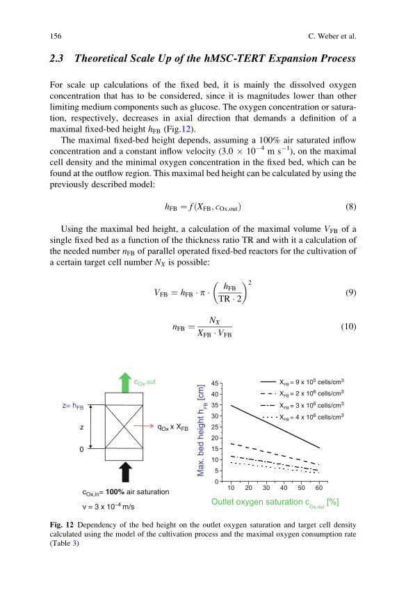
2.3 Theoretical Scale Up of the hMSC-TERT Expansion Process
For scale up calculations of the fixed bed, it is mainly the dissolved oxygen
concentration that has to be considered, since it is magnitudes lower than other
limiting medium components such as glucose. The oxygen concentration or satura-
tion, respectively, decreases in axial direction that demands a definition of a
maximal fixed-bed height hFB (Fig.12).
The maximal fixed-bed height depends, assuming a 100% air saturated inflow
concentration and a constant inflow velocity (3.0 � 10�4 m s�1), on the maximal
cell density and the minimal oxygen concentration in the fixed bed, which can be
found at the outflow region. This maximal bed height can be calculated by using the
previously described model:
hFB ¼ f ðXFB; cOx;outÞ (8)
Using the maximal bed height, a calculation of the maximal volume VFB of a
single fixed bed as a function of the thickness ratio TR and with it a calculation of
the needed number nFB of parallel operated fixed-bed reactors for the cultivation of
a certain target cell number NX is possible:
VFB ¼ hFB � p � hFBTR � 2
� �2
(9)
nFB ¼ NX
XFB � VFB
(10)
z= hFB
cOx,in= 100% air saturation
v = 3 x 10⁻⁴ m/s
z
0
XFB = 9 x 10⁵ cells/cm3
XFB = 2 x 10⁶ cells/cm3
XFB = 3 x 10⁶ cells/cm3
XFB = 4 x 10⁶ cells/cm3
qOx x XFB
cOx,out
Fig. 12 Dependency of the bed height on the outlet oxygen saturation and target cell density
calculated using the model of the cultivation process and the maximal oxygen consumption rate
(Table 3)
156 C. Weber et al.

The reactor system was scaled exemplarily for the cultivation of a target cell
number of 20 billion cells that is sufficient for approximately 200 single doses of
cell beads with a volume of about 5 mL per dose (Table 4).
The volume of a single fixed bed decreases with increasing cell number,
thickness ratio, and outlet oxygen saturation. Small reactor volumes means a
large number of parallel operated fixed-bed reactors, but this is very intricate and
non-practical regarding the handling and operation of the bioreactor system. There-
fore a reduction of the target cell density, as well as a reduction of the thickness
ratio and the outlet oxygen saturation, is recommended. As a result of this, small
numbers of parallel operated reactors which can be handled and operated more
easily are obtained. The oxygen outlet concentration, of course, can only be
decreased to an uncritical value.
3 Cultivation of Encapsulated Cells
3.1 The Reactor System
The cell bead cultivation system is based on single use plastic syringes in which the
cell beads themselves form the bed. The original piston is replaced by a custom
made insert, which enables perfusion of the fixed bed (Fig. 13).
Beside the cultivation on a single dose level, the advantages of this bioreactor
system are a cryopreservation of the cell beads by direct freezing of the syringe and
post-thaw an implantation of the cell beads using this syringe. This avoids contam-
ination risky transfer steps. For this purpose, the insert can be designed in such a
manner that it can act as the original syringe piston.
The reactor periphery is similar to that of the hMSC-TERT expansion system.
Furthermore, the previously described mathematical model can be used as well for
the simulation of the cell bead cultivation process.
Table 4 Numbers and volumes of parallel operated fixed beds needed for the cultivation of
2 � 1010 cells as a function of thickness ratio, target cell density, and outlet oxygen saturation
Thickness ratio ¼ 1 Thickness ratio ¼ 2
Target cell
density (cm�3)
Fixed-bed
volume
VFB (L)
Number of
fixed beds
nFB
Fixed-bed
volume
VFB (L)
Number of
fixed beds
nFB
Total
fixed-bed
volume (L)
Outlet oxygen saturation: 20%1 � 106 20 1.1 5.2 4.2 21.9
2 � 106 2.3 4.8 0.6 19.1 10.9
4 � 106 0.3 18.3 0.07 73.1 5.5
Outlet oxygen saturation: 30%1 � 106 13.4 1.6 3.4 6.5 21.9
2 � 106 1.5 7.3 0.4 29.0 10.9
4 � 106 0.2 27.8 0.05 111.4 5.5
Production Process for Stem Cell Based Therapeutic Implants 157

3.2 Cultivation of Encapsulated Cells
Cultivations of cell beads were exemplarily performed under adipogenic conditions
on a 1-cm3 scale (data not shown). Induction medium was applied for 3 days
followed by 4 days cultivation with maintenance medium (Table 5). This cycle
was repeated three times [14]. Medium, 50 mL per cycle, was perfused at a
superficial velocity of 1.28 � 10�4 m s�1, that is below the fluidization point.
Reference cultures were performed in 25-cm2 T-flasks using the same protocol.
Vitality was determined after 0, 100, 200, and 500 h with the Trypan blue
exclusion method after lysis of the cell beads using EDTA [28].
The vitality increased with advancing cultivation time, whereas the cell number
decreased (Fig. 14). This is explainable by decomposition of apoptotic cells.
Apoptosis may be triggered by the harvesting or the encapsulation process, or
Incubator37°C5% CO2
Conditioningvessel
WasteMedium Cryopro-tectivemedium
Inoculum(cell beads)
4°C
IPO2
IPO2
Fig. 13 System for the cultivation of cell beads in syringe based fixed reactors
Table 5 Composition of media for the adipogenic cultivation of cell beads [28]
Induction medium Maintenance medium
DMEM þ 10% FCS DMEM þ 10% FCS
100 U mL�1 penicillin 100 U mL�1 penicillin
0.1 mg mL�1 streptomycin 0.1 mg mL�1 streptomycin
0.01 mg mL�1 insulin 0.01 mg mL�1 insulin
0.5 mM 3-isobutyl-1-methyl-xanthin –
0.2 mM indomethacin –
1 mM dexamethason –
158 C. Weber et al.

dominant in this case, by the cryopreservation of the cell beads prior to the
cultivation. It could be shown that the vitality, and thus the quality of the beads,
is gradable during the cultivation process.
As an example, an adipogenic differentiation protocol was applied, whereby the
differentiation to adipocytes was verified by staining with the lipophilic fluorescence
dye Nile red (Fig. 15) [28]. Adipogenic cultured cell beads showed higher fluores-
cence intensity and thus are interpreted to be differentiated to adipocytes. No differ-
ences between the fixed-bed culture and the reference culture in T-flasks are detectable.
Table 6 shows the kinetics obtained by fitting the model parameter to the
experimental data which were used for a theoretical scale up of the system.
0
20
40
60
80
100 Fixed bed Reference (T25-Flask)
Vita
lity
[%]
Cel
l num
ber
Nx
[-]
0
2x10⁶
4x10⁶
6x10⁶
8x10⁶
1x10⁷
Time [h]5002001000
Time [h]
5002000
Fig. 14 Time dependent vitality and cell number of cell beads cultured in fixed-bed reactors and in
T-flasks (reference)
Adipogeniccultivation
Non-adipogeniccultivation
T25-Flask(Reference)
Fixed bed
Fig. 15 Nile red staining of cell beads which were cultured under adipogenic and non-adipogenic
conditions in fixed-bed reactors or T25-flasks
Production Process for Stem Cell Based Therapeutic Implants 159

3.3 Theoretical Scale Up of the Cell Bead Cultivation Process
A calculational scale up was carried out for a cultivation of 200 single doses of cell
beads each of 5 mL (Fig. 16). The inlet oxygen concentration was assumed to be air
saturated. This can be realized, for example, by using membrane oxygenators.
For a calculation of the oxygen or glucose concentration profile in the cell bead,
the following diffusion and diffusion-reaction equations were used:
DCC � d2cCCdr2
þ 2
r� dcCC
dr
� �¼ q � XCC (11)
DAC � d2cACdr2
þ 2
r� dcAC
dr
� �¼ 0 (12)
Table 6 Consumption kinetics of encapsulated hMSC-TERT (cell beads) which were obtained
by fitting of model parameters to the experimental data of the adipogenic cultivation in a 1-cm3
fixed-bed scale (Fig. 14)
Growth rate mmax 0 (1 day�1)
Maximal glucose consumption rate qGlc;max (7.3 – 9.4) � 10�8 (mg h�1 cell�1)
Monod constant kM;qGlc 0.06 (mg mL�1)
Oxygen consumption rate qOx 5.5 � 10�9 (mg h�1 cell�1)
0
1
2
3
4
5G
luco
se c
once
ntra
tion
cell
bead
cen
ter
[mg/
ml]
Glucose concentrationin medium [mg/ml]
10 20 30 40 50 600
10
20
30
40
50
Oxy
gen
satu
ratio
nce
ll be
ad c
ente
r [%
]
Oxygen saturationin medium [%]
0 1 2 3 4 50 100 200 300 400 500
0
1
2
3
4
5
inlet outlet
Glu
cose
con
cent
ratio
nin
med
ium
[mg/
ml]
Time [h]
20
40
60
80
100
outlet
inlet
Oxy
gen
satu
ratio
nin
med
ium
[%]
Fig. 16 Simulated adipogenic cultivation of 200 single doses (5 cm3) of cell beads as well as the
glucose and oxygen profile at the cell bead center. Medium volume per cycle: 40 L, superficial
velocity: 2.5 � 10�4 m s�1
160 C. Weber et al.

with the concentration in the cell containing core bead cCC as well as in the cell free
alginate capsule cAC, the effective diffusion coefficient in the core bead DCC and
alginate capsule DAC; and the cell density of the core bead XCC.
It could be shown that an oxygen saturation in the medium of 40% leads to an
oxygen saturation at the center of approximately 22% (Fig. 16). The differences in
glucose between the medium and the cell bead center is negligible. Thus, it can be
assumed that no limitations of the cells at the center of a cell bead are expectable.
3.4 Conclusion and Outlook
Two fixed-bed reactor systems for the production of stem cell based therapeutic
implants were introduced. One system was developed for the expansion of the
production cell line (hMSC-TERT) and a second for the cultivation of encapsulated
cells in order to increase their vitality and thus the quality of the implants.
The fixed-bed system for the expansion of the production cell line is based on
non-porous BSGS. Cells can be cultured and harvested with high yield and vitality.
The separation of the cells from the carrier can easily be performed by flushing
them out with the medium flow. This saves additional process steps.
The fixed-bed system for the cultivation of encapsulated cells is based on commer-
cially available syringes in which the cell beads represent the bed. The advantage of
this system is that it can be used as an implantation tool after the cultivation procedure.
It could be shown that the vitality is gradable by the cultivation process. Furthermore,
the application of an adipogenic differentiation protocol could be demonstrated.
Both systems can be automated and produced as disposable items due to their
simple design.
The next steps will concern the development of a GMP-conform cryopreser-
vation procedure for the cell beads and the implementation of the cultivation
systems to the overall GMP-process of cell bead production.
Acknowledgements The authors would like to thank the Federal Ministry of Economics and
Technology for financial support as well as the CellMed AG for providing the production cell line
hMSC-TERT and the CellBeads1.
References
1. Lanza RP, Hayes JL, Chick WL (1996) Encapsulated cell technology. Nature Biotech
14:1107–1111
2. Freimark D, Czermak P (2009) Cell-based regeneration of intervertebral disc defects: review
and concepts. Int J Artif Organs 32:197–203
3. Baksh D, Song L, Tuan RS (2004) Adult mesenchymal stem cells: characterization, differen-
tiation, and application in cell and gene therapy. J Cell Mol Med 8:301–316
4. Chiu RCJ (2003) Bone-marrow stem cells as a source for cell therapy. Heart Failure Rev
8:247–251
Production Process for Stem Cell Based Therapeutic Implants 161

5. Fraser JK et al (2004) Adult stem cell therapy for the heart. Int J Biochem Cell Biol
36:658–666
6. Mimeault M, Hauke R, Batra SK (2007) Stem cells: a revolution in therapeutics–recent
advances in stem cell biology and their therapeutic applications in regenerative medicine
and cancer therapies. Clin Pharmacol Ther 82:252–264
7. Simonsen JL et al (2002) Telomerase expression extends the proliferative life-span and
maintains the osteogenic potential of human bone marrow stromal cells. Nat Biotechnol
20:592–596
8. Heile AMB et al (2009) Cerebral Q1 transplantation of encapsulated mesenchymal stem cells
improves cellular pathology after experimental traumatic brain injury. Neurosci Lett
9. Aris R (1975) The mathematical theory of diffusion and reaction impermeable catalysts.
Clarendon, Oxford
10. Bailey J, Ollis DF (1986) Biochemical engineering fundamentals. McGraw-Hill, New York
11. Froment GF, Bischoff KB (1979) Chemical reactor analysis and design. Wiley, New York
12. Fassnacht D (2001) Fixed-bed reactors for the cultivation of animal cells. Fortschritt-Berichte
VDI, vol. 17, VDI-Verlag, Dusseldorf
13. Willaert RG, Baron GV, de Backer L (1996) Modelling of immobilised bioprocesses. In:
Willaert RG, Baron GV, de Backer L (eds) Immobilised living cell systems. Wiley, New
York, pp 237–254
14. Perry RH, Green DW (2007) Perry’s chemical engineers’ handbook. McGraw-Hill
15. Weber C, Gokorsch S, Czermak P (2007) Expansion and chondrogenic differentiation of
human mesenchymal stem cells. Int J Artif Organs 30:611–618
16. Schop D et al (2009) Growth, metabolism, and growth inhibitors of mesenchymal stem cells.
Tissue Eng A 15:1877–1886
17. Higuera G et al (2009) Qvantifying in vitro growth and metabolism kinetics of human
mesenchymal stem cells using a mathematical model. Tissue Eng Part A 15:1–11
18. Schop D et al (2008) Expansion of mesenchymal stem cells using a microcarrier-based
cultivation system: growth and metabolism. J Tissue Eng Regen Med 2:126–135
19. Lonergan T, Brenner C, Bavister B (2006) Differentiation-related changes in mitochondrial
properties as indicators of stem cell competence. J Cell Phys 208:149–153
20. Conget P, Minguell JJ (1999) Phenotypical and functional properties of human bone marrow
mesenchymal progenitor cells. J Cell Phys 181:67–73
21. Guo Z et al (2001) Biological features of mesenchymal stem cells from human bone marrow.
Chin Med J 114:950–953
22. Soukup T et al (2006) Mesenchymal stem cells isolated from human bone marrow: cultiva-
tion, phenotypic analysis and changes in proliferation kinetics. Acta Med 49:27–33
23. Peng CA, Palson BA (1996) Determination of specific oxygen uptake rates in human
hematopoietic cultures and implications for bioreactor design. Ann Biomed Eng 24:373–381
24. Portner R et al (2005) Bioreactor design for tissue engineering. J Biosci Bioeng 100:
235–245
25. Acevedo CA et al (2008) A mathematical model for the design of fibrin microcapsules with
skin cells. Bioprocess Biosyst Eng 32(3):341–351
26. De Leon A, Mayani H, Ramırez OT (1998) Design, characterization and application of a
minibioreactor for the culture of human hematopoietic cells under controlled conditions.
Cytotechnol 28:127–138
27. Youn BS, Sen A, Behie LA (2006) Scale-up of breast cancer stem cell aggregate cultures to
suspension bioreactors. Biotechnol Prog 22:801–810
28. Weber C et al (2007) Cultivation and differentiation of encapsulated hMSC-TERT in a
disposable small-scale syringe-like fixed bed reactor. Open Biomed Eng J 1:64–70
162 C. Weber et al.

Adv Biochem Engin/Biotechnol (2010) 123: 163–200DOI: 10.1007/10_2010_67# Springer-Verlag Berlin Heidelberg 2010Published online: 4 May 2010
Cartilage Engineering from Mesenchymal
Stem Cells
C. Goepfert, A. Slobodianski, A.F. Schilling, P. Adamietz, and R. Portner
Abstract Mesenchymal progenitor cells known as multipotent mesenchymal stro-
mal cells or mesenchymal stem cells (MSC) have been isolated from various
tissues. Since they are able to differentiate along the mesenchymal lineages of
cartilage and bone, they are regarded as promising sources for the treatment of
skeletal defects. Tissue regeneration in the adult organism and in vitro engineering
of tissues is hypothesized to follow the principles of embryogenesis. The embryonic
development of the skeleton has been studied extensively with respect to the
regulatory mechanisms governing morphogenesis, differentiation, and tissue for-
mation. Various concepts have been designed for engineering tissues in vitro based
on these developmental principles, most of them involving regulatory molecules
such as growth factors or cytokines known to be the key regulators in developmen-
tal processes. Growth factors most commonly used for in vitro cultivation of
cartilage tissue belong to the fibroblast growth factor (FGF) family, the transform-
ing growth factor-beta (TGF-b) super-family, and the insulin-like growth factor
(IGF) family. In this chapter, in vivo actions of members of these growth factors
described in the literature are compared with in vitro concepts of cartilage
engineering making use of these growth factors.
C. Goepfert (*) and R. Portner
Institute of Bioprocess and Biosystems Engineering, Hamburg University of Technology,
Hamburg, Germany
e-mail: [email protected]
A. Slobodianski
Kompetenzzentrum Tissue Engineering, Universitat zu Lubeck, Lubeck, Germany
P. Adamietz
Department of Biochemistry and Molecular Biology II: Molecular Cell Biology, University
Medical Center Hamburg-Eppendorf, Hamburg, Germany
A.F. Schilling
Biomechanics Section, Hamburg University of Technology, Hamburg, Germany

Keywords Bone morphogenetic protein (BMP), Cartilage, Chondrocytes, Differ-
entiation, Fibroblast growth factor (FGF), Growth factors, Indian hedgehog (Ihh),
Insulin like growth factor (IGF), Mesenchymal stem cells (MSC), Multipotent
mesenchymal stromal cells (MSC), PTH related peptide (PTHrP), Sonic hedgehog
(Shh), Transforming growth factor-beta (TGF)
Contents
1 Introduction . . . . . . . . . . . . . . . . . . . . . . . . . . . . . . . . . . . . . . . . . . . . . . . . . . . . . . . . . . . . . . . . . . . . . . . . . . . . . . . 165
1.1 The Term “Mesenchymal Stem Cells” . . . . . . . . . . . . . . . . . . . . . . . . . . . . . . . . . . . . . . . . . . . . . . 165
1.2 Concepts for Cartilage Cultivation In Vitro . . . . . . . . . . . . . . . . . . . . . . . . . . . . . . . . . . . . . . . . 167
2 Development of Cartilage Tissue In Vivo . . . . . . . . . . . . . . . . . . . . . . . . . . . . . . . . . . . . . . . . . . . . . . . . 168
2.1 Migration and Proliferation . . . . . . . . . . . . . . . . . . . . . . . . . . . . . . . . . . . . . . . . . . . . . . . . . . . . . . . . . 178
2.2 Cell Condensation . . . . . . . . . . . . . . . . . . . . . . . . . . . . . . . . . . . . . . . . . . . . . . . . . . . . . . . . . . . . . . . . . . 178
2.3 Chondrogenic Differentiation . . . . . . . . . . . . . . . . . . . . . . . . . . . . . . . . . . . . . . . . . . . . . . . . . . . . . . . 178
2.4 Endochondral Ossification . . . . . . . . . . . . . . . . . . . . . . . . . . . . . . . . . . . . . . . . . . . . . . . . . . . . . . . . . . 179
3 The Influence of Growth Factors on Cartilage
Development In Vivo . . . . . . . . . . . . . . . . . . . . . . . . . . . . . . . . . . . . . . . . . . . . . . . . . . . . . . . . . . . . . . . . . . . . . 180
3.1 Fibroblast Growth Factors in Early Skeletal Development . . . . . . . . . . . . . . . . . . . . . . . . . 180
3.2 The TGF-b Superfamily of Growth and Differentiation Factors . . . . . . . . . . . . . . . . . . . . 181
3.3 The Role of IGFs in the Development of Cartilage Tissue . . . . . . . . . . . . . . . . . . . . . . . . . 182
3.4 Terminal Differentiation or Development of Articular Cartilage . . . . . . . . . . . . . . . . . . . 182
4 Growth Factors in Adult Cartilage Tissue . . . . . . . . . . . . . . . . . . . . . . . . . . . . . . . . . . . . . . . . . . . . . . . . 183
4.1 The Role of FGF in Adult Articular Cartilage . . . . . . . . . . . . . . . . . . . . . . . . . . . . . . . . . . . . . . 183
4.2 Members of the TGF-b Superfamily in Adult Articular Cartilage . . . . . . . . . . . . . . . . . . 184
4.3 The Role of IGF in Adult Articular Cartilage . . . . . . . . . . . . . . . . . . . . . . . . . . . . . . . . . . . . . . 184
5 Engineering of Cartilage Tissue In Vitro . . . . . . . . . . . . . . . . . . . . . . . . . . . . . . . . . . . . . . . . . . . . . . . . . 185
5.1 Expansion of Progenitor Cells . . . . . . . . . . . . . . . . . . . . . . . . . . . . . . . . . . . . . . . . . . . . . . . . . . . . . . 185
5.2 Chondrogenic Differentiation In Vitro . . . . . . . . . . . . . . . . . . . . . . . . . . . . . . . . . . . . . . . . . . . . . . 186
5.3 Maintenance of the Hyaline Phenotype in Cultivated Cartilage Tissue . . . . . . . . . . . . . 188
6 Conclusion . . . . . . . . . . . . . . . . . . . . . . . . . . . . . . . . . . . . . . . . . . . . . . . . . . . . . . . . . . . . . . . . . . . . . . . . . . . . . . . . 188
References . . . . . . . . . . . . . . . . . . . . . . . . . . . . . . . . . . . . . . . . . . . . . . . . . . . . . . . . . . . . . . . . . . . . . . . . . . . . . . . . . . . . 189
Abbreviations
AER Apical ectodermal ridge
BMP Bone morphogenetic protein
ECM Extracellular matrix
FGF Fibroblast growth factor
FGFR FGF receptor
GF Growth factor
HA Hyaluronic acid
IGF Insulin-like growth factor
IGFBP IGF binding protein
IGFR IGF receptor
Ihh Indian hedgehog
164 C. Goepfert et al.

MSC Mesenchymal stem cells
OA Osteoarthritis
PTHrP PTH related peptide
RA Rheumatoid arthritis
Shh Sonic hedgehog
TGF-b Transforming growth factor-bZPA Zone of polarizing activity
1 Introduction
1.1 The Term “Mesenchymal Stem Cells”
For cartilage regeneration in vitro and in vivo, various strategies have been pursued
regarding the appropriate cell source, chemical and physical factors, and culture
conditions. It is widely agreed that tissue regeneration from autologous cells can be
achieved taking advantage of the natural course and progression of embryonic
development [1–4]. Therefore, autologous mesenchymal stem cells (MSCs) are
extensively investigated for their ability to regenerate articular cartilage tissue in
situ or in vitro.
Tissues forming the skeleton of the limbs originate from stem cells of the lateral
plate and the somitic mesenchyme [2]. These embryonal MSCs undergo a series of
differentiation steps, finally producing differentiated skeletal tissues such as bone
and cartilage. In the adult organism, there are a limited number of mesenchymal
progenitor cells residing in the bone marrow which give rise to the repair of
damaged tissue, for instance bone, and which can easily be obtained by marrow
aspiration and selected by their ability to adhere to culture vessels. To date, it is not
clear whether they represent cells remaining from the embryonic mesenchyme or
whether they are a heterogenous population of mesenchymal precursor cells [4, 5],
possibly originating from the invading blood vessels populating the newly formed
bone marrow space during endochondral ossification [6].
First evidence for a precursor pool within the bone marrow was given by Frieden-
stein et al. [7], who described ectopic osteogenic differentiation originating from
whole bone marrow. Based on these results, they hypothesized a population of cells
occurring in the bone marrow which are able to differentiate along the osteogenic
lineage. These cells were characterized in vitro as colony forming fibroblastic cells
(CFU-f), isolated by their adherence to culture vessels [8]. MSC were hypothesized
as progenitor cells of mesenchymal tissues residing in the bone marrow and perios-
teum, persisting throughout lifetime as a pool for tissue regeneration, and which
might be isolated, expanded, and used for autologous treatment of damaged tissues
[9]. In 1999, Pittenger et al. [10] demonstrated the ability of clonally expanded human
bone marrow cells to differentiate towards the osteogenic, chondrogenic, and adipo-
genic lineages and thereby making it possible to develop treatments with human
Cartilage Engineering from Mesenchymal Stem Cells 165

autologous cells for the repair of mesenchymal tissues such as bone, cartilage and
adipose tissue. Since that time, these so-called MSCs have been isolated from various
tissues including adipose tissue [11], muscle and brain [12], bone [13], synovium [14,
15], umbilical cord [16], and blood [17]. Cells with even higher differentiation
potential have been isolated from cord blood [18, 19] and from bone marrow [20].
In order to characterize the MSCs, several markers have been described, but none
of them proved to be unique and exclusively present on MSCs. Enrichment of MSCs
has been carried out using the Stro-1 monoclonal antibody [21, 22]. The resulting cell
population was shown to be able to differentiate into the mesenchymal lineages of
osteoblasts, chondrocytes, adipocytes, and stromal cells supporting hematopoiesis
[23]. Other antigens specific for undifferentiated precursor cells and absent on
differentiated cells have been identified using the monoclonal antibodies SH-2 (endo-
glin, co-receptor for TGF-b3) [24], SH-3, and SH-4, respectively [25]. But there are
no unique surface antigens so far defining “the” MSC.
Clonally derived MSCs have been extensively analyzed for their ability to
proliferate in vitro, retaining their multi-lineage differentiation potential upon
prolonged cultivation. It was shown that these clonally derived cells lost their
multi-lineage potential upon extended cultivation in vitro and thus behave like
plastic progenitor cells rather than stem cells [5, 26–28]. On the other hand, Jiang
et al. [20] were able to describe a subset of pluripotent cells in the mouse and the rat
bone marrow, virtually proliferating for up to 60 passages without losing their
characteristic growth rates and their potential even to transdifferentiate into the
endodermal and ectodermal lineages displaying some features of hepatocytes or
neuronal cell types [20, 29]. Thus it is likely that MSCs are a heterogenous
population of rare cells in the bone marrow compartment and other tissues, dis-
playing various stages of predifferentiation. Due to the heterogeneity of the cell
preparations commonly referred to as MSCs, Dominici et al. defined mesenchymal
stem cells as plastic adherent cells positive for CD105, CD73, CD90 lacking the
expression of hematopoetic markers CD45, CD34, CD14 or CD11, CD79 alpha or
CD19 and HLA-DR surface markers [30]. Finally, according to this definition,
MSCs must be shown to differentiate into osteoblasts, adipocytes and chondrocytes
in vitro. According to Horwitz et al. [31], fibroblast-like plastic adherent mesen-
chymal progenitor cells isolated from various tissues are termed multipotent mes-
enchymal stromal cells (also referred to as MSCs).
Recently, it has been proposed to define “stemness” as a state of cells which are
able to differentiate into various cell types rather than the cells themselves [32].
According to this view, plasticity would be the most prominent characteristic of
stem cells whereas self-renewal and hierarchical differentiation are regarded as
subordinate features of these cell populations. Therefore, dedifferentiated cells
capable of differentiating into more than their original cell type could also be
regarded as stem cells. Since in vitro-expanded and thus dedifferentiated articular
chondrocytes were shown to differentiate similarly to MSCs along the mesenchy-
mal lineages into osteoblasts, chondrocytes, including hypertrophic chondrocytes,
and adipocytes [33, 34], the term “secondary progenitor cells” could be appropriate
for dedifferentiated chondrocytes [35].
166 C. Goepfert et al.

1.2 Concepts for Cartilage Cultivation In Vitro
The regeneration of damaged tissues in the adult organism as well as in vitro
engineering of tissues is hypothesized to follow the principles of embryogenesis
[1–4]. Therefore it is assumed that cartilage formation by means of cell therapy or
tissue engineering is bound to recapitulate, at least in several aspects, the stages of
in vivo development. The development of the appendicular skeleton is initiated by
migration of the early mesenchymal progenitors of the skeletal tissue towards the
prospective limb regions and by proliferation of these undifferentiated progenitors.
The accumulation of high cell densities is the prerequisite of cell condensation as
the key event of cartilage tissue formation. Thus, the factors governing the migra-
tion and proliferation of the undifferentiated mesenchymal cells hold potential for
the expansion of undifferentiated precursor cells in vitro.
The morphogenesis of skeletal elements requires precartilage condensation
leading to chondrogenic differentiation and thus to the formation of cartilaginous
models of the skeletal elements. During chondrogenic differentiation, the cells
adopt a rounded morphology and start to synthesize cartilage specific matrix
molecules. Further shaping of the skeletal primordia involves the separation of
the digital rows by apoptosis of the cells within the interdigital mesenchyme. The
concomitant formation of the joints requires dedifferentiation of the cells in the
prospective joint regions transiently leading to high cell densities and finally to
apoptotic cell death and the development of the joint cavity.
Since the precursor cells are usually expanded in 2D systems in vitro where they
display a flattened spindle-shaped morphology, 3D culture systems are supposed to
be appropriate for chondrogenic differentiation. The naturally occurring events are
mimicked by cell–cell-contacts established in high density pellet culture [36] by
cultivation in hydrogels [37–41] or 3D matrices of biocompatible materials [42, 43].
A comparison between the in vivo stages of cartilage development and the strate-
gies used for transferring these stages into in vitro concepts and the inherent
challenges is given in Table 1.
Usually these culture systems taken alone are not sufficient to induce chondro-
genesis of precursor cells. Therefore, growth factors are included into the culture
medium and combined with physical factors such as reduction of oxygen supply
Table 1 Strategies for transferring in vivo development into in vitro concepts
In vivo development of cartilage tissue In vitro cultivation of cartilage tissue
Migration and proliferation Expansion of MSC in vitro (2D cultivation)
Prechondrogenic condensation 3D aggregate culture imitating cellular condensation
Chondrogenic differentiation Cultivation in 3D biomaterials mimicking the 3D
structure of the ECM
Transient growth plate cartilage:
hypertrophy and apoptosis
Permanent articular cartilage: no
hypertrophy, no apoptosis
Prevention of hypertrophy and apoptosis in articular
cartilage regeneration
Cartilage Engineering from Mesenchymal Stem Cells 167

which occurs naturally by exclusion of the blood vessels from forming limb buds.
Chondrogenic differentiation in vitro is achieved after cultivation for several weeks
resulting in the formation of collagen type II and proteoglycans along with other
cartilage specific tissue components.
In vivo, the articular cartilage is maintained in a stage in which progression
towards hypertrophy naturally occurring in the growth plate is prevented by defined
mechanisms. In the growth plate, hypertrophy and calcification of the cartilage
tissue precede vascular invasion, finally leading to tissue replacement by bone. The
inactivation of the differentiation program towards hypertrophy is particularly
important for in vitro cultivation of cartilage tissue when MSCs are used as the
cell source since terminally differentiated cells will undergo apoptosis. Unlike
MSCs, articular chondrocytes usually do not undergo lineage progression upon
extended cultivation [44] or when implanted into ectopic sites [45]. Maintenance of
the phenotype of articular cartilage therefore comprises the inactivation of the
developmental pathway leading to terminal differentiation, and a switch to sus-
tained maintenance of functional extracellular matrix. For this reason, the appro-
priate stage of in vitro cultivated cartilage tissue needs to be carefully evaluated
regarding the state of cellular phenotype of chondrocytes and the characteristics of
the extracellular matrix formed.
In this overview, regimes of growth factor treatments currently applied in
cartilage tissue engineering in vitro are compared to stage specific actions of growth
factors in vivo during cartilage differentiation. Since growth factors of the fibroblast
growth factor (FGF)-family, transforming growth factor-b (TGF-b) superfamily
and the insulin-like growth factor (IGF) family are widely used in cartilage engi-
neering in vitro, the overview will be narrowed to these major groups of growth
factors. An overview of in vivo actions with in vitro applications of growth factors
is given in Table 2.
2 Development of Cartilage Tissue In Vivo
The development of the skeleton in vivo is a multistep process tightly regulated
regarding the temporal and spatial distribution of the appropriate signals. Morpho-
genesis is evoked by gradients of specific signaling molecules along the axes of the
developing limbs. Following steps are triggered by the achievement of distinct
differentiation stages of the cells and by stage specific extracellular matrix synthe-
sis. Regulatory mechanisms such as feedback terminate a distinct developmental
stage and allow for further differentiation. Thus, tissue development depends on the
differentiation stage of the cells as well as on signals provided by paracrine,
autocrine or systemic factors, and the extracellular matrix (Fig. 1).
Taking into account the multi-stage process leading to functional tissues in vivo,
histogenesis in vitro also needs to be tightly regulated regarding the time sequence
of induction and the resulting consecutive stages of extracellular matrix synthesis
(Fig. 2).
168 C. Goepfert et al.

Table
2Occurrence
andactionsofgrowth
factors
indevelopingandmature
cartilage,
diseasedcartilagetissue,
andapplicationsin
cartilagecultivation
invitro
Growth
factor/
receptor
Role
inthedevelopmentof
cartilagetissuein
vivo
Role
inadultcartilagetissue
Role
incartilagelesionsorin
articularcartilagedisease
Invitro
applicationsandfunctions
FGFfamily
FGF-2
Presentin
theAER
Boundto
heparan
sulfatein
theECM,possiblerolein
mechanotransduction
[97–99]
RecruitmentofMSCsto
cartilagedefects
Stimulationofproliferationof
MSCin
vitro
[146]
StimulationofTenascinsynthesis
inmesenchymal
condensations[147]
–InductionofMMP-13[101]and
noggin
[104]
Maintenance
ofdifferentiation
potential
ofMSCs[126,127]
Stimulationoftenascinand
syndecan
synthesisin
condensationslimitingsize
of
condensations[64]
–Increasedproductionin
RA
[148]
Increasedintegrinalpha10-
expressiononculturedMSCs
invitro
[128]
Activationbyendothelial
proteases
inendochondral
ossification[149]
––
Maintenance
ofdifferentiation
potential
ofmam
malian
chondrocytesin
vitro
[123]
––
–“D
edifferentiation”of
chondrocytes[33]
FGF-4
Occurrence
intheAER,
chem
oattractantforlimbbud
mesenchymal
cells[46]
––
–
Occurrence
intheZPA,role
in
APaxisform
ation,controls
theform
ationofgap
junctions
[150,151]
––
–
FGF-8
Expressionin
theAER,
maintenance
offgf-10
expressionin
theunderlying
mesenchyme
[47,75,77]
––
–
(con
tinu
ed)
Cartilage Engineering from Mesenchymal Stem Cells 169

Table
2(continued)
Growth
factor/
receptor
Role
inthedevelopmentof
cartilagetissuein
vivo
Role
inadultcartilagetissue
Role
incartilagelesionsorin
articularcartilagedisease
Invitro
applicationsandfunctions
FGF-10
Lateral
plate
mesenchyme:
––
–
Proliferation,migration[73,77]
––
–
Expressionin
blastem
asof
regeneratinglimbsof
amphibia
[78]
––
–
FGF-18
Roleinendochondralossification,
coordinationof
chondrogenesisand
osteogenesis[152,153]
––
–
Signalingvia
FGFR3,promotion
ofchondrogenesis[154]
––
–
FGFreceptors
FGFRI
Mesenchymal
cellsofthelimb
budpriorto
condensation
[155,156]
––
Expressionin
proliferatingcells
ofmesenchymal
lineage
invitro
[157]
Absence
ofFGFR1causes
apoptosisin
limbbuds[158]
––
Expressionpriortodifferentiation
[130,131]
––
–Expressionin
hypertrophic
culturesofMSCs[130,131]
FGFRII
Mesenchymal
condensations
[156],essential
forlimb
outgrowth
[74]
Adultarticularcartilage
[130]
–Expressionin
condensationsof
mesenchymal
micromass
cultures[157]
Expressionin
limbbud
ectodermal
epithelium
[155]
––
Expressionduringcondensation
invitro
[131]
Ectodermal
FGF-2
and
FGF-8
inhibitchondrogenesis
throughFGFRIIsignaling
[159]
––
Downregulationduring
differentiationin
vitro
[130]
Inprechondrocyticcellspriorto
condensation[155]
––
–
170 C. Goepfert et al.

FGFRIII
Growth
plate
chondrocytes[155,
160]
––
Priorto
calcificationof
mesenchymal
micromass
cultures[157]
TGF-b
superfamily
TGF-b1
Expressionin
condensations,
stim
ulatessynthesisof
fibronectinTenascin,N-
CAM,N-cadherin
[50,64,65]
Presentin
adultcartilage,
boundto
cartilagematrix
[107]
InductionofOA-likechanges
uponoverexpression[111]
Inductionofchondrogenesisof
bonemarrow
MSC[133]and
synovium
derived
MSC[137]
Fnpositiveareasprecede
chondrogenic
differentiation
[86]
Stimulationoffibronectin
synthesisin
cartilage
explants[161]
Stimulationofproteoglycan
synthesisbyOA
chondrocytes[162,163]
Inhibitory
effect
onprimary
chondrocytes[164,165]
Inhibitionofterm
inal
differentiationofepiphyseal
chondrocytes[166]
TGFbindingdomainin
Procollagen
typeIIA[167]
–UpregulationofTGF-b
expressionin
OA
[168]
–
Storagein
growth
plate
matrix
boundto
LTBP,activationby
matrixvesicles[107,108]
––
–
StimulationofPTHrP
synthesisin
epiphyseal
chondrocytes
[169]
––
–
Proliferationofundifferentiated
mesenchymal
cells[79]
––
–
TGF-b2
Condensations
Presentin
adultcartilage
Proliferation
Inductionofmatrixsynthesis,
Endogenoussynthesisby
ATDC-5
aggregatecultures
[170]
––
–Inhibitory
effect
onproliferation
ofMSCs[146]
(con
tinu
ed)
Cartilage Engineering from Mesenchymal Stem Cells 171

Table
2(continued)
Growth
factor/
receptor
Role
inthedevelopmentof
cartilagetissuein
vivo
Role
inadultcartilagetissue
Role
incartilagelesionsorin
articularcartilagedisease
Invitro
applicationsandfunctions
TGF-b3
––
Proliferation,stim
ulationof
matrixsynthesis
Chondrogenic
differentiationof
MSCs[10,36]
––
InductionbyIL-1
[112]
–
BMP(general)
Negativeregulators
oftheAER
[81]
––
ATDC5cellline:
stagespecific
expressionofBMP-2,-4,-6,
-7[171]
FeedbackinhibitionbyNoggin
[172]
––
–
BMPsignalingrequired
for
maintenance
ofdifferentiated
phenotype,cellproliferation
andhypertrophy[173]
––
–
InhibitionofBMPsignalingby
noggin
inhibitscondensation
anddifferentiation[91]
––
–
BMPsactas
heterodim
ers[82]
––
–
Co-localizationofBMPs[174]
––
–
FeedbackinhibitionofBMP
signalingbynoggin
[175]
––
–
BMP-2
Expressionin
theAERandZPA
––
StimulationofN-cadherin
synthesisandchondrogenesis
inC3H10T1/2
cells[176]
Inductionofcollagen
typeX
synthesisandalkaline
phosphataseexpressionin
growth
plate
chondrocytes
[177]
––
Inductionofchondrogenesisof
bonemarrow
MSC[178]and
synovium
derived
MSC[15]
andsynovialexplants[137]
172 C. Goepfert et al.

BMP-4
Inductionofcollagen
typeX
synthesisandalkaline
phosphataseexpressionin
growth
plate
chondrocytes
[177]
––
–
BMP-6
Transientexpressionin
prehypertrophic
chondrocytes
[179,180]
––
Chondrogenic
differentiationof
MSCs,synthesisby
prehypertrophic
cellsin
vitro
[171]
BMP-7
(OP-1)
Expressionduring
chondrogenesis
Synovialfluid,cartilage
tissue
Reducedexpressionin
aged
cartilagetissue[113]
Synthesisofcollagen
typeII,
aggrecan,GAG
Inductionofcollagen
typeX
synthesisandalkaline
phosphataseexpressionin
growth
plate
chondrocytes
[177]
––
Inductionofchondrogenesisin
ATDC5cellline[181]
––
–Inductionofchondrogenesisof
synovialexplants[137]
––
–Redifferentiationofarticular
chondrocytes[182,183]
BMPreceptors
BMPRIB
expressionprefigures
thecartilageprimordia
[184]
––
–
BMPRIB
presenton
chondrogenic
cells,
stim
ulationofBMPRIB
expressionbyTGF-b
[65,
185]
––
–
InhibitionofBMPRIB
expression
byFGFcausesapoptosis[80]
––
–
GDFs
Form
ationofjointsandjoint
spaces
[186,187]
––
–
(con
tinu
ed)
Cartilage Engineering from Mesenchymal Stem Cells 173
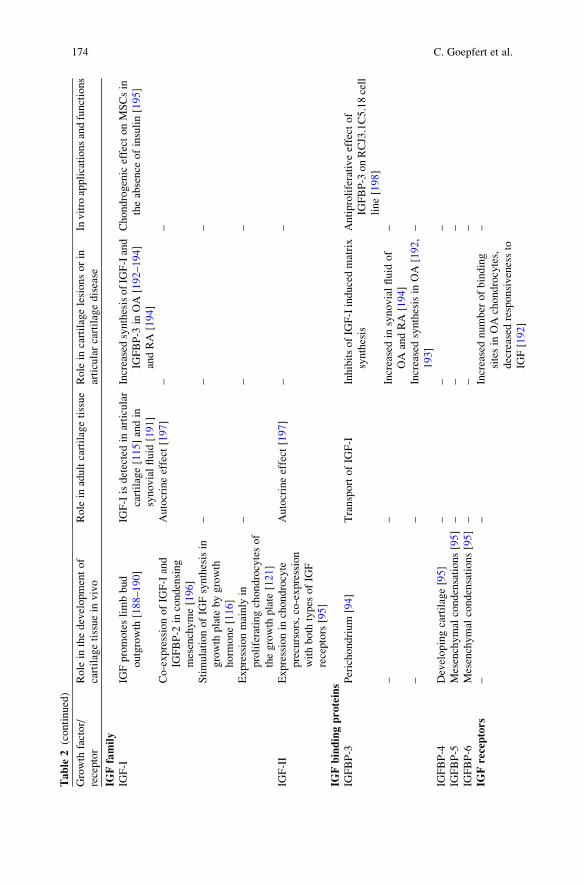
Table
2(continued)
Growth
factor/
receptor
Role
inthedevelopmentof
cartilagetissuein
vivo
Role
inadultcartilagetissue
Role
incartilagelesionsorin
articularcartilagedisease
Invitro
applicationsandfunctions
IGFfamily
IGF-I
IGFpromoteslimbbud
outgrowth
[188–190]
IGF-Iisdetectedin
articular
cartilage[115]andin
synovialfluid
[191]
IncreasedsynthesisofIG
F-Iand
IGFBP-3
inOA[192–194]
andRA
[194]
Chondrogenic
effect
onMSCsin
theabsence
ofinsulin[195]
Co-expressionofIG
F-Iand
IGFBP-2
incondensing
mesenchyme[196]
Autocrineeffect
[197]
––
StimulationofIG
Fsynthesisin
growth
plate
bygrowth
horm
one[116]
––
–
Expressionmainly
in
proliferatingchondrocytesof
thegrowth
plate
[121]
––
–
IGF-II
Expressionin
chondrocyte
precursors,co-expression
withboth
types
ofIG
F
receptors
[95]
Autocrineeffect
[197]
––
IGFbindingproteins
IGFBP-3
Perichondrium
[94]
TransportofIG
F-I
InhibitsofIG
F-Iinducedmatrix
synthesis
Antiproliferativeeffect
of
IGFBP-3
onRCJ3.1C5.18cell
line[198]
––
Increasedin
synovialfluid
of
OA
andRA
[194]
–
––
Increasedsynthesisin
OA[192,
193]
–
IGFBP-4
Developingcartilage[95]
––
–
IGFBP-5
Mesenchymal
condensations[95]
––
–
IGFBP-6
Mesenchymal
condensations[95]
––
–
IGFreceptors
––
Increasednumber
ofbinding
sitesin
OAchondrocytes,
decreased
responsivenessto
IGF[192]
–
174 C. Goepfert et al.

PTHrP
PTHrP
Synthesisin
epiphyseal
chondrocytes[67,169]
–Preventionofcollagen
typeX
synthesisbyMSCofOA
donors[144]
Preventionofcollagen
typeX
synthesisbybonemarrow
MSC[143,144]andby
adipose
tissuederived
MSC
[143]
Hedgehogproteins
Ihh
Productionbyprehypertrophic
chondrocytes,inductionof
PTHrP
inepiphyseal
cartilage
[68,69]
––
–
Shh
Expressionin
theZPA,rolein
AP
axisform
ation[89,199]
––
–
Growth
factorinteractions
FGF/TGF
StimulationofTGFsynthesisby
FGFin
progress
zone=>
promotestransitionof
precursorcellstowards
differentiation[46]
––
FGFsuppresses
senescence
inducedbyTGF-b2[129]
––
–“Secondaryprogenitorcells”
from
human
articular
chondrocytes[35]
––
–Stimulationofchondrocyte
growth
invitro
[203]
––
–Synergisticeffect
onchondrocyte
proliferationtogether
with
PDGF-BB[124]
FGF/BMP
FGFandBMPhaveopposite
effectsonlimboutgrowth
[208]
––
–
FGFandBMPinteract
toinduce
apoptosisoftheinterdigital
mesenchyme[80]
––
–
IGF/TGF
––
–Additiveeffectonchondrogenesis
ofMSC[138]andon
synovium
derived
MSC[200]
(con
tinu
ed)
Cartilage Engineering from Mesenchymal Stem Cells 175

Table
2(continued)
Growth
factor/
receptor
Role
inthedevelopmentof
cartilagetissuein
vivo
Role
inadultcartilagetissue
Role
incartilagelesionsorin
articularcartilagedisease
Invitro
applicationsandfunctions
––
–Enhancedchondrogenesisof
periostealexplants[201]
––
–Synergisticeffect
ofIG
F-Iand
TGF-b
onredifferentiationof
human
articularchondrocytes
[202]
IGF/BMP
––
Synergisticeffect
onmatrix
synthesisin
OA
chondrocytes[204]
Synergisticeffect
onmatrix
synthesis[204,206]
––
Inhibitory
effect
onMMP-13
expression[205]
IGF/TGF/FGF
––
–Enhance
proliferationof
synovium
derived
stem
cells
[200]
IGF/BMP/FGF
––
–Inhibitionofanaboliceffectsof
IGF-IandBMP-7
on
chondrocytesbyFGF[105]
TGF/BMP
––
–Inductionofchondrogenesisof
adipose-derived
stem
cells
byTGF-b3combined
with
BMP-6
[135]
––
–Inductionofchondrogenesisof
bonemarrow
andadipose
tissuederived
MSCby
TGF-b2in
combinationwith
BMP-7
[136]
––
–Synergisticeffect
ofTGF-b1and
BMP-2
onthechondrogenesis
ofbonemarrow
MSC[207]
176 C. Goepfert et al.

mesenchymal stem cells promotion oflineage progression
differentiated cells
paracrine / autocrinegrowth factors
extracellularmatrix
release of growthfactors from ECM
binding of growthfactors to ECM
proliferating /differentiating cells
stag
e sp
ecifi
c
synt
hesi
s of
par
acrin
e /
auto
crin
e G
F
GF
prom
otin
g
linea
ge p
rogr
essi
on
stage specific
synthesis / remodelling
of EC
M
feedback via
cell surface receptors
Fig. 1 Interaction of cellular differentiation, growth and differentiation factors, and specific
matrix molecules during histogenesis. Morphogenesis and histogenesis result from the interaction
of specific growth and differentiation factors acting in a stage dependent manner. Growth factor
actions are modulated by binding to ECM molecules and the expression of their receptors
depending on the developmental stage of the cells
proliferation anddifferentiation
terminaldifferentiation
calcification /vascularization
ossificationproliferationand migration
FGF-10FGF-2,4,8FGFR1
condensation
TGF-ßFGF-2shh
FGF-2/FGFR2IGF-IIBMP-2,4,7
FGF-18/FGFR3PTHrPBMP-2,7
FGF-2/FGFR1BMP-2,7
collagen I
collagen IIA collagen IIB collagen X collagen I
diaphysis
epiphysisperichondralbone collar
fibronectin
blood vesselcartilaginous
model
N-cadherin
aggrecan
tenascin
hyaluronan
growthplate
Fig. 2 Development of the long bones under the control of growth factors. The different develop-
mental stages of the skeletal primordia are shown together with the specific growth and differenti-
ation factors (above the arrows) which promote the stage specific synthesis of matrix molecules
(below) [6, 144]
Cartilage Engineering from Mesenchymal Stem Cells 177

2.1 Migration and Proliferation
In the initial phase of limb development, mesenchymal cells of the lateral plate
mesenchyme and of the somitic mesenchyme migrate towards the limb field [2].
The ectodermal cells are induced by the mesoderm to form a specialized epithelial
structure termed the apical ectodermal ridge (AER). The AER supports migration
and proliferation of the mesenchymal cells providing the cell mass for the forma-
tion of precartilage condensations [46, 47].
2.2 Cell Condensation
Cell condensation is characterized as a transient stage during the early morphogen-
esis which can be detected by PNA (peanut agglutinin) staining [48, 49]. During
cell condensation, cell density in the prospective limb regions is increased leading
to cell–cell contacts mediated by cell adhesion molecules such as N-cadherin and
N-CAM [50–52] and the formation of gap junctions [53, 54]. Cell adhesion
molecules are expressed specifically during the condensation phase and down
regulated subsequently upon chondrogenic differentiation of the prechondrogenic
cells. Prior to cell condensation, the extracellular matrix in the prospective limb
regions contains high amounts of collagen type I and hyaluronan [55, 56]. Hyaluronan
is supposed to prevent cell–cell interactions prior to the condensation phase [56].
During the condensation phase, hyaluronidase activity is detected, suggesting that
matrix remodeling takes place allowing for cell–cell interaction. During the con-
densation phase, a specific splice variant of fibronectin, FnEIIIA, is detected
throughout the condensations [57, 58]. Fibronectin was shown to be essential
for condensation and subsequent chondrogenesis. Furthermore, fibronectin distri-
bution during the condensation phase indicates the localization of skeletal elements
formed later on [55].
2.3 Chondrogenic Differentiation
Cellular differentiation is characterized by the increased synthesis of transcription
factors sox-5 and sox-6, and the appearance of the cartilage specific transcription
factor sox-9 [59–61]. Collagen type I and fibronectin are synthesized in the ECM
prior to condensation and reach a maximum density at the time of cellular differen-
tiation [55]. Chondrogenic differentiation of the condensing cells is characterized
by the appearance of collagen types II, IX, and XI, the characteristic components of
collagenous network of cartilage tissue. As a result of chondrogenic differentiation,
178 C. Goepfert et al.

remodeling of the extracellular matrix towards the cartilaginous composition of the
tissue takes place. Collagen type I consequently disappears from the tissue [55].
In adult articular cartilage, fibronectin is detected predominantly in the pericellular
area [62]. Collagen type I is usually not detected in articular cartilage unless repair
tissue is formed originating from the bone marrow. Mixed collagen type I and type
II formation resulting in mechanically inferior fibrocartilage is assumed to be an
intrinsic property of MSCs which cannot be avoided in mesenchymal cartilage
repair [63].
During cell differentiation, tenascin is transiently up-regulated in the developing
tissue. Tenascin and its receptor syndecan prevent further interaction of N-CAM
with fibronectin and thus terminate the condensation phase, allowing for further
progression of cellular differentiation [64].
Along with cellular differentiation and cartilage tissue formation, the skeletal
elements of the limbs are shaped into the precursors of the future skeleton. Joint
formation and the organization of the digital rows involve apoptosis of the cell
groups in the joint space and the interdigital regions. Therefore, the mesenchy-
mal cells of the limb bud either undergo chondrogenic differentiation or apo-
ptosis [65].
2.4 Endochondral Ossification
During the development of the appendicular skeleton in vivo, the cartilaginous
models of the long bones are replaced by bone tissue. Thus, lineage progression
towards terminal differentiation is an intrinsic program of the mesenchymal cells.
Articular cartilage, however, does not undergo the process of terminal differ-
entiation unless there are pathological conditions such as osteoarthritis (OA). In
OA, lineage progression is resumed leading to the formation of collagen type X
as a hypertrophy marker [66] and finally to abnormal calcification patterns of
the cartilage matrix. The mechanisms preventing the chondrocytes of the peri-
articular region from terminal differentiation and maintaining the cartilaginous
phenotype of articular cartilage are extensively studied in vivo [67–69]. In the
growth plate, chondrocytes resume proliferation leading to columnar cartilage
which represents the growth zone of the long bones in the embryo and the
juvenile organism. Chondrocytes leaving the growth zone start synthesizing
collagen type X which is the hallmark of hypertrophy. Morphologically, the
chondrocytes swell and adopt the hypertrophic phenotype. The hypertrophic
cartilage tissue is eroded by invading cells and replaced by bone marrow and in
the end by bone. Hypertrophic chondrocytes also secrete matrix degrading
enzymes such as MMP-13 which cleaves specifically collagen type II and thus
contributes to remodeling of the extracellular matrix. Hypertrophic chondrocytes
finally undergo apoptosis.
Cartilage Engineering from Mesenchymal Stem Cells 179

3 The Influence of Growth Factors on Cartilage
Development In Vivo
An overview of the occurrence and actions of growth factors in developing and
mature cartilage tissue is given in Table 2.
3.1 Fibroblast Growth Factors in Early Skeletal Development
The members of the FGF family share a homologous central core but differ in
their carboxyterminal and N-terminal regions due to alternative splicing [70]. The
FGFs are known as mitogens for mesodermal and neuroectodermal cell types.
They induce chemotaxis and angiogenesis [71] and bind to heparan sulfate with
high affinity [72]. Members of the FGF family of growth factors play a crucial
role in the development and morphogenesis of the appendical skeleton. They
are involved in all stages of tissue development as well as in the signal transduc-
tion during mechanical loading. FGFs are also involved in the remodeling and
degradation of articular cartilage matrix taking place in inflammatory joint
diseases.
FGFs are indispensable for outgrowth of limb buds preceding the development
of the appendical skeleton [47, 73]. The initial steps involve epithelial to mesen-
chymal interactions mediated by members of the FGF-family. Mesenchymal FGF-10
and ectodermal FGF-8 are the earliest factors involved in limb bud outgrowth. FGF-
10 is synthesized by the mesenchymal cells and induces the expression of FGF-
8 within the AER. The proximo-distal axis of the developing limb is established via
this feedback loop [74] which results in accumulation of proliferating cells in the
region of the prospective limb bud [46]. A specific splice variant of CD44 with the
ability to specifically bind FGF-8 mediates the presentation and thus the stimulation
of mesenchymal cells by FGF-8 [75]. FGF-4 and FGF-2 are co-expressed in the
AER [74, 76, 77]. Knowledge about these events arises from regeneration of limbs
in amphibians [78] and knockout models in mice [74]. Signal transduction is
mediated by FGFR2 [74].
During morphogenesis of the limb skeleton, the members of the FGF family of
growth factors interact with other growth and differentiation factors. After the
onset of condensation in the proximal region, a growth zone is formed between the
AER and the condensing cell mass, termed the progress zone (PZ). The dynamic
structure of the PZ results from the interaction of members of the FGF family with
TGF-b. Proliferating cells move away from the AER and leave the zone governed
by the growth promoting FGFs. Further proximally, members of the TGF-bsuperfamily are expressed within the condensing region of the limb bud. TGF-bpromotes chondrogenic differentiation of the cells leading to the expression of
cartilage matrix genes. Whereas TGF-b acts as growth stimulus in undifferentiated
mesenchymal cells [79], it stimulates the formation of N-cadherin and N-CAM and
180 C. Goepfert et al.

the synthesis of fibronectin and tenascin in the condensing regions, thereby
promoting the progression of mesenchymal cells towards chondrogenic differenti-
ation [50].
When limb outgrowth is completed, growth stimulation by the FGFs is
terminated by regression of the AER induced by members of the BMP family
[80, 81].
3.2 The TGF-b Superfamily of Growth and DifferentiationFactors
The TGF-b superfamily of growth factors comprises a number of growth and
differentiation factors characterized by their dimeric structure. Growth factors
belonging to the TGF-b superfamily are the transforming growth factors TGF-b1,b2 and b3, the bone morphogenetic proteins (BMP-2–BMP-15), growth and differ-
entiation factors (GDF), activin and inhibin, and Mullerian inhibitory substance.
BMPs often act as heterodimers which are known to have higher affinities for their
receptors [82]. Members of the TGF-b superfamily are involved in embryonic
development as well as in repair responses to tissue injuries, but also in pathological
tissue responses such as scarring and fibrosis [83].
Among the members of the TGF-b superfamily TGF-b1 to TGF-b3, BMP-2, -4,
and -7, as well as GDF-5 and GDF-6, play crucial roles during skeletal development.
After the establishment of the AER, TGF-b1 is expressed in the condensing
regions of the limb buds. It plays a crucial role in co-stimulating HA synthesis in the
sub-epidermal layer of the limb bud [84]. TGF-b also induces the synthesis of cell
adhesion molecules involved in prechondrogenic cell–cell interactions, N-CAM,
and N-cadherin, which are prerequisite for the induction of chondrogenic differen-
tiation [50, 85].
In the early phase of condensation, members of the TGF-b family stimulate the
synthesis of fibronectin [86] which is necessary for the condensation and differen-
tiation of the mesenchymal cells. Furthermore, members of the TGF-b family up-
regulate the synthesis of tenascin and collagen type I which is transiently expressed
during the early phase of condensation [50]. TGF-b is known to induce the
synthesis of the transcription factor sox-9 which is the key regulator of chondro-
genesis [87]. The expression of Sox-9 is regulated by other paracrine factors such as
FGF [88].
BMPs are co-expressed with FGFs in the AER but act as negative regulators of
the AER [81] and as morphogenic factors involved in the antero-posterior axis
formation induced by the zone of polarizing activity (ZPA) [89, 90]. BMPs origi-
nating from the AER are involved in shaping the autopod by inducing apoptosis in
the interdigital mesenchyme acting through the BMP receptor BMPR1A which is
expressed throughout the limb buds. In the digital elements, TGF-b1 induces the
synthesis of BMPR1B which allows for chondrogenic differentiation of the
Cartilage Engineering from Mesenchymal Stem Cells 181

condensing cells mediated by BMP. BMPR1B expression is inhibited in the inter-
digital mesenchyme by FGFs secreted by the AER [80].
BMP-2, -4, and -7 were shown to be co-expressed throughout the limb bud. They
are required during the condensation stage and later on for the induction of chondro-
genic differentiation. Inhibition of BMP signaling by misexpression of noggin causes
complete inhibition of prechondrogenic condensation [91]. After chondrogenic dif-
ferentiation, BMP actions in the limb cartilages are regulated by their endogenous
inhibitor noggin [91]. BMPs stimulate the synthesis of noggin and thus induces
limitation of the chondrogenic regions in the limb. At the same time, BMP expression
persists in the perichondrium and stimulates recruitment of mesenchymal cells for
chondrogenic differentiation [91]. Thus, TGF and BMP pathways interact in the
context of chondrogenic differentiation of mesenchymal cells.
3.3 The Role of IGFs in the Development of Cartilage Tissue
During embryogenesis, both isoforms of the IGF family, IGF-I and IGF-II, are
present in the skeletal system. Absence of IGF signalling exerted by knockout of
IGF receptor genes leads to severe skeletal defects [92]. Both receptors, IGFR1 and
IGFR2, are expressed in all stages of limb bud development [93]. The IGF actions
in the skeletal system are modulated by the IGF binding proteins (IGFBP) by
reducing or enhancing the bioavailability of the IGFs. IGF-I and IGF-II and the
IGFBP exhibit a specific pattern in the developing limb buds, suggesting a specific
role during chondrogenesis [94]. In prechondrogenic condensations, IGF-II is found
to be co-expressed with IGFBP-5 and IGFBP-6 [95]. During digit formation, IGFs
are also present in the interdigital mesenchyme undergoing apoptosis instead of
chondrogenic differentiation. IGFBP-2, -4, and -5 are found in the interdigital zone
whereas IGFBP-3, -4, and -5 are detected in the phalangeal joint areas [94] at the
time of joint formation. Components of the IGF-system are also detected in the
AER (IGFBP-2 and -4) and in the ZPA (IGF-I and IGFBP-4) [94].
3.4 Terminal Differentiation or Development ofArticular Cartilage
During further progression of skeletal development, two divergent lineages of
chondrocytic phenotypes emerge from the cartilaginous models of the skeleton.
Periarticular cartilage is prevented from terminal differentiation and develops into
hyaline articular cartilage. Cartilaginous tissue of the growth plate on the other hand
progresses towards terminal differentiation and is finally replaced by bone. The
regulation of terminal differentiation vs maintenance of epiphyseal cartilage has
been extensively studied in vivo. Vortkamp et al. have established a model of
182 C. Goepfert et al.

interdependent regulation by a negative feedback loop between the growth zone
and the epiphysis [69]. Prehypertrophic chondrocytes synthesize Ihh, which
induces PTHrP in epiphyseal chondrocytes. It is assumed that PTHrP is a key factor
preventing hypertrophy and terminal differentiation of the epiphyseal chondro-
cytes. TGF-b2 was shown to stimulate the synthesis of PTHrP in the perichondrium
of organ cultures of developing bones. TGF-b expression was induced by
Hedgehog proteins [96]. BMP acts independently of PTHrP on terminal differenti-
ation by delaying this process [68].
4 Growth Factors in Adult Cartilage Tissue
Growth and differentiation factors are also present in adult articular cartilage
performing distinct functions in tissue maintenance, transduction of mechanical
stimuli, and regeneration. Since these functions differ from the actions exerted
during development, the availability of growth factors is adjusted by specific binding
factors and antagonists, and by their affinity to extracellular matrix molecules such
as heparan sulfate, collagen, and fibronectin. These mechanisms make sure that the
growth factors are available to regulate the natural turnover of cartilage matrix or
induce tissue repair in cartilage injury for example caused by traumatic tissue
damage and disease.
4.1 The Role of FGF in Adult Articular Cartilage
Besides their role in the development of the skeleton, the FGFs are also detected in
hyaline articular cartilage. FGF-2 was found to be entrapped within the pericellular
matrix bound to heparan sulfate side chains of perlecan [97]. Perlecan was detected
in the pericellular matrix of the chondrocytes rich in collagen type VI. FGF-2 has
been demonstrated to exert significant functions during mechanical loading of
articular cartilage [98] and mediate the immediate response of articular cartilage
to mechanical injury [99]. Its role in cartilage injury and repair is still controversial,
since FGF-2 is known to have anabolic as well as catabolic effects. It was shown
that FGF-2 inhibited aggrecanolysis by ADAMTS, which is regarded as an initial
and still reversible step of matrix degradation in OA and rheumatoid arthritis (RA)
[100]. On the other hand, FGF-2 is known to induce matrix metalloproteinases
MMP-1 and MMP-3 and tissue inhibitor of metalloproteinase TIMP-1 [99]. FGF-2
stimulates the synthesis of MMP-13 [101], the major collagen type II degrading
enzyme which is found to be present in excess in OA and RA, but also plays a role
in tissue remodeling during skeletogenesis [102, 103]. Furthermore, FGF-2 induces
chondrocyte proliferation and thus might contribute to cluster formation in affected
cartilage tissue. Nucleus pulposus cells up-regulate the synthesis of noggin upon
stimulation with FGF-2, which might contribute to the reduced sensitivity to
Cartilage Engineering from Mesenchymal Stem Cells 183

BMP-7 observed in diseased tissue [104, 105]. Increased synthesis of FGF-2 by
cells of the synovial tissue contributes to the altered growth factor environment in
cartilage disease [103]. The multi-facetted actions of FGF-2 in healthy hyaline
cartilage and in trauma and disease demonstrate the imperative of tight regulation
of the availability of growth factors in the adult cartilage tissue.
4.2 Members of the TGF-b Superfamily in AdultArticular Cartilage
TGF-b is secreted together with its propeptide (latency associated peptide, LAP),
which binds with high affinity to TGF-b and thus limits its bioavailability. Further-
more, there are several members of LTBP proteins which bind to TGF-b and form
large latent complexes (LLCs). LTBPs contain binding domains for extracellular
matrix proteins such as collagen and fibronectin. LTBP-3 knockout mice show
skeletal abnormities which are due to impaired TGF-b signaling leading to early
OA [106]. Thus LTBP-3 is assumed to have a regulatory function in articular
cartilage. LTBP-1 plays a major role in the growth plate during endochondral
ossification [107, 108]. These observations indicate that local concentrations of
TGF-b are tightly regulated by binding factors and that sequestered growth factors
might be released upon demand [109]. TGF-b itself is known to be essential for the
homeostasis of adult articular cartilage, because the phenotype of TGF-b receptor
type II knockout mice displays signs of OA and impaired cartilage repair [110]. On
the other hand, overexpression TGF-b in mice leads to the formation of osteophytes
similar to osteophyte formation in OA [111]. Inflammatory cytokines such as IL-1
induce the synthesis of TGF-b3 in articular chondrocytes [112].
The role of BMPs is largely investigated during development of the skeleton
and well established in cell differentiation, histogenesis, and morphogenesis.
BMPs are also known to be present in synovial fluid and mature articular cartilage
tissue [113]. BMP signaling is known to be strictly regulated by negative feed-
back, for example by noggin, which is induced by various isoforms of BMP. BMP
overexpression leads to progression of differentiation and to matrix calcification,
even in articular cartilage. Therefore it can be hypothesized that there are mechan-
isms to limit BMP signaling in the adult articular cartilage tissue. TGF-bs are
known to inhibit partially excess BMP action, but the interactions of TGF-b and
BMP during development and in mature articular cartilage tissue have not been
fully elucidated.
4.3 The Role of IGF in Adult Articular Cartilage
IGF was first described to stimulate proteoglycan synthesis [114] and to maintain a
steady state of proteoglycan synthesis in articular cartilage explants [115]. In the
184 C. Goepfert et al.

growth plate, IGF was shown to be the local mediator of growth hormone action on
proliferative chondrocytes and thus to contribute to the longitudinal growth of the
long bones [116, 117]. The IGFs are found as complexes with their specific binding
proteins, themselves exerting distinct functions promoting or inhibiting IGF signal-
ing. Endocrine IGF-I in the bloodstream is complexed by IGFBP-3 as a carrier
protein. In articular cartilage, IGFBP-3, -4, and -5 were detected [118]. IGFBPs are
known to play significant roles in IGF transport in the articular cartilage [119] and
in transduction of mechanical stimulation [120]. IGFBPs bound to extracellular
matrix might represent a reservoir for IGF within the cartilage tissue. IGFBP-3 was
shown to bind to fibronectin of the pericellular matrix, but neither IGFBP-4 nor
IGFBP-5 were associated with these extracellular matrix components [118]. On the
other hand, in OA, IGF-signaling and thus matrix synthesis are impaired by the
increased levels of inhibitory IGFBP-3 in the synovial fluid [121]. Although IGF-I
is synthesized in the diseased articular cartilage at higher rates compared to healthy
cartilage tissue, the stimulatory effect on matrix synthesis is reduced compared to
normal articular cartilage tissue [122].
5 Engineering of Cartilage Tissue In Vitro
An overview of the applications and actions of growth factors in cartilage cultiva-
tion in vitro is given in Table 2.
5.1 Expansion of Progenitor Cells
For the expansion of chondrocytes or MSCs, various growth factors have been used
in order to optimize cell growth starting with a limited number of primary cells. The
expansion of chondrocytes usually implies a process of dedifferentiation, meaning
that matrix synthesis is down-regulated when chondrocytes are isolated from their
natural environment. Growth factor treatment stimulates the growth of chondro-
cytes in vitro, but also contributes to dedifferentiation [33].
Among the members of the FGF family, FGF-2 is the factor most studied in
cartilage tissue engineering. Treatment with FGF-2, which is a strong mitogen for
various mesenchymal cell types, leads to a rapid loss of collagen type II synthesis
during cell expansion. On the other hand, FGF-2 inhibits the formation of stress
fibers and the typical collagen type I expression of dedifferentiated articular
chondrocytes [123]. Furthermore, chondrocytes expanded in the presence of
FGF-2 are able to produce higher amounts of matrix constituents when culture
conditions permissive for redifferentiation are applied [123]. Various growth
factor regimes have been suggested for the expansion of articular chondrocytes,
most of them comprising the application of FGF-2, which allows for the expansion
of adult articular chondrocytes and at the same time priming the cells for
Cartilage Engineering from Mesenchymal Stem Cells 185

optimized redifferentiation. FGF-2 is also used in combination with TGF-b [35] or
TGF-b and PDGF-BB [124].
FGF-2 together with TGF-b and PDGF-BB was also shown to promote dediffer-
entiation towards the “mesenchymal” phenotype, thus leading to increased synthesis
of cartilage matrix components upon the application of differentiating conditions
[33]. This beneficial effect on chondrocytes is contributed to themaintenance of sox-
9 expression during expansion of articular chondrocytes and MSCs in vitro [88].
For mesenchymal progenitor cells, FGF-2 is a very powerful growth stimulating
factor, extending the life span of bone marrow MSC [125], but also helping to
maintain the multipotential properties of the precursor cells during prolonged
cultivation in vitro. This has been shown for the osteogenic [126] and for the
chondrogenic potential of bone marrow MSC [127]. The mechanism by which
treatment with FGF maintains the differentiation potential of MSCs and improves
the matrix synthesis of dedifferentiated chondrocytes is not yet clear. Varas et al.
have shown that treatment with FGF-2 during extended expansion of MSCs leads to
up-regulation of the cartilage specific integrin alpha10 while reducing the expres-
sion of integrin alpha 11 characteristic for fibroblasts [128]. FGF-2 was also shown
to slow down senescence of MSCs during expansion in vitro by inhibiting TGF-b2expression [129].
In chondrogenic cultures of MSCs, FGF receptors are expressed in a similar
manner as in vivo during chondrogenesis. FGFR1 was detected in MSCs prior to
chondrogenic differentiation and later on in hypertrophic constructs derived from
pellet cultures of humanMSCs [130, 131]. On the mRNA level, FGFR2 and FGFR3
expression was shown to increase over time after the onset of chondrogenic
differentiation [131], whereas on the protein level, FGFR2 and FGFR3 were not
detected in collagen type II synthesizing tissue or in articular cartilage at all. The
three types of FGF receptors were detected on the protein level in pellet cultures of
adult human MSCs, indicating a role for FGF during the late phase of cartilage
differentiation in vitro [130]. Hypertrophy and calcification were accompanied by
expression of MMP-13 and decreasing levels of proteoglycan and collagen content,
indicating that matrix degradation takes place in a way comparable to the in vivo
situation in the growth plate [131].
5.2 Chondrogenic Differentiation In Vitro
Chicken limb bud mesenchymal cells, long established as in vitro model of
chondrogenic differentiation, demonstrate the ability of mesenchymal cells to
undergo reversibly differentiation, terminal differentiation, and dedifferentiation
[28]. Suspension cultures of these mesenchymal cells undergo spontaneous
chondrogenesis and start collagen type II synthesis in vitro induced by cell–cell
contacts upon aggregation. The differentiating cells in aggregate cultures were
shown to proceed towards terminal differentiation and synthesize collagen type
186 C. Goepfert et al.

X [132]. On the other hand, chicken limb bud cells adopt a fibroblastic morphology
and synthesize collagen type I when transferred to monolayer culture at low cell
densities.
For chondrogenic induction of adult MSCs, high cell densities in pellet culture
or in appropriate 3D biomaterials in combination with growth factors are required.
Growth factors currently used for chondrogenic differentiation of MSCs belong to
the TGF-b superfamily. TGF-b and BMPs are well established in redifferentiation
of culture expanded chondrocytes [33, 123, 124]. The protocols for chondrogenic
differentiation of bone marrow MSCs make use of TGF-b1 [133], TGF-b3[10, 36] BMP-2, -4, -6, and -7, or combinations of these growth factors. Most of
these protocols use serum free conditions and include dexamethasone and ITS
(insulin, transferrin, and selenite) supplement in the culture medium [10]. Since
MSCs can be isolated from different tissues, the growth factors used for chondro-
genic induction are adapted to the different requirements. For the chondrogenic
differentiation of bone marrow MSCs, BMP-2 was most effective in comparison
with BMP-4 and BMP-6 [134].
Compared to bone marrow MSCs, adipose tissue-derived MSCs have a reduced
chondrogenic potential. In this cell culture system, the combined action of TGF-band BMP-6 was most effective in inducing chondrogenesis. This was due to
upregulation of the TGF-b receptor-1 (TbRI) by BMP-6. TbRI is usually absent
from a adipose tissue-derived MSCs [135]. A combination of TGF-b2 and BMP-7
also effectively induced chondrogenesis of adipose tissue-derived MSCs [136].
Bovine synovium derived MSCs cultivated in alginate gel underwent chondro-
genic differentiation upon treatment with BMP-2 but not in response to TGF-b[15]. Micromass cultures of synovium derived cells and explants of the synovial
membrane were differentiated towards the chondrogenic lineage using TGF-b,BMP-2, or BMP-7, in the absence of dexamethasone. These growth factors
induced different types of cartilaginous tissue, all showing synthesis of cartilage
matrix proteoglycans. Collagen type II was only obtained by treatment with
BMPs, but not with TGF-b [137]. Morphologically, BMP driven chondrogenesis
resulted in cartilage-like appearance of chondrocytes within lacunae, but similar to
hypertrophic cartilage, which was confirmed by expression analysis of collagen
type X. On the other hand, TGF-b1 failed to induce collagen type II synthesis and
showed lower levels of collagen type X. Morphologically, lacunae within the
tissue were not apparent.
Members of the IGF family have also been used in protocols for chondrogenic
induction of MSCs. Martin et al. have shown that IGF-I and -II stimulate the growth
of marrow derived cells in vitro, but have no influence on subsequent chondrogenic
or osteogenic differentiation [126]. In vitro induction of chondrogenesis usually is
carried out under serum free conditions using a pre-mix of ITS (insulin, transferrin,
selenite). Longobardi et al. pointed out [138] that high concentrations of insulin in
the culture medium might obscure the effect of IGF-I on chondrogenic differentia-
tion of MSCs since insulin also binds to IGF receptors, although with much lower
affinity. In the absence of insulin, IGF-I stimulated cell proliferation as well as
chondrogenesis of MSCs and induced Sox-9 and collagen type II expression and
Cartilage Engineering from Mesenchymal Stem Cells 187

proteoglycan synthesis in a way that was comparable to TGF-b. Effects of IGF-Iand TGF-b were additive. In contrast to IGF-I, TGF-b induced condensation as
shown by PNA staining and the expression of cell adhesion molecules mediating
cell–cell contacts such as N-cadherin. Although IGF-I induced the synthesis of
chondrogenic markers, this was carried out in a way that was independent of cell
condensation [138].
5.3 Maintenance of the Hyaline Phenotype in CultivatedCartilage Tissue
For future clinical applications of MSCs for the treatment of articular cartilage
defects, the characterization of cellular differentiation and the composition of the
resulting cartilage tissue are essential in order to make sure that implanted tissue
will meet the biochemical and biomechanical requirements. To date there are major
problems related to the characterization of the developmental stage of the resulting
tissue. In chondrogenic cultures of MSCs, collagen type I is usually expressed along
with collagen type II indicating a fibrocartilaginous phenotype of the cells in vitro
[139, 140]. Fibrocartilage is biomechanically inferior to hyaline cartilage and thus
the implantation of fibrocartilaginous tissue might lead to failure of the graft [63].
Another concern is the synthesis of collagen type X and the progression towards
terminal differentiation observed in cartilaginous tissue derived from various
sources of MSCs. Terminal differentiation might lead to calcification and invasion
of blood vessels and thus to the loss of the implanted tissue [45, 139, 140]. Early
induction of collagen type X was observed upon chondrogenic induction of MSCs,
even before the onset of collagen type II synthesis [141, 142]. In order to overcome
this problem, PTHrP was applied in chondrogenic cultures to prevent terminal
differentiation. In the presence of PTHrP, collagen type X expression was sup-
pressed and alkaline phosphatase activity was reduced in comparison with control
cultures [143] even when derived from OA patients [144].
6 Conclusion
The development of the limbs is one of the most studied models for the investiga-
tion of the mechanisms controlling morphogenesis and regeneration in vivo. In
vitro culture models have been derived from cell cultures of the limb mesenchyme
to explore the developmental mechanisms under defined conditions and knockout
mice have been generated to study in detail the factors contributing to tissue
development and morphogenesis.
In vitro synthesis of cartilage tissue, mainly driven by the emerging concepts of
regenerative medicine, relies on experimental approaches involving biomaterials
188 C. Goepfert et al.

and growth factors known to support chondrogenesis in vivo. Besides the actions
of growth factors, there are significant influences of the ECM components on
cartilage differentiation which might have a major impact on the design of new
biomimetic scaffolds. These effects mediated by the cooperation of growth
factors and their specific receptors with integrins and other cell surface receptors
were shown to play a major role in modulating growth factor actions during
differentiation.
Regarding future clinical applications of cartilage engineering fromMSCs, there
are still important questions to be answered regarding the differences between
permanent articular cartilage and the transient cartilage of the growth plate. Knowledge
about the mechanisms defining the type of cartilage formed in vivo might lead to
improved protocols for cartilage production in vitro.
References
1. Caplan AI (2003) Embryonic development and the principles of tissue engineering. Novartis
Found Symp 249:17–25, discussion 25–33, 170–174, 239–241
2. DeLise AM, Fischer L, Tuan RS (2000) Cellular interactions and signaling in cartilage
development. Osteoarthr Cartil 8:309–334
3. Ingber DE, van Mow C, Butler D, Niklason L, Huard J, Mao J, Yannas I, Kaplan D, Vunjak-
Novakovic G (2006) Tissue engineering and developmental biology: going biomimetic.
Tissue Eng 12:3265–3283
4. Tuan RS (2004) Biology of developmental and regenerative skeletogenesis. Clin Orthop
Relat Res 427:S105–S117
5. Phinney DG (2007) Biochemical heterogeneity of mesenchymal stem cell populations: clues
to their therapeutic efficacy. Cell Cycle 6:2884–2889
6. Bianco P, Cossu G (1999) Uno, nessuno e centomila: searching for the identity of meso-
dermal progenitors. Exp Cell Res 251:257–263
7. Friedenstein AJ, Piatetzky-Shapiro II, Petrakova KV (1966) Osteogenesis in transplants of
bone marrow cells. J Embryol Exp Morphol 16:381–390
8. Friedenstein AJ, Chailakhjan RK, Lalykina KS (1970) The development of fibroblast
colonies in monolayer cultures of guinea-pig bone marrow and spleen cells. Cell Tissue
Kinet 3:393–403
9. Caplan AI (1991) Mesenchymal stem cells. J Orthop Res 9:641–650
10. Pittenger MF, Mackay AM, Beck SC, Jaiswal RK, Douglas R, Mosca JD, Moorman MA,
Simonetti DW, Craig S, Marshak DR (1999) Multilineage potential of adult human mesen-
chymal stem cells. Science 284:143–147
11. Zuk PA, Zhu M, Ashjian P, de Ugarte DA, Huang JI, Mizuno H, Alfonso ZC, Fraser JK,
Benhaim P, Hedrick MH (2002) Human adipose tissue is a source of multipotent stem cells.
Mol Biol Cell 13:4279–4295
12. Jiang Y, Vaessen B, Lenvik T, Blackstad M, Reyes M, Verfaillie CM (2002) Multipotent
progenitor cells can be isolated from postnatal murine bone marrow, muscle, and brain. Exp
Hematol 30:896–904
13. Noth U, Osyczka AM, Tuli R, Hickok NJ, Danielson KG, Tuan RS (2002) Multilineage
mesenchymal differentiation potential of human trabecular bone-derived cells. J Orthop Res
20:1060–1069
14. Park K, Cho K, Kim J, Kim I, Han DK (2009) Functional PLGA scaffolds for chondrogenesis
of bone-marrow-derived mesenchymal stem cells. Macromol Biosci 9:221–229
Cartilage Engineering from Mesenchymal Stem Cells 189

15. Park Y, Sugimoto M, Watrin A, Chiquet M, Hunziker EB (2005) BMP-2 induces the
expression of chondrocyte-specific genes in bovine synovium-derived progenitor cells
cultured in three-dimensional alginate hydrogel. Osteoarthr Cartil 13:527–536
16. Sarugaser R, Lickorish D, Baksh D, Hosseini MM, Davies JE (2005) Human umbilical
cord perivascular (HUCPV) cells: a source of mesenchymal progenitors. Stem Cells
23:220–229
17. Zvaifler NJ, Marinova-Mutafchieva L, Adams G, Edwards CJ, Moss J, Burger JA, Maini
RN (2000) Mesenchymal precursor cells in the blood of normal individuals. Arthritis Res
2:477–488
18. Kogler G, Sensken S, Airey JA, Trapp T, Muschen M, Feldhahn N, Liedtke S, Sorg RV,
Fischer J, Rosenbaum C et al (2004) A new human somatic stem cell from placental cord
blood with intrinsic pluripotent differentiation potential. J Exp Med 200:123–135
19. Kogler G, Sensken S, Wernet P (2006) Comparative generation and characterization of
pluripotent unrestricted somatic stem cells with mesenchymal stem cells from human cord
blood. Exp Hematol 34:1589–1595
20. Jiang Y, Jahagirdar BN, Reinhardt RL, Schwartz RE, Keene CD, Ortiz-Gonzalez XR,
Reyes M, Lenvik T, Lund T, Blackstad M et al (2002) Pluripotency of mesenchymal stem
cells derived from adult marrow. Nature 418:41–49
21. Simmons PJ, Gronthos S, Zannettino A, Ohta S, Graves S (1994) Isolation, characterization
and functional activity of human marrow stromal progenitors in hemopoiesis. Prog Clin Biol
Res 389:271–280
22. Simmons PJ, Torok-Storb B (1991) Identification of stromal cell precursors in human bone
marrow by a novel monoclonal antibody, STRO-1. Blood 78:55–62
23. Dennis JE, Carbillet J, Caplan AI, Charbord P (2002) The STRO-1+ marrow cell population
is multipotential. Cells Tissues Organs (Print) 170:73–82
24. Barry FP, Boynton RE, Haynesworth S, Murphy JM, Zaia J (1999) The monoclonal antibody
SH-2, raised against human mesenchymal stem cells, recognizes an epitope on endoglin
(CD105). Biochem Biophys Res Commun 265:134–139
25. Barry F, Boynton R, Murphy M, Haynesworth S, Zaia J (2001) The SH-3 and SH-4
antibodies recognize distinct epitopes on CD73 from human mesenchymal stem cells.
Biochem Biophys Res Commun 289:519–524
26. Banfi A, Muraglia A, Dozin B, Mastrogiacomo M, Cancedda R, Quarto R (2000) Prolifera-
tion kinetics and differentiation potential of ex vivo expanded human bone marrow stromal
cells: implications for their use in cell therapy. Exp Hematol 28:707–715
27. Banfi A, Bianchi G, Notaro R, Luzzatto L, Cancedda R, Quarto R (2002) Replicative aging
and gene expression in long-term cultures of human bone marrow stromal cells. Tissue Eng
8:901–910
28. Cancedda R, Castagnola P, Cancedda FD, Dozin B, Quarto R (2000) Developmental control
of chondrogenesis and osteogenesis. Int J Dev Biol 44:707–714
29. Verfaillie CM, Schwartz R, Reyes M, Jiang Y (2003) Unexpected potential of adult stem
cells. Ann N Y Acad Sci 996:231–234
30. Dominici M, Le Blanc K, Mueller I, Slaper-Cortenbach I, Marini F, Krause D, Deans R,
Keating A, Prockop D, Horwitz E (2006) Minimal criteria for defining multipotent mesen-
chymal stromal cells. The International Society for Cellular Therapy position statement.
Cytotherapy 8:315–317
31. Horwitz EM, Le Blanc K, Dominici M,Mueller I, Slaper-Cortenbach I, Marini FC, Deans RJ,
Krause DS, Keating A (2005) Clarification of the nomenclature for MSC: The International
Society for Cellular Therapy position statement. Cytotherapy 7:393–395
32. Zipori D (2005) The stem state: plasticity is essential, whereas self-renewal and hierarchy are
optional. Stem Cells 23:719–726
33. Barbero A, Ploegert S, Heberer M, Martin I (2003) Plasticity of clonal populations of
dedifferentiated adult human articular chondrocytes. Arthritis Rheum 48:1315–1325
190 C. Goepfert et al.

34. Tallheden T, Dennis JE, Lennon DP, Sjogren-Jansson E, Caplan AI, Lindahl A (2003) Pheno-
typic plasticity of human articular chondrocytes. J Bone Joint Surg 85-A(Suppl 2):93–100,
American volume
35. Bradham DM, Horton WE (1998) In vivo cartilage formation from growth factor modulated
articular chondrocytes. Clin Orthop Relat Res 352:239–249
36. Mackay AM, Beck SC, Murphy JM, Barry FP, Chichester CO, Pittenger MF (1998)
Chondrogenic differentiation of cultured human mesenchymal stem cells from marrow.
Tissue Eng 4:415–428
37. Chung C, Beecham M, Mauck RL, Burdick JA (2009) The influence of degradation
characteristics of hyaluronic acid hydrogels on in vitro neocartilage formation by mesenchy-
mal stem cells. Biomaterials 30:4287–4296
38. Dickhut A, Gottwald E, Steck E, Heisel C, Richter W (2008) Chondrogenesis of mesenchy-
mal stem cells in gel-like biomaterials in vitro and in vivo. Front Biosci 13:4517–4528
39. Erickson IE, Huang AH, Sengupta S, Kestle S, Burdick JA, Mauck RL (2009) Macromer
density influences mesenchymal stem cell chondrogenesis and maturation in photocrosslinked
hyaluronic acid hydrogels. Osteoarthr, Cartil
40. Erickson IE, Huang AH, Chung C, Li RT, Burdick JA, Mauck RL (2009) Differential
maturation and structure-function relationships in mesenchymal stem cell- and chondro-
cyte-seeded hydrogels. Tissue Eng Part A 15:1041–1052
41. Mauck RL, Yuan X, Tuan RS (2006) Chondrogenic differentiation and functional maturation
of bovine mesenchymal stem cells in long-term agarose culture. Osteoarthr Cartil
14:179–189
42. Elisseeff J (2004) Injectable cartilage tissue engineering. Expert Opin Biol Ther 4:1849–1859
43. Stoddart MJ, Grad S, Eglin D, Alini M (2009) Cells and biomaterials in cartilage tissue
engineering. Regen Med 4:81–98
44. Binette F, McQuaid DP, Haudenschild DR, Yaeger PC, McPherson JM, Tubo R (1998)
Expression of a stable articular cartilage phenotype without evidence of hypertrophy by adult
human articular chondrocytes in vitro. J Orthop Res 16:207–216
45. Pelttari K, Winter A, Steck E, Goetzke K, Hennig T, Ochs BG, Aigner T, Richter W (2006)
Premature induction of hypertrophy during in vitro chondrogenesis of human mesenchymal
stem cells correlates with calcification and vascular invasion after ectopic transplantation in
SCID mice. Arthritis Rheum 54:3254–3266
46. Li S, Muneoka K (1999) Cell migration and chick limb development: chemotactic action of
FGF-4 and the AER. Dev Biol 211:335–347
47. Mahmood R, Bresnick J, Hornbruch A, Mahony C, Morton N, Colquhoun K, Martin P,
Lumsden A, Dickson C, Mason I (1995) A role for FGF-8 in the initiation and maintenance
of vertebrate limb bud outgrowth. Curr Biol 5:797–806
48. Hurle JM, Hinchliffe JR, Ros MA, Critchlow MA, Genis-Galvez JM (1989) The extracellu-
lar matrix architecture relating to myotendinous pattern formation in the distal part of the
developing chick limb: an ultrastructural, histochemical and immunocytochemical analysis.
Cell Differ Dev 27:103–120
49. Milaire J (1991) Lectin binding sites in developing mouse limb buds. Anat Embryol
184:479–488
50. Chimal-Monroy J, de Dıaz Leon L (1999) Expression of N-cadherin, N-CAM, fibronectin
and tenascin is stimulated by TGF-beta1, beta2, beta3 and beta5 during the formation of
precartilage condensations. Int J Dev Biol 43:59–67
51. Oberlender SA, Tuan RS (1994) Expression and functional involvement of N-cadherin in
embryonic limb chondrogenesis. Development 120:177–187
52. Oberlender SA, Tuan RS (1994) Spatiotemporal profile of N-cadherin expression in the
developing limb mesenchyme. Cell Adhes Commun 2:521–537
53. Coelho CN, Kosher RA (1991) Gap junctional communication during limb cartilage differ-
entiation. Dev Biol 144:47–53
Cartilage Engineering from Mesenchymal Stem Cells 191

54. Zhang W, Green C, Stott NS (2002) Bone morphogenetic protein-2 modulation of chondro-
genic differentiation in vitro involves gap junction-mediated intercellular communication.
J Cell Physiol 193:233–243
55. Dessau W, von der Mark H, von der Mark K, Fischer S (1980) Changes in the patterns of
collagens and fibronectin during limb-bud chondrogenesis. J Embryol Exp Morphol
57:51–60
56. Toole BP, Jackson G, Gross J (1972) Hyaluronate in morphogenesis: inhibition of chondro-
genesis in vitro. Proc Natl Acad Sci USA 69:1384–1386
57. Gehris AL, Oberlender SA, Shepley KJ, Tuan RS, Bennett VD (1996) Fibronectin mRNA
alternative splicing is temporally and spatially regulated during chondrogenesis in vivo and
in vitro. Dev Dyn 206:219–230
58. Gehris AL, Stringa E, Spina J, Desmond ME, Tuan RS, Bennett VD (1997) The region
encoded by the alternatively spliced exon IIIA in mesenchymal fibronectin appears essential
for chondrogenesis at the level of cellular condensation. Dev Biol 190:191–205
59. Ikeda T, Kamekura S, Mabuchi A, Kou I, Seki S, Takato T, Nakamura K, Kawaguchi H,
Ikegawa S, Chung U (2004) The combination of SOX5, SOX6, and SOX9 (the SOX trio)
provides signals sufficient for induction of permanent cartilage. Arthritis Rheum
50:3561–3573
60. Ikeda T, Kawaguchi H, Kamekura S, Ogata N, Mori Y, Nakamura K, Ikegawa S, Chung U
(2005) Distinct roles of Sox5, Sox6, and Sox9 in different stages of chondrogenic differenti-
ation. J Bone Miner Metab 23:337–340
61. Wright E, Hargrave MR, Christiansen J, Cooper L, Kun J, Evans T, Gangadharan U,
Greenfield A, Koopman P (1995) The Sry-related gene Sox9 is expressed during chondro-
genesis in mouse embryos. Nat Genet 9:15–20
62. Burton-Wurster N, Horn VJ, Lust G (1988) Immunohistochemical localization of fibronectin
and chondronectin in canine articular cartilage. J Histochem Cytochem 36:581–588
63. Johnstone B, Yoo JU (1999) Autologous mesenchymal progenitor cells in articular cartilage
repair. Clin Orthop Relat Res 367:S156–S162
64. Hall BK, Miyake T (2000) All for one and one for all: condensations and the initiation of
skeletal development. Bioessays 22:138–147
65. Merino R, Ganan Y, Macias D, Economides AN, Sampath KT, Hurle JM (1998) Morpho-
genesis of digits in the avian limb is controlled by FGFs, TGF betas, and noggin through
BMP signaling. Dev Biol 200:35–45
66. Walker GD, Fischer M, Gannon J, Thompson RC, Oegema TR (1995) Expression of type-X
collagen in osteoarthritis. J Orthop Res 13:4–12
67. Kronenberg HM, Chung U (2001) The parathyroid hormone-related protein and Indian
hedgehog feedback loop in the growth plate. Novartis Found Symp 232:144–152, discussion
152-7
68. Minina E, Wenzel HM, Kreschel C, Karp S, Gaffield W, McMahon AP, Vortkamp A (2001)
BMP and Ihh/PTHrP signaling interact to coordinate chondrocyte proliferation and differen-
tiation. Development 128:4523–4534
69. Vortkamp A, Lee K, Lanske B, Segre GV, Kronenberg HM, Tabin CJ (1996) Regulation of
rate of cartilage differentiation by Indian hedgehog and PTH-related protein. Science
273:613–622
70. Powers CJ, McLeskey SW, Wellstein A (2000) Fibroblast growth factors, their receptors and
signaling. Endocr Relat Cancer 7:165–197
71. Basilico C, Moscatelli D (1992) The FGF family of growth factors and oncogenes. Adv
Cancer Res 59:115–165
72. Burgess WH, Maciag T (1989) The heparin-binding (fibroblast) growth factor family of
proteins. Annu Rev Biochem 58:575–606
73. Sekine K, Ohuchi H, Fujiwara M, Yamasaki M, Yoshizawa T, Sato T, Yagishita N, Matsui
D, Koga Y, Itoh N et al (1999) Fgf10 is essential for limb and lung formation. Nat Genet
21:138–141
192 C. Goepfert et al.

74. Xu X, Weinstein M, Li C, Naski M, Cohen RI, Ornitz DM, Leder P, Deng C (1998)
Fibroblast growth factor receptor 2 (FGFR2)-mediated reciprocal regulation loop between
FGF8 and FGF10 is essential for limb induction. Development 125:753–765
75. Sherman L, Wainwright D, Ponta H, Herrlich P (1998) A splice variant of CD44 expressed in
the apical ectodermal ridge presents fibroblast growth factors to limb mesenchyme and is
required for limb outgrowth. Genes Dev 12:1058–1071
76. Niswander L, Tickle C, Vogel A, Martin G (1994) Function of FGF-4 in limb development.
Mol Reprod Dev 39:83–88, discussion 88-9
77. Ohuchi H, Nakagawa T, Yamamoto A, Araga A, Ohata T, Ishimaru Y, Yoshioka H,
Kuwana T, Nohno T, Yamasaki M et al (1997) The mesenchymal factor, FGF10, initiates
and maintains the outgrowth of the chick limb bud through interaction with FGF8, an apical
ectodermal factor. Development 124:2235–2244
78. Yokoyama H, Yonei-Tamura S, Endo T, Izpisua Belmonte JC, Tamura K, Ide H (2000)
Mesenchyme with fgf-10 expression is responsible for regenerative capacity in Xenopus
limb buds. Dev Biol 219:18–29
79. Sanders EJ, Prasad S, Hu N (1993) The involvement of TGF beta 1 in early avian develop-
ment: gastrulation and chondrogenesis. Anat Embryol 187:573–581
80. Ganan Y, Macias D, Basco RD, Merino R, Hurle JM (1998) Morphological diversity of the
avian foot is related with the pattern of msx gene expression in the developing autopod. Dev
Biol 196:33–41
81. Pizette S, Niswander L (1999) BMPs negatively regulate structure and function of the limb
apical ectodermal ridge. Development 126:883–894
82. Israel DI, Nove J, Kerns KM, Kaufman RJ, Rosen V, Cox KA, Wozney JM (1996) Hetero-
dimeric bone morphogenetic proteins show enhanced activity in vitro and in vivo. Growth
Factors 13:291–300
83. Derynck R, Akhurst RJ (2007) Differentiation plasticity regulated by TGF-beta family
proteins in development and disease. Nat Cell Biol 9:1000–1004
84. Knudson CB, Munaim SI, Toole BP (1995) Ectodermal stimulation of the production of
hyaluronan-dependent pericellular matrix by embryonic limb mesodermal cells. Dev Dyn
204:186–191
85. Tsonis PA, Del Rio-Tsonis K, Millan JL, Wheelock MJ (1994) Expression of N-cadherin and
alkaline phosphatase in chick limb bud mesenchymal cells: regulation by 1,25-dihydroxyvi-
tamin D3 or TGF-beta 1. Exp Cell Res 213:433–437
86. Downie SA, Newman SA (1995) Different roles for fibronectin in the generation of fore and
hind limb precartilage condensations. Dev Biol 172:519–530
87. Healy C, Uwanogho D, Sharpe PT (1999) Regulation and role of Sox9 in cartilage formation.
Dev Dyn 215:69–78
88. Murakami S, Kan M, McKeehan WL, de Crombrugghe B (2000) Up-regulation of the
chondrogenic Sox9 gene by fibroblast growth factors is mediated by the mitogen-activated
protein kinase pathway. Proc Natl Acad Sci USA 97:1113–1118
89. Bastida MF, Sheth R, Ros MA (2009) A BMP-Shh negative-feedback loop restricts Shh
expression during limb development. Development 136:3779–3789
90. Capdevila J, Izpisua Belmonte JC (2001) Patterning mechanisms controlling vertebrate limb
development. Annu Rev Cell Dev Biol 17:87–132
91. Pizette S, Niswander L (2000) BMPs are required at two steps of limb chondrogenesis:
formation of prechondrogenic condensations and their differentiation into chondrocytes. Dev
Biol 219:237–249
92. Zhang M, Xuan S, Bouxsein ML, von Stechow D, Akeno N, Faugere MC, Malluche H,
Zhao G, Rosen CJ, Efstradiadis A, Clemens TL (2002) Osteoblast specific knockout of the
insulin-like growth factor (IGF) receptor gene reveals an essential role of IGF signalling in
bone matrix mineralization. J Biol Chem 277(46):44005–44012
93. Bhaumick B, Bala RM (1987) Receptors for insulin-like growth factors I and II in developing
embryonic mouse limb bud. Biochim Biophys Acta 927:117–128
Cartilage Engineering from Mesenchymal Stem Cells 193

94. van Kleffens M, Groffen C, Rosato RR, van den Eijnde SM, van Neck JW, Lindenbergh-
Kortleve DJ, Zwarthoff EC, Drop SL (1998) mRNA expression patterns of the IGF system
during mouse limb bud development, determined by whole mount in situ hybridization. Mol
Cell Endocrinol 138:151–161
95. Wang E, Wang J, Chin E, Zhou J, Bondy CA (1995) Cellular patterns of insulin-like growth
factor system gene expression in murine chondrogenesis and osteogenesis. Endocrinology
136:2741–2751
96. Alvarez J, Sohn P, Zeng X, Doetschman T, Robbins DJ, Serra R (2002) TGFbeta2 mediates
the effects of hedgehog on hypertrophic differentiation and PTHrP expression. Development
129:1913–1924
97. Vincent TL, McLean CJ, Full LE, Peston D, Saklatvala J (2007) FGF-2 is bound to perlecan
in the pericellular matrix of articular cartilage, where it acts as a chondrocyte mechano-
transducer. Osteoarthr Cartil 15:752–763
98. Vincent TL, Hermansson MA, Hansen UN, Amis AA, Saklatvala J (2004) Basic fibroblast
growth factor mediates transduction of mechanical signals when articular cartilage is loaded.
Arthritis Rheum 50:526–533
99. Vincent T, Hermansson M, Bolton M, Wait R, Saklatvala J (2002) Basic FGF mediates an
immediate response of articular cartilage to mechanical injury. Proc Natl Acad Sci USA
99:8259–8264
100. Sawaji Y, Hynes J, Vincent T, Saklatvala J (2008) Fibroblast growth factor 2 inhibits induction
of aggrecanase activity in human articular cartilage. Arthritis Rheum 58:3498–3509
101. Salminen HJ, Saamanen AK, Vankemmelbeke MN, Auho PK, Perala MP, Vuorio EI (2002)
Differential expression patterns of matrix metalloproteinases and their inhibitors during
development of osteoarthritis in a transgenic mouse model. Ann Rheum Dis 61:591–597
102. Im H, Muddasani P, Natarajan V, Schmid TM, Block JA, Davis F, vanWijnen AJ, Loeser RF
(2007) Basic fibroblast growth factor stimulates matrix metalloproteinase-13 via the molec-
ular cross-talk between the mitogen-activated protein kinases and protein kinase Cdelta
pathways in human adult articular chondrocytes. J Biol Chem 282:11110–11121
103. Muddasani P, Norman JC, Ellman M, van Wijnen AJ, Im H (2007) Basic fibroblast growth
factor activates the MAPK and NFkappaB pathways that converge on Elk-1 to control
production of matrix metalloproteinase-13 by human adult articular chondrocytes. J Biol
Chem 282:31409–31421
104. Li X, An HS, Ellman M, Phillips F, Thonar EJ, Park DK, Udayakumar RK, Im H (2008)
Action of fibroblast growth factor-2 on the intervertebral disc. Arthritis Res Ther 10:R48
105. Loeser RF, Chubinskaya S, Pacione C, Im H (2005) Basic fibroblast growth factor inhibits
the anabolic activity of insulin-like growth factor 1 and osteogenic protein 1 in adult human
articular chondrocytes. Arthritis Rheum 52:3910–3917
106. Dabovic B, Chen Y, Colarossi C, Zambuto L, Obata H, Rifkin DB (2002) Bone defects in
latent TGF-beta binding protein (Ltbp)-3 null mice; a role for Ltbp in TGF-beta presentation.
J Endocrinol 175:129–141
107. Boyan BD, Schwartz Z, Park-Snyder S, Dean DD, Yang F, Twardzik D, Bonewald LF (1994)
Latent transforming growth factor-beta is produced by chondrocytes and activated by extra-
cellular matrix vesicles upon exposure to 1, 25-(OH)2D3. J Biol Chem 269:28374–28381
108. Pedrozo HA, Schwartz Z, Gomez R, Ornoy A, Xin-ShengW, Dallas SL, Bonewald LF, Dean
DD, Boyan BD (1998) Growth plate chondrocytes store latent transforming growth factor
(TGF)-beta 1 in their matrix through latent TGF-beta 1 binding protein-1. J Cell Physiol
177:343–354
109. Morales TI, Joyce ME, Sobel ME, Danielpour D, Roberts AB (1991) Transforming growth
factor-beta in calf articular cartilage organ cultures: synthesis and distribution. Arch Bio-
chem Biophys 288:397–405
110. Serra R, Johnson M, Filvaroff EH, LaBorde J, Sheehan DM, Derynck R, Moses HL (1997)
Expression of a truncated, kinase-defective TGF-beta type II receptor in mouse skeletal tissue
promotes terminal chondrocyte differentiation and osteoarthritis. J Cell Biol 139:541–552
194 C. Goepfert et al.

111. Bakker AC, van de Loo FA, van Beuningen HM, Sime P, van Lent PL, van der Kraan PM,
Richards CD, van den Berg WB (2001) Overexpression of active TGF-beta-1 in the murine
knee joint: evidence for synovial-layer-dependent chondro-osteophyte formation. Osteoarthr
Cartil 9:128–136
112. Villiger PM, Kusari AB, ten Dijke P, Lotz M (1993) IL-1 beta and IL-6 selectively induce
transforming growth factor-beta isoforms in human articular chondrocytes. J Immunol
151:3337–3344
113. Chubinskaya S, Kumar B, Merrihew C, Heretis K, Rueger DC, Kuettner KE (2002) Age-
related changes in cartilage endogenous osteogenic protein-1 (OP-1). Biochim Biophys Acta
1588:126–134
114. McQuillan DJ, Handley CJ, Campbell MA, Bolis S, Milway VE, Herington AC (1986)
Stimulation of proteoglycan biosynthesis by serum and insulin-like growth factor-I in
cultured bovine articular cartilage. Biochem J 240:423–430
115. Luyten FP, Hascall VC, Nissley SP, Morales TI, Reddi AH (1988) Insulin-like growth
factors maintain steady-state metabolism of proteoglycans in bovine articular cartilage
explants. Arch Biochem Biophys 267:416–425
116. Nilsson A, Carlsson B, Isgaard J, Isaksson OG, Rymo L (1990) Regulation by GH of insulin-
like growth factor-I mRNA expression in rat epiphyseal growth plate as studied with in-situ
hybridization. J Endocrinol 125:67–74
117. Nilsson A, Isgaard J, Lindahl A, Dahlstrom A, Skottner A, Isaksson OG (1986) Regulation
by growth hormone of number of chondrocytes containing IGF-I in rat growth plate. Science
233:571–574
118. Martin JA, Miller BA, Scherb MB, Lembke LA, Buckwalter JA (2002) Co-localization of
insulin-like growth factor binding protein 3 and fibronectin in human articular cartilage.
Osteoarthr Cartil 10:556–563
119. Bhakta NR, Garcia AM, Frank EH, Grodzinsky AJ, Morales TI (2000) The insulin-like
growth factors (IGFs) I and II bind to articular cartilage via the IGF-binding proteins. J Biol
Chem 275:5860–5866
120. Bonassar LJ, Grodzinsky AJ, Frank EH, Davila SG, Bhaktav NR, Trippel SB (2001) The
effect of dynamic compression on the response of articular cartilage to insulin-like growth
factor-I. J Orthop Res 19:11–17
121. Olney RC, Mougey EB (1999) Expression of the components of the insulin-like growth
factor axis across the growth-plate. Mol Cell Endocrinol 156:63–71
122. Verschure PJ, Marle JV, Joosten LA, Helsen MM, Lafeber FP, BergWB (1996) Localization
of insulin-like growth factor-1 receptor in human normal and osteoarthritic cartilage in
relation to proteoglycan synthesis and content. Br J Rheumatol 35:1044–1055
123. Martin I, Vunjak-Novakovic G, Yang J, Langer R, Freed LE (1999) Mammalian chon-
drocytes expanded in the presence of fibroblast growth factor 2 maintain the ability
to differentiate and regenerate three-dimensional cartilaginous tissue. Exp Cell Res
253:681–688
124. Jakob M, Demarteau O, Schafer D, Hintermann B, Dick W, Heberer M, Martin I (2001)
Specific growth factors during the expansion and redifferentiation of adult human articular
chondrocytes enhance chondrogenesis and cartilaginous tissue formation in vitro. J Cell
Biochem 81:368–377
125. Bianchi G, Banfi A, Mastrogiacomo M, Notaro R, Luzzatto L, Cancedda R, Quarto R (2003)
Ex vivo enrichment of mesenchymal cell progenitors by fibroblast growth factor 2. Exp Cell
Res 287:98–105
126. Martin I, Muraglia A, Campanile G, Cancedda R, Quarto R (1997) Fibroblast growth
factor-2 supports ex vivo expansion and maintenance of osteogenic precursors from
human bone marrow. Endocrinology 138:4456–4462
127. Mastrogiacomo M, Cancedda R, Quarto R (2001) Effect of different growth factors on the
chondrogenic potential of human bone marrow stromal cells. Osteoarthr Cartil 9(Suppl A):
S36–S40
Cartilage Engineering from Mesenchymal Stem Cells 195
128. Varas L, Ohlsson LB, Honeth G, Olsson A, Bengtsson T,Wiberg C, Bockermann R, Jarnum S,
Richter J, Pennington D et al (2007) Alpha10 integrin expression is up-regulated on fibroblast
growth factor-2-treated mesenchymal stem cells with improved chondrogenic differentiation
potential. Stem Cells Dev 16:965–978
129. Ito T, Sawada R, Fujiwara Y, Seyama Y, Tsuchiya T (2007) FGF-2 suppresses cellular
senescence of human mesenchymal stem cells by down-regulation of TGF-beta2. Biochem
Biophys Res Commun 359:108–114
130. Hellingman CA, Koevoet W, Kops N, Farrell E, Jahr H, Liu W, Baatenburg Jong RJ de,
Frenz D, van Osch G (2009) Fibroblast growth factor receptors in in vitro and in vivo
chondrogenesis: Relating tissue engineering using adult mesenchymal stem cells to embry-
onic development. Tissue Eng Part A
131. Mueller MB, Tuan RS (2008) Functional characterization of hypertrophy in chondrogenesis
of human mesenchymal stem cells. Arthritis Rheum 58:1377–1388
132. Castagnola P, Moro G, Descalzi-Cancedda F, Cancedda R (1986) Type X collagen
synthesis during in vitro development of chick embryo tibial chondrocytes. J Cell Biol
102:2310–2317
133. Johnstone B, Hering TM, Caplan AI, Goldberg VM, Yoo JU (1998) In vitro chondrogenesis
of bone marrow-derived mesenchymal progenitor cells. Exp Cell Res 238:265–272
134. Sekiya I, Larson BL, Vuoristo JT, Reger RL, Prockop DJ (2005) Comparison of effect of
BMP-2, -4, and -6 on in vitro cartilage formation of human adult stem cells from bone
marrow stroma. Cell Tissue Res 320:269–276
135. Hennig T, Lorenz H, Thiel A, Goetzke K, Dickhut A, Geiger F, Richter W (2007) Reduced
chondrogenic potential of adipose tissue derived stromal cells correlates with an altered
TGFbeta receptor and BMP profile and is overcome by BMP-6. J Cell Physiol 211:682–691
136. Kim H, Im G (2009) Combination of transforming growth factor-beta2 and bone morphoge-
netic protein 7 enhances chondrogenesis from adipose tissue-derived mesenchymal stem
cells. Tissue Eng Part A 15:1543–1551
137. Shintani N, Hunziker EB (2007) Chondrogenic differentiation of bovine synovium: bone
morphogenetic proteins 2 and 7 and transforming growth factor beta1 induce the formation
of different types of cartilaginous tissue. Arthritis Rheum 56:1869–1879
138. Longobardi L, O’Rear L, Aakula S, Johnstone B, Shimer K, Chytil A, HortonWA,Moses HL,
Spagnoli A (2006) Effect of IGF-I in the chondrogenesis of bone marrow mesenchymal stem
cells in the presence or absence of TGF-beta signaling. J Bone Miner Res 21:626–636
139. Dickhut A, Pelttari K, Janicki P, Wagner W, Eckstein V, Egermann M, Richter W (2009)
Calcification or dedifferentiation: requirement to lock mesenchymal stem cells in a desired
differentiation stage. J Cell Physiol 219:219–226
140. Pelttari K, Steck E, Richter W (2008) The use of mesenchymal stem cells for chondrogen-
esis. Injury 39(Suppl 1):S58–S65
141. Barry F, Boynton RE, Liu B, Murphy JM (2001) Chondrogenic differentiation of mesenchy-
mal stem cells from bone marrow: differentiation-dependent gene expression of matrix
components. Exp Cell Res 268:189–200
142. Mwale F, Stachura D, Roughley P, Antoniou J (2006) Limitations of using aggrecan and
type X collagen as markers of chondrogenesis in mesenchymal stem cell differentiation.
J Orthop Res 24:1791–1798
143. Kim Y, Kim H, Im G (2008) PTHrP promotes chondrogenesis and suppresses hypertrophy
from both bone marrow-derived and adipose tissue-derived MSCs. Biochem Biophys Res
Commun 373:104–108
144. Kafienah W, Mistry S, Dickinson SC, Sims TJ, Learmonth I, Hollander AP (2007) Three-
dimensional cartilage tissue engineering using adult stem cells from osteoarthritis patients.
Arthritis Rheum 56:177–187
145. Goldring MB, Tsuchimochi K, Ijiri K (2006) The control of chondrogenesis. J Cell Biochem
97:33–44
196 C. Goepfert et al.

146. Im G, Jung N, Tae S (2006) Chondrogenic differentiation of mesenchymal stem cells
isolated from patients in late adulthood: the optimal conditions of growth factors. Tissue
Eng 12:527–536
147. Tucker RP, Hammarback JA, Jenrath DA, Mackie EJ, Xu Y (1993) Tenascin expression in
the mouse: in situ localization and induction in vitro by bFGF. J Cell Sci 104(Pt 1):69–76
148. Manabe N, Oda H, Nakamura K, Kuga Y, Uchida S, Kawaguchi H (1999) Involvement of
fibroblast growth factor-2 in joint destruction of rheumatoid arthritis patients. Rheumatology
(Oxford) 38:714–720
149. Babarina AV, Mollers U, Bittner K, Vischer P, Bruckner P (2001) Role of the subchondral
vascular system in endochondral ossification: endothelial cell-derived proteinases derepress
late cartilage differentiation in vitro. Matrix Biol 20:205–213
150. Makarenkova H, Patel K (1999) Gap junction signalling mediated through connexin-43 is
required for chick limb development. Dev Biol 207:380–392
151. Warner A (1999) Interactions between growth factors and gap junctional communication in
developing systems. Novartis Found Symp 219:60–72, discussion 72-5
152. Liu Z, Lavine KJ, Hung IH, Ornitz DM (2007) FGF18 is required for early chondrocyte
proliferation, hypertrophy and vascular invasion of the growth plate. Dev Biol 302:80–91
153. Liu Z, Xu J, Colvin JS, Ornitz DM (2002) Coordination of chondrogenesis and osteogenesis
by fibroblast growth factor 18. Genes Dev 16:859–869
154. Davidson D, Blanc A, Filion D, Wang H, Plut P, Pfeffer G, Buschmann MD, Henderson JE
(2005) Fibroblast growth factor (FGF) 18 signals through FGF receptor 3 to promote
chondrogenesis. J Biol Chem 280:20509–20515
155. Delezoide AL, Benoist-Lasselin C, Legeai-Mallet L, LeMerrerM,MunnichA, VekemansM,
Bonaventure J (1998) Spatio-temporal expression of FGFR 1, 2 and 3 genes during human
embryo-fetal ossification. Mech Dev 77:19–30
156. Noji S, Koyama E, Myokai F, Nohno T, Ohuchi H, Nishikawa K, Taniguchi S (1993)
Differential expression of three chick FGF receptor genes, FGFR1, FGFR2 and FGFR3, in
limb and feather development. Prog Clin Biol Res 383B:645–654
157. Szebenyi G, Savage MP, Olwin BB, Fallon JF (1995) Changes in the expression of fibroblast
growth factor receptors mark distinct stages of chondrogenesis in vitro and during chick limb
skeletal patterning. Dev Dyn 204:446–456
158. Li C, Xu X, Nelson DK, Williams T, Kuehn MR, Deng C (2005) FGFR1 function at the
earliest stages of mouse limb development plays an indispensable role in subsequent autopod
morphogenesis. Development 132:4755–4764
159. Moftah MZ, Downie SA, Bronstein NB, Mezentseva N, Pu J, Maher PA, Newman SA (2002)
Ectodermal FGFs induce perinodular inhibition of limb chondrogenesis in vitro and in vivo
via FGF receptor 2. Dev Biol 249:270–282
160. Ornitz DM (2001) Regulation of chondrocyte growth and differentiation by fibroblast growth
factor receptor 3. Novartis Found Symp 232:63–76, discussion 76–80, 272–282
161. Burton-Wurster N, Zhang DW, Lust G (1995) Accumulation of fibronectin in articular
cartilage explants cultured with TGF beta 1 and fucoidan. Arch Biochem Biophys
316:452–460
162. Lafeber FP, van Roy HL, van der Kraan PM, van den Berg WB, Bijlsma JW (1997)
Transforming growth factor-beta predominantly stimulates phenotypically changed chon-
drocytes in osteoarthritic human cartilage. J Rheumatol 24:536–542
163. Lafeber FP, Vander Kraan PM, Huber-Bruning O, Vanden Berg WB, Bijlsma JW (1993)
Osteoarthritic human cartilage is more sensitive to transforming growth factor beta than is
normal cartilage. Br J Rheumatol 32:281–286
164. Glansbeek HL, van der Kraan PM, Vitters EL, van den Berg WB (1993) Variable TGF-beta
receptor expression regulates TGF-beta responses of articular chondrocytes. Agents Actions
Suppl 39:139–145
165. van der Kraan P, Vitters E, van den Berg W (1992) Differential effect of transforming
growth factor beta on freshly isolated and cultured articular chondrocytes. J Rheumatol
19:140–145
Cartilage Engineering from Mesenchymal Stem Cells 197

166. Ballock RT, Heydemann A, Wakefield LM, Flanders KC, Roberts AB, Sporn MB (1993)
TGF-beta 1 prevents hypertrophy of epiphyseal chondrocytes: regulation of gene expression
for cartilage matrix proteins and metalloproteases. Dev Biol 158:414–429
167. Zhu Y, Oganesian A, Keene DR, Sandell LJ (1999) Type IIA procollagen containing the
cysteine-rich amino propeptide is deposited in the extracellular matrix of prechondrogenic
tissue and binds to TGF-beta1 and BMP-2. J Cell Biol 144:1069–1080
168. van der Kraan PM, Glansbeek HL, Vitters EL, van den Berg WB (1997) Early elevation of
transforming growth factor-beta, decorin, and biglycan mRNA levels during cartilage matrix
restoration after mild proteoglycan depletion. J Rheumatol 24:543–549
169. Pateder DB, Rosier RN, Schwarz EM, Reynolds PR, Puzas JE, D’Souza M, O’Keefe RJ
(2000) PTHrP expression in chondrocytes, regulation by TGF-beta, and interactions between
epiphyseal and growth plate chondrocytes. Exp Cell Res 256:555–562
170. Kawai J, Akiyama H, Shigeno C, Ito H, Konishi J, Nakamura T (1999) Effects of transform-
ing growth factor-beta signaling on chondrogenesis in mouse chondrogenic EC cells,
ATDC5. Eur J Cell Biol 78:707–714
171. Akiyama H, Shukunami C, Nakamura T, Hiraki Y (2000) Differential expressions of BMP
family genes during chondrogenic differentiation of mouse ATDC5 cells. Cell Struct Funct
25:195–204
172. Nifuji A, Kellermann O, Noda M (1999) Noggin expression in a mesodermal pluripotent cell
line C1 and its regulation by BMP. J Cell Biochem 73:437–444
173. Enomoto-Iwamoto M, Iwamoto M, Mukudai Y, Kawakami Y, Nohno T, Higuchi Y,
Takemoto S, Ohuchi H, Noji S, Kurisu K (1998) Bone morphogenetic protein signaling is
required for maintenance of differentiated phenotype, control of proliferation, and hypertrophy
in chondrocytes. J Cell Biol 140:409–418
174. Lyons KM, Hogan BL, Robertson EJ (1995) Colocalization of BMP 7 and BMP 2 RNAs
suggests that these factors cooperatively mediate tissue interactions during murine develop-
ment. Mech Dev 50:71–83
175. Brunet LJ, McMahon JA, McMahon AP, Harland RM (1998) Noggin, cartilage morphogen-
esis, and joint formation in the mammalian skeleton. Science 280:1455–1457
176. Haas AR, Tuan RS (1999) Chondrogenic differentiation of murine C3H10T1/2 multipoten-
tial mesenchymal cells: II. Stimulation by bone morphogenetic protein-2 requires modula-
tion of N-cadherin expression and function. Differentiation 64:77–89
177. Volk SW, Luvalle P, Leask T, Leboy PS (1998) A BMP responsive transcriptional region in
the chicken type X collagen gene. J Bone Miner Res 13:1521–1529
178. Schmitt B, Ringe J, Haupl T, Notter M, Manz R, Burmester G, Sittinger M, Kaps C (2003)
BMP2 initiates chondrogenic lineage development of adult human mesenchymal stem cells
in high-density culture. Differentiation 71:567–577
179. Carey DE, Liu X (1995) Expression of bone morphogenetic protein-6 messenger RNA in
bovine growth plate chondrocytes of different size. J Bone Miner Res 10:401–405
180. Iwasaki M, Le AX, Helms JA (1997) Expression of indian hedgehog, bone morphogenetic
protein 6 and gli during skeletal morphogenesis. Mech Dev 69:197–202
181. Asahina I, Sampath TK, Hauschka PV (1996) Human osteogenic protein-1 induces chon-
droblastic, osteoblastic, and/or adipocytic differentiation of clonal murine target cells. Exp
Cell Res 222:38–47
182. Flechtenmacher J, Huch K, Thonar EJ, Mollenhauer JA, Davies SR, Schmid TM, Puhl W,
Sampath TK, Aydelotte MB, Kuettner KE (1996) Recombinant human osteogenic protein 1
is a potent stimulator of the synthesis of cartilage proteoglycans and collagens by human
articular chondrocytes. Arthritis Rheum 39:1896–1904
183. Reddi AH (2003) Cartilage morphogenetic proteins: role in joint development, homoeosta-
sis, and regeneration. Ann Rheum Dis 62(Suppl 2):ii73–ii78
184. Zou H,Wieser R,Massague J, Niswander L (1997) Distinct roles of type I bonemorphogenetic
protein receptors in the formation and differentiation of cartilage. Genes Dev 11:2191–2203
198 C. Goepfert et al.

185. Macias D, Ganan Y, Rodriguez-Leon J, Merino R, Hurle JM (1999) Regulation by members
of the transforming growth factor beta superfamily of the digital and interdigital fates of the
autopodial limb mesoderm. Cell Tissue Res 296:95–102
186. Dickman S (1998) Growing joints use their noggins. Science 280:1350
187. Storm EE, Kingsley DM (1996) Joint patterning defects caused by single and double
mutations in members of the bone morphogenetic protein (BMP) family. Development
122:3969–3979
188. Dealy CN, Clarke K, Scranton V (1996) Ability of FGFs to promote the outgrowth and
proliferation of limb mesoderm is dependent on IGF-I activity. Dev Dyn 206:463–469
189. Dealy CN, Kosher RA (1995) Studies on insulin-like growth factor-I and insulin in chick
limb morphogenesis. Dev Dyn 202:67–79
190. Dealy CN, Kosher RA (1996) IGF-I and insulin in the acquisition of limb-forming ability by
the embryonic lateral plate. Dev Biol 177:291–299
191. Matsumoto T, Gargosky SE, Iwasaki K, Rosenfeld RG (1996) Identification and characteri-
zation of insulin-like growth factors (IGFs), IGF-binding proteins (IGFBPs), and IGFBP
proteases in human synovial fluid. J Clin Endocrinol Metab 81:150–155
192. Dore S, Pelletier JP, DiBattista JA, Tardif G, Brazeau P, Martel-Pelletier J (1994) Human
osteoarthritic chondrocytes possess an increased number of insulin-like growth factor 1
binding sites but are unresponsive to its stimulation. Possible role of IGF-1-binding proteins.
Arthritis Rheum 37:253–263
193. Olney RC, Tsuchiya K, Wilson DM, Mohtai M, Maloney WJ, Schurman DJ, Smith RL
(1996) Chondrocytes from osteoarthritic cartilage have increased expression of insulin-like
growth factor I (IGF-I) and IGF-binding protein-3 (IGFBP-3) and -5, but not IGF-II or
IGFBP-4. J Clin Endocrinol Metab 81:1096–1103
194. Tavera C, Abribat T, Reboul P, Dore S, Brazeau P, Pelletier JP, Martel-Pelletier J (1996) IGF
and IGF-binding protein system in the synovial fluid of osteoarthritic and rheumatoid
arthritic patients. Osteoarthr Cartil 4:263–274
195. Spagnoli A, Longobardi L, O’Rear L (2005) Cartilage disorders: potential therapeutic use of
mesenchymal stem cells. Endocrine development 9:17–30
196. McQueeney K, Dealy CN (2001) Roles of insulin-like growth factor-I (IGF-I) and IGF-I
binding protein-2 (IGFBP2) and -5 (IGFBP5) in developing chick limbs. Growth Horm IGF
Res 11:346–363
197. Takigawa M, Okawa T, Pan H, Aoki C, Takahashi K, Zue J, Suzuki F, Kinoshita A (1997)
Insulin-like growth factors I and II are autocrine factors in stimulating proteoglycan synthe-
sis, a marker of differentiated chondrocytes, acting through their respective receptors on a
clonal human chondrosarcoma-derived chondrocyte cell line, HCS-2/8. Endocrinology
138:4390–4400
198. Spagnoli A, Hwa V, Horton WA, Lunstrum GP, Roberts CT, Chiarelli F, Torello M,
Rosenfeld RG (2001) Antiproliferative effects of insulin-like growth factor-binding
protein-3 in mesenchymal chondrogenic cell line RCJ3.1C5.18. relationship to differentia-
tion stage. J Biol Chem 276:5533–5540
199. Niswander L, Jeffrey S, Martin GR, Tickle C (1994) A positive feedback loop coordinates
growth and patterning in the vertebrate limb. Nature 371:609–612
200. Pei M, He F, Kish VL, Vunjak-Novakovic G (2008) Engineering of functional cartilage
tissue using stem cells from synovial lining: a preliminary study. Clin Orthop Relat Res
466:1880–1889
201. Fukumoto T, Sperling JW, Sanyal A, Fitzsimmons JS, Reinholz GG, Conover CA, O’Dris-
coll SW (2003) Combined effects of insulin-like growth factor-1 and transforming growth
factor-beta1 on periosteal mesenchymal cells during chondrogenesis in vitro. Osteoarthr
Cartil 11:55–64
202. Yaeger PC, Masi TL, de Ortiz JL, Binette F, Tubo R, McPherson JM (1997) Synergistic
action of transforming growth factor-beta and insulin-like growth factor-I induces expression
Cartilage Engineering from Mesenchymal Stem Cells 199

of type II collagen and aggrecan genes in adult human articular chondrocytes. Exp Cell Res
237:318–325
203. Pei M, Seidel J, Vunjak-Novakovic G, Freed LE (2002) Growth factors for sequential
cellular de- and re-differentiation in tissue engineering. Biochem Biophys Res Commun
294:149–154
204. Loeser RF, Pacione CA, Chubinskaya S (2003) The combination of insulin-like growth
factor 1 and osteogenic protein 1 promotes increased survival of and matrix synthesis by
normal and osteoarthritic human articular chondrocytes. Arthritis Rheum 48:2188–2196
205. Im H, Pacione C, Chubinskaya S, van Wijnen AJ, Sun Y, Loeser RF (2003) Inhibitory effects
of insulin-like growth factor-1 and osteogenic protein-1 on fibronectin fragment- and
interleukin-1beta-stimulated matrix metalloproteinase-13 expression in human chondro-
cytes. J Biol Chem 278:25386–25394
206. Chubinskaya S, Hakimiyan A, Pacione C, Yanke A, Rappoport L, Aigner T, Rueger DC,
Loeser RF (2007) Synergistic effect of IGF-1 and OP-1 on matrix formation by normal and
OA chondrocytes cultured in alginate beads. Osteoarthr Cartil 15:421–430
207. Toh WS, Liu H, Heng BC, Rufaihah AJ, Ye CP, Cao T (2005) Combined effects of
TGFbeta1 and BMP2 in serum-free chondrogenic differentiation of mesenchymal stem
cells induced hyaline-like cartilage formation. Growth Factors 23:313–321
208. Niswander L, Martin GR (1993) FGF-4 and BMP-2 have opposite effects on limb growth.
Nature 361:68–71
200 C. Goepfert et al.

Adv Biochem Engin/Biotechnol (2010) 123: 201–217DOI: 10.1007/10_2009_65# Springer-Verlag Berlin Heidelberg 2010Published online: 18 February 2010
Outgrowth Endothelial Cells: Sources,
Characteristics and Potential Applications in
Tissue Engineering and Regenerative Medicine
Sabine Fuchs, Eva Dohle, Marlen Kolbe, and Charles James Kirkpatrick
Abstract Endothelial progenitor cells from peripheral blood or cord blood are
attracting increasing interest as a potential cell source for cellular therapies aiming
to enhance the neovascularization of tissue engineered constructs or ischemic
tissues. The present review focus on a specific population contained in endothelial
progenitor cell cultures designated as outgrowth endothelial cells (OEC) or endo-
thelial colony forming cells from peripheral blood or cord blood. Special attention
will be paid to what is currently known in terms of the origin and the cell biological
or functional characteristics of OEC. Furthermore, we will discuss current concepts,
how OEC might be integrated in complex tissue engineered constructs based on
biomaterial or co-cultures, with special emphasis on their potential application in
bone tissue engineering and related vascularization strategies.
Keywords Bone tissue engineering, Co-culture models, Endothelial progenitor
cells, Vascularization
Contents
1 Introduction . . . . . . . . . . . . . . . . . . . . . . . . . . . . . . . . . . . . . . . . . . . . . . . . . . . . . . . . . . . . . . . . . . . . . . . . . . . . . . . 202
2 Endothelial Progenitor Cells in the Neovascularization Process . . . . . . . . . . . . . . . . . . . . . . . . . . . . . 202
3 Diversity of Endothelial Progenitor Cell Populations . . . . . . . . . . . . . . . . . . . . . . . . . . . . . . . . . . . . . 203
4 Origin of OEC and Enrichment Strategies . . . . . . . . . . . . . . . . . . . . . . . . . . . . . . . . . . . . . . . . . . . . . . . . 205
5 In Vivo Evaluations of Angiogenic Potential of OEC . . . . . . . . . . . . . . . . . . . . . . . . . . . . . . . . . . . . 208
6 OEC for Vascularization of Complex Tissue Engineered Constructs . . . . . . . . . . . . . . . . . . . . . 209
7 Self-Endothelialization Strategies . . . . . . . . . . . . . . . . . . . . . . . . . . . . . . . . . . . . . . . . . . . . . . . . . . . . . . . . . 212
8 Conclusions . . . . . . . . . . . . . . . . . . . . . . . . . . . . . . . . . . . . . . . . . . . . . . . . . . . . . . . . . . . . . . . . . . . . . . . . . . . . . . . 213
References . . . . . . . . . . . . . . . . . . . . . . . . . . . . . . . . . . . . . . . . . . . . . . . . . . . . . . . . . . . . . . . . . . . . . . . . . . . . . . . . . . . . 213
S. Fuchs (*), E. Dohle, M. Kolbe, and C.J. Kirkpatrick
Institute of Pathology, Universitatsmedizin der Johannes Gutenberg-Universitat, Langenbeck-
strasse 1, Mainz, Germany
e-mail: [email protected]

1 Introduction
The creation of adequate tissue equivalents and other therapeutical products in tissue
engineering and regenerative medicine is, for several reasons, a highly challenging
task. First of all, tissues reveal a highly complex structure, usually consisting of more
than a single cell type and a cellular organization in an individual, tissue specific
manner. In addition, tissue function is regulated by a close interaction of the individ-
ual cell types controlled via matrix components and cell to cell communication
mechanisms also underlying the control through physiological conditions such as
oxygen tension, state of inflammation, or mechanical stimulation. Last but not least,
tissues are integrated into the body and linked to central body functions, such as
vascularization, ensuring the supply with oxygen and nutrients, as well as the removal
of waste products. Furthermore, adequate vascularization is an essential prerequisite
allowing stem cells to approach the sites of tissue repair [1–4]. The development of
new therapies leading to a fast and successful vascularization is therefore one of the
most central and highly discussed subjects in tissue engineering and regenerative
medicine. In this context, the use of endothelial progenitor cells for proangiogenic
cell therapies has been proposed as a potential means to overcome the current
problems in the neovascularization of bioengineered or harmed tissues [4, 5]. Despite
high expectations, the use of endothelial progenitor cells is currently not yet feasible
for broad clinical applications, due to a series of open questions regarding the
definition of the relevant cell types and the transfer into practicable approaches,
which meet the clinical requirements.
2 Endothelial Progenitor Cells in the Neovascularization Process
For many years angiogenesis, the generation of blood vessels from the existing
vasculature through activation of proliferation and sprouting mechanisms in adult
endothelial cells, was considered as the exclusive pathway for blood vessel forma-
tion in an adult organism.
The understanding of blood vessel formation has changed in recent years,
mainly due to the discovery of so-called endothelial progenitor cells by Asahara
et al. [6]. They postulated that in the adult organism endothelial progenitor cells
contribute to de novo formation of blood vessels in a process described as vascu-
logenesis. Since then, vasculogenesis, the contribution of stem cells or endothelial
progenitor cells to de novo vascularization, has been discussed as a collateral
mechanism in neovascularization. Although the definition clearly differentiates
the two processes in the academic sense, both mechanisms seem to work hand
in hand in blood vessel formation and remodeling. Both pathways are coupled
through a series of mostly unknown signaling mechanisms guiding endothe-
lial progenitor cells to the sites of repair and inducing their differentiation and
functional integration into the vasculature [7].
202 S. Fuchs et al.

A significant number of studies on endothelial progenitor cells have been
published over the last decade, defining endothelial progenitor cells by surface
markers such as CD133 and CD34 [8–10], and a series of other characteristics
such as low density lipoprotein (LDL)-uptake [4] and binding of ulex europaeus
agglutinin (UEA) [6, 11]. The ability to form vascular structures in proangiogenic
matrices in vitro, as well as the potential to contribute to the vascularization
in vivo have been defined as functional key elements of endothelial progenitor
cells. Several sources, isolation procedures, and suggested marker profiles for
endothelial progenitor cells have been described, although there is still a lack of
consensus on which cell type would resemble the “true endothelial progenitor
cell” or might be preferred in terms of a therapeutical application. From a
practical point of view, such a cell would have to fulfill a series of prerequisites
including: (1) origin from an easy obtainable source in connection with a minimal
invasive procedure and (2) a good expansion capability not interfering with the
therapeutical potential. According to these requirements, endothelial progenitor
cells, in particular from peripheral blood or cord blood, have raised significant
interest.
3 Diversity of Endothelial Progenitor Cell Populations
Experimental evidence from studies by Lin [12], Gulati [13], Hur [14], and
Ingram [15] suggested that endothelial progenitor cells from the peripheral
blood are a heterogeneous population of cells. Endothelial progenitor cells
from peripheral blood were classified by several groups according to their
order of appearance and morphological characteristics in early or late endothe-
lial progenitor cells [14]. Other synonyms for late endothelial progenitor
cells include outgrowth endothelial cells (OEC) [12], late OEC, blood OEC,
and endothelial colony forming endothelial cells. Although the isolation proce-
dure or the nomination of these subsets differs slightly amongst the individual
studies, the phenotypic and functional key features described for those cells are
more or less identical and are summarized in Table 1. This rather simple classifi-
cation was at least a basis for the definition of blood derived endothelial progenitor
cells for subsequent studies using these cells for applications in tissue engineering
and regenerative medicine as described in this review.
The isolation of mononuclear cells from the blood by ficoll gradient centrifuga-
tion followed by culture in a commercial available cell culture medium resulted in
cellular colonies with cobble stone-like morphology showing remarkable simila-
rities to endothelial cells in terms of the marker profile and functional key elements
(Table 1).
Those cells appear at relatively low frequency in the peripheral blood [15, 27,
28]. In contrast to these cells, so-called early endothelial progenitor cells make up
the majority of mononuclear cells derived from the peripheral blood and show a
more or less rounded to fibroblast-like morphology. Although they express to some
Outgrowth Endothelial Cells: Sources, Characteristics and Potential Applications 203

Table
1Overview
ofcharacteristicsandfunctions
EPC
OEC
Morphology
Spindle
shaped
morphology[12,16,17]
Cobblestone-likemorphology[13,15,18,19]
Order
ofappearance
After
7daysin
culture
[14,16]
After
2–3weeksin
culture
[12–14]
Marker
profile
CD31+[6,11,17]
CD45+[17,20]
CD34+[6,11,16]
CD14+[13,16,17]
CD146�
[21]
CD133+[8,9,22]
Flt-1
[14]
eNos[14]
vWF[14,19,23]
VE-Cadherin
[11,14,17]
KDR[11,14,23]
CD36+[12]
Tie2+[6,13]
CD31+[13,19,24]
CD45�
[20]
CD34+[8,16,20]
CD14�
[13,16]
CD146+[21,24]
CD133�
[20]
Flt-1
[14]
eNos[13,14]
vWF[14,19,24]
VE-Cadherin
[12,14,24]
KDR[14,16,20]
CD36+[12]
Tie-2+[13]
Caveolin-1
[13,24]
Characteristics
Invitro
Low
proliferativepotential
[14,16,17]
Notubeform
ationonmatrigel
[16,25]
Highproliferativepotential
[14,16]
Tubeform
ationonmatrigel
[16,25]
Invivo
Vasculogenic
potential
throughparacrinemechanisms[13]
Secretionofproangiogenic
molecules[16]
Highvasculogenic
potential
[13,14,26]
204 S. Fuchs et al.
extent relevant endothelial markers such as CD31 (compare Table 1), these cells do
not show the full marker profile of endothelial markers or the functional features
typical for endothelial cells. Most of these cells still carry hematopoietic markers
such as CD45 (compare Table 1). Different studies have compared the early vs late
endothelial progenitor cells in terms of their characteristics. The isolation of OEC
has been reported from different sources such as human cord blood as well as adult
peripheral blood or from different species such as porcine [29], murine [30], and
canine [31].
Surprisingly, although only OEC showed the characteristics and angiogenic
potential of endothelial cells in vitro, both cell types contributed to the de novo
vascularization in vivo [14]. In a following study the same group provided addi-
tional data potentially explaining the discrepancies in the angiogenic potential of
early EPC in vitro and in vivo. In this study early EPC supported the angiogenic
activity of OEC in vitro [16] probably through paracrine mechanisms based on the
production of IL-8 and VEGF by early EPC. This cytokine production improved the
angiogenic activity of OEC in vitro. In addition, the invasion of OEC into angio-
genic matrices in vitro was positively influenced when both cell types were applied
mediated through activation of matrix metalloproteinases such as MMP-2 in OEC
and MMP-9 in early EPC. In the same study a synergistic effect was also docu-
mented for the neovascularization process in vivo by the application of both
populations in a hindlimb model. Subsequently, data from these studies are ques-
tioning the contribution of early endothelial progenitor cells to the neovasculariza-
tion process as a result of a true progenitor function in a strict sense including the
differentiation towards mature endothelial cells. It seems to be more adequate to
assume that only a small subset contained within this mixed population constitutes a
true endothelial stem progenitor cell with the ability to differentiate towards an
endothelial cell. On the other hand, the improvement of the vascularization pro-
cess by so-called early endothelial progenitor cells seems to be mainly the result of
other mechanisms such as the support of mature endothelial cells or true stem
cells by paracrine factors and other mechanisms which still need to be defined
further.
4 Origin of OEC and Enrichment Strategies
Although it has been widely accepted that peripheral blood contains stem cells with
the potential to differentiate towards cells with endothelial phenotype there are still
a series of difficulties identifying their distinct origin. Several markers have been
suggested in attempts to define the origin of OEC or to isolate them in a more
specific manner from diverse mononuclear cell fractions. Nevertheless, most of
these surface markers result only in an enrichment of cell populations containing
OEC. In addition, some of these applied surface markers are not suitable to
distinguish progenitor derived endothelial cells from mature endothelial cells
circulating in the blood stream, as discussed in the following section.
Outgrowth Endothelial Cells: Sources, Characteristics and Potential Applications 205

Gulati et al. [13] isolated and identified OEC from the CD 14 negative fraction of
mononuclear cells from the peripheral blood. Other approaches used CD146 to isolate
cells with the characteristics of OEC from the cord blood and adult peripheral blood
[12, 21]. The adhesion molecule CD146 is expressed in different types of endothelial
cells throughout the vascularization and is often used to identify circulating mature
endothelial cells (CEC) [32]. Delorme et al. used a combination of an adhesion step
in order to separate mature endothelial cells from the mononuclear fraction followed
by a magnetic separation step for CD146 positive cells. Using this approach they
identified two distinct populations in cord blood and in peripheral blood formingOEC
in culture CD146 positive EPC (CD146+, CD34+, CD45+, CD133+, or CD117+)
and CD146 CEC (CD146+, CD34+, CD45�, CD133�, or CD117�).
The complexity of the problem of how to differentiate between endothelial cells
from the circulation and from cells which have been mobilized from the bone
marrow was highlighted by a study of Lin et al. [12]. This group investigated
OEC from recipients of gender-mismatched bone marrow transplantations. By in
situ hybridization, Lin et al. were able to identify endothelial cells from the
recipient making up the majority of endothelial cells in the early phases of the
culture. On the other hand, the donor derived endothelial cells constitute the major
population after 1 month of the cultures which was due to their high expansion
capacity (over 1,000-fold over 2 months). It was not possible to distinguish donor or
recipient derived OEC on the basis of a marker profile, whereas the capability of
expansion was one of the remarkable differences of the two endothelial cell
populations found in the peripheral blood. Both endothelial cell types were positive
for CD36, indicating their microvascular endothelial phenotype. This microvascu-
lar phenotype of OEC was also supported by findings from other groups. Microvas-
cular characteristics of OEC were recently also supported by microarray data,
indicating that OEC are different from macrovascular cells, share similarities
with microvascular cells or might be even considered as an individual subclass of
endothelial cells [33].
Although CD133 is one of the markers suggested for endothelial progenitor
groups, cell sorting experiments provided evidence that OEC are not derived from
CD133 or CD45 positive cells but seem to be enriched in the CD34 positive, CD45
negative fraction [20].
Besides these surface marker based isolation strategies, another approach to
enrich OEC from the mononuclear fraction has been described recently [28]. By
including a passaging step (Fig. 1a) in the early phase of the culture of mononuclear
cells, the formation of OEC colonies was significantly improved in one group of
cultures classified as high colony forming cultures. This simple protocol modifica-
tion resulted in a significant enrichment of OEC (Fig. 1b) and in a higher number of
OEC gained per individual donor. Although the reason for this effect has to be
further defined, the protocol modification might exert a positive effect, specifically
on such OEC with a high clonogenic potential, which have been described by
several groups as stated above.
Other groups modified the isolation protocols by using full blood preparations
omitting widely used centrifugation steps which resulted in an improved OEC
206 S. Fuchs et al.

colony formation. For the further expansion of these cells the same group estab-
lished culture conditions designed for a potential therapeutical use of OEC in
humans [34].
Isolation of OEC
blood plasma
Centrifugation Standard protocol
Ficoll
erythrocytes
Mononuclear cells
p1 p2… px
Appearanceof OEC Expansion
day 0 7 x28
Discard non-adherent cells
Mononuclear cells
Modified protocol Passaging step
Passaging step
Standard protocol Modified protocol
*of seeded MNCgenerate OEC colonies
0,0131 %*
Low colony-forming culture1
High colony-forming culture2
0,0010 %* 0,0014 %* 0,0024 %*
Low colony-forming culture1
High colony-forming culture2
1Low colony-forming culture: < 0,004 %of seeded MNC generate OEC colonies inculture with passaging step (n = 23)
2High colony-forming culture: > 0,004 % of seeded MNC generate OEC colonies inculture with passaging step (n = 23)
a
b
Fig. 1 (a) Schematic overview of the standard and the modified culture protocol according to
Kolbe et al. (b) Schematic overview on the effects of protocol modification on the enrichment of
OEC from adult peripheral blood
Outgrowth Endothelial Cells: Sources, Characteristics and Potential Applications 207

Nevertheless, despite the advances in the understanding of endothelial progeni-
tor cell biology, the identification and specific isolation of OEC from heterogeneous
peripheral blood cell populations remains also a technical problem due to their very
low frequencies in heterogeneous cell populations isolated from the peripheral
blood. Steurer et al. [35] recently compared real time PCR and flow cytometry to
detect circulating endothelial cells and endothelial progenitor cells. Using defined
numbers of circulating endothelial cells added to heterogeneous blood mononuclear
cells, detection limits were in the range of 0.001% for quantitative real time PCR or
0.01% for flow cytometry. Despite the ten-times higher sensitivity of real time
PCR, the specificity of the real time PCR was lower due to the methodological
problems associated with this technology also due to the lack of markers exclu-
sively expressed on the relevant cell types.
5 In Vivo Evaluations of Angiogenic Potential of OEC
A series of studies investigated the in vivo potential of OEC for vascularization
strategies in tissue engineering and regenerative medicine. From these studies we
have learned that the successful formation of blood vessels depends strongly on the
experimental settings and the source of OEC as described in the following section.
Melero-Martin et al. [36] co-implanted OEC from the cord blood or adult peripheral
blood together with smooth muscle cells in matrigel plugs. Only in co-implantion
approaches blood vessel formation by OEC was observed, reflecting an active role
of the smooth muscle cells in the blood vessel formation or stabilization. The
angiogenic activity of the blood derived cells reduced with increasing numbers of
passages that the cells underwent during their expansion in vitro. Consistent with
the finding that the therapeutical potential correlates with cellular aging, cord blood
derived endothelial cells had a higher angiogenic potential then those derived from
adult blood. This observation was also confirmed by Au et al. [37] although this
group used different experimental settings and co-implanted the endothelial cells
with 10T1/2 mouse embryonic fibroblasts as stabilizing perivascular-like cells in
collagen gels. In this study, co-implantation with other cell types such as 10T1/2 had
no effect on the blood vessel formation itself. Nevertheless, the stability of newly
formed blood vessels formed by adult peripheral blood derived cells was relatively
impaired, so that these vessels regressed within the 3 weeks. Compared to this cord
blood cell derived vessels revealed a stability lasting over months. In another study
by Au et al. the critical role of stabilizing cells for the in vivo outcome of
transplanted endothelial cells was proven in another experimental set up based on
HUVEC as endothelial cell source and on bone marrow mesenchymal stem cells
functioning as cells with perivascular potential. Using collagen gels based
approaches, the blood vessel formation without mesenchymal cells (MSCs) was
negligible whereas the addition of those or of 10T1/2 fibroblasts leaded to blood
vessels with a long-term stability [38].
208 S. Fuchs et al.

6 OEC for Vascularization of Complex Tissue Engineered
Constructs
Successful regeneration or replacement of complex tissues combines challenges in
biomaterial design, evaluation of therapeutically relevant cell populations, as well
as an improved knowledge of cellular and molecular mechanisms involved in tissue
repair. A fast and successful vascularization of such complex constructs is essential
for the tissue survival but still associated with severe problems especially in term of
larger or highly vascularized tissues such as bone or muscle. To overcome these
limitations in tissue engineering, different strategies have been developed, includ-
ing diverse delivery systems for angiogenic growth factors [39–41], as well as the
generation of prevascularized tissues by incorporation of endothelial cells [42–45]
or endothelial progenitor cells, recently also reviewed by [46]. Prevascularization
and anastomosis of bioengineered vascular structures with those in the peri-implant
tissue might be a suitable therapeutic approach to enhance or to accelerate the
vascularization as recently shown by Levenberg et al. for a muscle construct [47].
Although OEC seem to possess a promising angiogenic potential, the question,
how OEC could be applied for complex and properly vascularized tissue constructs,
has to be addressed from case to case depending on the field of application. OEC
have been used to generate vascular network in skin substitutes based on decel-
lularized dermis [48, 49] leading to the active perfusion of the skin constructs by
OEC derived blood vessels. Another potential application of outgrowth endothelial
lies in the generation of artificial blood vessels. Several groups have shown that the
coverage of artificial blood vessels with OEC seems to be an effective way to
overcome problems associated with thrombogenicity [50, 51].
In other approaches, OEC have been combined with biomaterials serving as
scaffolds to deliver them to the site of the action. The bio-functionality of such
constructs has been assessed in vitro and in vivo. In a recent study the alginate
based delivery of OEC from human cord blood has been compared with the
application of OEC by bolus injection in an ischemic hindlimb model. Bolus
injection of OEC showed no therapeutical effect and resulted in necrosis and
foot loss. In contrast, the material based approach successfully induced the vascu-
lar reperfusion to normal levels within 40 days and prevented tissue necrosis [52].
This study showed a significant advantage of material based delivery of OEC for
therapeutical applications.
Most of the studies described above were performed in matrigel or other gel-like
matrices; nevertheless the use of biomaterials depends on the field of application
and is another critical element for successful vascularization strategies. In bone
tissue engineering, mechanical stability of scaffolding materials is considered as
one of the key features. Such scaffolding materials have to support the ingrowth of
vessels from the peri-implant tissue and should not interfere with the angiogenic
potential of endothelial cells included in the construct.
Furthermore, a series of studies emphasized the close association of bone devel-
opment and angiogenesis including a mutual information exchange of osteoblasts
Outgrowth Endothelial Cells: Sources, Characteristics and Potential Applications 209

and endothelial cells or their respective precursors [53–56] and a beneficial effect in
bone formation by strategies improving the vascularization process in general.
Although the detailed mechanisms are still unclear, it seems that the co-culture
with the primary osteoblasts (pOB) or MSCs provides both a proangiogenic matrix
based on components such as collagen and the supply with angiogenic growth
factors such as VEGF leading to an angiogenic activation of endothelial cells
[57, 58], as well as the inclusion of control mechanisms based on intercellular
communication [59–61]. These findings will probably result in new concepts in
tissue engineering, such as prevascularization strategies based on the natural inter-
action of cells as recently addressed by several authors [46, 62–64]. From this point
of view, OEC might serve as a potential source of autologous endothelial cells in
applications in tissue engineering and regenerative medicine.
At present there are several arguments supporting the potential of OEC for bone
tissue engineering applications. First, OEC grow according to our experience on
any type of scaffolding materials, which supports the growth and the functionality
of endothelial cells in general, so that they could be applied to a series of biomater-
ials developed for bone tissue engineering. Furthermore, co-cultures of OEC with
primary human osteoblasts induce the organization of OEC into prevascular struc-
tures characterized by a defined vascular lumen [65] also depicted in Fig. 2. This
beneficial effect on the angiogenic activity of OEC in co-cultures with osteoblasts
can also be transferred to biomaterial based tissue engineering approaches proven at
both levels of investigation in vitro [66] and in vivo [67].
In addition, subcutaneous co-implantation of both cell types, OEC and primary
human osteoblasts in a matrigel plugs, improved the formation of actively per-
fused blood vessels by OEC compared to controls implanting OEC alone [67].
These findings can also be confirmed in applications based on scaffolding mate-
rials such as starch polycaprolactone (SPCL) fiber meshes after subcutaneous
implantation [67].
Although the detailed mechanisms are still unclear, it seems that the co-culture
with the pOB provides both a proangiogenic matrix based on components such as
collagen [66] and the supply with angiogenic growth factors such as VEGF and
angiopoietin-1 [68], leading to an angiogenic activation of OEC. Another potential
effect in co-cultures of endothelial cells and other cells is the stabilization of
vascular structures mediated by the matrix components enabling the organization
of endothelial cells into matured tube-like structures with the basement proteins
collagen type-4 and laminin (Fig. 2), as has been shown before for co-cultures of
endothelial cells with fibroblasts [69], smooth muscle cells [36, 42], andMSCs [38].
Similar co-culture approaches by other groups using canine derived OEC and
mesenchymal stem cells co-implanted subcutaneously on collagen fiber mesh
scaffolds in a nude mouse showed both, a positive effect on the vascularization
process as well as on bone formation [70]. Although in this study no direct influence
of EPC on the osteogenic differentiation in terms of alkaline phosphatase activity
was observed, the improved vascularization per se seems to have a beneficial effect
on the bone formation process.
210 S. Fuchs et al.

a
c
e f
d
b
Fig. 2 Co-cultures consisting of outgrowth endothelial cells (OEC) and primary osteoblasts (pOB)
stained immunohistochemical for endothelial cell specific marker CD31 after 1 week: (a) cross
section of constructs derived from rotating cell culture vessels as described in Fuchs et al. 2007 and
after 3 weeks, (b) two-dimensional culture of co-cultivation. (c–f) Frozen sections of three-
dimensional co-cultures generated in a rotating cell culture vessel system for 3 weeks and stained
for laminin (c, d; green) and collagen IV (e, f; green). The endothelial marker vWF is stained in
red (c, e). Cell nuclei are counterstained with Hoechst (blue). Scale bars (a–f) = 75 mM
Outgrowth Endothelial Cells: Sources, Characteristics and Potential Applications 211

Nevertheless, despite their promising outcome, the above-mentioned studies
have to be considered as proof of principle studies. Many more studies are
needed, focusing on the underlying mechanisms of repair processes and the
refinement of protocols to optimize cell isolation efficacies and implantation
strategies before these approaches might be applied in clinical practice. In this
context, there is also an increasing demand for advanced test systems taking into
account the complexity of physiological processes during tissue regeneration.
Therefore, a series of co-culture models have been developed over recent years,
serving as therapeutical tools, but also as advanced in vitro models to mimic ways
of cellular interaction during the repair process and to identify potential targets
for therapeutical intervention.
The currently existing concept in tissue engineering is based on the compo-
nents scaffolds, cells, and growth factors to enhance and accelerate the repair
process. Some growth factors are already known as potential candidates for
a therapeutical intervention, other potential therapeutical options still have to
be identified, for instance by investigation of developmental processes often
recapitulated in repair processes in adult life [71–73]. Potential signaling path-
ways of interest in bone repair are wnt signaling pathways [74, 75] mediating
developmental processes by secreted morphogens, as well hedgehog mediated
pathways [74] which is involved in the two fundamental processes in bone repair
angiogenesis [76] and osteogenesis [77–79]. Sonic hedgehog enhances both
processes simultaneously as it has been recently shown in co-cultures of OEC
and pOB [68].
7 Self-Endothelialization Strategies
For all the applications based on complex constructs as described above, OEC
have to be isolated and expanded in vitro. This is associated with certain risks
such as karyotype aberrations during the expansion of OEC [80] in vitro. Last, but
not least, in vitro expansion of OEC is time and cost consuming and often
incompatible with acute cell therapy of ischemic tissues. Therefore, other strate-
gies aim at a self-endothelialization of functionalized biomaterial surfaces by
attracting endothelial progenitor cells from the peripheral bloodstream as recently
reviewed in [81]. These approaches include the application of RGD sequences,
antibodies, etc. to generate artificial surfaces with a selective affinity for circulat-
ing cells with endothelial capacity. Using phage display technologies to identify
peptide ligands that bind to OEC but not to human umbilical vein endothelial cells
[82], new potential approaches for material modification have been suggested
[83]. Other groups used aptamers, single stranded nucleic acids identified by
systematic evolution of ligands by exponential enrichment (SELEX) with a high
affinity to circulating CD31 positive cells to promote adhesion of cells with
endothelial characteristics [84].
212 S. Fuchs et al.
8 Conclusions
Endothelial progenitor cells and OEC in particular have raised new hopes as cell
sources for proangiogenic therapies. In recent years the picture of therapeutical
relevant cell types contained in heterogeneous endothelial progenitor cell cultures
became more evident. Nevertheless, although initial ideas concerning a potential
application in tissue engineering and regenerative medicine have been developed, a
lot of questions need to be answered. These includes aspects of basic endothelial
progenitor cell biology as well as the development of new therapeutical concepts by
the integration of multidisciplinary research areas.
References
1. Lapidot T, Dar A, Kollet O (2005) How do stem cells find their way home? Blood 106:
1901–1910
2. Ceradini DJ, Gurtner GC (2005) Homing to hypoxia: HIF-1 as a mediator of progenitor cell
recruitment to injured tissue. Trends Cardiovasc Med 15:57–63
3. Pelosi E, Valtieri M, Coppola S, Botta R, Gabbianelli M, Lulli V, Marziali G, Masella B,
Muller R, Sgadari C, Testa U, Bonanno G, Peschle C (2002) Identification of the hemangio-
blast in postnatal life. Blood 100:3203–3208
4. Shi Q, Rafii S, Wu MH, Wijelath ES, Yu C, Ishida A, Fujita Y, Kothari S, Mohle R, Sauvage
LR, Moore MA, Storb RF, Hammond WP (1998) Evidence for circulating bone marrow-
derived endothelial cells. Blood 92:362–367
5. Rafii S, Lyden D (2003) Therapeutic stem and progenitor cell transplantation for organ
vascularization and regeneration. Nat Med 9:702–712
6. Asahara T, Murohara T, Sullivan A, Silver M, van der Zee R, Li T, Witzenbichler B,
Schatteman G, Isner JM (1997) Isolation of putative progenitor endothelial cells for angio-
genesis. Science 275:964–966
7. Hristov M, Zernecke A, Liehn EA, Weber C (2007) Regulation of endothelial progenitor cell
homing after arterial injury. Thromb Haemost 98:274–277
8. Gehling UM, Ergun S, Schumacher U, Wagener C, Pantel K, Otte M, Schuch G, Schafhausen
P, Mende T, Kilic N, Kluge K, Schafer B, Hossfeld DK, Fiedler W (2000) In vitro differentia-
tion of endothelial cells from AC133-positive progenitor cells. Blood 95:3106–3112
9. Peichev M, Naiyer AJ, Pereira D, Zhu Z, Lane WJ, Williams M, Oz MC, Hicklin DJ, Witte L,
Moore MA, Rafii S (2000) Expression of VEGFR-2 and AC133 by circulating human
CD34(+) cells identifies a population of functional endothelial precursors. Blood 95:952–958
10. Salven P, Mustjoki S, Alitalo R, Alitalo K, Rafii S (2003) VEGFR-3 and CD133 identify a
population of CD34+ lymphatic/vascular endothelial precursor cells. Blood 101:168–172
11. Kalka C, Masuda H, Takahashi T, Kalka-Moll WM, Silver M, Kearney M, Li T, Isner JM,
Asahara T (2000) Transplantation of ex vivo expanded endothelial progenitor cells for
therapeutic neovascularization. Proc Natl Acad Sci USA 97:3422–3427
12. Lin Y, Weisdorf DJ, Solovey A, Hebbel RP (2000) Origins of circulating endothelial cells and
endothelial outgrowth from blood. J Clin Invest 105:71–77
13. Gulati R, Jevremovic D, Peterson TE, Chatterjee S, Shah V, Vile RG, Simari RD (2003)
Diverse origin and function of cells with endothelial phenotype obtained from adult human
blood. Circ Res 93:1023–1025
14. Hur J, Yoon CH, Kim HS, Choi JH, Kang HJ, Hwang KK, Oh BH, Lee MM, Park YB (2004)
Characterization of two types of endothelial progenitor cells and their different contributions
to neovasculogenesis. Arterioscler Thromb Vasc Biol 24:288–293
Outgrowth Endothelial Cells: Sources, Characteristics and Potential Applications 213

15. Ingram DA, Mead LE, Tanaka H, Meade V, Fenoglio A, Mortell K, Pollok K, Ferkowicz MJ,
Gilley D, Yoder MC (2004) Identification of a novel hierarchy of endothelial progenitor cells
using human peripheral and umbilical cord blood. Blood 104:2752–2760
16. Yoon C-H, Hur J, Park K-W, Kim J-H, Lee C-S, Oh I-Y, Kim T-Y, Cho H-J, Kang H-J,
Chae I-H, Yang H-K, Oh B-H, Park Y-B, Kim H-S (2005) Synergistic neovascularization by
mixed transplantation of early endothelial progenitor cells and late outgrowth endothelial cells:
the role of angiogenic cytokines and matrix metalloproteinases. Circulation 112:1618–1627
17. Rehman J, Li J, Orschell CM, March KL (2003) Peripheral blood “endothelial progenitor
cells” are derived from monocyte/macrophages and secrete angiogenic growth factors. Circu-
lation 107:1164–1169
18. Ingram DA, Mead LE, Moore DB, Woodard W, Fenoglio A, Yoder MC (2005) Vessel wall-
derived endothelial cells rapidly proliferate because they contain a complete hierarchy of
endothelial progenitor cells. Blood 105:2783–2786
19. Yoder MC, Mead LE, Prater D, Krier TR, Mroueh KN, Li F, Krasich R, Temm CJ, Prchal JT,
Ingram DA (2007) Redefining endothelial progenitor cells via clonal analysis and hemato-
poietic stem/progenitor cell principals. Blood 109:1801–1809
20. Timmermans F, Van Hauwermeiren F, De Smedt M, Raedt R, Plasschaert F, De Buyzere ML,
Gillebert TC, Plum J, Vandekerckhove B (2007) Endothelial outgrowth cells are not derived
from CD133+ cells or CD45+ hematopoietic precursors. Arterioscler Thromb Vasc Biol
27:1572–1579
21. Delorme B, Basire A, Gentile C, Sabatier F, Monsonis F, Desouches C, Blot-Chabaud M,
Uzan G, Sampol J, Dignat-George F (2005) Presence of endothelial progenitor cells, distinct
from mature endothelial cells, within human CD146+ blood cells. Thromb Haemost 94:
1270–1279
22. Urbich C, Dimmeler S (2004) Endothelial progenitor cells: characterization and role in
vascular biology. Circ Res 95:343–353
23. Assmus B, Schachinger V, Teupe C, Britten M, Lehmann R, Dobert N, Grunwald F, Aicher A,
Urbich C, Martin H, Hoelzer D, Dimmeler S, Zeiher AM (2002) Transplantation of progenitor
cells and regeneration enhancement in acute myocardial infarction (TOPCARE-AMI). Circu-
lation 106:3009–3017
24. Fuchs S, Hermanns MI, Kirkpatrick CJ (2006) Retention of a differentiated endothelial
phenotype by outgrowth endothelial cells isolated from human peripheral blood and expanded
in long-term cultures. Cell Tissue Res 326:79–92
25. Mukai N, Akahori T, Komaki M, Li Q, Kanayasu-Toyoda T, Ishii-Watabe A, Kobayashi A,
Yamaguchi T, Abe M, Amagasa T, Morita I (2008) A comparison of the tube forming
potentials of early and late endothelial progenitor cells. Exp Cell Res 314:430–440
26. Reyes M, Dudek A, Jahagirdar B, Koodie L, Marker PH, Verfaillie CM (2002) Origin of
endothelial progenitors in human postnatal bone marrow. J Clin Invest 109:337–346
27. Solovey A, Lin Y, Browne P, Choong S, Wayner E, Hebbel RP (1997) Circulating activated
endothelial cells in sickle cell anemia. N Engl J Med 337:1584–1590
28. Kolbe M, Dohle E, Katerla D, Kirkpatrick J, Fuchs S (2009) Enrichment of outgrowth
endothelial cells in high and low colony-forming cultures from peripheral blood progenitors.
Tissue Eng Part C Methods 2010
29. Allen J, Khan S, Serrano MC, Ameer G (2008) Characterization of porcine circulating
progenitor cells: toward a functional endothelium. Tissue Eng Part A 14:183–194
30. Somani A, Nguyen J, Milbauer LC, Solovey A, Sajja S, Hebbel RP (2007) The establishment
of murine blood outgrowth endothelial cells and observations relevant to gene therapy. Transl
Res 150:30–39
31. Wu H, Riha GM, Yang H, Li M, Yao Q, Chen C (2005) Differentiation and proliferation of
endothelial progenitor cells from canine peripheral blood mononuclear cells 1, 2. J Surg Res
126:193–198
32. Bardin N, Anfosso F, Masse J-M, Cramer E, Sabatier F, Bivic AL, Sampol J, Dignat-George F
(2001) Identification of CD146 as a component of the endothelial junction involved in the
control of cell-cell cohesion. Blood 98:3677–3684
214 S. Fuchs et al.

33. Jiang A, Pan W, Milbauer LC, Shyr Y, Hebbel RP (2007) A practical question based on cross-
platform microarray data normalization: are BOEC more like large vessel or microvascular
endothelial cells or neither of them? J Bioinform Comput Biol 5:875–893
34. Reinisch A, Hofmann NA, Obenauf AC, Kashofer K, Rohde E, Schallmoser K, Flicker K,
Lanzer G, Linkesch W, Speicher MR, Strunk D (2009) Humanized large-scale expanded
endothelial colony-forming cells function in vitro and in vivo. Blood 113(26):6716–6725
35. Steurer M, Kern J, Zitt M, Amberger A, Bauer M, Gastl G, Untergasser G, Gunsilius E (2008)
Quantification of circulating endothelial and progenitor cells: comparison of quantitative PCR
and four-channel flow cytometry. BMC Res Notes 1:71
36. Melero-Martin JM, Khan ZA, Picard A, Wu X, Paruchuri S, Bischoff J (2007) In vivo
vasculogenic potential of human blood-derived endothelial progenitor cells. Blood
109:4761–4768
37. Au P, Daheron LM, Duda DG, Cohen KS, Tyrrell JA, Lanning RM, Fukumura D, Scadden
DT, Jain RK (2008) Differential in vivo potential of endothelial progenitor cells from human
umbilical cord blood and adult peripheral blood to form functional long-lasting vessels. Blood
111:1302–1305
38. Au P, Tam J, Fukumura D, Jain RK (2008) Bone marrow derived mesenchymal stem cells
facilitate engineering of long-lasting functional vasculature. Blood
39. Geiger F, Bertram H, Berger I, Lorenz H, Wall O, Eckhardt C, Simank HG, Richter W (2005)
Vascular endothelial growth factor gene-activated matrix (VEGF165-GAM) enhances osteo-
genesis and angiogenesis in large segmental bone defects. J Bone Miner Res 20:2028–2035
40. Gu F, Amsden B, Neufeld R (2004) Sustained delivery of vascular endothelial growth factor
with alginate beads. J Control Release 96:463–472
41. Ribatti D, Nico B, Morbidelli L, Donnini S, Ziche M, Vacca A, Roncali L, Presta M (2001)
Cell-mediated delivery of fibroblast growth factor-2 and vascular endothelial growth factor
onto the chick chorioallantoic membrane: endothelial fenestration and angiogenesis. J Vasc
Res 38:389–397
42. Elbjeirami WM, West JL (2006) Angiogenesis-like activity of endothelial cells co-cultured
with VEGF-producing smooth muscle cells. Tissue Eng 12:381–390
43. Rouwkema J, de Boer J, Van Blitterswijk CA (2006) Endothelial cells assemble into a
3-dimensional prevascular network in a bone tissue engineering construct. Tissue Eng 12:
2685–2693
44. Grellier M, Granja PL, Fricain JC, Bidarra SJ, Renard M, Bareille R, Bourget C, Amedee J,
Barbosa MA (2009) The effect of the co-immobilization of human osteoprogenitors and
endothelial cells within alginate microspheres on mineralization in a bone defect. Biomater-
ials 30:3271–3278
45. Santos MI, Unger RE, Sousa RA, Reis RL, Kirkpatrick CJ (2009) Crosstalk between osteo-
blasts and endothelial cells co-cultured on a polycaprolactone-starch scaffold and the in vitro
development of vascularization. Biomaterials 30:4407–4415
46. Rivron NC, Liu JJ, Rouwkema J, de Boer J, van Blitterswijk CA (2008) Engineering
vascularised tissues in vitro. Eur Cell Mater 15:27–40
47. Levenberg S, Rouwkema J, Macdonald M, Garfein ES, Kohane DS, Darland DC, Marini R,
van Blitterswijk CA, Mulligan RC, D’Amore PA, Langer R (2005) Engineering vascularized
skeletal muscle tissue. Nat Biotechnol 23:879–884
48. Elaine F, Kung FWJSS (2008) In vivo perfusion of human skin substitutes with microvessels
formed by adult circulating endothelial progenitor cells. Dermatol Surg 34:137–146
49. Shepherd BR, Enis DR, Wang F, Suarez Y, Pober JS, Schechner JS (2006) Vascularization
and engraftment of a human skin substitute using circulating progenitor cell-derived endothe-
lial cells. FASEB J 20:1739–1741
50. Shirota T, Yasui H, Matsuda T (2003) Intralumenal tissue-engineered therapeutic stent using
endothelial progenitor cell-inoculated hybrid tissue and in vitro performance. Tissue Eng
9:473–485
51. Schmidt D, Asmis LM, Odermatt B, Kelm J, Breymann C, Gossi M, Genoni M, Zund G,
Hoerstrup SP (2006) Engineered living blood vessels: functional endothelia generated from
human umbilical cord-derived progenitors. Ann Thorac Surg 82:1465–1471
Outgrowth Endothelial Cells: Sources, Characteristics and Potential Applications 215

52. Silva EA, Kim E-S, Kong HJ, Mooney DJ (2008) Material-based deployment enhances
efficacy of endothelial progenitor cells. Proc Natl Acad Sci USA 105:14347–14352
53. Finkenzeller G, Arabatzis G, Geyer M, Wenger A, Bannasch H, Stark GB (2006) Gene
expression profiling reveals platelet-derived growth factor receptor alpha as a target of cell
contact-dependent gene regulation in an endothelial cell-osteoblast co-culture model. Tissue
Eng 12:2889–2903
54. Meury T, Verrier S, Alini M (2006) Human endothelial cells inhibit BMSC differentiation
into mature osteoblasts in vitro by interfering with osterix expression. J Cell Biochem 98:
992–1006
55. Guillotin B, Bourget C, Remy-Zolgadri M, Bareille R, Fernandez P, Conrad V, Amedee-
Vilamitjana J (2004) Human primary endothelial cells stimulate human osteoprogenitor cell
differentiation. Cell Physiol Biochem 14:325–332
56. Gerber HP, Vu TH, Ryan AM, Kowalski J, Werb Z, Ferrara N (1999) VEGF couples
hypertrophic cartilage remodeling, ossification and angiogenesis during endochondral bone
formation. Nat Med 5:623–628
57. Kaigler D, Krebsbach PH, Polverini PJ, Mooney DJ (2003) Role of vascular endothelial
growth factor in bone marrow stromal cell modulation of endothelial cells. Tissue Eng
9:95–103
58. Hubert Mayer HB, Lindenmaier W, Korff T, Weber H, Weich H (2005) Vascular endothelial
growth factor (VEGF-A) expression in human mesenchymal stem cells: autocrine and para-
crine role on osteoblastic and endothelial differentiation. J Cell Biochem 95:827–839
59. Stahl A, Wenger A, Weber H, Stark GB, Augustin HG, Finkenzeller G (2004) Bi-directional
cell contact-dependent regulation of gene expression between endothelial cells and osteoblasts
in a three-dimensional spheroidal coculture model. Biochem Biophys Res Commun 322:
684–692
60. Villars F, Guillotin B, Amedee T, Dutoya S, Bordenave L, Bareille R, Amedee J (2002) Effect
of HUVEC on human osteoprogenitor cell differentiation needs heterotypic gap junction
communication. Am J Physiol Cell Physiol 282:C775–785
61. Guillotin B, Bareille R, Bourget C, Bordenave L, Amedee J (2008) Interaction between
human umbilical vein endothelial cells and human osteoprogenitors triggers pleiotropic effect
that may support osteoblastic function. Bone 42(6):1080–1091
62. Grellier M, Bordenave L, Amedee J (2009) Cell-to-cell communication between osteogenic
and endothelial lineages: implications for tissue engineering. Trends Biotechnol 27:562–571
63. Carano RAD, Filvaroff EH (2003) Angiogenesis and bone repair. Drug Discov Today
8:980–989
64. Hofmann A, Ritz U, Verrier S, Eglin D, Alini M, Fuchs S, Kirkpatrick CJ, Rommens PM
(2008) The effect of human osteoblasts on proliferation and neo-vessel formation of human
umbilical vein endothelial cells in a long-term 3D co-culture on polyurethane scaffolds.
Biomaterials 29:4217–4226
65. Fuchs S, Hofmann A, Kirkpatrick CJ (2007) Microvessel-like structures from outgrowth
endothelial cells from human peripheral blood in 2-dimensional and 3-dimensional co-cul-
tures 03+with osteoblastic lineage cells. Tissue Eng 13:2577–2588
66. Fuchs S, Jiang X, Schmidt H, Dohle E, Ghanaati S, Orth C, Hofmann A, Motta A, Migliaresi C,
Kirkpatrick CJ (2009) Dynamic processes involved in the pre-vascularization of silk fibroin
constructs for bone regeneration using outgrowth endothelial cells. Biomaterials 30:1329–1338
67. Fuchs S, Ghanaati S, Orth C, Barbeck M, Kolbe M, Hofmann A, Eblenkamp M, Gomes M,
Reis RL, Kirkpatrick CJ (2009) Contribution of outgrowth endothelial cells from human
peripheral blood on in vivo vascularization of bone tissue engineered constructs based on
starch polycaprolactone scaffolds. Biomaterials 30:526–534
68. Dohle E, Fuchs S, Kolbe M, Hofmann A, Schmidt H, Kirkpatrick J (2009) Sonic Hedgehog
promotes angiogenesis and osteogenesis in a co-culture system consisting of primary osteo-
blasts and outgrowth endothelial cells. Tissue Eng Part A
216 S. Fuchs et al.

69. Berthod F, Germain L, Tremblay N, Auger FA (2006) Extracellular matrix deposition by
fibroblasts is necessary to promote capillary-like tube formation in vitro. J Cell Physiol
207:491–498
70. Usami K, Mizuno H, Okada K, Narita Y, Aoki M, Kondo T, Mizuno D, Mase J, Nishiguchi H,
Kagami H, Ueda M (2009) Composite implantation of mesenchymal stem cells with endothe-
lial progenitor cells enhances tissue-engineered bone formation. J Biomed Mater Res A
90:730–741
71. Kusano KF, Pola R, Murayama T, Curry C, Kawamoto A, Iwakura A, Shintani S, Ii M, Asai J,
Tkebuchava T, Thorne T, Takenaka H, Aikawa R, Goukassian D, von Samson P, Hamada H,
Yoon YS, Silver M, Eaton E, Ma H, Heyd L, Kearney M, Munger W, Porter JA, Kishore R,
Losordo DW (2005) Sonic hedgehog myocardial gene therapy: tissue repair through transient
reconstitution of embryonic signaling. Nat Med 11:1197–1204
72. Serrano MC, Pagani R, Ameer GA, Vallet-Regi M, Portoles MT (2008) Endothelial cells
derived from circulating progenitors as an effective source to functional endothelialization of
NaOH-treated poly(ϵ-caprolactone) films. J Biomed Mater Res A 87:964–971
73. Pola R, Ling LE, Aprahamian TR, Barban E, Bosch-Marce M, Curry C, Corbley M, Kearney
M, Isner JM, Losordo DW (2003) Postnatal recapitulation of embryonic hedgehog pathway in
response to skeletal muscle ischemia. Circulation 108:479–485
74. Deschaseaux F, Sensebe L, Heymann D (2009) Mechanisms of bone repair and regeneration.
Trends Mol Med 15:417–429
75. Yan C, Benjamin AA (2009) Wnt pathway, an essential role in bone regeneration. J Cell
Biochem 106:353–362
76. Pola R, Ling LE, Silver M, Corbley MJ, Kearney M, Blake Pepinsky R, Shapiro R, Taylor FR,
Baker DP, Asahara T, Isner JM (2001) The morphogen Sonic hedgehog is an indirect
angiogenic agent upregulating two families of angiogenic growth factors. Nat Med 7:706–711
77. Yuasa T, Kataoka H, Kinto N, Iwamoto M, Enomoto-Iwamoto M, Iemura S, Ueno N, Shibata
Y, Kurosawa H, Yamaguchi A (2002) Sonic hedgehog is involved in osteoblast differentiation
by cooperating with BMP-2. J Cell Physiol 193:225–232
78. Takahito Y, Hiroko K, Naoki K, Masahiro I, Motomi E-I, Shun-ichiro I, Naoto U, Yasuaki S,
Hisashi K, Akira Y (2002) Sonic hedgehog is involved in osteoblast differentiation by
cooperating with BMP-2. J Cell Physiol 193:225–232
79. Nakamura T, Aikawa T, Iwamoto-Enomoto M, Iwamoto M, Higuchi Y, Pacifici M, Kinto N,
Yamaguchi A, Noji S, Kurisu K, Matsuya T (1997) Induction of osteogenic differentiation by
hedgehog proteins. Biochem Biophys Res Commun 237:465–469
80. Corselli M, Parodi A, Mogni M, Sessarego N, Kunkl A, Dagna-Bricarelli F, Ibatici A, Pozzi S,
Bacigalupo A, Frassoni F, Piaggio G (2008) Clinical scale ex vivo expansion of cord blood-
derived outgrowth endothelial progenitor cells is associated with high incidence of karyotype
aberrations. Exp Hematol 36:340–349
81. Avci-Adali M, Paul A, Ziemer G, Wendel HP (2008) New strategies for in vivo tissue
engineering by mimicry of homing factors for self-endothelialisation of blood contacting
materials. Biomaterials 29:3936–3945
82. Anka N, Veleva SLCCP (2007) Selection and initial characterization of novel peptide ligands
that bind specifically to human blood outgrowth endothelial cells. Biotechnol Bioeng 98:
306–312
83. Veleva AN, Heath DE, Cooper SL, Patterson C (2008) Selective endothelial cell attachment to
peptide-modified terpolymers. Biomaterials 29:3656–3661
84. Hoffmann J, Paul A, Harwardt M, Groll J, Reeswinkel T, Klee D, Moeller M, Fischer H,
Walker T, Greiner T, Ziemer G, Wendel HP (2008) Immobilized DNA aptamers used as
potent attractors for porcine endothelial precursor cells. J Biomed Mater Res A 84:614–621
Outgrowth Endothelial Cells: Sources, Characteristics and Potential Applications 217

Adv Biochem Engin/Biotechnol (2010) 123: 219–263DOI: 10.1007/10_2010_66# Springer-Verlag Berlin Heidelberg 2010Published online: 23 March 2010
Basic Science and Clinical Application
of Stem Cells in Veterinary Medicine
I. Ribitsch, J. Burk, U. Delling, C. Geißler, C. Gittel,
H. Julke, and W. Brehm
Abstract Stem cells play an important role in veterinarymedicine in different ways.
Currently several stem cell therapies for animal patients are being developed and
some, like the treatment of equine tendinopathies with mesenchymal stem cells
(MSCs), have already successfully entered themarket.Moreover, animal models are
widely used to study the properties and potential of stem cells for possible future
applications in human medicine. Therefore, in the young and emerging field of stem
cell research, human and veterinary medicine are intrinsically tied to one another.
Many of the pioneering innovations in the field of stem cell research are achieved by
cooperating teams of human and veterinary medical scientists.
Embryonic stem (ES) cell research, for instance, is mainly performed in animals.
Key feature of ES cells is their potential to contribute to any tissue type of the body
(Reed and Johnson, J Cell Physiol 215:329–336, 2008). ES cells are capable of self-
renewal and thus have the inherent potential for exceptionally prolonged culture (up
to 1–2 years). So far, ES cells have been recovered and maintained from non-human
primate, mouse (Fortier, Vet Surg 34:415–423, 2005) and horse blastocysts (Guest
and Allen, Stem Cells Dev 16:789–796, 2007). In addition, bovine ES cells have
been grown in primary culture and there are several reports of ES cells derived from
mink, rat, rabbit, chicken and pigs (Fortier, Vet Surg 34:415–423, 2005). However,
clinical applications of ES cells are not possible yet, due to their in vivo teratogenic
degeneration. The potential to form a teratoma consisting of tissues from all three
germ lines even serves as a definitive in vivo test for ES cells.
Stem cells obtained from any postnatal organism are defined as adult stem cells.
Adult haematopoietic and MSCs, which can easily be recovered from extra embryonic
or adult tissues, possess a more limited plasticity than their embryonic counterparts
(Reed and Johnson, J Cell Physiol 215:329–336, 2008). It is believed that these
I. Ribitsch (*), J. Burk, U. Delling, C. Geißler, and H. Julke
Translational Centre for Regenerative Medicine, Leipzig, Germany
e-mail: [email protected]
U. Delling, C. Gittel, and W. Brehm
Large Animal Clinic for Surgery, Faculty of Veterinary Medicine, Leipzig, Germany

stem cells serve as cell source to maintain tissue and organ mass during normal cell
turnover in adult individuals. Therefore, the focus of attention in veterinary science
is currently drawn to adult stem cells and their potential in regenerative medicine.
Also experience gained from the treatment of animal patients provides valuable
information for human medicine and serves as precursor to future stem cell use in
human medicine.
Compared to human medicine, haematopoietic stem cells only play a minor role
in veterinary medicine because medical conditions requiring myeloablative chemo-
therapy followed by haematopoietic stem cell induced recovery of the immune
system are relatively rare and usually not being treated for monetary as well as
animal welfare reasons.
In contrast, regenerative medicine utilising MSCs for the treatment of acute
injuries as well as chronic disorders is gradually turning into clinical routine.
Therefore, MSCs from either extra embryonic or adult tissues are in the focus of
attention in veterinary medicine and research. Hence the purpose of this chapter is
to offer an overview on basic science and clinical application of MSCs in veterinary
medicine.
Keywords Animal models, Clinical stem cell applications, Embryonic stem cells,
Immunogenicity, Induced pluripotent stem cells, Mesenchymal stem cells, Regen-
erative medicine, Stem cell sources, Veterinary medicine
Contents
1 Basic Research: Origin, Functionality and Capacities of Mesenchymal Stem Cells . . . . . . 221
2 Stem Cell Sources . . . . . . . . . . . . . . . . . . . . . . . . . . . . . . . . . . . . . . . . . . . . . . . . . . . . . . . . . . . . . . . . . . . . . . . . 222
2.1 Bone Marrow . . . . . . . . . . . . . . . . . . . . . . . . . . . . . . . . . . . . . . . . . . . . . . . . . . . . . . . . . . . . . . . . . . . . . 222
2.2 Peripheral Blood . . . . . . . . . . . . . . . . . . . . . . . . . . . . . . . . . . . . . . . . . . . . . . . . . . . . . . . . . . . . . . . . . . 224
2.3 Umbilical Cord Blood . . . . . . . . . . . . . . . . . . . . . . . . . . . . . . . . . . . . . . . . . . . . . . . . . . . . . . . . . . . . 225
2.4 Stem Cell Recovery from Solid Mesenchymal Tissues . . . . . . . . . . . . . . . . . . . . . . . . . . . 226
2.5 Adipose Tissue . . . . . . . . . . . . . . . . . . . . . . . . . . . . . . . . . . . . . . . . . . . . . . . . . . . . . . . . . . . . . . . . . . . . 227
2.6 Umbilical Cord . . . . . . . . . . . . . . . . . . . . . . . . . . . . . . . . . . . . . . . . . . . . . . . . . . . . . . . . . . . . . . . . . . . 228
2.7 Synovial Membrane . . . . . . . . . . . . . . . . . . . . . . . . . . . . . . . . . . . . . . . . . . . . . . . . . . . . . . . . . . . . . . . 230
2.8 Periodontal Ligament . . . . . . . . . . . . . . . . . . . . . . . . . . . . . . . . . . . . . . . . . . . . . . . . . . . . . . . . . . . . . 231
2.9 Skin . . . . . . . . . . . . . . . . . . . . . . . . . . . . . . . . . . . . . . . . . . . . . . . . . . . . . . . . . . . . . . . . . . . . . . . . . . . . . . . 232
2.10 Other Potential MSC Sources . . . . . . . . . . . . . . . . . . . . . . . . . . . . . . . . . . . . . . . . . . . . . . . . . . . . . 233
3 Immunogenicity . . . . . . . . . . . . . . . . . . . . . . . . . . . . . . . . . . . . . . . . . . . . . . . . . . . . . . . . . . . . . . . . . . . . . . . . . . . 233
4 Clinical Applications of Stem Cells in Veterinary Medicine . . . . . . . . . . . . . . . . . . . . . . . . . . . . . 234
4.1 Tendon Injuries . . . . . . . . . . . . . . . . . . . . . . . . . . . . . . . . . . . . . . . . . . . . . . . . . . . . . . . . . . . . . . . . . . . . . 236
4.2 Osteoarthritis . . . . . . . . . . . . . . . . . . . . . . . . . . . . . . . . . . . . . . . . . . . . . . . . . . . . . . . . . . . . . . . . . . . . . . . . 242
4.3 MSCs in Bone Regeneration . . . . . . . . . . . . . . . . . . . . . . . . . . . . . . . . . . . . . . . . . . . . . . . . . . . . . . . . 247
4.4 Spinal Cord Injuries . . . . . . . . . . . . . . . . . . . . . . . . . . . . . . . . . . . . . . . . . . . . . . . . . . . . . . . . . . . . . . . . . 250
4.5 Liver Disease . . . . . . . . . . . . . . . . . . . . . . . . . . . . . . . . . . . . . . . . . . . . . . . . . . . . . . . . . . . . . . . . . . . . . . . 252
5 Future Prospects and Outlook . . . . . . . . . . . . . . . . . . . . . . . . . . . . . . . . . . . . . . . . . . . . . . . . . . . . . . . . . . . . . 253
6 Embryonic Stem Cells and Induced Pluripotent Stem Cells . . . . . . . . . . . . . . . . . . . . . . . . . . . . . . 254
7 Animal Models . . . . . . . . . . . . . . . . . . . . . . . . . . . . . . . . . . . . . . . . . . . . . . . . . . . . . . . . . . . . . . . . . . . . . . . . . . . 255
References . . . . . . . . . . . . . . . . . . . . . . . . . . . . . . . . . . . . . . . . . . . . . . . . . . . . . . . . . . . . . . . . . . . . . . . . . . . . . . . . . . . . 256
220 I. Ribitsch et al.

1 Basic Research: Origin, Functionality and Capacities
of Mesenchymal Stem Cells
Mesenchymal stem cells (MSCs) are a population of undifferentiated multipotent
cells isolated from adult tissue (e.g. bone marrow or fat), with the capacity to
differentiate into mesodermal lineages such as bone, cartilage, fat, and muscle
tissue [1–4] and the ability of self-renewal through replication [5].
MSCs participate in tissue regeneration by two distinct mechanisms. They
directly contribute to tissue repair by differentiating into specific cellular pheno-
types such as tendon or ligament fibroblasts. Of equal importance to the direct
differentiation and production of matrix is the production of bioactive proteins by
adult stem cells. These factors include various growth factors, anti-apoptotic factors
and chemotactic agents that have profound effects on the local cellular dynamics,
producing anabolic effects, stimulating neovascularisation, and recruiting addi-
tional stem cells to the site of injury. Recruited stem cells may in turn differentiate
and/or produce additional biologically active peptides [6].
MSCs can be isolated and expanded with high efficiency (Fig. 1) and induced to
differentiate into multiple lineages under defined culture conditions in vitro [2, 7].
MSCs are typically spindle shaped resembling fibroblasts [8].
Due to the lack of specific MSC markers which would allow an exclusive
definition of cells as completely undifferentiated stem cells or as lineage committed
cells [5], MSCs are identified through their ability to differentiate into multiple
lineages, their property to adhere to plastic in vitro and, in human medicine, through
a combination of positive expression (CD 105, CD 73, CD 90) or distinct lack (CD
34, CD 45) of typical cell surface markers [9]. However, in veterinary medicine the
characterization of stem cells is a bit more difficult because most of the commonly
available cell surface markers do not cross react with the animal cells. Thus it is not
clear if results indicating a lack of specific surface markers are based on a true lack
of these markers or if the human directed markers simply do not cross-react with
animal cells [10]. Currently, adherence to plastic and trilineage differentiation
potential are the only way to identify MSCs in veterinary medicine [11].
Within the last few years the name “mesenchymal stem cells” has been used
very generally for any sort of mesenchymal progenitor cells. Recently the term
“mesenchymal stem cells” has been reviewed by the International Society of
Cellular Therapy who suggested to rather use the name “multipotent mesenchymal
stromal cells” in order to ensure an accurate denomination in scientific literature.
The term “mesenchymal stem cells” should be exclusively used for cells with
proven in vivo potential of long-term survival with self-renewal ability and the
capacity to repopulate multilineage tissue. A precise nomenclature of cell popula-
tions will enable a much more accurate comparison of results from different
investigators [11]. This is of particular importance in veterinary medicine since
differences between species always need to be considered. However, this distinc-
tion has not yet been generally accepted and therefore the term used by each author
has been maintained in this article.
Basic Science and Clinical Application of Stem Cells in Veterinary Medicine 221

2 Stem Cell Sources
In animals, and in humans, a lot of different tissues representing potential sources of
adult stem cells have been identified. Currently, bone marrow (BM) is certainly the
best researched source. Even though a lot of alternative sources of MSCs have been
described and investigated, bone marrow is still the most commonly used source to
recover stem cells [12, 13].
In veterinary medicine not only the potential stem cell yield is important for the
definition of a good cell source but also species specific difficulties regarding
the process of stem cell recovery and the associated costs certainly need to be
taken into consideration. Therefore, the practicability of different stem cell sources
in veterinary clinical practice does not always correlate with the theoretically best
source from a cell yield and quality perspective. Nevertheless it has repeatedly been
described that there are significant differences inMSCs from different tissue sources
and that findings from one species cannot necessarily be extrapolated to another [14].
In future the identification of the optimal mesenchymal tissue as a source of
MSCs depending on the intended use and the animal species will play an important
role. Also the cell source to treat best a particular clinical condition is yet to be
discovered.
Until proven otherwise, bone marrow remains the most reliable source of
progenitor cells in veterinary medicine [15].
2.1 Bone Marrow
Isolation of bone marrow derived MSCs, also known as marrow stromal cells or
mesenchymal stromal cells [16, 17], has been described in several animal species
[18] such as rabbits, mice and rats, horses [3], dogs [9, 19], cats [20], pigs [21] and
cattle [22].
Bone marrow contains not only mesenchymal fibroblast-like stem cells but also
a high amount of haematopoietic stem cells. The MSCs can easily be separated
from the haematopoietic cell fraction by culture and adherence to plastic dishes.
Fig. 1 Different stages of MSC culture (immediately after seeding, 30%, 70% and 100% con-
fluency)
222 I. Ribitsch et al.

Primary bone marrow derived nucleated cells vary in morphology and include large
widespread, occasionally multi-nucleated cells and spindle shaped mononuclear
cells. With subsequent passages this degree of heterogeneity decreases and the
small spindle shaped fibroblast like cells predominate [16]. In contrast, Giovannini
et al. [18] reported that MSCs obtained from equine bone marrow always display
the same fibroblastic morphology.
Opinions regarding the quality of bone marrow as MSC source are controversial,
as there seem to be significant differences between different species.
Even though it is reported that in mature individuals bone marrow is generally
the richest source of stem cells presently known in humans as well as animals [15],
Yoshimura et al. [14] found that the colony formation rate of primary bone marrow
derived MSCs in rats and humans seems to be lower than that of MSC derived from
other mesenchymal tissues. The number of primary colonies per nucleated cell from
synovium, periosteum, adipose and muscle tissue seems to be much higher [14].
An advantage of bone marrow derived stem cells is that they can easily be
passaged many times and over long time periods [15, 23]. They also show good
differentiation potential. However, Vidal et al. [16] showed that adipogenesis of
equine bone marrow MSC is satisfying only after adding 5% rabbit serum to the
culture medium.
In bone marrow the achievable MSC yield also varies between different species:
canine and feline BM-derived MSC frequency, for example, is about 1 in 2.5 � 104
and 1 in 3.8 � 105 respectively, whereas in murine bone marrow MSC frequencies
range between 1 in 10.8 � 103 and 1 in 3.45 � 104. In horses an MSC frequency of
1 in 4.2 � 103 is reported [16].
Ultimately, bone marrow is not an optimal source for MSC, because the collec-
tion procedure is painful and contains a non-negligible risk of haemorrhage and
infection as well as sepsis [18]. Additionally, in veterinary medicine there is a
relatively high safety risk for the veterinarian due to the collection modality in
particular collecting bone marrow from horses, which is either performed from the
patient’s sternum or tuber coxae (Fig. 2 and 3).
Fig. 2 Bone marrow
collection from a horse’s
sternum
Basic Science and Clinical Application of Stem Cells in Veterinary Medicine 223

Bone marrow collection in a horse is usually conducted under standing sedation.
If bone marrow is collected from the tuber coxae the veterinarian is standing right
next to the patients hind leg and can easily be kicked. If the bone marrow is
aspirated from the horse’s sternum the operating veterinarian is kneeling under
the sedated horse and can therefore easily be kicked as well. Some horses also tend
to fall down either due to the relatively heavy sedation or in an attempt to hinder the
veterinarian from puncturing their sternum. This is associated with some risk for the
veterinarian (who might get caught underneath the horse) and the patient itself,
because the needle might be pushed further into the sternum when the horse’s chest
touches the ground.
In rodents, who play a major role in animal experiments, using MSCs we are
facing a different problem, as the collection and isolation of sufficient amounts of
bone marrow MSCs is quite difficult due to the small body size of the animals,
resulting in a limitation of the feasible number of in vivo experiments [14].
2.2 Peripheral Blood
Peripheral blood (PB) compared to bone marrow and solid tissue displays a safe and
virtually pain-free source for stem cell recovery.
Unfortunately, isolation and proliferation of fibroblast like cells from PB
requires very sophisticated techniques. Only in mice, guinea pigs and rabbits can
standard isolation protocols as known for bone marrow derived MSCs be used [15].
In horses, Smith et al. [24] processed blood samples using a slightly modified
method as for equine bone marrow, but were not able to isolate fibroblastoid cells.
This is in agreement with data obtained in human medicine and in canines,
where it was reported that the isolation and propagation of PB derived fibroblast
like cells from mature individuals is difficult [25]. A study on equine PB derived
stem cells performed by Koerner et al. [15] revealed that only 36.4% of the samples
Fig. 3 Bone marrow
collection from a horse’s
tuber coxae
224 I. Ribitsch et al.

gave rise to fibroblastoid cells. In these samples only 1–5 cell colonies were
observed after 14 days. Giovannini et al. [18] used more sophisticated isolation
techniques and were able to isolate successfully fibroblast like cells from 8 out of 12
PB samples (75% success rate).
Both Giovannini et al. [18] and Koerner et al. [15] found cells of different
morphologies in the initial culture. One group of colony forming units (CFUs)
consisted of cells with a more fibroblastic shape whereas other cells in other CFUs
showed a more distinct polygonal morphology. Nonetheless, similar to bone mar-
row, the morphologic differences observed in the initial culture were lost after the
first passage [18].
Another interesting finding was that, as a consequence of continued passaging,
equine PB derived stem cells either stopped proliferating or grew in a side by side
primary structure and a net-shaped secondary structure. After about five passages
the proliferation capacities of PB progenitors seem to cease [15].
In addition, the differentiation potential of blood derived MSCs seems to be
inferior compared to other stem cell sources as Koerner et al. [15] were not able to
induce chondrogenic differentiation. In contrast, Giovannini et al. [18] were able to
show that equine PB derived fibroblast like cells do have the potential to differenti-
ate into the three common mesodermal lineages but only under specially optimised
differentiation conditions.
Moreover, equine PB progenitor cells proved very sensitive to trypsinisation as
well as cryostorage in liquid nitrogen and thawing [15], which significantly alters
their usefulness in regenerative medicine in the long run. In addition, the limited
differentiation potential observed by Koerner et al. [15] as well as the slower
differentiation response towards cartilage and bone observed by Giovannini et al.
[18] indicate that PB derived fibroblastoid cells might not be the same cells as bone
marrow MSCs.
2.3 Umbilical Cord Blood
Compared to human medicine, umbilical cord blood (UCB) collection has only
recently moved into the focus of interest in veterinary medicine. Therefore, experi-
ence in this field is still very limited and mainly restricted to the horse. According to
our knowledge, UCB banking is only commercially available for horses.
In equine medicine it was shown that UCB can be collected without complica-
tions for either the foal or the mare at the time of foaling (Fig. 4a, b, c). The only
downside is that UCB can only be collected if a veterinarian or somebody else who
is capable of drawing blood is present at the birth, which often is not the case.
UCB stem cells show slightly different characteristics when compared to other
adult stem cells. They are proven to differentiate into cell types characteristic for
mesodermal and endodermal origins. Their ability to form hepatocytes suggests
that UCB derived cells may be more plastic than MSCs derived from other adult
Basic Science and Clinical Application of Stem Cells in Veterinary Medicine 225

tissues. In addition, Oct4 – a characteristic embryonic stem (ES) cell marker protein –
was identified in over 90% of the cell nuclei of equine UCB derived cells [26].
Compared to other cell sources like bone marrow and fat, the achievable MSC
yield from UCB is relatively low. Only in four out of seven UCB samples could
colonies with MSC morphology be detected. Additionally, and similar to humans,
the achievable number of primary cultures (between one and five) is relatively low
[13]. This was also confirmed by Reed and Johnson [26] who reported that the yield
of adherent cells was poor.
Similar to PB and bone marrow, the morphology of cultured UCB cells varies
[13, 26]. The recovered cell population was heterogeneous with typical slender and
elongated spindle shaped cells and cell clusters of cuboidal cells with shorter
cytoplasmatic extensions. It is not yet clear why undifferentiated cells show differ-
ent morphology and if they display different stem cell phenotypes [13, 26].
Equine UCB derived MSCs can be successfully differentiated towards the
osteogenic, chondrogenic and adipogenic cell fate [13, 26]. However, formation
of adipogenic [13, 26] and muscle cells was not efficient [26]. It was only possible
to induce adipogenic differentiation after adding rabbit serum to the culture
media [13].
In contrast to PB derived MSCs, it was reported that cryo preservation, thawing
and post thawing expansion had no negative influence on either cell morphology,
proliferation potential or differentiation capacity of UCB derived MSCs [13].
Based on that knowledge, and similar to human medicine, commercial storage of
equine UCB derived stem cells for later use for autologous stem cell transplantation
is available and might offer the same potential as in humans [13].
2.4 Stem Cell Recovery from Solid Mesenchymal Tissues
Sakaguchi et al. [27] reported that the colony forming efficiency of suspended cells
from solid mesenchymal tissues following collagenase digestion is about 100-fold
higher than that of bone marrow. This was confirmed by Yoshimura et al. [14] who
reported that the yield and proliferation capacity of MSCs from solid tissues was
much better than from bone marrow.
Fig. 4 (a, b, c): Collection of equine umbilical cord blood
226 I. Ribitsch et al.

2.5 Adipose Tissue
Together with bone marrow, fat, which is more abundant and more easily accessible
than bone marrow [28] (Fig. 5), is the most frequently used stem cell source in
veterinary medicine. Unfortunately, evidence that fat derived stem cells are quali-
tatively and quantitatively comparable to bone marrow derived stem cells is still
missing. Regardless of that, stem cell therapies for animals (dogs and horses) using
fat derived cells for the treatment of osteoarthritis (OA) as well as tendon and
ligament injuries are already on the market. However, commercially available
“stem cell therapies” using fat derived stem cells cannot always be referred to as
real stem cell therapy. The MSC rate in the nucleated cell fraction of fat is very low
and only culture purification and expansion leads to a sufficient MSC yield. Hence
the application of the nucleated cell fraction without prior cell purification and
expansion cannot be referred to as true stem cell therapy.
However, fat might be a useful alternative to bone marrow because it can easily
be obtained from subcutaneous tissue which is less invasive than a bone marrow
aspiration [13] and therefore associated with less risk and pain to the patient. In
addition it is usually available in large amounts [29].
Recently studies comparing MSCs from fat and bone marrow regarding their
quality and quantity performing FACS- and PCR-analysis as well as differentiation
and proliferation experiments were carried out in horses. It was confirmed that the
differentiation potential of fat derived stem cells is similar to bone marrow derived
MSCs [30, 31]. Comparing the yield of adherent cells, growth kinetics, cell
senescence and efficiency of gene transduction between MSCs from bone marrow
and MSCs from adipose tissue, it has been reported that there is no difference
between the cells derived from these two sources [32]. Interestingly, more recently
Conrad et al. [30] and Mundle et al. [31] in contrast demonstrated that MSCs
derived from fat show a twofold faster proliferation compared to bone marrow
MSCs in vitro. This was lately confirmed by Dahlgren [6] who reported about a
higher frequency of stem cells in fat compared to bone marrow (2% vs 0.002%)
with an average cell yield of 450.000 per gram of fat and a higher proliferation rate.
Also Reich et al. [28] found a shorter population doubling time and faster migration
into an artificial wound area when comparing fat with bone marrow derived stem
cells.
Interestingly, this seems to be in contrast to humans as Sakaguchi et al. [27]
reported that MSCs derived from human adipose tissue had a lower proliferation
potential than other mesenchymal tissue derived MSCs. On the other hand, Kern
et al. [33] demonstrated a higher proliferation potential of human adipose tissue
derived MSCs. Regarding the number of colony-forming units [33] and population
doublings [29], fat is reported to be a better source of progenitor cells as well.
Comparing the MSC differentiation potential from different sources in vitro and
in vivo, MSCs from adipose tissue of rodents seem to have the lowest chondrogenic
potential [14, 34] based on their reduced expression of bone morphogenetic protein
(BMP)-2, -4, -6 and lack of TGF-b receptors, which was also found in human adipose
Basic Science and Clinical Application of Stem Cells in Veterinary Medicine 227

tissue [35]. Also, canine adipose derived MSCs seem to have a lower chondrogenic
differentiation potential compared to bone marrow MSCs whereas the osteogenic
potential appeared to be comparable [28].
As expected, the adipogenic differentiation potential is obviously higher in
adipose derived MSCs than in MSCs from other sources [14].
2.6 Umbilical Cord
The umbilical cord (UC) matrix or Wharton’s jelly of humans and animals is
reported to be a particularly rich source of very young MSCs with high proliferation
ability (Fig. 6). The gelatinous connective tissue of the umbilical cord consists of
myofibroblast like stromal cells, collagen fibres and proteoglycans [10, 36].
Umbilical cord MSCs have so far only played a minor role in veterinary science
and no clinical applications are known to date. However, promising results in
human medicine are already available and have prompted the first veterinary
medical in vitro studies (mainly in equine medicine), which might serve as basis
for future studies and possible first clinical applications.
Stem cell isolation from umbilical cord tissue (Wharton’s jelly) is easy and
practicable as a simple collagenase digestion of small, blood vessel free matrix
pieces is performed [36]. The average number of cells and CFUs calculated at the
end of the primary culture and the population doubling as well as fold increase of
umbilical cord derived stem cells are reasonably high [36].
In addition, they can be cryogenically stored and brought back into culturewithout
obvious changes regarding their growth or phenotypic characteristics [10, 36].
Umbilical cord matrix cells show functional similarities to MSCs from other
sources [10, 36]: It could be shown that the fibroblast like cells can differentiate into
the three major mesenchymal lineages bone, cartilage and fat [36]. Interestingly,
again three morphological types of cells in the primary culture were observed
Fig. 5 Fat recovery location
in horses [145]
228 I. Ribitsch et al.

(Fig. 7): Large and occasionally multi-nucleated cells, small, spindle-shaped,
mononucleated cells and stellate cells. The large and occasionally multi-nucleated
cells disappeared after the first passage and the small, spindle-shaped fibroblastoid
cells predominated [36].
It was reported that UC MSCs express embryonic marker proteins like Oct-4,
SSEA-4 and c-Kit [10, 36]. Therefore, it is hypothesised that they represent a
primitive phenotype between embryonic and adult stem cells [10, 36]. This hypothesis
was supported by the findings of Mitchell et al. [37] who demonstrated that cells
isolated from porcine umbilical cord matrix are able to differentiate into cells that
morphologically resemble neurons and express proteins specific for neurons and
glia cells. In addition, Weiss et al. [38] showed that porcine umbilical cord matrix
cells express markers of mature neurons when transplanted into rat brain. Rat
umbilical cord matrix cells show similar properties and equine umbilical cord
matrix cells were also demonstrated to adopt a morphology typical for neurons
with axon and dendrite like processes upon culture in the right medium [10]. These
findings confirm that MSCs from extraembryonic tissues are able to differentiate
Fig. 6 Isolation of MSC from
umbilical cord tissue via
migration onto culture dish
Fig. 7 Different MSC
morphology in primary
culture
Basic Science and Clinical Application of Stem Cells in Veterinary Medicine 229

into cells from distinct germ layers like mesoderm and ectoderm. Another indica-
tion is that a large subset of the cultured umbilical cord matrix cells remain in the
quiescent state which is related to self renewal ability [10].
Stem cells from extraembryonic tissues are furthermore expected to express low
immunogenicity and may, therefore, potentially serve as allogeneic donor cells in
the future [10]. However, trials proving the allogeneic applicability of extra-embryonic
tissue derived stem cells in regenerative medicine are still missing. Hoynowski
et al. [10] evaluated the expression of markers related to immunogenicity such as
HLA-ABC, HLA-1AG and MHC-11. Unfortunately, they were unable to confirm
whether the lack of significant expression was truly negative or if equine cells
simply do not cross react with the reagents developed for human cells.
All these findings indicate that the umbilical cord matrix seems to be a good
alternative to bone marrow. MSCs from the umbilical cord can be collected in a
non-invasive manner at birth and stored for future use [10, 36]. The only downside
is that the maternal perineum and the delivery environment are certainly not sterile
[36], particularly referring to animals. Therefore, a sterile collection is challenging
and only samples cultured in a medium containing a relatively high amount of
antibiotics can be considered suitable for experiments and clinical applications.
2.7 Synovial Membrane
The synovial membrane (synovium) lines the inside of joint cavities, bursae and
tendon sheaths and regulates the content of the synovial fluid which is contained in
these cavities. The first successful extraction of MSCs from the synovium was
performed in humans by De Bari et al. [39]. According to Fan et al. [12] and
Yoshimura et al. [14], stem cells from the synovial membrane of humans and rats
excel other sources of MSCs in higher ability of proliferation and superiority in
chondrogenesis and adipogenesis. The achievable colony number per nucleated
cells was reported to be 100-fold higher than that of bone marrow derived rat MSCs.
Compared to other MSC sources, synovium-derived stem cells were also highest in
colony forming efficiency, fold increase and growth kinetics [14]. On the other
hand, they seem to be inferior in osteogenic capability compared to periosteal-
derived MSCs and bone marrow derived, but still superior in comparison to fat and
muscle derived stem cells [12, 27].
In addition to the in vitro results, multiple reasons why it is assumed that
synovium derived MSCs are especially superior in chondrogenesis compared to
MSCs from other sources were reported.
Synovium derived MSC have a higher hyaluronan receptor expression and
express enzymes involved in hyaluronan synthesis, the synovial membrane is further
believed to contribute to repair of partial thickness cartilage defects [12] and
cartilage can be formed in pathological synovial tissue (synovial chondromatosis)
and synovial pannus of rheumatoid arthritic knee joints [12, 14].
230 I. Ribitsch et al.

Moreover, synovium and cartilage originate from a common source of progeni-
tor cells and synovial tissue expresses a variety of cartilage specific markers [12].
A very interesting finding in particular for the treatment of tendon injuries is that
synovium derived MSCs can serve as hyaluronic acid blasters, avoiding adhesion, a
common complication in tendon injuries that can lead to scar tissue formation [12].
Although they can also be extracted from pathological synovium [12] and only a
minimal amount of synovial tissue is necessary for the extraction of a reportedly
high amount of MSCs, and in spite of the high regeneration rate of the synovium
which leads one to expect few complications at the donor site [12, 27], the cell
recovery process by arthroscopy requires general anaesthesia and is therefore
associated with a relatively high risk for the patient, especially in large animals.
Furthermore, it would also be expensive. In addition, preparation of the synovial
tissue for stem cell extraction is not easy. It was reported that the separation of the
subsynovial tissue from the synovial tissue – which is required in order to obtain
homogenous cell culture – is difficult. Another question regarding the quality of
MSCs from the synovium that needs to be addressed is the fact that some of them
seem to retain their fibroblastic characteristics even after differentiation induction
[12]. Therefore, synovium might be a practicable MSC source in human but not
necessarily in veterinary medicine.
2.8 Periodontal Ligament
In equine orthopaedics, MSCs have attracted much notice because of promising
results of MSC treatments of superficial digital flexor tendonitis. However, recov-
ered tendons have inferior biomechanical properties compared to healthy tendons.
Consequently, a source of MSCs is needed which guarantees a high tenogenic
differentiation capacity. In this regard, the periodontal ligament (PDL) earns much
attention. Under physiological conditions, the equine PDL combines two remark-
able characteristics. It withstands high biomechanical strains presenting character-
istics similar to a tendon, and at the same time it possesses a high regenerative
capacity [40].
The periodontial ligament is situated between the tooth and the jaw bone and is
part of the complex that keeps a tooth in place in its alveolar cavity. In veterinary
medicine the PDL as source for MSCs was only described in rodents and horses.
In order to compensate for rapid surface attrition the hypsodont equine cheek
tooth erupts continuously which is inevitably associated with permanent remodel-
ling of the periodontium. Therefore, the periodontium shows a rapid cell turnover
compared to other soft connective tissues. The equine PDL for example shows a
proliferation index of 1–3% [41]. In addition, the functional requirements of the
PDL depend on ample capacity for dynamic changes regarding tissue synthesis
remodelling and repair. This is only possible because of the inherent capacity of the
periodontial cells to differentiate into osteoblasts, collagen-forming fibroblasts or
cementoblast [42, 43].
Basic Science and Clinical Application of Stem Cells in Veterinary Medicine 231

It has been proposed that the periodontium comprises a population of undiffer-
entiated progenitor cells which migrate either towards the cementum to differenti-
ate into cementoblasts or towards the alveolar bone to become osteoblasts [41].
Gould [44] and McCulloch [42] showed typical characteristics of these cells in mice
which strongly indicate that they indeed are stem cells. A population of progenitor
cells which may be stem cells was also found in the paravascular sites of the mouse
molar PDL [42].
Only recently these suggestions were confirmed. MSCs in the periodontial
ligament of sheep and pigs – which were able to differentiate into a large variety
of cell lineages in vitro – were detected [45, 46]. At the same time it was shown that
there seems to be a considerable difference between PDL derived and bone marrow
derived MSCs based on their higher expression of tenocyte specific transcription
factors [47]. Periodontal cells also showed significantly higher capacities for self-
regeneration, i.e. number of CFUs, than cells from the subcutis, whereas the
population doubling time of subcutaneous cells seems to be significantly faster
than those of PDL cells. All cells showed osteogenic and adipogenic differentiation.
Marker mRNA for chondrogenic differentiation (Aggrecan, Collagen 2, COMP)
was highly expressed by cells from the middle and apical areas of the PDL. In
contrast, in subcutaneous cells and PDL cells from the subgingival area the expres-
sion of chondrogenic marker mRNA was limited to Aggrecan and COMP. The
equine PDL contains cell populations that exhibit typical properties defined for
MSCs. Cells from the apical and the middle areas showed higher differentiation
capacities than subgingival cells and subcutaneous cells [40].
Equine PDL cells might be a promising source for MSC-therapies in equine
musculoskeletal disorders [40]. However, the PDL contains only a small number of
progenitor cells [44], suggesting that the role as practicable MSC source in regen-
erative medicine needs to be questioned.
2.9 Skin
MSC derived from skin would be very easily accessible (regardless of the species)
with low costs and a low risk for both the patient and the veterinarian.
In juvenile and adult rodents it was shown that stem cells can be isolated from
the dermis. Interestingly, these cells seem to be able to differentiate into neuroec-
todermal and mesenchymal lineages, including neurons, glia, smooth muscle cells
and adipocytes. Based on these findings, rodent dermis derived stem cells are
distinct from MSCs. They can be passaged for at least one year without losing
their differentiation capacity and therefore probably represent a novel multipotent
adult stem cell type. They also clearly differ from adherent bone marrow derived
stem cells in the way that they require different growth factors to proliferate and
their selective ability to express proteins typical for neuronal precursors, as well as
their morphology and habit to grow in spheres. It is suggested that these precursor
cells represent a novel multipotent adult stem cell capable of generating cells from
232 I. Ribitsch et al.

more than one embryonic lineage [48]. However, further investigations will be
necessary to confirm these findings.
2.10 Other Potential MSC Sources
Other potential sources of MSCs are muscle, brain [15, 49], synovial fluid [50],
tendon [51] or periosteum [14]. Although rodent models for these tissues exist, the
practicable isolation of these cells as a common stem cell type used in regenerative
medicine is doubtful.
Regarding the differentiation ability, it is remarkable that MSCs derived from
muscle produce only tiny pellets after chondrogenic differentiation [34]. However,
they show a good calcification potential after osteogenic differentiation [14] as well
as an easy differentiation to adipocytes [52]. According to Koga et al. [34] muscle-
derived MSCs have a higher proliferation potential than other stem cells [34].
Although the isolation of brain-derived MSCs after enzymatic digestion of the
whole brain is not a practicable way to harvest MSCs for tissue engineering
purposes, it is interesting to see that this stem cell type has a less efficient adipogenic
proliferation potential [52].
MSCs isolated from the cambium layer of the periosteum have a high chondro-
genic proliferation potential which results in a greater production of cartilage
matrix. Regarding the calcification ability, it is not surprising that the periosteum-
derived MSCs have a high osteogenic proliferation potential [14].
3 Immunogenicity
The immunogenicity of adult MSCs is not completely understood. MSCs are said
to be hypo-immunogenic and to suppress T-cell activity and dendritic cell function
[8, 11] in humans and animal models [8].
Normally, allogeneic cells would be rejected by immune response. Surprisingly,
immunologists found that MSCs do not seem to obey the normal rules of allogeneic
rejection. Evidence indicates that the use of mismatched MSCs does not provoke a
proliferative T-cell response, thus suggesting an immunosuppressive potential [8].
Krampera et al. [53] found that murine MSCs lack MHC class II and inhibit
T-cell activity. Furthermore, Tse et al. [54] showed in humans that MSCs do not
elicit allogeneic T-cell response even when MHC class II was upregulated. It was
also reported that allogeneic baboon MSCs suppress lymphocyte activity in vitro
and prolong graft survival, indicating the anti inflammatory and pro healing effect
of MSCs which was later confirmed by Di Nicola et al. [55], Tse et al. [54] and
Krampera et al. [53].
MSCs appear to evade allogeneic rejection by being hypoimmunogenic, inter-
fering with maturation and function of dendritic cells, modulating CD4 and CD8
Basic Science and Clinical Application of Stem Cells in Veterinary Medicine 233

T-cell phenotype and response as well as natural killer cell activity and creating an
immunosuppressive milieu based on secretion of a variety of soluble factors [8].
Recently, allogeneic MSC transplantations were carried out in rabbits and
horses, showing no significant difference compared to autologous transplants.
Autologous and allogeneic bone marrow derived MSCs in a fibrin carrier were
implanted into rabbit Achilles tendons. In accordance with Guest et al. [56] it was
found that the distribution of inflammatory cells was similar in the allogeneic and
the autologous group. No apparent immune reaction such as lymphocyte infiltration
associated with the allogeneic transplantation was observed. Viable allogeneic
MSCs were detectable at 8 weeks post implantation [57].
In another study, autologous and allogeneic green fluorescent protein (GFP)
labelled mesenchymal progenitor cells (MPCs) were injected into artificially cre-
ated superficial digital flexor tendon (SDFT) lesions in horses. A very interesting
finding was that no differences in either the number or distribution of autologous
and allogeneic cells as well as in the density of leukocytes at the respective injection
sites were observed. Injection of allogeneic MSCs did not lead to any immune
response from the host. Neither external nor histological signs of increased inflam-
mation were found compared to the autologous injection site [56].
Therefore, MSCs really seem to be immunoprivileged and one could possibly
provide a readily available source of allogeneic MSCs for regenerative medicine
purposes at least in veterinary medicine. What needs to be kept in mind, though, is
the inherent risk of disease transmission from donor to recipient [5]. However,
some day regenerative veterinary medicine might be able to rely on allogeneic cells
to repair or replace tissue [8].
4 Clinical Applications of Stem Cells in Veterinary Medicine
Stem cell therapy in veterinary medicine is gradually turning into clinical reality.
Especially in equine orthopaedics and small animals stem cell treatments are being
commercially offered (Fig. 8).
It was demonstrated that special chemokine receptors enable MSCs to respond to
signals produced by damaged tissues [56]. As a response to these signals MSCs
migrate into the damaged tissue and seem to induce regeneration of the respective
tissue. Therefore, a lot of hope and research emphasis is put into the newly evolving
field of human as well as veterinary regenerative medicine.
However, not every treatment that is being advertised as stem cell therapy is
actually what it promises to be.
In many cases direct injection of crude bone marrow or nucleated cells isolated
from fat without further culture expansion is performed. This treatment is often
wrongly referred to as stem cell therapy, which might lead to misunderstandings [58].
As a matter of fact the transplant mainly consists of nucleated cells rather than
actual stem cells.
234 I. Ribitsch et al.

Herthel [59] for example reported that direct bone marrow injection for the
treatment of suspensory ligament (SL) injuries led to significantly better results and
a decreased reinjury rate � 92% of the bone marrow treated horses went back to
work compared to 84.8% that did not become sound or did not go back to work that
had received conventional treatment.
For multiple reasons the success of this treatment is questionable.
First of all, bone marrow contains only a small number of actual stem cells.
Therefore, the treatment cannot be referred to as actual stem cell treatment [5, 24,
58]. Convincing studies show that only about 0.001–0.01% of mononuclear cells
isolated from bone marrow aspirate using Ficoll density gradient are MSCs. Hence
the number of MSCs in crude bone marrow would actually be even less than 0.001–
0.01%. In horses under 5 years of age it was shown that only 1–2 � 105 adherent
cells can be obtained from 10mL of bone marrow aspirate after 3 days in culture [5].
Second, crude bone marrow might contain bone spicules and fat cells which can
be deleterious to tissue regeneration [24].
In contrast, injection of in vitro expandedMSCs provides a larger number ofMSCs
than endogenously available or delivered by direct bone marrow injection and addi-
tionally avoids the risk of adverse effects of other bone marrow constituents [24].
Therefore, it is important to interpret cautiously results from studies using stem
cell treatment because the term “stem cell therapy” is not always used in the correct
way and might be misleading.
Passaging andfurtherexpansion
Stem cell isolation
Proliferation
Cell har-vest
Biopsy
MSCsGenetic EngineeringTissue Engineering
Implantation
Adherent cells
Fig. 8 MSC therapy principle
Basic Science and Clinical Application of Stem Cells in Veterinary Medicine 235

4.1 Tendon Injuries
In addition to in vitro studies and small animal experiments, it is certainly the horse
that veterinary research is focussed on concerning stem cell treatment of tendon
injuries.
Tendon injuries are a frequently occurring problem in the equine athlete (Fig. 9).
Due to the equine quadruped-specific anatomy characterised by the proximally
located muscles and the distally located long SDFT, deep digital flexor tendon
(DDFT) and SL in combination with the hyper-extended metacarpophalangeal
joint, equine tendons and ligaments are exposed to enormous forces during athletic
workout. Maximal strains in the SDFT are reported to be at 16%, which comes up to
the functional limit, during galloping in thoroughbreds [60].
After suffering a clinical injury, a short inflammatory phase is observed in the
tendon, followed by the creation of fibrous scar tissue. This scar tissue lacks
elasticity compared to healthy tendon and therefore the risk of re-injury is high
[61]. Outcomes following conventional treatment regimes unfortunately are rather
poor [62]. As stem cell therapy encourages the regeneration of functional tendon
tissue rather than scar tissue, it is expected to reduce re-injury rates [56]. Two
possible theories regarding the effect of stem cells are discussed. One possibility is
that they differentiate into tenocytes within the tendon environment and support
healing via collagen production and remodelling activities. The second possibility
is that the injected cells supply growth factors rather than differentiate terminally
into the required tissue [57, 60].
It is proposed that the introduction of MSCs into the tissue which contains the
required cell type, in addition to the adequate mechanical environment, provides the
best stimulus for appropriate differentiation [24]. In case of stem cell treatments for
tendon or ligament lesions it is suggested that tensional mechanical load is neces-
sary for an optimal formation of organised tendon and ligament matrix [63].
Therefore, equine tendinopathy, with its typical centrally-positioned damage
surrounded by relatively intact tendon tissue or at least the thick paratenon offers
Fig. 9 Equine tendinitis of
the SDFT (courtesy of Dr.
Johannes Edinger)
236 I. Ribitsch et al.

perfect conditions for stem cell applications (Fig. 10, 11 and 12). Abundant growth
factors are also involved in early tendon healing and provide a perfect graft bed for
the injected MSCs. However, treatments of other forms of injuries are more
problematic mainly because accurate placement of the cells and cell retention is
more difficult [24].
In many cases direct intratendinous injection of crude bone marrow to support
tendon healing is performed, which was first reported by Herthel [59]. Although the
results of this study were favourable compared to conventional treatment, the
success of this technique is questionable for multiple reasons which have already
been discussed above. In addition, injection of large volumes of bone marrow might
even exacerbate the tendon injury, due to disruption of remaining intact tendon
tissue [24].
There are two different approaches of stem cell therapy that are clinically used
for the treatment of equine tendon disease: one is to apply isolated and expanded
bone marrow-derived MSCs, the other is to implant adipose-derived nucleated cell
(ADNC) fractions [60] or adipose derived expanded MSC.
The latter technique was tested in a small controlled experimental study with
eight horses suffering from collagenase-induced tendinitis. Five days after creation
of the SDFT lesions, adipose tissue was harvested from the paraxial caudodorsal
gluteal region under standing sedation and local anaesthesia. Collagenase digestion
and serial centrifugation was used to isolate and purify the ADNC fractions which
were then resuspended in phosphate buffered saline (PBS) solution, in order to be
injected only 2 days after adipose tissue harvest and 7 days after the lesions were
created. Four horses obtained ADNC-treatment, the others served as control.
Ultrasonographic and, 6 weeks later, gross and histologic examination revealed
an improvement in structural organisation and a reduction of inflammation in the
ADNC-treated tendons compared to the controls. Gene expression for COMP was
also significantly increased (concentrations of COMP, a noncollagenous glycopro-
tein, are positively correlated with ultimate tensile strength and stiffness in equine
tendons). However, analysis of collagen revealed no significant differences
between the two groups [64]. Unfortunately, although this technique is widely
Fig. 10 Core lesion in an
equine SDFT
Basic Science and Clinical Application of Stem Cells in Veterinary Medicine 237

used in the USA, there seems to be little information concerning the clinical
outcomes available so far. Dahlgren [6] reported that a total of 78% of sport horses
have returned to their previous level of performance and 69% of race horses have
returned to race more than once. Also Leppanen et al. [65] showed some promising
results after the application of enriched adipose derived stem cells in treatment of
equine tendon and ligament injuries. Significant improvement in ultrasonographic
fibre alignment scores and echogenicity scores were found during the follow-ups at
1, 3 and 6 months after the treatment. After a year from the injury 85% of the horses
in the recovery population (n ¼ 31) were back to competing and 75.9% of all
patient owners included in a survey (n ¼ 44) reported excellent or good satisfac-
tion, no significant adverse effects being reported [65].
These results are promising indications of good clinical success using the
procedure. The full potential of adipose-derived adult stem cell technology will
become evident in the coming years [6].
The major advantage of using ADNCs would be the immense reduction of the
interval from tissue harvest until cell application which minimises the cost and
simplifies laboratory procedures. Studies have revealed that approximately 80% of
the cells isolated from human lipid aspirates are multipotent MSCs [64]. However,
this has not yet been confirmed for the horse, and therefore this kind of treatment
should not be referred to as stem cell therapy in the narrow sense.
Another approach, using BM-derived MSCs, is performed according to a tech-
nique reported in [24]. By now, some aspects of this technique have been modified.
To name the most considerable ones, first the number of injected cells rose from
500,000 cells mentioned by [24] to approximately 10 � 106 cells [61, 66, 67].
Pacini et al. [66] observed that a cell number of less than 1 � 106 was insufficient
for tendon healing. Second, while Smith et al. [24] used fresh autologous plasma to
resuspend the cells before injection, nowadays citrated bone marrow supernatant is
applied, which has stimulatory effects on the injected cells and, due to the diffusion
of the citrate, clots after injection [68]. Nevertheless, there are also other
approaches, such as using PBS [67], autologous serum [66] or fibrinogen [69].
Fig. 11 Sonography of a core lesion in an equine SDFT. (a) Transversal view. (b) Longitudinal
view (courtesy of Prof. Roger Smith)
238 I. Ribitsch et al.

In a more recent study, eight horses with naturally occurring SDFT injury were
used. Autologous bone marrow derived MSCs were expanded in vitro, suspended in
citrated bone marrow supernatant and 1 � 107 implanted into the damaged SDFT
of four horses under ultrasound guidance. Saline was injected into four controls.
Horses received controlled exercise and were euthanised after 6 months. However
markers of regeneration in tendon were not identified but a normalisation of
biomechanical (reduced stiffness), histological (lower scores) and compositional
parameters (lower GAG content) towards those levels in normal (or less injured)
tendon could be considerable surrogate markers of regeneration. MSC implantation
results in a tissue more like normal matrix rather than fibrous scar tissue formed
after natural repair (Fig. 12a, b). MSC-treated SDFT had greater elasticity than
saline-treated SDFT (p < 0.05). Cross-sectional area of MSC-treated tendons was
lower than saline-treated tendons (p < 0.05). Histologically, MSC-treated tendons
had improved cellularity and organisation scores at the injured site and were
comparable to uninjured sites of the treated tendon. In the MSC-treated SDFT,
collagen-linked fluorescence was higher and DNA content lower than the saline-
treated SDFT (p < 0.05). Collagen and GAG content was lower in MSC-treated
SDFT but not significantly. The evidence of optimised healing seen experimentally
is supported clinically where a reduction in re-injury rate was found [70].
To date, initial reports describing long-term results of stem cell treatment of
tendinous lesions in horses as clinical patients have been published [61, 66]. Results
are favourable. Pacini et al. [66] reported a success rate of 90% following MSC
treatment of SDFT lesions in 10 race horses, showing that horses successfully
returned to their previous level of competition without re-injuring for more than
2 years, while in the non-MSC-treated control group, re-injury occurred in all
horses after a median time of 7 months [66]. The biggest clinical trial – with 500
cases of MSC-treated SDFT lesions involved, with long-term follow-up in 82 race
horses and in 24 other sports horses – was presented by Smith [61]. Investigating
re-injury rates after a 48-week rehabilitation, only 13–36% of the horses re-injured,
Fig. 12 (a) Equine tendon after conservative treatment – obvious scar tissue formation. (b) Equine
tendon after MSC therapy – no scar tissue formation (courtesy of Prof. Roger Smith)
Basic Science and Clinical Application of Stem Cells in Veterinary Medicine 239

including injuries to the contralateral untreated limbs. These results were compared
to success rates of 23–66% in all horses after more than 2 years of full work
following conventional treatment, published by Dyson [71] and Smith [61]. Own
experiences with MSC-treatment of equine tendinous lesions are based on 120
cases, whereof 35% had SDFT lesions and 56% were affected in the SL; success
rates for the first group were nearly 80% and over 70% for the second group, these
being horses that had returned to their previous level of performance and horses that
were in full training [72].
Considering these promising results, it is important to point out that the time of
cell injection plays an important role in the success of the treatment. Based on
clinical experience, it is suggested that the optimal time for implantation of MSCs is
1–2 months after injury, when a suitable granulation bed has formed and before
fibrosis is dominating [68].
Besides the encouraging clinical outcome, the ultrasonographic and post mortem
examinations of either clinical [61, 73] or experimental studies [56, 64, 67, 69] also
provide promising results. In most cases, ultrasonography revealed that MSC-
treated lesions filled in more quickly [73] and showed a linear striated pattern in
the longitudinal view [61]. However, Schnabel et al. [67] could not find any
significant differences between the treated and their control groups.
Histological findings showed that treated lesions appear to heal excellently and
organised collagen fibres in a crimp pattern were found [61, 67] (Fig. 13a, b, c, d).
In the study conducted by Schnabel et al. [67], the effect of Insulin-Like Growth
factor I gene transfer to the MSCs was tested additionally, but no significant
differences between tendons treated with IGF-I gene enhanced MSCs (AdIGF-
MSCs) and unmodified MSCs could be detected. Schnabel et al. [67] also examined
mechanical properties, anabolic and catabolic gene expressions, as well as DNA,
glycosaminoglycan and total collagen content. Although the treated tendons were
stiffer than the controls, and AdIGF-MSC-treated tendons showed an increased
gene expression of the catabolic MMP-13, there were no significant differences in
all of these parameters. These results suggest that the predominant effect of MSCs
on tendon healing is administered through structural organisation.
Another interesting study investigated the possibility of allogeneic MSC appli-
cation. Autologous and allogeneic GFP labelled MPCs, isolated from bone marrow,
were injected separately into SDFT lesions which had been artificially created using
a synovial resector blade. At 10 and 34 days after the treatment, no gross and
histological qualitative differences between the control lesions and those treated
with MPCs could be found in post mortem examinations, which might be due to the
short period of time after cell injection. In both cases large aggregations of
disorganised cells as well as completely acellular areas within the lesions were
detected with haematoxilin and eosin staining. Most labelled cells were located
within the MPC-treated lesions, and some were well integrated into the crimp
pattern of adjacent healthy tendon areas. A very interesting finding was that
no differences in either the number or distribution of autologous and allogeneic
cells as well as in the density of leukocytes observed at the respective injection sites
were observed, and neither external nor other histological signs of increased
240 I. Ribitsch et al.

inflammation were found compared to the autologous injection site. This indicates
that injection of allogeneic MSCs did not lead to any immune response from the
host [56].
In a similar small animal study, autologous and allogeneic bone marrow derived
MSCs in a fibrin carrier were implanted into rabbit Achilles tendons. In accordance
with Guest et al. [56] it was found that the distribution of inflammatory cells was
similar in the allogeneic and the autologous group. No apparent immune reaction
such as lymphocyte infiltration associated with the allogeneic transplantation was
observed. This seems to confirm that MSCs do not cause alloresponses due to
attributed mechanisms such as hypoimmunogenicity and the prevention of normal
T-cell responses. Viable allogeneic MSCs were detectable at 8 weeks post implan-
tation. At 3 and 6 weeks following implantation, the cells were shown to migrate
around the repair site but in contrast to Guest et al. [56] no migration into the
proximal or distal normal tendon was found. Collagen fibres seemed more orga-
nised with denser collagen I structures and better biomechanical properties in early
tendon healing. At 6 and 12 weeks, however, no differences were detected com-
pared to the group treated with the fibrin carrier alone [57].
Crovace et al. [74] also evaluated the efficacy of local injection of allogenic
MSC but in an ovine Achilles tendinitis model. The tendons injected with
Fig. 13 (a, c) Histology of an MSC treated tendon. (b, d) Histology of a normal tendon
Basic Science and Clinical Application of Stem Cells in Veterinary Medicine 241

allogeneic red fluorescent protein labelled stem cells in fibrin glue showed better
architecture of collagen fibres and higher expression of Collagen I compared to
control tendons. Moreover, no red fluorescent protein labelled cells were detected
in control tendons [74].
As in the studies mentioned above, current investigations of tissue engineered
tendons are based on histological and mechanical properties (Fig. 14a, b), due to a
lack of specific markers that characterise tendon fibroblasts. A recent study is now
analysing a panel of marker genes, which are, in combination, characteristic for
adult tendon tissue. Suggested markers are COL1A2, scleraxis and tenascin-C,
whereof low expression of tenascin-C and high expression of the former ones
distinguish tendon tissue from bone or cartilage [75].
Based on this knowledge, it might be possible to evaluate objectively tendon
neogenesis after stem cell application. Further controlled studies will reveal the
treatment success with different progenitor cell types, with or without modification,
so that optimal tendon tissue promoting MSCs can be identified.
4.2 Osteoarthritis
Osteoarthritis (OA) is a degenerative joint disease with intermittent inflammatory
episodes. It is induced by mechanical and biological factors interfering with the
normal balance between cartilage synthesis and degradation. These factors,
together with inflammatory episodes, lead to softening, fibrillation and degradation
of the cartilage surface, as well as to a loss of articular cartilage and sclerosis of the
subchondral bone in conjunction with osteophyte formation [146] (Fig. 15a, b, c).
The disease can be inherited or induced by one major trauma, several microtraumas
or strenuous exercise [76], and leads to pain and decreased range of motion.
OA is the most common human and animal joint disease encountered world-
wide. Therefore, MSC therapy for OA is of interest for both human and veterinary
medicine and results obtained from research in animals will serve as baseline for
clinical trials in humans. Unfortunately, no experimental models that really resem-
ble the pathology of spontaneous OA are available.
However, for single site cartilage defects, several animal models are available.
Fig. 14 (a) Core lesion in an equine SDFT – typical haemorrhage. (b) Normal equine SDFT
242 I. Ribitsch et al.

When choosing a certain species for an OA research study one needs to consider
anatomical, physiological and biomechanical aspects as well as availability,
handling, ethical concerns and, last but not least, economic aspects.
Rodents are rarely used for cartilage defect models, due to their knee joint size
and physiology (growth plates do not close). Rabbits are a useful species for early
cartilage defect research; however, important differences in size and physiology
minimise their applicability (spontaneous cartilage regeneration in young indivi-
duals). Furthermore, dogs can be used for OA research, since they can have defects
exclusively involving cartilage tissue (without damaging the subchondral plate),
second look arthroscopy can be performed and anatomy and weight bearing is
similar to human conditions. But relatively small defect volumes and ethical issues
make the dog a less often used species. Small ruminants (sheep and goat) are more
commonly employed for preclinical studies as joint anatomy and biomechanical
aspects resemble the human situation. A debatable issue is the sheep’s variable
cartilage thickness (0.4–1.68 mm in different studies) that can produce variable
results within the same study. Cartilage thickness seems to be less variable in goats,
allowing partial and complete thickness defects. Pigs are a seldom used species for
research, due to difficulties with handling and behaviour. The horse represents the
largest available animal model and probably the species with most anatomical
similarities to humans [77]. Comparable to humans, horses tend to develop sponta-
neous joint disorders. This is an essential aspect for clinically relevant OA as there
might be differences between spontaneous long-lasting and experimentally induced
development of OA [78]. Furthermore, cartilage thickness in the equine stifle joint
approximates 1.75–2 mm and is therefore comparable to cartilage thickness in the
knee of humans, which approximates 2.2 mm. Nevertheless, the differences in
human and equine body weights might result in different weight bearings and
biomechanical properties within the knee joints [77]. Frisbie et al. [76] compared
different animal models with regard to cartilage thickness in the knee joint and
observed that the horse is most similar to the human, followed by goat, sheep, dog
and finally rabbit.
Fig. 15 Extensive cartilage
defect (courtesy of
Dr. Johannes Edinger)
Basic Science and Clinical Application of Stem Cells in Veterinary Medicine 243

In veterinary orthopaedics in general, but especially in horses and dogs, joint
disease plays a major role. Joint diseases are the most prevalent causes of lameness
in horses [79, 80]. Degenerative forms of arthritis constitute approximately one
third of all equine lameness, and OA is certainly the most important one [81]. The
reasons for the development of OA are not yet fully understood. It is assumed that
injury, age and genetics are some of the risk factors [82].
Several epidemiologic studies have shown that lameness due to joint disease is
the most significant factor responsible for inability to race and loss of performance
in horses [83]. Thus, OA not only has a major impact on equine performance [84,
85], causing morbidity and pain, but is also a major cause of economic loss [86].
Unfortunately, articular cartilage shows only minimal regeneration potential as
there is a limited response of cartilage to tissue damage and an inability of natural
repair response from adjacent tissues to produce cartilage tissue with morphologic,
biochemical and biomechanical properties of healthy articular cartilage. Current
treatments include a wide range of non-pharmacological, pharmacological and
surgical modalities. Evidence to support the effectiveness of individual treatments,
however, is variable [87]. Therefore, the prognosis for patients suffering from OA is
still poor. The goals of contemporary management of the OA patient remain control
of pain and improvement of joint function as well as of quality of life. However,
there are no effective pharmacological therapies available that alter the pathobio-
logic course of the disease [88]. Therefore, major attempts have been made during
recent years to assess the efficacy of regenerative treatments for OA.
For cartilage repair, chondrocytes seem to be the preferred cell type. It is
possible to harvest cartilage, isolate the chondrocytes and expand them in vitro.
These cells can later be transplanted as fresh or cryopreserved cells. Seddighi et al.
[89] found that cartilage engineered with fresh chondrocytes contains more cells
and extracellular matrix than constructs engineered with cryopreserved cells. The
chondrocytes can be implanted into an existing cartilage defect under a periosteal
covering graft [90] or seeded on a collagen membrane which is then transplanted
into the cartilage defect [91]. Litzke et al. [92] performed autologous chondrocyte
transplantation (ACT) in an equine large animal model. They could show that in
comparison to untreated defects, ACT-treated defects had a significantly improved
defect filling with well integrated neocartilage.
However, because of the limited cell amount in donors, terminated life span and
possible de-differentiation of chondrocytes during the culture period, alternative
cell types with chondrogenic potential need to be found.
Currently, adult MSCs are being evaluated for various therapeutic approaches in
OA treatment [1, 93].
Well known and practicable sources for MSCs with promising chondrogenic
potential are bone marrow [3] and UCB [13, 18]. PB was also used but, in compari-
son to the other sources, yields were much lower and chondrogenic differentiation
was difficult to achieve [15].
It has been shown that MSCs in general – when exposed to TGF ß (Transforming
growth factor) – are capable of chondrogenic differentiation and production of
collagen type 2 and proteoglycan – two major factors needed for cartilage repair
244 I. Ribitsch et al.

[1, 3, 4]. Furthermore, Hegewald et al. [94] found that hyaluronic acid and autolo-
gous synovial fluid induce chondrogenic differentiation and collagen type 2-produc-
tion of equine MSCs. Chondrogenic differentiation is also supported by bone
morphogenetic protein-4 (BMP-4). In a study conducted by Kuroda et al. [95],
muscle derived stem cells, transduced to express BMP-4, were mixed with fibrin
glue and implanted into cartilage defects. The results of this treatment showed
improvement of cartilage repair up to 24 weeks after transplantation. All these
findings suggest that MSCs may be used as a therapeutic agent in OA.
Currently, different techniques exist for the transplantation of MSCs into carti-
lage defects.
There is the possibility of transplanting differentiated [96] or undifferentiated
MSCs, with or without a scaffold. For the implantation without a scaffold, cells can
be suspended in various fluids and injected blindly or arthroscopically. Further-
more, they can be fixed with fibrin glue to a certain location. Scaffolds consist of
natural or synthetic materials and are usually fixed onto the defect site by suturing,
press-fit and/or fibrin glue. MSCs are loaded onto the scaffold either before or
immediately after the implantation into the defect and are supposed to expand,
differentiate, and produce cartilage matrix.
Nowadays, the intra-articular injection of suspended cells is the most practicable
way and therefore most common cell application mode for veterinarians. Agung
et al. [1] injected fluorescent-labelled MSCs into rat knee joints with multiple
injured tissues (anterior cruciate ligament, medial meniscus and articular cartilage
of the femural condyles). Four weeks after injection, they found that MSCs mobi-
lised into some or even all injured tissues depending on the initial number of
injected cells. When 1 � 106 MSCs were injected cells migrated only into the
injured anterior cruciate ligament (ACL). When 1 � 107 MSCs were injected, cells
Fig. 16 (a) Naturally occurring osteoarthritis – extensive cartilage degeneration with typical
wearlines. (b) Naturally occurring osteoarthritis – extensive cartilage degeneration with typical
wearlines after Indian ink staining
Basic Science and Clinical Application of Stem Cells in Veterinary Medicine 245

were also found in the injured meniscus and articular cartilage, with extracellular
matrix present adjacent to the injected MSCs. However, it was shown that injection
of larger numbers of MSCs led to the formation of free scar tissue within the joint,
which might have adverse effects on cartilage regeneration. Therefore, determining
the optimal number of cells to be injected is essential to minimise problems
resulting from unrequested tissues. Regardless of the questions that still need to
be solved, Ferris et al. [97] reported about their results of a clinical evaluation of
bone marrow derived MSCs in naturally occurring joint disease in horses. Of the 40
horses integrated in the study, 72% returned to work. About half of them returned to
or even exceeded their previous level of work. They also found that age, sex, breed
and discipline were not significantly associated with outcome. Only the severity of
the injury, as classified by the attending veterinarian, was significantly associated
with a return to work as four horses who had severe cartilage damage were unable
to return to performance. This study confirms anecdotal reports of good clinical
outcome post MSC treatment for joint related lesions. Results of this study support
future controlled trials to be undertaken for the use of MSCs in horses [97]. Also
dogs suffering from OA in their elbow and hip joints were treated with adipose
tissue derived cells resulting in an overall clinical improvement of the patients [147,
148]. However, in an equine OA model no difference in lameness improvement
between horses treated with MSCs from bone marrow and nucleated cells from
adipose tissue [98] was found.
Oshima et al. [99] transplanted undifferentiated green fluorescence protein-
marked mesenchymal cells (MCs) rigidly into an osteochondral defect in rats
using fibrin glue. It was shown that there were still some marked MSCs in the
defect for as long as 24 weeks after transplantation. Also the defects showed better
repaired with hyaline-like cartilage than untreated defects.
Wilke et al. [100] implanted undifferentiated MSCs arthroscopically in a self-
polymerising autologous fibrin vehicle. The advantage of this technique is the one-
step surgical procedure, requiring only one arthroscopy under general anaesthesia.
They observed that MSC grafts in horses did improve early healing (1 month) of
full thickness cartilage lesions, but the long-term healing (8 months) did not
improve compared to untreated defects.
Recently, another new technique of transplanting cells into rabbit cartilage
defects, called local adherent technique, was described [34]. Undifferentiated
MSCs in suspension were directly placed on the cartilage defect. The defect
is pointing upwards and is held stationary for approximately 10 min to allow
cell adherence. This easy technique can also be performed via minimal invasive
surgery.
Although some studies have provided promising results [1, 93, 101], the efficacy
of MSCs in the treatment of OA is still controversial.
To date it is unknown whether tissue regeneration after MSC transplantation
originates from the transplanted cells themselves, or whether the transplanted
MSCs initiate and support local cells in regenerating the damaged tissue [102].
Another important factor which might contribute to therapeutic success is the anti-
inflammatory function assigned to MSCs [103].
246 I. Ribitsch et al.

4.3 MSCs in Bone Regeneration
The natural repair process of fractured bone occurs via primary and secondary bone
union. It is a complex process in which local MSCs generate various essential
progenies: chondroblasts, chondrocytes, fibroblasts and osteoblasts forming a frac-
ture callus. Cellular events during regeneration include MSC chemoattraction,
migration, proliferation and differentiation into osteoblastic, chondroblasic or
fibroblastic lineages depending on the local fracture environment [104]. New
extracellular matrix (ECM) is formed and comprises osteoids and cartilage that
undergo enchondral ossification and bone formation until the fracture gap is
bridged [105].
This natural repair process is efficient for most fractures since the mechanical
environment is maintained or created by internal fixation or adjustment. However,
specific situations such as tumour resection, trauma, arthrodesis, spinal fusions,
metabolic disease or insufficient healing capacities lead to substantial loss of bone.
They require augmentation of the natural healing process to regenerate larger
quantities of bone. The tissue engineering process of osseous tissue delivers some
or all elements required for the natural repair process directly to the site of the large
defect. Based on that, three general approaches have been applied to the art of tissue
engineering of bone: matrix based therapies that use scaffolding implants to replace
the missing bone, factor based therapies that directly provide osteoinductive stimuli
such as the family of BMPs and cell based therapies that transfer cells with
osteogenic potential directly to the repair site [106]. The latter is based on the
implantation of unfractionated fresh bone marrow, culture expanded MSCs, MSCs
differentiated towards osteoblastic and chondrogenic lineages or cells that have
been modified genetically to express a rhBMP [106]. In general, less differentiated
cells are easier to expand in vitro due to their high proliferation rate, while
differentiated cells are more effective in vivo due to their higher and rapid produc-
tion of mineralised ECM.
For both humans and animals, expanded MSCs derived from various tissues
(e.g. bone marrow, adipose tissue, periosteum, skeletal muscle) are confirmed to
possess osteogenic potential after culture in the presence of dexamethasone, ascor-
bic acid and glycerophosphate in vitro [15, 18, 107, 108]. Among all adult stem
cells, bone marrow-derived stem cells remain the most commonly used cell source
for bone regeneration and repair in studies using different animal models [109].
After in vitro findings, the first animal studies were conducted and indicated that
MSCs maintain their osteogenic capacity in vivo. Therefore, isolated and expanded
MSCs were loaded into porous scaffold matrices and implanted into the subcutane-
ous tissue of athymic murine hosts where the cells induced the formation of
vascularised bone [19]. Next steps comprised the implantation of expanded MSCs
and scaffolds into segmental defects in the femur of small animal models (e.g. rats)
as shown by Kadiyala and coworkers [19]. By 8 weeks, substantial new bone
formation occurred at the interface between the host tissue and the implant, leading
to a continuous span of bone across the defect. Furthermore, Richards and
Basic Science and Clinical Application of Stem Cells in Veterinary Medicine 247

coworkers injected murine MSCs into distracted femoral bones of rats. After
5 weeks they observed significant increase of new bone volume, formation of
new trabecular bone with marked osteoblastic activity and osteoid production
[110]. These studies established the proof of principle for MSC based tissue
regeneration therapy in bone.
So far, the bone regeneration capacity of MSCs to repair various damaged bone
tissues such as long bones, cranial bone, mandibular bone and alveolar bone as well
as for the enhancement of spinal fusion was examined.
For large segment defects of long bones, Bruder et al. [111] studied the healing
of critical-sized osteoperiosteal defects using porous ceramic implants loaded with
expanded MSCs. At 16 weeks, radiographic union was established at the interface
between the host bone and the implants in samples that had been loaded with MSCs.
Significantly more bone was found in the pores of the implants loaded with MSCs
than in the cell free implants. In addition, a large bone collar formed around the
MSC loaded implants which became integrated and contiguous with callus that
formed in the region of the periosteum of the host bone [111]. Other investigators
used sheep as alternative species for the segmental bone defect model and con-
firmed that, after a 2 months period, MSC loaded implants resulted in increased
bone formation and accelerated repair compared to unloaded scaffolds [112].
The group of Cui applied adipose derived stem cells and coral scaffolds to
repair a cranial bone defect in a canine model. Three-dimensional CT scans after
12 weeks showed that MSC loading of the scaffold resulted in new bone formation
while unloaded scaffolds were found partially degraded. Furthermore, radiographic
analysis after 24 weeks showed that MSC loaded scaffolds led to more than
threefold higher percentages of repair volume than unloaded scaffolds. This study
substantiates the potency to apply MSCs and coral scaffold for cranial bone
regeneration [113].
In terms of mandibular regeneration, Yuan and coworkers seeded osteogenically
induced bone marrow derived MSCs onto a porous beta-TCP scaffold. The cell-
scaffold-construct was implanted into critical-sized mandibular bone defects in
dogs. New bone formation was observed from 4 weeks after implantation and
bony union was achieved after 32 weeks. More importantly, the engineered bone
achieved a satisfactory biomechanical property in terms of bending load strength,
bending displacement and bending stress [114].
Alveolar bone resorption that is caused by periodontal disease is another field of
interest for the application of MSCs in bone regeneration. Weng and coworkers
mixed osteogenically induced bone marrow derived MSCs with calcium alginate to
create a cell-scaffold-construct in gel form. Those were implanted into alveolar
defects in dogs. After 4 weeks bone nodule structures were observed via histology
in the tissue. The engineered bone became more mature over 12 weeks, which was
similar to normal bone. At 24 weeks the repair level of the alveolus reached nearly
half of the height of the normal alveolus showing the applicability of MSCs for
alveolar bone regeneration [115].
Next to the described treatments for acute fractures, fracture nonunions and bone
defects, MSCs can further be used to achieve therapeutic arthrodesis as necessary
248 I. Ribitsch et al.

for spinal fusions. Muschler and coworkers developed a rapid, simple and effective
method to prepare cellular grafts containing enriched populations of bone marrow-
derived MSCs in an implantable matrix of demineralised cortical bone powder.
Afterwards, the MSCs enriched cellular graft was implanted into an established
canine spinal fusion model. The study showed that a simple aspirate of bone
marrow plus demineralised cortical bone powder resulted in an improvement in
bone union score, fusion area, and fusion volume compared to matrix alone and
matrix with pure bone marrow [116].
Crovace [117] also reported about enhanced bone healing using a resorbable
bioceramic based on silicon stabilised tricalcium phosphate and bone marrow
mononuclear cells, in a sheep model with a large-sized (4.8 cm), experimentally
induced defect in a weight-bearing long bone.
Gardel et al. [118] and McDuffee [119] on the other hand used MSCs which had
been differentiated into osteoblasts prior to direct injection into the fracture site in
canine patients and a horse model. The former successfully implanted osteoblasts
resuspended in PBS into a tibial fracture of a cat. The osteogenic behaviour of the
implanted cells was shown by the increased activity of serum ALP after the first and
second week of cell application and was in good agreement with the excellent
regeneration and bone healing characteristics of the fracture site. Based on the
results, MSC application may be considered a possible adjuvant therapy for a quick
and successful treatment of long-bone fracture in orthopaedic surgery of small
animals but requires further investigation [118].
According to McDuffee [119], periosteal tissue turned out to be the tissue of
choice to be used in the in vivo study in a large animal fracture model. Twenty
million labelled cells, stimulated to differentiate into osteoprogenitors, combined
with a fibrin glue were transplanted into the treatment limb. Fibrin glue alone
served as control. Results form five horses demonstrated enhanced bone formation
in simulated fractures which received the osteoprogenitor cell-based therapy.
Radiographic data showed an increase (p < 0.05) in the bone density and histolog-
ical data a greater percentage of bone area in the limbs which received osteopro-
genitor cells compared to control limbs [119].
Another possible indication for the application of MSCs in bone regeneration is
Legg–Calve–Perthes disease in dogs. Legg–Calve–Perthes syndrome, also known
as aseptic necrosis of the femoral head, is a degenerative disease of the hip joint,
characterised by loss of bone mass which may lead to a deformity of the femur head
and the surface of the hip socket. The disease is characterised by idiopathic
avascular osteonecrosis of the capital femoral epiphysis of the femoral head leading
to an interruption of the blood supply of the head of the femur close to the hip joint.
Small breeds are typically affected. Clinical symptoms are usually seen at a young
age (6–8 months). Radiographically the patients show increased opacity and focal
lysis in the head of the femur and, later in the disease, collapse and fracture of the
neck of the femur. The recommended treatment is surgical removal of the femur
head.
Lately Crovace and coworkers reported about the implantation of autologous bone
marrow mononuclear cells as a minimal invasive therapy of Legg–Calve–Perthes
Basic Science and Clinical Application of Stem Cells in Veterinary Medicine 249

disease in dogs. Prior to implantation the cells were suspended in fibrin glue. Implan-
tation was performed by transcutaneous injection, under CT or radiographic guide,
using a Jamshidi needle inserted through the femoral head and neck starting at the base
of the trochanter major.
In nine of the treated dogs the disappearance of pain was observed after about
3–4 weeks following cell administration. This also became obvious by a gradual
weightbearing on the affected limb up to a complete remission of the symptomatology.
In the other two cases a femoral head and neck ostectomy was performed because
the recovery proceeded too slowly. Histological and immunohistochemical studies
were performed on these samples and showed new formation of cartilage and
subchondral bone in the implantation area. Therefore cell therapy seems to be an
effective and minimal invasive therapeutic approach for the treatment of Legg–
Calve–Perthes disease. The efficacy is considered to be due to the osteogenetic as
well as anti-inflammatory capacity of the stromal cells which may first lead to pain
relieve and then to reparative activity within the bone causing a better sclerosis of
the femoral head [120].
Regardless of the clinical application, all mentioned studies share the common
observation of improved bone tissue formation upon local MSC application as an
essential part of the tissue engineering process. However, the application of MSCs
for bone repair in the veterinary implementation does not predominantly aim at the
clinical treatment of animal patients. More often, animals are used as appropriate
models to conduct preclinical studies before advancing to human clinical trials.
Still, the principles tested in a species like the dog can directly be clinically
translated in the patient of the respective species.
4.4 Spinal Cord Injuries
Acute spinal cord injuries affect many dogs and cats. It has been reported that about
1–2% of all dogs admitted to animal hospitals suffer from injuries to the spinal cord
only due to intervertebral disc disease. Clearly there are many other conditions that
can lead to compression, concussion or laceration of the spinal cord [121].
Traumatic spinal cord injury causes loss of tissue, including myelinated fibre
tracts responsible for carrying descending motor and ascending sensory informa-
tion. Reduced myelination could be due to either loss of myelinated cells or reduced
oligodendrocyte myelin synthesis [122].
Although animals tend to recover a substantial amount of locomotor ability after
spinal cord injury, the natural CNS capacity to recover from injury is unfortunately
limited. Neuroanatomical differences between species may also be an important
factor that needs to be considered in the assessment of the recovery of spinal cord
injuries [121].
After spinal cord injury, massive oligodendrocyte death attributed to apoptosis
occurs. It seems that a complete restoration of the lost myelin in the injury zone
by endogenous oligodendrocytes is not possible. Therefore transplantation of cells
250 I. Ribitsch et al.

with the ability to differentiate into oligodendrocytes may be a feasible method for
myelin replacement. It was reported that stem cells implanted into spinal cord
lesions not only differentiate into astrocytes and oligodendrocytes but also integrate
into axonal pathways and thus regenerate injured axons [122].
At the moment lots of different sources of cells for neurotransplantation are
being evaluated, e.g. embryonic, bone marrow, adipose and UCB stem cells. The
cells obtained from these sources can migrate and differentiate into neural pheno-
types in the damaged brain and spinal cord [123].
Jeffery et al. [124] showed that recovery of locomotor activity of dogs with
spinal cord injuries following autologous olfactory glial cell transplantation
appeared to be considerably faster than reported in historical cases.
Adel and Gabr [125] reported significant improvement in the motor power of six
dogs compared to the control group, based on intrathecal transplantation of autolo-
gous bone marrow derived MSCs 1 week after spinal cord injury.
It was also shown that allogeneic UCB derived MSC transplantation is feasible
to induce neuroregeneration using UCB MSCs derived from canine foetuses. UCB
contains more mesenchymal progenitor cells and is more pluripotent and geneti-
cally flexible than bone marrow derived stem cells. Based on the fact that they are
less mature than other adult stem cells they may not elicit alloreactive responses
that modulate the immune system.
Dogs included in the study had more than 75% of their spinal canal occluded
over a 12-h period. This resulted in a manifest lesion with histologically severe
haemorrhage and vacuole formation. The dogs showed paraplegia and were not
expected to regain a normal gait. In the group with UCB MSC treatment the gait
improved from 2 weeks and the weight bearing of the pelvic limbs improved from
10% to 50% of the time. Therefore, the group with UBC MSC treatment appeared
to have improved spinal cord function after the experimentally induced spinal cord
injury. It is concluded that MSCs might improve the functional outcome by creating
new neuronal pathways in the fibrous scar tissue. They have been observed to
integrate into the lesion in the central nervous tissue and a smaller percentage of
cavity formation was observed following UCB MSC injection. However, 8 weeks
after stem cell implantation magnetic resonance imaging and histology showed no
convincing evidence of spinal cord regeneration. Based on somatosensory evoked
potentials it was also demonstrated that the nerve conduction velocity was signifi-
cantly improved. In addition a distinct structural consistency of the nerve cell
bodies was observed in lesions treated with MSCs [123].
After human UCB stem cell implantation following spinal cord injury in rats,
locomotor function was significantly enhanced within 14 days after transplantation
as compared to the non-treated group. In contrast to the non-treated group, consis-
tent plantar stepping, forelimb-hindlimb coordination and no toe drag during
walking were observed. Findings demonstrated that hUCB stem cells differentiate
into oligodendrocytes and neurons in vivo and lead to improved locomotor function
[122]. Moreover, Dasari et al. [122] showed that the number of oligodendrocytes
as well as of myelinated axons was elevated in the treatment group compared to the
control group and that neuroptropins (NT3 and BDNF) secreted by these
Basic Science and Clinical Application of Stem Cells in Veterinary Medicine 251

oligodendrocytes in turn enhanced myelinogenesis as well as proliferation and
survival of oligodendrocyte precursors. Furthermore, hUCB stem cells producing
these neurotropins seem to promote neuritogenesis and axon myelination. Morpho-
logically normal appearing sheaths around the axons in the injured areas were found
as well, which was consistent with the observed rapid locomotor improvement.
These results are consistent with the hypothesised migration of stem cells to lesion
sites and their participation in healing of neurological defects caused by traumatic
injury. In the non-injured areas of the spinal cord no hUCB derived cells were
detectable [122].
First results from studies in primates (Rhesus monkeys) using bone marrow
derived MSCs were promising as well.
Corticosomatosensory evoked potential signals recovered significantly 3 months
after MSC injection whereas in control animals the signals remained flattened. The
same was observed for motorevoked potential. Healing and regeneration of the
spinal cords in animals transplanted with MSC derived cells was shown by H&E
staining. In contrast, the injured tissue of the control animals showed obvious
degeneration with the appearance of many holes and abundant dissolution of neural
tissue and cells. Re-establishment of the axon pathway across the contusive injury of
the spinal cord was evaluated by application of labelled cells that were later observed
in the rostral thoracic spinal cord, red nucleus and sensory motor cortex [126].
It is not clear, however, whether the therapeutic potential of stem cells is based
on their attributed inherent ability to replace injured tissues or if they repair
damaged tissue through the induction of neural protection and secretion of neuro-
trophic factors by various cell types within the graft. More precisely, stem cells
could either promote axonal regeneration by constituting a connection through a
lesion site which in turn supports axonal attachment or secrete certain growth
factors to attract injured axons. It also still needs to be determined if the enhanced
functional recovery is based on re-myelination of demyelinated axons or by trophic
support to prevent degeneration of the white matter [122].
MSCs have been shown to differentiate into neurons via ex vivo induction as
well as following in vivo transplantation. However, compared with native MSCs,
neural induced MSCs display a higher survival rate and support better functional
recovery after transplantation in rat models. As the microenvironment of acute
injury does not favour de novo neurogenesis, the brief induction of MSCs prior to
implantation might have a beneficial effect on their in vivo differentiation [126].
4.5 Liver Disease
Also so-called liver progenitor cells (LPC) are hoped to be able to support liver
regeneration. LPCs, undifferentiated epithelial cells lying at the interface of the
hepatic cords and the biliary tree, offer a promising target for therapeutic interven-
tion in severe liver diseases [127]. They are bipotential cells who express hepato-
cytic, biliary and progenitor cell markers and can also be isolated from the smallest
252 I. Ribitsch et al.

and most peripheral branches of the biliary tree (Hering canals) [128, 129]. These
cells are defined as side population. Side populations were identified in multiple
tissues and display an enriched population of authentic or potential tissue stem
cells. In vitro they show a greatly enriched haematopoietic stem cell potential
whereas in vivo they show haematopoietic reconstitution activity. In healthy livers,
LPCs remain in a quiescent stadium [129] and their presence is low, but they
proliferate and invade the liver parenchyma in several pathologic conditions
[128]. LPCs are only activated during liver regeneration when hepatocyte prolifer-
ation is insufficient [127]. Activated LPCs can either differentiate into haemato-
poietic lineages [129] or mature hepatocytes as well as cholangiocytes in order to
regenerate the pathological changes in the liver [128].
In animal models it was shown that MSCs induced to adopt a hepatocytic
phenotype as well as BM mononuclear MSC subpopulations contribute to a histo-
logic decrease in hepatic fibrosis and a rise in serum albumin level when infused
early enough after injury onset [130].
The results of the study performed by Arends et al. [129] provide a new option of
treatment approach in currently untreatable canine liver diseases. It is hypothesised
that liver reconstitution can be stimulated by injection of progenitor cells into
diseased livers or via stimulation of the endogenous progenitor cells. The potential
use of these cells for the treatment of naturally occurring liver disease in dogs is also
of interest for human medicine, as a high homology with human liver diseases at the
molecular as well as pathological level is described [128].
Another approach to achieve liver regeneration might be using bone marrow.
Bone marrow comprises hepatic stellate cells and myofibroblasts, which were
shown to be of MSC origin [131]. Based on these findings it is hypothesised that
some hepatocyte regeneration may be achieved through bone marrow MSC
transplantation and might induce measurable improvements in hepatic function
after damage. Whether engraftment and origin restitution continues in the long
term has not been described yet. Another possible explanation for the reduced
fibrosis is that hepatocyte proliferation and suppression of fibrogenesis are
induced by critical growth factors and cytokines supplied by migrating bone
marrow cells [130].
5 Future Prospects and Outlook
Based on all these reports it is obvious that regenerative medicine in the field of
veterinary medicine is making great steps to become clinical reality but it is also
shown that several important questions still remain to be answered. One of the
fundamental questions is the adequate number of cells that would need to be
implanted in order to achieve optimal results. Proving that the effect of cell based
treatment regimes is in fact caused by the administered stem cells and not by any
other cells or biological factors applied simultaneously is still outstanding as well [11].
It also needs to be answered whether stem cells really functionally incorporate into
Basic Science and Clinical Application of Stem Cells in Veterinary Medicine 253

the tissue that requires regeneration or whether they excite a conducting role
recruiting and controlling resident cells to regenerate the respective tissue [11].
Maybe they rather synthesise and secrete growth factors which in turn promote
tissue function [5].
Considering all the aspects discussed above, it also seems that MSCs obtained
from different sources may have different properties and it will be necessary to
define the best source depending on the intended treatment.
6 Embryonic Stem Cells and Induced Pluripotent Stem Cells
Embryonic stem (ES) cells are pluripotent stem cells obtained from the inner cell
mass of the blastocyst – an early-stage embryo. In humans, for example, embryos
reach the blastocyst stage about 4–5 days post fertilisation. ES cells are capable of
self-renewal and thus have the inherent potential for exceptionally prolonged
culture (up to 1–2 years). So far ES cells have been recovered and maintained
from non-human primate, mouse [5] and horse blastocysts [132]. In addition,
bovine ES cells have been grown in primary culture and there are several reports
of ES cells derived from mink, rat, rabbit, chicken and pigs [5]. Advances in the
laboratory have led to development of feeder and animal-sera – free cells lines.
However, clinical application of ES cells remains faced with practical and ethical
concerns [133]. Their potential for uncontrolled proliferation and immune rejection
[133] as well as their tendency towards teratogenic degeneration in vivo remain
major obstacles. The potential to form teratoma consisting of tissues from all three
germ lines even serves as a definitive in vivo test for ES cells.
Recently veterinary scientists started to develop several equine ES cells lines so
that they can be genetically matched to patients to eliminate immune rejection
[133]. Horse ES cells were found to express ES cell marker genes that differ from
both human and mouse ES cells, but that reflects the expression of these genes in the
inner cell mass of horse blastocysts. Therefore it may be concluded that species
differences exist even at this early stage of development and that horse ES cells may
provide a better tool to study early horse development than extrapolating data from
other species. Equine ES cells are able to generate derivatives of all three germ
layers upon differentiation in vitro. Interestingly, they seem not to generate terato-
mas upon implantation into severe combined immune deficient (SCID) mice. This,
combined with a lack of expression of MHC class II antigens, may make horse ES
cells more suitable for use in cell transplantation therapies [134] than ES cells from
other species. Based on that, at least two companies are currently developing equine
ES cells and pilot studies are being performed to determine the efficacy of equine
ES cells for tendon regeneration [133]. In addition, ES cells certainly remain an
important model system for studying cellular differentiation in relationship to
development and oncogenesis [133].
A major breakthrough in the field of stem cell research was achieved in 2006,
when it was shown that induced pluripotent stem (IPS) cells could be obtained from
254 I. Ribitsch et al.

adult somatic cells through expression of a set of transcription factors such as Oct4,
Sox2, Klf4, c-Myc, NANOG and Lin28 [133]. Also IPS cells are capable of
differentiating into all three embryonic germ layers and, because of this, they
have enormous potential for biomedical research and regenerative therapy. These
ES-like cells have been generated from rodent, human and porcine somatic cells by
forcing the ectopic expression of four transcription factors, Oct4, Sox2, Klf4 and
cMyc. These IPS cells may have a great potential in medicine because they can be
produced in a patient-specific manner [135]. Generation of IPS cells allows for
development of patient-specific cell populations without the ethical controversy of
ES cells. Prior to clinical application, an important next step will be to identify ways
of assessing which IPS cell lines are sufficiently reprogrammed and safe for
therapeutic applications [133]. In addition it will again be necessary to overcome
their potential to form teratoma in vivo.
A species, other than humans, that is likely to benefit from this potential is the
horse, particularly in regard to the treatment of musculoskeletal injuries. First
studies attempting to derive IPS cells from equine somatic cells have begun.
Putative equine IPS colonies were identified that tested positive for ALP and
Nanog. Clonal populations have continued to expand while maintaining their ES-
like morphology over several passages. Current efforts are focused on definitely
establishing the pluripotency of these cell lines, including their potential for
differentiation into cells of all three embryonic germ layers. Initial results are
very encouraging for the eventual generation of IPS cell lines that may have great
potential for equine regenerative medicine [135]. However, it was shown that age,
origin and cell type have a deep impact on the reprogramming efficiency [136] and
most likely also the quality of the obtained IPS cells. IPS cells obtained from
somatic cells of adult patients are IPS cells with the biological age of the donor.
Therefore, recently a study on the generation of IPS cells from human cord blood
was carried out in order to obtain IPS cells from young cells which can be expected
to carry minimal somatic mutations and the immunological immaturity of newborn
cells [136]. This might offer major advantages for the future use of IPS cells.
In summary, it can clearly be said that in the future a lot of effort still needs to be
put into all fields of stem cell research and veterinary medicine will also play an
important role because animals may serve as models for human medicine.
7 Animal Models
Cell therapy with adult pluripotent MSCs may revolutionise the treatment of a large
variety of diseases in veterinary as well as in human medicine in the future [7].
Veterinary medicine in the form of animal trials plays a major role in preclinical
and first clinical phases of human medical trials. In animal studies MSCs seem to
provide disease-ameliorating effects in conditions like Alzheimer’s disease [137],
Huntington’s disease [138], amyotrophic lateral sclerosis [139], spinal cord injury
[122, 140–142] and myocardial infarction [143, 144].
Basic Science and Clinical Application of Stem Cells in Veterinary Medicine 255

Also, the effect of MSCs in orthopaedic disorders like OA and tendon injuries is
being studied using animal models.
In general, it is important to define the questions and goals of a preclinical
animal study before the required species is chosen. However, successful laboratory
studies provide valuable proof of principal demonstrating statistical differences in
outcome between small groups of treated and control animals with highly uniform
injuries, but translational studies aiming to determine whether MSC transplantation
provides a medically useful effect in large patient populations that have some
variability in the degree of injury severity still need to be carried out [124].
References
1. Agung M, Ochi M, Yanada S, Adachi N, Izuta Y, Yamasaki T, Toda K (2006) Mobilization
of bone marrow-derived mesenchymal stem cells into the injured tissues after intraarticular
injection and their contribution to tissue regeneration. Knee Surg Sports Traumatol Arthrosc
14:1307–1314
2. Barry FP, Murphy JM (2004) Mesenchymal stem cells: clinical applications and biological
characterization. Int J Biochem Cell Biol 36:568–584
3. Fortier LA, Nixon AJ, Williams J, Cable CS (1998) Isolation and chondrocytic differentia-
tion of equine bone marrow-derived mesenchymal stem cells. Am J Vet Res 59:1182–1187
4. Worster AA, Nixon AJ, Brower-Toland BD, Williams J (2000) Effect of transforming
growth factor beta1 on chondrogenic differentiation of cultured equine mesenchymal stem
cells. Am J Vet Res 61:1003–1010
5. Fortier LA (2005) Stem cells: classifications, controversies, and clinical applications. Vet
Surg 34:415–423
6. Dahlgren LA (2009) Fat-derived mesenchymal stem cells for equine tendon repair. In: World
Conference on Regenerative Medicine. Regen Med Suppl, Vol.4, No.6 (Suppl. 2), Nov 2009.
Ref Type: Conference Proceeding
7. Prockop DJ, Gregory CA, Spees JL (2003) One strategy for cell and gene therapy: harnessing
the power of adult stem cells to repair tissues. Proc Natl Acad Sci USA 100(Suppl 1):11917–
11923
8. Ryan JM, Barry FP, Murphy JM, Mahon BP (2005) Mesenchymal stem cells avoid alloge-
neic rejection. J Inflamm (Lond) 2:8
9. Csaki C, Matis U, Mobasheri A, Ye H, Shakibaei M (2007) Chondrogenesis, osteogenesis
and adipogenesis of canine mesenchymal stem cells: a biochemical, morphological and
ultrastructural study. Histochem Cell Biol 128:507–520
10. Hoynowski SM, Fry MM, Gardner BM, Leming MT, Tucker JR, Black L, Sand T, Mitchell
KE (2007) Characterization and differentiation of equine umbilical cord-derived matrix
cells. Biochem Biophys Res Commun 362:347–353
11. Koch TG, Berg LC, Betts DH (2008) Concepts for the clinical use of stem cells in equine
medicine. Can Vet J 49:1009–1017
12. Fan J, Varshney RR, Ren L, Cai D, Wang DA (2009) Synovium-derived mesenchymal stem
cells: a new cell source for musculoskeletal regeneration. Tissue Eng Part B Rev 15:75–86
13. Koch TG, Heerkens T, Thomsen PD, Betts DH (2007) Isolation of mesenchymal stem cells
from equine umbilical cord blood. BMC Biotechnol 7:26
14. Yoshimura H, Muneta T, Nimura A, Yokoyama A, Koga H, Sekiya I (2007) Comparison of
rat mesenchymal stem cells derived from bone marrow, synovium, periosteum, adipose
tissue, and muscle. Cell Tissue Res 327:449–462
256 I. Ribitsch et al.

15. Koerner J, Nesic D, Romero JD, Brehm W, Mainil-Varlet P, Grogan SP (2006) Equine
peripheral blood-derived progenitors in comparison to bone marrow-derived mesenchymal
stem cells. Stem Cells 24:1613–1619
16. Vidal MA, Kilroy GE, Johnson JR, Lopez MJ, Moore RM, Gimble JM (2006) Cell growth
characteristics and differentiation frequency of adherent equine bone marrow-derived mes-
enchymal stromal cells: adipogenic and osteogenic capacity. Vet Surg 35:601–610
17. Vidal MA, Kilroy GE, Lopez MJ, Johnson JR, Moore RM, Gimble JM (2007) Characteriza-
tion of equine adipose tissue-derived stromal cells: adipogenic and osteogenic capacity and
comparison with bone marrow-derived mesenchymal stromal cells. Vet Surg 36:613–622
18. Giovannini S, Brehm W, Mainil-Varlet P, Nesic D (2008) Multilineage differentiation
potential of equine blood-derived fibroblast-like cells. Differentiation 76:118–129
19. Kadiyala S, Young RG, Thiede MA, Bruder SP (1997) Culture expanded canine mesenchy-
mal stem cells possess osteochondrogenic potential in vivo and in vitro. Cell Transplant
6:125–134
20. Martin DR, Cox NR, Hathcock TL, Niemeyer GP, Baker HJ (2002) Isolation and characteri-
zation of multipotential mesenchymal stem cells from feline bone marrow. Exp Hematol
30:879–886
21. Ringe J, Kaps C, Schmitt B, Buscher K, Bartel J, Smolian H, Schultz O, Burmester GR,
Haupl T, Sittinger M (2002) Porcine mesenchymal stem cells. Induction of distinct mesen-
chymal cell lineages. Cell Tissue Res 307:321–327
22. Bosnakovski D, Mizuno M, Kim G, Takagi S, Okumura M, Fujinaga T (2005) Isolation and
multilineage differentiation of bovine bone marrow mesenchymal stem cells. Cell Tissue
Res 319:243–253
23. Kulterer B, Friedl G, Jandrositz A, Sanchez-Cabo F, Prokesch A, Paar C, Scheideler M,
Windhager R, Preisegger KH, Trajanoski Z (2007) Gene expression profiling of human
mesenchymal stem cells derived from bone marrow during expansion and osteoblast differ-
entiation. BMC Genomics 8:70
24. Smith RK, Korda M, Blunn GW, Goodship AE (2003) Isolation and implantation of
autologous equine mesenchymal stem cells from bone marrow into the superficial digital
flexor tendon as a potential novel treatment. Equine Vet J 35:99–102
25. Huss R, Lange C, Weissinger EM, Kolb HJ, Thalmeier K (2000) Evidence of peripheral
blood-derived, plastic-adherent CD34(-low) hematopoietic stem cell clones with mesenchy-
mal stem cell characteristics. Stem Cells 18:252–260
26. Reed SA, Johnson SE (2008) Equine umbilical cord blood contains a population of stem cells
that express Oct4 and differentiate into mesodermal and endodermal cell types. J Cell
Physiol 215:329–336
27. Sakaguchi Y, Sekiya I, Yagishita K, Muneta T (2005) Comparison of human stem cells
derived from various mesenchymal tissues: superiority of synovium as a cell source. Arthritis
Rheum 52:2521–2529
28. Reich CM, Raabe O, Wenisch S, Bridger PS, Kramer M, Arnhold S (2009) Comparison of
canine adipose and bone marrow-derived mesenchymal stem cells. In: World Conference on
Regenerative Medicine. Regen Med Suppl,Vol.4, No.6 (Suppl. 2), Nov 2009. Ref Type:
Conference Proceeding
29. Colleoni S, Bottani E, Tessaro I, Mari G, Merlo B, Romagnoli N, Spadari A, Galli C, Lazzari
G (2009) Isolation, growth and differentiation of equine mesenchymal stem cells: effect of
donor, source, amount of tissue and supplementation with basic fibroblast growth factor. Vet
Res Commun 33:811–821
30. Conrad S, Nufer F, Mundle K, Ihring J, Seid K, Walliser U, Skutella T (2009) Mesenchymale
Stammzellen aus dem Fettgewebe des Pferdes – Isolation, Expansion und Charakterisierung.
18. In: Tagung uber Pferdekrankheiten im Rahmen der Equitana. 119. 2009, 20-3-2009. Ref
Type: Conference Proceeding
31. Mundle K, Conrad S, Skutella T, Walliser U (2009) Mesenchymale Stammzellen aus
Fettgewebe – Neue Anwendungsmoglichkeiten in der Orthopadie. 18. In: Tagung uber
Pferdekrankheiten im Rahmen der Equitana, pp. 120–121. Ref Type: Conference Proceeding
Basic Science and Clinical Application of Stem Cells in Veterinary Medicine 257

32. De Ugarte DA et al (2003) Comparison of multi-lineage cells from human adipose tissue and
bone marrow. Cells Tissues Organs 174:101–109
33. Kern S, Eichler H, Stoeve J, Kluter H, Bieback K (2006) Comparative analysis of mesen-
chymal stem cells from bone marrow, umbilical cord blood, or adipose tissue. Stem Cells
24:1294–1301
34. Koga H, Muneta T, Nagase T, Nimura A, Ju YJ, Mochizuki T, Sekiya I (2008) Comparison
of mesenchymal tissues-derived stem cells for in vivo chondrogenesis: suitable conditions
for cell therapy of cartilage defects in rabbit. Cell Tissue Res 333:207–215
35. Vidal MA, Robinson SO, Lopez MJ, Paulsen DB, Borkhsenious O, Johnson JR, Moore RM,
Gimble JM (2008) Comparison of chondrogenic potential in equine mesenchymal stromal
cells derived from adipose tissue and bone marrow. Vet Surg 37:713–724
36. Passeri S et al (2009) Isolation and expansion of equine umbilical cord-derived matrix cells
(EUCMCs). Cell Biol Int 33:100–105
37. Mitchell KE et al (2003) Matrix cells fromWharton’s jelly form neurons and glia. Stem Cells
21:50–60
38. Weiss ML, Mitchell KE, Hix JE, Medicetty S, El-Zarkouny SZ, Grieger D, Troyer DL
(2003) Transplantation of porcine umbilical cord matrix cells into the rat brain. Exp Neurol
182:288–299
39. De Bari C, Dell’Accio F, Tylzanowski P, Luyten FP (2001) Multipotent mesenchymal stem
cells from adult human synovial membrane. Arthritis Rheum 44:1928–1942
40. Staszyk C, Mensing N, Hambruch N, Hager J-D, Pfarrer C, Gasse H (2009) Equine
periodontal ligament: a source of mesenchymal stem cells for regenerative therapies in the
horse? In: World Conference on Regenerative Medicine. Regen Med Suppl, Vol. 4, No. 6
(Suppl.2), Nov 2009. Ref Type: Conference Proceeding
41. Warhonowicz M, Staszyk C, Rohn K, Gasse H (2006) The equine periodontium as a
continuously remodeling system: morphometrical analysis of cell proliferation. Arch Oral
Biol 51:1141–1149
42. McCulloch CA (1985) Progenitor cell populations in the periodontal ligament of mice. Anat
Rec 211:258–262
43. Staszyk C, Gasse H (2007) Primary culture of fibroblasts and cementoblasts of the equine
periodontium. Res Vet Sci 82:150–157
44. Gould TR (1983) Ultrastructural characteristics of progenitor cell populations in the peri-
odontal ligament. J Dent Res 62:873–876
45. Gronthos S, Mrozik K, Shi S, Bartold PM (2006) Ovine periodontal ligament stem cells:
isolation, characterization, and differentiation potential. Calcif Tissue Int 79:310–317
46. Shirai K, Ishisaki A, Kaku T, Tamura M, Furuichi Y (2009) Multipotency of clonal cells
derived from swine periodontal ligament and differential regulation by fibroblast growth
factor and bone morphogenetic protein. J Periodontal Res 44:238–247
47. Fujii S, Maeda H, Wada N, Tomokiyo A, Saito M, Akamine A (2008) Investigating a clonal
human periodontal ligament progenitor/stem cell line in vitro and in vivo. J Cell Physiol
215:743–749
48. Toma JG, Akhavan M, Fernandes KJ, Barnabe-Heider F, Sadikot A, Kaplan DR, Miller FD
(2001) Isolation of multipotent adult stem cells from the dermis of mammalian skin. Nat Cell
Biol 3:778–784
49. Jiang Y, Vaessen B, Lenvik T, Blackstad M, Reyes M, Verfaillie CM (2002) Multipotent
progenitor cells can be isolated from postnatal murine bone marrow, muscle, and brain. Exp
Hematol 30:896–904
50. Stewart A, Chen YJ, Caporali EH, Stewart A (2009) Isolation and chondrogenic differentia-
tion of cells isolated from equine synovial fluid. In: World Conference on Regenerative
Medicine. Regen Med Suppl Vol. 4, No. 6 (Suppl. 2), Nov 2009. Ref Type: Conference
Proceeding
51. Durgam SS, Stewart AA, Caporali EH, Karlin WM, Stewart MC (2009) Effect of tendon-
derived progenitor cells on a collagenase-induced model of tendinitis in horses. In: World
258 I. Ribitsch et al.
Conference on Regenerative Medicine. Regen Med Suppl, Vol.4, No.6 (Suppl.2), Nov 2009.
Ref Type: Conference Proceeding
52. da Silva ML, Chagastelles PC, Nardi NB (2006) Mesenchymal stem cells reside in virtually
all post-natal organs and tissues. J Cell Sci 119:2204–2213
53. Krampera M, Glennie S, Dyson J, Scott D, Laylor R, Simpson E, Dazzi F (2003) Bone
marrowmesenchymal stem cells inhibit the response of naive and memory antigen-specific T
cells to their cognate peptide. Blood 101:3722–3729
54. Tse WT, Pendleton JD, Beyer WM, Egalka MC, Guinan EC (2003) Suppression of alloge-
neic T-cell proliferation by human marrow stromal cells: implications in transplantation.
Transplantation 75:389–397
55. Di Nicola M, Carlo-Stella C, Magni M, Milanesi M, Longoni PD, Matteucci P, Grisanti S,
Gianni AM (2002) Human bone marrow stromal cells suppress T-lymphocyte proliferation
induced by cellular or nonspecific mitogenic stimuli. Blood 99:3838–3843
56. Guest DJ, Smith MR, Allen WR (2008) Monitoring the fate of autologous and allogeneic
mesenchymal progenitor cells injected into the superficial digital flexor tendon of horses:
preliminary study. Equine Vet J 40:178–181
57. Chong AK, Ang AD, Goh JC, Hui JH, Lim AY, Lee EH, Lim BH (2007) Bone marrow-
derived mesenchymal stem cells influence early tendon-healing in a rabbit Achilles tendon
model. J Bone Joint Surg Am 89:74–81
58. Brehm W (2006) Stammzellen, Stammzelltherapie – Begriffserklarung, Zusammenhange
und mogliche klinische Anwendungen. Pferdeheilkunde 22:259–267
59. Herthel DJ (2001) Enhanced suspensory ligament healing in 100 horses by stem cells and
other bone marrow components. Proc Am Ass equine Practnrs 47:319–321
60. Richardson LE, Dudhia J, Clegg PD, Smith R (2007) Stem cells in veterinary medicine–
attempts at regenerating equine tendon after injury. Trends Biotechnol 25:409–416
61. Smith RK (2008) Mesenchymal stem cell therapy for equine tendinopathy. Disabil Rehabil
30:1752–1758
62. Dowling BA, Dart AJ, Hodgson DR, Smith RK (2000) Superficial digital flexor tendonitis in
the horse. Equine Vet J 32:369–378
63. Taylor SE, Smith RK, Clegg PD (2007) Mesenchymal stem cell therapy in equine musculo-
skeletal disease: scientific fact or clinical fiction? Equine Vet J 39:172–180
64. Nixon AJ, Dahlgren LA, Haupt JL, Yeager AE, Ward DL (2008) Effect of adipose-derived
nucleated cell fractions on tendon repair in horses with collagenase-induced tendinitis. Am J
Vet Res 69:928–937
65. Leppanen M, Miettinen S, Makinen S, Wilpola P, Katiskalahti T, Heikkila P, Tulamo R-M
(2009) Management of equine tendon and ligament injuries with expanded autologous
adipose-derived mesenchymal stem cells: a clinical study. In: World Conference on Regen-
erative Medicine. Regen Med Suppl, Vol.4, No.6 (Suppl. 2), Nov 2009. Ref Type: Confer-
ence Proceeding
66. Pacini S, Spinabella S, Trombi L, Fazzi R, Galimberti S, Dini F, Carlucci F, Petrini M (2007)
Suspension of bone marrow-derived undifferentiated mesenchymal stromal cells for repair
of superficial digital flexor tendon in race horses. Tissue Eng 13:2949–2955
67. Schnabel LV, Lynch ME, van der Meulen MC, Yeager AE, Kornatowski MA, Nixon AJ
(2009) Mesenchymal stem cells and insulin-like growth factor-I gene-enhanced mesenchy-
mal stem cells improve structural aspects of healing in equine flexor digitorum superficialis
tendons. J Orthop Res 27:1392–1398
68. Smith RK, Webbon PM (2005) Harnessing the stem cell for the treatment of tendon injuries:
heralding a new dawn? Br J Sports Med 39:582–584
69. Crovace A, Lacitignola L, De SR, Rossi G, Francioso E (2007) Cell therapy for tendon repair
in horses: an experimental study. Vet Res Commun 31(Suppl. 1):281–283
70. Smith R, Young N, Dudhia J, Kasashima Y, Clegg PD, Goodship A (2009) Effectiveness of
bone-marrow-derived mesenchymal progenitor cells for naturally occurring tendinopathy in
the horse. In: World Conference on Regenerative Medicine. Regen Med Suppl, Vol. 4, No. 6
(Suppl. 2), Nov 2009. Ref Type: Conference Proceeding
Basic Science and Clinical Application of Stem Cells in Veterinary Medicine 259

71. Dyson SJ (2004) Medical management of superficial digital flexor tendonitis: a comparative
study in 219 horses (1992–2000). Equine Vet J 36:415–419
72. Brehm W (2008) Equine mesenchymal stem cells for the treatment of tendinous lesions in
the horse – cellular, clinical and histologic features. In: International Bone-Tissue-Engineer-
ing Congress. bone-tec, 2008. Ref Type: Conference Proceeding
73. Mountford DR, Smith RK, Patterson-Kane JC (2006) Mesenchymal stem cell treatment of
suspensory ligament branch desmitis; post mortem findings in a 10 year old Russian
Warmblood gelding – a case report. Pferdeheilkunde 22:559–563
74. Crovace A, Lacitignola L, Francioso E, Rossi G (2008) Histology and immunohistochem-
istry study of ovine tendon grafted with cBMSCs and BMMNCs after collagenase-induced
tendinitis. Vet Comp Orthop Traumatol 21:329–336
75. Taylor SE, Vaughan-Thomas A, Clements DN, Pinchbeck G, Macrory LC, Smith RK, Clegg
PD (2009) Gene expression markers of tendon fibroblasts in normal and diseased tissue
compared to monolayer and three dimensional culture systems. BMC Musculoskelet Disord
10:27
76. Frisbie DD, Kawcak CE, McIlwraith CW (2006) Evaluation of Bone Marrow Derived Stem
Cells and Adipose Derived Stromal Vascular Fraction for Treatment of Osteoarthitis Using
an Equine Experimental Model. AAEP Proceedings 52:420–421
77. Ahern BJ, Parvizi J, Boston R, Schaer TP (2009) Preclinical animal models in single site
cartilage defect testing: a systematic review. Osteoarthritis Cartilage 17:705–713
78. Koch TG, Betts DH (2007) Stem cell therapy for joint problems using the horse as a
clinically relevant animal model. Expert Opin Biol Ther 7:1621–1626
79. Brommer H, vanWeeren PR, Brama PA (2003) New approach for quantitative assessment of
articular cartilage degeneration in horses with osteoarthritis. Am J Vet Res 64:83–87
80. Todhunter RJ (1992) Synovial joint anatomy, biology and pathobiology. In: Auer JA (ed)
Equine surgery. Saunders, Philadelphia, pp 844–866
81. Alwan WH, Carter SD, Bennett D, Edwards GB (1991) Glycosaminoglycans in horses with
osteoarthritis. Equine Vet J 23:44–47
82. Chen FH, Tuan RS (2008) Mesenchymal stem cells in arthritic diseases. Arthritis Res Ther
10:223
83. Goodrich LR, Nixon AJ (2006) Medical treatment of osteoarthritis in the horse – a review.
Vet J 171:51–69
84. Jouglin M, Robert C, Valette JP, Gavard F, Quintin-Colonna F, Denoix JM (2000) Metallo-
proteinases and tumor necrosis factor-alpha activities in synovial fluids of horses: correlation
with articular cartilage alterations. Vet Res 31:507–515
85. Trumble TN, Trotter GW, Oxford JR, McIlwraith CW, Cammarata S, Goodnight JL,
Billinghurst RC, Frisbie DD (2001) Synovial fluid gelatinase concentrations and matrix
metalloproteinase and cytokine expression in naturally occurring joint disease in horses.
Am J Vet Res 62:1467–1477
86. Jeffcott LB, Rossdale PD, Freestone J, Frank CJ, Towers-Clark PF (1982) An assessment of
wastage in thoroughbred racing from conception to 4 years of age. Equine Vet J 14:185–198
87. Pendleton A et al (2000) EULAR recommendations for the management of knee osteoarthri-
tis: report of a task force of the Standing Committee for International Clinical Studies
Including Therapeutic Trials (ESCISIT). Ann Rheum Dis 59:936–944
88. Felson DT et al (2000) Osteoarthritis: new insights. Part 2: treatment approaches. Ann Intern
Med 133:726–737
89. Seddighi MR, Griffon DJ, Schaeffer DJ, Fadl-Alla BA, Eurell JA (2008) The effect of
chondrocyte cryopreservation on cartilage engineering. Vet J 178:244–250
90. Brittberg M, Lindahl A, Nilsson A, Ohlsson C, Isaksson O, Peterson L (1994) Treatment of
deep cartilage defects in the knee with autologous chondrocyte transplantation. N Engl J Med
331:889–895
91. Wakitani S, Imoto K, Yamamoto T, Saito M, Murata N, Yoneda M (2002) Human autolo-
gous culture expanded bone marrow mesenchymal cell transplantation for repair of cartilage
defects in osteoarthritic knees. Osteoarthritis Cartilage 10:199–206
260 I. Ribitsch et al.

92. Litzke LE, Wagner E, Baumgaertner W, Hetzel U, Josimovic-Alasevic O, Libera J (2004)
Repair of extensive articular cartilage defects in horses by autologous chondrocyte trans-
plantation. Ann Biomed Eng 32:57–69
93. Murphy JM, Fink DJ, Hunziker EB, Barry FP (2003) Stem cell therapy in a caprine model of
osteoarthritis. Arthritis Rheum 48:3464–3474
94. Hegewald AA, Ringe J, Bartel J, Kruger I, Notter M, Barnewitz D, Kaps C, Sittinger M
(2004) Hyaluronic acid and autologous synovial fluid induce chondrogenic differentiation of
equine mesenchymal stem cells: a preliminary study. Tissue Cell 36:431–438
95. Kuroda R, Usas A, Kubo S, Corsi K, Peng H, Rose T, Cummins J, Fu FH, Huard J (2006)
Cartilage repair using bone morphogenetic protein 4 and muscle-derived stem cells. Arthritis
Rheum 54:433–442
96. Jiang X, Cui PC, Chen WX, Zhang ZP (2003) In vivo chondrogenesis of induced human
marrow mesenchymal stem cells in nude mice. Di Yi Jun Yi Da Xue Xue Bao 23:766–769,
773
97. Ferris D et al. (2009) Clinical evaluation of bone marrow-derived mesenchymal stem cells in
naturally occurring joint disease. In: World Conference on Regenerative Medicine. Regen
Med Suppl, Vol.4, No.6 (Suppl.2), Nov 2009. Ref Type: Conference Proceeding
98. Frisbie DD, Kisiday JD, Kawcak CE, Werpy NM, McIlwraith CW (2009) Evaluation of
adipose-derived stromal vascular fraction or bone marrow-derived mesenchymal stem cells
for treatment of osteoarthritis. J Orthop Res 27:1675–1680
99. Oshima Y, Watanabe N, Matsuda K, Takai S, Kawata M, Kubo T (2005) Behavior of
transplanted bone marrow-derived GFP mesenchymal cells in osteochondral defect as a
simulation of autologous transplantation. J Histochem Cytochem 53:207–216
100. Wilke MM, Nydam DV, Nixon AJ (2007) Enhanced early chondrogenesis in articular
defects following arthroscopic mesenchymal stem cell implantation in an equine model.
J Orthop Res 25:913–925
101. Butnariu-Ephrat M, Robinson D, Mendes DG, Halperin N, Nevo Z (1996) Resurfacing of
goat articular cartilage by chondrocytes derived from bone marrow. Clin Orthop Relat Res
330:234–243
102. Frisbie DD (2005) Future directions in treatment of joint disease in horses. Vet Clin North
Am Equine Pract 21:713–724, viii
103. Chen YJ, Huang CH, Lee IC, Lee YT, Chen MH, Young TH (2008) Effects of cyclic
mechanical stretching on the mRNA expression of tendon/ligament-related and osteoblast-
specific genes in human mesenchymal stem cells. Connect Tissue Res 49:7–14
104. Carter DR, Beaupre GS, Giori NJ, Helms JA (1998) Mechanobiology of skeletal regenera-
tion. Clin Orthop Relat Res 355:S41–S55
105. Kraus KH, Kirker-Head C (2006) Mesenchymal stem cells and bone regeneration. Vet Surg
35:232–242
106. Bruder SP, Fox BS (1999) Tissue engineering of bone. Cell based strategies. Clin Orthop
Relat Res 367:S68–S83
107. Jaiswal N, Haynesworth SE, Caplan AI, Bruder SP (1997) Osteogenic differentiation of
purified, culture-expanded human mesenchymal stem cells in vitro. J Cell Biochem
64:295–312
108. Pittenger MF, Mackay AM, Beck SC, Jaiswal RK, Douglas R, Mosca JD, Moorman MA,
Simonetti DW, Craig S, Marshak DR (1999) Multilineage potential of adult human mesen-
chymal stem cells. Science 284:143–147
109. El Tamer MK, Reis RL (2009) Progenitor and stem cells for bone and cartilage regeneration.
J Tissue Eng Regen Med 3:327–337
110. Richards M, Huibregtse BA, Caplan AI, Goulet JA, Goldstein SA (1999) Marrow-derived
progenitor cell injections enhance new bone formation during distraction. J Orthop Res
17:900–908
111. Bruder SP, Kraus KH, Goldberg VM, Kadiyala S (1998) The effect of implants loaded with
autologous mesenchymal stem cells on the healing of canine segmental bone defects. J Bone
Joint Surg Am 80:985–996
Basic Science and Clinical Application of Stem Cells in Veterinary Medicine 261

112. Kon E et al (2000) Autologous bone marrow stromal cells loaded onto porous hydroxyapatite
ceramic accelerate bone repair in critical-size defects of sheep long bones. J Biomed Mater
Res 49:328–337
113. Cui L, Liu B, Liu G, Zhang W, Cen L, Sun J, Yin S, Liu W, Cao Y (2007) Repair of cranial
bone defects with adipose derived stem cells and coral scaffold in a canine model. Bioma-
terials 28:5477–5486
114. Yuan J, Cui L, Zhang WJ, Liu W, Cao Y (2007) Repair of canine mandibular bone defects
with bone marrow stromal cells and porous beta-tricalcium phosphate. Biomaterials
28:1005–1013
115. Weng Y, Wang M, Liu W, Hu X, Chai G, Yan Q, Zhu L, Cui L, Cao Y (2006) Repair of
experimental alveolar bone defects by tissue-engineered bone. Tissue Eng 12:1503–1513
116. Muschler GF, Matsukura Y, Nitto H, Boehm CA, Valdevit AD, Kambic HE, Davros WJ,
Easley KA, Powell KA (2005) Selective retention of bone marrow-derived cells to enhance
spinal fusion. Clin Orthop Relat Res 432:242–251
117. Crovace A (2009) Experimental and clinical application of BMSCs for the treatment of large
bone defects in animals. In: World Conference on Regenerative Medicine. Regen Med
Suppl, Vol.4, No.6 (Suppl.2), Nov 2009. Ref Type: Conference Proceeding
118. Gardel L, Frias C, Afonso M, Serra L, Rada T, Gomes M, Reis R (2009) Autologous stem
cell therapy for the treatment of bone fractures in cat: a case report. In: World Conference on
Regenerative Medicine. Regen Med Suppl, Vol.4, No.6 (Suppl.2), Nov 2009. Ref Type:
Conference Proceeding
119. McDuffee L (2009) Osteoprogenitors in bone repair. In: World Conference on Regenerative
Medicine. Regen Med Suppl, Vol.4, No.6 (Suppl.2), Nov 2009. Ref Type: Conference
Proceeding
120. Crovace A, Staffieri F, Rossi G, Francioso E (2009) Implantation of autologous bone marrow
mononuclear cells as a minimal invasive therapy of Legg-Calve-Perthes’ disease in the dog.
In: World Conference on Regenerative Medicine. Regen Med Suppl, Vol.4, No.6 (Suppl.2),
Nov 2009. Ref Type: Conference Proceeding
121. Webb AA, Jeffery ND, Olby NJ, Muir GD (2004) Behavioural analysis of the efficacy of
treatments for injuries to the spinal cord in animals. Vet Rec 155:225–230
122. Dasari VR, Spomar DG, Gondi CS, Sloffer CA, Saving KL, Gujrati M, Rao JS, Dinh DH
(2007) Axonal remyelination by cord blood stem cells after spinal cord injury. J Neuro-
trauma 24:391–410
123. Lim JH, Byeon YE, Ryu HH, Jeong YH, Lee YW, Kim WH, Kang KS, Kweon OK (2007)
Transplantation of canine umbilical cord blood-derived mesenchymal stem cells in experi-
mentally induced spinal cord injured dogs. J Vet Sci 8:275–282
124. Jeffery ND, Lakatos A, Franklin RJ (2005) Autologous olfactory glial cell transplantation
is reliable and safe in naturally occurring canine spinal cord injury. J Neurotrauma
22:1282–1293
125. Adel N, Gabr H (2007) Stem cell therapy of acute spinal cord injury in dogs. Third World
Congress of Renerative Medicine. Regen Med 2(5):523, Ref Type: Conference Proceeding
126. Deng YB, Liu XG, Liu ZG, Liu XL, Liu Y, Zhou GQ (2006) Implantation of BM mesen-
chymal stem cells into injured spinal cord elicits de novo neurogenesis and functional
recovery: evidence from a study in rhesus monkeys. Cytotherapy 8:210–214
127. Penning LC, Schotanus BA, Spee B, Rothuizen J (2009) Increased Wnt and Notch signaling
in activated canine liver progenitor cells. In: World Conference on Regenerative Medicine.
Regen Med Suppl, Vol.4, No.6 (Suppl.2), 23, Nov 2009. Ref Type: Conference Proceeding
128. Arends B, Spee B, Schotanus BA, Roskams T, van den Ingh TS, Penning LC, Rothuizen J
(2009) In vitro differentiation of liver progenitor cells derived from healthy dog livers. Stem
Cells Dev 18:351–358
129. Arends B, Vankelecom H, Vander BS, Roskams T, Penning LC, Rothuizen J, Spee B (2009)
The dog liver contains a “side population” of cells with hepatic progenitor-like character-
istics. Stem Cells Dev 18:343–350
262 I. Ribitsch et al.

130. Kallis YN, Alison MR, Forbes SJ (2007) Bone marrow stem cells and liver disease. Gut
56:716–724
131. Russo FP, Alison MR, Bigger BW, Amofah E, Florou A, Amin F, Bou-Gharios G, Jeffery R,
Iredale JP, Forbes SJ (2006) The bone marrow functionally contributes to liver fibrosis.
Gastroenterology 130:1807–1821
132. Guest DJ, Allen WR (2007) Expression of cell-surface antigens and embryonic stem cell
pluripotency genes in equine blastocysts. Stem Cells Dev 16:789–796
133. Fortier LA (2009) Equine embryonic stem and induced pluripotent stem cells . In: World
Conference on Regenerative Medicine. Regen Med Suppl, Vol. 4, No. 6 (Suppl. 2), Nov
2009. Ref Type: Conference Proceeding
134. Guest DJ, Li X, Allen WR (2009) Establishing an equine embryonic stem cell line. In: World
Conference on Regenerative Medicine. Regen Med Suppl, Vol. 4, No. 6 (Suppl. 2), Nov
2009. Ref Type: Conference Proceeding
135. Donadeu X, Breton A, Diaz C (2009) Transgene-induced reprogramming of equine fibro-
blasts. In: World Conference on Regenerative Medicine. Regen Med Suppl, Vol. 4, No. 6
(Suppl. 2), Nov 2009. Ref Type: Conference Proceeding
136. Giorgetti A et al (2009) Generation of induced pluripotent stem cells from human cord blood
using OCT4 and SOX2. Cell Stem Cell 5:353–357
137. Ende N, Chen R, Ende-Harris D (2001) Human umbilical cord blood cells ameliorate
Alzheimer’s disease in transgenic mice. J Med 32:241–247
138. Jacobs VR, Schneider KTM (2009) Steigende klinische Anwendung von Stammzellen aus
Nabelschnurblut und Konsequenzen fur den Umgang mit diesem Biomaterial. Zeitschrift fur
Geburtshilfe und Neonatologie 213:49–55
139. Chen R, Ende N (2000) The potential for the use of mononuclear cells from human umbilical
cord blood in the treatment of amyotrophic lateral sclerosis in SOD1 mice. J Med 31:21–30
140. Cao FJ, Feng SQ (2009) Human umbilical cord mesenchymal stem cells and the treatment of
spinal cord injury. Chin Med J (Engl) 122:225–231
141. Lee SH et al (2009) Effects of human neural stem cell transplantation in canine spinal cord
hemisection. Neurol Res 31(9):996–1002
142. Yang CC, Shih YH, Ko MH, Hsu SY, Cheng H, Fu YS (2008) Transplantation of human
umbilical mesenchymal stem cells from Wharton’s jelly after complete transection of the rat
spinal cord. PLoS One 3:e3336
143. Ma N, Stamm C, Kaminski A, Li W, Kleine HD, Muller-Hilke B, Zhang L, Ladilov Y, Egger
D, Steinhoff G (2005) Human cord blood cells induce angiogenesis following myocardial
infarction in NOD/scid-mice. Cardiovasc Res 66:45–54
144. Shake JG, Gruber PJ, Baumgartner WA, Senechal G, Meyers J, Redmond JM, Pittenger MF,
Martin BJ (2002) Mesenchymal stem cell implantation in a swine myocardial infarct model:
engraftment and functional effects. Ann Thorac Surg 73:1919–1925
145. www.evostem.com, http://www.evostem.com/owners.php?lang¼en, 01.12.2009
146. Fritz J, Gaissmaier B, Weise K (2006) Biologische Knorpelrekonstruktion im Kniegelenk
WHO-Definition der Arthrose Der Unfallchirurg 7:563–574
147. Black LL, Gaynor J, Gahring D, Adams C, Aron D, Harman S, Gingerich DA, Harman R
(2007) Effect of adipose-derived mesenchymal stem and regenerative cells on lameness in
dogs with chronic osteoarthritis of the coxofemoral joints: a randomized, double-blinded,
multicenter, controlled trial. Vet Ther 8(4):272–284
148. Black LL, Gaynor J, Adams C, Dhupa S, Sams AE, Taylor R, Harman S, Gingerich DA,
Harman R (2008) Effect of intraarticular injection of autologous adipose-derived mesenchy-
mal stem and regenerative cells on clinical signs of chronic osteoarthritis of the elbow joint in
dogs. Vet Ther 9(3):192–200
Basic Science and Clinical Application of Stem Cells in Veterinary Medicine 263

Adv Biochem Engin/Biotechnol (2010) 123: 265–292DOI: 10.1007/10_2010_78# Springer‐Verlag Berlin Heidelberg 2009Published online: 27 August 2010
Bone Marrow Stem Cells in Clinical
Application: Harnessing Paracrine Roles
and Niche Mechanisms
Rania M. El Backly and Ranieri Cancedda
Abstract The being of any individual throughout life is a dynamic process relying
on the capacity to retain processes of self-renewal and differentiation, both of which
are hallmarks of stem cells. Although limited in the adult human organism, regen-
eration and repair do take place in virtue of the presence of adult stem cells. In the
bone marrow, two major populations of stem cells govern the dynamic equilibrium
of both hemopoiesis and skeletal homeostasis; the hematopoietic and the mesen-
chymal stem cells. Recent cell based clinical trials utilizing bone marrow-derived
stem cells as therapeutic agents have revealed promising results, while others have
failed to display as such. It is therefore imperative to strive to understand the
mechanisms by which these cells function in vivo, how their properties can be
maintained ex-vivo, and to explore further their recently highlighted immunomod-
ulatory and trophic effects.
Keywords Bone marrow stem cells, Homing and recruitment, Paracrine roles,
Regenerative medicine, Stem cell niche
Contents
1 Bone Marrow Stem Cell Based Clinical Trials: Lessons Learned . . . . . . . . . . . . . . . . . . . . . . . . 266
1.1 Stem Cell Based Therapies for Cardiovascular Disease . . . . . . . . . . . . . . . . . . . . . . . . . . . . 266
1.2 Diligent Candidates for Treatment of a Multitude
of Systemic Diseases . . . . . . . . . . . . . . . . . . . . . . . . . . . . . . . . . . . . . . . . . . . . . . . . . . . . . . . . . . . . . . . . 268
R. Cancedda (*)
Istituto Nazionale per la Ricerca sul Cancro, and Dipartimento di Oncologia, Biologia e Genetica
dell’Universita’ di Genova, Genova, Italy
e-mail: [email protected]
R.M. El Backly
Istituto Nazionale per la Ricerca sul Cancro, and Dipartimento di Oncologia, Biologia e Genetica
dell’Universita’ di Genova, Genova, Italy
Dentistry, Alexandria University, Alexandria, Egypt

1.3 From Tissue Engineering to Regenerative Medicine . . . . . . . . . . . . . . . . . . . . . . . . . . . . . . . 270
2 Redefining the Bone Marrow Niche: Implications for Clinical Application . . . . . . . . . . . . . . 271
2.1 The Bone Marrow Niche: An Orchestra of Cells and Signals . . . . . . . . . . . . . . . . . . . . . . 272
2.2 Niche Mechanisms and Bases for Stem Cell Homing and Recruitment . . . . . . . . . . . . 276
3 Immunomodulation, Trophic Effects, and Angiogenic Supporting Role of Bone Marrow
Derived Stem Cells . . . . . . . . . . . . . . . . . . . . . . . . . . . . . . . . . . . . . . . . . . . . . . . . . . . . . . . . . . . . . . . . . . . . . . . 282
4 Concluding Remarks . . . . . . . . . . . . . . . . . . . . . . . . . . . . . . . . . . . . . . . . . . . . . . . . . . . . . . . . . . . . . . . . . . . . . . 284
References . . . . . . . . . . . . . . . . . . . . . . . . . . . . . . . . . . . . . . . . . . . . . . . . . . . . . . . . . . . . . . . . . . . . . . . . . . . . . . . . . . . . 285
Abbreviations
BM-MSCs Bone marrow derived mesenchymal stem cells
BMSCs Bone marrow derived stem cells
HSCs Hematopoietic stem cells
MSCs Mesenchymal stem cells
1 Bone Marrow Stem Cell Based Clinical Trials: Lessons
Learned
Innovative in-depth research in the field of stem cell biology has paved the way for
the initiation of numerous clinical trials using bone marrow stem cells as therapeu-
tic agents, many of which are already in phases II and III. Currently more than 1,280
clinical trials involving the use of bone marrow stem cells are listed under service of
the US National Institutes of Health alone (www.clinicaltrials.gov). Clinical studies
published in the past couple of years showed promising results and offered hope for
major leaps, particularly in the treatment of cardiovascular disease [1, 2]. However,
it is difficult to extract information from many of these trials due to the small
number of cases and varying benefits to the patients [3–5]. Perhaps the most
limiting factors seem to be the lack of protocol standardization and the shortage
of in vivo tracking assays, which make understanding the mechanisms by which
these effects are brought about difficult to analyze in human subjects.
1.1 Stem Cell Based Therapies for Cardiovascular Disease
In spite of some inconsistencies, the fact that some beneficial outcomes were noted
cannot be ignored. Acute myocardial infarction (AMI) has probably received the
most attention of clinically based studies using bone marrow stem cell based
therapies [6]. A clinical study conducted on AMI in 60 patients receiving intracor-
onary injections of autologous mononuclear bone marrow stem cells revealed
improvement of both systolic and diastolic left ventricular functions after 6 months.
266 R.M. El Backly and R. Cancedda

The injected cells were phenotypically characterized, the majority of those trans-
planted being CD34+ cells and a smaller fraction CD133+ cells. In contrast to other
studies which have shown minimal improvements, the conductors of this study
relate positive alleviation of symptoms to several points: the method of introduction
of the cells which in this case was intracoronary, the high number of cells used in
this study including a major fraction of CD34+ cells, and the elevated initial
severity of the condition. However, the number of patients was too low to extrapo-
late sufficient clinical data [5]. The route of cell-delivery appears as a determining
factor in interpreting beneficial results. Preclinical studies indicated that direct
intramyocardial cell injection could provide higher efficacy [7] and a recently
conducted phase I clinical trial displayed substantial patient improvement up to
12 months post-injection [8]. However, the lack of sham controls and placebos
leave unanswered questions, such as to what extent the observed improvement is
due to the injected cells.
The time-line over which some of these studies have been conducted also
appears to have an effect on the extent of improvements witnessed. While short-
term studies have advocated the therapeutic regimen and report significant effects,
it has been suggested that multiple injections could be required [9, 10]. Some
longer-term studies varying from 18 month to 3 year follow-up of patients who
received transplants showed disparate results [2, 4, 11]. However, results of the
REPAIR-AMI trial have dissected the processes by which left ventricular remodel-
ing took place within 4 months after injection and have revealed that bone marrow
cell injection resulted in earlier remodeling after AMI compared to the placebo
group [12]. In these patients, adverse cardiovascular events were significantly
reduced and functional improvements persisted for at least 2 years. Most benefits
appeared for patients who had presented with a more severe initial status [2].
A meta-analysis conducted over seven controlled clinical studies on patients
with AMI revealed that bone marrow stem cell administration significantly
enhanced left ventricular functions although two of these studies reported no effect
[13]. Although inclusion and exclusion criteria were met, mild variations between
these studies did exist, such as differences in cell fraction isolation techniques as
well as in the time of bone marrow stem cell administration, number of cells given,
methods of evaluation, and follow-up intervals.
Based on recent evidence of the importance of CXCR12/CXCR4 interactions in
homing and recruitment, the REGENT clinical trial was conducted using CD34(+)
CXCR4(+) bone marrow cells vs an unselected and control group, respectively [3].
After 6 months, the differences in left ventricular function, however, were not
significant between the groups, although in patients with more severe illness, results
were better in the cell therapy group. In another study, an antagonist of CXCR4
systemically injected in patients with AMI displayed an enhanced capacity of
mobilizing CD133+ cells [14]. These effects were observed following a single
injection as compared to the need for multiple injections of G-CSF, and did not
cause systemic activation of inflammation.
Other approaches have involved the use of precultured allogeneic bone
marrow mesenchymal stem cells. A phase I clinical trial using allogeneic BMScs
Bone Marrow Stem Cells in Clinical Application: Harnessing Paracrine Roles 267

(prochymal) in patients with AMI has shown a significant increase in ejection
fraction that was sustained up to 6 months [1]. The use of more sensitive analytical
techniques, such as the cardiac MRI, revealed that some improvements maintained
up to 12 months could only be seen in the groups that had received the hBMSCs.
Reverse remodeling was also noted in the cell-therapy group, as compared to the
placebo group which showed a continuous chamber enlargement; however, these
effects were nonsignificant. Although the cells were apparently not retained at
the site of damage for prolonged periods of time, the therapeutic effects appeared
to be most important in the early post-injury period.
It is highly probable that the paracrine and immunomodulatory effects elicited
by these cells could be as important as the exact number of cells engrafted, and
whether or not these cells directly contribute to tissue remodeling remains to be
seen. A significant improvement of cardiac function has been shown in a rat model
of myocardial infarction, whereas the number of cells found in the ischemic heart
decreased after 60 min of inoculation and most of the cells were entrapped in the
lungs [15]. However, homing was more efficient in ischemic rat hearts as compared
to the sham-operated controls. This in fact could be attributed to changes in the
local microenvironment and the interaction with the cells which are then capable of
activating and recruiting other cells as well as secreting biochemical factors and
providing cardioprotective functions. Studies on mice using allogeneic bone mar-
row stem cells have shown similar detainment times of both syngeneic and alloge-
neic cells emphasizing the immunomodulatory properties of mesenchymal stem
cells and that cell number reduction was not due to immune rejection [16].
Clinical trials using stem cell therapy in AMI have been categorized according to
three general approaches; direct injection, indirect cell mobilization using G-CSF,
or a combinational approach by first mobilizing the cells and then performing a
direct cell injection [6]. In light of the moderate improvements shown from trials
using the first and second approaches, the combinational approach has gained favor.
This technique may overcome the hurdle of myocardial homing of small numbers
of cells by indirect mobilization through the introduction of a successive stem cell
injection, thereby enhancing chances of engraftment [17]. A multitude of factors
still need to be addressed, such as mechanism of action, lineage of the stem cells
and characterization, number of cells, time and method of delivery, homing, follow-
up, and imaging of biologic effects [6]. Other applications of stem cell based
cardiac therapy are also currently targeting nonischemic dilated cardiomyopathy
[18], peripheral arterial disease [19], and ischemic heart failure [20].
1.2 Diligent Candidates for Treatment of a Multitudeof Systemic Diseases
Stem cell based clinical trials are also underway for the treatment of a substantially
diverse group of systemic diseases ranging from neurodegenerative disorders,
stroke, graft-versus-host disease (GVHD), and lately to diabetes mellitus [21].
268 R.M. El Backly and R. Cancedda

This is in addition to the use of allogeneic MSCs in the treatment of osteogenesis
imperfecta and ankle arthritis [22].
It has been proposed that bone marrow stem cells can be used in the treatment of
acute GVHD [23]. Allogeneic mesenchymal stem cells have been shown to
enhance engraftment of cotransplanted hematopoietic stem cells in leukemia
patients presenting with GVHD as a consequence of a previous MSC injection
which resulted in hematopoietic recovery of patients [24]. In a clinical trial involv-
ing a group of patients with steroid resistant acute GVHD, half of the patients were
found responsive to at least a single injection of bone marrow stem cells. The cells
appeared to have a multiorgan effect on reversing GVHD perhaps by suppression of
donor T-cell responses to recipient alloantigen [23].
Chronic obstructive pulmonary disease (COPD) and Crohn’s disease have also
been addressed by stem cell based approaches and are just embarking on the clinical
trial phase [25]. Autoimmune diseases such as systemic lupus erythematosus (SLE)
may also benefit from allogeneic bone marrow stem cell therapy. SLE has been
shown in mice to be associated with osteoblastic niche deficiency which is thereby
detrimental to maintenance of the hematopoietic stem cell niche and may contribute
to the pathology of SLE [26]. Allogeneic MSCs may act by reestablishing the
osteoblastic niche, recovering Foxp3+ cells, and down-regulating Th17 cell levels
which are important entities in combating autoimmune diseases. Based on these
conclusions, a patient based study was conducted revealing improvements of SLE
symptoms upon short-term follow-up [26]. Similar results were displayed in
patients with systemic sclerosis receiving autologous hematopoietic stem cell
transplantation (HSCT) [27].
The emergence of clinical studies directed towards treatment of neurodegenera-
tive diseases using stem cell based therapy has launched aspiration for many
patients. A study on seven patients with Parkinson’s disease receiving autologous
BM-MSCs has indicated subjective improvements in some symptoms [28]. These
are initial results that cannot be generalized at this time but offer immense hope for
the future. Other areas include targeting amytrophic lateral sclerosis [29] and stroke
[30, 31].
Treatment of diabetes mellitus has lately acquired recognition as a substantial
goal of bone marrow stem cell based therapies using different approaches. Type I
diabetes mellitus has been shown to be reversible following nonmyeloablative
HSCT. The majority of the patients in this study could remain insulin-free for
periods as long as 4 years. C-peptide levels were elevated and, even though patients
had to resume insulin treatments, this could account for a decreased rate of future
complications from diabetes mellitus [32]. Autologous nonmyeloablative HSCT
currently appears to be the sole modality capable of reversing insulin dependent
diabetes. Consistent positive effects were noted as well in type II diabetic patients
[33]. Local cell therapies aiming towards the treatment of chronic diabetic ulcers
revealed an improved healing in response to the autologous BM-MSC application
in conjunction with wound dressings although biochemical parameters remained
unaltered [34].
Bone Marrow Stem Cells in Clinical Application: Harnessing Paracrine Roles 269

1.3 From Tissue Engineering to Regenerative Medicine
In the context of tissue engineering approaches, clinical trials utilizing bone
marrow derived stem cell/scaffold based strategies are lacking. The bone tissue
engineering category, which has been the pioneer field explored, is evidently
lagging behind [35]. This can probably be accounted for by several reasons
including, but not limited to, the absence of a general consensus on (1) the type
of scaffold to be used for distinct bone defects, (2) the type of cell(s) providing
optimum bone regeneration in the shortest time, (3) the appropriate biomolecule
cocktails, including their time and mode of delivery, the capacity to overcome the
revascularization hurdle met with especially in large defects, (4) optimal high-
resolution noninvasive method(s) for the evaluation of the implant results, and
(5) clinically feasible, safe, and efficient protocol(s) that will result in fully func-
tional dynamic tissues mimicking those of developmental origin.
Long-term follow up of successful reconstruction of challenging long bone
defects was documented in four patients who received macroporous bioceramic
scaffolds implanted with autologous BM-MSCs [36, 37]. The 6- to 7-year outcome
of these patients revealed good integration with complete bone healing and evi-
dence of revascularization in the graft area yet, the scaffolds remained unresorbed.
This could later compromise the biomechanical integrity of the regenerated tissue
in addition to hindering objective follow-up of the healing process. However,
recently a modified silicon stabilized tricalcium phosphate based scaffold has
been introduced to address these limitations [38, 39].
Subsequent to these studies, there has been an array of isolated clinical case
reports in different application areas. Some aimed to enhance the functionality of
allogenic bone grafts by incorporating bone marrow mononuclear cells [40], while
others have reported successful long-term results using a combination of bone
marrow derived stromal cells and platelet rich plasma as injectable tissue engi-
neered bone for maxillary sinus augmentation [41]. In the latter, alveolar bone
height showed consistent increase over 24 months compared to baseline values,
although no controls were incorporated in this study.
A similar study showed that bone marrow derived stem cells delivered on a
biphasic hydroxyapatite–tricalcium phosphate (HA/TCP) material successfully
elevated the sinus compared to initial bone heights and facilitated implant place-
ment in six patients [42]. However in a second study by the same group, little bone
formation was seen when BMSCs were combined with a conventional bone substi-
tute in alveolar clefts [43]. It is the recommendation by the investigators that the use
of human serum rather than conventional fetal calf serum could have hindered the
ability of the BM-MSCs to form bone. Transfer of the cells to the graft substitute as
well as the difficulty in establishing bone continuity could be hindering parameters
in alveolar cleft regeneration by MSCs based modalities.
A major drawback in these studies is the absence of control patients. Of course,
this is not an easy task in a clinical set-up which again reemphasizes the need for
controlled clinical trials mimicking the same parameters applied in successful case
270 R.M. El Backly and R. Cancedda

reports. Only then can an objective unbiased evaluation be made. Another impor-
tant setback is the use of heterogeneous populations of stem cells that did not go
through in vitro characterization before patient testing [41–43].
A more insightful clinical study investigating the use of autologous BM-MSCs
in conjunction with a hydroxyapatite based scaffold for regenerating osseous jaw
bone defects prior to dental implant placement documented de novo bone formation
in only one of six patients after 4 months [44]. At the same time, a set of
synchronized experiments were performed implanting cells from each patient in
immunodeficient mice. These revealed ectopic bone formation with cells from all
patients;, however, the failure to confirm this clinically was explained by a lack of
sufficient vascular supply leading to immediate death of the cells following trans-
plantation. It is hence plausible that bone marrow stem cell–scaffold based strate-
gies need to address the issue of reconstituting a developmentally conductive
“niche” which would ensure establishment of a vascular network while maintaining
a bed of self-renewing stem cells ensuring dynamic turnover of the tissue [45].
It is apparent that stem cell based clinical therapy is steadfastly gaining momen-
tum yet, until stringent parameters are applied and generalized, a standard clinical
application will remain far-fetched. On this basis, more in-depth basic studies have
resurfaced, aiming to arrive at more comprehensive explanations for clinical
observations.
2 Redefining the Bone Marrow Niche: Implications
for Clinical Application
A clinically appealing concept for the use of stem cells is one that allows manipula-
tion of these cells in vivo rather than relying solely on the cumbersome process of
ex vivo culture and expansion. These newly founded methodologies would thereby
be capable of triggering in-house recruitment and expansion of stem cell populations
in away that would boost the body’s own regenerative capacity. This entails a deeper
probing of the bone marrow stem cell microenvironment (the bone marrow niche).
A niche is a local microenvironment within which one or more stem cells are
housed and maintained. Initially, the niche concept was defined by Grinnell and it
was introduced in mammals by Schofield to delineate a microenvironment capable
of supporting hematopoiesis [46, 47]. An ideal niche is one that, after a complete
elimination of its host stem cell population, could retake a new stem cell and in turn
maintain it. Hence, a niche is difficult to replicate in in vitro cultures since these
newly introduced environments can alter the patterning of the cells and modify their
behavior later in vivo. The existence of facultative niches is a facilitating mecha-
nism to allow homing of stem cells in response to stress or injury. Indeed, signaling
profiles of stem cells vary according to the neighboring cells and the physical
environment, which further complicates the identification and purification of a
purely genuine stem cell [48].
Bone Marrow Stem Cells in Clinical Application: Harnessing Paracrine Roles 271

The existence of stem cell niches in the organism is a vital prerequisite to
maintaining a constant supply of naıve, undifferentiated stem cells while maintain-
ing lineage development required for the individual’s long-term survival. For
successful cell based therapy, transplanted stem cells must be capable of homing
to appropriate niches, thereby maintaining their lineage development potential and
at the same time a constant supply of native cells. In a tissue engineering approach,
the ultimate goal would be to engineer an appealing niche within the scaffold,
thereby creating a suitable microenvironment for the delivered cells which can be
sustained in vivo [49].
2.1 The Bone Marrow Niche: An Orchestra of Cells and Signals
Although stem cell niches should by concept exist in all organs and tissues, little
information exists on the nature and mechanism that control these niches. So far,
most studies have been concerned with the bone marrow stem cell niche. This is the
particular niche within the bone marrow representing a harmonious microenviron-
ment whereby the coexistence of hematopoietic stem cells within their physical
microenvironment with bone marrow derived mesenchymal stem cells brings about
this balance. In the following section, we will present available information on the
bone marrow niche as well as a paradigm of other stem cell niches. Indeed the stem
cell niche represents the physical 3D microenvironment within which stem cells are
either maintained in a quiescent state, protecting the stem cell reservoir from
exhaustion, or under triggering circumstances are prompted to enter the cell cycle
and proliferate, mature, or differentiate. From a bone engineering perspective,
recreating the stem cell niche is required if a truly hematopoiesis supporting stroma
is to be developed within the newly regenerating bone [50–52].
Mesenchymal stem cells have been redefined on a more precise basis as being
cells that display plastic adherence, express CD105, CD90, and CD73 in greater
than 95% of the culture, and display a lack of expression of markers including
CD34, CD45, CD14 or CD11b, CD79a or CD19, and HLA-DR in greater than 95%
of the culture, in addition to their capacity to differentiate into bone, fat and
cartilage [53].
Hematopoietic stem cells are cells capable of self-renewal and giving rise to a
cascade of differentiation leading to the creation of all types of blood cells [48].
Hematopoietic stem cells found in adult bone marrow develop from preexisting
hematopoietic stem cells that emerged early in ontogenesis, when the bone
marrow had not yet formed. In mouse bone marrow, genuine hematopoietic
stem cells appear in the bone marrow only after 4–5 days of birth, meaning that
they are not responsible for the initial establishment of hematopoiesis but play a
major role in its long-term sustenance [54]. During human embryonic develop-
ment, hematopoiesis sequentially includes the yolk sac, an area surrounding the
dorsal aorta termed the aorta–gonad mesonephros (AGM) region, the fetal liver,
the bone marrow, and the placenta. However, the properties of hematopoietic
272 R.M. El Backly and R. Cancedda

stem cells differ in each of these sites, shining additional evidence on the
influential effects of various niches [55].
Within adult bone marrow, hematopoietic stem cells can be more precisely
described as groups of cells with varying developmental potentials depending
upon signals derived from their cellular niches. It is within this microenvironment
that they receive prompting “instructions” either towards blood lineage develop-
ment or maintenance of self-renewal, i.e., there is a presence of a continuous pool of
undifferentiated cells [55].
Identification of hematopoietic stem cells within their niches has been facilitated
by the evolution of SLAM family proteins [48, 56]. The identification of SLAM
family receptors, including CD150, CD244, and CD48 on the cell surface allowed
the definition of the majority of hematopoietic stem cells as related to endothelial
cells in vivo [46].
Four possible models of a bone marrow stem cell niche have been depicted:
(1) the first relies on adherence of HSCs to perivascular cells and is influenced by
nearby endosteal cells; (2) according to the second model stem cells may reside in
endosteal niches but can migrate and are subsequently controlled in the perivascular
microenvironment by perivascular cells; (3) in the third, stem cells reside in
spatially distinct endosteal and perivascular niches; (4) in the last model the stem
cells exist in a niche with equal contributions from endosteal and perivascular
cells [48].
A positive role of osteoblasts (osteogenic endosteal lining cells) has been
depicted using constitutively active PPR (col1-caPPR) under the control of the
a1 (I) collagen promoter active in osteoblastic cells in a transgenic mouse. These
mice, which had an increased number of trabeculae and trabecular osteoblasts,
presented a significantly higher stem-cell-enriched lineage negative (Lin�) Sca-1+
c-Kit+ subpopulation of cells as compared to the wild type animals. This increase
was found to be stroma-determined, yet the number of cells in G0 vs G1 was not
different between the two types. Furthermore, the PPR activation on the osteoblasts
increased the overall production of Jag1 thereby activating Notch signals which led
to the expansion of the stem cell fraction [57].
Furthermore, it has been shown that cell-to-cell contact between osteoblasts and
hematopoietic stem cells ensures hemopoietic stem cell survival. The physical
adjacency of CD34+ bone marrow cells to the osteoblasts triggers the release of
several cytokines such as interleukin (IL)-6, leukemia inhibitory factor (LIF),
transforming growth factor-b1 (TGF-b1), macrophage inhibitory protein-1a,hepatocyte growth factor (HGF), CXCL12, and IL-7. It has been suggested that
quiescent hematopoietic stem cells are maintained by close contact with osteoblasts
while their proliferation and differentiation is a function of endothelial cells [46].
Quiescent long-term populating hematopoietic stem cells (LT-HSCs) were
found to express Tie 2 tyrosine kinase receptor that interacts with Angiopoietin-1
(Ang)-1 secreted by osteoblasts [58]. They have also been found to be attached to
N-cadherin osteoblasts where increased numbers of LT-HSCs were found to be
associated with an increase of CD45� N-cadherin+ osteoblastic cells presenting
evidence for the role of N-cadherin in supporting HSCs [59]. However, others have
Bone Marrow Stem Cells in Clinical Application: Harnessing Paracrine Roles 273

shown that N-cadherin is not essential for proper hematopoiesis. Through genetic
deletion of N-cadherin from HSCs in adult Mx-1� Cre+N-cadherinfl/� mice, no
effects on hematopoietic stem cell maintenance were found. It remains possible that
the effect of N-cadherin deficiency acts indirectly or that a subset of HSCs do not
rely on N-cadherin to localize to the endosteum, and again raises the possibility of
perivascular niches [60, 61]. Supportive facts seem to point to hematopoietic stem
cell localization to different niches with diverse effects on their properties.
Further evidence for the role of osteoblasts in preserving the quiescent state of
HSCs comes from models aiming to devise the osteoblastic niche in vitro. By
coculturing osteoblastically differentiated human mesenchymal stem cells with
megakaryocytes in the presence of hypoxia, maturation and differentiation of
megakaryoctyes into proplatelets was prevented. At the same time, this dynamic
interaction led to the deposition of more regularly oriented fibrillar collagen by the
human osteoblasts. This in turn led to a feedback inhibitory effect on proplatelet
formation mediated through a binding with the integrin a2b1 receptors [62].
In considering the role played by the cells of the osteoblastic cell lineage in the
HSC niche, one should notice the major role which is apparently played by stromal
preosteoblast cells rather than mature osteoblasts [63]. This sheds light on the rather
crucial role that bone marrow mesenchymal stem cells play in regulatory mechan-
isms of the hematopoietic stem cell niche.
The endochondral ossification route to bone formation also provides additional
evidence for modulator functions within the hematopoietic stem cell niche where
the formation of a hematopoietic territory appears to take place only via endochon-
dral ossification [16]. Upon ectopic transplantation of mesenchymal stem cells,
only CD105+Thy1� mesenchymal stem cells were found to reconstitute both bone
and marrow, i.e., they reconstituted a niche generating environment. This was
explained by the fact that CD105+Thy1� formed bone through a cartilage interme-
diate whereas CD105+Thy1+ cells did not. Expression of osteoblastic markers was
found to be fivefold higher for the latter cells. The mechanism of niche generation
was initiated by formation of donor-derived chondrocytes which then recruited
host-derived vasculature into the center of the developing graft. As endochondral
ossification progressed, hematopoietic centers began to appear first by appearance
of erythroid and myeloid, followed by c-kit+ progenitors, and finally the HSCs [16].
This evidence also poses questions as to the optimal differentiation route
required to optimize bone engineering in a bone marrow mesenchymal stem cell
based approach in vivo [64–67]. This is of the utmost clinical relevance and should
be used in the future to develop more targeted strategies for tissue engineering, in
particular by providing enhanced vascularization. Prepriming of bone marrow
mesenchymal stem cells for bone engineering is a rapidly evolving issue for clinical
exploitation but it is beyond the scope of this review.
Cross-talk between hematopoietic stem cells and various niche cells has also
been demonstrated through other models [62, 68]. Ex vivo real time imaging of
stem cells has shown dynamic interaction between HSCs and the bone marrow
upon their transplantation in irradiated mice. The HSCs preferentially homed to the
endosteal region, yet this preference disappeared in the absence of bone marrow
274 R.M. El Backly and R. Cancedda

damage. A mechanism was proposed through expression of SDF-1(CXCL12)
which had an increased expression in the trabecular bone area in response to
irradiation. In the central marrow zone, vascular signals appear to predominate
and the presence of bone marrow damage may give rise to a transient stimulatory
environment where osteoblastic signals are reduced and vascular signals are
enhanced [60].
The correlation between HSCs and MSCs has likewise been studied. The spatial
relationships within the niche through cell-to-cell contacts studied in a three-com-
partment coculture system of HSCs and MSCs provide insight into their behavioral
interconnectivity. Within this system, the cellular localization of HSCs in relation to
MSCs affected their expansion. HSCs that had migrated beneath the MSCs retained
their stem cell characteristics and proliferated more slowly. b1 integrins and the
SDF-1/CXCR4 axis were involved in their migration beneath the feeder layer of
MSCs [69]. It has also been shown that contact with MSCs alters the migratory
behavior and genetic profiles of CD133+ HSCs ex vivo [70]. Others showed the
importance of MSCs in maintaining the hematopoietic environment [71].
Nonetheless, the intricate bond between HSCs and MSCs seems to rely on
more than just their physical coexistence. Cotransplantation of HSCs with naıve
MSCs alone did not seem to support their self-renewal whereas b-catenin-acti-vated MSCs gave rise to a 4.5-fold increase in the frequency of competitive
repopulating units (CRUs) while bone marrow cellularity remained normal.
This implies activation of Wnt/b-catenin signals, a concept which may be
employed to enhance engraftment of allogeneic transplanted HSCs for patient
therapy. It also denotes that successful engraftment may require the preexistence
of an activated niche environment [72].
Furthermore, three-dimensional spheroidal culture encompassing noninduced
and 1-week osteoblastic induced human bone marrow stromal cells were con-
structed. In this model, hematopoietic CD34+ cells were seen to migrate freely
and lodge to and from the spheroids and could maintain a hematopoietic conductive
environment; however, the osteo-induced BM-MSCs displayed more strained
migration. Specific localization of the CD34+ cells was shown only in mixed
spheroids containing both BM-MSCs and osteo-induced BMSCs, showing that
both BM-MSCs and active osteoblasts are required for an informative microenvi-
ronment. CXCL12 expression increased in the BM-MSCs in the presence of
hypoxia [73].
Recent relevance has been given to oxygen levels in the niche microenvironment
with discernible proof of the detrimental effects of high oxygen levels on self-
renewal of HSCs. Engraftment potential and primitive phenotypes of HSCs appear
to be maintained in a hypoxic environment [58]. Slow-cycling HSCs appear to exist
in hypoxic zones close to the bone surface and distant from capillaries [74]. SDF-1,
which has been shown to be important for HSC homing, is induced by hypoxia
inducible factor-1 (HIF-1) and has been found to be abundantly expressed in
hypoxic areas of the bone marrow [75].
It is possible that a hypoxic environment functions as a protective mechanism to
maintain a pool of quiescent stem cells [58, 62]. This knowledge can again be
Bone Marrow Stem Cells in Clinical Application: Harnessing Paracrine Roles 275

tailored to clinical application as many of the diseases showing beneficial effects
with bone marrow stem cell therapy share a common phenomenon of oxygen
deprivation. Creation of hypoxic zones could provide an effective method of
enhancing stem cell recruitment to ischemic tissues and improving repair capabil-
ities. However, the exact oxygen concentration and its duration are not irrefutable and
require additional studies, although MSCs cultured in 1% O2 appear to have reduced
proliferation in culture supplemented with platelet lysate over prolonged durations
and this appears to be a protective mechanism against DNA damage that may arise
with successive replications as well as from free oxygen radical species [76].
Concomitantly shown is the fact that the alterations in the bone marrow micro-
environment may be a causative factor in the development of diseases with osteo-
lytic bone lesions such as multiple myeloma. However, repair of these bone lesions,
that should operate through mesenchymal stem cells, does not occur in these
patients [77]. Similar observations on disease associated changes of the bone
marrow niche have been reflected through the altered colony forming efficiency
(CFE) of bone marrow stromal cells in a multitude of metabolic, skeletal, and
hematological pathologies [78].
As for the long-standing debate as to the location and origin of the bone marrow
mesenchymal stem cell and, in turn, the definition of its niche, the European
GENOSTEM consortium has recently published an extensive report. They accu-
rately define native bone marrow mesenchymal stem cells to be located on the
abluminal side of endothelial cells in sinusoids and that they are the same entity as
the stromal cells forming the hematopoietic stem cell niche [79]. It has also been
demonstrated by others that mesenchymal stem cells may have originated from the
pericytes. Isolated purified pericytes display multipotency and secretion of multiple
growth factors similar to those secreted by MSCs. They also express all commonly
accepted MSC markers, including CD44, CD73, CD90, and CD105. This could
explain the continued presence of progenitor cells with multilineage potential found
in virtually all organs. They go further as to illuminate the possibility of the
existence of an even more primitive stem cell in human vascular structures [80, 81].
Based on the GENOSTEM experience, they deduce that bone marrow mesen-
chymal stem cells may in the future be selected using markers of marrow mural/
pericyte cells as they have been shown to be multipotential yet preferentially
primed to differentiate along mesenchymal and vascular smooth muscle lineages.
They also conclude that bona fide stem cells are in fact those that represent clonal
highly proliferative culture expandable cells [79].
2.2 Niche Mechanisms and Bases for Stem Cell Homingand Recruitment
Migration, homing, and recruitment of bone marrow stem cells are reliant on their
respective niches and their interaction mechanisms. In the previous sections, cell–
cell communications within the niche have been discussed with some clarifications
276 R.M. El Backly and R. Cancedda

as regards mechanisms controlling these interactions, yet stem cell trafficking is a
whole new face of the coin.
It has been shown from a number of clinical trials that bone marrow derived stem
cell therapy may provide an efficient means of reconstituting host bone marrow.
This may occur through mechanisms involving the recapitulation of signals
required to reestablish an appealing host niche, as successful engraftment relies
on the availability of open niches with low turnover rates that will support self-
renewal, maturation, and differentiation (Fig. 1). Homing is essentially a multi-
cascade process that involves intravascular dissemination of stem cells coupled
with active migration occurring both before and after the dissemination step. For
efficient homing to take place, the cells arriving at the target site must distinguish
target-specific signals and enter into a multistep adhesion cascade to adhere to
vessel walls in the target organ. Interstitial migration, which is another trafficking
mechanism, differs from homing in that it does not require blood flow yet necessi-
tates active ameboid movement of the cells [82].
CXCL12 and angiopoietin-1 expression has been found in endosteal as well as
perivascular cells and are thought to be important regulators important for their
maintenance [50]. Notch and Wnt signaling have also been suggested although they
Fig. 1 Schematic diagram depicting some of the numerous stem cell niche interactions and
specific roles within played by hematopoietic and mesenchymal stem cells during homeostasis
and injury
Bone Marrow Stem Cells in Clinical Application: Harnessing Paracrine Roles 277

may not be necessary for adult HSC maintenance in stable conditions but rather
upon stress induction. Coordinated processes of symmetric and asymmetric divi-
sion could also contribute to maintenance of HSCs [48].
A concise review has summed up the major cell-extrinsic factors within the bone
marrow microenvironment that are mostly responsible for hematopoietic stem cell
regulation. The CXCL12/CXCR4 axis is important for controlling retention of
HSCs within the bone marrow as well as the presence of calcium sensing receptors
on the surface of HSCs, and a lack of osteopontin may lead to increase in the HSC
pool. N-Cadherin appears to play a role although it continues to be controversial
and so does the role of Jagged-1 in the activation of Notch1 pathways. On the other
hand, maintenance of a quiescent population of HSCs appears to be clearly linked to
stem cell factor (SCF), Ang-1, and thrombopoietin [63]. These are in addition to
Annexin II, very late antigen-4 (VLA-4)/fibronectin (FN) or vascular cell adhesion
molecule-1 (VCAM-1) and leukocyte function associated antigen-1 (LFA-1)/inter-
cellular adhesion molecule-1 (ICAM-1) [46].
Some cell intrinsic factors have also been identified. Profound exploration of
intercellular signals reveals molecular mechanisms involved in cellular crosstalk
in the bone marrow niche. Upon hematopoietic progenitor and osteoblast cell
contact, intercellular transfer takes place. Parts of the hematopoietic progenitor
cell membrane are endocytosed at the interface by osteoblasts and delivered to
SARA (Smad Anchor for Receptor Activation) – positive signaling endosomes.
SARA endosomes specialize in the propagation of extracellular signals such as
TGFb3 and are known to signal through SMAD activation. In response to intercel-
lular transfer, the osteoblasts exhibit greater production of SDF-1 as consequence of
a decreased SMAD signaling. This probably occurs because the transferred material
in the SARA endosomes sequesters SARA away from its cofactor function in
SMAD activation leading to increased SDF-1 production. The cumulative result
of these events may influence migration, homing and function of hematopoietic
progenitors [83].
Microvesicles (MVs) which are vehicles for mRNA transport have been incri-
minated in intracellular niche communications as well. They interact with cells
through specific receptor ligand interactions leading to direct cell stimulation or by
cell surface receptor transfer. Endothelial stem cells (ESCs) are an ample source of
MVs and ESC derived MVs can reprogram hematopoietic progenitors by a hori-
zontal transfer of mRNA and protein delivery. The ESC derived MVs can shuttle a
specific subset of cellular mRNA such as that associated with eNOS and P13K/
AKT pathways [84].
Hematopoietic stem cell homing and migration show a strong involvement of
CD41/integrinab2 during mouse embryogenesis stem cell trafficking. CXCR7 may
also be involved. A switch from rapid proliferation to quiescence takes place
shortly after HSC homing to bone marrow. The Egr1 transcription factor is the
direct molecular link between HSC proliferation and in vivo localization [69, 82].
In addition, CCR2 has been identified as a possible player during hematopoietic
stem cell recruitment to the damaged liver in mice, as active recruitment occurred
only in wild type mice and not in CCR2�/� mice [85].
278 R.M. El Backly and R. Cancedda

For hematopoietic stem cell reengraftment in the host bone marrow, it is likely
that preexisting pathways normally used to support HSC physiological circulation
to maintain hematopoiesis are also involved to guide efficient engraftment. In vivostem cell homing and migration patterns, however, vary between stem cell lineages
and rely to a great extent on how they normally interact with their niches. By
understanding these innate migratory mechanisms, stem cells may be exploited as
clinical drug or gene delivery vehicles with precise aiming properties [82].
A unique multistep adhesion cascade for HSC homing involves, first, free-
flowing HSCs being tethered to the vessel by the vascular selectins, E- and
P-selectin, which bind to sialyl-Lewis-like carbohydrate ligands that are associated
with PSGL-1 and CD44 on HSCs. Selectin binding, together with engagement of
endothelial VCAM-1 with the integrin VLA-4 (a4b1), mediates HSC rolling in
marrow sinusoids. The rolling HSCs are then activated by the chemokine CXCL12,
which binds to the G protein-coupled receptor, CXCR4. The chemokine signal is
thought to induce a rapid conformational change in the VLA-4 heterodimer (VLA-4)
that results in increased affinity for VCAM-1 and permits the rolling cells to arrest.
Adherent HSCs then migrate into the extravascular bone marrow compartment.
Some blood-borne HSCs exit the blood in various peripheral organs where they
spend almost 36 h before entering the draining lymphatics. While in peripheral
tissues, HSCs can divide and differentiate, presumably to replenish tissue-resident
myeloid cells. Through this mechanism, migratory HSCs contribute to immune
surveillance by the innate immune system [82].
Hematopoietic stem and progenitor cells (HSPCs) apparently also follow extra-
medullary traffic routes shown by the presence of clonogenic HSPCs in mouse
thoracic duct lymph, which are capable of short and long-term multilineage recon-
stitution. HSPCs travel to extramedullary sites where they remain for 2 days before
they enter the draining lymphatics and return to the blood. The release of tissue-
residing HSPCs into lymphatics seems to occur in response to a lipid S1P gradient
which in a similar way regulates the egress of lymphocytes from thymus, spleen,
and lymph nodes. This mechanism may serve as part of the innate immune system
by which quiescent HPSCs which express TLRs (TLRs recognize foreign mole-
cules such as the bacterial outer membrane component LPS) are forced to enter the
cell cycle of myeloid differentiation upon TLR-LPS binding to provide large
numbers of cells to boost the number of innate immune effector cells in response
to infection or damage [86].
As for migratory mechanisms invoked by mesenchymal stem cells injected in
AMI, these maybe confronted with those utilized by leucocytes in response to
inflammation. Inflammation-released chemokines trigger an intense release of
integrins which propagate firm adhesion to extracellular components followed by
their migration from the endothelium through the extracellular matrix (ECM) via
the action of ECM degrading matrix metalloproteinases (MMPs). Of these MMPs,
MT1-MMP appears to control human MSC collagenolysis and invasion as well as
controlling MSC differentiation in 3D in a specific fashion [87]. The adhesion
cascade constitutes several steps which start with tethering and rolling, followed
by a chemotactic/activating stimulus provided by soluble or surface-bound
Bone Marrow Stem Cells in Clinical Application: Harnessing Paracrine Roles 279

chemoattractants, and finally sticking. Both selectin and integrin mediation appear
crucial for adhesion [82, 88, 89].
Integrin-mediated adhesion is mandatory if the cells are to cope with shear
stresses encountered associated with transendothelial migration. Yet although this
maybe the probable mechanism, critical chemokines specifically responsible for
MSC migration remain under speculation. It is factual that MSCs have been shown
to express various adhesion molecules including CD106 (VCAM-1), CD54
(ICAM-1), CD50 (ICAM-3), CD166 (ALCAM), CD44, and integrins including
a1, a2, a3, a4, a5, av, b1, b3, and b4, many of which are thought to be involved in
migration. In particular, high levels of expression of CD44 by MSCs may be
directly responsible as blocking CD44 expression markedly reduces the migration
of MSCs to damaged kidneys in mice. Signal transduction pathways have gained
less attention although Wnt signaling has lately been pinpointed as vital for
migration, yet may negatively affect self-renewal properties [88, 89].
In animal models of AMI, myocardial ischemia is found to be responsible for
the release of CCL2 (MCP-1), CCL3 (MIP-1a/), CCL4 (MIP-1b), CXCL8 (IL-8),
CXCL10 (IP-10), and CXCL12 (SDF-1). At the same time, MSCs have been found
to express CXCR4 which allow them to migrate in response to CXCL12. However,
their expression of CXCR4 appears to be reduced with ex vivo expansion, yet can
be enhanced by stimulation with cytokines Flt-3 ligand, SCF, interleukin (IL)-6,
HGF, and IL-3. Electin-mediated adhesion has also been suggested to be involved
despite the presence of fucosyl transferase in MSCs; an enzyme necessary to
generate functional P and E-selectin receptors, remains contradictory in the sense
that some researchers have found that MScs have fucosyl transferase (necessary for
functional P and E selectin binding) while others (discussed in this reference) have
found that they DO NOT have the enzyme and so doubt the involvment of P and E
selectin adhesion in MSc migration [88].
High mobility group box 1 (HMGB-1) as well as SDF-1a act as strong chemoat-
tractants for a variety of cell types including stemcells. HMGB-1 is a chemoattractant
released during inflammation and cell necrosis and may be involved in recruitment.
Furthermore, Rho GTPases which function during adhesion and migration events
through actin cytoskeletal regulation have been investigated in trafficking of MSCs.
However, neither the Rho nor the Rho effector Rho kinase (ROCK) were found
crucial for migration of MSCs in a 3D model. Although others have shown that Rho
inhibition induced cytoskeletal reorganization in MSCs, rendering them more sus-
ceptible to induction of migration, data remain inconclusive. On the other hand,
enhanced migration velocity of MSCs in response to PDGF-B activated fibroblasts
points to a positive role of growth factor (bFGF) and epithelial neutrophil activating
peptide-78 (ENA-78 or CXCL5) in mediatingMSC trafficking. Blocking both bFGF
and CXCL5 inhibited both trafficking and differentiation of MSCs while invasion
and migration were enhanced when these factors were added exogenously [89].
The SDF-1/CXCR4 axis has repeatedly been shown to play a major role
in migration and homing of both mesenchymal and hematopoietic stem cells
[82, 90–93]. Hematopoietic stem cells are retained within the bone marrow in a
quiescent state by virtue of the SDF-1a/CXCR4 axis, and their mobilization may be
280 R.M. El Backly and R. Cancedda

effectively brought about by CXCR4 antagonist coupled with G-CSF treatment,
while this same regimen did not effectively mobilize endothelial or stromal pro-
genitor cells. However, when pretreatment with VEGF was administered, hemato-
poietic stem cell mobilization was suppressed while the mobilization of endothelial
progenitor cells was enhanced. Exogenous VEGF stimulated the hematopoietic
stem cells to enter into the cell cycle, thereby hindering their migration affinity,
whereas it had no effect on endothelial or stromal progenitor cells that retained their
ability to migrate in response to administration of CXCR4 antagonist. The effect is
only noticed after a few days of pretreatment with VEGF as acute administration
did not interfere with endothelial progenitor cell mobilization [91]. Such selective
recruitment patterns raise questions as to the therapeutic implications of clinically
established protocols and may offer some explanation for some of the disappointing
results obtained from some clinical trials.
SDF-1 is up-regulated at sites of bone injury and partakes in endochondral bone
repair. Using live and dead bone graft models, SDF-1 expression was shown to be
increased markedly in the acute phase of repair in only the live graft periosteum,
certifying its role for successful bone healing. Intravenous injection of BMSCs leads
to their rapid recruitment to the live graft lesion while blocking CXCR4 using an
antagonist inhibited their migration. CXCR4+/� and SDF-1+/� mice show marked
reduction in callus formation and exchanging live grafts between CXCR4+/� and
SDF-1+/� mice revealed that, while live bone grafts from CXCR4+/� into SDF-1+/�
mice could restore decreased bone formation by asmuch as 52%, the opposite had no
such effect [92].
Also worth noting is the migration of osteogenic bone marrow MSCs during the
bone remodeling cycle to sites where bone resorption is underway to start a coupling
process between bone resorption and formation. Active TGFb1 is released from the
bone matrix in response to osteoclastic bone resorption which induces migration of
BMSCs through SMAD family transduction proteins. Depletion of TGF-b1 in mice
resulted in massive trabecular bone volume loss, and when these mice received
BMSC transplants the cells failed to migrate to the bone surface, showing the
essential role of TGF-b1 in inducing migration [94]. From the clinical perspective,
the use of bone marrow derived mesenchymal stem cells as a therapy approach for
skeletal diseases such as osteoporosis, arthritis, infantile hypo-phosphasia, and
osteogenesis imperfecta may be useful, whether these cells are to be exploited by
virtue of their capacity to enhance bone repair and regeneration directly or indi-
rectly, or by their spectrum of trophic and immunomodulatory functions [95].
Mechanisms of mobilization and homing/recruitment are vital if stem cells are to
be therapeutically used in the clinic. When the mode of delivery is systemic,
homing/recruitment mechanisms are the main mechanisms that must be optimized
if the cells are to reach the target organs in high enough levels to bring about the
desired effect. However, when stem cells are to be utilized as local delivery
systems, the need for recruitment signals is surmounted by the need to sustain
survival, differentiation, and proliferation if these cells are to contribute directly to
local tissue repair. Whether the positive effects brought about by the cells are due
to a direct contribution to local tissue repair is uncertain as extensive evidence
Bone Marrow Stem Cells in Clinical Application: Harnessing Paracrine Roles 281
seems to point strongly to the paracrine role of injected MSCs as the main reason
for the therapeutic effects noticed.
3 Immunomodulation, Trophic Effects, and Angiogenic
Supporting Role of Bone Marrow Derived Stem Cells
The trophic effects of mesenchymal stem cells are portrayed by their ability to home
to injured tissues and secrete an array of bioactive macromolecules or paracrine
factors that are both immunoregulatory as well as regenerative. These capacities lie
at the very heart of what is regenerative medicine. It is possible that human MSCs
undergoing logarithmic growth are comparable to those that arrive in vivo to sites of
injury or ischemia aiming to recuperate lost tissue, since 24 h of MSC culture under
osteogenic (+dex, +ascorbate) or stromagenic conditions (+IL-1a) yields
conditioned medium with a variety of secreted bioactive molecules such as G-
CSF, GM-CSF, M-CSF, LIF, IL-6, IL-11, SCF, IL-3, TGFb2, and OSM [96].
MSCs have been described to have strong immunosuppressive effects displayed
through their ability to inhibit TNFa and INFg secretion, thus increasing IL-10
secretion, thereby inhibiting T-cell response. This has been supported by the positive
effects of allogeneic MSC transplantation to combat GVHD. However, the behavior
of MSCs towards the immune system is context sensitive. Low doses of INFg cause
MSCs to express class II major histocompatibility complex (MHC) causing them to
behave as antigen presenting cells, while high doses decrease the surface expression
of class II MHC and lead to secretion of antiinflammatory factors [96, 97]. The
presence of proinflammatory cytokines is necessary to trigger the immunosuppres-
sive function of MSCs [98]. Several reports have noted the need for INFg to promote
the immunosuppressive effects of MSCs; however, others have shown that INFgalone is insufficient and that the costimulatory activity of either TNFa or IL-1 is
required. Nitric oxide (NO) appears to mediate this function. Inducible nitric oxide
synthase (iNOS) is upregulated in MSCs stimulated by inflammatory cytokines and
the immunosuppressive properties of MSCs are not observed in iNOS deficient mice.
The inherent secretory active nature of MSCs allows them to render a regenera-
tive microenvironment at the site of tissue injury or destruction. This has been duly
shown in models of myocardial ischemia whereby the trophic effects of MSCs
served to inhibit apoptosis and scarring, stimulated angiogenesis, as well as induced
mitosis of tissue residing stem or progenitor cells [96]. Activation of the paracrine
pathway of autologous transplanted BMSCs has been achieved in a pig model
with acute myocardial ischemia upon the coimplantation of a (bFGF)-incorporated
degradable stent (TMDRSI). This method enhanced survival and differentiation of
transplanted cells, thereby augmenting their effects on myocardial remodeling [99].
In a similar manner, specific populations of bone marrow derived mesen-
chymal stem cells have been shown to have superior paracrine effects. Stro-1+
mesenchymal precursor cells isolated from BM have a tenfold enhanced clonogenic
efficiency compared to their plastic adherence selected counterparts. This specific
282 R.M. El Backly and R. Cancedda

subpopulation has enhanced paracrine effects as conditioned media from these cells
have amuch higher capacity to induce endothelial cell migration and endothelial tube
formation in vitro as well as superior effects on target cardiac muscle cells [100].
Trophic effects of bone marrow derived mesenchymal stem cells can also be
harnessed to promote evolution of noninvasive myocardial stem therapy regimens.
Available data from several reports supports the contribution of injected cells,
mainly through their paracrine effects [101, 102]. Intramuscular injection of
BMSCs and BMSC-conditioned media in a hamster model of heart failure showed
that, although the cells appeared to be trapped in the muscle, they were capable of
bringing about a multitude of effects that ultimately stimulated active heart regen-
eration. Myocyte regeneration was evident by the expression of cell cycle markers.
Circulating levels of HGF, LIF, and macrophage colony-stimulating factor were
associated with the mobilization of c-Kit-positive, CD31-positive, and CD133-
positive progenitor cells and with the subsequent increase in myocardial c Kit-
positive cells. These should be c-Kit+, CD31+, CD133+. Trophic effects of BMSCs
further activated the expression of HGF, IGF-II, and VEGF in the myocardium,
emphasizing the precise molecular cross-talks that took place between the injected
BMSCs and the host bone marrow compartment and heart. Hopefully, by accurately
evaluating effectiveness of this regimen, in the future one may overcome present
clinical drawbacks of intramyocardial or intracoronary injection [102].
Such intricate cross-talks can also be displayed in ectopic mouse models of bone
regeneration. It has been reported that most of the bone marrow derived sheep
mesenchymal stem cells incorporated within ceramic scaffolds disappeared shortly
after implantation, and that within 48 h a large percentage of apoptotic cells could
be observed [103]. In spite of this fact, bone regeneration still occurred in the
ectopic implants as possibly due to the presence of a small number of apoptosis-
resistant BMSCs capable of surviving and regaining their proliferative nature.
However, mouse BMSCs implanted in ceramic scaffolds into syngeneic mice
gave rise to bone of host origin [104, 105]. Host cells recruited to the implants at
7 days were shown to be a CD31+ enriched population while 11 days after implan-
tation a CD146+ enriched population could be recovered. The latter was dependent
on the preoccurrence of the first wave of recruited endothelial progenitor rich
population. It was proposed that the exogenous BMSCs release numerous factors
in the immediate implant vicinity, thus creating a likable microenvironment to
support recruitment of host cells. This fact, combined with newly formed vascular
networks, can facilitate the cellular cross-talk between the implant milieu and the
host circulation system facilitating the recruitment process. The strong trophic
effects noticed in this work with the formation of bone from host origin may be
attributed to the implantation of less committed MSCs that are capable of boosting
the host response to trigger intrinsic repair as opposed to a more osteogenic com-
mitted phenotype that would be tempted to initiate a bone formation cascade.
In addition to the previously mentioned effects, MSC conditioned media has also
been found to stimulate differentiation processes of certain cell types such as from
neural progenitor cells to oligodendricytes. Therefore, the trophic effects of MSCs
can be summed up into antiapoptotic, supportive (stimulate mitosis, proliferation,
and differentiation), and angiogenic [96, 97].
Bone Marrow Stem Cells in Clinical Application: Harnessing Paracrine Roles 283
The antiapoptotic effects of MSCs are a function of increased levels of vascular
endothelial growth factor (VEGF), insulin-like growth factor 1 (IGF-1), and HGF
all of which enhance endothelial cell growth and survival. Secretion of GM-CSF,
bFGF, and TGF-b has also been reported. Hypoxic culture conditions increase the
production of these growth factors, hence explaining antiapoptotic roles of MSCs
aiming to minimize cell death as hypoxia occurs in the early stages of injury. bFGF
and HGF also contribute to the anti-scaring effects of MSCs by reducing fibrosis,
while VEGF, bFGF, placental growth factor (PlGF), and MCP-1 secretion enhance
angiogenesis. The secretion of SDF-1 by MSCs aims to retain a pool of quiescent
hematopoietic stem cells while the secretion of a larger spectrum of chemoattrac-
tant chemokines accounts for the MSC involvement in homing and recruitment
mechanisms of a variety of cell types [97].
A major trophic effect of bone marrow derived stem cells deals with their direct
and indirect involvement in angiogenesis. Cotransplantation of human bone marrow
derived hematopoietic and mesenchymal stem cells in an ectopic model of bone
regeneration has been shown to enhance angiogenesis in the implants. The human
hematopoietic cells formed stable anastomosis with host vasculature enlightening
vascular signaling provided by transplanted cells [52]. When VEGFwas added to this
model, vessel number and diameter increased; however, bone regeneration capacity
was reduced. This is possible as VEGF stimulates endothelial differentiation of CD34+
hematopoietic stem cells which have been previously shown to contribute themselves
to osteocalcin expression and engraft at fracture healing sites [106].
In a novel 3D coculture system, BM-MSCs were cultured with human umbilical
vein endothelial cells (HUVECs). In this model, 3D lawns of fibroblasts (multi-
layers) were first created, then on top of these lawns HUVECs were seeded in
conjunction with BMSCs. The incorporation of BM-MSCs along with HUVECs led
to development and stabilization of tube-like vascular structures in vitro. Further-
more, a subset of the BMSCs was closely coaligned with the newly formed vascular
structures [107]. In parallel studies, coculturing BM-MSCs with endothelial cells
elicited a time- and dose-dependent increase in vessel formation. The number of
sable vessels formed increased when higher proportions of BM-MSCs were used
and with longer culture times [108]. As strategies aiming to enhance angiogenesis
are at the core of regenerative medicine, discovering the active contribution of bone
marrow derived stem cells to angiogenesis is of major interest in the field.
4 Concluding Remarks
The various effects of bone marrow stem cells make them likely candidates for the
leap into clinical regenerative medicine and tissue engineering (Fig. 2). The appli-
cations are innumerable and will be surpassed only by the further discovery of
additional roles for BMSCs in development, disease, and tissue repair [109].
To arrive at defined clinical applications of bone marrow stem cell therapy,
meticulous reappraisal appears necessary and the lack of wider scale standardized
284 R.M. El Backly and R. Cancedda

clinical trials makes it a difficult task. The exact mechanisms by which these cells
function in vivo after transplantation remain highly enigmatic. Data from clinical
trials show that there are undoubted benefits and the safety of the procedures has
been well established, yet, a more profound understanding of the multidimensional
aspects of bone marrow stem cell homing, recruitment, and engraftment in vivo is
mandatory. More stringent parameters need to be applied to isolate, expand, and
characterize these cells if these methods are to move forward as established
therapeutic regimens suitable for clinical use. Regenerative medicine has evolved
as a growing field aiming to harness the body’s innate capacity for regeneration;
yet, the comprehensive understanding of these processes still requires delving deep
into the basic blocks of developmental biology.
References
1. Hare JM, Traverse JH, Henry TD, Dib N, Strumpf RK, Schulman SP, Gerstenblith G,
DeMaria AN, Denktas AE, Gammon RS, Hermiller JB Jr, Reisman MA, Schaer GL,
Sherman W (2009) A randomized, double-blind, placebo-controlled, dose-escalation study
of intravenous adult human mesenchymal stem cells (prochymal) after acute myocardial
infarction. J Am Coll Cardiol 54(24):2277–2286
Bone MarrowDerived
MesenchymalStem Cells
For reg
enerat
ive medi
cine a
pplica
tions
For tissue engineering applications
Local delivery
Systemic injection
migration
homingengraftment
Damaged Tissue
Paracrine effects
Creating an appealing tissueregeneration specific niche within the
scaffold
Portion of implanted cells differentiate insitu to regeneratedamaged tissue
Recruitment of host cells via implantedcells which in turn regenerate
damaged tissue
Muscle
Fat
!!
SCAFFOLD + CELLS and/orBIOACTIVE MOLECULES
Cartilage
Bone
Fig. 2 Possible roles of bone marrow derived mesenchymal stem cells in regenerative medicine vs
tissue engineering applications
Bone Marrow Stem Cells in Clinical Application: Harnessing Paracrine Roles 285

2. Assmus B, Rolf A, Erbs S, Elsasser A, Haberbosch W, Hambrecht R, Tillmanns H, Yu J,
Corti R, Mathey DG, Hamm CW, Suselbeck T, Tonn T, Dimmeler S, Dill T, Zeiher AM,
Schachinger V, REPAIR-AMI Investigators (2010) Clinical outcome 2 years after intracor-
onary administration of bone marrow-derived progenitor cells in acute myocardial infarc-
tion. Circ Heart Fail 3(1):89–96
3. Tendera M, Wojakowski W, Ruzyłło W, Chojnowska L, Kepka C, Tracz W, Musiałek P,
Piwowarska W, Nessler J, Buszman P, Grajek S, Breborowicz P, Majka M, Ratajczak MZ,
REGENT Investigators (2009) Intracoronary infusion of bone marrow-derived selected
CD34+CXCR4+ cells and non-selected mononuclear cells in patients with acute STEMI
and reduced left ventricular ejection fraction: results of randomized, multicentre myocardial
regeneration by intracoronary infusion of selected population of stem cells in acute myo-
cardial infarction (REGENT) trial. Eur Heart J 30(11):1313–21
4. Beitnes JO, Hopp E, Lunde K, Solheim S, Arnesen H, Brinchmann JE, Forfang K, Aakhus S
(2009) Long-term results after intracoronary injection of autologous mononuclear bone
marrow cells in acute myocardial infarction: the ASTAMI randomised, controlled study.
Heart 95(24):1983–1989
5. PlewkaM, Krzeminska-Pakuła M, Lipiec P, Peruga JZ, Jezewski T, KidawaM,Wierzbowska-
Drabik K, Korycka A, Robak T, Kasprzak JD (2009) Effect of intracoronary injection of
mononuclear bone marrow stem cells on left ventricular function in patients with acute
myocardial infarction. Am J Cardiol 104(10):1336–1342
6. George JC (2010) Stem cell therapy in acute myocardial infarction: a review of clinical trials.
Transl Res 155:10–19
7. Hou D, Youssef EA, Brinton TJ et al (2005) Radiolabeled cell distribution after intramyo-
cardial, intracoronary, and interstitial retrograde coronary venous delivery: implications for
current clinical trials. Circulation 112(Suppl):I150–I156
8. Krause K, Jaquet K, Schneider C, Haupt S, Lioznov MV, Otte K-M, Kuck K-H (2009)
Percutaneous intramyocardial stem cell injection in patients with acute myocardial infarction:
first-in man study. Heart 95:1145–1152
9. Herbots L, D’hooge J, Eroglu E, Thijs D, Ganame J, Claus P, Dubois C, Theunissen K,
Bogaert J, Dens J, Kalantzi M, Dymarkowski S, Bijnens B, Belmans A, Boogaerts M,
Sutherland G, Van de Werf F, Rademakers F, Janssens S (2009) Improved regional function
after autologous bone marrow-derived stem cell transfer in patients with acute myocardial
infarction: a randomized, double-blind strain rate imaging study. Eur Heart J 30(6):662–670
10. Guhathakurta S, Subramanyan UR, Balasundari R, Das CHK, Madhusankar N, Cherian KM
(2009) Stem cell experiments and initial clinical trial of cellular cardiomyoplasty. Asian
Cardiovasc Thorac Ann 17:581–586
11. Wollert KC, Meyer GP, Lotz J et al (2004) Intracoronary autologous bone-marrow cell
transfer after myocardial infarction: the BOOST randomised controlled clinical trial. Lancet
364:141–148
12. Schachinger V, Assmus B, Erbs S, Elsasser A, Haberbosch W, Hambrecht R, Yu J, Corti R,
Mathey DG, Hamm CW, Tonn T, Dimmeler S, Zeiher AM, REPAIR-AMI investigators
(2009) Intracoronary infusion of bone marrow-derived mononuclear cells abrogates adverse
left ventricular remodelling post-acute myocardial infarction: insights from the reinfusion of
enriched progenitor cells and infarct remodelling in acute myocardial infarction (REPAIR-
AMI) trial. Eur J Heart Fail 11(10):973–979
13. Singh S, Arora R, Handa K, Khraisat A, Nagajothi N, Molnar J, Khosla S (2009) Stem
cells improve left ventricular function in acute myocardial infarction. Clin Cardiol
32(4):176–180
14. Blum A, Childs RW, Smith A, Patibandla S, Zalos G, Samsel L, McCoy JP, Calandra G,
Csako G, Cannon RO 3rd (2009) Targeted antagonism of CXCR4 mobilizes progenitor cells
under investigation for cardiovascular disease. Cytotherapy 11(8):1016–1019
15. Assis ACM, Carvalho JL, Jacoby BA, Ferreira RLB, Castanheira P, Diniz SOF, Cardoso VN,
Goes AM, Ferreira AJ (2010) Time-dependent migration of systemically delivered bone
286 R.M. El Backly and R. Cancedda

marrow mesenchymal stem cells to the infarcted heart. Cell Transplant. doi: 10.3727/
096368909X479677
16. Chan Ch KF, Chen Ch-Ch, Luppen CA, Kraft DL, Kim J-B, DeBoer A, Wei K, Weissman IL
(2009) Endochondral ossification is required for hematopoietic stem cell niche formation.
Nature 457(7228):490–494
17. Kang HJ, Kim HS, Zhang SY et al (2004) Effects of intracoronary infusion of peripheral
blood stem-cells mobilised with granulocyte- colony stimulating factor on left ventricular
systolic function and restenosis after coronary stenting in myocardial infarction: the MAGIC
cell randomised clinical trial. Lancet 363:751–756
18. Fischer-Rasokat U, Assmus B, Seeger FH, Honold J, Leistner D, Fichtlscherer S,
Schachinger V, Tonn T, Martin H, Dimmeler S, Zeiher AM (2009) A pilot trial to assess
potential effects of selective intracoronary bone marrow-derived progenitor cell infusion
in patients with nonischemic dilated cardiomyopathy: final 1-year results of the trans-
plantation of progenitor cells and functional regeneration enhancement pilot trial in
patients with nonischemic dilated cardiomyopathy. Circ Heart Fail 2(5):417–423
19. Subramaniyam V, Waller EK, Murrow JR, Manatunga A, Lonial S, Kasirajan K, Sutcliffe D,
Harris W, Taylor WR, Alexander RW, Quyyumi AA (2009) Bone marrow mobilization with
granulocyte macrophage colony-stimulating factor improves endothelial dysfunction and
exercise capacity in patients with peripheral arterial disease. Am Heart J 158(1):53–60
20. Yeo C, Mathur A (2009) Autologous bone marrow-derived stem cells for ischemic heart
failure: REGENERATE-IHD trial. Regen Med 4(1):119–127
21. Trounson A (2009) New perspectives in human stem cell therapeutic research. BMC Med
7:29
22. Abdallah BM, Kassem M (2009) The use of mesenchymal (skeletal) stem cells for treatment
of degenerative diseases: current status and future perspectives. J Cell Physiol 218:9–12
23. Le Blanc K, Frassoni F, Ball L, Locatelli F, Roelofs H, Lewis I, Lanino E, Sundberg B,
Bernardo ME, Remberger M, Dini G, Egeler RM, Bacigalupo A, FibbeW, Ringden O (2008)
Mesenchymal stem cells for treatment of steroid-resistant, severe, acute graft-versus-host
disease: a phase II study. Lancet 371:1579–1586
24. LeBlancK, SamuelssonH,GustafssonB, RembergerM, SundbergB,Arvidson J, Ljungman P,
Lonnies H, Nava S, Ringden O (2007) Transplantation of mesenchymal stem cells to enhance
engraftment of hematopoietic stem cells. Leukemia 21:1733–1738
25. Sueblinvong V, Weiss DJ (2009) Cell therapy approaches for lung diseases: current status.
Curr Opin Pharmacol 9(3):268–273
26. Sun L, Akiyama K, Zhang H, Yamaza T, Hou Y, Zhao S, Xu T, Le A, Shi S (2009)
Mesenchymal stem cell transplantation reverses multiorgan dysfunction in systemic lupus
erythematosus mice and humans. Stem Cells 27(6):1421–1432
27. Bohgaki T, Atsumi T, Bohgaki M, Furusaki A, Kondo M, Sato-Matsumura KC, Abe R,
Kataoka H, Horita T, Yasuda S, Amasaki Y, Nishio M, Sawada K, Shimizu H, Koike T
(2009) Immunological reconstitution after autologous hematopoietic stem cell transplanta-
tion in patients with systemic sclerosis: relationship between clinical benefits and intensity of
immunosuppression. J Rheumatol 36(6):1240–1248
28. Venkataramana NK, Kumar SK, Balaraju S, Radhakrishnan RC, Bansal A, Dixit A, Rao DK,
Das M, Jan M, Gupta PK, Totey SM (2010) Open-labeled study of unilateral autologous
bone-marrow-derived mesenchymal stem cell transplantation in Parkinson’s disease. Transl
Res 155(2):62–70
29. Deda H, Inci MC, Kurekci AE, Sav A, Kayihan K, Ozgun E, Ustunsoy GE, Kocabay S
(2009) Treatment of amyotrophic lateral sclerosis patients by autologous bone marrow-
derived hematopoietic stem cell transplantation: a 1-year follow-up. Cytotherapy
11(1):18–25
30. Suarez-Monteagudo C, Hernandez-Ramırez P, Alvarez-Gonzalez L, Garcıa-Maeso I,
de la Cuetara-Bernal K, Castillo-Dıaz L, Bringas-Vega ML, Martınez-Aching G, Morales-
Chacon LM, Baez-Martın MM, Sanchez-Catasus C, Carballo-Barreda M, Rodrıguez-Rojas
Bone Marrow Stem Cells in Clinical Application: Harnessing Paracrine Roles 287

R, Gomez-Fernandez L, Alberti-Amador E, Macıas-Abraham C, Balea ED, Rosales LC,
Del Valle PL, Ferrer BB, Gonzalez RM, Bergado JA (2009) Autologous bone marrow stem
cell neurotransplantation in stroke patients. An open study. Restor Neurol Neurosci 27
(3):151–161
31. Wechsler L, Steindler D, Borlongan C, Chopp M, Savitz S, Deans R, Caplan L, Hess D,
Mays RW, Boltze J, Boncoraglio G, Borlongan CV, Caplan LR, Carmichael ST, Chopp M,
Davidoff AW, Deans RJ, Fisher M, Hess DC, Kondziolka D,Mays RW, Norrving B, Parati E,
Parent J, Reynolds BA, Gonzalez-Rothi LJ, Savitz S, Sanberg P, Schneider D, Sinden JD et al
(2009) Stem cell therapies as an emerging paradigm in stroke (STEPS): bridging basic
and clinical science for cellular and neurogenic factor therapy in treating stroke. Stroke
40:510–515
32. Couri CE, Oliveira MC, Stracieri AB, Moraes DA, Pieroni F, Barros GM, Madeira MI,
Malmegrim KC, Foss-Freitas MC, Simoes BP, Martinez EZ, Foss MC, Burt RK,
Voltarelli JC (2009) C-peptide levels and insulin independence following autologous non-
myeloablative hematopoietic stem cell transplantation in newly diagnosed type 1 diabetes
mellitus. JAMA 301(15):1573–1579
33. Bhansali A, Upreti V, Khandelwal N, Marwaha N, Gupta V, Sachdeva N, Sharma RR,
Saluja K, Dutta P, Walia R, Minz R, Bhadada S, Das S, Ramakrishnan S (2009) Efficacy of
autologous bone marrow-derived stem cell transplantation in patients with type 2 diabetes
mellitus. Stem Cells Dev 18(10):1407–1416
34. Dash NR, Dash SN, Routray P, Mohapatra S, Mohapatra PC (2009) Targeting nonhealing
ulcers of lower extremity in human through autologous bone marrow-derived mesenchymal
stem cells. Rejuvenation Res 12(5):359–366
35. Bueno EM, Glowacki JM (2009) Cell-free and cell-based approaches for bone regeneration.
Nat Rev Rheumatol 5(12):685–697
36. Quarto R, Mastrogiacomo M, Cancedda R, Kutepov SM, Mukhachev V, Lavroukov A,
Kon E, Marcacci M (2001) Repair of large bone defects with the use of autologous bone
marrow stromal cells. N Engl J Med 344:385–386
37. MarcacciM, Kon EV,MoukhachevV, Lavroukov A,Kutepov S, Quarto R,MastrogiacomoM,
Cancedda R (2007) Stem cells associated with macroporous bioceramics for long bone repair:
6- to 7-year outcome of a pilot clinical study. Tissue Eng 13(5):947–955
38. Mastrogiacomo M, Corsi A, So EF, Di Comite MS, Monetti F, Scaglione S, Favia A,
Crovace A, Bianco P, Cancedda R (2006) Reconstruction of extensive long bone defects
in sheep using resorbable bioceramics based on silicon stabilized tricalcium phosphate.
Tissue Eng 12(5):1–13
39. Komlev VS, MastrogiacomoM, Peyrin F, Cancedda R, Rustichelli F (2009) X-ray synchrotron
radiation pseudo holotomography as a new imaging technique to investigate angio- and micro-
vasculogenesis with no usage of contrast agents. Tissue Eng C 15:1–6
40. Filho Cerruti H, Kerkis I, Kerkis A, Tatsui NH, da Costa Neves A, Bueno DF, da Silva MC
(2007) Allogenous bone grafts improved by bone marrow stem cells and platelet growth
factors: clinical case reports. Artif Organs 31(4):268–273
41. Yamada Y, Nakamura S, Ito K, Kohgo T, Hibi H, Nagasaka T, Ueda M (2008) Injectable
tissue-engineered bone using autogenous bone marrow-derived stromal cells for maxillary
sinus augmentation: clinical application report from a 2–6-year follow-up. Tissue Eng A 14
(10):1699–1707
42. Shayesteh YS, Khojasteh A, Soleimani M, Alikhasi M, Khoshzaban A, Ahmadbeigi N
(2008) Sinus augmentation using human mesenchymal stem cells loaded into a beta-trical-
cium phosphate/hydroxyapatite scaffold. Oral Med Oral Pathol Oral Radiol Endod 106:203–
209
43. Behnia H, Khojasteh A, Soleimani M, Tehranchi A, Khoshzaban A, Keshel SH, Atashi R
(2009) Secondary repair of alveolar clefts using human mesenchymal stem cells. Oral Surg
Oral Med Oral Pathol Oral Radiol Endod 108:e1–e6
288 R.M. El Backly and R. Cancedda

44. Meijer GJ, De Bruijn JD, Koole R, Van Blitterswijk CA (2008) Cell based bone tissue
engineering in jaw defects. Biomaterials 29:3053–3061
45. Dawson JI, Oreffo ROC (2008) Bridging the regeneration gap: stem cells, biomaterials and
clinical translation in bone tissue engineering. Arch Biochem Biophys 473:124–131
46. Shiozawa Y, Havens AM, Pienta KJ, Taichman RS (2008) The bone marrow niche: habitat to
hematopoietic and mesenchymal stem cells, and unwitting host to molecular parasites.
Leukemia 22:941–950
47. Schofield R (1978) The relationship between the spleen colony-forming cell and the hae-
mopoietic stem cell. Blood Cells 4:7–25
48. Morrison SJ, Spradling AC (2008) Stem cells and niches: mechanisms that promote stem cell
maintenance throughout life. Cell 132:598–611
49. Burdick JA, Vunjak-Novakovic G (2009) Engineered microenvironments for controlled
stem cell differentiation. Tissue Eng A 15(2):205–219
50. Sacchetti B, Funari A, Michienzi S, Di Cesare S, Piersanti S, Saggio I, Tagliafico E, Ferrari S,
Robey PG, Riminucci M, Paolo Bianco P (2007) Self-renewing osteoprogenitors in bone
marrow sinusoids can organize a hematopoietic microenvironment. Cell 131:324–336
51. Mankani MH, Kuznetsov SA, Robey PG (2007) Formation of hematopoietic territories and
bone by transplanted human bone marrow stromal cells requires a critical cell density. Exp
Hematol 35:995–1004
52. Moioli EK, Clark PA, Chen M, Dennis JE, Erickson HP, Gerson SL, Mao JJ (2008)
Synergistic actions of hematopoietic and mesenchymal stem/progenitor cells in vasculariz-
ing bioengineered tissues. PLoS One 3(12):e3922
53. Dominici M, Le Blanc K, Mueller I, Slaper-Cortenbach I, Marini F, Krause D, Deans R,
Keating A, Prockop D, Horwitz E (2006) Minimal criteria for defining multipotent mesen-
chymal stromal cells. The International Society for Cellular Therapy position statement.
Cytotherapy 8:315–317
54. Tavian M, Peault B (2005) Embryonic development of the human hematopoietic system. Int
J Dev Biol 49:243–250
55. Orkin SH, Zon LI (2008) Hematopoiesis: an evolving paradigm for stem cell biology. Cell
132(4):631–644
56. Kiel MJ, Yilmaz OH, Iwashita T, Terhorst C, Morrison SJ (2005) SLAM family receptors
distinguish hematopoietic stem and progenitor cells and reveal endothelial niches for stem
cells. Cell 121:1109–1121
57. Calvi LM, Adams GB, Weibrecht KW, Weber JM, Olson DP, Knight MC, Martin RP,
Schipani E, Divieti P, Bringhurst FR, Milner LA, Kronenberg HM, Scadden DT (2003)
Osteoblastic cells regulate the haematopoietic stem cell niche. Nature 425:841–846
58. Eliasson P, Jonsson J-I (2010) The hematopoietic stem cell niche: low in oxygen but a nice
place to be. J Cell Physiol 222:17–22
59. Zhang J, Niu Ch, Ye L, Huang H, He X, Tong W-G, Ross J, Haug J, Johnson T, Feng Jq,
Harris S, Wiedemann Lm, Mishina Y, Li L (2003) Identification of the haematopoietic stem
cell niche and control of the niche size. Nature 425(6960):778–779
60. Xie Y, Yin T, Wiegraebe W, He XC, Miller D, Stark D, Perko K, Alexander R, Schwartz J,
Grindley JC, Park J, Haug JS, Wunderlich JP, Li H, Zhang S, Johnson T, Feldman RA, Li L
(2009) Detection of functional haematopoietic stem cell niche using real-time imaging.
Nature 457(7225):97–101
61. Kiel MJ, Acar M, Radice GL, Morrison SJ (2009) Hematopoietic stem cells do not depend on
N-cadherin to regulate their maintenance. Cell Stem Cell 4(2):170–179
62. Pallotta I, Lovett M, Rice W, Kaplan DL, Balduini A (2009) Bone marrow osteoblastic
niche: a new model to study physiological regulation of megakaryopoiesis. PLoS One 4(12):
e8359
63. Askmyr M, Sims NA, Martin TJ, Purton LE (2009) What is the true nature of the osteoblastic
hematopoietic stem cell niche? Trends Endocrinol Metab 20(6):303–309
Bone Marrow Stem Cells in Clinical Application: Harnessing Paracrine Roles 289

64. Chang S Ch-N, Tai Ch-L, Chung H-Y, Lin T-M, Jeng L-B (2009) Bone marrow mesenchy-
mal stem cells form ectopic woven bone in vivo through endochondral bone formation. Artif
Organs 33(4):301–308
65. Farrell E, van der Jagt OP, Koevoet W, Kops N, van Manen ChJ, Hellingman CA, Jahr H,
O’Brien FJ, Verhaar JAN, Weinans H, van Osch GJVM (2009) Chondrogenic priming of
human bone marrow stromal cells: a better route to bone repair? Tissue Eng C 15(2):285–295
66. Tortelli F, Tasso R, Loiacono F, Cancedda R (2010) The development of tissue-engineered
bone of different origin through endochondral and intramembranous ossification following
the implantation of mesenchymal stem cells and osteoblasts in a murine model. Biomaterials
31:242–249
67. Janicki P, Kasten P, Kleinschmidt K, Luginbuehl R, Richter W (2010) Chondrogenic
preinduction of human mesenchymal stem cells on b-TCP: enhanced bone quality by
endochondral heterotopic bone formation, Acta Biomater. doi: 10.1016/j.actbio.2010.01.037
68. Jung Y, Song J, Shiozawa Y, Wang J, Wang Z, Williams B, Havens A, Schneider A, Ge C,
Franceschi RT, McCauley LK, Krebsbach PH, Taichman RS (2008) Hematopoietic stem
cells regulate mesenchymal stromal cell induction into osteoblasts thereby participating in
the formation of the stem cell niche. Stem Cells 26:2042–2051
69. Jing D, Fonseca A-V, Alakel N, Fierro FA, Muller K, Bornhauser M, Ehninger G,
Corbeil D, Ordemann R (2010) Hematopoietic stem cells in coculture with mesenchymal
stromal cells – modelling the niche compartments in vitro. Haematologica. doi: 10.3324/
haematol.2009.010736
70. Alakel N, Jing D, Muller K, Bornhauser M, Ehninger G, Ordemann R (2009) Direct contact
with mesenchymal stromal cells affects migratory behavior and gene expression profile of
CD133+ hematopoietic stem cells during ex vivo expansion. Exp Hematol 37(4):504–513
71. Valtieri M, Sorrentino A (2008) The mesenchymal stromal cell contribution to homeostasis.
J Cell Physiol 217(2):296–300
72. Ahn JY, Park G, Shim JS, Lee JW, Oh IH (2010) Intramarrow injection of beta-catenin-
activated, but not naıve mesenchymal stromal cells stimulates self-renewal of hematopoietic
stem cells in bone marrow. Exp Mol Med 42(2):122–131
73. De Barros AP, Takiya CM, Garzoni LR, Leal-Ferreira ML, Dutra HS, Chiarini LB,
Meirelles MN, Borojevic R, Rossi MI (2010) Osteoblasts and bone marrow mesenchymal
stromal cells control hematopoietic stem cell migration and proliferation in 3D in vitro
model. PLoS One 5(2):e9093
74. Kubota Y, Takubo K, Suda T (2008) Bone marrow long label-retaining cells reside in the
sinusoidal hypoxic niche. Biochem Biophys Res Commun 366(2):335–339
75. Levesque JP, Winkler IG, Hendy J, Williams B, Helwani F, Barbier V, Nowlan B,
Nilsson SK (2007) Hematopoietic progenitor cell mobilization results in hypoxia with
increased hypoxia-inducible transcription factor-1 alpha and vascular endothelial growth
factor A in bone marrow. Stem Cells 25(8):1954–1965
76. Holzwarth C, Vaegler M, Gieseke F, Pfister SM, Handgretinger R, Kerst G, Muller I (2010)
Low physiologic oxygen tensions reduce proliferation and differentiation of human multi-
potent mesenchymal stromal cells. BMC Cell Biol 11:11
77. Gunn WG, Conley A, Deininger L, Olson SD, Prockop DJ, Gregory CA (2006) A crosstalk
between myeloma cells and marrow stromal cells stimulates production of Dkk1 and
interleukin-6: a potential role in the development of lytic bone disease and tumor progression
in multiple myeloma. Stem Cells 24:986–991
78. Kuznetsova SA, Mankani MH, Bianco P, Robey PG (2009) Enumeration of the colony-
forming units fibroblast from mouse and human bone marrow in normal and pathological
conditions. Stem Cell Res 2(1):83–94
79. Charbord P, Livne E, Gross G, Haupl Th, Neves NM, Marie P, Bianco P, Jorgensen Ch
(2010) Human bone marrow mesenchymal stem cells: a systematic reappraisal via the
genostem experience. Stem Cell Rev Rep. doi: 10.1007/s12015-010-9125-6
290 R.M. El Backly and R. Cancedda

80. Chen Ch-W, Montelatici E, Crisan M, Corselli M, Huard J, Lazzari L, Peault B (2009)
Perivascular multi-lineage progenitor cells in human organs: regenerative units, cytokine
sources or both? Cytokine Growth Factor Rev 20:429–434
81. Chen L, Tredget EE, Liu C,Wu Y (2009) Analysis of allogenicity of mesenchymal stem cells
in engraftment and wound healing in mice. PLoS One 4(9):e7119
82. Laird DJ, von Andrian UH, Wagers AJ (2008) Stem cell trafficking in tissue development,
growth, and disease. Cell 132:612–630
83. Gillette JM, Larochelle A, Dunbar CE, Lippincott-Schwartz J (2009) Intercellular transfer to
signaling endosomes regulates an ex vivo bone marrow niche. Nat Cell Biol 11(3):303–311
84. Deregibus MC, Tetta C, Camussi G (2010) The dynamic stem cell microenvironment is
orchestrated by microvesicle-mediated transfer of genetic information. Histol Histopathol 25
(3):397–404
85. Si Y, Tsou CL, Croft K, Charo IF (2010) CCR2 mediates hematopoietic stem and progenitor
cell trafficking to sites of inflammation in mice. J Clin Invest 120(4):1192–1203
86. Massberg S, von Andrian UH (2009) Novel trafficking routes for hematopoietic stem and
progenitor cells hematopoietic stem cells VII. Ann NY Acad Sci 1176:87–93
87. Lu C, Li XY, Hu Y, Rowe RG, Weiss SJ (2010) MT1-MMP controls human mesenchymal
stem cell trafficking and differentiation. Blood 115(2):221–229
88. Kollar K, Cook MM, Atkinson K, Brooke G (2009) Molecular mechanisms involved in
mesenchymal stem cell migration to the site of acute myocardial infarction. Int J Cell Biol.
doi: 10.1155/2009/904682
89. Liu Z-J, Zhuge Y, Velazquez OC (2009) Trafficking and differentiation of mesenchymal
stem cells. J Cell Biochem 106:984–991
90. Chavakis E, Urbich C, Dimmeler S (2008) Homing and engraftment of progenitor cells: a
prerequisite for cell therapy. J Mol Cell Cardiol 45:514–522
91. Pitchford SC, Furze RC, Jones CP, Wengner AM, Rankin SM (2009) Differential mobiliza-
tion of subsets of progenitor cells from the bone marrow. Cell Stem Cell 4:62–72
92. Kitaori T, Ito H, Schwarz EM, Tsutsumi R, Yoshitomi H, Oishi S, Nakano M, Fujii N,
Nagasawa T, Nakamura T (2009) Stromal cell–derived factor 1/CXCR4 signaling is critical
for the recruitment of mesenchymal stem cells to the fracture site during skeletal repair in a
mouse model. Arthritis Rheum 60(3):813–823
93. He X, Ma J, Jabbari E (2010) Migration of marrow stromal cells in response to sustained
release of stromal-derived factor-1alpha from poly(lactide ethylene oxide fumarate) hydro-
gels. Int J Pharm 390(2):107–116
94. Tang Y, Wul X, Lei W, Pang L, Wan Ch, Shi Z, Zhao L, Nagy TR, Peng X, Hu J, Feng X,
Van Hul W, Wan M, Cao X (2009) TGF-b1–induced migration of bone mesenchymal stem
cells couples bone resorption with formation. Nat Med 15(7):757–766
95. Chanda D, Kumar S, Ponnazhagan S (2010) Therapeutic potential of adult bone marrow
derived mesenchymal stem cells in diseases of the skeleton. J Cell Biochem. doi: 10.1002/
jcb.22701
96. Caplan AI (2007) Adult mesenchymal stem cells for tissue engineering versus regenerative
medicine. J Cell Physiol 213:341–347
97. Meirelles LD, Fontes AM, Covas DT, Caplan AI (2009) Mechanisms involved in the
therapeutic properties of mesenchymal stem cells. Cytokine Growth Factor Rev 20:419–427
98. Ren G, Zhang L, Zhao X, Xu G, Zhang Y, Roberts AI, Zhao RCh, Shi Y (2008) Mesenchy-
mal stem cell-mediated immunosuppression occurs via concerted action of chemokines and
nitric oxide. Cell Stem Cell 2:141–150
99. Zhang GW, Liu XC, Li-Ling J, Luan Y, Ying YN, Wu XS, Zhao CH, Liu TJ, Lu F (2010)
Mechanisms of the protective effects of BMSCs promoted by TMDRwith heparinized bFGF-
incorporated stent in pig model of acute myocardial ischemia. J Cell Mol Med. Apr 7. [Epub
ahead of print]
100. Psaltis PJ, Paton S, See F, Arthur A, Martin S, Itescu S, Worthley SG, Gronthos S,
Zannettino ACW (2010) Enrichment for STRO-1 expression enhances the cardiovascular
Bone Marrow Stem Cells in Clinical Application: Harnessing Paracrine Roles 291

paracrine activity of human bone marrow-derived mesenchymal cell populations. J Cell
Physiol 223:530–540
101. Iso Y, Spees JL, Serrano C, Bakondi B, Pochampally R, Song Y-H, Sobel BE, Delafontaine P,
Prockop DJ (2007) Multipotent human stromal cells improve cardiac function after myocar-
dial infarction in mice without long-term engraftment. Biochem Biophys Res Commun 354
(3):700–706
102. Shabbir A, Zisa D, Suzuki G, Lee T (2009) Heart failure therapy mediated by the trophic
activities of bone marrow mesenchymal stem cells: a noninvasive therapeutic regimen. Am J
Physiol Heart Circ Physiol 296:H1888–H1897
103. Giannoni P, Scaglione S, Daga A, Ilengo C, Cilli M, Quarto R (2010) Short-time survival and
engraftment of bone marrow stromal cells in an ectopic model of bone regeneration. Tissue
Eng A 16(2):489–499
104. Tasso R, Augello A, Boccardo S, Salvi S, Carida M, Postiglione F, Fais F, Truini M,
Cancedda R, Pennesi G (2009) Recruitment of a host’s osteoprogenitor cells using exoge-
nous mesenchymal stem cells seeded on porous ceramic. Tissue Eng A 15(8):2203–2212
105. Tasso R, Fais F, Reverberi D, Tortelli F, Cancedda R (2010) The recruitment of two
consecutive and different waves of host stem/progenitor cells during the development of
tissue-engineered bone in a murine model. Biomaterials 31:2121–2129
106. Graziano A, d’Aquino R, Laino G, Proto A, Giuliano MT, Pirozzi G, De Rosa A, Di Napoli D,
PapaccioG (2008)Human CD34+ stem cells produce bone nodules in vivo. Cell Prolif 41:1–11
107. Sorrell JM, Baber MA, Caplan AI (2009) Influence of adult mesenchymal stem cells on
in vitro vascular formation. Tissue Eng A 15:1–11
108. Duffy GP, Ahsan T, O’Brien T, Barry F, Nerem RM (2009) Bone marrow-derived
mesenchymal stem cells promote angiogenic processes in a time- and dose-dependent
manner in vitro. Tissue Eng A 15(9):2459–2470
109. Parekkadan B, Milwid JM (2010) Mesenchymal stem cells as therapeutics. Annu Rev
Biomed Eng. Apr 20 [Epub ahead of print]
292 R.M. El Backly and R. Cancedda
Adv Biochem Engin/Biotechnol (2010) 123: 293–317DOI: 10.1007/10_2010_77# Springer-Verlag Berlin Heidelberg 2010Published online: 27 August 2010
Clinical Application of Stem Cells
in the Cardiovascular System
Christof Stamm, Kristin Klose, and Yeong-Hoon Choi
Abstract Regenerative medicine encompasses “tissue engineering” – the in vitro
fabrication of tissues and/or organs using scaffold material and viable cells – and
“cell therapy” – the transplantation or manipulation of cells in diseased tissue
in vivo. In the cardiovascular system, tissue engineering strategies are being
pursued for the development of viable replacement blood vessels, heart valves,
patch material, cardiac pacemakers and contractile myocardium. Anecdotal clinical
applications of such vessels, valves and patches have been described, but informa-
tion on systematic studies of the performance of such implants is not available, yet.
Cell therapy for cardiovascular regeneration, however, has been performed in large
series of patients, and numerous clinical studies have produced sometimes
conflicting results. The purpose of this chapter is to summarize the clinical experi-
ence with cell therapy for diseases of the cardiovascular system, and to analyse
possible factors that may influence its outcome.
Keywords Cell therapy, Heart, Regeneration, Stem cells
C. Stamm (*)
Department of Cardiothoracic and Vascular Surgery, Deutsches Herzzentrum Berlin, Augusten-
burger Platz 1, 13352 Berlin, Germany
Berlin-Brandenburg Center for Regenerative Therapies, Berlin, Germany
e-mail: [email protected]
K. Klose
Department of Cardiothoracic and Vascular Surgery, Deutsches Herzzentrum Berlin, Augusten-
burger Platz 1, 13352 Berlin, Germany
Y.-H. Choi
Department of Cardiothoracic Surgery, Heart Center and Center of Molecular Medicine Cologne,
University of Cologne, Cologne, Germany

Contents
1 Clinical Background . . . . . . . . . . . . . . . . . . . . . . . . . . . . . . . . . . . . . . . . . . . . . . . . . . . . . . . . . . . . . . . . . . . . 294
2 Myocardial Regeneration Concepts . . . . . . . . . . . . . . . . . . . . . . . . . . . . . . . . . . . . . . . . . . . . . . . . . . . . . 295
2.1 Direct Regeneration . . . . . . . . . . . . . . . . . . . . . . . . . . . . . . . . . . . . . . . . . . . . . . . . . . . . . . . . . . . . . . . 295
2.2 Intrinsic Cardiac Regeneration . . . . . . . . . . . . . . . . . . . . . . . . . . . . . . . . . . . . . . . . . . . . . . . . . . . . 297
2.3 Indirect Regeneration . . . . . . . . . . . . . . . . . . . . . . . . . . . . . . . . . . . . . . . . . . . . . . . . . . . . . . . . . . . . . 297
3 The Development of Cardiac Cell Therapy . . . . . . . . . . . . . . . . . . . . . . . . . . . . . . . . . . . . . . . . . . . . . 297
4 Clinical Cardiovascular Cell Therapy . . . . . . . . . . . . . . . . . . . . . . . . . . . . . . . . . . . . . . . . . . . . . . . . . . . 298
4.1 Skeletal Myoblasts . . . . . . . . . . . . . . . . . . . . . . . . . . . . . . . . . . . . . . . . . . . . . . . . . . . . . . . . . . . . . . . 298
4.2 Bone Marrow Mononuclear Cells in Acute Infarction . . . . . . . . . . . . . . . . . . . . . . . . . . . 299
4.3 MNC in Chronic Ischaemia . . . . . . . . . . . . . . . . . . . . . . . . . . . . . . . . . . . . . . . . . . . . . . . . . . . . . . 299
4.4 MNC in Non-Ischaemic Heart Disease . . . . . . . . . . . . . . . . . . . . . . . . . . . . . . . . . . . . . . . . . . 301
4.5 Purified Stem Cell Products . . . . . . . . . . . . . . . . . . . . . . . . . . . . . . . . . . . . . . . . . . . . . . . . . . . . . . 301
4.6 Cytokine-Induced Bone Marrow Cell Mobilization . . . . . . . . . . . . . . . . . . . . . . . . . . . . . 302
4.7 Second Generation Clinical Cell Products . . . . . . . . . . . . . . . . . . . . . . . . . . . . . . . . . . . . . . . 303
4.8 Combination Treatments . . . . . . . . . . . . . . . . . . . . . . . . . . . . . . . . . . . . . . . . . . . . . . . . . . . . . . . . . 304
4.9 Foetal or Neonatal Stem Cells . . . . . . . . . . . . . . . . . . . . . . . . . . . . . . . . . . . . . . . . . . . . . . . . . . . 305
4.10 Paediatric Heart Disease . . . . . . . . . . . . . . . . . . . . . . . . . . . . . . . . . . . . . . . . . . . . . . . . . . . . . . . . . 305
5 Clinical Translation Problems . . . . . . . . . . . . . . . . . . . . . . . . . . . . . . . . . . . . . . . . . . . . . . . . . . . . . . . . . . 306
5.1 Patient Selection . . . . . . . . . . . . . . . . . . . . . . . . . . . . . . . . . . . . . . . . . . . . . . . . . . . . . . . . . . . . . . . . . . 306
5.2 Cell Delivery . . . . . . . . . . . . . . . . . . . . . . . . . . . . . . . . . . . . . . . . . . . . . . . . . . . . . . . . . . . . . . . . . . . . . . 306
5.3 Timing . . . . . . . . . . . . . . . . . . . . . . . . . . . . . . . . . . . . . . . . . . . . . . . . . . . . . . . . . . . . . . . . . . . . . . . . . . . . 307
5.4 Cell Survival . . . . . . . . . . . . . . . . . . . . . . . . . . . . . . . . . . . . . . . . . . . . . . . . . . . . . . . . . . . . . . . . . . . . . . 307
5.5 Dose . . . . . . . . . . . . . . . . . . . . . . . . . . . . . . . . . . . . . . . . . . . . . . . . . . . . . . . . . . . . . . . . . . . . . . . . . . . . . . 307
5.6 Age . . . . . . . . . . . . . . . . . . . . . . . . . . . . . . . . . . . . . . . . . . . . . . . . . . . . . . . . . . . . . . . . . . . . . . . . . . . . . . . 308
5.7 Disease . . . . . . . . . . . . . . . . . . . . . . . . . . . . . . . . . . . . . . . . . . . . . . . . . . . . . . . . . . . . . . . . . . . . . . . . . . . . 308
5.8 Legal Framework . . . . . . . . . . . . . . . . . . . . . . . . . . . . . . . . . . . . . . . . . . . . . . . . . . . . . . . . . . . . . . . . . 308
6 Future Clinical Cell Therapy Studies . . . . . . . . . . . . . . . . . . . . . . . . . . . . . . . . . . . . . . . . . . . . . . . . . . . 309
References . . . . . . . . . . . . . . . . . . . . . . . . . . . . . . . . . . . . . . . . . . . . . . . . . . . . . . . . . . . . . . . . . . . . . . . . . . . . . . . . . . . . 310
1 Clinical Background
Heart failure is not a uniform disease but has a variety of causes. Quantitatively, the
most important is clearly ischaemic heart disease, i.e. coronary artery disease.
Occlusion of a coronary vessel results in irreversible necrosis of the downstream
myocardium unless the vessel can be reopened within a few hours. Tissue remodel-
ling processes later lead to fibrotic scar formation in the infarct area, but can also
affect neighbouring areas of still viable myocardium and may further impair
contractile function of the heart. The other causes of heart failure are usually
grouped together as “non-ischaemic” and include genetic predisposition, inflam-
matory heart disease (viral myocarditis, Chagas disease), toxic myocardial damage
(doxorubicin, alcohol, cocaine) and structural defects such as valvar disease or
congenital abnormalities. Often, however, the underlying cause cannot be clearly
established, and the term “idiopathic dilated cardiomyopathy” is used. If the onset
294 C. Stamm et al.
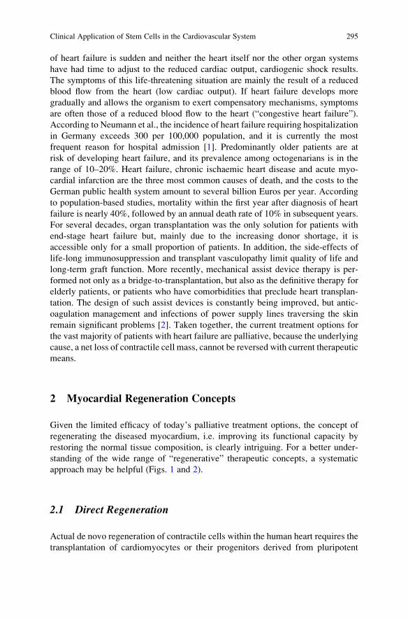
of heart failure is sudden and neither the heart itself nor the other organ systems
have had time to adjust to the reduced cardiac output, cardiogenic shock results.
The symptoms of this life-threatening situation are mainly the result of a reduced
blood flow from the heart (low cardiac output). If heart failure develops more
gradually and allows the organism to exert compensatory mechanisms, symptoms
are often those of a reduced blood flow to the heart (“congestive heart failure”).
According to Neumann et al., the incidence of heart failure requiring hospitalization
in Germany exceeds 300 per 100,000 population, and it is currently the most
frequent reason for hospital admission [1]. Predominantly older patients are at
risk of developing heart failure, and its prevalence among octogenarians is in the
range of 10–20%. Heart failure, chronic ischaemic heart disease and acute myo-
cardial infarction are the three most common causes of death, and the costs to the
German public health system amount to several billion Euros per year. According
to population-based studies, mortality within the first year after diagnosis of heart
failure is nearly 40%, followed by an annual death rate of 10% in subsequent years.
For several decades, organ transplantation was the only solution for patients with
end-stage heart failure but, mainly due to the increasing donor shortage, it is
accessible only for a small proportion of patients. In addition, the side-effects of
life-long immunosuppression and transplant vasculopathy limit quality of life and
long-term graft function. More recently, mechanical assist device therapy is per-
formed not only as a bridge-to-transplantation, but also as the definitive therapy for
elderly patients, or patients who have comorbidities that preclude heart transplan-
tation. The design of such assist devices is constantly being improved, but antic-
oagulation management and infections of power supply lines traversing the skin
remain significant problems [2]. Taken together, the current treatment options for
the vast majority of patients with heart failure are palliative, because the underlying
cause, a net loss of contractile cell mass, cannot be reversed with current therapeutic
means.
2 Myocardial Regeneration Concepts
Given the limited efficacy of today’s palliative treatment options, the concept of
regenerating the diseased myocardium, i.e. improving its functional capacity by
restoring the normal tissue composition, is clearly intriguing. For a better under-
standing of the wide range of “regenerative” therapeutic concepts, a systematic
approach may be helpful (Figs. 1 and 2).
2.1 Direct Regeneration
Actual de novo regeneration of contractile cells within the human heart requires the
transplantation of cardiomyocytes or their progenitors derived from pluripotent
Clinical Application of Stem Cells in the Cardiovascular System 295

stem cells. A large amount of experimental data exists for embryonic stem cells
(ESC), which can effectively re-generate myocardium but provoke ethical contro-
versies, a risk of teratoma formation and immunologic problems when used in
humans [3, 4]. However, the possibility of producing pluripotent stem cells by
genetic reprogramming of somatic cells (induced pluripotent stem cells, iPSC) has
recently opened up exiting new possibilities [5].
Mec
hani
sm
Angiogenesis (growth of new blood vessels from pre-existing vessels)
• Vasculogenesis (spontaneous blood-vessel formation) and
• Intussusception (blood vessel formation by branching of existing vessels)
Arteriogenesis (increase in the diameter of existing arterial vessels)
Direct suppport of ischemic end-organ cells/tissues
Immunomodulation
The
rape
utic
com
poun
ds
Physcial stimuli (hypoxia, hyperoxygenation, heat, ultrasound etc.)
Growth factors (gene therapy, protein delivery)
Cytokines
Somatic stem cells (HSC, MSC, Myoendothelial cells)
Endothelial progenitor cells
Cells overexpressing growth factors and/or cytokines)
Vasculature
Fig. 2 Regeneration of the vascular system. Here, the systematic approach shown for myocardial
regeneration is not applicable. The concepts shown are not only valid for the vasculature of the
heart, but also that of any other ischaemic tissue
Angiogenesis
Extracellular matrix
Protection of ischemic cardiomyocytes
Immunomodulation
Contractile cellreplacement
Increased number of functionalcardiomyocytes
Cardiomyocytesproliferation
Activation and differentiation of resident CPCM
echa
nism
Myocardium
ESC
iPS-derivedcardiomyocytes
Somatic stem cells / progenitor cells
Growth factors
Laser revascularization etc.
Genetic cell cyclemanipulation (Cyclins)
CPC activation
The
rape
utic
com
poun
ds
Direct regenerationIndirect regeneration Intrinsic regeneration
Angiogenesis
Extracellular matrix
Protection of ischemic cardiomyocytes
Immunomodulation
Contractile cellreplacement
Increased number of functionalcardiomyocytes
Cardiomyocytesproliferation
Activation and differentiation of resident CPCM
echa
nism
ESC
iPS-derivedcardiomyocytes
Somatic stem cells / progenitor cells
Growth factors
Laser revascularization etc.
Genetic cell cyclemanipulation (Cyclins)
CPC activation
The
rape
utic
com
poun
ds
Fig. 1 Systematic representation of myocardial regeneration strategies. Examples of the proposed
mechanism, its biological mediators, and the respective therapeutic compounds are listed
296 C. Stamm et al.

2.2 Intrinsic Cardiac Regeneration
Specific progenitor cells that reside in the heart (i.e. cardiac stem cells, cardiac
progenitor cells) may possess a therapeutically exploitable cardiomyogenic poten-
tial [6–8]. Alternatively, mature cardiomyocytes, which are terminally differen-
tiated and do not proliferate, might be forced to leave the G0 resting phase and
re-enter the S-phase (DNA synthesis) of the cell cycle [9, 10]. However, DNA
synthesis does not necessarily result in genome duplication, and genome duplica-
tion does not necessarily result in cytokinesis – division of the cell [9, 11]
2.3 Indirect Regeneration
Somatic human stem cell and progenitor cell products cannot compensate for a loss
of contractile tissue in terms of de novo formation of cardiomyocytes [12]. They
are, however, able to exert a number of indirect beneficial effects on the diseased
myocardium, including the support of angiogenesis and arteriogenesis, modulation
of extracellular matrix composition and manipulation of local immune processes
[13–16]. Moreover, somatic stem cells seem to be able to protect directly ischaemic
cardiomyocytes cell death and/or loss-of-function, but it is unclear whether this
phenomenon is clinically relevant [17, 18].
3 The Development of Cardiac Cell Therapy
The goal of cardiovascular regenerative medicine is to restore heart function
primarily by biologic means such as the support of intrinsic regeneration processes
or the transplantation of exogenous cells. In the 1990s, the concept of cell trans-
plantation for heart failure evolved [19–21]. Initially, myocyte cultures and neona-
tal rodent cardiomyocytes were used for transplantation in animal hearts [22–25],
and the finding that transplanted contractile cells are able to incorporate in postnatal
myocardium was ground-breaking. Later, those cells were applied to experimental
models of myocardial infarction or non-ischaemic cryolesions, and it was shown
that there may indeed be a benefit in terms of contractile function. However,
transplanted cardiomyocytes may not be able to survive in terminally ischaemic
infarcted tissue [26]. Skeletal muscle progenitor cells (satellite cells, skeletal
myoblasts), however, have a very high tolerance to ischemia, the capacity to
maintain contractile work even through prolonged periods of anaerobic metabo-
lism, and were shown to form contractile neo-tissue even in scar tissue after
myocardial infarction [20, 27]. Indeed, skeletal myoblasts were the first cells to
be used for clinical cell therapy, but they were found not to integrate into recipient
myocardium and the functional benefit was very limited (see below).
Clinical Application of Stem Cells in the Cardiovascular System 297

In parallel to the transplantation of contractile cells, marrow-derived cells of
haematopoietic–pre-endothelial lineage were shown to be useful for the induc-
tion of angiogenesis in the ischaemic heart. The potent pro-angiogenic capacity
of marrow-derived cells was initially shown in the mouse hindlimb ischaemia
model, and Kocher and colleagues successfully used human CD34+ cells in a rat
model of myocardial infarction [13]. Here, the increased growth of small blood
vessels in the infarcted heart was associated with a marked improvement of
contractility. In large animals, however, the impact of neo-angiogenesis on
contractility is less pronounced, while in humans it is often negligible. The
most significant – apparent – breakthrough, however, was reported in 2001
when C-kit+ lin� cells were isolated from the bone marrow of GFP expressing
transgenic mice and implanted in the infarcted myocardium of non-GFP-expressing
animals, and indeed both GFP+ blood vessels and contractile cells were visualized
[28]. The conclusion was that adult bone marrow stem cells can differentiate
into both endothelial cells and cardiomyocytes, driven by factors present in the
surrounding infarcted host myocardium. This report led to clinical pilot studies, in
which mainly autologous bone marrow mononuclear cells were delivered to the
hearts of patients with myocardial infarction. Other investigators doubted the
surprising plasticity of unmodified adult bone marrow stem cells, and reports
were published that, using state-of-the-art methods, failed to detect relevant
cardiomyocyte differentiation of murine bone marrow stem cells in vivo [29].
Controversy on this issue remains and variations in the technical details of the
various experiments are used to help explain the different outcomes. However,
even if some haematopoietic stem cells can be driven to express myocyte-specific
markers, the frequency of such events in humans is surely too small to produce
a significant clinical effect [30]. Mesenchymal stem cells (MSC) from rodents
appear to have a greater myogenic potential, provided they are manipulated
genetically and/or epigenetically, but only a few studies have provided evidence
of functioning cardiomyocytes produced from human MSC in vitro [31–33]. In
addition, multipotent cells have been described that seem to belong to none of the
typical bone marrow cell compartments but do have a strong regenerative impact
on the heart [34]. It is, however, unclear whether such “stemness” characteristics
are true features of primary cells or have been induced by long-term cell culture.
4 Clinical Cardiovascular Cell Therapy
4.1 Skeletal Myoblasts
Clinical myoblast transplantation as part of a cardiac surgical procedure was first
performed in 2001 [35]. Initial safety and feasibility studies were successful and
laid the foundation for the avalanche of cell therapy studies that were to come later
[36]. Once a large number of patients had undergone CABG and myoblast
298 C. Stamm et al.

transplantation, however, problems with ventricular arrhythmia were noted. It soon
became clear that skeletal myoblasts lack the capacity to couple electrically with
surrounding cardiomyocytes because they do not express the intercellular commu-
nication protein connexin 43 and thus do not form “connexon” ion channels that are
part of the gap junction typical for cardiomyocytes [37]. Several clinical trials using
skeletal myoblasts and catheter-based delivery devices are still ongoing [38–40],
but the majority of clinicians have abandoned skeletal myoblasts for treatment of
heart failure.
4.2 Bone Marrow Mononuclear Cells in Acute Infarction
In theory, no potentially important cell population is missed by transplanting the
entirety of nucleated marrow cells into the heart. On the other hand, vast numbers of
leukocyte progenitor cells are being delivered in addition to the actual stem cells,
and some have argued that more inflammation than regeneration is induced in
the myocardium. A clinically very relevant argument in favour of mononuclear
cells (MNCs) is their simple and speedy preparation. Traditionally, density gradient
centrifugation is used to separate bone marrow mononuclear cells (MNCs) from
other marrow components, and industry has developed several easy-to-use devices
for one-step preparation of MNC products in closed systems. Bone marrow
MNCs were the first cell products to be used in patients with acute myocardial
infarction, where they are injected into the infarct vessel that has before been
reopened by percutaneous balloon dilation and stent placement (Fig. 3). Following
several small-scale pilot trails [41, 42], the first randomized, placebo-controlled
study comparing intracoronary MNC injection with standard treatment of acute
myocardial infarction was the Hannover BOOST trial [43–45]. At 6 month follow-
up, cell-treated patients had a significantly higher left ventricular ejection fraction
than control patients. Subsequently, a number of similar studies were conducted by
other groups, including a multicenter study that enrolled 200 patients [46–51].
Some of those trials clearly produced a negative result in that there was no
difference in outcome between cell-treated and placebo-treated patients [52, 53].
In the multicenter trial coordinated by the Frankfurt group, LVEF rose by 5.5% in
cell-treated patients, and by 3.0% in the placebo-treated control group [54]. The
difference proved statistically significant, but it remains controversial whether such
a small effect will translate into a relevant clinical benefit. Other reports focused on
clinical exercise tolerance and quality-of-life data, and again there seems to be a
slight advantage for patients who have received cell therapy [55].
4.3 MNC in Chronic Ischaemia
Patients with chronic myocardial ischaemia have also been treated with bone marrow
MNC products in several clinical studies. Again, some trials on catheter-based
Clinical Application of Stem Cells in the Cardiovascular System 299
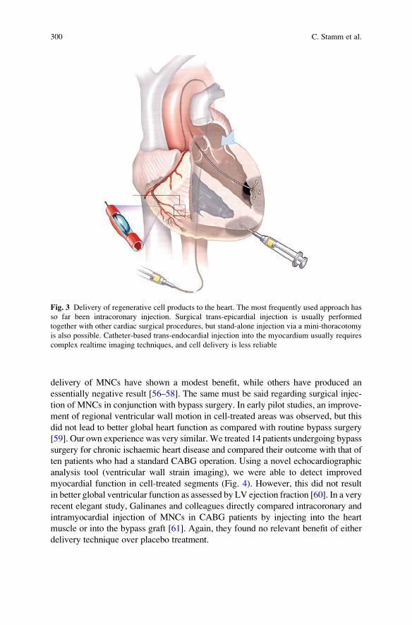
delivery of MNCs have shown a modest benefit, while others have produced an
essentially negative result [56–58]. The same must be said regarding surgical injec-
tion of MNCs in conjunction with bypass surgery. In early pilot studies, an improve-
ment of regional ventricular wall motion in cell-treated areas was observed, but this
did not lead to better global heart function as compared with routine bypass surgery
[59]. Our own experience was very similar. We treated 14 patients undergoing bypass
surgery for chronic ischaemic heart disease and compared their outcome with that of
ten patients who had a standard CABG operation. Using a novel echocardiographic
analysis tool (ventricular wall strain imaging), we were able to detect improved
myocardial function in cell-treated segments (Fig. 4). However, this did not result
in better global ventricular function as assessed by LV ejection fraction [60]. In a very
recent elegant study, Galinanes and colleagues directly compared intracoronary and
intramyocardial injection of MNCs in CABG patients by injecting into the heart
muscle or into the bypass graft [61]. Again, they found no relevant benefit of either
delivery technique over placebo treatment.
Fig. 3 Delivery of regenerative cell products to the heart. The most frequently used approach has
so far been intracoronary injection. Surgical trans-epicardial injection is usually performed
together with other cardiac surgical procedures, but stand-alone injection via a mini-thoracotomy
is also possible. Catheter-based trans-endocardial injection into the myocardium usually requires
complex realtime imaging techniques, and cell delivery is less reliable
300 C. Stamm et al.

4.4 MNC in Non-Ischaemic Heart Disease
Initial experimental and clinical cardiac cell therapy studies focussed on ischaemic
heart disease, but more recently the potential therapeutic usefulness of cell
transplantation for other types of heart failure has been investigated [62–64]. In
the recent trial by the Frankfurt group, the effects of selective intracoronary
bone marrow cell infusion in 33 patients with non-ischaemic dilated cardiomyo-
pathy were studied. Overall, microvascular function in the coronary system was
improved, and the increase of regional contractile function correlated with the
functionality of the infused cells as measured by their colony-forming capacity
[65]. A decrease in brain natriuretic peptide (NT-proBNP) serum levels also
suggested a beneficial effect on left ventricular remodelling processes, and con-
trolled studies are planned to validate these findings.
4.5 Purified Stem Cell Products
Progenitor cell products can be prepared using clinical-grade immunomagnetic selec-
tion for either CD34 or CD133, and negative selection for CD45 is also possible.
Human bonemarrow stem cells of haematopoietic-endothelial lineage, progeny of the
primitive haemangioblast, the common precursor of blood and blood vessel-forming
cells, can be isolated for clinical use based on the expression of CD34 and CD133 [66,
67]. Theoretically, these cells are potent supporters of angiogenesis processes
and hence may be useful for the relief of myocardial ischaemia. Another strategy
is the in vitro expansion of bone marrow mononuclear cells, with or without addition
of differentiation-inducing or differentiation-suppressing substances. Alternatively,
SEPT
a bANT_SEPT
3 –6
–9
–22
–13
–20
–17
–12
–17
–17
–22
–13
–11
–11–16
–19 –18
–2
5
–11
–11
–14
–15
–15
–11
3
2
–6
–9 –8
–7–13
–9 –16
ANT_SEPT
ANT ANTSEPT
LAT LATINF INF
POST POST
Fig. 4 Two-dimensional longitudinal strain echocardiography analysis of regional left ventricular
contractile performance, depicted as “bull’s eye view”. Preoperatively (a) the strain data are
inhomogenous, with impaired myocardial motion shown inblue. After bypass surgery and bone
marrow cell injection (b), performance has largely normalized
Clinical Application of Stem Cells in the Cardiovascular System 301

progenitor cells can be identified based on intracellular enzyme activities, such as
aldehyde dehydrogenase (ALDH) activity. ALHD+ cell products from peripheral
blood and bone marrow correlate closely with those enriched for CD34 or CD133
but may have superior functional characteristics [68]. Few investigators have used
purified haematopoietic stem cell products for the treatment of acute or subacute
myocardial infarction. In one such study, concerns were raised about a higher rate of
stent occlusion following intracoronary injection of CD133+ cells. Notably, Hofmann
et al. studied the cardiac retention of bone marrow cells after intracoronary injection
using radioactively labelled cells [69]. When mononuclear marrow cells were used,
approximately 2% of the activity was retained in the heart, while the vast majority of
the cells accumulated in liver and spleen. However, when CD34 selected cells were
used, cardiac cell retention was between 14% and 39%. In chronic ischaemia,
however, CD34 or CD133 enriched cells products have found more widespread
application [70, 71]. In conjunction with CABG surgery, intramyocardial injection
of CD34+ bone marrow cells resulted in nearly 10% higher LV ejection fraction than
CABG surgery alone [67]. Our group has focussed on CD133+ cells given during
CABG surgery, because they are believed to contain a subpopulation of cells that are
even more immature than CD34+ cells. In 2001, we started a feasibility and safety
study in ten patients, and no procedure-related adverse events were observed [72, 73].
Subsequently, we conducted a controlled study in 40 patients. Here, CABG and
CD133+ cell injection led to a significantly higher LVEF at 6 month follow-up than
CABG surgery alone [66]. A pronounced effect on the blood supply to the ischaemic
myocardium was evident in numerous patients, indicating the potent angiogenesis
support of CD133+ cells (Fig. 5). Other groups have isolated CD133 cells for surgical
delivery from peripheral blood, following mobilization from the marrow with G-CSF
[74, 75]. This procedure yields a substantially higher cell dose but requires several
days for cell preparation. Enriched bonemarrow stem cell products (here CD34+ cells)
have also been administered by intramuscular injection using catheter-based systems.
In a phase I/II pilot trial, this procedure has been shown to be safe, and preliminary
data indicate an improvement of LV function over placebo treatment [76].
4.6 Cytokine-Induced Bone Marrow Cell Mobilization
Another strategy aims at circumventing any mechanically invasive procedure for
cell delivery and minimizing the interval between the onset of myocardial infarc-
tion and cell therapy by mobilizing marrow cells using granulocyte stimulating
factor (G-CSF). The idea is that stem/progenitor cells mobilized from marrow will
be attracted to the ischaemic heart and initiate regeneration processes [77]. That the
number of circulating progenitor cells can be greatly enhanced by G-CSF stimula-
tion has been well established [78]. However, the number of mature leukocytes
also rises markedly, and this has raised concerns regarding the safety of C-CSF
treatment. In patients with myocardial infarction treated with G-CSF a high rate
302 C. Stamm et al.

of in-stent restenosis has been observed. Moreover, incidences such as acute
re-infarction and sudden death have occurred in other studies [79]. A number of
controlled clinical efficacy studies have subsequently been performed, but the
outcome data in terms of heart function improvement are rather disappointing.
4.7 Second Generation Clinical Cell Products
Given the modest improvement of heart function after intracardiac delivery of
unmodified bone marrow cells, clinical studies are currently being conducted
with cell products that have been subjected to in vitro modifications or have been
isolated from alternative sources. Among those are mononuclear cells conditioned
and expanded in specific bioreactor systems where, theoretically, cells with greater
stemness are enriched and their secretory activity is augmented [80, 81]. The
frequency of CD34+ or CD133+ cells that are not committed towards a leukocyte
phenotype (CD45�) is very low in adult human bone marrow [82, 83], but expan-
sion of haematopoietic stem cells in vitro induces spontaneous differentiation and
impairs stem cell function [84]. Bioreactor systems that may be able to suppress
stem cell differentiation during proliferation have been developed, but their clinical
use in cardiovascular medicine has been limited [85, 86]. The second cellular
compartment within the bone marrow, the stroma, contains stem cells that are
easier to handle and to manipulate. Multipotent stromal cells, a.k.a. MSC, make
Fig. 5 Perfusion scans of the heart of a patient who had myocardial infarction of the inferior wall
of the left ventricle (arrow). Preoperatively (a), perfusion is several impaired. Two weeks after
bypass grafting and transplantation of CD133+ cells in the infarct area, there is no obvious change
in blood supply (b). Six months later, however, perfusion is effectively restored (c), probably via a
pro-angiogenic effect of the cell transplantation. This effect is maintained at 1 year follow-up (d)
Clinical Application of Stem Cells in the Cardiovascular System 303

up only 0.01% of the nucleated bone marrow cells but are important supporters of
haematopoiesis and act as an independent stem cell pool involved in the mainte-
nance of tissues derived from the embryonic mesenchyme [87]. Moreover, they
have been shown to be able to trans-differentiate across lineage boundaries in
experimental models, and possess potent immuno-modulatory properties that may
also be useful for cardiovascular regenerative medicine [88–91]. Several clinical
pilot studies have evaluated the effect of autologous bone marrow MSC in patients
with heart disease, but results have been equivocal [92, 93]. Allogeneic donor MSC
are also being used in patients with heart disease [94]. They can escape detection
and elimination by the immune system, because they express no MHC class II, low
levels of MHC class I, and none of the T cell co-stimulatory antigens CD40, CD80
or CD86. Definitive clinical efficacy data, however, are not available yet. MSC
from non-marrow tissues are also being evaluated in clinical pilot studies in both
autologous and allogeneic fashion. Autologous MSC from adipose tissue can be
isolated by plastic adherence in cell culture before they are injected into the heart,
while in other trials cells are freshly obtained from a large volume of lipo-aspirate
by digestion, washing and centrifugation, and injected intramyocardially without
prior cell cultivation [95].
Another interesting development that may help optimize the clinical usefulness
of MSC-like cells is the identification of perivascular cells that co-express myo-
genic and endothelial cell markers and have a robust myocyte differentiation
capacity (myoendothelial cells). They are believed to represent the solid-organ
reservoir of tissue-specific MSC [96, 97], and have been shown to be very efficient
for myocardial regeneration in experimental models.
4.8 Combination Treatments
Numerous strategies to enhance the regenerative capacity of adult stem cells in the
heart have been developed. For instance, genetic manipulation of mesenchymal
stem cells inducing overexpression of anti-apoptotic proteins such as AKT or Bcl-2,
or cytokines such as HGF, IGF or VEGF, not only leads to a marked improvement
of MSC survival following transplantation into the heart, but also has beneficial
effects on the surrounding host myocardium that comes in contact with secreted
anti-apoptotic factors [98, 99]. However, due to patient safety concerns and regu-
latory restrictions, genetically modified adult stem cells have not yet been tested in
the clinical setting. Clinically better applicable strategies to improve cell therapy in
the heart are, for instance, hypoxic preconditioning, heat shock treatment or stimu-
lation of cells with cytokines [100, 101], which increase both cellular resilience and
paracrine activity. Alternatively, pre-treatment of cells with pharmaceutical agents
such as erythropoietin, parathyroid hormone, statins or nitric oxide synthase enhan-
cers has also been applied [102–104]. For both approaches, clinical trials have been
initiated. The same is the case for the combination of cell therapy and ultrasound
304 C. Stamm et al.

shock wave treatment [105], which was shown to increase the functional capacity
of bone marrow and endothelial progenitor cells [106]. Transplanted cells may
survive and function better when they are embedded in an adequate extracellular
matrix (ECM) [107], and a clinical trial has been set up where bone marrow cells
are mixed with collagen-rich semi-liquid hydrogel prior to implantation in the
heart [108]. In this context, the recent development of decellularized cardiac
ECM should also be mentioned, which can not only be used as a scaffold for
myocardial tissue engineering but will also help understand the interactions
between cardiac ECM and transplanted cells [109]. Finally, the combination of
transmural laser revascularization (TMLR) and cell therapy deserves to be noted,
TMLR is believed to induce a local inflammatory stimulus that supports marrow
cells injected into the heart. Again, clinical studies are in progress [110].
4.9 Foetal or Neonatal Stem Cells
The use of juvenile stem cells for cardiac regenerationmay solve the problem of age-
related and disease-related impairment of stem cell function, and experimental
studies have been conducted with cells derived from cord blood, umbilical cord
or placenta [5, 111]. However, autologous cells will only be available for patients
who had cord blood (or other cells) banked at the time of birth, and large-scale cord
blood banking for autologous use has just begun. Some stem cell types from neonatal
tissues, however, may have a sufficiently immature immunophenotype to allow
allogeneic application [112, 113]. For instance, a clinical trial of allogeneic placenta
MSC in patients with severe limb ischaemia is currently going on in our institution
[114]. Therapeutic applications for heart disease have not yet been described.
4.10 Paediatric Heart Disease
For children with structural congenital heart disease, tissue engineering may offer
new therapeutic options in the future, including the implantation of viable, auto-
logous valves and blood vessels. A number of children, however, suffer from
acquired or congenital heart failure without structural defects, and require assist
device support and/or heart transplantation. Several cases of cell therapy for
myocardial regeneration in children have been reported [115, 116]. Some of these
reports were very encouraging, and it has been speculated that the young myocar-
dium may have a greater regenerative potential, and that juvenile stem cells possess
greater plasticity and proliferative capacity. Systematic studies, however, have not
been performed so far.
Clinical Application of Stem Cells in the Cardiovascular System 305

5 Clinical Translation Problems
Attention usually focuses on the cell product, but there are many other factors
that need to be considered to maximize the likelihood of successful cell-based
myocardial regeneration.
5.1 Patient Selection
Every novel therapeutic approach is best evaluated in a uniform cohort of patients
with well-defined patient-related and disease-related characteristics. Regarding cell
therapy for heart disease, however, this is more difficult than it first seems. In the
majority of patients, myocardial cell therapy has been performed within several
days after the onset of myocardial infarction symptoms, when patients cannot be
selected according to their pre-treatment heart function [43, 47, 53, 54]. Conse-
quently, such patient cohorts cover a wide range of left ventricular contractility at
baseline, impeding data analysis and interpretation. In patients with chronic myo-
cardial ischaemia, the degree of contractile dysfunction is usually better defined
[66, 67, 74]. Patients with non-ischaemic heart disease have also been subject to cell
therapy approaches, although the body of preclinical data is much smaller than for
ischaemic heart disease [117]. Here, one problem is the diversity of underlying
aetiologies for non-ischaemic heart failure, including inflammatory processes,
genetic diseases and a large group of “idiopathic” cases where no cause can be
established.
5.2 Cell Delivery
Injection into the coronary arteries requires transmigration of cells through the
vascular wall into the myocardium. Catheter-based direct intramyocardial cell
delivery is also possible, often combined with intracardiac NOGA mapping of the
left ventricle to identify the area of interest (Fig. 3) [56]. A needle-tipped catheter
can also be guided into the epicardial veins for injection into the myocardium
(reviewed in [118]). For cell delivery under direct vision using surgical techniques,
any commercially available syringe and needle system can be used, but industry has
also developed special cell injection systems with side-holes. In some trials, cells
are being delivered by peripheral venous injection, relying on myocardial chemo-
kines to attract cells to the heart, but the majority undergo first-pass trapping in the
lung or are eliminated by the reticuloendothelial system [119]. Each of these cell
delivery techniques involves biologic processes that are incompletely understood
but probably have a major impact on the therapeutic efficacy.
306 C. Stamm et al.

5.3 Timing
It is not known whether there is an ideal time point for cell therapy in patients with
ischaemic heart disease. Emergency treatment of acute infarction is usually done by
the interventional cardiologist, who may also decide to perform intracoronary injec-
tion of a rapidly available cell product. The situation in chronic ischaemic heart
disease is very different. In the post-infarct or chronically ischaemic myocardium, a
substantial net loss of contractile tissue mass has occurred and there is diffuse or
localized scar formation. Intuitively, the longer the interval between myocardial
infarction and cell treatment, the smaller the chance to achieve a beneficial effect
becomes. This notion, however, is presently not supported by solid data.
5.4 Cell Survival
When cells are injected into diseased myocardium, most of them do not survive.
The magnitude of cell death upon intramyocardial transplantation is difficult to
measure, but the suggested survival rate ranges between 0.1 and 10% [26, 120].
The ischaemic heart is a hostile environment, due to local hypoxia, acidosis, lack
of substrates and accumulation of metabolites. Moreover, it is infiltrated by
phagocytic cells that remove cell debris, and many transplanted cells are probably
lost in this “clean-up” process. Third, the mechanic forces in the myocardium are
substantial. Transmural pressure is high during systole, there are shear forces
between contracting myofibers and layers, and a marrow cell is not well equipped
to withstand such stress. However, the rate of cell death can be slowed. Transfec-
tion of marrow stromal cells with genes encoding for the anti-apototic proteins
AKT or Bcl-2 has been shown to improve greatly cell survival and regenerative
capacity [98, 99]. Pre-treatment of endothelial progenitor cells with eNOS-
enhancing substances also appears to have a beneficial effect [102], similar to
the effects observed with statins [103]. Hypoxic preconditioning or heat shock
prior to cell injection might also help, since both activate anti-apoptotic and
NO-related signalling pathways [100].
5.5 Dose
The normal adult heart weighs 250–350 g, and around 80% of the myocardial mass
consists of approximately 10 � 109 cardiomyocytes [121]. Assuming that 20% of
the cardiomyocytes are lost following myocardial infarction, one would ultimately
need 2 � 109 surviving neo-myocytes weighing nearly 50 g to reconstitute the
myocardium completely. Given the high rate of cell death upon transplantation into
the heart and the presumably very low number of adult stem cells that actually
differentiate into myocytes, it becomes clear that, with currently available cell
products, we are far from being able to replace all lost heart muscle tissue.
Clinical Application of Stem Cells in the Cardiovascular System 307

5.6 Age
Theoretically, stem cells constantly renew themselves, but it has become clear that
bone marrow stem cells undergo ageing processes and are affected by remote
diseases [122]. Ageing may not just be imposed on marrow cells by external and
internal stressors, but seems to be an active process that helps protect the organism.
For instance, upregulation of cell cycle inhibitors of the cip/kip and the INK4a/ARF
family reduces the risk of malignancy in the ageing organism. On the other hand,
this protective mechanism reduces the stem cell’s capacity for self-renewal and
proliferation. Indeed, when the cyclin-dependent kinase inhibitor p16INK4a, which
accumulates with age, is knocked out in ageing mice, tissue regeneration processes
involving haematopoietic stem cells are much improved, but the animals also
develop various kinds of malignancies [123]. In essence, the organism sacrifices
its regenerative capacity in order to counteract the increasing tendency to develop
cancer.
5.7 Disease
It has been well established that ischaemic heart disease is associated with impaired
endothelial progenitor cell number and function [124], but it remains unclear
whether progenitor cell dysfunction is a cause or an effect of heart disease. On
the other hand, exercise has beneficial effects on the somatic stem cell pool. It may
be argued that the attempt to repair heart failure with autologous cells that are
affected by age and disease is a futile undertaking. The use of allogenic, juvenile
stem cells from healthy individuals may be an elegant solution and is propagated by
those involved in cell banking activities. However, disease processes in the recipi-
ent organism may influence even the behaviour of healthy allogenic cells. Humoral
factors in the serum of patients with heart failure have been shown to impair the
in vitro proliferation of allogenic bone marrow MSC [125]. Our group has studied
the effect of heart failure patient serum on neonatal cord blood mesenchymal stem
cells, and we found that, in some patients, MSC proliferation is accelerated, while
in others it is severely impaired (Fig. 6). The identification of patients who are
likely to benefit from cell therapy and those who will probably not respond is still a
largely neglected field of research. Given the biologic complexity of viable cells as
therapeutic compounds as well as the organism’s response to those clearly requires
an individualized approach to maximize the efficacy of cardiac cell therapy
5.8 Legal Framework
Today, the legal situation regarding the production and the therapeutic use of viable
cells and related products has been clarified in many countries. Within the EU, this
308 C. Stamm et al.

was completed with the Regulation on Advanced Therapies No 1394/2007, and the
situation is similar in North America. Cells used for tissue regeneration are now
considered medicinal products. Hence, their production and clinical application
must comply with Good Manufacturing (GMP) and Good Clinical Practice (GCP).
Essentially, the same rules are applied that were originally designed to supervise
the development in production processing of pharmaceutical compounds. One
exception is when autologous cells are harvested and implanted within the same
surgical procedure, as is sometimes done in bedside bone marrow or adipose tissue
cell transplantation. However, even such “bedside cell therapy products” may be
considered medicinal products if the cells undergo more-than-minimal manipula-
tion or their use is non-homologous (meaning the cells are meant to behave in a way
that differs from their original function). This regulatory framework has greatly
helped optimize the safety of patients who are subjected to still experimental
cellular therapies. On the other hand, it has significantly slowed down the clinical
translation process.
6 Future Clinical Cell Therapy Studies
Previous and ongoing clinical studies using somatic cells from marrow, adipose
tissue and other sources were relatively easy to conduct in accordance with the cell
therapy-specific regulations and the standards of evidence-based medicine. The
0
0.1
0.2
0.3
0.4
0.5
0 1 2 3 4 5 6
time [days]
Via
ble
cel
ls (
MT
S t
est)
0
0.1
0.2
0.3
0.4
0.5
0 1 2 3 4 5 6
time [days]V
iab
le c
ells
(M
TS
tes
t)Fig. 6 Serum of patient with heart failure influences stem cell behaviour. (a) The in vitro
proliferation rate of neonatal cord blood mesenchymal stem cells from a healthy donor, in the
presence (black) and absence (grey) of serum from a patient with ischaemic heart failure.
Compared to the standard medium (foetal calf serum), cell proliferation and viability is much
reduced. On the other hand, (b) shows the in vitro proliferation rate of the same cell type, in the
presence of serum from a patient who underwent surgery for ischaemic heart disease (black),compared to that of a healthy individual (grey). Here, the patient serum clearly has activating
effects
Clinical Application of Stem Cells in the Cardiovascular System 309

majority of these approaches were shown to be safe, albeit also with limited
efficacy, because they mainly induce indirect regeneration and do not directly
interfere with genomic integrity and cardiomyocyte biology. The clinical evalua-
tion of stem cell-derived contractile cells for direct regeneration, however, carries
much greater risks and needs a different approach. Because of the tumour and
arrhythmia risk when, for instance, iPS cell-derived myocytes are transplanted, we
feel that feasibility and safety studies should initially be performed in no-option
patients who have or require a mechanical ventricular assist device (VAD).
Here, ventricular arrhythmia is usually not life-threatening, and myocardium for
histology studies can be collected upon VAD explanation or heart transplantation.
Cell therapy has led to a new level of complexity in both experimental cardio-
vascular research and clinical practice. The successful use of viable cells for
regeneration of diseased tissue requires a completely novel scientific and clinical
armamentarium, and it cannot be foreseen whether the efforts will be ultimately
rewarded by efficacious and safe regenerative therapies.
References
1. Neumann T, Biermann J, Erbel R, Neumann A, Wasem J, Ertl G et al (2009) Heart failure:
the commonest reason for hospital admission in Germany: medical and economic perspec-
tives. Dtsch Arztebl Int 106(16):269–275
2. Slaughter MS, Rogers JG, Milano CA, Russell SD, Conte JV, Feldman D et al (2009)
Advanced heart failure treated with continuous-flow left ventricular assist device. N Engl J
Med 361:2241–2251
3. Behfar A, Perez-Terzic C, Faustino RS, Arrell DK, Hodgson DM, Yamada S et al (2007)
Cardiopoietic programming of embryonic stem cells for tumor-free heart repair. J Exp Med
204(2):405–420
4. Menard C, Hagege AA, Agbulut O, Barro M, Morichetti MC, Brasselet C et al (2005)
Transplantation of cardiac-committed mouse embryonic stem cells to infarcted sheep myo-
cardium: a preclinical study. Lancet 366(9490):1005–1012
5. Takahashi K, Yamanaka S (2006) Induction of pluripotent stem cells from mouse embryonic
and adult fibroblast cultures by defined factors. Cell 126(4):663–676
6. Stamm C, Choi YH, Nasseri B, Hetzer R (2009) A heart full of stem cells: the spectrum of
myocardial progenitor cells in the postnatal heart. Ther Adv Cardiovasc Dis 3(3):215–229
7. Laugwitz KL, Moretti A, Lam J, Gruber P, Chen Y, Woodard S et al (2005) Postnatal isl1+
cardioblasts enter fully differentiated cardiomyocyte lineages. Nature 433(7026):647–653
8. Smith RR, Barile L, Cho HC, Leppo MK, Hare JM, Messina E et al (2007) Regenerative
potential of cardiosphere-derived cells expanded from percutaneous endomyocardial biopsy
specimens. Circulation 115(7):896–908
9. Pasumarthi KB, Field LJ (2002) Cardiomyocyte cell cycle regulation. Circ Res 90(10):
1044–1054
10. Soonpaa MH, Field LJ (1997) Assessment of cardiomyocyte DNA synthesis in normal and
injured adult mouse hearts. Am J Physiol 272(1 Pt 2):H220–H226
11. Meckert PC, Rivello HG, Vigliano C, Gonzalez P, Favaloro R, Laguens R (2005) Endomi-
tosis and polyploidization of myocardial cells in the periphery of human acute myocardial
infarction. Cardiovasc Res 67(1):116–123
310 C. Stamm et al.

12. Stamm C, Nasseri B, Choi YH, Hetzer R (2009) Cell therapy for heart disease: great
expectations, as yet unmet. Heart Lung Circ 18(4):245–256
13. Kocher AA, Schuster MD, Szabolcs MJ, Takuma S, Burkhoff D, Wang J et al (2001)
Neovascularization of ischemic myocardium by human bone-marrow-derived angioblasts
prevents cardiomyocyte apoptosis, reduces remodeling and improves cardiac function. Nat
Med 7(4):430–436
14. Du YY, Zhou SH, Zhou T, Su H, Pan HW, Du WH et al (2008) Immuno-inflammatory
regulation effect of mesenchymal stem cell transplantation in a rat model of myocardial
infarction. Cytotherapy 10(5):469–478
15. Molina EJ, Palma J, Gupta D, Torres D, Gaughan JP, Houser S et al (2009) Reverse
remodeling is associated with changes in extracellular matrix proteases and tissue inhibitors
after mesenchymal stem cell (MSC) treatment of pressure overload hypertrophy. J Tissue
Eng Regen Med 3(2):85–91
16. Jin B, Luo XP, Ni HC, Li Y, Shi HM (2009) Cardiac matrix remodeling following intracor-
onary cell transplantation in dilated cardiomyopathic rabbits. Mol Biol Rep
17. Lai VK, Ang KL, Rathbone W, Harvey NJ, Galinanes M (2009) Randomized controlled trial
on the cardioprotective effect of bone marrow cells in patients undergoing coronary bypass
graft surgery. Eur Heart J 30(19):2354–2359
18. Lai VK, Linares-Palomino J, Nadal-Ginard B, Galinanes M (2009) Bone marrow cell-
induced protection of the human myocardium: characterization and mechanism of action.
J Thorac Cardiovasc Surg 138(6):1400–1408
19. Reinecke H, ZhangM, Bartosek T, Murry CE (1999) Survival, integration, and differentiation
of cardiomyocyte grafts: a study in normal and injured rat hearts. Circulation 100(2):193–202
20. Chiu RC, Zibaitis A, Kao RL (1995) Cellular cardiomyoplasty: myocardial regeneration
with satellite cell implantation. Ann Thorac Surg 60(1):12–18
21. Marelli D, Desrosiers C, el-Alfy M, Kao RL, Chiu RC (1992) Cell transplantation for
myocardial repair: an experimental approach. Cell Transplant 1(6):383–390
22. Scorsin M, Hagege AA, Marotte F, Mirochnik N, Copin H, Barnoux M et al (1997) Does
transplantation of cardiomyocytes improve function of infarcted myocardium? Circulation
96(9 Suppl):II-188–II-193
23. Reffelmann T, Dow JS, Dai W, Hale SL, Simkhovich BZ, Kloner RA (2003) Transplantation
of neonatal cardiomyocytes after permanent coronary artery occlusion increases regional
blood flow of infarcted myocardium. J Mol Cell Cardiol 35(6):607–613
24. Roell W, Lu ZJ, Bloch W, Siedner S, Tiemann K, Xia Y et al (2002) Cellular cardiomyo-
plasty improves survival after myocardial injury. Circulation 105(20):2435–2441
25. Leor J, Patterson M, Quinones MJ, Kedes LH, Kloner RA (1996) Transplantation of fetal
myocardial tissue into the infarcted myocardium of rat. A potential method for repair of
infarcted myocardium? Circulation 94(9 Suppl):II332–II336
26. Zhang M, Methot D, Poppa V, Fujio Y, Walsh K, Murry CE (2001) Cardiomyocyte grafting
for cardiac repair: graft cell death and anti-death strategies. J Mol Cell Cardiol 33(5):
907–921
27. Taylor DA, Atkins BZ, Hungspreugs P, Jones TR, Reedy MC, Hutcheson KA et al (1998)
Regenerating functional myocardium: improved performance after skeletal myoblast trans-
plantation. Nat Med 4(8):929–933
28. Orlic D, Kajstura J, Chimenti S, Jakoniuk I, Anderson SM, Li B et al (2001) Bone marrow
cells regenerate infarcted myocardium. Nature 410(6829):701–705
29. Murry CE, Soonpaa MH, Reinecke H, Nakajima H, Nakajima HO, Rubart M et al (2004)
Haematopoietic stem cells do not transdifferentiate into cardiac myocytes in myocardial
infarcts. Nature 428(6983):664–668
30. Rota M, Kajstura J, Hosoda T, Bearzi C, Vitale S, Esposito G et al (2007) Bone marrow
cells adopt the cardiomyogenic fate in vivo. Proc Natl Acad Sci USA 104(45):
17783–17788
31. Makino S, Fukuda K, Miyoshi S, Konishi F, Kodama H, Pan J et al (1999) Cardiomyocytes
can be generated from marrow stromal cells in vitro. J Clin Invest 103(5):697–705
Clinical Application of Stem Cells in the Cardiovascular System 311

32. Hakuno D, Fukuda K, Makino S, Konishi F, Tomita Y, Manabe T et al (2002) Bone marrow-
derived regenerated cardiomyocytes (CMG Cells) express functional adrenergic and musca-
rinic receptors. Circulation 105(3):380–386
33. Toma C, Pittenger MF, Cahill KS, Byrne BJ, Kessler PD (2002) Human mesenchymal stem
cells differentiate to a cardiomyocyte phenotype in the adult murine heart. Circulation 105
(1):93–98
34. Yoon YS, Wecker A, Heyd L, Park JS, Tkebuchava T, Kusano K et al (2005) Clonally
expanded novel multipotent stem cells from human bone marrow regenerate myocardium
after myocardial infarction. J Clin Invest 115(2):326–338
35. Menasche P, Hagege AA, Scorsin M, Pouzet B, Desnos M, Duboc D et al (2001) Myoblast
transplantation for heart failure. Lancet 357(9252):279–280
36. Hagege AA, Marolleau JP, Vilquin JT, Alheritiere A, Peyrard S, Duboc D et al (2006)
Skeletal myoblast transplantation in ischemic heart failure: long-term follow-up of the first
phase I cohort of patients. Circulation 114(1 Suppl):I108–I113
37. Rubart M, Soonpaa MH, Nakajima H, Field LJ (2004) Spontaneous and evoked intracellular
calcium transients in donor-derived myocytes following intracardiac myoblast transplanta-
tion. J Clin Invest 114(6):775–783
38. Dib N, Michler RE, Pagani FD, Wright S, Kereiakes DJ, Lengerich R et al (2005) Safety and
feasibility of autologous myoblast transplantation in patients with ischemic cardiomyopathy:
four-year follow-up. Circulation 112(12):1748–1755
39. Pagani FD, DerSimonian H, Zawadzka A, Wetzel K, Edge AS, Jacoby DB et al (2003)
Autologous skeletal myoblasts transplanted to ischemia-damaged myocardium in humans.
Histological analysis of cell survival and differentiation. J Am Coll Cardiol 41(5):879–888
40. Siminiak T, Kalawski R, Fiszer D, Jerzykowska O, Rzezniczak J, Rozwadowska N et al
(2004) Autologous skeletal myoblast transplantation for the treatment of postinfarction
myocardial injury: phase I clinical study with 12 months of follow-up. Am Heart J 148
(3):531–537
41. Strauer BE, Brehm M, Zeus T, Kostering M, Hernandez A, Sorg RV et al (2002) Repair of
infarcted myocardium by autologous intracoronary mononuclear bone marrow cell trans-
plantation in humans. Circulation 106(15):1913–1918
42. Strauer BE, BrehmM, Zeus T, Gattermann N, Hernandez A, Sorg RV et al (2001) Intracoro-
nary, human autologous stem cell transplantation for myocardial regeneration following
myocardial infarction. Dtsch Med Wochenschr 126(34–35):932–938
43. Wollert KC, Meyer GP, Lotz J, Ringes-Lichtenberg S, Lippolt P, Breidenbach C et al (2004)
Intracoronary autologous bone-marrow cell transfer after myocardial infarction: the BOOST
randomised controlled clinical trial. Lancet 364(9429):141–148
44. Meyer GP, Wollert KC, Lotz J, Steffens J, Lippolt P, Fichtner S et al (2006) Intracoronary
bone marrow cell transfer after myocardial infarction: eighteen months’ follow-up data from
the randomized, controlled BOOST (BOne marrOw transfer to enhance ST-elevation infarct
regeneration) trial. Circulation 113(10):1287–1294
45. Schaefer A, Meyer GP, Fuchs M, Klein G, Kaplan M, Wollert KC et al (2006) Impact of
intracoronary bone marrow cell transfer on diastolic function in patients after acute myo-
cardial infarction: results from the BOOST trial. Eur Heart J 27(8):929–935
46. Assmus B, Fischer-Rasokat U, Honold J, Seeger FH, Fichtlscherer S, Tonn T et al (2007)
Transcoronary transplantation of functionally competent BMCs is associated with a decrease
in natriuretic peptide serum levels and improved survival of patients with chronic postinfarc-
tion heart failure: results of the TOPCARE-CHD registry. Circ Res 100(8):1234–1241
47. Assmus B, Honold J, Schachinger V, Britten MB, Fischer-Rasokat U, Lehmann R et al
(2006) Transcoronary transplantation of progenitor cells after myocardial infarction. N Engl
J Med 355(12):1222–1232
48. Schachinger V, Tonn T, Dimmeler S, Zeiher AM (2006) Bone-marrow-derived progenitor
cell therapy in need of proof of concept: design of the REPAIR-AMI trial. Nat Clin Pract
Cardiovasc Med 3 Suppl 1:S23–S28
312 C. Stamm et al.

49. Schachinger V, Assmus B, Britten MB, Honold J, Lehmann R, Teupe C et al (2004)
Transplantation of progenitor cells and regeneration enhancement in acute myocardial infarc-
tion: final one-year results of the TOPCARE-AMI trial. J Am Coll Cardiol 44(8):1690–1699
50. Erbs S, Linke A, Schachinger V, Assmus B, Thiele H, Diederich KW et al (2007) Restoration
of microvascular function in the infarct-related artery by intracoronary transplantation of
bone marrow progenitor cells in patients with acute myocardial infarction: the Doppler
Substudy of the Reinfusion of Enriched Progenitor Cells and Infarct Remodeling in Acute
Myocardial Infarction (REPAIR-AMI) trial. Circulation 116(4):366–374
51. Schachinger V, Erbs S, Elsasser A, Haberbosch W, Hambrecht R, Holschermann H et al
(2006) Improved clinical outcome after intracoronary administration of bone-marrow-
derived progenitor cells in acute myocardial infarction: final 1-year results of the
REPAIR-AMI trial. Eur Heart J 27(23):2775–2783
52. Janssens S, Dubois C, Bogaert J, Theunissen K, Deroose C, Desmet W et al (2006)
Autologous bone marrow-derived stem-cell transfer in patients with ST-segment elevation
myocardial infarction: double-blind, randomised controlled trial. Lancet 367(9505):113–121
53. Lunde K, Solheim S, Aakhus S, Arnesen H, Abdelnoor M, Egeland T et al (2006) Intracor-
onary injection of mononuclear bone marrow cells in acute myocardial infarction. N Engl J
Med 355(12):1199–1209
54. Schachinger V, Erbs S, Elsasser A, Haberbosch W, Hambrecht R, Holschermann H et al
(2006) Intracoronary bone marrow-derived progenitor cells in acute myocardial infarction.
N Engl J Med 355(12):1210–1221
55. Lunde K, Solheim S, Aakhus S, Arnesen H, Moum T, Abdelnoor M et al (2007) Exercise
capacity and quality of life after intracoronary injection of autologous mononuclear bone
marrow cells in acute myocardial infarction: results from the Autologous Stem cell Trans-
plantation in Acute Myocardial Infarction (ASTAMI) randomized controlled trial. Am Heart
J 154(4):710e1–710e8
56. Perin EC, Dohmann HF, Borojevic R, Silva SA, Sousa AL, Silva GV et al (2004) Improved
exercise capacity and ischemia 6 and 12 months after transendocardial injection of auto-
logous bone marrow mononuclear cells for ischemic cardiomyopathy. Circulation 110
(11 Suppl 1):II213–II218
57. Brehm M, Strauer BE (2006) Stem cell therapy in postinfarction chronic coronary heart
disease. Nat Clin Pract Cardiovasc Med 3 Suppl 1:S101–S104
58. Strauer BE, Brehm M, Zeus T, Bartsch T, Schannwell C, Antke C et al (2005) Regeneration
of human infarcted heart muscle by intracoronary autologous bone marrow cell transplanta-
tion in chronic coronary artery disease: the IACT study. J Am Coll Cardiol 46(9):1651–1658
59. Galinanes M, Loubani M, Davies J, Chin D, Pasi J, Bell PR (2004) Autotransplantation of
unmanipulated bone marrow into scarred myocardium is safe and enhances cardiac function
in humans. Cell Transplant 13(1):7–13
60. Nasseri BA, Kukucka M, Dandel M, Knosalla C, Choi YH, Ebell W et al (2009) Two-
dimensional speckle tracking strain analysis for efficacy assessment of myocardial cell
therapy. Cell Transplant 18(3):361–370
61. Ang KL, Chin D, Leyva F, Foley P, Kubal C, Chalil S et al (2008) Randomized, controlled trial
of intramuscular or intracoronary injection of autologous bone marrow cells into scarred
myocardiumduringCABGversusCABGalone.Nat Clin Pract CardiovascMed 5(10):663–670
62. Yamada S, Nelson TJ, Crespo-Diaz RJ, Perez-Terzic C, Liu XK, Miki T et al (2008)
Embryonic stem cell therapy of heart failure in genetic cardiomyopathy. Stem Cells (Dayton,
Ohio) 26(10):2644–2653
63. Suzuki K, Murtuza B, Suzuki N, Smolenski RT, Yacoub MH (2001) Intracoronary infusion
of skeletal myoblasts improves cardiac function in doxorubicin-induced heart failure. Circu-
lation 104(12 Suppl 1):I213–I217
64. Ishida M, Tomita S, Nakatani T, Fukuhara S, Hamamoto M, Nagaya N et al (2004) Bone
marrow mononuclear cell transplantation had beneficial effects on doxorubicin-induced
cardiomyopathy. J Heart Lung Transplant 23(4):436–445
Clinical Application of Stem Cells in the Cardiovascular System 313

65. Fischer-Rasokat U, Assmus B, Seeger FH, Honold J, Leistner D, Fichtlscherer S et al (2009)
A pilot trial to assess potential effects of selective intracoronary bone marrow-derived
progenitor cell infusion in patients with nonischemic dilated cardiomyopathy: final 1-year
results of the transplantation of progenitor cells and functional regeneration enhancement
pilot trial in patients with nonischemic dilated cardiomyopathy. Circ Heart Fail 2(5):
417–423
66. Stamm C, Kleine HD, Choi YH, Dunkelmann S, Lauffs JA, Lorenzen B et al (2007)
Intramyocardial delivery of CD133+ bone marrow cells and coronary artery bypass grafting
for chronic ischemic heart disease: safety and efficacy studies. J Thorac Cardiovasc Surg
133(3):717–725
67. Patel AN, Geffner L, Vina RF, Saslavsky J, Urschel HC Jr, Kormos R et al (2005) Surgical
treatment for congestive heart failure with autologous adult stem cell transplantation: a
prospective randomized study. J Thorac Cardiovasc Surg 130(6):1631–1638
68. Povsic TJ, Zavodni KL, Kelly FL, Zhu S, Goldschmidt-Clermont PJ, Dong C et al (2007)
Circulating progenitor cells can be reliably identified on the basis of aldehyde dehydrogenase
activity. J Am Coll Cardiol 50(23):2243–2248
69. Hofmann M, Wollert KC, Meyer GP, Menke A, Arseniev L, Hertenstein B et al (2005)
Monitoring of bone marrow cell homing into the infarcted human myocardium. Circulation
111(17):2198–2202
70. Erbs S, Linke A, Adams V, Lenk K, Thiele H, Diederich KW et al (2005) Transplantation of
blood-derived progenitor cells after recanalization of chronic coronary artery occlusion: first
randomized and placebo-controlled study. Circ Res 97(8):756–762
71. Erbs S, Linke A, Schuler G, Hambrecht R (2006) Intracoronary administration of circulating
blood-derived progenitor cells after recanalization of chronic coronary artery occlusion
improves endothelial function. Circ Res 98(5):e48
72. Stamm C, Kleine HD, Westphal B, Petzsch M, Kittner C, Nienaber CA et al (2004) CABG
and bone marrow stem cell transplantation after myocardial infarction. Thorac Cardiovasc
Surg 52(3):152–158
73. Stamm C, Westphal B, Kleine HD, Petzsch M, Kittner C, Klinge H et al (2003) Autologous
bone-marrow stem-cell transplantation for myocardial regeneration. Lancet 361(9351):
45–46
74. Pompilio G, Cannata A, Peccatori F, Bertolini F, Nascimbene A, Capogrossi MC et al (2004)
Autologous peripheral blood stem cell transplantation for myocardial regeneration: a novel
strategy for cell collection and surgical injection. Ann Thorac Surg 78(5):1808–1812
75. Pompilio G, Cannata A, Pesce M, Capogrossi MC, Biglioli P (2005) Long-lasting improve-
ment of myocardial perfusion and chronic refractory angina after autologous intramyocardial
PBSC transplantation. Cytotherapy 7(6):494–496
76. Losordo DW, Schatz RA, White CJ, Udelson JE, Veereshwarayya V, Durgin M et al (2007)
Intramyocardial transplantation of autologous CD34+ stem cells for intractable angina: a
phase I/IIa double-blind, randomized controlled trial. Circulation 115(25):3165–3172
77. Orlic D, Kajstura J, Chimenti S, Limana F, Jakoniuk I, Quaini F et al (2001) Mobilized bone
marrow cells repair the infarcted heart, improving function and survival. Proc Natl Acad Sci
USA 98(18):10344–10349
78. Honold J, Lehmann R, Heeschen C, Walter DH, Assmus B, Sasaki K et al (2006) Effects
of granulocyte colony simulating factor on functional activities of endothelial progenitor
cells in patients with chronic ischemic heart disease. Arterioscler Thromb Vasc Biol
26(10):2238–2243
79. Ince H, Stamm C, Nienaber CA (2006) Cell-based therapies after myocardial injury. Curr
Treat Options Cardiovasc Med 8(6):484–495
80. Prahalad AK, Hock JM (2009) Proteomic characteristics of ex vivo-enriched adult human
bone marrow mononuclear cells in continuous perfusion cultures. J Proteome Res 8
(4):2079–2089
314 C. Stamm et al.

81. Gastens MH, Goltry K, Prohaska W, Tschope D, Stratmann B, Lammers D et al (2007) Good
manufacturing practice-compliant expansion of marrow-derived stem and progenitor cells
for cell therapy. Cell Transplant 16(7):685–696
82. Kuci S, Wessels JT, Buhring HJ, Schilbach K, SchummM, Seitz G et al (2003) Identification
of a novel class of human adherent CD34-stem cells that give rise to SCID-repopulating
cells. Blood 101(3):869–876
83. Handgretinger R, Gordon PR, Leimig T, Chen X, Buhring HJ, Niethammer D et al (2003)
Biology and plasticity of CD133+ hematopoietic stem cells. Ann N Y Acad Sci 996:141–151
84. Hofmeister CC, Zhang J, Knight KL, Le P, Stiff PJ (2007) Ex vivo expansion of umbilical
cord blood stem cells for transplantation: growing knowledge from the hematopoietic niche.
Bone Marrow Transplant 39(1):11–23
85. Koestenbauer S, Zisch A, Dohr G, Zech NH (2009) Protocols for hematopoietic stem cell
expansion from umbilical cord blood. Cell Transplant 18(10):1059–1068
86. Peled T, Landau E, Mandel J, Glukhman E, Goudsmid NR, Nagler A et al (2004) Linear
polyamine copper chelator tetraethylenepentamine augments long-term ex vivo expansion of
cord blood-derived CD34+ cells and increases their engraftment potential in NOD/SCID
mice. Exp Hematol 32(6):547–555
87. Friedenstein AJ, Petrakova KV, Kurolesova AI, Frolova GP (1968) Heterotopic of bone
marrow. Analysis of precursor cells for osteogenic and hematopoietic tissues. Transplanta-
tion 6(2):230–247
88. Pittenger MF, Mackay AM, Beck SC, Jaiswal RK, Douglas R, Mosca JD et al (1999)
Multilineage potential of adult human mesenchymal stem cells. Science 284(5411):143–147
89. Le Blanc K, Ringden O (2007) Immunomodulation by mesenchymal stem cells and clinical
experience. J Intern Med 262(5):509–525
90. Amado LC, Saliaris AP, Schuleri KH, St John M, Xie JS, Cattaneo S et al (2005) Cardiac
repair with intramyocardial injection of allogeneic mesenchymal stem cells after myocardial
infarction. Proc Natl Acad Sci USA 102(32):11474–11479
91. Ren G, Zhang L, Zhao X, Xu G, Zhang Y, Roberts AI et al (2008) Mesenchymal stem cell-
mediated immunosuppression occurs via concerted action of chemokines and nitric oxide.
Cell Stem Cell 2(2):141–150
92. Chin SP, Poey AC, Wong CY, Chang SK, Teh W, Jon Mohr T et al (2010) Cryopreserved
mesenchymal stromal cell treatment is safe and feasible for severe dilated ischemic cardio-
myopathy. Cytotherapy 12:31–37
93. Chen S, Liu Z, Tian N, Zhang J, Yei F, Duan B et al (2006) Intracoronary transplantation of
autologous bone marrow mesenchymal stem cells for ischemic cardiomyopathy due to
isolated chronic occluded left anterior descending artery. J Invasive Cardiol 18(11):552–556
94. Hare JM, Traverse JH, Henry TD, Dib N, Strumpf RK, Schulman SP et al (2009)
A randomized, double-blind, placebo-controlled, dose-escalation study of intravenous
adult human mesenchymal stem cells (prochymal) after acute myocardial infarction. J Am
Coll Cardiol 54(24):2277–2286
95. Hong SJ, Traktuev DO, March KL (2010) Therapeutic potential of adipose-derived stem
cells in vascular growth and tissue repair. Curr Opin Organ Transplant 15:86–91
96. Caplan AI (2008) All MSCs are pericytes? Cell Stem Cell 3(3):229–230
97. Crisan M, Yap S, Casteilla L, Chen CW, Corselli M, Park TS et al (2008) A perivascular
origin for mesenchymal stem cells in multiple human organs. Cell Stem Cell 3(3):301–313
98. Mangi AA, Noiseux N, Kong D, He H, Rezvani M, Ingwall JS et al (2003) Mesenchymal
stem cells modified with Akt prevent remodeling and restore performance of infarcted hearts.
Nat Med 9(9):1195–1201
99. Li W, Ma N, Ong LL, Nesselmann C, Klopsch C, Ladilov Y et al (2007) Bcl-2 engineered
MSCs inhibited apoptosis and improved heart function. Stem Cells 25(8):2118–2127
100. Maurel A, Azarnoush K, Sabbah L, Vignier N, Le Lorc’h M, Mandet C et al (2005) Can cold
or heat shock improve skeletal myoblast engraftment in infarcted myocardium? Transplan-
tation 80(5):660–665
Clinical Application of Stem Cells in the Cardiovascular System 315

101. Abarbanell AM, Coffey AC, Fehrenbacher JW, Beckman DJ, Herrmann JL, Weil B et al
(2009) Proinflammatory cytokine effects on mesenchymal stem cell therapy for the ischemic
heart. Ann Thorac Surg 88(3):1036–1043
102. Sasaki K, Heeschen C, Aicher A, Ziebart T, Honold J, Urbich C et al (2006) Ex vivo
pretreatment of bone marrow mononuclear cells with endothelial NO synthase enhancer
AVE9488 enhances their functional activity for cell therapy. Proc Natl Acad Sci USA 103
(39):14537–14541
103. Spyridopoulos I, Haendeler J, Urbich C, Brummendorf TH, Oh H, Schneider MD et al (2004)
Statins enhance migratory capacity by upregulation of the telomere repeat-binding factor
TRF2 in endothelial progenitor cells. Circulation 110(19):3136–3142
104. Westenbrink BD, Lipsic E, van der Meer P, van der Harst P, Oeseburg H, Du Marchie
Sarvaas GJ et al (2007) Erythropoietin improves cardiac function through endothelial
progenitor cell and vascular endothelial growth factor mediated neovascularization. Eur
Heart J 28(16):2018–2027
105. Aicher A, Heeschen C, Sasaki K, Urbich C, Zeiher AM, Dimmeler S (2006) Low-energy shock
wave for enhancing recruitment of endothelial progenitor cells: a new modality to increase
efficacy of cell therapy in chronic hind limb ischemia. Circulation 114(25):2823–2830
106. Nurzynska D, Di Meglio F, Castaldo C, Arcucci A, Marlinghaus E, Russo S et al (2008)
Shock waves activate in vitro cultured progenitors and precursors of cardiac cell lineages
from the human heart. Ultrasound Med Biol 34(2):334–342
107. Christman KL, Vardanian AJ, Fang Q, Sievers RE, Fok HH, Lee RJ (2004) Injectable fibrin
scaffold improves cell transplant survival, reduces infarct expansion, and induces neovascu-
lature formation in ischemic myocardium. J Am Coll Cardiol 44(3):654–660
108. Chachques JC, Trainini JC, Lago N, Cortes-Morichetti M, Schussler O, Carpentier A (2008)
Myocardial assistance by grafting a new bioartificial upgraded myocardium (MAGNUM
trial): clinical feasibility study. Ann Thorac Surg 85(3):901–908
109. Ott HC, Matthiesen TS, Goh SK, Black LD, Kren SM, Netoff TI et al (2008) Perfusion-
decellularized matrix: using nature’s platform to engineer a bioartificial heart. Nat Med 14
(2):213–221
110. Klein HM, Ghodsizad A, Borowski A, Saleh A, Draganov J, Poll L et al (2004) Autologous
bonemarrow-derived stem cell therapy in combination with TMLR. A novel therapeutic option
for endstage coronary heart disease: report on 2 cases. Heart Surg Forum 7(5):E416–E419
111. Ma N, Stamm C, Kaminski A, Li W, Kleine HD, Muller-Hilke B et al (2005) Human cord
blood cells induce angiogenesis following myocardial infarction in NOD/scid-mice. Cardio-
vasc Res 66(1):45–54
112. Oh W, Kim DS, Yang YS, Lee JK (2008) Immunological properties of umbilical cord blood-
derived mesenchymal stromal cells. Cell Immunol 251(2):116–123
113. Weiss ML, Anderson C, Medicetty S, Seshareddy KB, Weiss RJ, VanderWerff I et al (2008)
Immune properties of human umbilical cord Wharton’s jelly-derived cells. Stem Cells
(Dayton, Ohio) 26(11):2865–74
114. Prather WR, Toren A, Meiron M, Ofir R, Tschope C, Horwitz EM (2009) The role of
placental-derived adherent stromal cell (PLX-PAD) in the treatment of critical limb ische-
mia. Cytotherapy 11(4):427–434
115. Rupp S, Bauer J, Tonn T, Schachinger V, Dimmeler S, Zeiher AM et al (2009) Intracoronary
administration of autologous bone marrow-derived progenitor cells in a critically ill two-yr-
old child with dilated cardiomyopathy. Pediatr Transplant 13(5):620–623
116. Rupp S, Zeiher AM, Dimmeler S, Tonn T, Bauer J, Jux C et al (2010) A regenerative strategy
for heart failure in hypoplastic left heart syndrome: intracoronary administration of auto-
logous bone marrow-derived progenitor cells. J Heart Lung Transplant 29(5):574–577
117. Nasseri BA, Kukucka M, Dandel M, Knosalla C, Potapov E, Lehmkuhl HB et al (2007)
Intramyocardial delivery of bone marrow mononuclear cells and mechanical assist device
implantation in patients with end-stage cardiomyopathy. Cell Transplant 16(9):941–949
316 C. Stamm et al.

118. Sherman W, Martens TP, Viles-Gonzalez JF, Siminiak T (2006) Catheter-based delivery of
cells to the heart. Nat Clin Pract Cardiovasc Med 3 Suppl 1:S57–S64
119. American College of Cardiology (2007) First human trial tests stem-cell-based treatment for
heart attacks. ScienceDaily. Retrieved September 26, 2008, from http://209.85.135.104/
search?q=cache:2bHT8K26Ug0J:www.sciencedaily.com/releases/2007/03/070326121246.
htm+American+College+of+Cardiology+Innovation+in+Intervention:+i2+Summit+hare&
amp;hl=de&ct=clnk&cd=2&gl=de
120. Yau TM, Kim C, Ng D, Li G, Zhang Y, Weisel RD et al (2005) Increasing transplanted cell
survival with cell-based angiogenic gene therapy. Ann Thorac Surg 80(5):1779–1786
121. Mayhew TM, Pharaoh A, Austin A, Fagan DG (1997) Stereological estimates of nuclear
number in human ventricular cardiomyocytes before and after birth obtained using physical
disectors. J Anat 191(Pt 1):107–115
122. Stamm C, Nasseri B, Drews T, Hetzer R (2008) Cardiac cell therapy: a realistic concept for
elderly patients? Exp Gerontol 43(7):679–690
123. Janzen V, Forkert R, Fleming HE, Saito Y, Waring MT, Dombkowski DM et al (2006) Stem-
cell ageing modified by the cyclin-dependent kinase inhibitor p16INK4a. Nature 443
(7110):421–426
124. Hill JM, Zalos G, Halcox JP, Schenke WH, Waclawiw MA, Quyyumi AA et al (2003)
Circulating endothelial progenitor cells, vascular function, and cardiovascular risk. N Engl J
Med 348(7):593–600
125. Yamahara K, Min KD, Tomoike H, Kangawa K, Kitamura S, Nagaya N (2009) Pathological
role of angiostatin in heart failure: an endogenous inhibitor of mesenchymal stem-cell
activation. Heart (British Cardiac Society) 95(4):283–289
Clinical Application of Stem Cells in the Cardiovascular System 317

Index
AActivin, 181
Activin A/transforming growth factor
beta (Tgfb), 129
Acute myocardial infarction (AMI), 266
Adipogenesis, 1
Adipose tissue, 91, 227
Adipose-derived stem cells (ASC), 5, 55, 57
Alginate capsules, 145
Aminopeptidase, 10
Amnion-derived cells, 4
Amytrophic lateral sclerosis (ALS), 117
Angiogenesis, 76, 202
Angiopoietin-1, 277
Animal models, 220
BBasic fibroblast growth factor (bFGF), 129
Blood vessel formation, 202
Bone marrow, collection, 223
mononuclear cells, 299
stem cells, 265, 271, 285
Bone marrow mesenchymal stem cells
(BMSC), 3, 31
Bone morphogenetic proteins (BMP),
72, 163, 181
receptors, 12
Bone tissue engineering, 92, 201
Brain, 233
Brain-derived neurotrophic factor
(BDNF), 75
CCadherins, 68
Cardiac cell therapy, 297
Cardiomyoblast-like cells (CLC), 79
Cardiomyocytes (CMs), 109
Cardiomyopathy, 294
Cardiovascular disease, 266
Cartilage, 163
cultivation, 167
repair, 244
tissue, 93
engineering, 185
Caveolin, 68
Cell-assisted lipotransfer (CAL), 90
Cell beads, 144
Cell condensation, PNA (peanut agglutinin)
staining, 178
Cell delivery, coronary arteries, 306
Cell survival, 307
Cell therapy, 143, 293
Cell transplantation, 107
Cholangiocytes, 253
Chondroblasts, 41
Chondrocytes, 163
Chondrogenesis, 73
Chondrogenic differentiation, 178, 186
Chronic obstructive pulmonary disease
(COPD), 269
Clinical stem cell applications, 220
Clonogenic myeloid cells (CMC), 75
Co-culture models, 201
Collagen, 68
319

Collagen type 1 subtype A1 (COL1A1), 72
Combination treatments, 304
Counterflow centrifugal elutriation (CCE),
29, 35
Crohn’s disease, 89, 269
CXCL12, 277
Cytokines, 74
DDelivery methods, 132
Dexamethasone, 72
Diabetes, 117, 144, 268
Differentiation, 107, 163
Disease modeling, 116, 127, 133
DNA methylation, 135
Dofetilide, 115
Doxycycline, 111
EEmbryonic stem cells, 31, 128, 220
Encapsulated cells, cultivation, 157
Encapsulation, 143
Endochondral ossification, 179
Endothelial progenitor cells, 201, 202
Endothelial stem cells (ESCs), 278
Epigenetic reprogramming, 134
Equine orthopaedics, 234
Estradiol, 72
Expansion, 1
Extracellular matrix (ECM), 247
Extracellular signal-regulated kinase-1
(ERK), 136
FFanconi anemia (FA), 118
Fat harvesting, 55, 60
Fibrin glue, 82
Fibroblast growth factors (FGF), 263, 180
Fibronectin, 68, 178
Fixed bed bioreactor, 143
GGlass carrier, 143
Glial fibrillary acidic protein (GFAP), 80
GLP-1, 144
Glucose transporter 4, 72
Glycerol-3-phosphate dehydrogenase
(GAPDH), 72
Glycogen synthase kinase 3 beta (GSK3B)
inhibitors, 136
Good Clinical Practice (GCP), 309
Graft-versus-host disease, 45, 86, 268
Granulocyte stimulating factor
(G-CSF), 302
Growth and differentiation factors
(GDF), 181
Growth factors, 163
HHeart, 109, 293
Heart disease, 308
paediatric, 305
Heart failure, 79, 294
Hematopoietic stem and progenitor cells
(HSPCs), 279
Hematopoietic stem cell transplantation
(HSCT), 269
Hepatocytes, 252
High mobility group box 1 (HMGB-1), 280
Homing and recruitment, 265
Human amniotic cells, 1
Human amniotic epithelial cells (hAEC), 4
Human amniotic mesenchymal stromal cells
(hAMSC), 4
Human telomerase reverse transcriptase
(hTERT), 1, 6
Human umbilical vein endothelial cells
(HUVECs), 284
Hyaline phenotype, 188
Hyaluronan, 178
Hyaluronic acid (HA), 32
Hypoxia, 284, 307
Hypoxia-inducible factor (HIF), 75
IImmortalization, 1
Immunogenicity, 220, 233
Immunomodulation, 1, 8, 44, 86, 282
Immunophenotype, 1
In vitro differentiation, 40
Indian hedgehog (Ihh), 163
Indomethacin, 72
Induced pluripotent stem cells (iPS), 107,
127, 220
320 Index

Inhibin, 181
Insulin, 72
Insulin-like growth factor (IGF), 75, 163,
168, 182, 284
Integral membrane protein 2A (ITM2A), 73
Integrins, 68
Intervertebral disc disease, 250
Ischaemia, myocardial, 299
Ischemic muscle tissue, 76
Isobutyl-1-methylxanthine (IBMX), 72
Isolation protocol, 55
JJuvenile stem cells, cardiac regeneration,
305
LLaminin, 68
Leukemia, 87, 129, 269
Leukemia inhibiting factor (LIF), 129, 273
Lipectomy, 58
Lipoatrophy, 90
Lipoprotein lipase, 72
Liposuction, 58
Liver disease, 109, 116, 252
Liver progenitor cells (LPC), 252
MMarmoset (Callithrix jacchus), 108Marrow-derived stroma (MdS), 75
Matrix metalloproteinases (MMPs), 76, 279
Mesenchymal markers, 1
Mesenchymal stem cells (MSC), 29, 143,
163, 165, 220
Mesenchymal stromal cell, 29, 163
Microtubule-associated protein
2 (MAP2), 80
Microvesicles (MVs), 278
Middle cerebral artery occlusion
(MCAO), 80
Mononuclear cells, 299
MSC, 29, 143, 163, 165, 220
Mullerian inhibitory substance, 181
Multipotency, 163
Murine embryonic fibroblasts (MEFs), 111
Myelination, 250
Myeloperoxidase (MPO), 81
Myeloproliferative disorders (MPDs), 116
Myocardial infarction (MI), 79
Myocardial regeneration, 295
NNatriuretic peptide, 301
Neovascularization, 202
Neuroptropins, 251
Nitric oxide, 73, 282
Nonhuman primate (NHP) ES cell
cultures, 108
OOsteoarthritis (OA), 179, 242
Osteogenesis, 1, 10, 212
Osteonectin, 68
Osteopontin (OPN), 72
Outgrowth endothelial cells (OEC), 203
OX-42, 81
PPaediatric heart disease, 305
Paracrine roles, 265
Parkinsons disease, 117, 144, 269
Patient-derived expandable cells, 108
Periodontal ligament (PDL), 231
Periosteum, 233
Peripheral blood, 224
mononuclear cells (PBMC), 7
Photoreceptor progenitor cells, 119
Pioglitazone, 72
Placental growth factor (PlGF), 284
Platelet lysate, 1, 12
Pluripotency induction, 110
Proliferator-activated receptor, 72
PTH related peptide (PTHrP), 163
PTHrP, 183
Pure red cell aplasia (PRCA), 88
RRegeneration, 293
Regenerative medicine, 127, 220, 265, 270
Reprogramming, 107, 110, 127, 134
Rhesus monkey (Macaca mulatta), 108, 136Rosiglitazone, 72
Index 321

SScarpa’s facia, 56
Secreted frizzled-related protein (sFRP), 72
Self-endothelialization, 212
Sickle cell anemia, 118
Skeletal myoblasts, 298
Skin, 232
SKOM, 132
Somatic cell nuclear transfer (SCNT), 130
Sonic hedgehog (Shh), 163
Spermidine/spermine N1-acetyltransferase
(SSAT), 73
Spinal cord injuries, 250
Stem cells, application, 55
homing, 276
markers, 1
niche, 265
sources, 220
Steroid hormones, 72
Stromal cell-derived factor 1 (SDF-1), 78
Stromal-vascular fraction (SVF), 57
Superficial digital flexor tendonitis, 231
Superparamagnetic iron oxide (SPIO), 78
Synovial fluid, 233
Synovial membrane, 230
TTelomerase, 1, 15, 40, 145
Tenascin, 179
Tendon, 233
injuries, 236
Thiazolidinediones, 72
Thomson factors, 110
Thrombopoietin, 278
Tissue engineering, 91, 107, 270, 293
TNF a, 75Transferase dUTP nick end labeling
(TUNEL), 81
Transforming growth factor (TGF), 163,
168, 181
Transmural laser revascularization (TMLR),
305
Trogitazone, 72
UUmbilical cord, 29, 228
blood (UCB), 225
VVascularization, 201
Vasculogenesis, 202
VEGF, 75, 77, 284
Veterinary medicine, 220, 234
WWharton’s jelly (WJ), 32
YYamanaka factors, 110
ZZone of polarizing activity (ZPA), 181
322 Index
Copyright © 2022 FDOKUMEN