Comparative analysis of highly defined proteases for the isolation of adipose tissue-derived stem...
11
R ESEARCH A RTICLE 10.2217/17460751.3.5.705 © 2008 Future Medicine Ltd ISSN 1746-0751 Regen. Med. (2008) 3(5), 705–715 705 part of Comparative analysis of highly defined proteases for the isolation of adipose tissue-derived stem cells Linda Pilgaard 1 , Pia Lund 1 , Jeppe G Rasmussen 2 , Trine Fink 1 & Vladimir Zachar 1,3† † Author for correspondence 1 Laboratory for Stem Cell Research, Aalborg University, Fredrik Bajers Vej 3B, 9220 Aalborg, Denmark 2 Department of Pharmacology, University of Aarhus, 8000 Aarhus C, Denmark 3 Tel.: +45 99 407 556; Fax: +45 9940 7816; E-mail: [email protected] Keywords: adipose-derived stem cells, collagenase, colony-forming unit, enzymatic dissociation, protease Background: Before the potential of adipose tissue-derived stem cells can fully be exploited for a broad scope of tissue-engineering and cell-based therapeutical applications, an effective and reproducible method for isolation is needed. Aim: To comparatively analyze five highly defined protease formulations, Blendzyme 1–4, liberase H1 and a crude collagenase mixture in the course of digestion that consisted of three 1-h intervals. Methods: The resulting digests of human adipose tissue aspirates were evaluated for the yield of nucleated cells, viability and frequency of specific lineages, in particular CD90, CD34 and CD45, by flow cytometry. The functionality of the cells was assessed as to the colony-forming capacity in limiting dilution assays. Results: Based on all evaluation criteria, Blendzymes 1 and 2 and liberase H1 demonstrated a superior performance and highest consistency. Blendzyme 3 clearly underperformed compared with all other enzymes, and the performance of the rest of enzymes appeared erratic. As for the length of digestion, a 2-h interval appeared optimal when weighing both the yield and functionality of the cells in the stromal vascular fractions obtained from different adipose tissue samples. Conclusion: Our results demonstrate that the highly purified proteases provide a valuable alternative to crude collagenase preparations, especially in scenarios where a high definition and reproducibility of the digestion process is of importance. Custom-designed regenerative medicine based on autologous stem cells is a rapidly growing field of research within stem cell biology. Bone marrow stromal cells (BMSCs) that comprise mesenchymal stem cells have previously proven their potential in a number of therapeutical applications [1–4]. The harvest of bone marrow demands an invasive procedure causing a signifi- cant amount of discomfort for the patient, and the number of isolated cells is restricted owing to limitations in dispensable bone marrow volume. Adipose tissue-derived stem cells (ASCs) have been characterized and found to have a differen- tiation potential and phenotype similar to that of BMSCs [5]. The aspiration of adipose tissue inflicts a minimum of discomfort on the patient and the volume of tissue that can be aspirated is less restricted. These benefits make ASCs highly suitable for tissue-engineering applications. In order to implement new therapies, effective and reproducible methods for isolation and expansion of ASCs are needed. It has been shown that parameters, such as digest time and enzyme activity, applied in the isolation procedure of pri- mary cells have a significant effect on cell yield, viability and phenotype [6–9]. In addition, in vitro culture time and conditions affect stem cell via- bility and differentiation potential. In vitro cul- ture introduces increased risk of contamination and cell senescence with culture time [10–12], thus it is crucial that in order to prevent variations in protocols that may produce suboptimal cell pop- ulations, the enzymatic release of ASCs are opti- mized. The adipose tissue aspirate is most often digested by collagenase. Typically, either a puri- fied type of collagenase or a crude collagenase mix (CCM) is applied. The collagenases most often originate from Clostridium histolyticum. In the case of CCMs, the collagenase mixture con- tains a variation of proteases in addition to the collagenase, and hence varies in collagenase activity and enzyme composition between the lots [7,8,13]. The use of purified collagenase is insufficient, as additional protease activity has proven to be essential for optimal tissue digest efficacy and cell yield [8]. The challenge is to find the best combination of enzymes and digest time, and balance these against the desired cell yield and cell function for the appli- cation. Additionally, the necessity and risk of in vitro expansion has to be considered. In the current investigation, we set out to identify the enzyme and digestion interval to yield the highest number of cells with colony- forming capacity when isolating the ASCs. Six different enzyme blends of equal collagenase activity but variable protease composition and activity were tested on human adipose tissue
-
Upload
independent -
Category
Documents
-
view
0 -
download
0
Transcript of Comparative analysis of highly defined proteases for the isolation of adipose tissue-derived stem...

RESEARCH ARTICLE
Comparative analysis of highly defined proteases for the isolation of adipose tissue-derived stem cells
Linda Pilgaard1, Pia Lund1, Jeppe G Rasmussen2, Trine Fink1 & Vladimir Zachar1,3††Author for correspondence1Laboratory for Stem Cell Research, Aalborg University, Fredrik Bajers Vej 3B, 9220 Aalborg, Denmark2Department of Pharmacology, University of Aarhus, 8000 Aarhus C, Denmark3Tel.: +45 99 407 556;Fax: +45 9940 7816;E-mail: [email protected]
part of
Keywords: adipose-derived stem cells, collagenase, colony-forming unit, enzymatic dissociation, protease
10.2217/17460751.3.5.705 © 2
Background: Before the potential of adipose tissue-derived stem cells can fully be exploited for a broad scope of tissue-engineering and cell-based therapeutical applications, an effective and reproducible method for isolation is needed. Aim: To comparatively analyze five highly defined protease formulations, Blendzyme 1–4, liberase H1 and a crude collagenase mixture in the course of digestion that consisted of three 1-h intervals. Methods: The resulting digests of human adipose tissue aspirates were evaluated for the yield of nucleated cells, viability and frequency of specific lineages, in particular CD90, CD34 and CD45, by flow cytometry. The functionality of the cells was assessed as to the colony-forming capacity in limiting dilution assays. Results: Based on all evaluation criteria, Blendzymes 1 and 2 and liberase H1 demonstrated a superior performance and highest consistency. Blendzyme 3 clearly underperformed compared with all other enzymes, and the performance of the rest of enzymes appeared erratic. As for the length of digestion, a 2-h interval appeared optimal when weighing both the yield and functionality of the cells in the stromal vascular fractions obtained from different adipose tissue samples. Conclusion: Our results demonstrate that the highly purified proteases provide a valuable alternative to crude collagenase preparations, especially in scenarios where a high definition and reproducibility of the digestion process is of importance.
Custom-designed regenerative medicine basedon autologous stem cells is a rapidly growingfield of research within stem cell biology. Bonemarrow stromal cells (BMSCs) that comprisemesenchymal stem cells have previously proventheir potential in a number of therapeuticalapplications [1–4]. The harvest of bone marrowdemands an invasive procedure causing a signifi-cant amount of discomfort for the patient, andthe number of isolated cells is restricted owing tolimitations in dispensable bone marrow volume.Adipose tissue-derived stem cells (ASCs) havebeen characterized and found to have a differen-tiation potential and phenotype similar to that ofBMSCs [5]. The aspiration of adipose tissueinflicts a minimum of discomfort on the patientand the volume of tissue that can be aspirated isless restricted. These benefits make ASCs highlysuitable for tissue-engineering applications.
In order to implement new therapies, effectiveand reproducible methods for isolation andexpansion of ASCs are needed. It has been shownthat parameters, such as digest time and enzymeactivity, applied in the isolation procedure of pri-mary cells have a significant effect on cell yield,viability and phenotype [6–9]. In addition, in vitroculture time and conditions affect stem cell via-bility and differentiation potential. In vitro cul-ture introduces increased risk of contamination
and cell senescence with culture time [10–12], thusit is crucial that in order to prevent variations inprotocols that may produce suboptimal cell pop-ulations, the enzymatic release of ASCs are opti-mized. The adipose tissue aspirate is most oftendigested by collagenase. Typically, either a puri-fied type of collagenase or a crude collagenasemix (CCM) is applied. The collagenases mostoften originate from Clostridium histolyticum. Inthe case of CCMs, the collagenase mixture con-tains a variation of proteases in addition to thecollagenase, and hence varies in collagenaseactivity and enzyme composition between thelots [7,8,13]. The use of purified collagenase isinsufficient, as additional protease activity hasproven to be essential for optimal tissue digestefficacy and cell yield [8]. The challenge is tofind the best combination of enzymes anddigest time, and balance these against thedesired cell yield and cell function for the appli-cation. Additionally, the necessity and risk ofin vitro expansion has to be considered.
In the current investigation, we set out toidentify the enzyme and digestion interval toyield the highest number of cells with colony-forming capacity when isolating the ASCs.Six different enzyme blends of equal collagenaseactivity but variable protease composition andactivity were tested on human adipose tissue
008 Future Medicine Ltd ISSN 1746-0751 Regen. Med. (2008) 3(5), 705–715 705
RESEARCH ARTICLE – Pilgaard, Lund, Rasmussen, Fink & Zachar
706
aspirate during the course of 1-, 2- or 3-h diges-tion. Although relatively long compared with thestandard of 30–60 min [10,14,15], these digestionintervals were chosen to exhaustively isolate theASCs. Highly defined Blendzyme 1–4 (B1–4),as well as human liberase H1 (all from RocheApplied Sciences, Hvidovre, Denmark) that havebeen approved for clinical use were selected. As areference, a CCM from C. histolyticum that wasoptimized and regularly used for ASC isolationin our laboratory was included. The resultingstromal vascular fraction (SVF) of the cells wasassayed for viability and function with regard tothe colony-forming capacity along with thephenotypic characteristics.
Materials & methodsDonors & adipose tissue samplingSamples of subcutaneous fat were obtained afterinformed consent from six female patients (age:26–45; mean: 35 years) undergoing elective sur-gery at the Grymer Private Hospital, Skejby, Den-mark (Table 1). The liposuctions were allperformed identically using the same tumescenttechnique [16] and pump-assisted aspiration wascarried out by one surgeon. Within 12 h of collec-tion, the adipose tissue was transported at roomtemperature to the laboratory and processed. Allprotocols have been reviewed and approved bythe regional Committee on Biomedical ResearchEthics in Northern Jutland, Denmark.
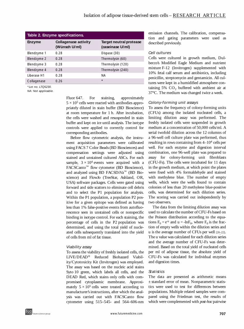
Isolation of stromal vascular fraction from adipose tissueThe cells making up the adipose stroma were iso-lated according to a previously described proto-col [14] with slight modifications. Six differentprotease mixtures, including B1–4, liberase H1(Roche Applied Sciences) and CCM (lot no.LTQ5230; Wako, Neuss, Germany), wereemployed (Table 2). The working solution foreach enzyme was freshly prepared prior to use bydissolving to a final collagenase activity of
0.28 Wünsch U/ml. This concentration wasselected based on the activity of our collagenasereference, which also fits well within the com-monly used 0.075–0.1% concentration range[17–19]. For enzymes, the concentration of whichwas expressed in Mandl units, a conversion fac-tor 1000 Mandl units = 1 Wünsch was used. Inthe case of CCM, the enzyme solution was dis-solved in Dulbecco’s phosphate-buffered saline(D-PBS) containing Ca2+ and Mg2+ (Invitrogen,Taastrup, Denmark) and supplemented with 2%bovine serum albumin (BSA) according to man-ufacturer instructions for optimal enzyme availa-bility and cell stabilization. The B1–4 andliberase H1 with included buffer salts werereconstituted in the delivery vial as recom-mended by the producer. No additional BSA wassupplemented with the latter enzymes.
The fat tissue was, prior to digestion, washedthree times with equal volumes of prewarmedD-PBS, and for digestion, 10 ml of the adiposetissue was mixed with an equal volume ofenzyme buffer. The incubation proceeded at37°C under gentle agitation for 1, 2 or 3 h. Thedissociated tissue was fractionated by sedimenta-tion centrifugation at 400 g for 10 min and thepelleted cells were filtered through a 70-µmmesh cell strainer (BD Bioscience, Broendby,Denmark) to remove debris. Contaminatingerythrocytes were lyzed using sterile water andthe remaining nucleated cells were further puri-fied through a second round of centrifugationand filtration. The total yield was determined ina hemocytometer after the cells had been stainedwith acetic methylene violet.
Immunophenotyping & flow cytometryThe flow cytometric analysis for a mesenchymalstem cell marker, CD90, hematopoetic progeni-tor cell marker, CD34, and a leukocyte lineagemarker, CD45, was performed with freshly iso-lated cells in a triple immunostaining procedureto assess coexpression of the assayed epitopes.The primary antibodies were mouse mono-clonals from Dako (Glostrup, Denmark), CD34(#M7080), CD45 (#MB0742), IgG1 (#X0931)and IgG2a (#X0943) or Abcam (Cambridge,UK) and CD90 (#ab11153). The Zenon labe-ling system (Invitrogen), including AlexaFluor 488 (#Z25002), Alexa Fluor 647(#Z25008) and R-Phycoerythrin (# Z25055),was used to discriminate the primary antibodies.The antibodies and fluorophores were combinedto provide conjugates CD34–Phycoerythrin,CD45–Alexa Fluor 488 and CD90–Alexa
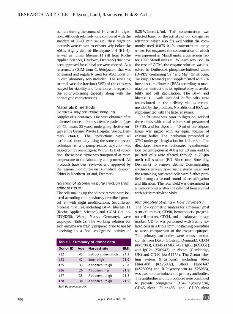
Table 1. Summary of donor data.
Donor ID Age Harvest site BMI
#22 45 Buttocks, inner thigh 24.3
#23 42 Inner thigh 21.0
#25 33 Abdomen, thigh 25.8
#26 26 Abdomen, hip 21.5
#27 35 Abdomen, thigh 21.2
#28 30 Abdomen, thigh 21.5
BMI: Body mass index.
Regen. Med. (2008) 3(5) future science groupfuture science group
Isolation of adipose tissue-derived stem cells – RESEARCH ARTICLE
future science groupfuture science group
Table 2. Enzyme spe
Enzyme Colla(Wü
Blendzyme 1 0.28
Blendzyme 2 0.28
Blendzyme 3 0.28
Blendzyme 4 0.28
Liberase H1 0.28
Collagenase 0.26
*Lot no. LTQ5230.NA: Not applicable.
Fluor 647. For staining, approximately5 × 105 cells were reacted with antibodies appro-priately diluted in stain buffer (BD Bioscience)at room temperature for 1 h. After incubation,the cells were washed and resuspended in stainbuffer and kept on ice until analysis. The isotypecontrols were applied to correctly control forcorresponding antibodies.
Before flow cytometric analysis, the instru-ment acquisition parameters were calibratedusing FACS 7 Color Beads (BD Biosciences) andcompensation settings were adjusted usingstained and unstained cultured ASCs. For eachsample, 3 × 10⁴ events were acquired with aFACSCanto™ flow cytometer (BD Bioscience),and analyzed using BD FACSDiVa™ (BD Bio-science) and FlowJo (TreeStar, Ashland, OR,USA) software packages. Cells were gated usingforward and side scatters to eliminate cell debrisand to select the P1 population for analysis.Within the P1 population, a population P2 pos-itive for a given epitope was defined as havingless than 1% false-positive events from autofluo-rescence seen in unstained cells or nonspecificbinding in isotype control. For each staining, thepercentage of cells in the P2 population wasdetermined, and using the total yield of nucle-ated cells subsequently translated into the yieldof cells from ml of fat tissue.
Viability assayTo assess the viability of freshly isolated cells, theLIVE/DEAD® Reduced Biohazard Viabil-ity/Cytotoxicity Kit (Invitrogen) was employed.The assay was based on the nucleic acid stainsSyto 10 green, which labels all cells, and theDEAD Red, which stains only cells with com-promised cytoplasmic membrane. Approxi-mately 5 × 10⁵ cells were treated according tomanufacturer’s instructions, after which the anal-ysis was carried out with FACSCanto flowcytometer using 515–545- and 564–606-nm
emission channels. The calibration, compensa-tion and gating parameters were used asdescribed previously.
Cell culturesCells were cultured in growth medium, Dul-becco’s Modified Eagle Medium and nutrientmixture F-12 (Invitrogen) supplemented with10% fetal calf serum and antibiotics, includingpenicillin, streptomycin and gentamicin. All cul-tures were kept in a humidified atmosphere con-taining 5% CO2 buffered with ambient air at37°C. The medium was changed twice a week.
Colony-forming unit assaysTo assess the frequency of colony-forming units(CFUs) among the isolated nucleated cells, alimiting dilution assay was performed. Thefreshly isolated cells were suspended in growthmedium at a concentration of 50,000 cells/ml. Aserial twofold dilution across the 12 columns ofa 96-well cell culture plate was performed, thusresulting in rows containing from 4–104 cells perwell. For each enzyme and digestion intervalcombination, one 96-well plate was prepared toassay for colony-forming unit fibroblasts(CFU-Fs). The cells were incubated for 11 daysin the growth medium, at which point the plateswere fixed with 4% formaldehyde and stainedwith methylene blue. The number of emptywells, which were the wells found to containcolonies of less than 20 methylene blue-positivecells, was determined for each dilution series.The scoring was carried out independently bytwo observers.
The data from the limiting dilution assay wasused to calculate the number of CFU-Fs based onthe Poisson distribution according to the equa-tions F0 = e-u and u = -lnF0, where F0 is the frac-tion of empty wells within the dilution series andu is the average number of CFUs per well [20–22].The u value was calculated for each dilution seriesand the average number of CFU-Fs was deter-mined. Based on the total yield of nucleated cellsper ml of adipose tissue, the absolute yield ofCFU-Fs was calculated for individual enzymesand digestion times.
StatisticsThe data are presented as arithmetic means± standard error of mean. Nonparametric statis-tics were used to test for differences betweenpopulations. Multiple related samples were com-pared using the Friedman test, the results ofwhich were complemented with post-hoc pairwise
cifications.
genase activity nsch U/ml)
Target neutral protease (caseinase U/ml)
Dispase (30)
Thermolysin (60)
Thermolysin (120)
Thermolysin (240)
NA
*
707www.futuremedicine.com

RESEARCH ARTICLE – Pilgaard, Lund, Rasmussen, Fink & Zachar
708
analysis by Wilcoxon signed-rank test. The rou-tines were carried out with the aid of theSPSS v.14 software package (SPSS, Chicago, IL,USA) and the statistical significance was assignedto the differences at p <0.05.
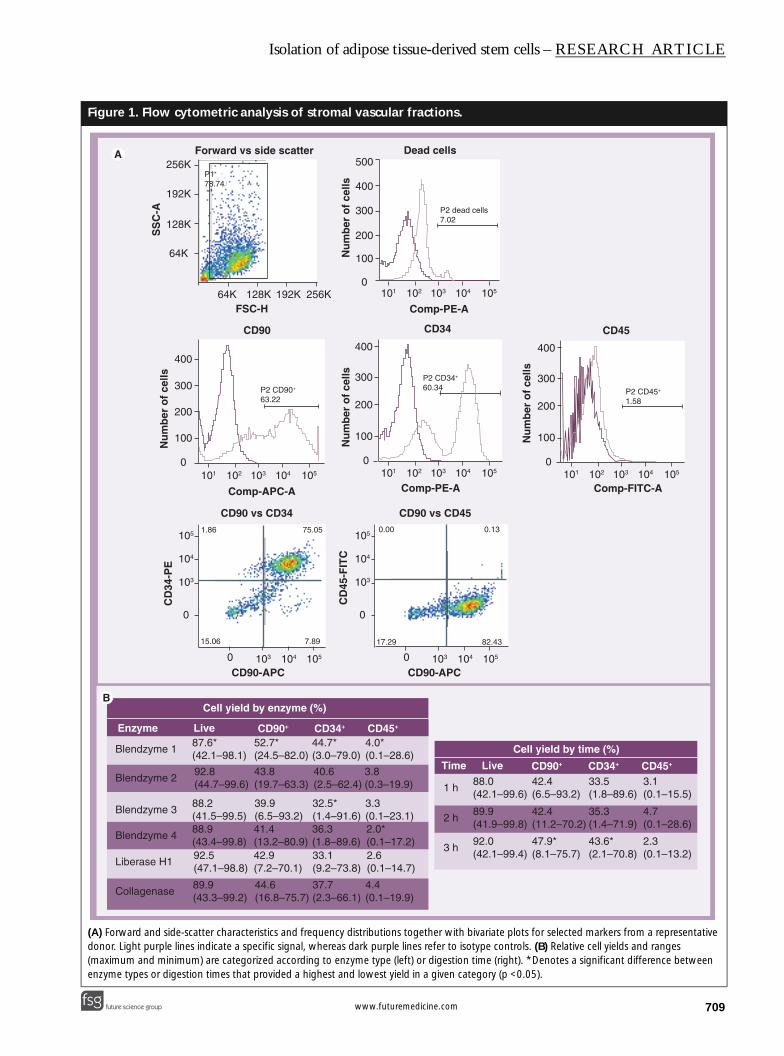
ResultsViability & phenotypic analysis of crude adipose tissue digestsA representative distribution of analyzed sub-populations within crude SVFs is presented inFigure 1A. The forward versus side scatter wasused to discriminate from cell debris. The gatedpopulation (P1) comprised approximately 80%of all events, approximately 10% of which con-stituted dead cells. In order to correctly deter-mine the proportion of CD34+, CD90+ andCD45+ cells, the range of specific signal intensi-ties was determined from the background stain-ing by isotype control relevant for each of thespecific antibodies. Since the experimental pro-cedure allowed for a simultaneous identificationof all three epitopes, bivariate plots representinga measure of CD90 and CD34, and CD90 andCD45 coexpression were possible.
As to the viability of isolated cells, approxi-mately 90% survived the procedure, based on anaverage across all enzymes and digestion times(Figure 1B). There was only one protease, B1,which resulted in a significantly lower propor-tion of live cells, irrespective of the length ofdigestion period. The same enzyme, however,yielded a significantly higher proportion of spe-cific precursors, CD90+ and CD34+. In general,the percentage of live cells in general did notchange significantly with longer incubationtime. By contrast, the relative yields of CD90-
and CD34+ cells increased significantly withtime. The percentage of leukocyte lineage cellsdid not change with prolonged digestion. Thisfraction represented on average 3.4% of the totalyield of nucleated cells in the SVF. Overall, it isstriking to notice the broad range of the obtainedrelative yields that underscores the existence ofremarkably high interdonor variability.
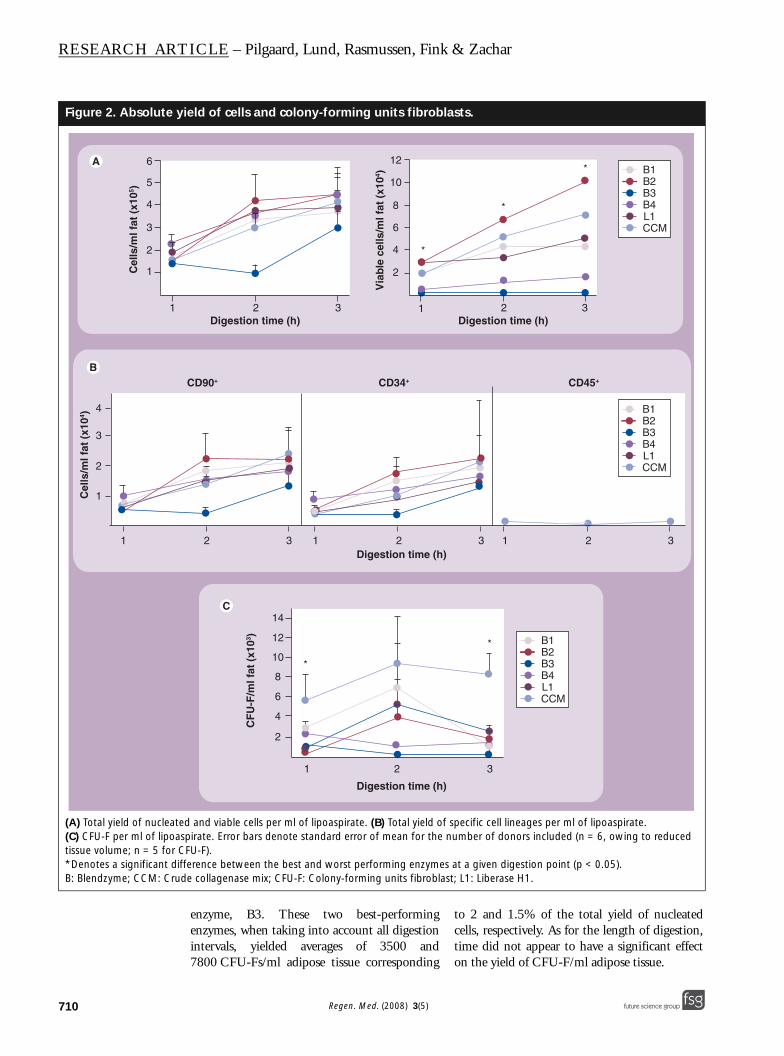
Total & phenotype-specific cell yieldsQuantitative data pertaining to the total andphenotype-specific cell yield are displayed inFigure 2. The total yield of freshly isolated nucle-ated cells was determined in six donor cases foreach combination of enzyme and digestion time(Figure 2A). Looking at the individual enzymes,B3 consistently yielded lower numbers at allthree digestion times. Nonetheless, the difference
in total cell yield was not significant betweenenzymes within each of the three evaluateddigestion periods. Interestingly, the total cellyield increased significantly with the digestiontime. Disregarding the applied enzyme, the over-all average yield of nucleated cells increased from1.7 × 105 at the first hour to 4.0 × 10⁵ cells perml adipose tissue after 3 h of digestion. As to thepotential to recover viable cells, a significant dif-ference was observed between the enzymes. B2and the CCM yielded significantly higher num-bers of viable cells at all time points as comparedwith B3 and 4.
The duration of digestion also had a signifi-cant impact, as evident from differences betweencell yields at the first and last hour of incubation.
With regard to the yield of CD90+ cells, nosingle enzyme could be indentified as signifi-cantly outperforming or underperforming,although B3 was clearly found trailing perform-ances of competitor proteases (Figure 2B). Whentaking into account the duration of digestion,the yield had a tendency to increase with time.Specifically, this increase was significant for B2and collagenase, and it corresponded to morethan a threefold rise during an interval from thefirst to the third hour. Regarding the yield ofCD34+ cells, there was no significant differencewhen comparing the performance of individualenzymes at different digestion times (Figure 2B). Itis noteworthy that for B1–3 and collagenase, asignificant increase in the yield of CD34+ cellswas obtained by prolonging the digestion from1–3 h. When taking into account all enzymes,the yield increase was more than threefold andcorresponded to a rise from 5300 cells/ml of adi-pose tissue in the first hour to approximately18,000 cells/ml of fat sample at the completionof digestion. The yield of CD45+ cells repre-sented only a nonsignificant contamination dur-ing the digestion procedure (Figure 2B), which isconsistent with only a very low proportion ofthese cell in the SVF (Figure 1A & B).
Yield of nonspecific colony-forming precursors The functionality of the isolated nucleated cellswas tested by their capacity to support out-growth of colonies in a limited dilution assay.Among the enzymes, there was a significant dif-ference in CFU-F yield, both when comparingwithin the individual digestion times and irrespec-tive of digestion periods (Figure 2C). B1 and colla-genase yielded the highest numbers of CFU-Fs, allsignificantly higher than the worst-performing
Regen. Med. (2008) 3(5) future science groupfuture science group
Isolation of adipose tissue-derived stem cells – RESEARCH ARTICLE
future science groupfuture science group
Figure 1. Flow cytom
(A) Forward and side-scatdonor. Light purple lines in(maximum and minimum)enzyme types or digestion
FS
SC
-A256K
192K
128K
64K
Nu
mb
er o
f ce
lls
400
300
200
100
0
0
103
104
105
CD
34-P
E
1
Enzyme Li
Blendzyme 1
Blendzyme 2
Blendzyme 3
Blendzyme 4
Liberase H1
Collagenase
87(4
92(4
88(488(492(4
89(4
etric analysis of stromal vascular fractions.
ter characteristics and frequency distributions together with bivariate plots for selected markers from a representative dicate a specific signal, whereas dark purple lines refer to isotype controls. (B) Relative cell yields and ranges are categorized according to enzyme type (left) or digestion time (right). *Denotes a significant difference between times that provided a highest and lowest yield in a given category (p <0.05).
orward vs side scatter
FSC-H256K192K128K64K
CD90N
um
ber
of
cells
Nu
mb
er o
f ce
lls
Nu
mb
er o
f ce
lls
Comp-APC-A
0
400
300
200
100
0101 102 103
103
104
104
105
105
101 102 103 104 105
101 102 103 104 105
P2 CD90+
63.22
Dead cells
Comp-PE-A
Comp-PE-A
500
400
300
200
100
0
P2 dead cells7.02
CD34
400
300
200
100
0101 102 103 104 105
P2 CD34+
60.34
CD45
Comp-FITC-A
P2 CD45+
1.58
CD90 vs CD34
0
0
103
103
104
104
105
105
CD90-APC CD90-APC
CD90 vs CD45
CD
45-F
ITC
1.86 75.05
5.06 7.89 17.29
0.00 0.13
82.43
Cell yield by enzyme (%)
Cell yield by time (%)
ve CD90+ CD34+ CD45+
.6*2.1–98.1)
52.7*(24.5–82.0)
44.7*(3.0–79.0)
4.0*(0.1–28.6)
.84.7–99.6)
.21.5–99.5).93.4–99.8).57.1–98.8)
.93.3–99.2)
43.8(19.7–63.3)
39.9(6.5–93.2)41.4(13.2–80.9)42.9(7.2–70.1)
44.6(16.8–75.7)
40.6(2.5–62.4)
32.5*(1.4–91.6)36.3(1.8–89.6)33.1(9.2–73.8)
37.7(2.3–66.1)
3.8(0.3–19.9)
3.3(0.1–23.1)2.0*(0.1–17.2)2.6(0.1–14.7)
4.4(0.1–19.9)
Time Live CD90+ CD34+ CD45+
1 h
2 h
3 h
88.0(42.1–99.6)
42.4(6.5–93.2)
33.5(1.8–89.6)
3.1(0.1–15.5)
89.9(41.9–99.8)
92.0(42.1–99.4)
47.9*(8.1–75.7)
42.4(11.2–70.2)
35.3(1.4–71.9)
43.6*(2.1–70.8)
4.7(0.1–28.6)
2.3(0.1–13.2)
P178.74
709www.futuremedicine.com
RESEARCH ARTICLE – Pilgaard, Lund, Rasmussen, Fink & Zachar
710
Figure 2. Absolute y
(A) Total yield of nucleat(C) CFU-F per ml of lipoatissue volume; n = 5 for C*Denotes a significant diB: Blendzyme; CCM: Cru
Cel
ls/m
l fat
(x1
05 )
6
5
4
3
2
1
1
1
1
2
3
Cel
ls/m
l fat
(x1
04 ) 4
enzyme, B3. These two best-performingenzymes, when taking into account all digestionintervals, yielded averages of 3500 and7800 CFU-Fs/ml adipose tissue corresponding
to 2 and 1.5% of the total yield of nucleatedcells, respectively. As for the length of digestion,time did not appear to have a significant effecton the yield of CFU-F/ml adipose tissue.
ield of cells and colony-forming units fibroblasts.
ed and viable cells per ml of lipoaspirate. (B) Total yield of specific cell lineages per ml of lipoaspirate. spirate. Error bars denote standard error of mean for the number of donors included (n = 6, owing to reduced FU-F).
fference between the best and worst performing enzymes at a given digestion point (p < 0.05). de collagenase mix; CFU-F: Colony-forming units fibroblast; L1: Liberase H1.
12 3
2 3 1 2 3
1 2 3
1 2 3
Digestion time (h)2 3
Digestion time (h)
B1B2B3B4L1CCM
*
*
*
Via
ble
cel
ls/m
l fat
(x1
04 )
12
2
4
6
8
10
B1B2B3B4L1CCM
B1B2B3B4L1CCM
CD90+ CD34+ CD45+
Digestion time (h)
Digestion time (h)
CF
U-F
/ml f
at (
x103 )
14
12
10
8
6
4
2
*
*
Regen. Med. (2008) 3(5) future science groupfuture science group
Isolation of adipose tissue-derived stem cells – RESEARCH ARTICLE
future science groupfuture science group
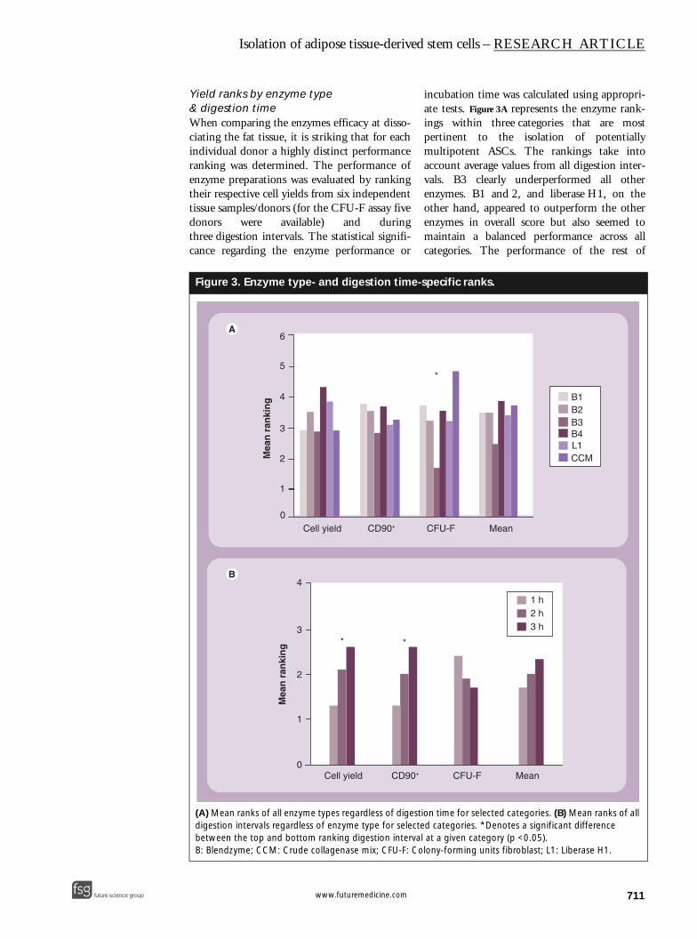
Yield ranks by enzyme type & digestion timeWhen comparing the enzymes efficacy at disso-ciating the fat tissue, it is striking that for eachindividual donor a highly distinct performanceranking was determined. The performance ofenzyme preparations was evaluated by rankingtheir respective cell yields from six independenttissue samples/donors (for the CFU-F assay fivedonors were available) and duringthree digestion intervals. The statistical signifi-cance regarding the enzyme performance or
incubation time was calculated using appropri-ate tests. Figure 3A represents the enzyme rank-ings within three categories that are mostpertinent to the isolation of potentiallymultipotent ASCs. The rankings take intoaccount average values from all digestion inter-vals. B3 clearly underperformed all otherenzymes. B1 and 2, and liberase H1, on theother hand, appeared to outperform the otherenzymes in overall score but also seemed tomaintain a balanced performance across allcategories. The performance of the rest of
Figure 3. Enzyme type- and digestion time-specific ranks.
(A) Mean ranks of all enzyme types regardless of digestion time for selected categories. (B) Mean ranks of all digestion intervals regardless of enzyme type for selected categories. *Denotes a significant difference between the top and bottom ranking digestion interval at a given category (p <0.05).B: Blendzyme; CCM: Crude collagenase mix; CFU-F: Colony-forming units fibroblast; L1: Liberase H1.
Cell yield CD90+ CFU-F Mean
**
Mea
n r
anki
ng
4
3
2
1
0
B1B2B3B4L1CCM
1 h2 h3 h
Cell yield CD90+ CFU-F Mean
*
Mea
n r
anki
ng
6
5
4
3
2
1
0
711www.futuremedicine.com

RESEARCH ARTICLE – Pilgaard, Lund, Rasmussen, Fink & Zachar
712
enzymes appeared more erratic. It is importantto note, however, that the ranking order couldnot be supported at a statistically significantlevel. In Figure 3B, the ranking of digestion inter-vals, which take into account average valuesfrom all enzymes, is listed. Although it is appar-ent that extended digestion yields significantlymore nucleated as well as CD90+ cells, there isno evidence that such correlation also applies tothe yield of CFU-F. Considering the averagescore, it seems that 2-h incubation would be themost suitable interval to provide for optimalyields and CFU-F numbers.
DiscussionIn the current investigation, we set out to stand-ardize the enzymatic dissociation step of theisolation procedure for adipose-derived mesen-chymal stem cells. Previous investigations haveaccumulated evidence with regard to a substan-tial interindividual variability of SVF parame-ters, such as total cell yield or phenotypical andfunctional characteristics. The great variabilityis assumed to arise from specifics in donordemographics, such as body mass index and age[11,16,17,23]. Our own data confirm great discrep-ancy in enzyme performance between differentindividuals. In order to minimize the individualspecific factors, we ranked the data for donorsseparately and subsequently used the averageranking across donors to test for the significanceof the relevant analysis result. Friedman’s statis-tical routine was conveniently used for this pur-pose. Since the approach also accommodates forrepeated measurements, it was possible to simul-taneously take into account different digestionintervals and thus increase the power of analysis.Our comprehensive comparison of six differentenzyme preparations indicates that B1 and 2,and liberase H1 are most universal when abroad spectrum of goals, ranging from a quanti-tative yield of cells to the yield of biologicallyactive progenitors, is to be met. The analysis ofdigestion times revealed that although the yieldof specific cell types increases with time, this isnot true for biologically active precursors. Thus,it appears that for this set-up a digestion periodof 2 h would satisfy most of the experimentaldemands.
In the present study, we implemented longerdigestion intervals than traditionally used andadditionally, the enzyme activity was lower thanthat commonly used in protocols based onshorter intervals and higher enzyme concentra-tions. The set-up was inspired by the work of
Wang et al. who found that although the fre-quency of cultivable dermal fibroblast and theirproliferation rate decreased with time, the totalcell outcome was elevated owing to the increaseof the cell yield [7]. In order to exhaustively iso-late the ASC population, we opted to prolongthe digestion period threefold. The more effec-tive tissue dissociation was apparent from theincrease in the relative as well as absolute cellyields. The higher yield of both CD90+ andCD34+ cells indicates a more efficient cell releasein general, and in the case of the hematopoieticprogenitors, a progressive disintegration of tissuemicrovasculature.
Conversely, the low yield of CD45+ leukocytelineage, reaching consistently around 3.4% ofthe total yield of nucleated cells in the SVF, con-firms that these cells are not associated with theextracellular matrix of the adipose tissue. Ourresults are in agreement with some [17,24,25] butnot all [18] of the previous studies. The discrep-ancy is likely to reflect differences between thetissue samples themselves, since the anatomicalsites differ with respect to vascularization, tissueharvest methods and isolation procedures.
Regarding the biological activity of isolatedcells, the capability of supporting the outgrowthof colonies (CFU-F) averaged 1% of the SVFpool in our study. This number was very similarfor all enzymes as well as digestion periods, andwas also in agreement with previously publishedfigures [11]. With the stable fraction of approxi-mately 90% viable cells, this result wasexpected. It is interesting that the low yield ofCFU-Fs is in contrast with the high frequencyof progenitor CD90+ cells, which in additionappear to assume an increasing fraction duringthe course of digestion.
Similar results were previously demonstratedby Oedayrajsingh-Varma et al., where three tis-sue harvest methods yielded similar numbers ofviable cells but differed in the frequency ofASCs [19]. An explanation could be the lateonset of apoptosis caused by the extended expo-sure to proteolytic enzymes. The LIVE/DEADassay employed in current investigation is onlyable to distinguish between intact cells and cellswith compromised membrane integrity. Inaddition, the SVF cells were plated for theCFU-F assay according to the number of livecells based on trypan blue staining, which iden-tifies dead cells with compromised membranes.Sufficient amounts of stress and/or DNAdamage as a result of the enzyme treatmentcombined with removal from the natural
Regen. Med. (2008) 3(5) future science groupfuture science group
Isolation of adipose tissue-derived stem cells – RESEARCH ARTICLE
future science groupfuture science group
Executive summary
Key findings
• The forward versus sidevents and 90% of th
• The relative yields of Crepresented only a no
• Blendzyme 2 and the Blendzymes 3 and 4.
• Blendzyme 1 and crudworst performing enz
Conclusions
• Blendzymes 1 and 2, cells and potential pre
• The analysis of digest• The highly purified pr
definition and reprod• By using selected form
tissue-derived stem ce
microenvironment could initiate an apoptoticreaction in the SVF cells. Provided the presenceof a very initial phase of apoptosis, the results inthe CFU-F assay may have been affected, sincethe used methods are not effective in such a sce-nario. Further analysis would be necessary todetermine a relationship between enzymatictreatment, culture conditions and the occurrenceof apoptosis.
Despite the increased cell yield with pro-longed digestion, the results indicate that thecell source had not been fully exhausted fromcells, as no plateau in cell yields was reached.Hence, more progenitor cells could potentiallybe isolated. The challenge is to isolate the maxi-mum number of functional progenitors withoutdamaging the cell population in the process. Toachieve this, one could vary the combination ofenzyme activity with different digestion periods.Inferring from the interindividual variability,the ideal set-up would include a test to deter-mine the most optimal enzyme formulation ineach case. Subsequently, a long-term digestwould be performed in a sequential manner,where released cells are retrieved continuously,thus avoiding proteolytic overexposure.
The main goal of the current study was tocomparatively analyze a complement of highlydefined proteases with the crude collagenasetype I enzyme, which hitherto was a mainstaytool to obtain SVFs. Surprisingly, such an inves-tigation has not been carried out before, butwith the prospect of exploiting of ASCs in clini-cal applications [26], it appears critical that
highly reproducible protocols are developed.Crude collagenase, unfortunately, is subject to asubstantial lot-to-lot variation and standardiza-tion is hardly achievable, in addition to the factthat its composition is too poorly defined toconform with good manufacturing practicestandards. Nevertheless, it is interesting that thehighly defined enzymatic formulations did notsignificantly outperform crude collagenase fromour study, or for that matter in the previousinvestigations, as far as the yield of precursors isconcerned [10,11,14–17,22,23]. Moreover, the inter-individual variability appeared to be of such amagnitude that the data to identify the best-per-forming enzyme with a statistical significancecould not be obtained. Thus, despite the factthat the new generation of enzyme mixes doesnot offer substantial benefit over the commonlyused collagenase with regard to the quantitativeyields, the purified proteases still appear to be anappealing alternative maybe in a new sequentialapproach and especially in cases where the diges-tion process needs to be highly defined andreproducible.
ConclusionIn the current investigation, five highly definedprotease formulations and a CCM were compar-atively evaluated at 1, 2 and 3 h of digestion.The resulting SVFs of human adipose tissueaspirates were analyzed for the yield of nucleatedcells, viability and frequency of specific lineagesby flow cytometry. The functionality of the cellswas assessed as to the colony forming capacity in
e-gated population within crude stromal vascular fraction cells (P1) comprised approximately 80% of all e cells were viable. The total cell yield increased significantly with the digestion time.D90+ and CD34+ cells increased significantly with time, whereas the percentage of leukocyte lineage cells
nsignificant contamination during the digestion procedure and did not change with prolonged digestion. crude collagenase mix yielded significantly higher numbers of viable cells at all time points as compared with
e collagenase yielded the highest numbers of colony-forming unit fibroblats, all significantly higher than the yme, the Blendzyme 3.
and liberase H1, are most universal when a broad spectrum of goals, ranging from a quantitative yield of cursor to the yield of biologically active progenitors, is to be met.
ion times revealed that a period of 2 h would satisfy most of the experimental demands.oteases provide a valuable alternative to crude collagenase preparations, especially in scenarios where high ucibility of the digestion process is of high priority.
ulations of highly defined protease mixtures, along with well-tuned growth conditions, an adipose ll population highly suitable for regenerative clinical applications may readily become available.
713www.futuremedicine.com
RESEARCH ARTICLE – Pilgaard, Lund, Rasmussen, Fink & Zachar
714
a limiting dilution assay. Our data provide evi-dence that B1 and 2, and liberase H1 outper-form other enzyme preparations, whereas B3clearly trails in performance. As for the length ofdigestion, a 2-h interval appeared optimal whenconsidering both the yield and functionality ofthe cells in the SVF. In conclusion, our resultsdemonstrate that the highly purified proteasesprovide a valuable alternative to crude colla-genase preparations, especially in scenarioswhere high definition and reproducibility of thedigestion process is of high priority.
AcknowledgementsThe authors wish to thank plastic surgeons Frants Grymerand Christian Bang, their office and nursing staff, and theirpatients at Grymer Private Hospital, Aarhus, Denmark, fordonation of the liposuction material. The expert technicalassistance of Helle Skjødt Møller is highly appreciated.
Financial & competing interests disclosure The authors greatly appreciate support from the Carlsberg,John and Birthe Meyer’s, Toyota Foundations and the Dan-ish Medical Research Council grants no. 271-06-0283 and2052-01-0045. The authors have no other relevant affilia-tions or financial involvement with any organization orentity with a financial interest in or financial conflict withthe subject matter or materials discussed in the manuscriptapart from those disclosed.
No writing assistance was utilized in the production ofthis manuscript.
Ethical conduct of research The authors state that they have obtained appropriate insti-tutional review board approval or have followed the princi-ples outlined in the Declaration of Helsinki for all human oranimal experimental investigations. In addition, for investi-gations involving human subjects, informed consent has beenobtained from the participants involved.
BibliographyPapers of special note have been highlighted as either of interest (•) or of considerable interest (••) to readers.1. Wollert KC, Meyer GP, Lotz J et al.:
Intracoronary autologous bone-marrow cell transfer after myocardial infarction: the BOOST randomised controlled clinical trial. Lancet 364, 141–148 (2004).
2. Horwitz EM, Prockop DJ, Fitzpatrick LA et al.: Transplantability and therapeutic effects of bone marrow-derived mesenchymal cells in children with osteogenesis imperfecta. Nat. Med. 5, 309–313 (1999).
3. Aviles FF, San Roman JA, Garcia Frade J et al.: Intracoronary stem cell transplantation in acute myocardial infarction. Rev. Esp. Cardiol. 57, 201–208 (2004).
4. Kuroda R, Ishida K, Matsumoto T et al.: Treatment of a full-thickness articular cartilage defect in the femoral condyle of an athlete with autologous bone-marrow stromal cells. Osteoarthr. Cartil. 15, 226–231 (2007).
5. De Ugarte DA, Morizono K, Elbarbary A et al.: Comparison of multi-lineage cells from human adipose tissue and bone marrow. Cells Tissues Organs 174, 101–109 (2003).
6. Georges P, Muirhead RP, Williams L et al.: Comparison of size, viability, and function of fetal pig islet-like cell clusters after digestion using collagenase or liberase. Cell Transplant. 11, 539–545 (2002).
7. Wang H, Van Blitterswijk CA, Bertrand-De Haas M, Schuurman AH, Lamme EN: Improved enzymatic isolation of fibroblasts for the creation of autologous skin substitutes. In Vitro Cell Dev. Biol. Anim. 40, 268–277 (2004).
• Comprehensively analyzes the effect of various Clostridium histolyticum collagenase preparations, the addition of neutral proteases and variable digestion periods in the isolation procedure for human fibroblasts. They demonstrate that the right balance in digestion parameters not necessarily lies where cell viability is highest or tissue digestion most efficient. The final and total cell outcome after cultivation is the main evaluation criteria not regarding the functionality of the cells.
8. Williams SK, McKenney S, Jarrell BE: Collagenase lot selection and purification for adipose tissue digestion. Cell Transplant. 4, 281–289 (1995).
• The use of crude Clostridial collagenase in the digestion of human adipose tissue is evaluated with respect of cell yield and cell adherence. They convincingly point out lot-to-lot variation, test collagenase preparations subjected to different degrees of purification and prove that neutral proteases are essential for optimal digestion. They conclude that purified collagenase added to a specified protease will be optimal in applications where control and reproducibility are of the essence.
9. Hayman DM, Blumberg TJ, Scott CC, Athanasiou KA: The effects of isolation on chondrocyte gene expression. Tissue Eng. 12, 2573–2581 (2006).
10. Varma MJ, Breuls RG, Schouten TE et al.: Phenotypical and functional characterization of freshly isolated adipose tissue-derived stem cells. Stem Cells Dev. 16, 91–104 (2007).
• Through triple-color fluorescence-activated cell sorting (FACS) analysis, this study demonstrates the applicability of the stromal vascular fraction (SVF) cells in engineering purposes involving bone repair. Freshly isolated SVF cells are compared with cultured adipose tissue-derived stem cells (ASCs), and through critical examination it was shown that the loss of CD34+ cells during culture was caused by a reduction in expression as opposed to an overgrowth of CD34- cells.
11. Mitchell JB, McIntosh K, Zvonic S et al.: Immunophenotype of human adipose-derived cells: temporal changes in stromal-associated and stem cell-associated markers. Stem Cells 24, 376–385 (2006).
• Applying limited dilution assays and FACS analysis, this study elegantly demonstrates the selection of a homogeneous cell population through adherence of SVF cells and subsequent serial passaging. During expansion, ASC cultures are enhanced for stromal colony-forming cells with at least osteogenic and adipogenic capacity.
12. Sekiya I, Larson BL, Smith JR, Pochampally R, Cui JG, Prockop DJ: Expansion of human adult stem cells from bone marrow stroma: conditions that maximize the yields of early progenitors and evaluate their quality. Stem Cells 20, 530–541 (2002).
Regen. Med. (2008) 3(5) future science groupfuture science group
Isolation of adipose tissue-derived stem cells – RESEARCH ARTICLE
13. Kin T, Johnson PR, Shapiro AM, Lakey JR: Factors influencing the collagenase digestion phase of human islet isolation. Transplantation 83, 7–12 (2007).
14. Zuk PA, Zhu M, Mizuno H et al.: Multilineage cells from human adipose tissue: implications for cell-based therapies. Tissue Eng. 7, 211–228 (2001).
• Study forming the basic theory of presences and characteristics of ASCs that states the basic definition of ASCs regarding differentiation potential and surface markers. The author healthily questions the existence of multipotential mesenchymal stem cells in adipose tissue as opposed to several different lineage precursor cells.
15. Guilak F, Lott KE, Awad HA et al.: Clonal analysis of the differentiation potential of human ASCs. J. Cell. Physiol. 206(1), 229–237 (2005).
• Investigation takes up the question raised by [14] and proves the presence of a real stem cell with multilineage potential in adipose tissue. Through ring-cloning expansion and differentiation assays, they show that ASC cultures contain cells of
varying potential in adipogenic, osteogenic, chondrogenic and neurogenic differentiation.
16. Meyerrose TE, De Ugarte DA, Hofling AA et al.: In vivo distribution of human adipose-derived mesenchymal stem cells in novel xenotransplantation models. Stem Cells 25(1), 220–227 (2007).
17. Yoshimura K, Shigeura T, Matsumoto D et al.: Characterization of freshly isolated and cultured cells derived from the fatty and fluid portions of liposuction aspirates. J. Cell. Physiol. 208, 64–76 (2006).
18. Aust L, Devlin B, Foster SJ et al.: Yield of human adipose-derived adult stem cells from liposuction aspirates. Cytotherapy 6, 7–14 (2004).
19. Oedayrajsingh-Varma MJ, van Ham SM, Knippenberg M et al.: Adipose tissue-derived mesenchymal stem cell yield and growth characteristics are affected by the tissue-harvesting procedure. Cytotherapy 8, 166–177 (2006).
20. Bellows CG, Aubin JE: Determination of numbers of osteoprogenitors present in isolated fetal rat calvaria cells in vitro. Dev. Biol. 133, 8–13 (1989).
21. Wu X, Peters JM, Gonzalez FJ, Prasad HS, Rohrer MD, Gimble JM: Frequency of stromal lineage colony forming units in bone marrow of peroxisome proliferator-activated receptor-α-null mice. Bone 26, 21–26 (2000).
22. Castro-Malaspina H, Gay RE, Resnick G et al.: Characterization of human bone marrow fibroblast colony-forming cells (CFU-F) and their progeny. Blood 56, 289–301 (1980).
23. Sen A, Lea-Currie YR, Sujkowska D et al.: Adipogenic potential of human adipose derived stromal cells from multiple donors is heterogeneous. J. Cell. Biochem. 81, 312–319 (2001).
24. Gronthos S, Franklin DM, Leddy HA, Robey PG, Storms RW, Gimble JM: Surface protein characterization of human adipose tissue-derived stromal cells. J. Cell. Physiol. 189, 54–63 (2001).
25. Katz AJ, Tholpady A, Tholpady SS, Shang H, Ogle RC: Cell surface and transcriptional characterization of human adipose-derived adherent stromal (hADAS) cells. Stem Cells 23, 412–423 (2005).
26. Daniels E: Cytori Therapeutics, Inc. Regen. Med. 2(3), 317–320 (2007).
715future science groupfuture science group www.futuremedicine.com