Proteasome Inhibition by Fellutamide B Induces Nerve Growth Factor Synthesis
33
Proteasome Inhibition by Fellutamide B Induces Nerve Growth Factor Synthesis John Hines 1 , Michael Groll 4 , Margaret Fahnestock 5 , and Craig M. Crews 1,2,3* 1Dept. of Molecular, Cellular and Developmental Biology, Yale University, New Haven, CT 06511, USA 2Dept. of Chemistry, Yale University, New Haven, CT 06511, USA 3Dept. of Pharmacology, Yale University, New Haven, CT 06511, USA 4Center for Integrated Protein Science at the Department Chemie, Technische Universität München, Lichtenbergstrasse 4, Garching D-85747, Germany 5Dept. of Psychiatry & Behavioural Neurosciences, McMaster University, Hamilton, ON L8N 3Z5, Canada SUMMARY Neurotrophic small molecules have the potential to aid in the treatment of neuronal injury and neurodegenerative diseases. The natural product fellutamide B, originally isolated from Penicillium fellutanum, potently induces nerve growth factor (NGF) release from fibroblasts and glial-derived cells, although the mechanism for this neurotrophic activity has not been elucidated. Here, we report that fellutamide B potently inhibits proteasome catalytic activity. High resolution structural information obtained from co-crystallization of the 20S proteasome reveals novel aspects regarding β-subunit binding and adduct formation by fellutamide B to inhibit their hydrolytic activity. We demonstrate that fellutamide B and other proteasome inhibitors increased NGF gene transcription via a cis-acting element (or elements) in the promoter. These results demonstrate an unrecognized connection between proteasome inhibition and NGF production, suggesting a possible new strategy in the development of neurotrophic agents. The development of neurotrophic therapeutics for treatment of neuronal injury or the neurodegenerative effects of stroke, ischemia and CNS diseases (e.g. Parkinson’s Disease, Alzheimer’s Disease) has attracted much attention. In particular, the neuroprotective and restorative effects of nerve growth factor or other neurotrophins (reviewed in (Huang and Reichardt, 2003)) in ameliorating the symptoms or pathophysiology in animal models of the disease(s) have been documented in the literature (Castellanos-Ortega et al., 1999). For example, co-transplantation of NGF along with fetal ventral mesencephalic cells into the striatum of lesioned rats (a model of Parkinson's Disease) significantly restored spontaneous locomotor activity and striatal and nigral dopamine levels compared to those in rats receiving transplanted cells alone (Chaturvedi et al., 2006). These results suggested that NGF exhibited neuroprotective effects on the transplanted cells as well as helped rescue remaining host dopaminergic neurons from cell death. Similarly, NGF can attenuate lesion-induced *Corresponding author: Craig M. Crews phone: (203) 432-9364; fax: (203) 432-6161; email: [email protected]. COMPETING INTERESTS STATEMENT C.M.C. is a co-founder of Proteolix, Inc., which currently is exploring the use of proteasome inhibitors in oncology. Publisher's Disclaimer: This is a PDF file of an unedited manuscript that has been accepted for publication. As a service to our customers we are providing this early version of the manuscript. The manuscript will undergo copyediting, typesetting, and review of the resulting proof before it is published in its final citable form. Please note that during the production process errors may be discovered which could affect the content, and all legal disclaimers that apply to the journal pertain. NIH Public Access Author Manuscript Chem Biol. Author manuscript; available in PMC 2009 May 1. Published in final edited form as: Chem Biol. 2008 May ; 15(5): 501–512. NIH-PA Author Manuscript NIH-PA Author Manuscript NIH-PA Author Manuscript
Transcript of Proteasome Inhibition by Fellutamide B Induces Nerve Growth Factor Synthesis

Proteasome Inhibition by Fellutamide B Induces Nerve GrowthFactor Synthesis
John Hines1, Michael Groll4, Margaret Fahnestock5, and Craig M. Crews1,2,3*
1Dept. of Molecular, Cellular and Developmental Biology, Yale University, New Haven, CT 06511, USA
2Dept. of Chemistry, Yale University, New Haven, CT 06511, USA
3Dept. of Pharmacology, Yale University, New Haven, CT 06511, USA
4Center for Integrated Protein Science at the Department Chemie, Technische Universität München,Lichtenbergstrasse 4, Garching D-85747, Germany
5Dept. of Psychiatry & Behavioural Neurosciences, McMaster University, Hamilton, ON L8N 3Z5, Canada
SUMMARYNeurotrophic small molecules have the potential to aid in the treatment of neuronal injury andneurodegenerative diseases. The natural product fellutamide B, originally isolated from Penicilliumfellutanum, potently induces nerve growth factor (NGF) release from fibroblasts and glial-derivedcells, although the mechanism for this neurotrophic activity has not been elucidated. Here, we reportthat fellutamide B potently inhibits proteasome catalytic activity. High resolution structuralinformation obtained from co-crystallization of the 20S proteasome reveals novel aspects regardingβ-subunit binding and adduct formation by fellutamide B to inhibit their hydrolytic activity. Wedemonstrate that fellutamide B and other proteasome inhibitors increased NGF gene transcriptionvia a cis-acting element (or elements) in the promoter. These results demonstrate an unrecognizedconnection between proteasome inhibition and NGF production, suggesting a possible new strategyin the development of neurotrophic agents.
The development of neurotrophic therapeutics for treatment of neuronal injury or theneurodegenerative effects of stroke, ischemia and CNS diseases (e.g. Parkinson’s Disease,Alzheimer’s Disease) has attracted much attention. In particular, the neuroprotective andrestorative effects of nerve growth factor or other neurotrophins (reviewed in (Huang andReichardt, 2003)) in ameliorating the symptoms or pathophysiology in animal models of thedisease(s) have been documented in the literature (Castellanos-Ortega et al., 1999). Forexample, co-transplantation of NGF along with fetal ventral mesencephalic cells into thestriatum of lesioned rats (a model of Parkinson's Disease) significantly restored spontaneouslocomotor activity and striatal and nigral dopamine levels compared to those in rats receivingtransplanted cells alone (Chaturvedi et al., 2006). These results suggested that NGF exhibitedneuroprotective effects on the transplanted cells as well as helped rescue remaining hostdopaminergic neurons from cell death. Similarly, NGF can attenuate lesion-induced
*Corresponding author: Craig M. Crews phone: (203) 432-9364; fax: (203) 432-6161; email: [email protected] INTERESTS STATEMENTC.M.C. is a co-founder of Proteolix, Inc., which currently is exploring the use of proteasome inhibitors in oncology.Publisher's Disclaimer: This is a PDF file of an unedited manuscript that has been accepted for publication. As a service to our customerswe are providing this early version of the manuscript. The manuscript will undergo copyediting, typesetting, and review of the resultingproof before it is published in its final citable form. Please note that during the production process errors may be discovered which couldaffect the content, and all legal disclaimers that apply to the journal pertain.
NIH Public AccessAuthor ManuscriptChem Biol. Author manuscript; available in PMC 2009 May 1.
Published in final edited form as:Chem Biol. 2008 May ; 15(5): 501–512.
NIH
-PA Author Manuscript
NIH
-PA Author Manuscript
NIH
-PA Author Manuscript
cholinergic deficits and cognitive impairments in animal models of Alzheimer’s Disease:chronic NGF treatment induced increased blood flow and nicotine uptake in the cerebral cortex(Lapchak, 1993), and implantation of genetically engineered NGF-secreting fibroblasts intoearly Alzheimer’s patients brains significantly slowed cholinergic nerve deterioration(Tuszynski et al., 2005). However, the requirement for direct administration of neurotrophinsand/or large molecules into the CNS in order to circumvent the blood-brain barrier severelylimits their therapeutic utility, and is not without side effects (Venero et al., 1996). Efforts topromote penetration of NGF across the blood-brain barrier by conjugating it to transferrin(Liao et al., 2001) have been made; however, the amount of modified NGF detected in the CNSwas very small.
The development of small molecule neurotrophic compounds capable of entering the brain is,therefore, an attractive therapeutic strategy. Literature reports have described small moleculesthat are able to upregulate selected neuronal proteins and/or induce neurite outgrowth ofcultured preneuronal cells (Cheng et al., 2006; Warashina et al., 2006). However, themechanism(s) whereby such molecules act are yet unknown, nor is the extent to which theycan completely mimic the actions of endogenous neurotrophins. The fellutamides are marinefungal metabolites (Shigemori et al., 1991) reported to induce the synthesis and secretion ofNGF (Yamaguchi et al., 1993) from cultured brain cells and fibroblasts. Thus, fellutamide Bmight be considered an “indirect neurotrophin”, in much the same manner as a drug likereserpine, which triggers the release of norepinephrine from presynaptic vesicles, is consideredan indirect adrenergic agonist. An indirect neurotrophic small molecule is likely to reap thecombined benefits of easier CNS access and as well the ultimate therapeutic effects of theinduced endogenous proteins. We recently reported on the first total synthesis of fellutamideB (Schneekloth et al., 2006). Here, we explore the mechanism by which fellutamide B exertsits neurotrophin-inducing effect. We show that fellutamide B potently inhibits the 20Sproteasome leading to increased NGF gene expression and secretion.
RESULTSFunctional and structural evidence of proteasome inhibition by fellutamide B
Given the similarity in chemical structure between fellutamide B and peptide aldehydeproteasome inhibitors (as exemplified by MG132, see Figure 1a), we investigated whetherfellutamide B could inhibit the three hydrolytic activities of the 20S proteasome. As shown inFigure 1b, fellutamide B potently inhibits the chymotryptic-like activity with an IC50 value of9.4 ± 2.5 nM. The tryptic-like and caspase-like activities were also inhibited by fellutamide B,albeit less potently (2.0 ± 0.4 µM and 1.2 ± 0.8 µM , respectively). The potency of fellutamideB against the chymotryptic-like activity was, in fact, greater than that of the peptide aldehydeinhibitor MG132 (40 ± 3.3 nM) and approached the high potency seen with the irreversibleproteasome inhibitor (Meng et al., 1999b) epoxomicin (5.7 ± 1.3 nM) (Figure 1c). As with theother two established proteasome inhibitors, treatment of L-M mouse fibroblasts withfellutamide B led to accumulation of ubiquitinated proteins (Figure 1d), confirming its abilityto inhibit the proteasome in intact cells.
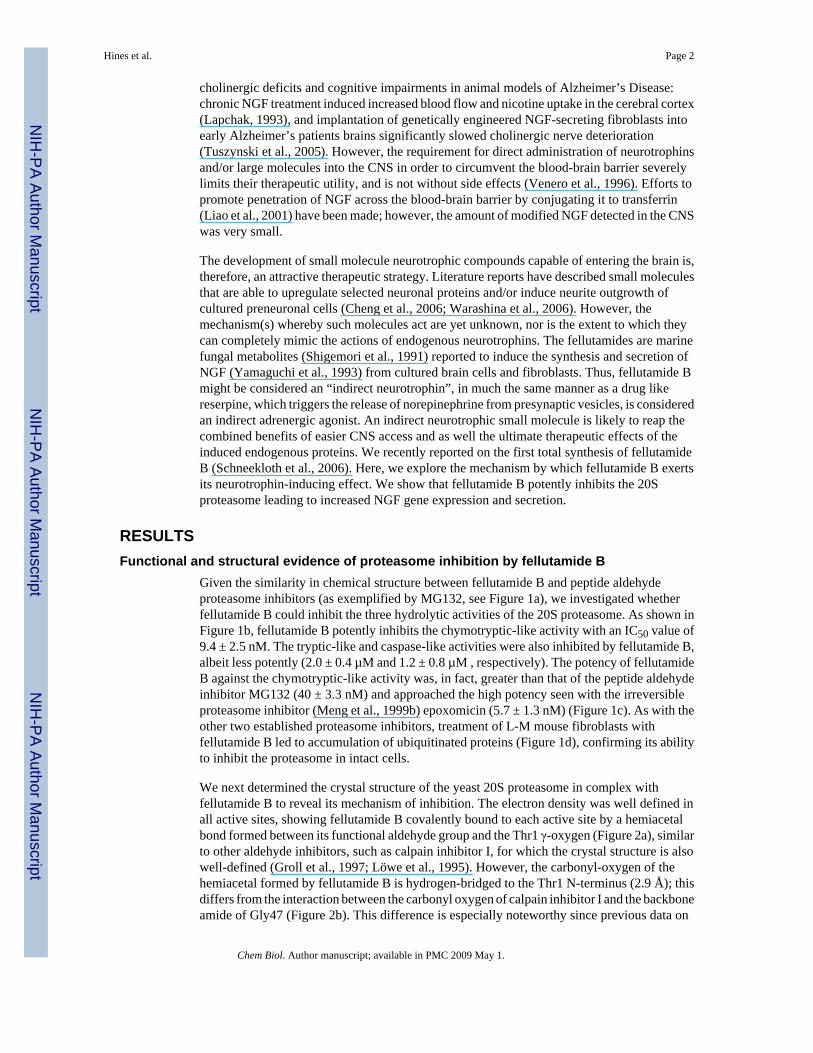
We next determined the crystal structure of the yeast 20S proteasome in complex withfellutamide B to reveal its mechanism of inhibition. The electron density was well defined inall active sites, showing fellutamide B covalently bound to each active site by a hemiacetalbond formed between its functional aldehyde group and the Thr1 γ-oxygen (Figure 2a), similarto other aldehyde inhibitors, such as calpain inhibitor I, for which the crystal structure is alsowell-defined (Groll et al., 1997; Löwe et al., 1995). However, the carbonyl-oxygen of thehemiacetal formed by fellutamide B is hydrogen-bridged to the Thr1 N-terminus (2.9 Å); thisdiffers from the interaction between the carbonyl oxygen of calpain inhibitor I and the backboneamide of Gly47 (Figure 2b). This difference is especially noteworthy since previous data on
Hines et al. Page 2
Chem Biol. Author manuscript; available in PMC 2009 May 1.
NIH
-PA Author Manuscript
NIH
-PA Author Manuscript
NIH
-PA Author Manuscript

aldehyde inhibitors had defined the backbone amide of Gly47 as the stereotypical oxyanionhole stabilizing the transition state intermediate by hydrogen bonding to the carbonyl group ofthe peptide bond undergoing hydrolysis.
It is known that aldehydes act as proteasome inhibitors only if they contain a peptide backbonethat allows their stabilization at the proteasomal active sites through formation of an anti-parallel β-sheet structure. These interactions are necessary to increase the mean residence timeof the ligand at the active center. By varying their peptide backbone, proteasome inhibitorssharing the same reactive functional group show preference for certain active sites (Myung etal., 2001). Compared to other aldehyde proteasome inhibitors, fellutamide B also contains anextended β-hydroxy aliphatic tail. Surprisingly, the whole aliphatic tail, which shows highflexibility in solution, has well defined electron density at all proteolytic active sites. Thisstands in contrast to synthetic long chain aldehyde inhibitors, which are structurally orderedonly in their first three residues (Loidl et al., 1999). Interestingly, structural superposition offellutamide B bound to the proteolytic active sites indicates that while the peptide backboneadopts similar conformations, the orientation of the aliphatic tail differs completely (Figure2c). In particular, and uniquely at the chymotryptic-like subunit, C24 to C29 of the fellutamideB aliphatic tail (see Figure 1a) interact with a hydrophobic groove through van der Waals forceswith protein residues Pro95, Tyr96, Pro115 and Val116 of subunit β6 (Figure 2d). Interestingly,the hydrophobic groove to which the aliphatic tail is bound is formed only during ligandbinding, following concerted movements of the aliphatic tail and protein side chain residues.
Neurotrophic effects of fellutamide B and other proteasome inhibitors on mammalian cellsFellutamide B induces NGF secretion from a number of cells that previously have been usedas models to study NGF synthesis and secretion (Table 1). All five of these cell lines (L-M,NIH-3T3, S-180, A172 and C6-2B cells) responded to 10 µM fellutamide B treatment byupregulating NGF secretion, as detected by ELISA of culture medium. L-M cells secreted themost NGF overall and also exhibited the most robust response to the drug, a 16.8-fold averageinduction, while NIH-3T3 cells also responded to fellutamide B with an average 15.8-foldincrease in NGF secretion. S-180 sarcoma cells increased NGF secretion by only 4.0-fold,although perhaps these tumor cells by their very nature are already operating at near theirmaximum capacity to produce this neurotrophin. By comparison, the two glial-derived celllines, A172 and C6-2B, secreted NGF on a much smaller scale; however, both responded tofellutamide B by increasing their NGF production 4.9-fold and 6.6-fold, respectively. Not everycell type tested responded to fellutamide B. KB epidermal carcinoma cells and P-388 leukemiacells were used in the original report on the discovery of this small molecule (Shigemori et al.,1991). Both failed to show a response to the drug in terms of NGF secretion, despite theirreported sensitivity to the growth inhibitory effects of fellutamide B. Likewise, the preneuronalcell line PC-12 (rat pheochromocytoma) which can differentiate into a neuronal phenotype inthe presence of NGF (Obin et al., 1999) also failed to show detectable NGF secretion in thepresence of fellutamide B either by ELISA (Table 1) or by neurite outgrowth (unpublishedobservation). Fellutamide B causes cell cycle arrest (measured by [3H]-thymidineincorporation into cellular DNA) in all the cell lines on which it was tested (Table 1). In mostcell lines, this growth arrest resulted from the cytoxicity of fellutamide B (measured by MTSconversion by active mitochondria in Table 1), although in a few instances – L-M cells, andto lesser extent C6-2B and A172 cells – the IC50 for growth arrest was below that observedfor cytotoxicity. Thus, while the growth inhibitory/cytotoxic activity of fellutamide B appearsto affect all cells, its activity to elicit NGF upregulation is restricted to a subset of cell types.
Since L-M cells displayed the most robust NGF response, this cell line was selected for furtheranalysis of fellutamide B activities: as shown in Figure 3a, the potency with which fellutamideB induces NGF in L-M cells is similar to its cytotoxicity in MTS conversion assay (Figure 3b).
Hines et al. Page 3
Chem Biol. Author manuscript; available in PMC 2009 May 1.
NIH
-PA Author Manuscript
NIH
-PA Author Manuscript
NIH
-PA Author Manuscript

The bond formed between proteasome catalytic subunits and peptide aldehyde inhibitors likefellutamide B is reversible. Thus, it was hypothesized that a brief administration of fellutamideB for a few hours (a “pulse”) followed by a drug-free “recovery” interval -- rather than acontinuous 24 hr. fellutamide B treatment -- might result in significant NGF induction withreduced cytotoxicity compared to continuous drug treatment. It was found that 7 hr. pulse of10 µM fellutamide B was sufficient to elicit 90% of the maximal NGF upregulation that hadbeen induced by 24 hr of continuous treatment (Figure 3c). However, the reduction in cellviability (Figure 3d) associated with the 7 hr pulse was only 35.0%, which was markedlyreduced compared to the 60.0% reduction seen with the continuous 24 hr treatment. Thus,although an exclusively neurotrophic and non-toxic concentration of fellutamide B was notapparent, the two activities of fellutamide B can yet be largely dissociated from each other bythe proper duration of treatment.
Having now confirmed the NGF-inducing activity of fellutamide B, and having also shown itto be a proteasome inhibitor, we tested whether this NGF-inducing activity could be generalizedto other proteasome inhibitors. Similar to fellutamide B, the established proteasome inhibitorsepoxomicin and MG132 triggered NGF upregulation in the L-M cells with EC50 values of 127± 13.4 nM and 11.8 ± 3.7 µM, respectively (Figures 3e and 3f). This confirms that proteasomeinhibition is the key activity of fellutamide B, since epoxomicin is a highly specific inhibitorof the proteasome that does not inhibit other proteases. While we observed that fellutamide Band the other proteasome inhibitors less potently induced upregulation of NGF than theyinhibited the purified proteasomes, this is likely due to the inherent differences between thetwo assays. Intact cells were required in the NGF secretion assay (as well as the anti-ubiquitinwesterns in Figure 1d), and thus factors like membrane permeation, drug-efflux pumps (e.g.P-glycoprotein) and de-ubiquitinases that often reduce the apparent potency of these drugswould have attenuating effect; such factors are absent from our measurements on the purifiedproteasome activity, which cannot be accurately, specifically made in intact cells due to theinterfering presence of free proteases. Nevertheless, the rank order potency with which thesecompounds induce NGF secretion corresponds to their rank order potency for inhibiting theproteasomal chymotryptic-like activity. The biological activity of the secreted NGF wasconfirmed by incubating undifferentiated PC12 cells in medium that had been conditioned byfellutamide B- or epoxomicin-treated L-M cells. PC12 neurite outgrowth was readilyobservable after only 24 hrs. incubation (not shown) and pronounced by 48 hrs. incubation inconditioned medium (Figure 3g).
Given the cytotoxicity accompanying fellutamide B treatment, the question remained whetherNGF induction was a consequence of proteasome inhibition specifically or rather a responseto general cytotoxic insult. To answer this, L-M cells were separately exposed (Figure 4a) tothree different toxic treatments: 1) a combination of veratridine and ouabain, to collapse themembrane potential; 2) a combination of cytochalasin D and myoseverin, to destroy thecytoskeleton; and 3) epoxomicin, a structurally dissimilar proteasome inhibitor. In the case offellutamide B and epoxomicin, these agent(s) induced the formation of long cell processeswhich in the past had been misinterpreted as neurites (Fenteany et al., 1994) when observedon neuronal cell lines. All treatments were comparably cytotoxic as measured by MTSconversion assay (Figure 4b), which western blotting for cleavage of the caspase 3 substratePARP (Figure 4c) showed was due to apoptotic cell death. However, only fellutamide B andepoxomicin triggered upregulation of NGF (Figure 4d). Thus, the neurotrophic effects offellutamide B appear to be a specific consequence of proteasome inhibition rather than a cellularresponse to general cytotoxicity.
Hines et al. Page 4
Chem Biol. Author manuscript; available in PMC 2009 May 1.
NIH
-PA Author Manuscript
NIH
-PA Author Manuscript
NIH
-PA Author Manuscript

Upregulation of NGF occurs by induction of new gene transcription from both NGFpromoters
To provide further mechanistic insight into proteasome inhibitor-induced NGF upregulation,we next examined the effect of fellutamide B on NGF gene transcription. Specifically, thelevels of NGF mRNA from fellutamide B- versus vehicle-treated cells were measured usingRT-PCR. These results (Figure 5a) showed a robust upregulation of NGF mRNA in responseto the proteasome inhibitor, while measurements of the housekeeping transcript GAPDHshowed no drug-associated changes. There are, in fact, two known promoters for the NGF gene(Racke et al., 1996): one upstream of the first exon, and another that lies in between exons 2and 3, with the entire protein coding region located in exon 4. The mRNAs produced from thetwo NGF promoters can be distinguished by their differentially retained exons. Our RT-PCRresults show that NGF mRNA transcripts from both known promoters are strongly upregulatedfollowing fellutamide B treatment (Figure 5a); note that two mRNA transcripts of slightlydifferent sizes are detected from the upstream promoter, as previously seen (Racke et al.,1996). The magnitude of this increase in NGF mRNA following proteasome inhibitor treatmentmay be sufficient to account for the increases in NGF at the protein level. To confirm that theupregulation of NGF was due to NGF mRNA transcription, a time course of fellutamide-triggered NGF induction in the absence/presence of the RNA polymerase II inhibitor, α-amanitin, was performed. Following 12 hr. of continuous fellutamide B treatment, the levelsof secreted NGF began to rise (Figure 5b); this was completely blocked at all times by thepresence of α-amanitin. This result is consistent with upregulation due to de novo NGF mRNAsynthesis due upon fellutamide B administration, and rules out any independent role for post-translational upregulation of NGF secretion. A parallel MTS assay showed conclusively thatthe inhibition of NGF upregulation by α-amanitin cannot be attributed to additive cytotoxicityin combination with fellutamide B, as there was none (Figure 5c). Furthermore, measurementsof NGF mRNA stability in the absence or presence of fellutamide B (Figure 5d) showed nodifferences in rate of decay, demonstrating that upregulation of NGF mRNA in response to thesmall molecule was due only to enhanced transcription and not diminished transcriptdegradation.
In order to focus in on the region(s) of the NGF promoter mediating the transcriptionalactivation response to proteasome inhibitors, we subcloned the entire 5 kb mouse NGFpromoter (D'Mello and Heinrich, 1991) including the first 120 bp downstream of thetranscription start site (defined as position +1) into a promoter-less firefly luciferase plasmidfor gene reporter assays. Upstream regions of the NGF promoter were progressively removedto create plasmids (Figure 6a) which were stably transfected into cells, along with SV40 earlypromoter-driven renilla luciferase co-reporter, and tested for the ability of fellutamide B toinduce firefly luciferase activity (Figure 6b). To ensure that any effects on reporter expression/regulation arising from differing genomic integration points would be balanced out betweenthe different reporter transfectants, pools of stably-transfected clones for each reporter wereused. The full 5 kb NGF promoter (−5000 bp) was responsive to fellutamide B, showing anearly six-fold induction of luciferase activity. While truncation of the promoter down to 1.8kb (−1800 bp) increased overall luciferase transcription (basal and drug-induced), the level ofinduction by fellutamide B remained approximately the same. Further truncation of thepromoter down to 750 bp (−750 bp), and then 250 bp (−250 bp) continued the trend ofincreasing overall luciferase transcription, indicating the removal of postulated genesuppressive elements from the promoter (D'Mello and Heinrich, 1991). Yet, these reporterswere still responsive to fellutamide B and showed increased luciferase activity in its presence.Given the importance of the AP-1 site at position +35 in mediating the stimulatory effects ofphorbol esters (Omae et al., 1994) and dihydroxyvitamin D3 (Veenstra et al., 1998) on NGFsynthesis, and the fact that fellutamide B treatment causes the stabilization and activation ofc-Jun (unpublished observation), we tested whether this AP-1 site is also crucial for the effects
Hines et al. Page 5
Chem Biol. Author manuscript; available in PMC 2009 May 1.
NIH
-PA Author Manuscript
NIH
-PA Author Manuscript
NIH
-PA Author Manuscript

of proteasome inhibitors. Reporter vectors were constructed lacking the AP-1 site (−1800 w/o AP-1; −750 w/o AP-1; and −250 w/o AP-1), but assays on cells transfected with theseconstructs continued to show increased luciferase activity in response to fellutamide B, albeitremoval of the AP-1 site had a diminishing effect on overall luciferase transcription. Truncationof the promoter down to 150 bp retained fellutamide B responsiveness, indicating that the cis-acting element or elements lie close to the transcription start site. Epoxomicin, MG 132 andclasto-lactacystin β-lactone all induced luciferase (Figure 6c) from this minimal reporterplasmid, verifying that this region of the NGF promoter was responsive to proteasomeinhibitors other than fellutamide B.
DISCUSSIONOur finding that proteasome inhibition leads to the production and secretion of NGF revealsanother potential therapeutic utility of this class of small molecules. Proteasome inhibitors havealready been identified as candidate anti-inflammatory (Meng et al., 1999b) and anti-tumordrugs (Meng et al., 1999a). They have also been implicated in stimulating bone formation(Garrett et al., 2003), the treatment of stroke (Phillips et al., 2000), and as antiparasitic agents(Lindenthal et al., 2005). While proteasome inhibition can lead to cytotoxicity, therapeuticwindows have been established that nonetheless permit their use clinically. Indeed, while acontinuous 24 hr treatment of the cells with fellutamide B resulted in maximum NGFupregulation and toxicity, “pulse” treatment with fellutamide B for only 7 hours followed bya 17 hr. recovery interval resulted in a nearly equivalent induction of NGF with markedlyreduced toxicity. The separation of these two activities of fellutamide B – and, by extension,other reversible proteasome inhibitors ‒ is an important step in their development as potentialneurotrophic therapeutics. This is less of a concern with fellutamide B than it will be with otherproteasome inhibitors: epoxomicin was the most potent inducer of NGF, but was also the mostpotently cytotoxic. This greater toxicity is almost certainly attributable to the fact thatepoxomicin, unlike the fellutamide B and MG132, irreversibly inhibits the proteasome, whichplaced an upper limit on the epoxomicin concentrations that could be used on whole cells(e.g. Figure 1d and Figure 3e) and still produce the biological effect without killing them first.This irreversible inhibition by epoxomicin ultimately makes it unsuitable for the same “pulse”treatments that reduced the toxicity of fellutamide B.
Application of proteasome inhibitors such as MG132 to transected axons significantly delaysthe onset of Wallerian degeneration (Zhai et al., 2003) both in vitro and in vivo. Given thatpersistent activation of erk1/2 (MacInnis and Campenot, 2005), a known downstream effectorof NGF receptors, was also observed in these studies was, it is possible that the degenerationwas delayed by proteasome inhibitor-triggered upregulation of NGF. The ability of someproteasome inhibitors to spur “neurite outgrowth” from preneuronal cells has been reportedmany times (Fenteany et al., 1994; Inoue et al., 2004); however, this effect is not specific topreneuronal cells, and has been shown to also occur in endothelial cells (Meng et al., 1999a)and, in the present study, in L-M fibroblasts (Figure 4a). Mitchison and colleagues havereported that proteasome inhibitors arrest dividing cells in cytokinesis (Straight et al., 2003),which may be the basis for the spindle-like morphology seen in treated cells. Thus, it is perhapsmore accurate to describe this proteasome inhibitor effect as a general induction of a bipolarcellular elongation; which in preneuronal cells was misinterpreted as “neurites”. However,NGF induction by proteasome inhibition is a process that should elicit neuronal differentiation,maintenance and neuroprotection given that it upregulates the natural neurotrophin. Furtherevidence for the independence of proteasome inhibitor-triggered process extension from NGFsecretion is that fellutamide B failed to trigger the latter in PC12 cells (data not shown), asystem in which neurite extension following proteasome inhibition has been studiedextensively.
Hines et al. Page 6
Chem Biol. Author manuscript; available in PMC 2009 May 1.
NIH
-PA Author Manuscript
NIH
-PA Author Manuscript
NIH
-PA Author Manuscript

The structural data from the co-crystallization of fellutamide B with the 20S proteasome revealssome interesting and unexpected insights into how inhibition is achieved. It illustrates thepossibility of two different hemiacetal adduct enantiomers (R,S) formed by peptide aldehydeinhibitors: the customary adduct with the planar aldehyde group oxygen atom pointing into theoxyanion hole formed by Gly47-N; or, as now seen with fellutamide B, an alternativeorientation of the aldehyde group oxygen towards Thr1-NH. Although hydrogen bondingbetween the Thr1-N terminus and the active group of a small molecule inhibitor has beenobserved for other classes of inhibitors (e.g. β-lactones (Borissenko and Groll, 2007)), it isnonetheless an unexpected finding for a peptide aldehyde proteasomal inhibitor that eitherorientiation can and will stabilize the hemiacetal oxygen atom and thereby block catalyticactivity (Borissenko and Groll, 2007; Groll et al., 1997; Löwe et al., 1995). In addition to thebinding of fellutamide B’s P1 and P3 side chains to the cognate S1 and S3 specificity pockets,the structural basis for its preferential blockade of the chymotryptic-like active site may alsoreside in the interaction of its distinctive aliphatic tail with several residues in an adjacenthydrophobic groove. The aliphatic tail adopts dissimilar conformations when bound to thetryptic-like and caspase-like active sites, such that the stabilization of the aliphatic tail to thehydrophobic groove is peculiar to the chymotryptic-like active site. This may contributesignificantly to the tighter binding of fellutamide B to that subunit, translating into the observed200- to 400-fold greater potency to inhibit this activity (Figure 1b).
An earlier study (Yamaguchi et al., 1993) had shown decreasing NGF secretion triggered bya maximum dose of fellutamide in the presence of ever-increasing concentrations ofactinomycin D. This raised two possibilities: either the increasing, combined cytotoxicity fromblockade of both the proteasome and transcription killed the cells before NGF could beproduced, or de novo NGF mRNA transcription is a necessary element in the mechanism offellutamide. We have, in fact, observed that fellutamide B and other proteasome inhibitorsexert a biphasic effect on NGF secretion: increasing it until a critical concentration ofproteasome inhibitor is reached, after which further increases become overwhelmingly toxicand NGF secretion diminishes. Thus, a more rigorous examination of the involvement ofmRNA upregulation in the effect of fellutamide B seemed particularly compelling. The datahere show conclusively that increased transcription of NGF mRNA is, indeed, necessary – infact, the increase in NGF mRNA following fellutamide B treatment was so dramatic that itrecommended upregulation occurs exclusively at the mRNA level. This increase in NGFmRNA involves activation of both known promoter regions for the NGF gene, consistent withan earlier report suggesting coordinate regulation of these two promoters (Racke et al., 1996).Since the ultimate NGF protein products from these two promoters are believed to bebiologically equivalent, the combined effect of proteasome inhibition would be that muchgreater.
While our own RT-PCR results clearly showed that NGF mRNA was being upregulated byfellutamide B, it was possible that either degradation of the transcripts had been attenuated ortheir synthesis had been stimulated, or some combination of both. The blockade of theneurotrophic response to fellutamide B by α-amanitin suggested transcriptional activation ofthe NGF gene and ruled out the possibility that increased NGF secretion could be largely post-translational (i.e. NGF protein trafficking). Still, it did not eliminate the possibility thatstabilization of NGF mRNA contributed to the transcript upregulation. Only after showing thatthe decay of NGF mRNA remains the same in the presence or absence of fellutamide B couldwe unambiguously conclude that transcriptional activation of the NGF gene is the solemechanism for increased production of the protein. Furthermore, we were able to narrow theregion of the NGF promoter crucial for transcriptional activation down to within 150 bp of thetranscription start site and rule out participation of an important AP-1 site at position +35.
Hines et al. Page 7
Chem Biol. Author manuscript; available in PMC 2009 May 1.
NIH
-PA Author Manuscript
NIH
-PA Author Manuscript
NIH
-PA Author Manuscript

Changes in levels of other proteins besides NGF are to be expected following treatment withproteasome inhibitors, which is why these small molecules have therapeutic value for treatingmany disease states. However, the broad effects of these small molecules can also lead topotential limitations on their usefulness. For example, some uncertainty exists regarding thestabilizing effect of proteasome inhibition on cytoplasmic levels of the non-infectious form ofthe prion protein, PrPC. While transmission of prion disease states depends on the presence ofthe misfolded “scrapie” form of the protein (PrPSc), Lindquist and colleagues reported thataccumulation of PrPC following proteasome inhibition can itself result in neuronal toxicity(Ma and Lindquist, 2001). Although subsequent reports (Fioriti et al., 2005; Kristiansen et al.,2005; Roucou et al., 2003) strongly dispute the neurotoxic effect of elevated cytoplasmicPrPC, the effects of proteasome inhibitors on protein levels other than NGF need to be carefullyconsidered as their potential for neuronal therapeutics is evaluated. Separating the toxic effectof fellutamide B and other proteasome inhibitors from their NGF-inducing properties is acrucial barrier towards their ultimate development for any clinical use, neurotrophic orotherwise. While it was demonstrated that toxicity is not a precondition for upregulating NGFfrom the cell types tested, toxicity is a side effect of proteasome inhibitors. There are, however,reasons to believe that these two activities of fellutamide B are independent. For instance, notevery cell tested herein responded to fellutamide B by upregulating NGF; yet all the cells testedwere sensitive to the cytotoxic effect of the proteasome inhibitor. In addition, theaforementioned pulse-recovery strategy of administering fellutamide B produced nearmaximum NGF upregulation with attenuated toxicity. Moreover, the dose-responserelationship of fellutamide B cytotoxicity clearly differs from that of NGF induction in that thelatter reaches maximum efficacy over a more narrow concentration range: as shown in Figure3b, the Hill slope for fellutamide B cytotoxicity fits to unity, while that for its NGF induction(as well as MG132 and epoxomicin) is 3.5 (as shown in Figures 3a, e and f). While the molecularbasis for this pharmacodynamic difference is not known, it suggests the possibilities of positivecooperativity or other positive feedback mechanism in the upregulation of NGF by fellutamideB or the activation of multiple cis-elements over a narrow concentration range of fellutamideB (non-first order induction); this is not seen with cytotoxicity. Since the blockade of theproteasome affects the levels of multiple proteins within the cell, it is reasonable to hypothesizethat the protein(s) that cause upregulation of NGF production will be different from those thatmediate the toxic effect. Thus, maximum NGF induction can be achieved at proteasomeinhibitor conditions that do not elicit maximum toxicity.
It is encouraging that fellutamide B exerts its effects on both glial-type and fibroblast-typecells. In terms of therapeutic potential, a broader tissue effect would enable proteasomalinhibition to treat both peripheral nerve injury, where fibroblasts would be the predominantcell type to secrete the trophic factors, as well as neurodegeneration in the brain, where glialtype cells are more plentiful. Moreover, as many neurotrophins appear to be coordinatelyregulated in the CNS (Maisonpierre et al., 1990; Takeda et al., 1993), the possibility thatproteasome inhibition might have a similar trophic effect on other neurotrophins (e.g. NT-3,BDNF) is an interesting possibility that remains to be investigated.
SIGNIFICANCEThere is great interest in the fields of neuroscience and chemical biology to identify compoundsthat either trigger neuronal differentiation and survival or induce neurotrophin expression. Suchcompounds would have great therapeutic potential for the treatment of neuronal injury orneurodegenerative diseases. While reports that identify such compounds from library screensor natural sources are increasing in frequency, none of these emerging studies have identifieda protein receptor and/or mechanism of action to which the neurotrophic activity can beascribed. We report here that fellutamide B, one of the earliest compounds identified to haveneurotrophin-inducing activity, does so by binding to and inhibiting the 20S proteasome. While
Hines et al. Page 8
Chem Biol. Author manuscript; available in PMC 2009 May 1.
NIH
-PA Author Manuscript
NIH
-PA Author Manuscript
NIH
-PA Author Manuscript

the manner in which fellutamide B binds to the proteasome is distinct from other peptidealdehyde inhibitors, its ability to upregulate NGF is shared not only by other peptide aldehydes,but other mechanistically different classes of proteasome inhibitors as well. Proteasomeinhibitors as well as NGF administration are known to attenuate Wallerian axonal degenerationand our results now suggest a direct connection between these two neuroprotective approaches.
The proteasome has previously been targeted in the treatment of cancer, inflammation andstroke – herein, we have identified another potential medical application for proteasomeinhibitors, some of which are already in clinical trials. With the demonstrated efficacy ofneurotrophins to alleviate symptoms of neurodegenerative diseases, fellutamide B and otherproteasome inhibitors deserve further attention as potential neuronal therapeutics.
EXPERIMENTAL PROCEDURESReagents
Fellutamide B (Schneekloth et al., 2006) and epoxomicin (Sin et al., 1999) were synthesizedas previously described. MG 132, myoseverin, cytochalasin D, clasto-lactacystin β-lactoneand α-amanitin were purchased from Calbiochem (La Jolla, CA). Veratridine and ouabain wereobtained from Sigma (St. Louis, MO). Fluorogenic substrate peptides for proteasome activityassay were from Bachem Bioscience (King of Prussia, PA). Purified human 20S proteasomewere purchased from Boston Biochemical (Cambridge, MA)
Cell cultureC6-2B cells were a generous gift from Satya Kunapuli (Temple University, Philadelphia, PA),and PC12 cells were a gift from Randy Pittman (University of Pennsylvania, Philadelphia,PA); all other cell lines were purchased from ATCC (Manassas, VA). Undifferentiated PC12cells were grown in RPMI 1640 medium supplemented with 10% heat-inactivated horse serum,5% heat-inactivated fetal bovine serum. L-M mouse fibroblasts were cultured in Medium 199supplemented with 0.5% peptone. NIH-3T3, C6-2B and A172 cells were grown in high glucoseDulbecco’s Modified Eagle Medium supplemented with 10% heat-inactivated fetal bovineserum and S-180 cells were cultivated in minimal essential medium supplemented with Earle’ssalts, 2 mM glutamine, 1 mM sodium pyruvate and 0.1 mM non-essential amino acids. Allculture media was supplemented with 100 units/ml penicillin G and 100 µg/ml streptomycinsulfate.
Cytotoxicity assayFollowing drug treatment of cells as indicated, culture medium was supplemented with 330µg/ml MTS (Promega Corp., Madison, WI) and 25 µM phenazine methosulfate and incubatedat 37°C. Mitochondrial reduction of MTS to the formazan derivative was monitored bymeasuring the medium’s absorbance at 490 nm.
Proliferation assayPerformed as previously described (Yeh et al., 2000). [3H]-Thymidine was purchased fromPerkin-Elmer Life Sciences (Boston, MA).
Neurite outgrowth assayL-M cells were treated with either 10 µM fellutamide B, 250 nM epoxomicin, or vehicle control(0.1% DMSO) for 24 hr. Afterwards conditioned medium was collected and dialyzed (MWCO= 10 kDa) against RPMI 1640 for 24 hrs. at 4°C. The dialyzed, conditioned medium was thensupplemented with 2% heat-inactivated horse serum and 1% heat-inactivated fetal bovine
Hines et al. Page 9
Chem Biol. Author manuscript; available in PMC 2009 May 1.
NIH
-PA Author Manuscript
NIH
-PA Author Manuscript
NIH
-PA Author Manuscript

serum and applied to undifferentiated PC12 cells growing on collagen-covered 60 mm dishes.The development of neurites was monitored over the following 48 hrs.
NGF ELISAELISA for NGF was performed in accordance with the kit manufacturer’s recommendedinstructions (Promega Corp., Madison, WI). Data were analyzed using PRISM software(GraphPad Software, San Diego, CA)
Western BlottingPerformed as previously described (Meng et al., 1999a). Protein samples were resolved by 8%SDS-PAGE, transferred to nitrocellulose and probed with antibodies to ubiquitin (CellSignaling Tech., Danvers, MA), α-tubulin (Sigma, St. Louis, MO) or PARP (Zymed Labs, SanFrancisco, CA).
Proteasome activity assayPerformed according to previously described detailed method (Kim et al., 2005).
RT-PCRmRNA was extracted from fellutamide B-treated and vehicle-treated L-M cells using Trizolreagent. First strand synthesis of cDNA was performed using SuperScript III first strandsynthesis kit (Invitrogen, Carlsbad, CA). PCR measurements of NGF and GAPDH wereperformed using the following primers:
total NGF forward primer: 5’-GCAGTGAGGTGCATAGCGTA-3’
upstream promoter-derived NGF forward primer: 5’-AGAGAGCGCCTGGAGCCG-3’
downstream promoter-driven NGF forward primer: 5’-CTTCCTGGGCTCTAATGATGC-3’
total NGF reverse primer: 5’-CACTGAGAACTCCCCCATGT-3’
GAPDH forward primer: 5’-AACTTTGGCATTGTGGAAGG-3’
GAPDH reverse primer: 5’-ACACATTGGGGGTAGGAACA-3’
PCRs were performed for 30 cycles using decreasing titrations of template cDNA to verifychanges (or lack thereof) in target abundance.
Co-crystallization of fellutamide B and 20S proteasomeCrystals of 20S proteasome from S. cerevisiae were grown in hanging drops at 24°C asdescribed previously (Groll and Huber, 2005) and incubated for 60 min with fellutamide B (10mM in DMSO). The protein concentration used for crystallization was 40 mg/ml in 10 mMTris.HCl, pH 7.5, and 1 mM EDTA. Drops contained 3 µl of protein and 2µl of reservoirsolution (30 mM magnesium acetate, 100 mM morpholino-ethane-sulphonic acid (MES) pH7.2, and 10% 2-methyl-2,4-pentanediol (MPD)). The space group of proteasomal complexcrystals belongs to P21 with cell dimensions of a = 134.3 Å, b = 301.6 Å, c = 143.5 Å and β =112.7°. Data to 2.6 Å were collected using synchrotron radiation with λ = 1.05 Å at the BW6-beamline at DESY, Hamburg, Germany. Crystals were soaked in cryoprotecting buffer (30%MPD, 20 mM magnesium acetate, 100 mM MES, pH 6.9) and frozen in a stream of liquidnitrogen gas at 90 K (Oxford Cryo Systems, Oxford, UK). X-ray intensities were evaluatedusing the DENZO program package and data reduction was performed with SCALEPACK(Otwinowski et al., 2003; Otwinowski and Minor, 1997). Anisotropy of diffraction wascorrected by an overall anisotropic temperature factor, comparing observed and calculated
Hines et al. Page 10
Chem Biol. Author manuscript; available in PMC 2009 May 1.
NIH
-PA Author Manuscript
NIH
-PA Author Manuscript
NIH
-PA Author Manuscript

structure amplitudes using the program CNS (Brünger et al., 1998). A total of 796875reflections yielded 331875 unique reflections (98.7% completeness). The correspondingRmerge was 6.6% at 2.6 Å resolution (40.2% for the last resolution shell). Electron density wasimproved by averaging and back-transforming reflections 10 times over the two-fold non-crystallographic symmetry axis using the program package MAIN (Turk, 1992). Conventionalcrystallographic rigid body, positional and temperature factor refinements were carried outwith CNS using the yeast 20S proteasome structure as the starting model (Groll et al., 1997).For model building the program MAIN was used. The structure was refined to acrystallographic R-factor of 24.0% (free R-factor 26.5%(Brünger, 1993)) with rms-deviationsfrom target values of 0.007 Å for bonds and 1.37° for angles(Brünger, 1993). Modelingexperiments were performed using the coordinates of yeast 20S proteasome (Groll et al.,1997) with the program MAIN.
Luciferase reporter promoter activity assayUsing primer directed PCR, truncated versions of the previously cloned NGF promoter werecreated and inserted into the promoterless luciferase expression vector pGL4 (Promega Corp.).Reporter constructs were then stably transfected into NIH-3T3 cells using LipofecatAMINE2000; pools of stably-transfected clones were used for the analysis of each reporter construct.The stably-transfected cells were tested for their ability to upregulate luciferase activity inresponse to proteasome inhibitors using the Dual Luciferase Reporter Assay System (PromegaCorp.). An SV40 early promoter-driven renilla luciferase construct (Promega Corp.) was usedas a co-reporter to normalize results.
ACKNOWLEDGMENTS
The authors acknowledge the NIH (GM062120) for funding.
REFERENCESBorissenko L, Groll M. 20S proteasome and its inhibitors: crystallographic knowledge for drug
development. Chem. Rev 2007;107:687–717. [PubMed: 17316053]Brünger A, Adams P, Clore G, DeLano W, Gros P, Grosse-Kunstleve R, Jiang J, Kuszewski J, Nilges
M, Pannu N, et al. Crystallography & NMR system: a new software suite for macromolecular structuredetermination. Acta Crystallogr. D Biol. Crystallogr 1998;1:905–921.
Brünger AT. Assessment of phase accuracy by cross validation: the free R value. Methods andapplications. Acta Crystallogr. D Biol. Crystallogr 1993;49:24–36. [PubMed: 15299543]
Castellanos-Ortega MR, Cruz-Aguado R, Martinez-Martí L. Nerve growth factor: possibilities andlimitations of its clinical application. Rev. Neurol 1999;29:439–447. [PubMed: 10584248]
Chaturvedi RK, Shukla S, Seth K, Agrawal AK. Nerve growth factor increases survival of dopaminergicgraft, rescue of nigral dopaminergic neurons and restores functional deficits in rat model of Parkison'sdisease. Neurosci. Lett 2006;398:44–49. [PubMed: 16423459]
Cheng Y, Schneider B, Riese U, Schubert B, Li Z, Hamburger M. (+)-N-Deoxymilitarinone A, aneuritogenic pyridone alkaloid from the insect pathogenic fungus, Paecilomyces farinosus. J. Nat.Prod 2006;69:436–438. [PubMed: 16562854]
D'Mello SR, Heinrich G. Structural and functional identification of regulatory regions and cis elementssurrounding the nerve growth factor gene promoter. Mol. Brain. Res 1991;11:255–264. [PubMed:1661823]
Fenteany G, Standaert RF, Reichard GA, Corey EJ, Schreiber SL. A β-lactone related to lactacystininduces neurite outgrowth in a neuroblastoma cell line and inhibits cell cycle progression in anosteosarcome cell line. Proc. Natl. Acad. Sci. USA 1994;91:3358–3362. [PubMed: 8159752]
Fioriti L, Dossena S, Stewart LR, Stewart RS, Harris DA, Forloni G, Chiesa R. Cytosolic prion protein(PrP) is not toxic in N2A cells and primary neurons expressing pathogenic PrP mutations. J. Biol.Chem 2005;280:11320–11328. [PubMed: 15632159]
Hines et al. Page 11
Chem Biol. Author manuscript; available in PMC 2009 May 1.
NIH
-PA Author Manuscript
NIH
-PA Author Manuscript
NIH
-PA Author Manuscript

Garrett IR, Chen D, Gutierrez G, Zhao M, Escobedo A, Rossini G, Harris SE, Gallwitz W, Kim KB, HuS, et al. Selective inhibitors of the osteoblast proteasome stimulate bone formation in vivo and invitro. J. Clin. Invest 2003;111:1771–1782. [PubMed: 12782679]
Groll M, Ditzel L, Lowe J, Stock D, Bochtler M, Bartunik HD, Huber R. Structure of 20S proteasomefrom yeast at 2.4 Å resolution. Nature 1997;386:463–471. [PubMed: 9087403]
Groll M, Huber R. Purification, crystallization and X-ray analysis of the yeast 20S proteasomes. MethodsEnzymol 2005;398:329–336. [PubMed: 16275340]
Huang EJ, Reichardt LF. Trk receptors: roles in neuronal signal transduction. Ann. Rev. Biochem2003;72:609–642. [PubMed: 12676795]
Inoue M, Zhai H, Sakazaki H, Furuyama H, Fukuyama Y, Hirama M. TMC-95A, a reversible proteasomeinhibitor, induces neurite outgrowth in PC12 cells. Bioorg. Med. Chem. Lett 2004;14:663–665.[PubMed: 14741264]
Kim, K-B.; Fonseca, FN.; Crews, CM. Development and characterization of proteasome inhibitors. In:Deshaies, RJ., editor. Methods in Enzymology v.399. London, U.K.: Elsevier Academic Press; 2005.p. 585-569.
Kristiansen M, Messenger MJ, Klöhn P-C, Brandner S, Wadsworth JDF, Collinge J, Tabrizi SJ. Disease-related prion protein forms aggresomes in neuronal cells leading to caspase activation and apoptosis.J. Biol. Chem 2005;280:38851–38861. [PubMed: 16157591]
Lapchak PA. Nerve growth factor pharmacology: application to the treatment of choliergicneurodegeneration in Alzheimer's disease. Exp. Neurol 1993;124:16–20. [PubMed: 8282073]
Liao GS, Li XB, Zhang CY, Shu YY, Tang SX. Pharmacological actions of nerve growth factor-transferrin conjugate on the central nervous system. J. Nat. Toxins 2001;10:291–297. [PubMed:11695818]
Lindenthal C, Weich N, Chia YS, Heussler V, Klinkert MQ. The proteasome inhibitor MLN-273 blocksexoerythrocytic and erythrocytic development of Plasmodium parasites. Parasitology 2005;131:37–44. [PubMed: 16038394]
Loidl G, Groll M, Musiol HJ, Huber R, Moroder L. Bivalency as a principle for proteasome inhibition.Proc. Natl. Acad. Sci. USA 1999;96:5418–5422. [PubMed: 10318898]
Löwe J, Stock D, Jap B, Zwicki P, Baumeister W, Huber R. Crystal structure of the 20S proteasome fromthe archaeon T. acidophilum at 3.4 Å resolution. Science 1995;268:533–539. [PubMed: 7725097]
Ma J, Lindquist S. Wild-type PrP and a mutant associated with prion disease are subject to retrogradetransport and proteasome degradation. Proc. Natl. Acad. Sci. USA 2001;98:14955–14960. [PubMed:11742063]
MacInnis BL, Campenot RB. Regulation of Wallerian degeneration and nerve growth factor withdrawal-induced pruning of axons of sympathetic neurons by the proteasome and the MEK/Erk pathway.Mol. Cell. Neurosci 2005;28:430–439. [PubMed: 15737734]
Maisonpierre PC, Belluscio L, Friedman B, Alderson RF, Wiegand SJ, Furth ME, Lindsay RM,Yancopoulos GD. NT-3, BDNF and NGF in the developing rat nervous system: parallel as well asreciprocal patterns of expression. Neuron 1990;5:501–509. [PubMed: 1688327]
Meng L, Kwok BHB, Sin N, Crews CM. Eponemycin exerts its antitumor effect through the inhibitionof proteasome function. Cancer Res 1999a;59:2798–2801. [PubMed: 10383134]
Meng L, Mohan R, Kwok BHB, Elofsson M, Sin N, Crews CM. Epoxomicin, a potent and selectiveproteasome inhibitor, exhibits in vivo antiinflammatory activity. Proc. Natl. Acad. Sci. USA 1999b;96:10403–10408. [PubMed: 10468620]
Myung J, Kim KB, Lindsten K, Dantuma NP, Crews CM. Lack of proteasome active site allostery asrevealed by subunit-specific inhibitors. Mol. Cell 2001;7:411–420. [PubMed: 11239469]
Obin M, Mesco E, Gong X, Haas AL, Joseph J, Taylor A. Neurite outgrowth in PC12 cells. J. Biol. Chem1999;274:11789–11795. [PubMed: 10206996]
Omae F, Katsumata T, Sakuma M, Furukawa Y, Furukawa S. Prolonged alkylcatechol-inducedexpression of c-Jun proto-oncongene followed by elevation of NGF mRNA in cultured astroglialcells. J. Neurosci. Res 1994;39:290–297. [PubMed: 7869421]
Otwinowski Z, Borek D, Majewski W, Minor W. Multiparametric scaling of diffraction intensities. ActaCrystallogr. A 2003;59:228–234. [PubMed: 12714773]
Hines et al. Page 12
Chem Biol. Author manuscript; available in PMC 2009 May 1.
NIH
-PA Author Manuscript
NIH
-PA Author Manuscript
NIH
-PA Author Manuscript

Otwinowski Z, Minor W. Processing of X-ray diffraction data collected in oscillation mode. MethodsEnzymol 1997;276:307–326.
Phillips JB, Williams AJ, Adams J, Elliott PJ, Tortella FC. Proteasome inhibitor PS519 reduces infarctionand attenuates leukocyte infiltration in a rat model of focal cerebral ischemia. Stroke 2000;31:1686–1693. [PubMed: 10884474]
Racke MM, Mason PJ, Johnson MP, Brankamp RG, Linnik MD. Demonstration of a secondpharmacologically active promoter region in the NGF gene that induces transcription at exon 3. Mol.Brain Res 1996;41:192–199. [PubMed: 8883952]
Roucou X, Guo Q, Zhang Y, Goodyer CG, LeBlanc A. Cytosolic prion protein is not toxic and protectsagainst Bax-mediated cell death in human primary neurons. J. Biol. Chem 2003;278:40877–40881.[PubMed: 12917444]
Schneekloth JS, Sanders JL, Hines J, Crews CM. Neurotrophic peptide aldehydes: solid phase synthesisof fellutamide B and a simplified analog. Bioorg. Med. Chem. Lett 2006;16:3855–3858. [PubMed:16697191]
Shigemori H, Wakuri S, Yazawa K, Nakamura T, Sasaki T, Kobayashi Ji. Fellutamides A and B, cytotoxicpeptides from a marine fish-possessing fungus Penicillium fellutanum. Tetrahedron 1991;47:8529–8534.
Sin N, Kim K-B, Elofsson M, Meng L, Auth H, Kwok BH, Crews CM. Total synthesis of the potentproteasome inhibitor epoxomicin: a useful tool for understanding proteasome biology. Biorg. Med.Chem. Lett 1999;9:2283–2288.
Straight AF, Cheung A, Limouze J, Chen I, Westwood NJ, Sellers JR, Mitchison TJ. Dissecting temporaland spatial control of cytokinesis with a myosin II inhibitor. Science 2003;299:1743–1747. [PubMed:12637748]
Takeda A, Onodera H, Sugimoto A, Kogure K, Obinata M, Shibahara S. Coordinated expression ofmessenger RNAs for nerve growth factor, brain-derived neurotrophic factor, and neurotrophin-3 inthe rat hippocampus following transient forebrain ischemia. Neuroscience 1993;55:23–31. [PubMed:8350988]
Turk, D. Thesis in Biochemistry and Biophysics. Munich, Germany: Technische Universität Müenchen;1992. Improvement of a program for molecular graphics and manipulation of electron densities andits application for protein structure determination.
Tuszynski MH, Thai L, Pay M, Salmon DP, U HS, Bakay R, Patel P, Blesch A, Vahlsing HL, Ho G, etal. A phase 1 clinical trial of nerve growth factor gene therapy for Alzheimer disease. Nat. Med2005;11:551–555. [PubMed: 15852017]
Veenstra TD, Fahnestock M, Kumar R. An AP-1 site in the nerve growth factor promoter is essential for1,25-dihydroxyvitamin D3-mediated nerve growth factor expression in osteoblasts. Biochemistry1998;37:5988–5994. [PubMed: 9558335]
Venero JL, Hefti F, Knusel B. Trophic effect of exogenous nerve growth factor on rat striatal cholinergicneuros: comparison between intraparenchymal and intraventricular administration. Mol. Pharmacol1996;49:303–310. [PubMed: 8632763]
Warashina M, Hoon Min K, Kuwabara T, Huynh A, Gage FH, Schultz PG, Ding S. A synthetic smallmolecule that induces neuronal differentiation of adult hippocampal neural progenitor cells. Angew.Chem. Int. Ed 2006;45:591–593.
Yamaguchi K, Tsuji T, Wakuri S, Yazawa K, Kondo K, Shigemori H, Kobayashi Ji. Stimulation of nervegrowth factor synthesis and secretion by fellutamide A in vitro. Biosci. Biotech. Biochem1993;57:195–199.
Yeh J-R, Mohan R, Crews CM. The antiangiogenic agent TNP-470 requires p53 and p21CIP/WAF forendothelial growth arrest. Proc. Natl. Acad. Sci. USA 2000;97:12782–12787. [PubMed: 11070090]
Zhai Q, Wang J, Kim A, Liu Q, Watts R, Hoopfer E, Mitchison T, Luo L, He Z. Involvement of theubiquitin-proteasome system in the early stages of Wallerian Degeneration. Neuron 2003;39:217–225. [PubMed: 12873380]
Hines et al. Page 13
Chem Biol. Author manuscript; available in PMC 2009 May 1.
NIH
-PA Author Manuscript
NIH
-PA Author Manuscript
NIH
-PA Author Manuscript

Hines et al. Page 14
Chem Biol. Author manuscript; available in PMC 2009 May 1.
NIH
-PA Author Manuscript
NIH
-PA Author Manuscript
NIH
-PA Author Manuscript

Hines et al. Page 15
Chem Biol. Author manuscript; available in PMC 2009 May 1.
NIH
-PA Author Manuscript
NIH
-PA Author Manuscript
NIH
-PA Author Manuscript

Figure 1. Inhibition of proteasome activities by fellutamide B and other inhibitorsA. Chemical structures of fellutamide B and the known peptide-aldehyde proteasome inhibitor,MG132. Their respective active aldehyde groups are boxed in red. B. Fellutamide B inhibitsthe chymotryptic-like, the tryptic-like and the caspase-like activities of the mammalianproteasome. Proteolytic reactions were initiated by addition of proteasomes to pre-mixedsubstrate and inhibitor. Results presented are the mean ± standard error of three independentexperiments. C. Potency of fellutamide B versus other proteasome inhibitors to inhibitchymotryptic-like activity of mammalian proteasome. Proteolytic reactions were initiated byaddition of proteasomes to pre-mixed substrate and inhibitor. Results presented are the mean± standard error of three independent experiments. D. Fellutamide B treatment causesaccumulation of ubiquitinated proteins in vivo similar to other proteasome inhibitors (toppanel). L-M cells were treated for 2 hr. with either 10 µM fellutamide B, 500 nM epoxomicin,25 µM MG 132 or 0.1% DMSO (vehicle control); corresponding α-tubulin levels (bottompanel).
Hines et al. Page 16
Chem Biol. Author manuscript; available in PMC 2009 May 1.
NIH
-PA Author Manuscript
NIH
-PA Author Manuscript
NIH
-PA Author Manuscript

Hines et al. Page 17
Chem Biol. Author manuscript; available in PMC 2009 May 1.
NIH
-PA Author Manuscript
NIH
-PA Author Manuscript
NIH
-PA Author Manuscript

Figure 2. Structural data of fellutamide B co-crystallized with S. cerevisiae 20S proteasomeA. Fellutamide B (yellow) bound to the chymotryptic-like subunit (β5) of the proteasome(space filling model). B. Distinct stabilization of hemiacetal carbonyl-oxygen (red) offellutamide B (green) by Thr1 versus that of calpain inhibitor I (yellow) by Gly47. Thechymotryptic-like subunit is grey and the hemiacetal bond itself is colored pink. The inhibitorhydrogen-bonding elements within the chymotryptic-like subunit -- Thr1 and Gly47 -- arecolored black. C. Different, subunit-specific orientations of the aliphatic tail of fellutamide B.Fellutamide B is colored green, yellow and blue, when bound to the chymotryptic-like (β5),tryptic-like (β1) and caspase-like (β2) proteasomal subunits, respectively. D. Electron density
Hines et al. Page 18
Chem Biol. Author manuscript; available in PMC 2009 May 1.
NIH
-PA Author Manuscript
NIH
-PA Author Manuscript
NIH
-PA Author Manuscript

diagram showing fellutamide B (green) interacting with designated residues (black) along thespecificity pocket of the chymotryptic-like active site of the proteasome.
Hines et al. Page 19
Chem Biol. Author manuscript; available in PMC 2009 May 1.
NIH
-PA Author Manuscript
NIH
-PA Author Manuscript
NIH
-PA Author Manuscript

Hines et al. Page 20
Chem Biol. Author manuscript; available in PMC 2009 May 1.
NIH
-PA Author Manuscript
NIH
-PA Author Manuscript
NIH
-PA Author Manuscript

Hines et al. Page 21
Chem Biol. Author manuscript; available in PMC 2009 May 1.
NIH
-PA Author Manuscript
NIH
-PA Author Manuscript
NIH
-PA Author Manuscript

Hines et al. Page 22
Chem Biol. Author manuscript; available in PMC 2009 May 1.
NIH
-PA Author Manuscript
NIH
-PA Author Manuscript
NIH
-PA Author Manuscript

Figure 3. Treatment of L-M cells with fellutamide B or other proteasome inhibitors inducessecretion of NGFA. Fellutamide B treatment for 24 hr. induces dose-dependent secretion of NGF from L-Mcells. B Cytotoxicity of 24 hr. fellutamide B treatment against L-M cells. C. Effectiveness ofshort (“pulse”) fellutamide B treatments to induce NGF upregulation in L-M cells. D.Cytotoxicity of short (“pulse”) fellutamide B treatments against L-M cells. E. Epoxomicininduces secretion of NGF from L-M cells. F. MG132 induces secretion of NGF from L-Mcells. Data presented are the means ± standard deviation of three independent experiments. G.Conditioned medium from L-M cells treated with proteasome inhibitors causes differentiationof preneuronal PC12 cells. Conditioned medium from vehicle-treated L-M cells (top panel);
Hines et al. Page 23
Chem Biol. Author manuscript; available in PMC 2009 May 1.
NIH
-PA Author Manuscript
NIH
-PA Author Manuscript
NIH
-PA Author Manuscript

from 10 µM fellutamide B-treated L-M cells (center panel); and from 250 nM epoxomicin-treated L-M cells (bottom panel). Representative images presented.
Hines et al. Page 24
Chem Biol. Author manuscript; available in PMC 2009 May 1.
NIH
-PA Author Manuscript
NIH
-PA Author Manuscript
NIH
-PA Author Manuscript

Hines et al. Page 25
Chem Biol. Author manuscript; available in PMC 2009 May 1.
NIH
-PA Author Manuscript
NIH
-PA Author Manuscript
NIH
-PA Author Manuscript

Hines et al. Page 26
Chem Biol. Author manuscript; available in PMC 2009 May 1.
NIH
-PA Author Manuscript
NIH
-PA Author Manuscript
NIH
-PA Author Manuscript

Figure 4. Secretion of NGF is not a response to general cytotoxicityA. Morphological changes to L-M cells in response to 24 hr. treatment with depicted toxins(representative images presented) B. MTS conversion assay of L-M cells treated overnightwith indicated toxins. C. Fellutamide B and other toxins trigger apoptotic cleavage of poly(ADP-ribose) polymerase (treatment for 24 hr.). D. ELISA measurements of secreted NGFfrom L-M cells treated overnight with indicated toxins. Data presented are the means ± standarddeviation of three independent experiments.
Hines et al. Page 27
Chem Biol. Author manuscript; available in PMC 2009 May 1.
NIH
-PA Author Manuscript
NIH
-PA Author Manuscript
NIH
-PA Author Manuscript

Hines et al. Page 28
Chem Biol. Author manuscript; available in PMC 2009 May 1.
NIH
-PA Author Manuscript
NIH
-PA Author Manuscript
NIH
-PA Author Manuscript

Figure 5. Proteasome inhibition causes upregulation of NGF gene transcriptionA. RT-PCR for NGF and GAPDH transcripts from extracted mRNA from 24 hr. fellutamideB-treated and untreated cells: total NGF mRNA (top left), GAPDH (bottom left), NGF mRNAtranscribed from upstream promoter (top, right), and NGF mRNA transcribed fromdownstream promoter (bottom, right). Representative results shown. B. Blockade of RNApolymerase II with α-amanitin (20 µg/ml) abolishes upregulation of NGF secretion byfellutamide B (10 µM). White bars correspond to control cells, black bars to fellutamide B-treated cells, and grey bars to fellutamide B + α-amanitin-treated cells. C. α-Amanitin andfellutamide B co-treatment does not result in additive cytotoxicity to L-M cells. Colored barsrepresent the same drug treatment as in B. For B and C, data presented are the means ± standard
Hines et al. Page 29
Chem Biol. Author manuscript; available in PMC 2009 May 1.
NIH
-PA Author Manuscript
NIH
-PA Author Manuscript
NIH
-PA Author Manuscript

deviation of three independent experiments. D. Upregulation of NGF mRNA levels byfellutamide B does not involve enhanced stabilization of NGF mRNA transcripts. RT-PCRwas performed on mRNA isolated from L-M cells treated with α–amanitin (20 µg/ml) andmaintained with or without 10 µM fellutamide B for the times indicated. A representative time-dependent decay of NGF mRNA from vehicle-treated (top panel) and fellutamide B-treated(bottom panel) cells is shown.
Hines et al. Page 30
Chem Biol. Author manuscript; available in PMC 2009 May 1.
NIH
-PA Author Manuscript
NIH
-PA Author Manuscript
NIH
-PA Author Manuscript

Hines et al. Page 31
Chem Biol. Author manuscript; available in PMC 2009 May 1.
NIH
-PA Author Manuscript
NIH
-PA Author Manuscript
NIH
-PA Author Manuscript

Figure 6. A cis-acting element within the NGF promoter is induced by proteasome inhibitorsA. Schematic of NGF promoter-driven luciferase reporters. White regions represent the NGFpromoter and black regions represents firefly luciferase gene (not to scale). B. Fellutamideactivates a cis-acting element adjacent to the transcription start site. Data presented are NGFpromoter-driven firefly luciferase reporter activity normalized to SV40 early promoter-drivenrenilla luciferase co-reporter. Results are the mean ± standard error of 3 to 5 independentexperiments. C. Proteasome inhibitors other than fellutamide B induce via the same cis-actingelement in the NGF promoter. Cells were stably-transfected with the luciferase reporter drivenby the 150 bp upstream of the transcription start site in the NGF promoter (a.k.a. “-150 bp w/o AP1”). Results are the mean ± standard error of 3 to 5 independent experiments.
Hines et al. Page 32
Chem Biol. Author manuscript; available in PMC 2009 May 1.
NIH
-PA Author Manuscript
NIH
-PA Author Manuscript
NIH
-PA Author Manuscript

NIH
-PA Author Manuscript
NIH
-PA Author Manuscript
NIH
-PA Author Manuscript
Hines et al. Page 33Ta
ble
1B
iolo
gica
l act
iviti
es o
f fel
luta
mid
e B
at v
ario
us c
ell l
ines
. Fel
luta
mid
e B
trea
tmen
t was
for 2
4 hr
. and
val
ues
repo
rted
are
the
mea
n ±
stan
dard
err
or o
f at l
east
thre
e in
depe
nden
t exp
erim
enta
l det
erm
inat
ions
.C
ell L
ine
Tis
sue
Typ
eN
GF
secr
etio
n: v
ehic
le-tr
eate
d(0
.1%
DM
SO)
NG
F se
cret
ion:
10
µMfe
lluta
mid
e B
-trea
ted
Gro
wth
Arr
est (
[3 H]-
thym
idin
e) IC
50
Cyt
otox
icity
(MT
S co
nver
sion
)IC
50S1
80sa
rcom
a1.
1 ±
0.4
ng/m
l4.
5 ±
1.5
ng/m
l3.
39 ±
0.5
9 µM
4.12
± 0
.34
µMN
IH-3
T3fib
robl
ast
0.3
± 0.
2 ng
/ml
4.7
± 2.
0 ng
/ml
3.40
± 0
.79
µM4.
62 ±
0.4
4 µM
L-M
fibro
blas
t3.
0 ±
1.6
ng/m
l50
.3 ±
14.
9 ng
/ml
397
± 44
.9 n
M2.
24 ±
0.2
8 µM
A17
2gl
iobl
asto
ma
19.4
± 4
.7 p
g/m
l95
.9 ±
35.
8 pg
/ml
347
± 59
.1 n
M85
2 ±
77.5
nM
C6-
2Bgl
iom
a9.
4 ±
4.4
pg/m
l61
.7 ±
29.
1 pg
/ml
473
± 14
3 nM
1.52
± 0
.25
µMK
Bep
ider
moi
d ca
rcin
oma
none
det
ecte
dno
ne d
etec
ted
257
± 46
.1 n
M34
2 +
82.8
nM
P-38
8le
ukem
iano
ne d
etec
ted
none
det
ecte
d4.
26 ±
0.8
5 µM
7.80
± 1
.9 µ
MPC
12ph
eoch
rom
ocyt
oma
none
det
ecte
dno
ne d
etec
ted
403
± 19
.9 n
M56
5 ±
106
nM
Chem Biol. Author manuscript; available in PMC 2009 May 1.