Profiling the Specificity of Neutralizing Antibodies in a Large Panel of Plasmas from Patients...
18
JOURNAL OF VIROLOGY, Dec. 2008, p. 11651–11668 Vol. 82, No. 23 0022-538X/08/$08.000 doi:10.1128/JVI.01762-08 Copyright © 2008, American Society for Microbiology. All Rights Reserved. Profiling the Specificity of Neutralizing Antibodies in a Large Panel of Plasmas from Patients Chronically Infected with Human Immunodeficiency Virus Type 1 Subtypes B and C † James M. Binley, 1 Elizabeth A. Lybarger, 2 Emma T. Crooks, 1 Michael S. Seaman, 3 Elin Gray, 4 Katie L. Davis, 5 Julie M. Decker, 5 Diane Wycuff, 2 Linda Harris, 6 Natalie Hawkins, 6 Blake Wood, 6 Cory Nathe, 6 Douglas Richman, 7 Georgia D. Tomaras, 8 Frederic Bibollet-Ruche, 5 James E. Robinson, 9 Lynn Morris, 4 George M. Shaw, 5 David C. Montefiori, 8 and John R. Mascola 2 * Torrey Pines Institute for Molecular Studies, 3550 General Atomics Court, San Diego, California 92121 1 ; Vaccine Research Center, National Institute of Allergy and Infectious Diseases, NIH, 40 Convent Drive, Bethesda, Maryland 20892 2 ; Division of Viral Pathogenesis, Beth Israel Deaconess Medical Center, 330 Brookline Ave., RE-204, Boston, Massachusetts 02215 3 ; National Institute for Communicable Diseases, Private Bag X4, Sandringham 2131, Johannesburg, South Africa 4 ; Division of Hematology and Oncology, University of Alabama at Birmingham, 720 20th Street South, Kaul 816, Birmingham, Alabama 35294-0024 5 ; Statistical Center for HIV/AIDS Research and Prevention, Fred Hutchinson Cancer Research Center, 1100 Fairview Avenue N., LE-400, Seattle, Washington 98109 6 ; Department of Pathology, 9500 Gilman Drive, University of California, San Diego, California 92093 7 ; Department of Surgery, Department of Molecular Genetics and Microbiology, and Duke Human Vaccine Institute, Duke University Medical Center, Durham, North Carolina 27710 8 ; and Department of Pediatrics, Tulane University Medical Center, 1430 Tulane Avenue, New Orleans, Louisiana 70112 9 Received 20 August 2008/Accepted 18 September 2008 Identifying the viral epitopes targeted by broad neutralizing antibodies (NAbs) that sometimes develop in human immunodeficiency virus type 1 (HIV-1)-infected subjects should assist in the design of vaccines to elicit similar responses. Here, we investigated the activities of a panel of 24 broadly neutralizing plasmas from subtype B- and C-infected donors using a series of complementary mapping methods, focusing mostly on JR-FL as a prototype subtype B primary isolate. Adsorption with gp120 immobilized on beads revealed that an often large but variable fraction of plasma neutralization was directed to gp120 and that in some cases, neutralization was largely mediated by CD4 binding site (CD4bs) Abs. The results of a native polyacrylamide gel electrophoresis assay using JR-FL trimers further suggested that half of the subtype B and a smaller fraction of subtype C plasmas contained a significant proportion of NAbs directed to the CD4bs. Anti-gp41 neutralizing activity was detected in several plasmas of both subtypes, but in all but one case, constituted only a minor fraction of the overall neutralization activity. Assessment of the activities of the subtype B plasmas against chimeric HIV-2 viruses bearing various fragments of the membrane proximal external region (MPER) of HIV-1 gp41 revealed mixed patterns, implying that MPER neutralization was not dominated by any single specificity akin to known MPER-specific monoclonal Abs. V3 and 2G12-like NAbs appeared to make little or no contribution to JR-FL neutralization titers. Overall, we observed significant titers of anti-CD4bs NAbs in several plasmas, but approximately two-thirds of the neutralizing activity remained undefined, suggesting the existence of NAbs with specificities unlike any characterized to date. Broadly neutralizing antibodies (NAbs) are likely to be a key component of protective immunity conferred by an effective vaccine for human immunodeficiency virus (HIV) (26, 46, 48). However, methods for inducing NAbs that are effective against isolates characteristic of HIV type 1 (HIV-1) transmission (28, 34, 49, 53) have eluded vaccine developers thus far, despite considerable effort. NAbs generated in natural infection are invariably more effective than those elicited by vaccines. Early HIV-1 infection frequently results in highly isolate-specific NAbs that target viruses that were circulating 6 to 12 months prior to sampling rather than contemporaneous viruses, sug- gesting that the virus evolves to escape neutralization (9, 50, 65). Later in infection, some subjects go on to develop broadly cross-reactive NAb responses (32, 35). As this neutralizing ac- tivity perhaps best represents the type of response we would like to induce by a vaccine, efforts to understand its nature are vital. Several conserved regions of the surface gp120 and trans- membrane gp41 Env glycoproteins are potential targets for NAbs. Most important are the binding sites for the primary receptor CD4 (CD4bs) and the chemokine receptor binding site, both on gp120, and domains of gp41 involved in viral fusion with target cells, all of which need to preserve their antigenic structure to maintain viral fitness. The few neu- tralizing monoclonal antibodies (MAbs) isolated so far, all from infected patients, include MAb immunoglobulin G 1 b12 * Corresponding author. Mailing address: Vaccine Research Center, National Institute of Allergy and Infectious Diseases, NIH, 40 Convent Drive, Bethesda, MD 20892. Phone: (301) 594-8484. Fax: (301) 480- 2788. E-mail: [email protected]. † Supplemental material for this article may be found at http://jvi .asm.org/. Published ahead of print on 24 September 2008. 11651
-
Upload
independent -
Category
Documents
-
view
0 -
download
0
Transcript of Profiling the Specificity of Neutralizing Antibodies in a Large Panel of Plasmas from Patients...
JOURNAL OF VIROLOGY, Dec. 2008, p. 11651–11668 Vol. 82, No. 230022-538X/08/$08.00�0 doi:10.1128/JVI.01762-08Copyright © 2008, American Society for Microbiology. All Rights Reserved.
Profiling the Specificity of Neutralizing Antibodies in a Large Panel ofPlasmas from Patients Chronically Infected with Human
Immunodeficiency Virus Type 1 Subtypes B and C�†James M. Binley,1 Elizabeth A. Lybarger,2 Emma T. Crooks,1 Michael S. Seaman,3 Elin Gray,4
Katie L. Davis,5 Julie M. Decker,5 Diane Wycuff,2 Linda Harris,6 Natalie Hawkins,6 Blake Wood,6Cory Nathe,6 Douglas Richman,7 Georgia D. Tomaras,8 Frederic Bibollet-Ruche,5
James E. Robinson,9 Lynn Morris,4 George M. Shaw,5David C. Montefiori,8 and John R. Mascola2*
Torrey Pines Institute for Molecular Studies, 3550 General Atomics Court, San Diego, California 921211; Vaccine Research Center,National Institute of Allergy and Infectious Diseases, NIH, 40 Convent Drive, Bethesda, Maryland 208922; Division of Viral
Pathogenesis, Beth Israel Deaconess Medical Center, 330 Brookline Ave., RE-204, Boston, Massachusetts 022153; NationalInstitute for Communicable Diseases, Private Bag X4, Sandringham 2131, Johannesburg, South Africa4; Division ofHematology and Oncology, University of Alabama at Birmingham, 720 20th Street South, Kaul 816, Birmingham,
Alabama 35294-00245; Statistical Center for HIV/AIDS Research and Prevention, Fred Hutchinson CancerResearch Center, 1100 Fairview Avenue N., LE-400, Seattle, Washington 981096; Department of Pathology,
9500 Gilman Drive, University of California, San Diego, California 920937; Department of Surgery,Department of Molecular Genetics and Microbiology, and Duke Human Vaccine Institute, Duke
University Medical Center, Durham, North Carolina 277108; and Department of Pediatrics,Tulane University Medical Center, 1430 Tulane Avenue, New Orleans, Louisiana 701129
Received 20 August 2008/Accepted 18 September 2008
Identifying the viral epitopes targeted by broad neutralizing antibodies (NAbs) that sometimes develop inhuman immunodeficiency virus type 1 (HIV-1)-infected subjects should assist in the design of vaccines to elicitsimilar responses. Here, we investigated the activities of a panel of 24 broadly neutralizing plasmas fromsubtype B- and C-infected donors using a series of complementary mapping methods, focusing mostly onJR-FL as a prototype subtype B primary isolate. Adsorption with gp120 immobilized on beads revealed that anoften large but variable fraction of plasma neutralization was directed to gp120 and that in some cases,neutralization was largely mediated by CD4 binding site (CD4bs) Abs. The results of a native polyacrylamidegel electrophoresis assay using JR-FL trimers further suggested that half of the subtype B and a smallerfraction of subtype C plasmas contained a significant proportion of NAbs directed to the CD4bs. Anti-gp41neutralizing activity was detected in several plasmas of both subtypes, but in all but one case, constituted onlya minor fraction of the overall neutralization activity. Assessment of the activities of the subtype B plasmasagainst chimeric HIV-2 viruses bearing various fragments of the membrane proximal external region (MPER)of HIV-1 gp41 revealed mixed patterns, implying that MPER neutralization was not dominated by any singlespecificity akin to known MPER-specific monoclonal Abs. V3 and 2G12-like NAbs appeared to make little orno contribution to JR-FL neutralization titers. Overall, we observed significant titers of anti-CD4bs NAbs inseveral plasmas, but approximately two-thirds of the neutralizing activity remained undefined, suggesting theexistence of NAbs with specificities unlike any characterized to date.
Broadly neutralizing antibodies (NAbs) are likely to be a keycomponent of protective immunity conferred by an effectivevaccine for human immunodeficiency virus (HIV) (26, 46, 48).However, methods for inducing NAbs that are effective againstisolates characteristic of HIV type 1 (HIV-1) transmission (28,34, 49, 53) have eluded vaccine developers thus far, despiteconsiderable effort. NAbs generated in natural infection areinvariably more effective than those elicited by vaccines. EarlyHIV-1 infection frequently results in highly isolate-specific
NAbs that target viruses that were circulating 6 to 12 monthsprior to sampling rather than contemporaneous viruses, sug-gesting that the virus evolves to escape neutralization (9, 50,65). Later in infection, some subjects go on to develop broadlycross-reactive NAb responses (32, 35). As this neutralizing ac-tivity perhaps best represents the type of response we would liketo induce by a vaccine, efforts to understand its nature are vital.
Several conserved regions of the surface gp120 and trans-membrane gp41 Env glycoproteins are potential targets forNAbs. Most important are the binding sites for the primaryreceptor CD4 (CD4bs) and the chemokine receptor bindingsite, both on gp120, and domains of gp41 involved in viralfusion with target cells, all of which need to preserve theirantigenic structure to maintain viral fitness. The few neu-tralizing monoclonal antibodies (MAbs) isolated so far, allfrom infected patients, include MAb immunoglobulin G1b12
* Corresponding author. Mailing address: Vaccine Research Center,National Institute of Allergy and Infectious Diseases, NIH, 40 ConventDrive, Bethesda, MD 20892. Phone: (301) 594-8484. Fax: (301) 480-2788. E-mail: [email protected].
† Supplemental material for this article may be found at http://jvi.asm.org/.
� Published ahead of print on 24 September 2008.
11651
(IgG1b12), which binds an epitope that overlaps the CD4bs ofgp120 MAbs; MAb 2G12, which recognizes a cluster of oligo-mannose residues on gp120; and MAbs 2F5, Z13, and 4E10, allof which recognize the tryptophan-rich membrane-proximalexternal region (MPER) of gp41 that is thought to play a keyrole in the fusion process. The incidence of NAbs with similarspecificities and their relative contributions to broad plasmaneutralization remain largely unknown. In fact, other as-yet-uncharacterized specificities could conceivably make majorcontributions (9).
Until quite recently, epitope-mapping methodology hasbeen limited largely to peptide competition assays and bindingassays using panels of gp120 mutants. These provide littleinformation regarding neutralization specificity. Recently,more sophisticated methods have permitted a detailed analysisof a variety of key epitopes (2, 14, 18, 35, 69). One study (35)described the mapping of broadly neutralizing sera taken fromtwo slow-progressing patients from a cohort infected with awell-characterized subtype B virus (39). Fractionation usinggp120 immobilized on beads revealed that gp120 Abs wereresponsible for most of the broad neutralizing activity. Furtherexperiments using immobilized gp120 that was either dena-tured or contained a mutation at residue 368 that eliminatesthe binding of CD4bs Abs did not effectively deplete neutral-izing activity, suggesting that neutralizing CD4bs Abs wereresponsible for a substantial portion of the neutralizing activityin both plasmas. However, in one plasma, NAbs could beeluted from the gp120 with the mutated residue 368, suggest-ing that specificities distinct from the CD4bs may also contrib-ute to the overall neutralization activity. Indeed, some of theviruses neutralized by the sera were known to be b12 resistant,further suggesting the presence of neutralizing specificities dis-tinct from the b12 epitope.
A separate mapping study also employed gp120 fraction-ation to analyze the neutralizing activities in long-term non-progressor sera from two subtype B-infected donors and onesubtype A-infected donor (18). Again, gp120 Abs accountedfor the neutralization of some, but not all, isolates, consistentwith data from a previous analysis of one of the subtype Bplasmas (14). Plasmas differed in their sensitivities to gp120adsorption, depending on the strain from which the gp120 wasgenerated and the virus used to test neutralization. Further-more, a gp120 core protein exhibited a diminished ability toadsorb neutralization activity compared to the ability of a full-length gp120. In contrast, this core protein effectively adsorbedthe b12 MAb. Collectively, these observations provide moreevidence for the presence of CD4bs NAbs recognizingepitopes somewhat distinct from b12 that recognize structuresthat are absent from the gp120 core. In contrast, 2G12-com-peting Abs were not found in these sera. Moreover, a separatestudy reported weak or undetectable reactivity of HIV-1 donorsera with a gp120 outer domain fragment containing the 2G12epitope, further emphasizing the paucity of 2G12-like Abs innatural infection (68). 2G12-competing Abs were observed inanother study, however, and were more frequent in sera fromlong-term nonprogressors (7). It remains unclear whetherthese latter Abs actually contribute to neutralization or merelyoverlap the 2G12 epitope without neutralizing the virus.
Other mapping approaches reported to date include peptideinterference assays for measuring V3 Ab-mediated neutraliza-
tion (35, 36, 38) and assays employing HIV-2 in the presence ofsubinhibitory doses of soluble CD4 (sCD4) for measuringCD4-induced Abs (16). Gp41-specific NAbs can be measuredin several ways. At a fundamental level, gp120 beads provide away to fractionate gp41- and gp120-directed activities. Peptideinterference neutralization assays provide some insight intogp41 NAbs that target the contiguous MPER region of gp41.Neutralization assays using either an HIV-2 or simian immu-nodeficiency virus (SIV) scaffold virus with the HIV-1 MPERregion replacing the corresponding parental sequence havealso been described (18, 23, 35, 69). Another assay for mea-suring MPER activity uses a disulfide-shackled gp120-gp41mutant virus termed “SOS” (1, 3, 14). This virus can engageCD4 and CCR5, but fusion requires exposure to a low con-centration of reducing agent to break the gp120-gp41 bridge.Only gp41 MPER NAbs can effectively neutralize the receptor-engaged intermediate (3, 14). In the few studies reported sofar, little or no MPER activity was observed with most HIV-positive (HIV�) plasmas (3, 14, 18, 35, 69). MPER epitopes,including that of 4E10, have been reported to mimic cardio-lipin, and as a result, B-cell responses against this region maybe regulated by immune tolerance (25), which might also ex-plain the difficulties in formulating vaccine candidates to in-duce anti-MPER NAbs (26, 47, 48). However, there have beenexceptional cases where relatively high MPER titers were de-tected in HIV-1 infections (23). Indeed, the results of an anal-ysis of sera from a cohort of more than 200 patients whoseinfections were of several subtypes suggested that about one-third of HIV-1 sera have detectable levels of MPER NAbs (2).Whether the high frequency of activity observed in the latterstudy is attributable to the particulars of the cohort of plasmasinvestigated or to the exquisite sensitivity of the assay remainsunclear.
The assays described to date focus entirely on knownepitopes and do not account for possible NAbs that recognizequaternary trimer-specific epitopes not represented by gp120or peptides. Recently, a native polyacrylamide gel electro-phoresis (PAGE) method was described that visualizes Abbinding to Env trimers exposed on pseudovirions (12–14, 42).Only NAbs were found to bind to these trimers, confirming therole of trimer binding as a key requirement for neutralization,as suggested previously by others (19, 56). Here, we adaptedthis assay for mapping by employing trimers in which neutral-izing epitopes were selectively eliminated by point mutations.This assay, together with a series of other mapping methods asdescribed above, was used to comprehensively characterize apanel of 24 broadly neutralizing plasmas from subtype B- andC-infected donors.
MATERIALS AND METHODS
MAbs, sCD4, and guinea pig serum. A panel of MAbs directed to HIV-1 Envincluded MAb b12, directed to an epitope overlapping the CD4bs of gp120 (10);2G12, directed to a unique glycan-dependent epitope on gp120 (55, 57); E51,directed to a CD4-inducible (CD4i) epitope on gp120 (67); 447-52D, F425, and39F directed to the gp120 V3 loop (14, 45, 58, 60); and MAbs 2F5, Z13e1, and4E10, directed to the gp41 MPER (44, 71). Recombinant sCD4 consisting of allfour external domains was purchased from Progenics Pharmaceuticals (Tarry-town, NY). A guinea pig immune serum, WGP 102-4, was obtained by immu-nizing guinea pigs with a soluble BaL gp145 protein, termed B-gp145�CFI�V12(also known as 1AB subtype A), as described previously (66). This 1AB subtypeA protein had been modified to eliminate the gp120/gp41 cleavage site, fusion
11652 BINLEY ET AL. J. VIROL.
peptide, the interhelical gp41 connector, and the V1 and V2 loops. The nativeBaL V3 sequence was also replaced with a subtype A V3 loop that was truncatedat the stem.
Human plasmas. HIV-1 donor plasmas were obtained from various subtype B-and C-infected donors. One subtype B plasma, LT2, also known as N308, camefrom a long-term nonprogressor described previously (14, 18). Seven other sub-type B plasmas, from United States donors 1648, 1652, 1685, 1686, 1687, 1688,and 1702, were obtained from Zeptometrix Corporation (Buffalo, NY). Thepatients were attending clinics enrolling patients for apheresis, and the samplesobtained therefore correspond to the time of initial diagnosis. Two HIV-negativecontrol plasmas, 210 and 211, were also obtained from Zeptometrix.
Plasmas from sixteen subtype C donors were purchased from the South Afri-can Blood Bank (Johannesburg) and were selected from an initial panel ofplasmas from 107 donors, some of which have been described previously (23, 34).Like the commercial subtype B plasmas, these subtype C plasmas are likely to befrom the first diagnosis of chronic infection. Clinical information was unavailablebecause of the policy of anonymity of Blood Bank material. The entire panel willbe described in detail in a separate article (E. Gray and L. Morris, unpublisheddata).
Enzyme-linked immunosorbent assay (ELISA). Recombinant consensusgp140 (CONS gp140CFI) oligomers (provided by H. X. Liao and B. F. Haynes)(21, 37) and gp41 (Immunodiagnostics, Inc.) were precoated overnight ontomicrotiter wells (NUNC) and washed with an automated plate washer (Bio-Tek).For IgA and IgM detection, IgG was first absorbed out by protein G columns(protein G MultiTrap; GE Healthcare). Plasmas were then diluted and incu-bated with the immobilized antigens on the microwells. The plates were washedand then probed with subclass-specific anti-human Ig conjugated to alkalinephosphatase (anti-IgG1, anti-IgG2, anti-IgG3, anti-IgG4, and anti-IgM fromSouthern Biotech and anti-IgA from Jackson ImmunoResearch Laboratories).These conjugates were tested for specificity and chosen based on their on lackof cross-reactivity. Optical density (OD) readings at 410 nm were measured byusing a VersaMax plate reader (Molecular Devices), and an average OD readingfor each pair of replicates, with the background subtracted, was calculated. Foreach test, duplicate antigen-containing and blank wells were examined. A score(i.e., OD of antigen � OD of nonantigen) was positive if greater than or equalto an OD of 0.1 after the background was subtracted. A standardized HIV�
positive control was titrated in each assay (tracked with a Levy-Jennings plot withacceptance of titer only within 3 standard deviations of the mean), and theaverage OD was plotted as a function of plasma dilution to determine the Abtiter using a four-parameter logistic equation (SoftMax Pro; Molecular Devices).The concentration of isotype- and subclass-specific Ab was calculated by linearregression using a concentration curve of purified IgG1, IgG2, IgG3, IgG4, IgA,or IgM proteins (Sigma) in each plate, allowing a comparison of the relativelevels of plasma Abs. The two negative-control plasmas (210 and 211) wereincluded in each assay to ensure specificity, consistency, and reproducibility. Thecontrols for the detection of HIV-1-specific IgM Abs included a test for rheu-matoid factor. The integrity of raw data acquisition and data analysis was elec-tronically tracked (21 CFR part 11 compliant).
Live HIV-1, HIV-2, and SIV viral isolates and pseudoviruses. Live viral stocksand Env pseudotypes of various isolates were generated for neutralization assays.Pseudoviruses were produced by cotransfection of 293T cells with an Env-ex-pressing plasmid and one of two Env-deficient HIV-1 genomic backbone plas-mids, pNL-LucR-E- (for CF2 neutralization assays) or pSG3�Env (for TMZ-blneutralization assays) (3, 33, 34). A repertoire of Env plasmids included subtypeB and C tier 2 reference strains (33, 34). Several other Env plasmids and virusstocks have been described previously (3, 35, 59). The SHIV-89.6P and SHIV-SF162P3 viruses were uncloned live viruses grown on human peripheral bloodmononuclear cells (PBMCs) (24). A mutant JR-FL Env plasmid was generated,JR-FL.d301, with an asparagine-to-glutamine exchange at position 301, resultingin the removal of an N-linked carbohydrate moiety. Tier 1 virus plasmids forSF162.LS and MW965.26 Envs (11) were obtained from L. Stamatatos (SeattleBiomedical Research Institute) (61) and the NIH AIDS Reference and ReagentProgram (originally provided by B. Hahn) (20), respectively. Chimeric HIV-2Envs containing various segments of the HIV-1 MPER (see Fig. S3 in thesupplemental material) were constructed from a parental HIV-2/7312A plasmid(2, 23). An Env plasmid expressing another HIV-2 isolate, KR, has been de-scribed elsewhere (K. L. Davis, F. Bibollet-Ruche, H. Li, J. M. Decker, O.Kutsch, L. Morris, A. Strenger, A. Pinter, J. A. Hoxie, B. H. Hahn, P. D. Kwong,and G. M. Shaw, submitted for publication). The SIVmac239CS.23 Env plasmidwas subcloned from an animal challenge stock of molecularly cloned SIVmac239/nef-open provided by R. Desrosiers.
Neutralization assays. Neutralization assays were performed using eitherTZM-bl or CF2 target cells. Each assay was performed in duplicate and repeatedat least twice.
(i) Neutralization assays using TZM-bl target cells. The single-cycle TZM-blneutralization assay has been described previously (33). To determine the plas-ma-neutralizing 50% infective dose (ID50), serial dilutions of heat-inactivatedplasma or MAb were incubated with virus and the neutralization dose-responsecurves were fitted by nonlinear regression using a four-parameter hill slopeequation.
In peptide interference neutralization assays, a control or test peptide wasadded to the plasma 30 min prior to the addition of virus. The final concentrationof peptide in mixtures with plasma and virus was 25 �g/ml, except for peptide4E10.22, which was used at a concentration of 12.5 �g/ml (36). To control forpossible direct effects of peptides on infection (i.e., in the absence of MAb orplasma), assays were conducted in the presence of peptides at the same concen-trations as those used in interference assays. In these experiments, the peptideshad either little or no effect on virus infectivity. This internal control level ofinfection with each peptide was used as a baseline reference for peptide com-petition assays with plasmas. The MPER and V3 loop-based peptides based onthe YU2 and TV1 isolates are depicted in Fig. S3 and S6 in the supplementalmaterial and were synthesized by Biosynthesis (Lewisville, TX) and solubilized indimethyl sulfoxide and water.
(ii) Neutralization assays using CF2 target cells. Neutralization assays usingcanine CF2 cell lines in standard and post-CD4/CCR5 formats have been de-scribed previously (3, 14). Briefly, in the standard format, virus was incubatedwith graded dilutions of Ab for 1 h at 37°C. The mixture was then added to CF2cells, spinoculated, and incubated for 2 h at 37°C, after which the medium waschanged. After 3 days of culture, luciferase activity was measured. Post-CD4/CCR5 binding neutralization was measured using SOS mutant particles (1, 3).The particles were allowed to attach to target cells for 2 h, after which unboundparticles were washed away and graded concentrations of Abs were added for1 h. Infection was then activated by using 5 mM dithiothreitol for 10 min toreduce the gp120-gp41 disulfide bond (SOS), allowing infection to proceed.
Plasma fractionation using paramagnetic-bead-immobilized gp120. Plasmafractionation by using paramagnetic bead-immobilized gp120 has been describedpreviously (35). The two forms of YU2 gp120 used were the parental wild type(WT) and a D368R mutant gp120 that eliminates the binding of most known Absto the CD4bs. Beads coated with bovine serum albumin (purchased from Sigma)and blank beads were used as controls. Briefly, each protein was covalentlycoupled to 1.1 �M p-toluenesulfonyl (tosyl)-activated paramagnetic beads(MyOne tosylactivated Dynalbeads; Invitrogen) according to the manufacturer’sinstructions. Beads were used at a concentration of 40 mg/ml. Generally, 40 mgof beads was reacted with 2 mg of protein. The antigenic integrity of each proteinafter bead coupling was verified by flow cytometry using known MAbs b12, 2G12,447-52D, 17b, and polyclonal HIV Ig. For adsorptions, plasmas were diluted tobetween 1:20 to 1:30 in Dulbecco’s modified Eagle’s medium (DMEM)–10%fetal bovine serum and 800 to 1,000 �l of diluted plasma was incubated with 400to 500 �l of beads at room temperature for 30 min. Beads were separated witha magnet, centrifuged, separated from the supernatant, and stored in phosphate-buffered saline (PBS)–0.2% bovine serum albumin–0.02% sodium azide buffer at4°C. The adsorbed plasma supernatant was then characterized in ELISAs andneutralization assays. To elute functional Abs from beads, the beads were firstwashed in PBS containing 500 mM NaCl and then exposed to a series ofincreasingly acidic conditions. The following elution procedure was deter-mined from optimization experiments to effectively elute bound Ab whilemaintaining its functional neutralizing activity. Beads were first reacted witha 100 mM glycine-HCl elution buffer (pH 2.7) for 30 s during vortexing andthen pelleted by centrifugation and held in place with a magnet; the separatedIgG was removed and placed into a separate tube where the pH was adjustedto between pH 7.0 and 7.4 with 1 M Tris (pH 9.0) buffer. The eluted IgG wasthen diluted in DMEM, washed over a 30-kDa Centricon plus filter (MilliporeCorp.), and resuspended in DMEM. The beads were exposed to a glycine-HCl buffer at a pH of 2.3 and then at pH 1.7. After all three elutions, theeluted IgG fractions were combined. The concentration of IgG was measuredby using a commercial radial immunodiffusion kit (Binding Site Corp.). Neu-tralization data were processed as follows: if the unbound fractions from adsorptionsagainst blank, WT gp120, and D368R mutant gp120 beads are assigned as a, b, andc, then the percent gp120-directed activity � [(a � b)/a] � 100 and the percentCD4bs activity of the total neutralization � [(c � b)/a] � 100.
Native PAGE trimer shifts. Blue native PAGE (BN-PAGE) band shifts wereused to analyze Ab binding to native, virion-derived Env trimers as describedpreviously (12–14, 42). Briefly, concentrated JR-FL SOS pseudovirions weremixed with graded amounts of MAbs, plasmas, or sCD4 for 5 min. The particles
VOL. 82, 2008 NEUTRALIZING SPECIFICITIES OF A PANEL OF HIV� PLASMAS 11653
were then washed with PBS and gently solubilized in 0.12% Triton X-100 in 1mM EDTA–1.5 M aminocaproic acid and one microliter of a protease inhibitorcocktail (Sigma). An equal volume of 2� sample buffer containing 100 mMmorpholinepropanesulfonic acid (MOPS), 100 mM Tris-HCl, pH 7.7, 40% glyc-erol, and 0.1% Coomassie blue was then added. Samples were then loaded ontoa 4-to-12% N,N-methylenebisacrylamide-Tris NuPAGE gel (Invitrogen), usingferritin (Amersham) as a size standard, and were electrophoresed at 4°C for 3 hat 100 V with 50 mM MOPS–50 mM Tris, pH 7.7, containing 0.002% Coomassieblue as the cathode buffer and the same buffer without Coomassie blue as theanode buffer. The gel was then blotted onto polyvinylidene difluoride, destained,transferred to blocking buffer (4% nonfat milk in PBS), and probed with MAbsb12, 2G12, E51, 39F, 2F5, and 4E10, followed by an anti-human Fc alkalinephosphatase conjugate (Jackson). Trimer binding was measured via a depletionof the unliganded trimer using UN-SCAN-IT densitometry software (Silk Sci-entific) (12). Briefly, ID50s were calculated with reference to the control lanes ineach gel, in which the density of a uncomplexed control trimer band is taken as100% and one that is fully liganded by NAb (either b12, 2G12, or a highconcentration of plasma) is taken as 0%. The experiments were run a minimumof three times to confirm the accuracy of the 50% inhibitory concentration (IC50)estimates. The molecular masses of trimer-Ab complexes were estimated relativeto the estimated molecular masses of 420 and 140 kDa for JR-FL gp160�CTtrimers and monomers, respectively (12).
Virus capture competition. In virus capture assays (13, 17, 42), MAbs werecoated onto ELISA microwells overnight at 5 �g/ml. The wells were then washedand blocked with 3% bovine serum albumin in PBS. Graded dilutions of Abswere added to JR-FL pseudoviruses bearing surface vesicular stomatitis virus G.These virus-Ab mixtures were then added to MAb-coated ELISA wells for 3 h,followed by washing with PBS. For E51 MAb capture, a fixed concentration of 5�g/ml sCD4 was added throughout. CF2-CD4/CCR5 cells (29) were then over-laid, and 2 days later, the level of infection was measured by assaying luciferase.The reciprocal plasma dilution that inhibited MAb-mediated virus capture by50% was recorded.
RESULTS
Identification of broadly neutralizing plasmas from donorschronically infected with HIV-1 subtypes B and C. Preliminaryscreening of the neutralizing activity of a panel of 27 subtype Bplasmas against five primary isolates was performed by Mono-gram BioSciences using the PhenoSense assay (5). This led tothe selection of seven with considerable neutralizationbreadth. Together with the broadly neutralizing subtype Bplasma, LT2, these made up our panel of eight neutralizingsubtype B plasmas. A similar screening of 107 subtype C plas-mas against seven subtype C and three subtype B primaryviruses led to the selection of 14 with the greatest neutraliza-tion breadth and 2 more (BB21 and BB81) with moderatebreadth.
To provide a frame of reference for mapping studies, weinitially assessed the neutralization activities of these selectedplasmas against a panel of 40 viruses, including 20 subtype B,17 subtype C, and HIV-2 and SIV controls (Fig. 1). Insufficientquantities of the other four subtype B plasmas, 1685, 1688,1687, and LT2, were available to include them in this analysis.However, the results of a separate investigation of these plas-mas show that their neutralization titers against a separatepanel of mostly subtype B, C, and AE viruses are comparableto those of the other plasmas shown in Fig. 1 (J. M. Binley, T.Wrin, and D. Burton, unpublished data). An analysis of plasmaLT2’s neutralization breadth has also been published previ-
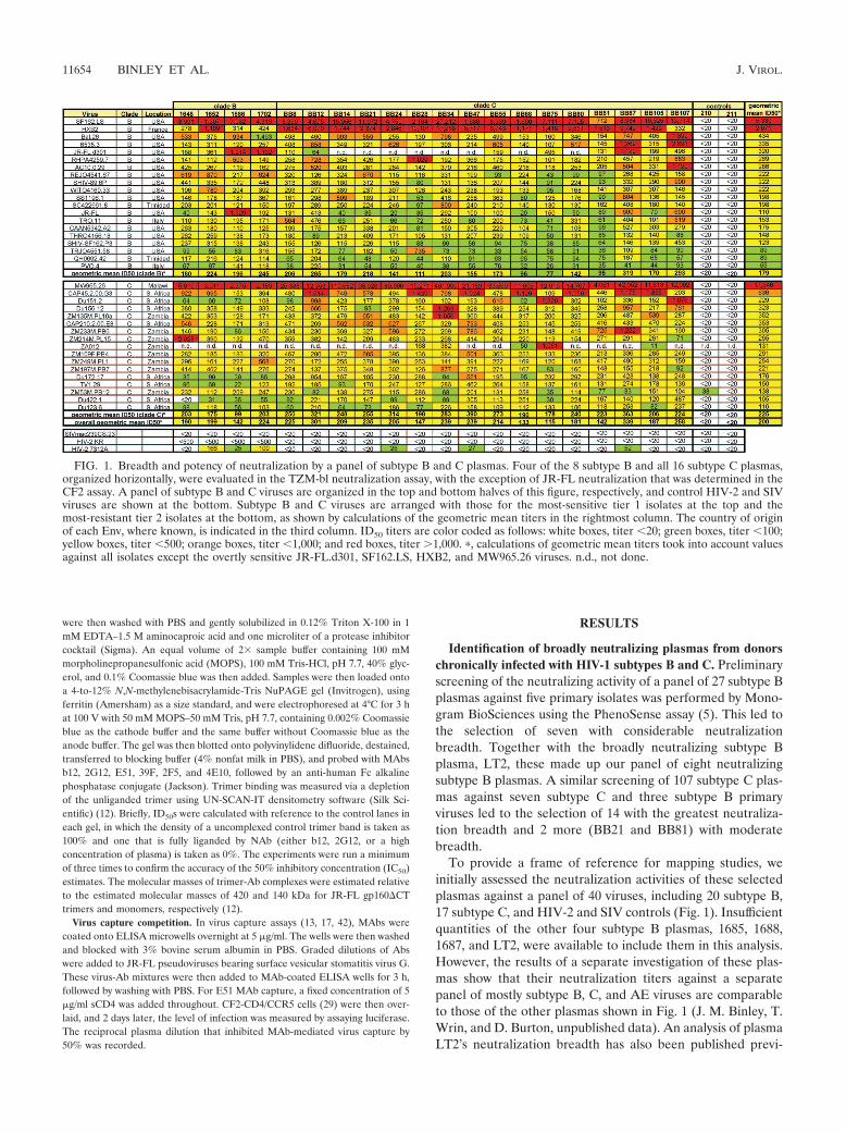
FIG. 1. Breadth and potency of neutralization by a panel of subtype B and C plasmas. Four of the 8 subtype B and all 16 subtype C plasmas,organized horizontally, were evaluated in the TZM-bl neutralization assay, with the exception of JR-FL neutralization that was determined in theCF2 assay. A panel of subtype B and C viruses are organized in the top and bottom halves of this figure, respectively, and control HIV-2 and SIVviruses are shown at the bottom. Subtype B and C viruses are arranged with those for the most-sensitive tier 1 isolates at the top and themost-resistant tier 2 isolates at the bottom, as shown by calculations of the geometric mean titers in the rightmost column. The country of originof each Env, where known, is indicated in the third column. ID50 titers are color coded as follows: white boxes, titer �20; green boxes, titer �100;yellow boxes, titer �500; orange boxes, titer �1,000; and red boxes, titer �1,000. �, calculations of geometric mean titers took into account valuesagainst all isolates except the overtly sensitive JR-FL.d301, SF162.LS, HXB2, and MW965.26 viruses. n.d., not done.
11654 BINLEY ET AL. J. VIROL.
ously (18). The panel of viruses used for the assays whoseresults are shown in Fig. 1 included those used in the mappinganalyses described below and subtype B and subtype C tier 2reference panel viruses described previously (33, 34).
Inspection of the rows in Fig. 1 reveals the inherent sensi-tivities of each virus, providing context for each value in ref-erence to the larger data set. The viruses are organized withthe most sensitive at the top of each subtype (see right-handcolumn of Fig. 1). As expected, all the HIV� plasmas effec-tively neutralized the tier 1 sensitive isolates SF162, HXB2,and MW965.26. The subtype B plasmas neutralized the d.301JR-FL virus somewhat more potently than the JR-FL parent.This mutant lacks a carbohydrate moiety that protects the V3loop in the parental version. Conversely, the subtype C plasmasneutralized these two viruses with largely equivalent titers.Subtype differences were even more apparent for neutraliza-tion of the MW965.26 isolate, which was neutralized with anaverage titer of 1:60,534 by subtype C plasmas but only 1:3,298by the subtype B plasmas. Collectively, this suggests somesubtype-based preference in the neutralization of certain iso-lates. However, this idea is complicated by the relatively potentneutralization of the subtype B isolate HXB2 and, to a lesserextent, SF162.LS by the subtype C plasmas.
The columns in Fig. 1 reveal the potency and breadth ofplasma neutralization. Importantly, all the plasmas neutralizedthe tier 2 reference viruses. The geometric mean ID50s rangedfrom 1:133 to 1:339, excluding negative controls, the d.301JR-FL mutant, and the SF162, HXB2, and MW965.26 viruses.Despite the general breadth of neutralization observed in Fig.1, varied neutralization titers against the refractive primaryisolate JR-FL were observed. For example, plasma 1686’s titerof 1:1,329 against JR-FL was significantly higher than its geo-metric mean titer of 1:142. This activity is important, as wefocus most of our attention on JR-FL in our attempts to dissectthe specificities contributing to neutralization. Although wedid not observe clear subtype-restricted patterns against tier 2viruses (8), on average, intrasubtype neutralization was slightlymore potent than intersubtype neutralization, particularly forsome subtype C plasmas. By using generalized estimatingequations to evaluate intra- versus interclade neutralization,excluding tier 1 and sensitive isolates, subtype B plasmas neu-tralized matched-subtype viruses 1.28-fold (P � 0.3) more po-tently and subtype C plasmas neutralized them 1.53-fold (P �0.015) more potently. Notably, the subtype B plasmas did notneutralize the Durban subtype C viruses as effectively as theother subtype C viruses, while subtype C plasmas did not ap-pear to discriminate between these and other subtype C Envs.However, the P values for these differences are, at best, mar-ginally significant (P � 0.3 and P � 0.02, respectively, usinggeneralized estimating equations of linear regression and fac-toring the correlation between multiple plasmas values foreach virus).
Neutralizing activity against the SIV and the HIV-2 KRcontrol viruses was undetectable, as expected. However, weakactivity against the HIV-2 7312A isolate was detected in somecases, particularly subtype B plasmas 1652 and 1702. This ac-tivity probably stems from Abs directed to a CD4i epitope thatis partially conserved between HIV-1 and HIV-2 isolates (16).
Collectively, the data shown in Fig. 1 suggest that any at-tempt to map neutralization is likely to be constrained by the
coexistence of broadly cross-reactive NAbs and NAbs withlittle cross-reactivity. However, mature anti-gp120 Ab re-sponses have been reported to be oligoclonal rather than poly-clonal (15), providing some hope regarding the practicality ofattempts to deconvolute the neutralizing activity and the fea-sibility of fine mapping of polyclonal plasma neutralization.
ELISA binding of Gp41 and gp140 by different IgG sub-classes and IgA. To profile the distribution of Env-specific Abs,we assessed the reactivities of the IgG1, IgG2, IgG3, and IgG4isotypes and IgA and IgM subclasses to a consensus gp140 Envglycoprotein. Fig. S1 in the supplemental material shows a boxplot of the relative distribution of Ab types present in plasmas.All plasmas had detectable IgG1 and IgA, but IgG2, IgG3,IgG4, and IgM were detected less frequently. The heavilybiased IgG1 profile (see Fig. S1 in the supplemental material;similar gp41 data are not shown) was perhaps consistent witha Th2-like response, typical of HIV-1 infection. There were noobservable differences in isotype or subclass responses betweenthe subtype B and C plasmas. However, the individual plasmasthat scored positive for either anti-gp140 IgG2 or IgG3 werediscordant, suggesting differences in the regulation or stimu-lation of these Abs. Anti-gp140 IgA Abs were detectable in allthe plasmas, but the relative concentration was significantlylower than the level of IgG1 (median, 3 �g/ml versus 94 �g/ml;Student’s t test, P �0.001).
Plasma fractionation using WT and CD4bs knockout mu-tant YU2 gp120 beads. For the purpose of the mapping anal-yses, we used the subtype B isolate JR-FL as a common pro-totype virus. The basis for this choice is that it has been awidely used primary isolate in the field. In addition, the gp120/gp41 cleavage site of JR-FL Env is effectively processed andtrimers are expressed efficiently, both of which are importantadvantages for the native PAGE assay described below.
To determine the contribution of gp120 Abs to JR-FL neu-tralization, plasmas from four subtype B- and four subtypeC-infected individuals were adsorbed with WT YU2 gp120covalently linked to paramagnetic beads (35). Plasmas werealso fractionated on beads coupled to a YU2 gp120 with aD368R mutation that is known to exclude the binding of mostCD4bs Abs (35, 51). The efficiencies of gp120 adsorptions wereverified by the results of WT gp120 and D368R proteinELISAs which demonstrated that each gp120 adsorbed all, ornearly all, detectable binding Abs to the corresponding gp120(data not shown). However, adsorption with the D368R mu-tant left variable amounts of WT gp120 binding activity insome plasmas. This residual gp120 binding most likely repre-sented CD4bs-specific Abs that were not adsorbed by thegp120 mutant.
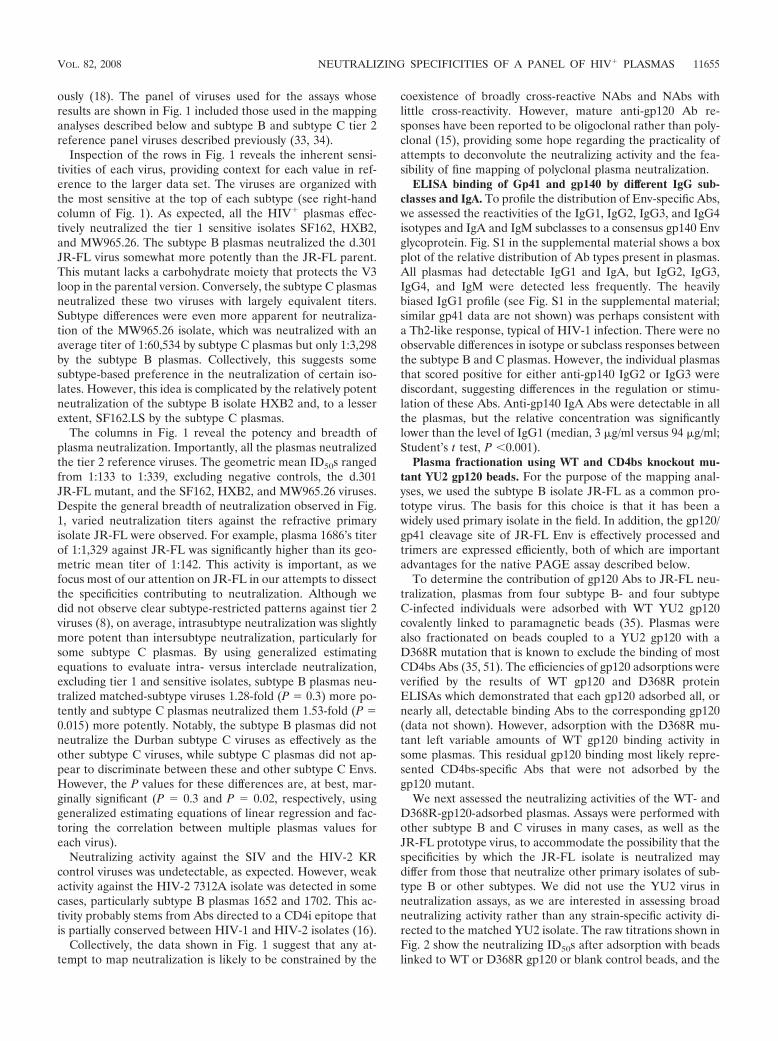
We next assessed the neutralizing activities of the WT- andD368R-gp120-adsorbed plasmas. Assays were performed withother subtype B and C viruses in many cases, as well as theJR-FL prototype virus, to accommodate the possibility that thespecificities by which the JR-FL isolate is neutralized maydiffer from those that neutralize other primary isolates of sub-type B or other subtypes. We did not use the YU2 virus inneutralization assays, as we are interested in assessing broadneutralizing activity rather than any strain-specific activity di-rected to the matched YU2 isolate. The raw titrations shown inFig. 2 show the neutralizing ID50s after adsorption with beadslinked to WT or D368R gp120 or blank control beads, and the
VOL. 82, 2008 NEUTRALIZING SPECIFICITIES OF A PANEL OF HIV� PLASMAS 11655
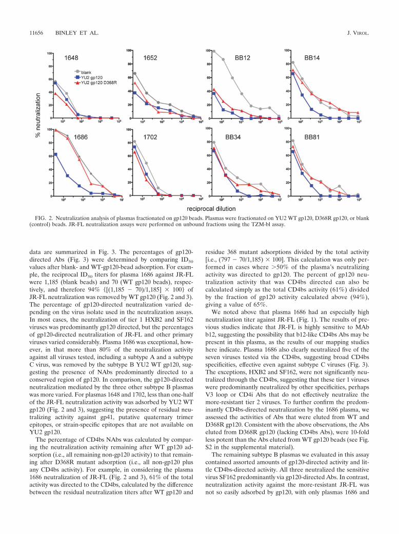
data are summarized in Fig. 3. The percentages of gp120-directed Abs (Fig. 3) were determined by comparing ID50
values after blank- and WT-gp120-bead adsorption. For exam-ple, the reciprocal ID50 titers for plasma 1686 against JR-FLwere 1,185 (blank beads) and 70 (WT gp120 beads), respec-tively, and therefore 94% {[(1,185 � 70)/1,185] � 100} ofJR-FL neutralization was removed by WT gp120 (Fig. 2 and 3).The percentage of gp120-directed neutralization varied de-pending on the virus isolate used in the neutralization assays.In most cases, the neutralization of tier 1 HXB2 and SF162viruses was predominantly gp120 directed, but the percentagesof gp120-directed neutralization of JR-FL and other primaryviruses varied considerably. Plasma 1686 was exceptional, how-ever, in that more than 80% of the neutralization activityagainst all viruses tested, including a subtype A and a subtypeC virus, was removed by the subtype B YU2 WT gp120, sug-gesting the presence of NAbs predominantly directed to aconserved region of gp120. In comparison, the gp120-directedneutralization mediated by the three other subtype B plasmaswas more varied. For plasmas 1648 and 1702, less than one-halfof the JR-FL neutralization activity was adsorbed by YU2 WTgp120 (Fig. 2 and 3), suggesting the presence of residual neu-tralizing activity against gp41, putative quaternary trimerepitopes, or strain-specific epitopes that are not available onYU2 gp120.
The percentage of CD4bs NAbs was calculated by compar-ing the neutralization activity remaining after WT gp120 ad-sorption (i.e., all remaining non-gp120 activity) to that remain-ing after D368R mutant adsorption (i.e., all non-gp120 plusany CD4bs activity). For example, in considering the plasma1686 neutralization of JR-FL (Fig. 2 and 3), 61% of the totalactivity was directed to the CD4bs, calculated by the differencebetween the residual neutralization titers after WT gp120 and
residue 368 mutant adsorptions divided by the total activity[i.e., (797 � 70/1,185) � 100]. This calculation was only per-formed in cases where �50% of the plasma’s neutralizingactivity was directed to gp120. The percent of gp120 neu-tralization activity that was CD4bs directed can also becalculated simply as the total CD4bs activity (61%) dividedby the fraction of gp120 activity calculated above (94%),giving a value of 65%.
We noted above that plasma 1686 had an especially highneutralization titer against JR-FL (Fig. 1). The results of pre-vious studies indicate that JR-FL is highly sensitive to MAbb12, suggesting the possibility that b12-like CD4bs Abs may bepresent in this plasma, as the results of our mapping studieshere indicate. Plasma 1686 also clearly neutralized five of theseven viruses tested via the CD4bs, suggesting broad CD4bsspecificities, effective even against subtype C viruses (Fig. 3).The exceptions, HXB2 and SF162, were not significantly neu-tralized through the CD4bs, suggesting that these tier 1 viruseswere predominantly neutralized by other specificities, perhapsV3 loop or CD4i Abs that do not effectively neutralize themore-resistant tier 2 viruses. To further confirm the predom-inantly CD4bs-directed neutralization by the 1686 plasma, weassessed the activities of Abs that were eluted from WT andD368R gp120. Consistent with the above observations, the Abseluted from D368R gp120 (lacking CD4bs Abs), were 10-foldless potent than the Abs eluted from WT gp120 beads (see Fig.S2 in the supplemental material).
The remaining subtype B plasmas we evaluated in this assaycontained assorted amounts of gp120-directed activity and lit-tle CD4bs-directed activity. All three neutralized the sensitivevirus SF162 predominantly via gp120-directed Abs. In contrast,neutralization activity against the more-resistant JR-FL wasnot so easily adsorbed by gp120, with only plasmas 1686 and
FIG. 2. Neutralization analysis of plasmas fractionated on gp120 beads. Plasmas were fractionated on YU2 WT gp120, D368R gp120, or blank(control) beads. JR-FL neutralization assays were performed on unbound fractions using the TZM-bl assay.
11656 BINLEY ET AL. J. VIROL.
1652 demonstrating a majority fraction of gp120-directed neu-tralization (Fig. 2 and 3).
Like the subtype B plasmas, the subtype C plasmas displayedvarious levels of gp120-directed neutralization. The subtype BYU2 WT gp120 protein beads adsorbed the majority of theneutralizing activity from plasmas BB12 and BB14 against bothsubtype B and C viruses, suggesting NAbs directed to con-served regions of gp120. However, these beads were somewhatless efficient at adsorbing the neutralizing activity from plasmasBB34 and BB81. While much of the neutralization of subtypeB viruses HXB2 and SF162.LS was gp120 directed, relativelylarge proportions of the neutralizing activities against subtypeC viruses Du151 and Du156 remained after WT gp120 adsorp-tion. Thus, the specificities in plasmas BB34 and BB81 that
mediate neutralization of these two primary C viruses remainundefined.
Fractionation of subtype C plasmas on WT and D368Rgp120 revealed mixed levels of anti-CD4bs NAbs. PlasmaBB12 contained CD4bs NAbs that were effective against thetier 1 virus HXB2 but not against the other viruses tested,including subtype C viruses (Fig. 3). It is possible that subtypeC plasmas contain CD4bs Abs that are not adsorbed by thesubtype B YU2 gp120. Further studies with subtype C gp120will be required to address this point (Gray and Morris, un-published data). As for plasma BB12, the CD4bs-neutralizingactivity in plasmas BB14, BB81, and BB34 explained a sub-stantial fraction of HXB2 virus neutralization and also ac-counted for some neutralization of primary subtype B and Cviruses. However, in all cases, this was less than 50% of thetotal activity (Fig. 3).
Evaluation of CD4bs NAbs by native PAGE. BN-PAGEusing native trimers may provide a way to evaluate NAbsdirected to quaternary epitopes (22, 27) that may be difficult todetect by other methods. We previously used this assay toinvestigate monoclonal Fab, IgG, and polyclonal sera bindingto functional WT trimers (12–14, 42). BN-PAGE exploits thetendency of hydrophobic surfaces to associate with Coomassiedye that imparts a uniform negative charge that is proportionalto the Stokes hydrodynamic radius. This allows separationunder nondenaturing conditions determined largely by the mo-lecular weight of the protein or protein complex. In previousstudies, only NAbs, and not nonneutralizing NAbs, could bindto trimers, confirming the relationship of trimer binding andneutralization (12–14, 42) suggested previously by others (56).
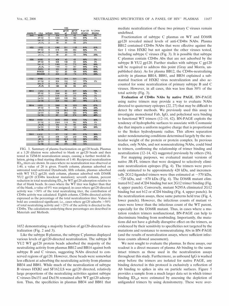
For mapping purposes, we evaluated mutant versions ofnative JR-FL trimers that were designed to selectively elimi-nate neutralization epitopes (Fig. 4). The trimers were previ-ously estimated to be approximately 420 kDa, and incremen-tally 2G12-liganded trimers were thus estimated at 570 kDa,720 kDa, and 870 kDa (Fig. 4). The D368R mutant elim-inated b12 and sCD4 binding but not 2G12 trimer binding (Fig.4, upper panels). Conversely, mutant N295A eliminated 2G12binding but not b12 or sCD4 binding (Fig. 4, upper panels). Inthe neutralization assays, these same patterns held true (Fig. 4,lower panels). However, the infectious counts of mutant vi-ruses were lower than the infectious count of the WT parent,especially for the D368R mutant. Thus, in cases where a mu-tation renders trimers nonfunctional, BN-PAGE can help todiscriminate binding from nonbinding. Importantly, the muta-tions did not have a globally disruptive effect on the trimers, asevidenced by their sensitivity to specificities not targeted by themutations and resistance to nonneutralizing Abs in BN-PAGE(and the results of neutralization assays, where sufficient infec-tious counts allowed assessment).
We next sought to evaluate the plasmas. In these assays, ourreadout is a direct measure of plasma Ab binding to the sameintact trimers as those used in the neutralization assaysthroughout this study. Furthermore, as unbound IgG is washedaway before the trimers are isolated for native PAGE, anybinding detected in this protocol is exclusively a reflection ofAb binding to spikes in situ on particle surfaces. Figure 5provides a sample from a much larger data set in which trimerbinding ID50s were estimated by assessing the depletion ofunliganded trimers by using densitometry. These were aver-
FIG. 3. Summary of plasma fractionation on gp120 beads. Plasmasat a 1:20 dilution were adsorbed to blank or gp120 beads and thentested in TZM-bl neutralization assays, causing a further twofold di-lution, giving a final starting dilution of 1:40. Reciprocal neutralizationID50 titers are shown. In cases where no neutralization was observed at1:40, a value of 20 is given. Fourth column, plasmas adsorbed onuntreated tosyl-activated Dynabeads; fifth column, plasmas adsorbedwith WT YU2 gp120; sixth column, plasmas adsorbed with D368RYU2 gp120 (CD4bs knockout mutation); seventh column, percentreduction in total neutralizing ID50 by WT gp120 adsorption relative tothat of blank beads. In cases where the WT titer was higher than thatof the blank, a value of 0% was assigned; in cases where gp120-directedactivity was �50% of the total neutralizing titer, the contribution ofCD4bs activity was calculated. Eighth column, CD4bs-directed activityexpressed as the percentage of the total neutralization titer. Values inbold are considered significant; i.e., cases where gp120 adsorbs �50%of total neutralizing activity and �25% of the activity is directed to theCD4bs. The calculations underlying these percentages are described inMaterials and Methods.
VOL. 82, 2008 NEUTRALIZING SPECIFICITIES OF A PANEL OF HIV� PLASMAS 11657
aged from at least two repeat titrations in addition to the datashown.
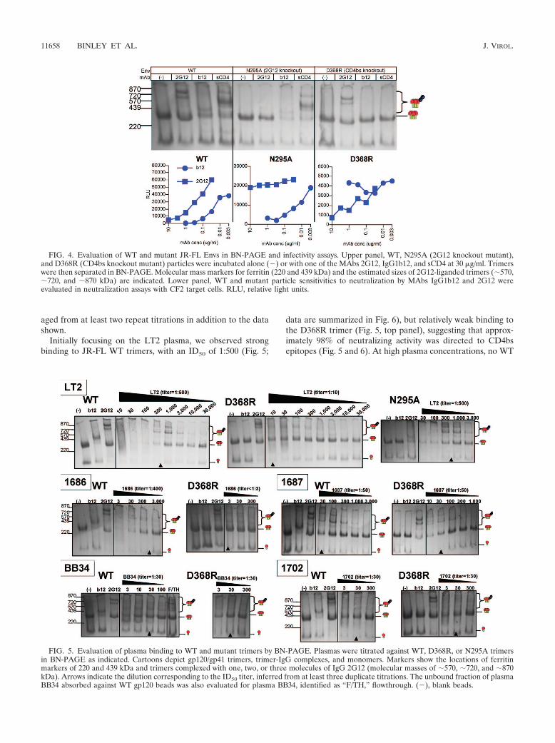
Initially focusing on the LT2 plasma, we observed strongbinding to JR-FL WT trimers, with an ID50 of 1:500 (Fig. 5;
data are summarized in Fig. 6), but relatively weak binding tothe D368R trimer (Fig. 5, top panel), suggesting that approx-imately 98% of neutralizing activity was directed to CD4bsepitopes (Fig. 5 and 6). At high plasma concentrations, no WT
FIG. 4. Evaluation of WT and mutant JR-FL Envs in BN-PAGE and infectivity assays. Upper panel, WT, N295A (2G12 knockout mutant),and D368R (CD4bs knockout mutant) particles were incubated alone (�) or with one of the MAbs 2G12, IgG1b12, and sCD4 at 30 �g/ml. Trimerswere then separated in BN-PAGE. Molecular mass markers for ferritin (220 and 439 kDa) and the estimated sizes of 2G12-liganded trimers (570,720, and 870 kDa) are indicated. Lower panel, WT and mutant particle sensitivities to neutralization by MAbs IgG1b12 and 2G12 wereevaluated in neutralization assays with CF2 target cells. RLU, relative light units.
FIG. 5. Evaluation of plasma binding to WT and mutant trimers by BN-PAGE. Plasmas were titrated against WT, D368R, or N295A trimersin BN-PAGE as indicated. Cartoons depict gp120/gp41 trimers, trimer-IgG complexes, and monomers. Markers show the locations of ferritinmarkers of 220 and 439 kDa and trimers complexed with one, two, or three molecules of IgG 2G12 (molecular masses of 570, 720, and 870kDa). Arrows indicate the dilution corresponding to the ID50 titer, inferred from at least three duplicate titrations. The unbound fraction of plasmaBB34 absorbed against WT gp120 beads was also evaluated for plasma BB34, identified as “F/TH,” flowthrough. (�), blank beads.
11658 BINLEY ET AL. J. VIROL.
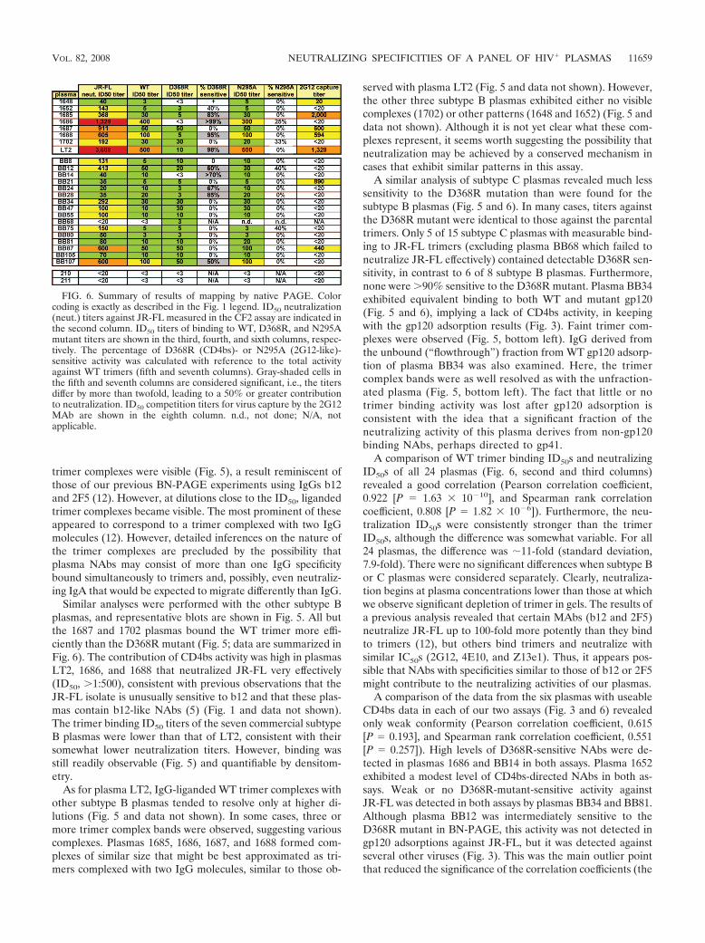
trimer complexes were visible (Fig. 5), a result reminiscent ofthose of our previous BN-PAGE experiments using IgGs b12and 2F5 (12). However, at dilutions close to the ID50, ligandedtrimer complexes became visible. The most prominent of theseappeared to correspond to a trimer complexed with two IgGmolecules (12). However, detailed inferences on the nature ofthe trimer complexes are precluded by the possibility thatplasma NAbs may consist of more than one IgG specificitybound simultaneously to trimers and, possibly, even neutraliz-ing IgA that would be expected to migrate differently than IgG.
Similar analyses were performed with the other subtype Bplasmas, and representative blots are shown in Fig. 5. All butthe 1687 and 1702 plasmas bound the WT trimer more effi-ciently than the D368R mutant (Fig. 5; data are summarized inFig. 6). The contribution of CD4bs activity was high in plasmasLT2, 1686, and 1688 that neutralized JR-FL very effectively(ID50, �1:500), consistent with previous observations that theJR-FL isolate is unusually sensitive to b12 and that these plas-mas contain b12-like NAbs (5) (Fig. 1 and data not shown).The trimer binding ID50 titers of the seven commercial subtypeB plasmas were lower than that of LT2, consistent with theirsomewhat lower neutralization titers. However, binding wasstill readily observable (Fig. 5) and quantifiable by densitom-etry.
As for plasma LT2, IgG-liganded WT trimer complexes withother subtype B plasmas tended to resolve only at higher di-lutions (Fig. 5 and data not shown). In some cases, three ormore trimer complex bands were observed, suggesting variouscomplexes. Plasmas 1685, 1686, 1687, and 1688 formed com-plexes of similar size that might be best approximated as tri-mers complexed with two IgG molecules, similar to those ob-
served with plasma LT2 (Fig. 5 and data not shown). However,the other three subtype B plasmas exhibited either no visiblecomplexes (1702) or other patterns (1648 and 1652) (Fig. 5 anddata not shown). Although it is not yet clear what these com-plexes represent, it seems worth suggesting the possibility thatneutralization may be achieved by a conserved mechanism incases that exhibit similar patterns in this assay.
A similar analysis of subtype C plasmas revealed much lesssensitivity to the D368R mutation than were found for thesubtype B plasmas (Fig. 5 and 6). In many cases, titers againstthe D368R mutant were identical to those against the parentaltrimers. Only 5 of 15 subtype C plasmas with measurable bind-ing to JR-FL trimers (excluding plasma BB68 which failed toneutralize JR-FL effectively) contained detectable D368R sen-sitivity, in contrast to 6 of 8 subtype B plasmas. Furthermore,none were �90% sensitive to the D368R mutant. Plasma BB34exhibited equivalent binding to both WT and mutant gp120(Fig. 5 and 6), implying a lack of CD4bs activity, in keepingwith the gp120 adsorption results (Fig. 3). Faint trimer com-plexes were observed (Fig. 5, bottom left). IgG derived fromthe unbound (“flowthrough”) fraction from WT gp120 adsorp-tion of plasma BB34 was also examined. Here, the trimercomplex bands were as well resolved as with the unfraction-ated plasma (Fig. 5, bottom left). The fact that little or notrimer binding activity was lost after gp120 adsorption isconsistent with the idea that a significant fraction of theneutralizing activity of this plasma derives from non-gp120binding NAbs, perhaps directed to gp41.
A comparison of WT trimer binding ID50s and neutralizingID50s of all 24 plasmas (Fig. 6, second and third columns)revealed a good correlation (Pearson correlation coefficient,0.922 [P � 1.63 � 10�10], and Spearman rank correlationcoefficient, 0.808 [P � 1.82 � 10�6]). Furthermore, the neu-tralization ID50s were consistently stronger than the trimerID50s, although the difference was somewhat variable. For all24 plasmas, the difference was 11-fold (standard deviation,7.9-fold). There were no significant differences when subtype Bor C plasmas were considered separately. Clearly, neutraliza-tion begins at plasma concentrations lower than those at whichwe observe significant depletion of trimer in gels. The results ofa previous analysis revealed that certain MAbs (b12 and 2F5)neutralize JR-FL up to 100-fold more potently than they bindto trimers (12), but others bind trimers and neutralize withsimilar IC50s (2G12, 4E10, and Z13e1). Thus, it appears pos-sible that NAbs with specificities similar to those of b12 or 2F5might contribute to the neutralizing activities of our plasmas.
A comparison of the data from the six plasmas with useableCD4bs data in each of our two assays (Fig. 3 and 6) revealedonly weak conformity (Pearson correlation coefficient, 0.615[P � 0.193], and Spearman rank correlation coefficient, 0.551[P � 0.257]). High levels of D368R-sensitive NAbs were de-tected in plasmas 1686 and BB14 in both assays. Plasma 1652exhibited a modest level of CD4bs-directed NAbs in both as-says. Weak or no D368R-mutant-sensitive activity againstJR-FL was detected in both assays by plasmas BB34 and BB81.Although plasma BB12 was intermediately sensitive to theD368R mutant in BN-PAGE, this activity was not detected ingp120 adsorptions against JR-FL, but it was detected againstseveral other viruses (Fig. 3). This was the main outlier pointthat reduced the significance of the correlation coefficients (the
FIG. 6. Summary of results of mapping by native PAGE. Colorcoding is exactly as described in the Fig. 1 legend. ID50 neutralization(neut.) titers against JR-FL measured in the CF2 assay are indicated inthe second column. ID50 titers of binding to WT, D368R, and N295Amutant titers are shown in the third, fourth, and sixth columns, respec-tively. The percentage of D368R (CD4bs)- or N295A (2G12-like)-sensitive activity was calculated with reference to the total activityagainst WT trimers (fifth and seventh columns). Gray-shaded cells inthe fifth and seventh columns are considered significant, i.e., the titersdiffer by more than twofold, leading to a 50% or greater contributionto neutralization. ID50 competition titers for virus capture by the 2G12MAb are shown in the eighth column. n.d., not done; N/A, notapplicable.
VOL. 82, 2008 NEUTRALIZING SPECIFICITIES OF A PANEL OF HIV� PLASMAS 11659
other, less-significant outlier was plasma BB81). One possibleexplanation for the discrepancy is that the D368R mutation ofmonomeric gp120 and trimers may have slightly different con-sequences for NAb binding. In addition, the use of mismatchedEnvs (YU2 and JR-FL) in these two methods might also havecontributed to the somewhat-different outcomes. The discrep-ancy is, however, unlikely to be a native PAGE artifact, since,as mentioned above, the protocol exclusively measures bindingto intact trimers in situ on particle membranes, so the resultscan be directly cross-referenced with neutralization data.
Mapping of 2G12-like Abs by native PAGE and virus cap-ture competition. MAb 2G12 does not compete with any otherknown MAbs directed to gp120 or gp41 (41). Therefore, com-petitive binding assays may be useful in assessing 2G12-likeactivity, even when the readout is not associated with neutral-ization (7, 17). In competitive virus capture assays on 2G12-coated plates, some of the subtype B plasmas (1648, 1686,1687, 1702, and LT2) and two of the subtype C samples (BB21and BB87) were found to inhibit virus capture (Fig. 6) butcontrol HIV-negative plasmas were not (not shown).
Another way to examine 2G12-like Abs is by BN-PAGEshift assays using N295A mutant trimers. In many cases, in-cluding plasma LT2, the titers against the 295 mutant werevery similar to those against the WT parent (Fig. 5 and 6),suggesting that few, if any, 2G12-like NAbs were present, evenin those plasmas that competed with 2G12 in virus capture(Fig. 5 and 6). Overall, these results suggest that a fraction ofpatients develop binding Abs that overlap the 2G12 epitope,but unlike 2G12, these Abs tend not to neutralize the virus (7).
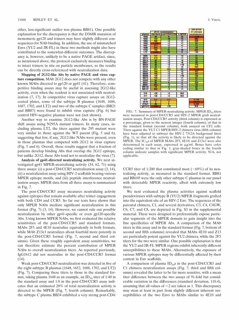
Analysis of gp41-directed neutralizing activity. We next in-vestigated gp41 MPER-neutralizing activity (54, 62, 71) usingthree assays: (i) a post-CD4/CCR5 neutralization assay (3, 14),(ii) a neutralization assay using HIV-2 scaffolds bearing variousMPER epitope motifs, and (iii) peptide interference neutral-ization assays. MPER data from all three assays is summarizedin Fig. 7.
The post-CD4/CCR5 assay measures neutralizing activityagainst epitopes that remain available after trimer engagementwith both CD4 and CCR5. So far our tests have shown thatonly MPER NAbs mediate significant neutralization in thisformat (Fig. 7) (3, 14). However, we cannot rule out possibleneutralization by other gp41-specific or even gp120-specificAbs. Using known MPER NAbs, we first evaluated the relativesensitivities of the post-CD4/CCR5 and standard formats.MAbs 2F5 and 4E10 neutralize equivalently in both formats,while MAb Z13e1 neutralizes about fourfold more potently inthe post-CD4/CCR5 format (Fig. 7, second and third col-umns). Given these roughly equivalent assay sensitivities, wecan therefore estimate the percent contribution of MPERNAbs to overall neutralization titers. As reported previously,IgG1b12 did not neutralize in the post-CD4/CCR5 format(14).
Weak post-CD4/CCR5 neutralization was detected in five ofthe eight subtype B plasmas (1648, 1652, 1688, 1702, and LT2)(Fig. 7). Comparing these titers to those in the standard for-mat, taking plasma 1648 as an example, an ID50 titer of 1:40 inthe standard assay and 1:8 in the post-CD4/CCR5 assay indi-cates that an estimated 20% of total neutralization activity isdirected to the MPER (Fig. 7, fourth column). Remarkably,the subtype C plasma BB34 exhibited a very strong post-CD4/
CCR5 titer of 1:200 that constituted most (69%) of its neu-tralizing activity, as measured in the standard format. BB81and BB105 were the only other subtype C plasmas in our panelwith detectable MPER reactivity, albeit with extremely lowtiters.
We next evaluated the plasma activities against scaffoldpseudoviruses with subtype B (YU2) MPER fragments graftedinto the equivalent site of an HIV-2 Env. The sequences of theparental chimera, C1, and several derivatives, C3, C4, C4GW,C6, C7, and C8, are depicted in Fig. S3 in the supplementalmaterial. These were designed to preferentially expose partic-ular segments of the MPER domain to gain insight into thefine specificities of MPER Abs. A comparison of the MAbtiters in this assay and in the standard format (Fig. 7, bottom ofsecond and fifth columns) revealed that MAbs 4E10 and Z13are particularly potent against the YU2 chimera, while the 2F5titers for the two were similar. One possible explanation is thatthe YU2 and JR-FL MPER regions exhibit inherently differentsusceptibilities to these MAbs. Alternatively, the exposure ofvarious MPER epitopes may be differentially affected by theircontext in Env scaffolds.
A comparison of plasma ID50s in the post-CD4/CCR5 andC1 chimera neutralization assays (Fig. 7, third and fifth col-umns) revealed the latter to be far more sensitive, with a meantiter difference between the two assays of 91-fold but consid-erable variation in the differences (standard deviation, 110.4),assuming that all values of �2 are taken as 1. This discrepancymay stem at least in part from slightly different inherent sus-ceptibilities of the two Envs to MAbs similar to 4E10 and
FIG. 7. Summary of MPER-neutralizing activity. MPER ID50 titerswere measured in post-CD4/CCR5 and HIV-2 MPER graft neutral-ization assays. Post-CD4/CCR5 activity (third column) is expressed asa percentage, given to the nearest integer (fourth column), of that inthe standard format (second column), both assayed on CF2 cells.Titers against the YU2 C1 MPER/HIV-2 chimera virus (fifth column)have been adjusted to subtract the HIV-2 7312A background titers(Fig. 1), so that all the activity is likely to be directed against theMPER. The IC50s of MPER MAbs 2F5, 4E10, and Z13e1 were alsodetermined in each assay, expressed in �g/ml. Boxes have colorcoding similar to that in Fig. 1; gray-shaded boxes in the fourthcolumn indicate samples with significant MPER activity. N/A, notapplicable.
11660 BINLEY ET AL. J. VIROL.

Z13e1, as discussed above. Nevertheless, some weak confor-mity was observed between the results of the two assays, asjudged by a Kendall’s rank correlation of 0.36 (P � 0.026) anda Spearman’s rank correlation of 0.44 (P � 0.031) (in which allvalues of �2 were ranked equally). The C1 chimera neutral-ization titers were particularly strong with plasmas 1648, 1652,1702, and BB34 (all ID50s were �1:500), all of which also haddetectable activity in the post-CD4/CCR5 assay. However,MPER activity against the C1 chimera was undetectable inplasma LT2, consistent with the results of a previous reportusing the same assay (18) but inconsistent with the low butnevertheless detectable MPER activity in the post-CD4/CCR5assay. It is possible that the activity in this plasma is directedagainst an N-terminal MPER epitope like 2F5 that is detectedrelatively inefficiently by the C1 chimeras.
We next inspected the activities of the subtype B plasmasagainst the various MPER-engrafted mutant viruses (depictedin Fig. S3 in the supplemental material) with reference to theactivities of MPER MAbs 2F5, 4E10, and Z13e1. The results ofthis analysis are shown in Fig. S4 in the supplemental material.Each MAb prototype exhibited a different pattern of activityagainst this panel of chimeras. MAb 2F5 neutralized the C3and C7 chimeras; 4E10 neutralized the C4, C4GW, C6, and C8chimeras; and Z13e1 neutralized only the C4GW and C8 chi-meras. The subtype B plasmas exhibited various neutralizationpatterns that in no case exactly recapitulated those of any oneof the prototypes. This may suggest the coexistence of multipleknown MPER specificities and/or new specificities unlike anyof the prototype MAbs. Despite the overall complexity of theplasma MPER responses, the potent activities of plasmas 1648and 1702 against the C4GW mutant suggest Z13-like NAbs,whereas the activity of 1652 against C4 and C6 suggests 4E10-like NAbs. We did not address the fine mapping of the subtypeC plasmas here, as this will form part of a separate study (Grayand Morris, unpublished).
We further assessed MPER activity in neutralization inter-ference assays using the peptides depicted in Fig. S3 in thesupplemental material. The full analysis is shown in Fig. S5,and essential data are summarized in the 10th column of Fig.S4, both of which are in the supplemental material. TheMPR.03 peptide covers the entire MPER domain, and theHXB2.2F5.01 and 4E10.22 peptides cover 2F5- and 4E10-likespecificities, respectively (see Fig. S3 in the supplemental ma-terial). Peptides designed for Z13e1 adsorption markedly af-fected virus infection and were therefore not investigated fur-ther. We first validated the peptides using known MPERMAbs. MPR.03 effectively blocked the neutralizing activities of2F5, 4E10, and Z13e1 against the 7312A C1 chimeric virus,demonstrating its ability to capture all known MPER Abs. The4E10.22 peptide blocked 4E10 and Z13e1 but not 2F5 neutral-ization, whereas the HXB2 2F5.01 peptide inhibited 2F5 butnot 4E10 or Z13e1 neutralization (see Fig. S5 in the supple-mental material).
Having established the peptide specificities, we next assessedtheir effects, if any, on subtype B plasma neutralization. Thedata are shown in Fig. S5 in the supplemental material; twofoldor greater changes in ID50 indicate that more than one-half ofthe NAb titer was blocked. For all four plasmas, competitionwith the MPR.03 peptide reduced JR-FL neutralization bytwofold or less (Fig. S5 in the supplemental material), consis-
tent with their relatively weak or absent activity in post-CD4/CCR5 assays (Fig. 7, third column) in proportion to the totalneutralization activity in each case.
We next assessed peptide interference in neutralization as-says with the MPER-grafted HIV-2 7312A C1 chimera thatshould only be susceptible to MPER-directed NAbs. Neutral-ization by all four plasmas was substantially inhibited by theMPR.03 and 4E10 peptides but not by the 2F5 peptide (seeFig. S5 in the supplemental material; data are summarized inFig. S4, 10th column, in the supplemental material), confirm-ing the presence of Z13e1 and/or 4E10-like Abs. This wasconsistent with the strong activity detected with three out offour of these plasmas against the C8 chimera, which encom-passes a 3 part of the MPER region similar to the 4E10.22peptide and can detect 4E10- and Z13-like activity. The excep-tion was plasma 1686, which had somewhat lower but detect-able anti-C8 activity that could conceivably contribute to theC1 mutant neutralization observed in the peptide interferenceassays in this study.
Peptide interference of HXB2 neutralization was weakerand only reached significance for plasma 1686. This may reflectthe sensitivity of HXB2 to neutralization by specificities that donot recognize the 7312A chimera, thereby emasculating thepotency of MPER peptides in neutralization interference.
Summarizing the results for MPER activities, we detectedlow titers of post-CD4/CCR5 NAbs against JR-FL in almost allcases, the specificities of which appeared to be quite complexand variable. The results of peptide interference assays sug-gested the presence of Z13- or 4E10-like rather than 2F5-likeactivity in four of the subtype B plasmas.
Neutralizing activity directed against the V3 loop. There arereasonable arguments for and against a role for V3 Abs inbroad plasma neutralization. While the V3 loop is largely oc-cluded on primary isolate trimers such that V3 Abs tend toneutralize primary isolates rather weakly (6), the high titers ofV3 Abs often observed in natural infection could conceivablyfactor into overall neutralization titers (40). Furthermore, al-though V3 loop Abs are classically considered to be highlystrain specific, certain V3 Abs react with more conserved ele-ments and, therefore, may in some cases be capable of cross-neutralizing activity (45, 70).
Three approaches were used to measure V3 loop activity inour plasma panel. One was to measure the abilities of plasmasto inhibit the capture of JR-FL virus particles by a V3 MAb,39F. Subtype B plasmas exhibited modest titers in this assay(Fig. 8, third column) and lower activities were found in thesubtype C plasmas, suggesting subtype-specific V3 loop bind-ing. A caveat regarding this assay is that capture may beblocked by non-V3 specificities, as well as V3 Abs. Therefore,in a second assay, we evaluated V3 reactivity using a chimericHIV-2 virus engrafted with the HIV-1 YU2 or subtype Cconsensus V3 loop in place of the equivalent parental sequence(see Fig. S6 in the supplemental material) (Davis et al., sub-mitted). High titers of V3 loop Abs were observed in mostcases (Fig. 8, fourth and fifth columns). The V3 reactivity wasagain found to be somewhat subtype restricted, so that plasmasbetter neutralized the matched chimera, as reflected by thebehavior of subtype B patient-derived MAbs 447-52D andF425. However, the subtype C plasmas showed higher overallV3 reactivity in this assay, in many cases giving higher titers
VOL. 82, 2008 NEUTRALIZING SPECIFICITIES OF A PANEL OF HIV� PLASMAS 11661

than the subtype B plasmas against the subtype B chimera.This finding contrasted with the relatively weak ability of sub-type C plasmas to block the capture of the subtype B isolateJR-FL by a V3 MAb. The underlying reason for this discrep-ancy is not clear but could be related to the different contextsin which the V3 sequences are presented (Fig. 8, comparebottom and top halves of third and fourth columns).
A caveat regarding this second assay is that the chimeras areacutely sensitive to the V3 MAbs 447-52D and F425 fromsubtype B donors (Fig. 8). Therefore, in a third approach toinvestigate the significance of the V3 activity, we evaluated theabilities of V3 peptides to interfere with the neutralization ofseveral HIV-1 isolates, including several primary isolates (Fig.8, 6th to 14th columns). Two types of V3 peptides were syn-thesized, matching each virus (see Fig. S6 in the supplementalmaterial). The V3.01 series were derived from the N-terminalpart of the loop, including the crown, and the V3.02 series werederived from the C-terminal part of the loop. Using MAb447-52D and the V3-sensitive d.301 derivative of JR-FL, wefound that the V3.01 peptide potently inhibited neutralizationbut the V3.02 peptide had no effect. The V3.01 peptide alsoblocked BaL.26 and SF162.LS neutralization by a panel of sixother V3 MAbs (data not shown), indicating that V3 Abs arecommonly directed to the N-terminal part of the V3 loop. Inthe examination of the subtype B plasmas, neither peptide con-vincingly interfered with WT JR-FL neutralization (changes ofless than twofold) (Fig. 8), consistent with the general V3 re-sistance of this primary isolate. However, the V3.01 peptidestrongly inhibited neutralization by plasma 1702 against d.301JR-FL (a 5.5-fold decrease in ID50 titer) and had some effect(1.9-fold) on plasma 1648. A twofold effect on the ID50 titerindicates that 50% of virus neutralization can be assigned tothe V3 loop. JR-FL d.301 neutralization by the remaining
subtype B plasmas was not significantly affected by V3 pep-tides.
We next evaluated V3 peptide interference against the sub-type C virus TV1. TV1 is comparable to JR-FL in terms ofoverall neutralization sensitivity (Fig. 1), but the results ofprevious studies have indicated that it is partially sensitive toV3 neutralization (66). In contrast to JR-FL d.301, the V3.02peptide rather than the V3.01 peptide blocked a substantialportion of subtype B plasma neutralization against TV1 (Fig. 8and 9). Therefore, cross-subtype Abs targeted to the C-termi-nal portion of the V3 loop contributed markedly to TV1 neu-tralization. Thus, for plasma 1686, the highly potent CD4bsactivity found to be effective against JR-FL was relatively in-effective against TV1 (1:22 for TV1 versus 1:1,329 for JR-FL)(Fig. 1), which instead appears to be neutralized in large partby NAbs directed to the C-terminal portion of the V3 loop. Incontrast to TV1, neutralization of the sensitive MW956 sub-type C isolate could not be inhibited by either peptide (Fig. 8).This is probably because MW965 is highly sensitive to mostknown MAbs, and non-V3 specificities may therefore dictatethe neutralization of this tier 1 isolate.
We next investigated V3 peptide interference in neutraliza-tion by the subtype C plasmas. V3 peptides were unable tointerfere with either JR-FL or its sensitive d.301 derivative,suggesting that cross-subtype neutralization of these isolates isnot mediated by V3-specific NAbs. As with the subtype Bplasmas, TV1 neutralization by subtype C plasmas was sensi-tive to adsorption by V3 peptides. Here, however, either orboth of the peptides V3.01 and V3.02 were effective, as exem-plified by the results for plasmas BB21 and BB68 shown in Fig.9. Therefore, neutralization was not restricted to the V3 Cterminus as with the subtype B plasmas (Fig. 8). In contrast,neutralization of the MW956 isolate was blocked by the V3.01
FIG. 8. Summary of V3 activity. V3 activity was measured by (i) competition of virus capture by a V3 MAb 39F (third column), (ii)neutralization of the HIV-2 KR virus engrafted with either the YU2 or a consensus subtype C V3 loop (fourth and fifth columns), and (iii) bypeptide interference assays (6th to 14th columns). Virus capture and chimera neutralization data are given as ID50 titers. Peptide interference wasrecorded as the severalfold change in ID50 titer with reference to the titer of controls to which no peptide was added. Virus capture and chimeraneutralization assay data are color coded as described in the Fig. 1 legend. Boxes depicting the results of peptide interference experiments in whichsignificant competition was observed (more-than-twofold effect) are shaded gray; results indicating no significant competition (less-than-twofoldeffect) are unshaded. Controls included V3 MAbs 447-52D, F425, and guinea pig serum WGP 102-4 (66). n.d., not done.
11662 BINLEY ET AL. J. VIROL.
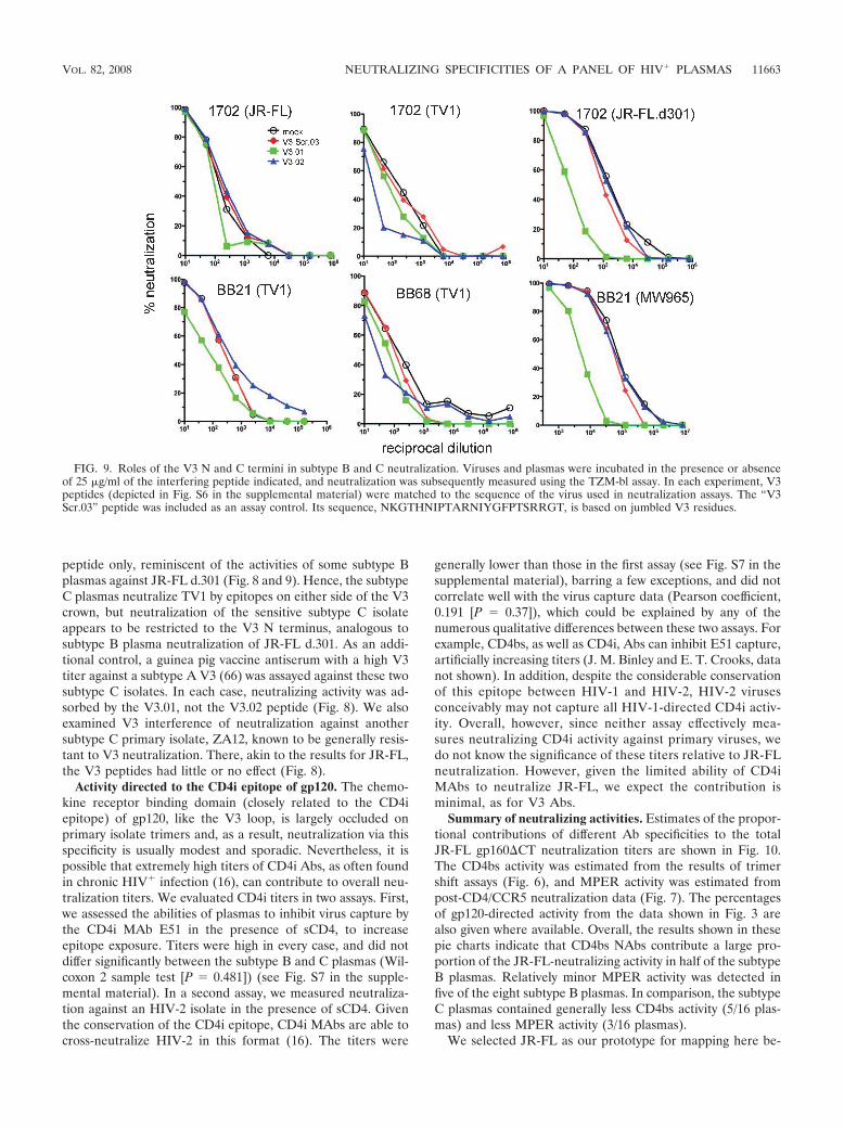
peptide only, reminiscent of the activities of some subtype Bplasmas against JR-FL d.301 (Fig. 8 and 9). Hence, the subtypeC plasmas neutralize TV1 by epitopes on either side of the V3crown, but neutralization of the sensitive subtype C isolateappears to be restricted to the V3 N terminus, analogous tosubtype B plasma neutralization of JR-FL d.301. As an addi-tional control, a guinea pig vaccine antiserum with a high V3titer against a subtype A V3 (66) was assayed against these twosubtype C isolates. In each case, neutralizing activity was ad-sorbed by the V3.01, not the V3.02 peptide (Fig. 8). We alsoexamined V3 interference of neutralization against anothersubtype C primary isolate, ZA12, known to be generally resis-tant to V3 neutralization. There, akin to the results for JR-FL,the V3 peptides had little or no effect (Fig. 8).
Activity directed to the CD4i epitope of gp120. The chemo-kine receptor binding domain (closely related to the CD4iepitope) of gp120, like the V3 loop, is largely occluded onprimary isolate trimers and, as a result, neutralization via thisspecificity is usually modest and sporadic. Nevertheless, it ispossible that extremely high titers of CD4i Abs, as often foundin chronic HIV� infection (16), can contribute to overall neu-tralization titers. We evaluated CD4i titers in two assays. First,we assessed the abilities of plasmas to inhibit virus capture bythe CD4i MAb E51 in the presence of sCD4, to increaseepitope exposure. Titers were high in every case, and did notdiffer significantly between the subtype B and C plasmas (Wil-coxon 2 sample test [P � 0.481]) (see Fig. S7 in the supple-mental material). In a second assay, we measured neutraliza-tion against an HIV-2 isolate in the presence of sCD4. Giventhe conservation of the CD4i epitope, CD4i MAbs are able tocross-neutralize HIV-2 in this format (16). The titers were
generally lower than those in the first assay (see Fig. S7 in thesupplemental material), barring a few exceptions, and did notcorrelate well with the virus capture data (Pearson coefficient,0.191 [P � 0.37]), which could be explained by any of thenumerous qualitative differences between these two assays. Forexample, CD4bs, as well as CD4i, Abs can inhibit E51 capture,artificially increasing titers (J. M. Binley and E. T. Crooks, datanot shown). In addition, despite the considerable conservationof this epitope between HIV-1 and HIV-2, HIV-2 virusesconceivably may not capture all HIV-1-directed CD4i activ-ity. Overall, however, since neither assay effectively mea-sures neutralizing CD4i activity against primary viruses, wedo not know the significance of these titers relative to JR-FLneutralization. However, given the limited ability of CD4iMAbs to neutralize JR-FL, we expect the contribution isminimal, as for V3 Abs.
Summary of neutralizing activities. Estimates of the propor-tional contributions of different Ab specificities to the totalJR-FL gp160�CT neutralization titers are shown in Fig. 10.The CD4bs activity was estimated from the results of trimershift assays (Fig. 6), and MPER activity was estimated frompost-CD4/CCR5 neutralization data (Fig. 7). The percentagesof gp120-directed activity from the data shown in Fig. 3 arealso given where available. Overall, the results shown in thesepie charts indicate that CD4bs NAbs contribute a large pro-portion of the JR-FL-neutralizing activity in half of the subtypeB plasmas. Relatively minor MPER activity was detected infive of the eight subtype B plasmas. In comparison, the subtypeC plasmas contained generally less CD4bs activity (5/16 plas-mas) and less MPER activity (3/16 plasmas).
We selected JR-FL as our prototype for mapping here be-
FIG. 9. Roles of the V3 N and C termini in subtype B and C neutralization. Viruses and plasmas were incubated in the presence or absenceof 25 �g/ml of the interfering peptide indicated, and neutralization was subsequently measured using the TZM-bl assay. In each experiment, V3peptides (depicted in Fig. S6 in the supplemental material) were matched to the sequence of the virus used in neutralization assays. The “V3Scr.03” peptide was included as an assay control. Its sequence, NKGTHNIPTARNIYGFPTSRRGT, is based on jumbled V3 residues.
VOL. 82, 2008 NEUTRALIZING SPECIFICITIES OF A PANEL OF HIV� PLASMAS 11663
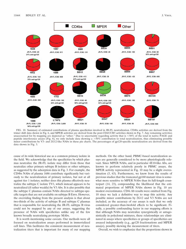
cause of its wide historical use as a common primary isolate inthe field. We acknowledge that the specificities by which plas-mas neutralize the JR-FL isolate may differ from those thatneutralize other primary subtype B isolates or other subtypes,as suggested by the adsorption data in Fig. 3. For example, theCD4bs NAbs of plasma 1686 contribute significantly but vari-ously to the neutralization of primary isolates, but not at allagainst tier 1 isolates; neither does this plasma effectively neu-tralize the subtype C isolate TV1, which instead appears to beneutralized (if rather weakly) by V3 Abs. It is also possible thatthe subtype C plasmas contain NAbs directed to subtype-spe-cific targets that are not available on subtype B Envs. However,the overriding finding from the present analysis is that abouttwo-thirds of the activity of subtype B and subtype C plasmasthat is responsible for neutralizing the JR-FL subtype B viruscould not be mapped by any of our assays, suggesting thepresence of NAbs with specificities unlike any of the fewknown broadly neutralizing prototype MAbs.
It is worth mentioning some caveats. Our methods were allfocused on neutralization assays performed in immortalizedcell lines. This facilitates the consistent measurement of neu-tralization titers that is important for many of our mapping
methods. On the other hand, PBMC-based neutralization as-says are generally considered to be more physiologically rele-vant. Since MPER NAbs, and in particular 4E10-like Abs, areknown to perform relatively poorly in PBMC assays, theMPER activity represented in Fig. 10 may be a slight overes-timation (5, 63). Furthermore, we know from the results ofprevious studies that the truncated gp160 mutant virus is some-what more sensitive to MPER NAbs than its full-length coun-terpart (14, 23), compounding the likelihood that the esti-mated proportions of MPER NAbs shown in Fig. 10 aremodest overestimates. CD4i Ab results were omitted from Fig.10 since we lack a definitive way to map this activity. Anypossible minor contributions by 2G12 and V3 NAbs are notincluded, as the accuracy of our assays is such that we onlyconsidered greater-than-twofold effects to be significant. Fi-nally, a possible confounding factor in our mapping efforts isthat although NAbs may neutralize synergistically or antago-nistically in polyclonal mixtures, these relationships are elimi-nated in assays where specificities or groups of specificities areassayed independently (e.g., gp120 fractionations and MPERassays), possibly skewing the measurement of titers.
Overall, we wish to emphasize that the proportions shown in
FIG. 10. Summary of estimated contributions of plasma specificities involved in JR-FL neutralization. CD4bs activities are derived from thetrimer shift data shown in Fig. 6, and MPER activities are derived from the post-CD4/CCR5 activities shown in Fig. 7. Any remaining activitiesunaccounted for by mapping are depicted as “other.” Due to uncertainty regarding activity that is �50% of the total in native PAGE andpeptide interference assays (Fig. 8), we only include data showing a �50% contribution to total neutralization, thus eliminating possibleminor contributions by V3- and 2G12-like NAbs in these pie charts. The percentages of gp120-specific neutralization are derived from thedata shown in Fig. 3.
11664 BINLEY ET AL. J. VIROL.
Fig. 10 are estimates influenced both by the accuracy of eachassay (although most assays were repeated several times toensure consistency) and by interassay protocol differences. Aclear example of the limits of our estimates is the apparentcontradiction that approximately 70% of the neutralizing ac-tivity of plasma BB34 apportioned to the gp41 MPER in thepost-CD4/CCR5 assay but 53% was allocated to gp120 in ad-sorption experiments. We interpret this by suggesting that ap-proximately half the activity against JR-FL is directed againstMPER and half against gp120.
DISCUSSION
We attempted to define the basis of broad neutralization ina collection of subtype B and C HIV-1 plasmas. We intendedto illuminate the relative importance of various known neu-tralizing epitopes as potential targets for vaccine development,as well as to provide insights into possible novel neutralizingepitopes. This is the largest and most-comprehensive data setof its kind to be generated to date. The results of the initialgp120 adsorption experiments (Fig. 2 and 3) revealed thatgp120 activity accounts for a significant but variable fraction ofthe neutralization, depending on the virus isolate. For someplasmas, broad neutralization was mediated by anti-gp120 Abs,and in some cases, CD4bs-directed NAbs. In these cases, neu-tralizing activity against gp41 or quaternary epitopes may playa relatively small role in virus neutralization. However, a largefraction of the neutralizing activity in many plasmas could notbe ascribed to Abs directed toward any of the known epitopes.This was particularly true for the subtype C plasmas. It ispossible that antigenic differences in subtype B and C Envstranslate into differences in the specificities of the broadlycross-reactive neutralizing activity that appears later in infec-tion (43, 52). For example, subtype C plasmas may containCD4bs NAb activity that is generally insensitive to the D368Rmutation, unlike subtype B plasmas. We therefore tested aD368R E370A double mutant in native PAGE that might beexpected to more completely knock out the CD4bs, but this didnot reveal any additional CD4bs activity beyond that revealedby the D368R single mutant (not shown). We are now evalu-ating the effects of subtype-specific D368R mutations in sub-type C Envs in gp120 adsorptions and trimer binding assays.
We considered activity directed to the C3/V4 domain as onepossible explanation for the uncharacterized subtype C activ-ity. The C3/V4 domain of subtype C Envs has been implicatedin autologous neutralization of subtype C viruses followingseroconversion (43, 52), but given the variability of this regionin subtype C Envs, it was not clear if these NAbs might becomecross-reactive during chronic infection. The results of prelim-inary studies indicated that Env-matched C3 peptides (see Fig.S6 in the supplemental material) did not interfere with subtypeC neutralization in any case (data not shown), suggesting thatC3 epitopes are not sufficiently conserved to be targets ofbroad neutralization activity. However, because we lack effec-tive control NAbs directed to the C3 region, we have no way toensure that our peptides can effectively adsorb C3 NAb activityper se.
The general conformity of the data from two assays designedto detect CD4bs NAbs reinforces and extends the findings ofprevious studies showing that CD4bs Abs are the only speci-
ficity defined to date that constitutes a significant portion ofplasma NAb activity in broadly neutralizing plasmas (18, 35).The contributions of CD4bs Abs to the neutralization of dif-ferent viruses also varied considerably. This is exemplified bythe behavior of plasma 1686 (Fig. 3), in which CD4bs activityis responsible for the neutralization of tier 2 but not subtype Btier 1 isolates, where the effect of V3 Abs probably eclipses theeffect of CD4bs NAbs. Therefore, the evaluation of any NAbspecificity’s contribution to neutralization should be carefullyconsidered in context with other Abs that may also neutralizea given virus isolate.
We observed several cases in which plasmas were unusuallypotent against a particular tier 2 virus (Fig. 1). This may stemfrom the acute sensitivity of certain viruses to the predominantcross-neutralizing activity, which in some cases may be medi-ated by CD4bs NAbs (e.g., the potent neutralization of JR-FLby plasma 1686). However, there are other cases where theplasma neutralization specificity remains largely unidentified.For example, plasma BB75 neutralized viruses Du123 andZA12 with ID50s of 9,320 and 1,081, respectively, much higherthan its titers of 1:100 against most other viruses (Fig. 1). Itseems likely that this arises from virus sensitivity to a particularspecificity, which in these cases remains unknown.
Of the remaining known neutralizing specificities that mightexplain broad plasma neutralization, little evidence was foundfor 2G12-like NAbs. This is perhaps not entirely surprising,given that 2G12 exhibits a highly unusual domain swap con-figuration and targets a carbohydrate epitope that would beexpected to be regulated by tolerance. Likewise, very little Abagainst the gp41 MPER was detected in most cases. This latterfinding may be consistent with the hypothesis that certainMPER Abs are regulated by tolerance due to their potentialautoreactive capacity (25).
Plasma BB34 was exceptional in that it exhibited an ex-tremely high titer of MPER-specific NAbs. The observed titerof 1:200 would require an estimated 67 �g/ml of 2F5, 600�g/ml of 4E10, or 960 �g/ml of Z13e1. If the MPER NAb(s) inBB34 is similar to the latter two prototypes, then they wouldlikely constitute an overwhelmingly dominant fraction of theoverall anti-Env Ab response, considering previous estimatesof 370 to 1,600 �g/ml of gp120 Ab in chronic HIV� plasmas(4). In practice, it is unlikely that NAbs exactly similar to 2F5or Z13e1 are responsible here, since the precise epitopes forthese NAbs are absent in most subtype C Envs. This is consis-tent with results of a study of plasmas from patients infectedwith various subtypes against the MPER graft mutants thatrevealed common specificities that were somewhat distinctfrom the three well-known prototypes (2). Fine mapping of theBB34 plasma using the various MPER mutants will appearelsewhere (Gray and Morris, unpublished).
The results of simple calculations suggest that V3 Abs arelikely to play little or no role in JR-FL neutralization in ouranalysis. Taking MAb 447-52D as an example, with a neutral-ization IC50 against JR-FL of 30 �g/ml, the concentrationthat would be required to mediate a neutralization titer of 1:30is 900 �g/ml, which is near the upper limit of total estimatedanti-gp120 binding Abs found in patients with typical chronicinfections. In contrast, for the primary subtype C isolate TV1,V3 loop adsorptions were effective regardless of the presenceof any CD4bs NAbs (Fig. 8). This indicates that the TV1 virus,
VOL. 82, 2008 NEUTRALIZING SPECIFICITIES OF A PANEL OF HIV� PLASMAS 11665
in contrast to JR-FL, is largely insensitive to CD4bs NAbs inpatient plasmas but is sensitive to V3 NAbs (Fig. 1). Thesubtype-specific differences in V3-mediated neutralization ob-served in the peptide interference studies were somewhat un-expected (Fig. 8). Though the V3 N terminus is well known toplay a dominant role in the neutralization of subtype B tier 1viruses (17, 36), the broad susceptibility of subtype C viruses tospecificities targeting either V3 terminus is less well appreci-ated. Further studies with additional subtype C isolates will berequired to confirm whether this observation indicates a sub-type-specific difference in V3 loop orientation. However, theresults of a recent study of polyvalent vaccine sera showed thatsubtype B immunogens elicit V3 loop Abs focused on the Nterminus, while subtype C immunogens elicit V3 Abs that aremore focused on the V3 crown (64). Other studies have alsoindicated that V3 Abs generated against other subtypes, in-cluding subtype C, can be more cross-subtype reactive (30, 31,66). Overall, our data imply that only the N-terminal portion ofthe V3 loop of JR-FL.d301 is exposed, while both the N and Ctermini of the TV1 V3 loop are accessible.
In summary, we found that NAbs directed against the YU2gp120 accounted for a substantial, although variable, fractionof the NAbs in many plasmas. A significant fraction of subtypeB and a smaller fraction of subtype C plasma neutralizationwas directed against the CD4bs. This subtype difference couldstem simply from the use of subtype B reagents in most of ourassays, thereby preventing the identification of subtype C-spe-cific epitope targets. Our prime intent here was to gain anappreciation of breadth both within and outside subtypes. Weare now adapting our assays to subtype C to identify intra-subtype activity. MPER NAbs were generally rare, with onenotable exception. As a result, our main finding was that themajority of neutralizing activity against JR-FL, particularly ofsubtype C plasmas, was unlike any of the few known broadlyneutralizing MAbs that our mapping assays were designed todetect. Clearly, it would be of great interest to identify thisactivity, to provide an impetus for new vaccine designs. Ac-cordingly, we are now developing panels of Env mutants, Envfragments, and modified assays to dissect these unknown spec-ificities, which appear to be largely directed to gp120 (Fig. 3).These efforts may also assist in identifying Env constructs andmethods that are well suited for selecting new NAbs frominfected donors. In the future, we anticipate that a compre-hensive mapping algorithm may assist the analysis of vaccinesera, such that informed adjustments can then be made to thevaccine to help refocus or amplify effective NAb responses.
ACKNOWLEDGMENTS
This study was supported by Bill and Melinda Gates Collaborationfor AIDS Vaccine Discovery Vaccine Immune Monitoring Consortiumgrant number 38619. Additional support was provided by NIH RO1AI58763 (J.M.B.), California HIV/AIDS Research Program grantID06-TPI-211 at the University of California (J.M.B.), the AIDS andInfectious Disease Science Center at the Torrey Pines Institute forMolecular Studies (J.M.B.), Grand Challenges Grant number 37874(G.M.S.), the South African AIDS Vaccine Initiative (SAAVI), andthe intramural research program of the Vaccine Research Center,NIAID (E.A.L., D.W. and J.R.M.).
We thank T. Wrin for providing assistance in selecting subtype Bplasmas; D. Burton for providing MAbs b12 and X5; H. Katinger forproviding MAbs 2G12, 2F5, and 4E10; Susan Zolla-Pazner for provid-ing MAb 447-52D; B. F. Haynes and H. X. Liao for providing consen-
sus gp140 proteins; M. Zwick for providing MAb Z13e1; L. Stamata-tos, B. Hahn, R. Desrosiers, and the NIH AIDS Repository forproviding virus stocks and Env plasmids; and G. Nabel for the guineapig serum. We thank Judy T. Lucas and Vicki C. Ashley for experttechnical assistance and Brenda Hartman for assistance with graphics.
REFERENCES
1. Abrahamyan, L. G., R. M. Markosyan, J. P. Moore, F. S. Cohen, and G. B.Melikyan. 2003. Human immunodeficiency virus type 1 Env with an inter-subunit disulfide bond engages coreceptors but requires bond reduction afterengagement to induce fusion. J. Virol. 77:5829–5836.
2. Bibollet-Ruche, F., L. Hui, J. M. Decker, P. A. Geopfert, B. H. Hahn, E.Delaporte, M. Peeters, S. Allen, E. Hunter, J. Robinson, P. D. Kwong, andG. M. Shaw. 2006. Detection of novel neutralizing antibody responses to themembrane proximal external region (MPER) or gp41 following infection byHIV-1 subtypes A, B, C, D, F, G, H, CRF01, CRF02, or CRF11, abstr. 110,p. 196. Keystone Symposium X6, HIV Vaccines, Keystone, CO, 27 March to2 April.
3. Binley, J. M., C. S. Cayanan, C. Wiley, N. Schulke, W. C. Olson, and D. R.Burton. 2003. Redox-triggered infection by disulfide-shackled human immu-nodeficiency virus type 1 pseudovirions. J. Virol. 77:5678–5684.
4. Binley, J. M., P. J. Klasse, Y. Cao, I. Jones, M. Markowitz, D. D. Ho, andJ. P. Moore. 1997. Differential regulation of the antibody responses to Gagand Env proteins of human immunodeficiency virus type 1. J. Virol. 71:2799–2809.
5. Binley, J. M., T. Wrin, B. Korber, M. B. Zwick, M. Wang, C. Chappey, G.Stiegler, R. Kunert, S. Zolla-Pazner, H. Katinger, C. J. Petropoulos, andD. R. Burton. 2004. Comprehensive cross-clade neutralization analysis of apanel of anti-human immunodeficiency virus type 1 monoclonal antibodies.J. Virol. 78:13232–13252.
6. Bou-Habib, D. C., G. Roderiquez, T. Oravecz, P. W. Berman, P. Lusso, andM. A. Norcross. 1994. Cryptic nature of envelope V3 region epitopes pro-tects primary monocytotropic human immunodeficiency virus type 1 fromantibody neutralization. J. Virol. 68:6006–6013.
7. Braibant, M., S. Brunet, D. Costagliola, C. Rouzioux, H. Agut, H. Katinger,B. Autran, and F. Barin. 2006. Antibodies to conserved epitopes of theHIV-1 envelope in sera from long-term non-progressors: prevalence andassociation with neutralizing activity. AIDS 20:1923–1930.
8. Brown, B. K., L. Wieczorek, E. Sanders-Buell, A. R. Borges, M. L. Robb,D. L. Birx, N. L. Michael, F. E. McCutchan, and V. R. Polonis. 2008.Cross-clade neutralization patterns among HIV-1 strains from the six majorclades of the pandemic evaluated and compared in two different models.Virology 375:529–538.
9. Bunnik, E. M., L. Pisas, A. C. van Nuenen, and H. Schuitemaker. 2008.Autologous neutralizing humoral immunity and evolution of the viral enve-lope in the course of subtype B human immunodeficiency virus type 1infection. J. Virol. 82:7932–7941.
10. Burton, D. R., J. Pyati, R. Koduri, S. J. Sharp, G. B. Thornton, P. W. Parren,L. S. Sawyer, R. M. Hendry, N. Dunlop, P. L. Nara, et al. 1994. Efficientneutralization of primary isolates of HIV-1 by a recombinant human mono-clonal antibody. Science 266:1024–1027.
11. Cheng-Mayer, C., R. Liu, N. R. Landau, and L. Stamatatos. 1997. Macro-phage tropism of human immunodeficiency virus type 1 and utilization of theCC-CKR5 coreceptor. J. Virol. 71:1657–1661.
12. Crooks, E. T., P. Jiang, M. Franti, S. Wong, M. B. Zwick, J. A. Hoxie, J. E.Robinson, P. L. Moore, and J. M. Binley. 2008. Relationship of HIV-1 andSIV envelope glycoprotein trimer occupation and neutralization. Virology377:364–378.
13. Crooks, E. T., P. L. Moore, M. Franti, C. S. Cayanan, P. Zhu, P. Jiang, R. P.de Vries, C. Wiley, I. Zharkikh, N. Schulke, K. H. Roux, D. C. Montefiori,D. R. Burton, and J. M. Binley. 2007. A comparative immunogenicity studyof HIV-1 virus-like particles bearing various forms of envelope proteins,particles bearing no envelope and soluble monomeric gp120. Virology 366:245–262.
14. Crooks, E. T., P. L. Moore, D. Richman, J. Robinson, J. A. Crooks, M.Franti, N. Schulke, and J. M. Binley. 2005. Characterizing anti-HIV mono-clonal antibodies and immune sera by defining the mechanism of neutral-ization. Hum. Antibodies 14:101–113.
15. D’Amelio, R., R. Biselli, R. Nisini, P. M. Matricardi, A. Aiuti, I. Mezzaroma,E. Pinter, O. Pontesilli, and F. Aiuti. 1992. Spectrotype of anti-gp120 anti-bodies remains stable during the course of HIV disease. J. Acquir. ImmuneDefic. Syndr. 5:930–935.
16. Decker, J. M., F. Bibollet-Ruche, X. Wei, S. Wang, D. N. Levy, W. Wang, E.Delaporte, M. Peeters, C. A. Derdeyn, S. Allen, E. Hunter, M. S. Saag, J. A.Hoxie, B. H. Hahn, P. D. Kwong, J. E. Robinson, and G. M. Shaw. 2005.Antigenic conservation and immunogenicity of the HIV coreceptor bindingsite. J. Exp. Med. 201:1407–1419.
17. Derby, N. R., Z. Kraft, E. Kan, E. T. Crooks, S. W. Barnett, I. K. Srivastava,J. M. Binley, and L. Stamatatos. 2006. Antibody responses elicited in ma-caques immunized with human immunodeficiency virus type 1 (HIV-1)SF162-derived gp140 envelope immunogens: comparison with those elicited
11666 BINLEY ET AL. J. VIROL.
during homologous simian/human immunodeficiency virus SHIVSF162P4and heterologous HIV-1 infection J. Virol. 80:8745–8762.
18. Dhillon, A. K., H. Donners, R. Pantophlet, W. E. Johnson, J. M. Decker,G. M. Shaw, F.-H. Lee, D. D. Richman, R. W. Doms, G. Vanham, and D. R.Burton. 2007. Dissecting the neutralizing antibody specificities of broadlyneutralizing sera from human immunodeficiency virus type 1-infected do-nors. J. Virol. 81:6548–6562.
19. Fouts, T. R., J. M. Binley, A. Trkola, J. E. Robinson, and J. P. Moore. 1997.Neutralization of the human immunodeficiency virus type 1 primary isolateJR-FL by human monoclonal antibodies correlates with antibody binding tothe oligomeric form of the envelope glycoprotein complex. J. Virol. 71:2779–2785.
20. Gao, F., S. G. Morrison, D. L. Robertson, C. L. Thornton, S. Craig, G.Karlsson, J. Sodroski, M. Morgado, B. Galvao-Castro, and H. von Briesen,S. Beddows, J. Weber, P. M. Sharp, G. M. Shaw, B. H. Hahn, and the WHOand NIAID Networks for HIV Isolation and Characterization. 1996. Molec-ular cloning and analysis of functional envelope genes from human immu-nodeficiency virus type 1 sequence subtypes A through G. J. Virol. 70:1651–1667.
21. Gao, F., E. A. Weaver, Z. Lu, Y. Li, H.-X. Liao, B. Ma, S. M. Alam, R. M.Scearce, L. L. Sutherland, J.-S. Yu, J. M. Decker, G. M. Shaw, D. C.Montefiori, B. T. Korber, B. H. Hahn, and B. F. Haynes. 2005. Antigenicityand immunogenicity of a synthetic human immunodeficiency virus type 1group M consensus envelope glycoprotein. J. Virol. 79:1154–1163.
22. Gorny, M. K., L. Stamatatos, B. Volsky, K. Revesz, C. Williams, X. H. Wang,S. Cohen, R. Staudinger, and S. Zolla-Pazner. 2005. Identification of a newquaternary neutralizing epitope on human immunodeficiency virus type 1virus particles. J. Virol. 79:5232–5237.
23. Gray, E. S., P. L. Moore, F. Bibollet-Ruche, H. Li, J. M. Decker, T. Meyers,G. M. Shaw, and L. Morris. 2008. 4E10-resistant variants in a human im-munodeficiency virus type 1 subtype C-infected individual with an anti-membrane-proximal external region-neutralizing antibody response. J. Virol.82:2367–2375.
24. Harouse, J. M., A. Gettie, T. Eshetu, R. C. Tan, R. Bohm, J. Blanchard, G.Baskin, and C. Cheng-Mayer. 2001. Mucosal transmission and induction ofsimian AIDS by CCR5-specific simian/human immunodeficiency virusSHIV(SF162P3). J. Virol. 75:1990–1995.
25. Haynes, B. F., J. Fleming, E. W. St. Clair, H. Katinger, G. Stiegler, R.Kunert, J. Robinson, R. M. Scearce, K. Plonk, H. F. Staats, T. L. Ortel, H. X.Liao, and S. M. Alam. 2005. Cardiolipin polyspecific autoreactivity in twobroadly neutralizing HIV-1 antibodies. Science 308:1906–1908.
26. Haynes, B. F., and D. C. Montefiori. 2006. Aiming to induce broadly reactiveneutralizing antibody responses with HIV-1 vaccine candidates. Expert Rev.Vaccines 5:347–363.
27. He, Y., W. J. Honnen, C. P. Krachmarov, M. Burkhart, S. C. Kayman, J.Corvalan, and A. Pinter. 2002. Efficient isolation of novel human monoclo-nal antibodies with neutralizing activity against HIV-1 from transgenic miceexpressing human Ig loci. J. Immunol. 169:595–605.
28. Keele, B. F., E. E. Giorgi, J. F. Salazar-Gonzalez, J. M. Decker, K. T. Pham,M. G. Salazar, C. Sun, T. Grayson, S. Wang, H. Li, X. Wei, C. Jiang, J. L.Kirchherr, F. Gao, J. A. Anderson, L. H. Ping, R. Swanstrom, G. D. Toma-ras, W. A. Blattner, P. A. Goepfert, J. M. Kilby, M. S. Saag, E. L. Delwart,M. P. Busch, M. S. Cohen, D. C. Montefiori, B. F. Haynes, B. Gaschen, G. S.Athreya, H. Y. Lee, N. Wood, C. Seoighe, A. S. Perelson, T. Bhattacharya,B. T. Korber, B. H. Hahn, and G. M. Shaw. 2008. Identification and char-acterization of transmitted and early founder virus envelopes in primaryHIV-1 infection. Proc. Natl. Acad. Sci. USA 105:7552–7557.
29. Kolchinsky, P., E. Kiprilov, and J. Sodroski. 2001. Increased neutralizationsensitivity of CD4-independent human immunodeficiency virus variants.J. Virol. 75:2041–2050.
30. Krachmarov, C., A. Pinter, W. J. Honnen, M. K. Gorny, P. N. Nyambi, S.Zolla-Pazner, and S. C. Kayman. 2005. Antibodies that are cross-reactive forhuman immunodeficiency virus type 1 clade A and clade B V3 domains arecommon in patient sera from Cameroon, but their neutralization activity isusually restricted by epitope masking. J. Virol. 79:780–790.
31. Krachmarov, C. P., W. J. Honnen, S. C. Kayman, M. K. Gorny, S. Zolla-Pazner, and A. Pinter. 2006. Factors determining the breadth and potency ofneutralization by V3-specific human monoclonal antibodies derived fromsubjects infected with clade A or clade B strains of human immunodeficiencyvirus type 1. J. Virol. 80:7127–7135.
32. Kraft, Z., N. R. Derby, R. A. McCaffrey, R. Niec, W. M. Blay, N. L. Haigwood,E. Moysi, C. J. Saunders, T. Wrin, C. J. Petropoulos, M. J. McElrath, andL. Stamatatos. 2007. Macaques infected with a CCR5-tropic simian/humanimmunodeficiency virus (SHIV) develop broadly reactive anti-HIV neutral-izing antibodies. J. Virol. 81:6402–6411.
33. Li, M., F. Gao, J. R. Mascola, L. Stamatatos, V. R. Polonis, M. Koutsoukos,G. Voss, P. Goepfert, P. Gilbert, K. M. Greene, M. Bilska, D. L. Kothe, J. F.Salazar-Gonzalez, X. Wei, J. M. Decker, B. H. Hahn, and D. C. Montefiori.2005. Human immunodeficiency virus type 1 env clones from acute and earlysubtype B infections for standardized assessments of vaccine-elicited neu-tralizing antibodies. J. Virol. 79:10108–10125.
34. Li, M., J. F. Salazar-Gonzalez, C. A. Derdeyn, L. Morris, C. Williamson, J. E.
Robinson, J. M. Decker, Y. Li, M. G. Salazar, V. R. Polonis, K. Mlisana, S. A.Karim, K. Hong, K. M. Greene, M. Bilska, J. Zhou, S. Allen, E. Chomba, J.Mulenga, C. Vwalika, F. Gao, M. Zhang, B. T. Korber, E. Hunter, B. H.Hahn, and D. C. Montefiori. 2006. Genetic and neutralization properties ofsubtype C human immunodeficiency virus type 1 molecular env clones fromacute and early heterosexually acquired infections in Southern Africa. J. Vi-rol. 80:11776–11790.
35. Li, Y., S. Migueles, B. Welcher, K. Svehla, A. Phogat, M. K. Louder, X. Wu,G. M. Shaw, M. Connors, R. T. Wyatt, and J. M. Mascola. 2007. BroadHIV-1 neutralization mediated by CD4 binding site antibodies. Nat. Med.13:1032–1034.
36. Li, Y., K. Svehla, N. L. Mathy, G. Voss, J. R. Mascola, and R. Wyatt. 2006.Characterization of antibody responses elicited by human immunodeficiencyvirus type 1 primary isolate trimeric and monomeric envelope glycoproteinsin selected adjuvants. J. Virol. 80:1414–1426.
37. Liao, H. X., L. L. Sutherland, S. M. Xia, M. E. Brock, R. M. Scearce, S.Vanleeuwen, S. M. Alam, M. McAdams, E. A. Weaver, Z. Camacho, B. J. Ma,Y. Li, J. M. Decker, G. J. Nabel, D. C. Montefiori, B. H. Hahn, B. T. Korber,F. Gao, and B. F. Haynes. 2006. A group M consensus envelope glycoproteininduces antibodies that neutralize subsets of subtype B and C HIV-1 primaryviruses. Virology 353:268–282.
38. Mascola, J. R., P. D’Souza, P. Gilbert, B. H. Hahn, N. L. Haigwood, L.Morris, C. J. Petropoulos, V. R. Polonis, M. Sarzotti, and D. C. Montefiori.2005. Recommendations for the design and use of standard virus panels toassess neutralizing antibody responses elicited by candidate human immu-nodeficiency virus type 1 vaccines. J. Virol. 79:10103–10107.
39. Migueles, S. A., A. C. Laborico, W. L. Shupert, M. S. Sabbaghian, R. Rabin,C. W. Hallahan, D. Van Baarle, S. Kostense, F. Miedema, M. McLaughlin,L. Ehler, J. Metcalf, S. Liu, and M. Connors. 2002. HIV-specific CD8� Tcell proliferation is coupled to perforin expression and is maintained innonprogressors. Nat. Immunol. 3:1061–1068.
40. Moore, J. P., Y. Cao, D. D. Ho, and R. A. Koup. 1994. Development of theanti-gp120 antibody response during seroconversion to human immunode-ficiency virus type 1. J. Virol. 68:5142–5155.
41. Moore, J. P., and J. Sodroski. 1996. Antibody cross-competition analysis ofthe human immunodeficiency virus type 1 gp120 exterior envelope glyco-protein. J. Virol. 70:1863–1872.
42. Moore, P. L., E. T. Crooks, L. Porter, P. Zhu, C. S. Cayanan, H. Grise, P.Corcoran, M. B. Zwick, M. Franti, L. Morris, K. H. Roux, D. R. Burton, andJ. M. Binley. 2006. Nature of nonfunctional envelope proteins on the surfaceof human immunodeficiency virus type 1. J. Virol. 80:2515–2528.
43. Moore, P. L., E. S. Gray, I. A. Choge, N. Ranchobe, K. Mlisana, S. S. A.Karim, C. Williamson, L. Morris, and the CAPRISA 002 Study Team. 2008.The C3-V4 region is a major target of autologous neutralizing antibodies inhuman immunodeficiency virus-1 subtype C infection. J. Virol. 82:1860–1869.
44. Nelson, J. D., F. M. Brunel, R. Jensen, E. T. Crooks, R. M. Cardoso, M.Wang, A. Hessell, I. A. Wilson, J. M. Binley, P. E. Dawson, D. R. Burton, andM. B. Zwick. 2007. An affinity-enhanced neutralizing antibody against themembrane-proximal external region of human immunodeficiency virus type1 gp41 recognizes an epitope between those of 2F5 and 4E10. J. Virol.81:4033–4043.
45. Pantophlet, R., R. O. Aguilar-Sino, T. Wrin, L. A. Cavacini, and D. R.Burton. 2007. Analysis of the neutralization breadth of the anti-V3 antibodyF425-B4e8 and re-assessment of its epitope fine specificity by scanning mu-tagenesis. Virology 364:441–453.
46. Pantophlet, R., and D. R. Burton. 2006. GP120: target for neutralizingHIV-1 antibodies. Annu. Rev. Immunol. 24:739–769.
47. Phogat, S., K. Svehla, M. Tang, A. Spadaccini, J. Muller, J. Mascola, I.Berkower, and R. Wyatt. 2008. Analysis of the human immunodeficiencyvirus type 1 gp41 membrane proximal external region arrayed on hepatitis Bsurface antigen particles. Virology 373:72–84.
48. Phogat, S., R. T. Wyatt, and G. B. Karlsson Hedestam. 2007. Inhibition ofHIV-1 entry by antibodies: potential viral and cellular targets. J. Intern. Med.262:26–43.
49. Quakkelaar, E. D., F. P. van Alphen, B. D. Boeser-Nunnink, A. C. vanNuenen, R. Pantophlet, and H. Schuitemaker. 2007. Susceptibility of re-cently transmitted subtype B human immunodeficiency virus type 1 variantsto broadly neutralizing antibodies. J. Virol. 81:8533–8542.
50. Richman, D. D., T. Wrin, S. J. Little, and C. J. Petropoulos. 2003. Rapidevolution of the neutralizing antibody response to HIV type 1 infection.Proc. Natl. Acad. Sci. USA 100:4144–4149.
51. Roben, P., J. P. Moore, M. Thali, J. Sodroski, C. F. Barbas III, and D. R.Burton. 1994. Recognition properties of a panel of human recombinant Fabfragments to the CD4 binding site of gp120 that show differing abilities toneutralize human immunodeficiency virus type 1. J. Virol. 68:4821–4828.
52. Rong, R., S. Gnanakaran, J. M. Decker, F. Bibollet-Ruche, J. Taylor, J. N.Sfakianos, J. L. Mokili, M. Muldoon, J. Mulenga, S. Allen, B. H. Hahn,G. M. Shaw, J. L. Blackwell, B. T. Korber, E. Hunter, and C. A. Derdeyn.2007. Unique mutational patterns in the envelope alpha 2 amphipathic helixand acquisition of length in gp120 hypervariable domains are associated with
VOL. 82, 2008 NEUTRALIZING SPECIFICITIES OF A PANEL OF HIV� PLASMAS 11667
resistance to autologous neutralization of subtype C human immunodefi-ciency virus type 1. J. Virol. 81:5658–5668.
53. Salazar-Gonzalez, J. F., E. Bailes, K. T. Pham, M. G. Salazar, M. B. Guffey,B. F. Keele, C. A. Derdeyn, P. Farmer, E. Hunter, S. Allen, O. Manigart, J.Mulenga, J. A. Anderson, R. Swanstrom, B. F. Haynes, G. S. Athreya, B. T.Korber, P. M. Sharp, G. M. Shaw, and B. H. Hahn. 2008. Decipheringhuman immunodeficiency virus type 1 transmission and early envelope di-versification by single-genome amplification and sequencing. J. Virol. 82:3952–3970.
54. Sanchez-Martinez, S., M. Lorizate, H. Katinger, R. Kunert, and J. L. Nieva.2006. Membrane association and epitope recognition by HIV-1 neutralizinganti-gp41 2F5 and 4E10 antibodies. AIDS Res. Hum. Retroviruses 22:998–1006.
55. Sanders, R. W., M. Venturi, L. Schiffner, R. Kalyanaraman, H. Katinger,K. O. Lloyd, P. D. Kwong, and J. P. Moore. 2002. The mannose-dependentepitope for neutralizing antibody 2G12 on human immunodeficiency virustype 1 glycoprotein gp120. J. Virol. 76:7293–7305.
56. Sattentau, Q. J., and J. P. Moore. 1995. Human immunodeficiency virus type1 neutralization is determined by epitope exposure on the gp120 oligomer. J.Exp. Med. 182:185–196.
57. Scanlan, C. N., R. Pantophlet, M. R. Wormald, E. Ollmann Saphire, R.Stanfield, I. A. Wilson, H. Katinger, R. A. Dwek, P. M. Rudd, and D. R.Burton. 2002. The broadly neutralizing anti-human immunodeficiency virustype 1 antibody 2G12 recognizes a cluster of �132 mannose residues on theouter face of gp120. J. Virol. 76:7306–7321.
58. Schulke, N., M. S. Vesanen, R. W. Sanders, P. Zhu, M. Lu, D. J. Anselma,A. R. Villa, P. W. Parren, J. M. Binley, K. H. Roux, P. J. Maddon, J. P.Moore, and W. C. Olson. 2002. Oligomeric and conformational properties ofa proteolytically mature, disulfide-stabilized human immunodeficiency virustype 1 gp140 envelope glycoprotein. J. Virol. 76:7760–7776.
59. Seaman, M. S., D. F. Leblanc, L. E. Grandpre, M. T. Bartman, D. C.Montefiori, N. L. Letvin, and J. R. Mascola. 2007. Standardized assessmentof NAb responses elicited in rhesus monkeys immunized with single- ormulti-clade HIV-1 envelope immunogens. Virology 367:175–186.
60. Sharon, M., N. Kessler, R. Levy, S. Zolla-Pazner, M. Gorlach, and J.Anglister. 2003. Alternative conformations of HIV-1 V3 loops mimic betahairpins in chemokines, suggesting a mechanism for coreceptor selectivity.Structure 11:225–236.
61. Stamatatos, L., M. Wiskerchen, and C. Cheng-Mayer. 1998. Effect of majordeletions in the V1 and V2 loops of a macrophage-tropic HIV type 1 isolate
on viral envelope structure, cell entry, and replication. AIDS Res. Hum.Retrovir. 14:1129–1139.
62. Suarez, T., S. Nir, F. M. Goni, A. Saez-Cirion, and J. L. Nieva. 2000. Thepre-transmembrane region of the human immunodeficiency virus type-1glycoprotein: a novel fusogenic sequence. FEBS Lett. 477:145–149.
63. Trkola, A., H. Kuster, P. Rusert, B. Joos, M. Fischer, C. Leemann, A.Manrique, M. Huber, M. Rehr, A. Oxenius, R. Weber, G. Stiegler, B. Vcelar,H. Katinger, L. Aceto, and H. F. Gunthard. 2005. Delay of HIV-1 reboundafter cessation of antiretroviral therapy through passive transfer of humanneutralizing antibodies. Nat. Med. 11:615–622.
64. Vaine, M., S. Wang, E. T. Crooks, P. Jiang, D. C. Montefiori, J. Binley, andS. Lu. 2008. Improved induction of antibodies against key neutralizingepitopes by human immunodeficiency virus type 1 gp120 DNA prime-proteinboost vaccination compared to gp120 protein only vaccination. J. Virol.82:7369–7378.
65. Wei, X., J. M. Decker, S. Wang, H. Hui, J. C. Kappes, X. Wu, J. F. Salazar-Gonzalez, M. G. Salazar, J. M. Kilby, M. S. Saag, N. L. Komarova, M. A.Nowak, B. H. Hahn, P. D. Kwong, and G. M. Shaw. 2003. Antibody neutral-ization and escape by HIV-1. Nature 422:307–312.
66. Wu, L., Z. Y. Yang, L. Xu, B. Welcher, S. Winfrey, Y. Shao, J. R. Mascola,and G. J. Nabel. 2006. Cross-clade recognition and neutralization by the V3region from clade C human immunodeficiency virus-1 envelope. Vaccine24:4995–5002.
67. Xiang, S.-H., N. Doka, R. K. Choudhary, J. Sodroski, and J. E. Robinson.2002. Characterization of CD4-induced epitopes on the HIV-1 gp120 enve-lope glycoprotein recognized by neutralizing human monoclonal antibodies.AIDS Res. Hum. Retrovir. 18:1207–1217.
68. Yang, X., V. Tomov, S. Kurteva, L. Wang, X. Ren, M. K. Gorny, S. Zolla-Pazner, and J. Sodroski. 2004. Characterization of the outer domain of thegp120 glycoprotein from human immunodeficiency virus type 1. J. Virol.78:12975–12986.
69. Yuste, E., H. B. Sanford, J. Carmody, J. Bixby, S. Little, M. B. Zwick, T.Greenough, D. R. Burton, D. D. Richman, R. C. Desrosiers, and W. E.Johnson. 2006. Simian immunodeficiency virus engrafted with human im-munodeficiency virus type 1 (HIV-1)-specific epitopes: replication, neutral-ization, and survey of HIV-1-positive plasma. J. Virol. 80:3030–3041.
70. Zolla-Pazner, S., S. S. Cohen, C. Krachmarov, S. Wang, A. Pinter, and S. Lu.2008. Focusing the immune response on the V3 loop, a neutralizing epitopeof the HIV-1 gp120 envelope. Virology 372:233–246.
71. Zwick, M. B., E. O. Saphire, and D. R. Burton. 2004. gp41: HIV’s shyprotein. Nat. Med. 10:133–134.
11668 BINLEY ET AL. J. VIROL.