New emerging trends in synthetic biodegradable polymers – Polylactide: A critique
Upload
khangminh22Category
view
4download
0
Polylactide microcapsules and films:
preparation and properties

Promotor: Prof. dr. ir. R.M. Boom Hoogleraar Levensmiddelenproceskunde, Wageningen Universiteit Copromotor: Dr. ir. C.G.P.H. Schroën Universitair docent, sectie Proceskunde, Wageningen Universiteit Promotiecommissie: Prof. dr. M.A. Cohen Stuart Wageningen Universiteit Prof. dr. ing. M. Wessling Universiteit Twente Dr. G.T. Vladisavljević Loughborough University, UK Dr. ir. M.R. Böhmer Philips Research, Eindhoven Dit onderzoek is uitgevoerd binnen de onderzoekschool VLAG

Hassan Ismaiel Mahmoud Sawalha
Polylactide microcapsules and films:
preparation and properties
Proefschrift Ter verkrijging van de graad van doctor
op gezag van de rector magnificus van Wageningen Universiteit,
Prof. dr. M.J. Kropff, in het openbaar te verdedigen op maandag 16 maart 2009
des namiddags te vier uur in de aula

Polylactide microcapsules and films: preparation and properties PhD thesis, Food and bioprocess engineering group, Wageningen University, the Netherlands, with summaries in Arabic and Dutch (2008) ISBN: 978-90-8585-315-2
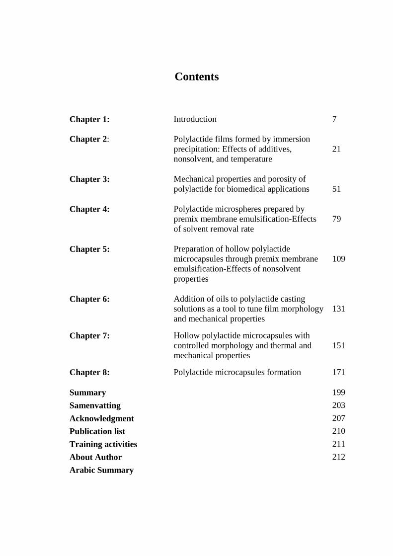
Contents
Chapter 1:
Introduction
7
Chapter 2:
Polylactide films formed by immersion precipitation: Effects of additives, nonsolvent, and temperature
21
Chapter 3:
Mechanical properties and porosity of polylactide for biomedical applications
51
Chapter 4:
Polylactide microspheres prepared by premix membrane emulsification-Effects of solvent removal rate
79
Chapter 5:
Preparation of hollow polylactide microcapsules through premix membrane emulsification-Effects of nonsolvent properties
109
Chapter 6:
Addition of oils to polylactide casting solutions as a tool to tune film morphology and mechanical properties
131
Chapter 7:
Hollow polylactide microcapsules with controlled morphology and thermal and mechanical properties
151
Chapter 8:
Polylactide microcapsules formation 171
Summary 199
Samenvatting 203
Acknowledgment 207
Publication list 210
Training activities 211
About Author 212
Arabic Summary


Chapter 1
Introduction

Chapter 1
8
Introduction
In this thesis, microparticles and films made of the biopolymer polylactide (PLA)
are investigated (see Figure 1). The structure in these systems was induced by
dissolving this polymer in a mixture of solvents and inducing phase separation.
In this introduction, first the properties of polymer itself are discussed, followed by
those of the two main products which were investigated: films and solid and hollow
particles (microbubbles).
We will then discuss the main target of this thesis, which is to prepare PLA-based
products with specific properties, followed by an outline of the chapters of the
thesis.
Figure1: examples of PLA films (a) and hollow-microcapsules (b) prepared in this study
Polylactide
PLA is a linear aliphatic thermoplastic polyester that can be derived from
renewable resources [1, 2]. Production of PLA starts with fermentation of food
stocks like starch, corn, or sugar beets into lactic acid (2-hydroxy propionic acid),
which is the basic building block of PLA [3, 4]. After fermentation and separation,
the lactic acid is converted into the cyclic di-ester lactide, using a combined process
of oligomerization and cyclization [3, 4]. The lactide is then polymerized through
ring-opening into polylactide [3, 4].
a b

Introduction
9
There are two optical isomers of lactide, the L (Levorotary) isomer, and the D
(Dexterotary) isomer [1, 3, 5]. Polymers prepared from the L and D isomers are
usually called poly(L-lactide), PLLA, and poly(D-lactide), PDLA, respectively.
Both homopolymers are highly (semi-)crystalline, while the random copolymer
poly (DL-lactide) PDLLA is completely amorphous (see Figure 2 for structural
formulae). Hence, the properties of DL-based products are very different from
those made of one of the homopolymers [1, 3, 5].
Figure 2: structural formulae of PLLA, PDLA and PDLLA [13].
The unique properties of PLA such as high mechanical strength and good
biodegradability and biocompatibility have made it a popular polymer for many
applications in the biomedical, pharmaceutical, as well as environmental fields [5-
11]. In the biomedical field, PLA is used in different types of biomaterials; i.e.
sutures, scaffolds for heart, bone and cartilage tissue engineering [8-10]. Besides, it
is also used in films and membranes for cell culturing [11], and in microcapsules
for controlled delivery of several types of drugs, antigens, and vaccines [7, 12].
Another big application area is in packaging, where PLA serves as an
environmental-friendly alternative for conventional petrochemical-based packaging
materials [6].
The mechanical and thermal properties of PLA are important for its application.
PLA is generally brittle and stiff at room or body temperature with a glass
transition temperature of around 60 oC [14, 15]. The lack of toughness is a
bottleneck for expanding the fields of applications of PLA. Its flexibility can be
improved by using different techniques such as copolymerization, blending, and
plasticization with other polymers and low molecular weight compounds [14, 16-

Chapter 1
10
23]. Even though some interesting materials were reported in literature, it is
obvious that optimization of the mechanical properties of PLA for the application
in mind is still of prime importance.
Preparation of PLA films
PLA films have found different applications like cell scaffolding and packaging
material [6, 11, 24, 25]. PLA films can be fabricated with melt or solution
processing [4, 26, 27]. In melt processing, PLA is heated above its melting point,
shaped into the desired shape, and then cooled to solidify the product. The
techniques used for melt processing include extrusion, film blowing, injection
moulding, and (thermal) compression [4]. A disadvantage of melt processing is that
PLA degrades at elevated temperature [4]. The solution processing involves
dissolving the polymer in a proper solvent, casting of the solution into a mold (e.g.,
a film), and subsequent solidification by removing the solvent or by reducing the
solvent quality. The most commonly used techniques for PLA films and
membranes are immersion precipitation and film casting; both techniques will be
discussed more elaborately in the following chapters. In evaporative film casting,
the polymer solution is cast on a flat mold and then exposed to air to evaporate the
solvent. With immersion precipitation, the cast film is immersed in a coagulation
bath filled with a nonsolvent for the polymer which is however miscible with the
solvent. The solvent diffuses out of the film into the nonsolvent bath, while the
nonsolvent diffuses into the polymer solution. This exchange results in a net
reduction of the overall solvent quality in the polymer solution which induces
nucleation and subsequent growth of a polymer poor phase, and enrichment and
ultimately solidification of the solution of the solution surrounding these nuclei
[27]. The polymer poor droplets eventually form the pores in a polymeric matrix.
The properties of the resulting films, i.e. morphology, porosity, and mechanical and
thermal properties are strongly dependent on the polymer concentration, polymer
crystallinity, and thermodynamic and kinetic interactions between polymer solvent
and nonsolvent [27] (see chapters 2 and 3). Mixtures of solvents and nonsolvents
instead of a single solvent and a single nonsolvent can be therefore used to modify
the properties of the films [28, 29].

Introduction
11
Preparation of PLA microbubbles and particles
PLA microparticles (and bubbles) have received increased attention in recent years
because of their applications in the pharmaceutical field. Microparticles have been
widely investigated as delivery carriers for bioactive therapeutic agents and
vaccines [7, 12]. In addition, hollow microparticles (bubbles) can be used as
contrast agents in ultrasound imaging. Ultrasound contrast agents (UCA’s) are
small gas bubbles stabilized with a thin polymer or protein shell [30]. These agents
are efficient ultrasound reflectors when subjected to an acoustic field: they enhance
the ultrasound signal and consequently can facilitate visualization of organs and
(soft) tissues of the body during ultrasound treatment [30, 31]. The mechanical and
chemical properties of microparticles and UCA’s are very important in medical
applications. For example, when particles are small and have uniform size they are
more biocompatible and induce less inflammatory response from the tissues
compared to larger and more polydisperse particles [32, 33]. Furthermore, small
particles can pass easier through narrow blood vessels and have longer circulation
time in the blood than big particles, since they are taken up less quickly by liver
and/or pancreas [34]. Preparation of microparticles and UCA’s with well-defined
properties is therefore very relevant, and it is obvious that preparation techniques
and conditions need to be chosen carefully.
Microparticles or microbubbles can be prepared using different techniques such as
solvent extraction/evaporation, coacervation, and spray-drying [35]. The solvent
extraction/evaporation method, which is a crossover between evaporative casting
and immersion precipitation, gives better control over the size and size distribution
of the particles than the other techniques [35]. Preparation of microparticles
through solvent extraction/evaporation starts with emulsification of a homogenous
polymer solution in a continuous phase that consists of nonsolvent (e.g., water),
which is immiscible with the solvent, and possibly a stabilizer to keep the droplets
apart. After emulsification, the solvent slowly diffuses out of the particles and
through the nonsolvent bath, and then evaporates at the surface of the bath (see
Figure 3). Removal of the solvent causes the polymer to solidify by glassification
or crystallization [35].

Chapter 1
12
solvent PLA oil
solvent PLA oil solvent
PLA oil
air
nonsolvent
The final particle size is determined by the initial droplet size and the concentration
of the polymer in the casting solution. It is thus important to start with a narrowly
dispersed emulsion of the casting solution. Standard emulsification techniques such
as sonication, high-pressure homogenizers and colloid mills, have as main
disadvantage that they give poor control over the size and size distribution of the
particles, and are energy intensive, which may result in damage when fragile
components are present in the droplets [36]. With newer emulsification methods
such as membrane emulsification, monodisperse emulsions and particles can be
effectively prepared [36], as is also the case for microchannel based emulsification
techniques [37, 38].
Figure 3: schematic representation of the extraction and evaporation of the solvent after emulsification.
For this study we decided to use premix membrane emulsification which combines
high throughput with good control on droplet size. In premix membrane
emulsification, a course pre-mix emulsion of the casting solution in the nonsolvent
continuous phase is pressed through a complex network of branching and joining
microchannels (e.g., a porous membrane matrix) (see Figure 4). The branching of
the channels causes large droplets to divide over the channels, and slowly reduce in
size, approximately down to the diameter of the channels [39]. Passage of the
emulsion through the membrane is repeated several times (see also chapters 4 and
5) [36].

Introduction
13
Figure 4: Schematic representation of premix membrane emulsification process.
When microbubbles are to be produced (see Figure 5), a mixture of a good and a
poor solvent is used in the casting solution, both poorly soluble in the nonsolvent
bath. Unlike the solvent for the polymer, the second, poor solvent (often called oil)
does not diffuse out, since it is not volatile, and remains in the polymer droplet. As
the good solvent slowly diffuses out of the droplets and evaporates, the
concentrations of polymer and the oil become higher and higher, until the solution
becomes unstable. The oil now forms a droplet inside the original droplet (being
poorly compatible with the nonsolvent bath, it will be at the inside of the droplet),
while the polymer will be in between the internal oil droplet and the outside
nonsolvent bath. This will ultimately form a solid shell around the oil droplet,
which can be removed by freeze-drying [40].
The shell properties are dependent on the precipitation process, which is strongly
determined by the solvent removal rate and on the choice of oil (see chapters 2, 3,
4, 7 and 8).
nonsolvent
polymer solution
premixing
premix emulsion
membrane

Chapter 1
14
Figure 5: Schematic representation of the evaporative immersion precipitation process of hollow PLA microparticles.
The shell properties are dependent on the precipitation process, which is strongly
determined by the solvent removal rate and on the choice of oil (see chapters 2, 3,
4, 7 and 8).
Aim and outline of the thesis
The aim of this study is to design PLA microbubbles (ultimately for use as UCA’s)
with well-defined size, size distribution, structure and mechanical properties. For
this purpose, the phase behavior of PLA, solvent, oil, and nonsolvent was first
studied using thin PLA films. The morphology and the mechanical properties of the
films were investigated, and the insight obtained was used as a tool to improve the
properties of films and microparticles and microbubbles.
Chapter 2 mainly focuses on the effects of nonsolvent, on the morphology of thin
PLA films prepared through immersion precipitation and the results are discussed
in relation to the phase separation behaviour of PLA.
In Chapter 3 the mechanical properties, structure, and porosity of PLA films
prepared through immersion precipitation and film casting are evaluated for various
nonsolvents. Amongst others, the effect of addition of dodecane is discussed and
potential uses for various biomedical applications are discussed.
hollow microaprticle
PLA solvent
oil
nonsolvent
oil
nonsolvent + solvent
PLA shell
extraction of solvent
gas
Freeze drying
solidification of the
polymer
emulsification

Introduction
15
Chapter 4 highlights the effects of solvent removal rates on the size, size
distribution, and morphology of solid PLLA microspheres prepared with premix
membrane emulsification. Both experimental and computer simulation results,
based on a Maxwell-Stefan model for non-ideal, multi-component mass transfer,
are presented.
The effects of the nonsolvent properties on the size and size distribution of hollow
PLLA microparticles prepared with premix membrane emulsification are discussed
in chapter 5, and linked to process conditions such as number of emulsification
cycles and transmembrane flux.
In chapter 6, results are shown for mechanical, thermal and structure properties of
PLLA films prepared through film casting when various oils were added to the
polymer solution. The use of different oils for creating different film properties is
discussed.
Chapter 7 summarizes the effects of the oils used in chapter 6 on the mechanical,
thermal and structure properties of hollow PLLA microparticles.
In chapter 8 the main results and conclusions obtained from the films and
microparticles are highlighted, summarized, and compared. Furthermore,
possibilities for future development in microparticle and film research based on the
current results are discussed, together with possible options for other fields of
research.

Chapter 1
16
References
1. Maillard, D., Prud'homme, R.E., Chirality Information Transfer in
Polylactides: From Main-Chain Chirality to Lamella Curvature. Macromolecules, 2006. 39(13): p. 4272.
2. Vaidya, A.N., Pandey, R.A., Mudliar, S., Kumar, M.S., Chakrabarti, T.,
Devotta, S. , Production and recovery of lactic acid for polylactide - An overview. Critical Reviews in Environmental Science and Technology, 2005. 35(5): p. 429.
3. Jacobsen, S., Degée, Ph., Fritz, H.G., Dubois, Ph., Jérôme, R., Polylactide
(PLA) - a new way of production. Polymer engineering and science, 1999. 39(7): p. 1311.
4. Lim, L.-T., Auras, R., Rubino, M., Processing technologies for poly(lactic
acid). Progress in polymer science, 2008. 33(8): p. 820. 5. Wuisman, P.I.J.M., Smit, T.H., Bioresorbable polymers: Heading for a new
generation of spinal cages. European spine journal, 2006. 15(2): p. 133-148. 6. Auras, R., Harte, B., Selke, S., An overview of polylactides as packaging
materials. Macromolecular bioscience, 2004. 4(9): p. 835. 7. Mohamed, F., Van Der Walle, C.F., Engineering biodegradable polyester
particles with specific drug targeting and drug release properties. Journal of Pharmaceutical Sciences, 2008. 97(1): p. 71.
8. Karageorgiou, V., Kaplan, D., Porosity of 3D biomaterial scaffolds and
osteogenesis. Biomaterials, 2005. 26(27): p. 5474. 9. Lam, K.H., Nijenhuis, A.J., Bartels, H., Postema, A.R., Jonkman, M.F.,
Pennings, A.J., Nieuwenhuis, P., Reinforced poly(L-lactic acid) fibres as suture material. Journal of applied biomaterials, 1995. 6(3): p. [d]191.
10. Lee, S. H., Kim, B. S., Kim, S. H., Kang, S. W., Kim, Y. H., Thermally
produced biodegradable scaffolds for cartilage tissue engineering. Macromolecular bioscience, 2004. 4(8): p. 802.
11. Luciano, R. M., Zavaglia, C. A. C., Duek, E. A. R., Alberto-Rincon, M. C.,
Synthesis and characterization of poly(L-lactic acid) membranes: Studies in vivo and in vitro. Journal of materials science. Materials in medicine, 2003. 14(1): p. 87.
12. Freiberg, S., Zhu, X. X., Polymer microspheres for controlled drug release.
International Journal of Pharmaceutics, 2004. 282(1-2): p. 1.

Introduction
17
13. Zhu, B., Li, J., He, Y., Yamane, H., Kimura, Y., Nishida, H., Inoue, Y., Effect
of steric hindrance on hydrogen-bonding interaction between polyesters and natural polyphenol catechin. Journal of Applied Polymer Science, 2004. 91(6): p. 3565.
14. Ljungberg, N., Wesslén, B., Preparation and properties of plasticized
poly(lactic acid) films. Biomacromolecules, 2005. 6(3): p. 1789. 15. Martin, O., Avérous, L., Poly(lactic acid): plasticization and properties of
biodegradable multiphase systems. Polymer, 2001. 42(14): p. 6209. 16. Broström, J., Boss, A., Chronakis, I.S., Biodegradable films of partly branched
poly(L-lactide)-co-poly(ε-caprolactone) copolymer: Modulation of phase morphology, plasticization properties and thermal depolymerization. Biomacromolecules, 2004. 5(3): p. 1124.
17. Jacobsen, S., Fritz, H.G., Plasticizing polylactide - the effect of different
plasticizers on the mechanical properties. Polymer engineering and science, 1999. 39(7): p. 1303.
18. Jiang, L., Wolcott, M.P., Zhang, J. , Study of biodegradable
polylactide/poly(butylene adipate-co-terephthalate) blends. Biomacromolecules, 2006. 7(1): p. 199.
19. Ke, T., Sun, S.X., Seib, P., Blending of poly(lactic acid) and starches
containing varying amylose content. Journal of applied polymer science, 2003. 89(13): p. 3639.
20. Kulinski, Z., Piorkowska, E., Gadzinowska, K., Stasiak, M., Plasticization of
poly(L-lactide) with poly(propylene glycol). Biomacromolecules, 2006. 7(7): p. 2128.
21. Labrecque, L.V., Kumar, R.A., Davé, V., Gross, R.A., Mccarthy, S.P. , Citrate
esters as plasticizers for poly(lactic acid). Journal of applied polymer science, 1997. 66(8): p. 1507.
22. Lu, J., Qiu, Z., Yang, W., Fully biodegradable blends of poly(l-lactide) and
poly(ethylene succinate): Miscibility, crystallization, and mechanical properties. Polymer, 2007. 48(14): p. 4196.
23. Sheth, M., Kumar, R.A., Davé, V., Gross, R.A., Mccarthy, S.P., Biodegradable
polymer blends of poly(lactic acid) and poly(ethylene glycol). Journal of applied polymer science, 1997. 66(8): p. 1495.

Chapter 1
18
24. Zoppi, R. A., Contant, S., Duek, E. A. R., Marques, F. R., Wada, M. L. F., Nunes, S. P., Porous poly(-lactide) films obtained by immersion precipitation process: morphology, phase separation and culture of VERO cells. Polymer, 1999. 40(12): p. 3275.
25. Liu, H.-C., Lee, I.-C., Wang, J.-H., Yang, S.-H., Young, T.-H., Preparation of
PLLA membranes with different morphologies for culture of MG-63 Cells. Biomaterials, 2004. 25(18): p. 4047.
26. Rhim, J.-W., Mohanty, A.K., Singh, S.P., Ng, P.K.W., Effect of the processing methods on the performance of polylactide films: Thermocompression versus solvent casting. Journal of applied polymer science, 2006. 101(6): p. 3736.
27. Van de Witte, P., Dijkstra, P. J., Van den Berg, J. W. A., Feijen, J., Phase
separation processes in polymer solutions in relation to membrane formation. Journal of Membrane Science, 1996. 117(1-2): p. 1.
28. Chuang, W. Y., Young, T. H., Chiu, W. Y., Lin, C. Y., The effect of polymeric
additives on the structure and permeability of poly(vinyl alcohol) asymmetric membranes. Polymer, 2000. 41(15): p. 5633.
29. Van de Witte, P., Esselbrugge, H., Peters, A. M. P., Dijkstra, P. J., Feijen, J.,
Groenewegen, R. J. J., Smid, J., Olijslager, J., Schakenraad, J. M., Eenink, M. J. D., Sam, A. P., Formation of porous membranes for drug delivery systems. Journal of Controlled Release, 1993. 24(1-3): p. 61.
30. Lathia, J. D., Leodore, L., Wheatley, M. A., Polymeric contrast agent with
targeting potential. Ultrasonics, 2004. 42(1-9): p. 763. 31. Dayton, P. A., Chomas, J. E., Lum, A. F. H., Allen, J. S., Lindner, J. R., Simon,
S. I., Ferrara, K. W., Optical and acoustical dynamics of microbubble contrast agents inside neutrophils. Biophysical journal, 2001. 80(3): p. 1547.
32. Chu, L. Y., Park, S. H., Yamaguchi, T., Nakao, S. I., Preparation of Micron-
Sized Monodispersed Thermoresponsive Core-Shell Microcapsules. Langmuir, 2002. 18(5): p. 1856.
33. Liu, R., Ma, G., Meng, F.-T., Su, Z.-G., Preparation of uniform-sized PLA
microcapsules by combining Shirasu Porous Glass membrane emulsification technique and multiple emulsion-solvent evaporation method. Journal of Controlled Release, 2005. 103(1): p. 31.
34. Gref, R. R., Minamitake, Y., Peracchia, M. T., Trubetskoy,V., Torchilin,V.,
Langer, R., Biodegradable long-circulating polymeric nanospheres. Science, 1994. 263(5153): p. 1600.

Introduction
19
35. Freitas, S., Merkle, H.P., Gander, B., Microencapsulation by solvent extraction/evaporation: Reviewing the state of the art of microsphere preparation process technology. Journal of Controlled Release, 2005. 102(2): p. 313.
36. Vladisavljević, G. T., Williams, R. A., Recent developments in manufacturing
emulsions and particulate products using membranes. Advances in colloid and interface science, 2005. 113(1): p. 1.
37. Van Der Graaf, S., Steegmans, M.L.J., Van Der Sman, R.G.M., Schroën,
C.G.P.H., Boom, R.M., Droplet formation in a T-shaped microchannel junction: A model system for membrane emulsification. Colloids and Surfaces A: Physicochemical and Engineering Aspects, 2005. 266(1-3): p. 106.
38. Utada, A.S., Lorenceau, E., Link, D.R., Kaplan, P.D., Stone, H.A., Weitz, D.A., Monodisperse Double Emulsions Generated from a Microcapillary Device. Science, 2005. 308(5721): p. 537.
39. Van Der Zwan, E., Schroën, K., Van Dijke, K., Boom, R., Visualization of
droplet break-up in pre-mix membrane emulsification using microfluidic devices. Colloids and Surfaces A: Physicochemical and Engineering Aspects, 2006. 277(1-3): p. 223.
40. Böhmer, M. R., Schroeders, R., Steenbakkers, J. A. M., de Winter, S. H. P. M.,
Duineveld, P. A., Lub, J., Nijssen, W. P. M., Pikkemaat, J. A., Stapert, H. R., Preparation of monodisperse polymer particles and capsules by ink-jet printing. Colloids and surfaces. A, Physicochemical and engineering aspects, 2006. 289(1-3): p. 96.

Chapter 1
20

Chapter 2
Polylactide films formed by immersion precipitation:
effects of additives, nonsolvent and temperature*
*This chapter has published as: Hassan Sawalha, Karin Schroën and Remko Boom, Polylactide films formed by immersion precipitation: Effects of additives, nonsolvent, and temperature. Journal of applied polymer science, 2007. 104(2): p. 959-971.

Chapter 2
22
Abstract
The influence of nonsolvent, crystallinity of the polymer film, and addition of
dodecane (a poor solvent for the polymer and for the nonsolvent), on the morphology
of polylactides films has been investigated, and was related to phase separation
behaviour. Both amorphous poly-DL-lactide (PDLLA) and crystalline poly-L-lactide
(PLLA) were dissolved in dichloromethane (DCM), and subsequently films were
made by immersion in non-solvent baths. PDLLA gave dense films without any
internal structure, since the structure was not solidified by crystallization or
glassification. PLLA films show varying structure depending on the non-solvent. With
methanol, asymmetric morphologies were observed as a result from combined liquid-
liquid demixing and crystallization, while with water symmetric spherulitic structures
were formed.
As a next step, dodecane was added, which is not miscible with the nonsolvent; and
we found it to have a strong influence on the morphology of the films. The PDLLA
films with dodecane did not collapse: a closed cell structure was obtained. In PLLA
films, dodecane speeds up phase separation and induces faster crystallization in the
films, and the porosity, size of the pores, and interconnectivity increased. When the
PLLA solutions were subjected to a heat pretreatment, crystallization could be
postponed, which yielded a cellular structure around dodecane, which did not contain
spherulites anymore.

Polylactide films formed by immersion precipitation
23
Introduction
Phase separation of polymer solutions is one of the most popular techniques used for
e.g. the preparation of porous polymeric membranes or dense or hollow particles.
Different methods are known such as: thermally induced phase separation, air-casting
of a polymer solution, precipitation from the vapour phase, and immersion
precipitation [1, 2]. All these methods are used to produce commercial membranes.
For the production of flat sheet membranes, a solution that consists of polymer and
solvent is cast on an inert support and subsequently immersed in a coagulation bath
filled with a non-solvent [3]. For the production of hollow fibre membranes, the
support is not required because of the construction of the nozzle that shapes the
membrane directly.
Due to the exchange of solvent and non-solvent, phase separation occurs. Two main
types of phase transitions are responsible for this, liquid-liquid demixing, and solid-
liquid demixing [4, 5]. Liquid-liquid demixing in polymer solutions that are relatively
concentrated (typically > 10 weight %), generally takes place by nucleation and
growth of the polymer poor phase. Solid-liquid demixing mainly happens in
crystalline and semi-crystalline polymers, and occurs because of crystallization,
gelation, or vitrification [1, 6, 7]. The resulting morphology is strongly determined by
the aforementioned processes. Generally, liquid-liquid demixing produces porous and
cellular structures, while crystallization forms interlinked particle-based structures [8-
10]. Many parameters such as concentration of the polymeric solution, crystallinity of
the polymer, temperature of the casting solution, and coagulation bath, type of solvent,
and non-solvent, and their mutual diffusivities [5, 11-15] influence demixing, and
consequently the final morphology. Some investigators have reported that additives in
the casting solution can be used to modify the structure obtained. As additives, a
second polymer, acids, alcohols, or inorganic salts have been reported. Obviously, the
resulting morphology strongly depends on the type of additive and the interactions
with the polymer, solvent, and non-solvent [13, 16-19].

Chapter 2
24
In the study reported here, we chose Polylactic acid (PLA) which is a biodegradable
polymer that has wide applications in the medical and pharmaceutical fields [20, 21].
PLA films were formed by means of immersion precipitation which has, for instance,
been proposed as a method for the preparation of biodegradable scaffolds for blood
vessels, but also for preparation of drug delivery devices [18]. Two types of PLA were
used: Poly (D50,L50) lactide PDLLA, and(P(L)LA) PLLA. PDLLA is a random co-
polymer that cannot crystallize and thus is either in the rubbery or in the glassy state,
while PLLA is in optically pure form and crystallizes readily [11, 22, 23].
The effects of non-solvent quality and PLA crystallinity on the resulting film
morphology were studied separately. Unlike most studies, in which additives are used,
that are soluble in the non-solvent [9, 12, 18], we have used dodecane as an additive
which is not soluble in the non-solvent. The effect on the resulting structures is
unknown, but it is to be expected that different morphologies can be obtained. The
morphology of the films was investigated visually with scanning electron microscopy.
Light transmission experiments were performed to monitor and characterise the film
formation process itself.
Experimental
Materials
Poly-L-lactide (PLLA) and poly-DL-lactide (PDLLA), with an intrinsic viscosity of
1.21 and 0.49 dl/g respectively, were supplied by PURAC Biochem B.V., Gorinchem,
the Netherlands. Dichloromethane (DCM), (HPLC, gradient grade) was obtained from
Merck and used as the solvent for the polymer. Dodecane (≥99%) was purchased from
Sigma-Aldrich and added to the casting solution as a poor solvent for the polymer.
Methanol (HPLC, gradient grade, ≥99.9%) (Aldrich) was used with Milli-Q water as a
non-solvent. All chemicals were used as received.
Film preparation
The casting solutions were prepared by dissolving different amounts of polymer in
various DCM-dodecane mixtures to obtain the desired concentrations. The solution
Polylactide films formed by immersion precipitation
25
was kept at the required temperature under stirring for 1-2 days and then cooled down
to room temperature before use. Solutions with concentrations (w/w/w) of 20:0:80,
20:5:75, and 20:10:70, or 20:0:80, and 20:10:70 PDLLA-dodecane-DCM were used.
The polymer solution was cast in the form of a thin film on a glass plate, and
subsequently immersed in the coagulation bath for 30 minutes, after which the films
were ready. All the experiments were done at room temperature. As non-solvents, the
following methanol-water mixtures were used: 100:0, 60:40, 30:70, 0:100.
Light transmission experiments
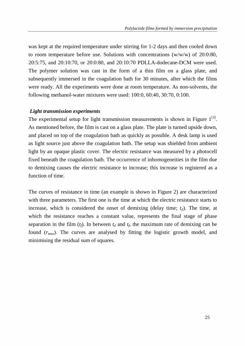
The experimental setup for light transmission measurements is shown in Figure 1[3].
As mentioned before, the film is cast on a glass plate. The plate is turned upside down,
and placed on top of the coagulation bath as quickly as possible. A desk lamp is used
as light source just above the coagulation bath. The setup was shielded from ambient
light by an opaque plastic cover. The electric resistance was measured by a photocell
fixed beneath the coagulation bath. The occurrence of inhomogeneities in the film due
to demixing causes the electric resistance to increase; this increase is registered as a
function of time.
The curves of resistance in time (an example is shown in Figure 2) are characterized
with three parameters. The first one is the time at which the electric resistance starts to
increase, which is considered the onset of demixing (delay time; td). The time, at
which the resistance reaches a constant value, represents the final stage of phase
separation in the film (tf). In between td and tf, the maximum rate of demixing can be
found (rmax). The curves are analysed by fitting the logistic growth model, and
minimising the residual sum of squares.

Chapter 2
26
Scanning electron microscope (SEM)
The morphology of polymer films was investigated with SEM (JEOL, JSM-5600 LV).
To prepare cross section samples, sections of the films were cut, dried, and fractured
in liquid nitrogen. The top and bottom surfaces and the cross sections were coated
with a very thin platinum layer using a sputter-coater (JEOL, JFC-1300) before
viewing with SEM.
Figure 1: Experimental setup for light transmission measurement: 1- plastic cover, 2- light source, 3- glass plate, 4- polymer film, 5- coagulation bath, and 6- photocell.
Figure 2: Interpretation of light transmission results; R is the electric resistance (Ω) at time t, R0 the initial resistance (Ω), td the delay time of demixing (second), rmax the maximum demixing rate (1/second), and tf, is the time where demixing is complete (second). The actual data were measured for a film of 20:10:70 PLLA:dodecane:DCM immersed in a water bath.

Polylactide films formed by immersion precipitation
27
Method - Calculation of phase diagrams
As many others, we have used the Flory-Huggins theory for evaluating the
thermodynamics of the (quaternary) systems used [1,3,4,6,10,11]. The Gibbs energy of
mixing is described by
433442243223411431132112
44332211 lnlnlnln
φχφχφχφχφχφχ
φφφφ
nnnnnn
nnnnRT
Gm
++++++
+++=∆
in which, ni is the number of moles of component i, and φi is the volume fraction of
component i, and χij is the Flory-Huggins interaction parameter (see Table 1). Index 1
represents the nonsolvent, 2 = solvent, 3 = polymer and 4 = additive. Because of the
complexity of such quaternary systems, we used constant interaction parameters. The
main aim of the phase diagrams is to show the various trends that are present and not
to quantitatively describe all the effects in detail.
The chemical potentials for each component were determined by taking the derivative
of the Gibbs energy to ni. Phase equilibria were calculated by equating the chemical
potential of each component in each phase. This results for a two-phase equilibrium in
m-1 equations (m is the number of components present), and for a three-phase
equilibrium in 2m – 2 equations. Solving these equations yields the coexisting
compositions, and therewith the binodals. The phase diagrams were shown as limiting
ternary phase diagrams, linked together to form the sides of a folded-out pyramidal
quaternary phase diagram. For the limiting ternary phase diagram, the volume fraction
of the excluded component was set to zero. The ternary phase diagrams (without
dodecane) are primarily used in the results section. The interested reader can find the
quaternary phase diagrams in the appendix, together with a more elaborate explanation
for the phase behaviour.

Chapter 2
28
Table 1: Values of the input parameters used in the equations.
Parameter Value Parameter Value
χ12 (methanol-DCM) 0.5 [24] v1 (methanol) 40.46 cm3/mol
χ12 (water-DCM) 3.3 v1 (water) 10.00 cm3/mol
χ13 (methanol-PLA) 1.5 [24] v2(DCM) 64.10 cm3/mol
χ13 (water-PLA) 3.4 [24] v4 (dodecane)
226.67
cm3/mol
χ14 (methanol-
dodecane) 2.5
r*( vnonsolvent/
vPLA) 0.00085 [24]
χ14 (water-dodecane) 3.4 ∆Hm PLA
81 - 140 J/g
[24]
χ23 (DCM-PLA) 0.2 [24] Tm0 PLA 480 K [24]
χ24 (DCM-dodecane) 0.5 T 298 K
χ34 (PLA-dodecane) 1.5
* The value of r is based on the number average degree of polymerization of PLLA with respect to the molar volume of water. This value has to be calculated for each polymer-nonsolvent combination; but because these values have negligible influence in the location of the phase boundaries, r was taken as a constant value [24].
The crystallization equilibriums were described with the Flory equation for quaternary
systems:
( )
4224411421124344
1223
2
1113
44
12
2
113
3
11
3
10
1
1ln11
φφχφφχφφχφχφχφχ
φφφφφ
−−−
+++
−−−−+=
−
∆
v
v
v
v
v
v
v
v
v
v
v
v
TTR
H
v
v
m
mu
in which v3 is the molar volume of the repeating unit of component 3 (PLA), and vi the
molar volume of component i; ∆Hm is the melting enthalpy, and Tm0 the melting
temperature of pure PLA. Once more, for the limiting ternary phase diagrams, the
Polylactide films formed by immersion precipitation
29
volume fraction of the excluded component was set to zero. Values of the parameters
used are summarized in Table 1.
Results and discussion
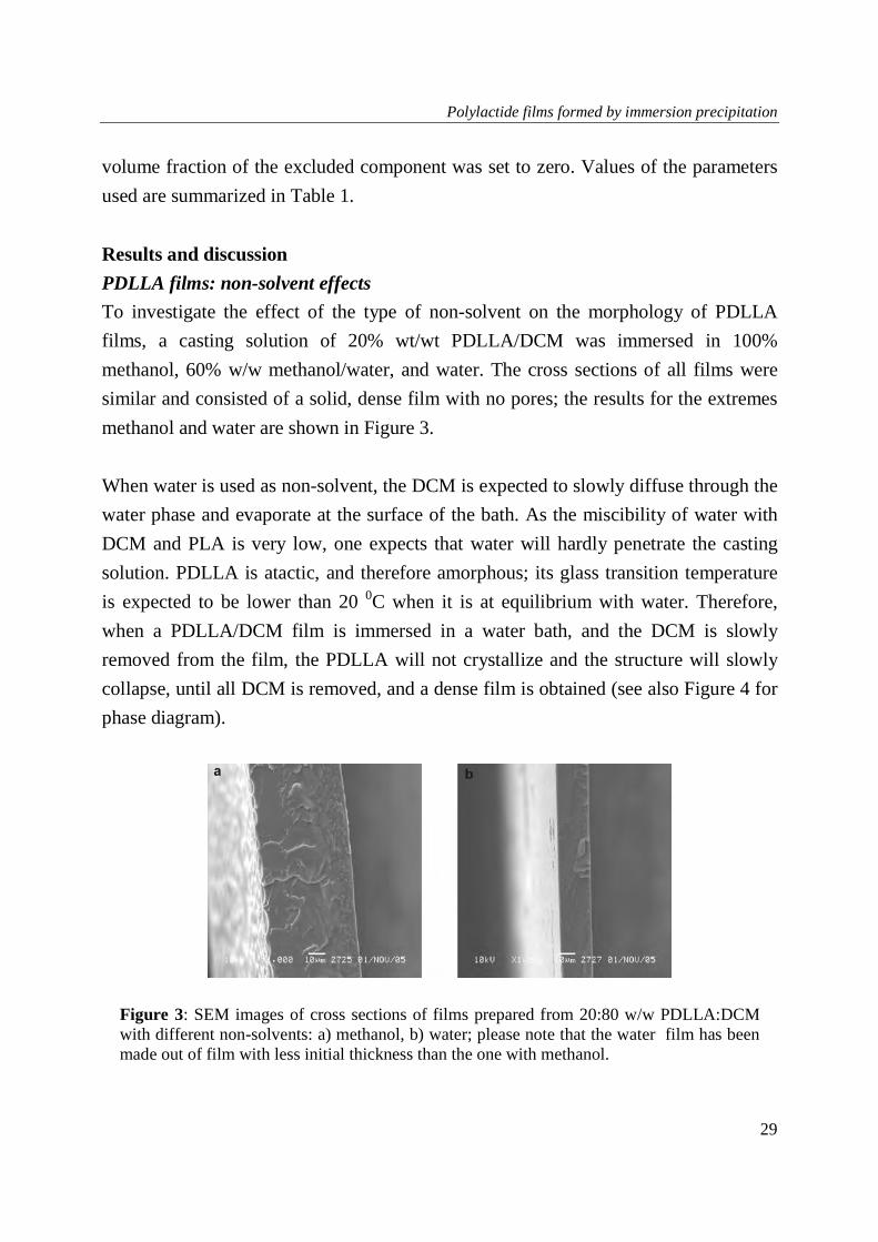
PDLLA films: non-solvent effects
To investigate the effect of the type of non-solvent on the morphology of PDLLA
films, a casting solution of 20% wt/wt PDLLA/DCM was immersed in 100%
methanol, 60% w/w methanol/water, and water. The cross sections of all films were
similar and consisted of a solid, dense film with no pores; the results for the extremes
methanol and water are shown in Figure 3.
When water is used as non-solvent, the DCM is expected to slowly diffuse through the
water phase and evaporate at the surface of the bath. As the miscibility of water with
DCM and PLA is very low, one expects that water will hardly penetrate the casting
solution. PDLLA is atactic, and therefore amorphous; its glass transition temperature
is expected to be lower than 20 0C when it is at equilibrium with water. Therefore,
when a PDLLA/DCM film is immersed in a water bath, and the DCM is slowly
removed from the film, the PDLLA will not crystallize and the structure will slowly
collapse, until all DCM is removed, and a dense film is obtained (see also Figure 4 for
phase diagram).
Figure 3: SEM images of cross sections of films prepared from 20:80 w/w PDLLA:DCM with different non-solvents: a) methanol, b) water; please note that the water film has been made out of film with less initial thickness than the one with methanol.

Chapter 2
30
Figure 4: Schematic equilibrium phase diagram for: (a) PDLLA-DCM-methanol and (b) PDLLA-DCM-water. The phase diagrams were calculated with the parameters as mentioned in Table 1.
DCM is only marginally miscible with water but readily miscible with methanol. With
methanol, a PDLLA-DCM solution will exhibit so-called delayed demixing (formation
of droplets of a polymer lean phase inside the polymer solution, after some time for
indiffusion has elapsed) [4]. Therefore, one would expect a film containing at least
some closed-cell pores because of the presence of the polymer lean phase. This is not
what we observed. We expect that the porous structure may have been formed during
the process, but as the ultimately formed film is still highly swollen with methanol
(PDLLA swells 22 % w/w in methanol), it will never reach the glass transition [11].
Thus, the film will never fixate, and the porous structure that is formed initially will
have collapsed into a completely dense film when the residual DCM evaporates.
It is known from literature that fixation of the cellular structure obtained by liquid-
liquid demixing requires a solidification step [11]. This can take place via solid-liquid
demixing (i.e. crystallization), or via glassification. If neither of these transitions
occurs, liquid-liquid demixing will proceed until two completely separated layers are
obtained [11]. Since PDLLA cannot crystallize [11, 22], and its glass transition line
does not cross the binodal for either methanol or water (this is illustrated in Figure 4),
it will not be solidified but will collapse given sufficient time. Van de Witte an co-
workers have shown with differential scanning calorimetry (DSC) that in a PDLLA-
Methanol0.1 0.2 0.3 0.4 0.5 0.6 0.7 0.8 0.9
PDLLA
0.1
0.2
0.3
0.4
0.5
0.6
0.7
0.8
0.9
DCM
0.1
0.2
0.3
0.4
0.5
0.6
0.7
0.8
0.9
2-phase L-L region
Glass transition
Ultimate film composition
Tie-lines
Initial film composition
Critical point
Water0.1 0.2 0.3 0.4 0.5 0.6 0.7 0.8 0.9
PDLLA
0.1
0.2
0.3
0.4
0.5
0.6
0.7
0.8
0.9
DCM
0.1
0.2
0.3
0.4
0.5
0.6
0.7
0.8
0.9
2-phase L-L region
Glass transitionUltimate film composition
Initial film composition
Miscibility gap between DCM/dodecane and water
(a) (b)

Polylactide films formed by immersion precipitation
31
chloroform-methanol system, phase transition occurred only by liquid-liquid demixing
and no signs of crystallites or transitions due to verification were observed [25], which
is in line with our findings.
For a film made out of 5% w/w PDLLA/Chloroform which was immersed and kept in
methanol for one day or longer Van de Witte and co-workers [11] found that no
structure was preserved. Comparison with our results shows that increasing the
polymer concentration reduces the time required for phase separation, and results in
faster loss of structure.
PLLA: non-solvent effects
In contrast to PDLLA, PLLA is a rapidly crystallizing polymer [11, 22, 25]. Therefore
one may expect a strong influence of polymer crystallisation, which will influence the
morphology of the films as was reported in the literatures [6, 9-11, 25]. Films with
polymer concentrations of 20% w/w PLLA/DCM were prepared using 100% methanol,
60%, 30% w/w methanol/water and water as non-solvents; the cross sections of the
films are shown in Figure 5. The film prepared with methanol as nonsolvent consists
of dense blobs surrounded by semi-circular closed cells (Figure 5a). The top layer at
the left of the image (the side in contact with the non-solvent) has a very densely
packed structure without pores. During film formation (see also Figure 6 for the phase
diagrams), the initial in-diffusion of non-solvent is much smaller than the out-diffusion
of the solvent [3]. Therefore, the polymer concentration in the top layer of the film
rises quickly, which will bring the composition in this layer far inside the
crystallization region of the phase diagram. Thus, the polymer will crystallize rapidly.
The out-diffusion of DCM from the sublayer to the non-solvent bath is reduced
significantly due to the additional mass transfer resistance created by the dense top
layer. In spite of this, in time the concentration of DCM in the sublayer will be
reduced, the solution will become more enriched with polymer, and the composition
will approach the liquid-liquid miscibility gap. As soon as the miscibility gap is
reached (after 16 seconds of immersion, see also Table 2), liquid-liquid demixing by
nucleation and growth of a polymer-lean phase will take place and a cellular structure

Chapter 2
32
is formed. The polymer concentration in the continuous phase will increase
continuously until the solid-liquid demixing region is entered and crystallization of the
polymer rich phase occurs, which will form the dense blobs, and pore walls. It is
expected that these solid blobs contain spherulites to such an extent that no distinction
of the individual spherulites is possible anymore. This becomes also clear from the top
view of the film (Figure 5b), which shows a dense, non-porous film full of spherulites.
The occurrence of both phase separation processes (liquid-liquid demixing and
crystallization) was observed for crystalline systems in general [6, 10] and specifically
for PLLA. For the PLLA-chloroform-methanol system, Van de Witte and co-workers
demonstrated by DSC the presence of crystallites during the formation of PLLA
membranes, Further, they stated that at high PLLA concentration (>20%w/w)
crystallization becomes the main demixing process, which affects to a large extent the
morphology of the product [25]. Our results are in agreement with those of Van de
Witte for the same polymer concentrations [11].
When water/methanol mixtures were used as non-solvent, the in-diffusion of methanol
mixture and the out-diffusion of DCM are slowed down, and the crystallization
process has more time to proceed. Thus, we see that for higher water concentrations,
the spherulites are more pronounced, larger and further apart (Figure 5 c-f).
Crystallization is expected to have taken place because of the slow exchange of the
solvent and non-solvent; the time available was long enough to initiate growth of the
solid crystals. This case is schematically illustrated in the phase diagram (see Figure
6); where the polymer concentration is slowly increased and after a relatively long
time, the solid-liquid demixing region is entered and crystallization occurred in the
film. The structure of the spherulites shows no signs of phase separation due to liquid-
liquid demixing. It has been reported in literature that slow exchange rates between
solvent and non-solvent promote solid-liquid demixing over liquid-liquid demixing [9,
10]; our findings are in line with this.

Polylactide films formed by immersion precipitation
33
Figure 5: SEM images of cross sections of films prepared from 20:80 w/w PLLA:DCM with different non-solvents: a) methanol, b) methanol, top surface, c) 60:40 w/w methanol:water, d) 30:70 w/w methanol:water; e) water, f) water, top surface.
Chapter 2
34
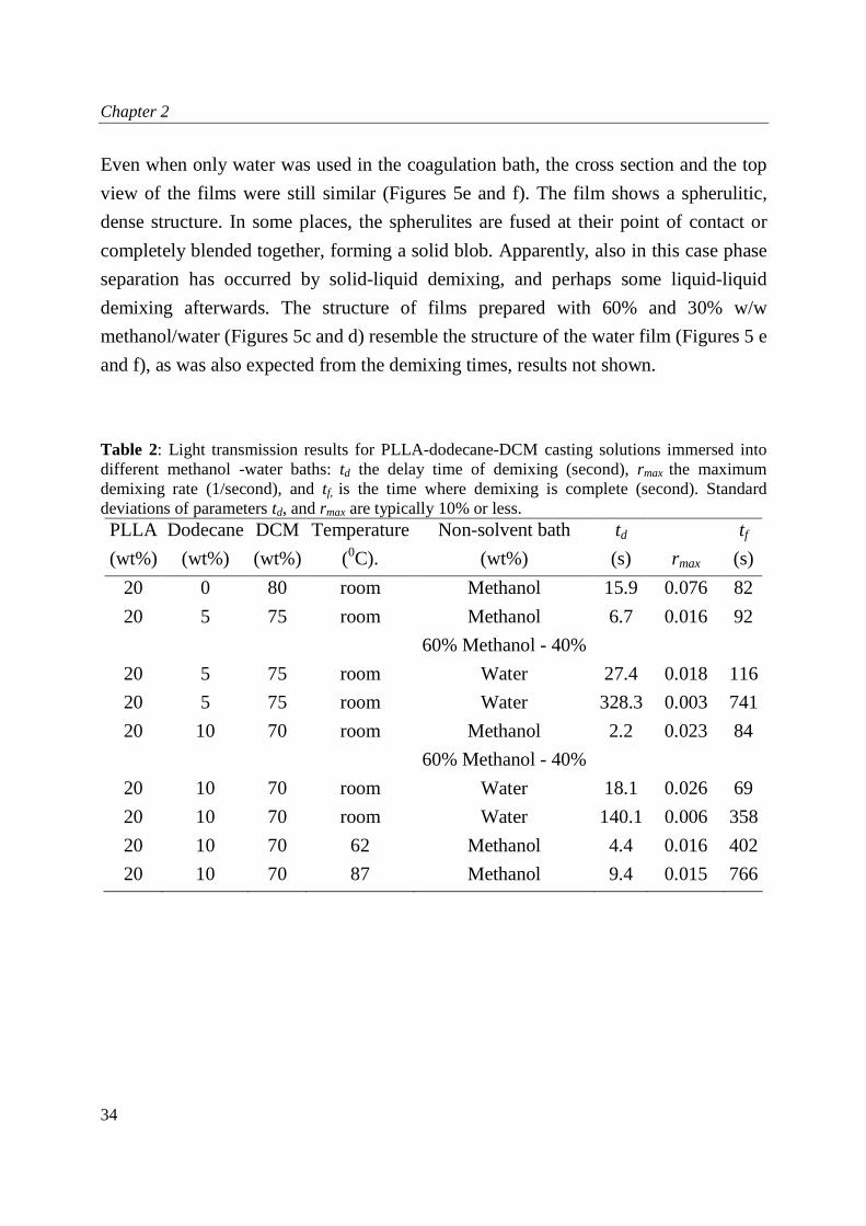
Even when only water was used in the coagulation bath, the cross section and the top
view of the films were still similar (Figures 5e and f). The film shows a spherulitic,
dense structure. In some places, the spherulites are fused at their point of contact or
completely blended together, forming a solid blob. Apparently, also in this case phase
separation has occurred by solid-liquid demixing, and perhaps some liquid-liquid
demixing afterwards. The structure of films prepared with 60% and 30% w/w
methanol/water (Figures 5c and d) resemble the structure of the water film (Figures 5 e
and f), as was also expected from the demixing times, results not shown.
Table 2: Light transmission results for PLLA-dodecane-DCM casting solutions immersed into different methanol -water baths: td the delay time of demixing (second), rmax the maximum
demixing rate (1/second), and tf, is the time where demixing is complete (second). Standard deviations of parameters td, and rmax are typically 10% or less. PLLA
(wt%)
Dodecane
(wt%)
DCM
(wt%)
Temperature
(0C).
Non-solvent bath
(wt%)
td
(s) rmax
tf
(s)
20 0 80 room Methanol 15.9 0.076 82
20 5 75 room Methanol 6.7 0.016 92
20 5 75 room
60% Methanol - 40%
Water 27.4 0.018 116
20 5 75 room Water 328.3 0.003 741
20 10 70 room Methanol 2.2 0.023 84
20 10 70 room
60% Methanol - 40%
Water 18.1 0.026 69
20 10 70 room Water 140.1 0.006 358
20 10 70 62 Methanol 4.4 0.016 402
20 10 70 87 Methanol 9.4 0.015 766

Polylactide films formed by immersion precipitation
35
Figure 6: Schematic phase diagrams of (a) PLLA-DCM-methanol and (b) PLLA-DCM-water systems. The crystallization line indicates fixation of the polymer rich matrix by formation of crystals. The phase diagrams were calculated with the parameters as mentioned in Table 1.
Addition of dodecane
PDLLA
The effect of an additive that is not miscible with either the polymer or with the
nonsolvent applied in the coagulation bath was investigated by addition of 10% w/w
dodecane to the casting solution. Compared to the situation without dodecane, the
morphology of the films dramatically changed into a typical asymmetric morphology.
The cross sections of these films show a dense skin layer with only little, small pores,
and a porous sublayer with a fairly uniform closed cellular morphology (Figure 7 a-d).
In the film prepared from methanol, the dense skin is thicker and has no pores, while
the porous sublayer contains big, irregular pores (Figure 7 a). The explanation is the
high exchange rate between methanol and solvent compared to the other non-solvents.
For methanol, the polymer concentration increased quickly at the film-bath interface
resulting in a dense toplayer. In the sublayer, the diffusion of solvent and non-solvent
slowed down because of the presence of the dense toplayer, however, slowly but
surely the dodecane concentration increased in the sublayer. As PDLLA is an
amorphous polymer, liquid-liquid demixing was the predominant phase separation
Methanol0.1 0.2 0.3 0.4 0.5 0.6 0.7 0.8 0.9
PLLA
0.1
0.2
0.3
0.4
0.5
0.6
0.7
0.8
0.9
DCM
0.1
0.2
0.3
0.4
0.5
0.6
0.7
0.8
0.9
2-phase L-L region
Crystallization line
Glass transition
Ultimate film composition
Tie-lines
2-phase S-L region
Initial film composition
Water0.1 0.2 0.3 0.4 0.5 0.6 0.7 0.8 0.9
PLLA
0.1
0.2
0.3
0.4
0.5
0.6
0.7
0.8
0.9
DCM
0.1
0.2
0.3
0.4
0.5
0.6
0.7
0.8
0.9
2-phase L-L region
2-phase S-L region
Glass transitionUltimate film composition
Initial film composition
Miscibility gap between DCM/dodecane and water
(a) (b)

Chapter 2
36
process, which resulted in the porous structure. As the methanol concentration inside
the film slowly increased, the solubility of dodecane in the solution decreased
accordingly, which ultimately resulted in the formation of droplets of dodecane; these
droplets were the precursors of the cellular pores observed. It is remarkable that
without dodecane the film collapses completely, while now some structure is
preserved. The dodecane phase is trapped while the polymer solution surrounding it
slowly becomes more viscous. As the out-diffusion of dodecane is extremely slow, the
collapse becomes too slow. Thus, when we would have extended the residence time in
the bath considerably, we would possibly have seen a slow reduction of the porosity as
a function of the immersion time.
Figure 7: SEM images of cross sections of films prepared from 20:10:70 w/w PDLLA:dodecane:DCM with different non-solvents: a) methanol, b) 60:40 w/w methanol:water, c) 30:70 w/w methanol:water, d) water; please note that the water film has been made out of film with a thinner initial thickness than the other films.

Polylactide films formed by immersion precipitation
37
If the dodecane in PDLLA solution was replaced with an additive that is miscible with
the nonsolvent as conventionally used, one would expect that the additive will diffuse
out of the film, along with the solvent. The film would have collapsed into a dense
structure, as without using an additive; no stabilization of the structure could have
taken place. The immiscibility of the additive ensures that it stays inside the film,
forming the cellular pores. In the appendix, the addition of dodecane and its effect on
the phase behaviour of the system is discussed in more detail for the interested reader.
PLLA
To investigate the effects by crystallinity of the polymer, also PLLA films with
different dodecane concentrations were prepared. In Figures 8 and 9, the SEM
micrographs are shown for dodecane concentrations of 5 and 10% (w/w), respectively.
Figure 8 shows the cross sections of films prepared from 20:5:75
PLLA:dodecane:DCM. Compared to those without dodecane (e.g. Figure 5.a), the
porosity of the films increased, the pores became larger and better connected, and
consequently the structure was more open.
Demixing set in approximately 7 seconds after immersion into the methanol bath (see
Table 2). The resulting film has an asymmetric structure consisting of a dense top
layer and a porous sublayer, which consists of a bicontinuous network. The
morphologies observed suggest a particular series of occurrences of liquid-liquid
demixing and crystallization. We expect that initially, crystallization will set in, which
depletes the surrounding solution of polymer, and which becomes more susceptible to
liquid-liquid demixing, as they simultaneously become more concentrated in DCM
and dodecane. This implies that the concentration of dodecane is expected to influence
the structure as well.
When the same polymer solution was immersed in water, demixing occurred only very
slowly (>6 minutes) as indicated in Table 2. The obtained film morphology differs
strongly from the one formed with methanol (Figure 8c and d). The dense skin layer
has disappeared and the structure consists of a few blobs embedded in a distorted,

Chapter 2
38
bicontinuous porous matrix. The pores were more open, interconnected, and irregular
in shape and size. When a solution of 60%w/w methanol in water was used as non-
solvent, the delay time was in between those of methanol and water (see Table 2). The
observed morphology was to some extent similar to the one obtained with water, but
the structure is less porous and the pores are smaller, more closed, and less
interconnected (Figure 8e).
The effect of the dodecane concentration was investigated further, because we
expected it to have an important role in the formation of the films. Figures 9a-b shows
the morphologies of films prepared with more dodecane (10% w/w) in the casting
solution.
The porosity and the size of the pores increase by increasing the dodecane
concentration. In case of methanol as nonsolvent (Figure 9a), pores can be observed in
the top layer, and the film contains some dense areas embedded in a more regular
cellular structure with bigger pores, compared to the film prepared with 5% dodecane
(Figure 8a). With water, the film has a more open morphology with high
interconnectivity and big spherical pores as shown in Figure 9b. This could be related
to an increased probability of coalescence of dodecane droplets due to the long
diffusion times, resulting in bigger pores. Similar effects as described for methanol
occurred for the film prepared from 60% methanol (result not shown).
From the light transmission results, it is clear that increasing the amount of dodecane
in the casting solution decreases the delay time for demixing (Table 2). As the solution
is less stable with the non-solvent dodecane present (i.e., the starting composition is
closer to the border of the demixing gap in the phase diagram), phase separation will
start at an earlier stage, at which droplets of a dodecane rich phase will be formed (see
Figure 9a). The remaining PLA-DCM solution will then demix according to a normal
(delay of) demixing regime with methanol (Figure 6), which will result in smaller
pores in the matrix surrounding the larger pores formed by the dodecane. In the
appendix, the addition of dodecane and its effect on the phase behaviour of the system
is discussed in more detail for the interested reader.

Polylactide films formed by immersion precipitation
39
Figure 8: SEM images of cross sections of films prepared from 20:5:75 w/w PLLA:dodecane:DCM with different non-solvents: a) methanol, b) magnification of a, c) water, d) magnifications of c, and e ) 60:40 w/w methanol:water.

Chapter 2
40
Figure 9: SEM images of cross sections of films prepared from 20:10:70 w/w PLLA:dodecane:DCM with different non-solvents: a) methanol, b) water.
Effect of temperature
Casting solutions with 10% w/w dodecane were heated up and after some time cooled
down to room temperature, before immersion in the non-solvent bath. When the film
was produced from a solution that was incubated at 87 oC, crystallization set in after
longer delay time (see Table 2) and the skin layer was thinner than with a solution that
was incubated at 62 oC. Besides that, the porous sublayer contained a closed cellular
structure (see Figures 10a and b). This indicates that the crystallization process
depends on nuclei already present in the casting solution. Heating the solution before
casting, results in melting of many of the nuclei. This suppresses the crystallization
process. Therefore, liquid-liquid demixing is relatively faster in these films. Therewith,
a cellular morphology was obtained and the crystallization-associated structures, such
as the observed solid blobs (compare with Figure 9a) were not present. We now see
structures that are similar to the ones observed with the amorphous PDLLA; the
structures are now fixated after some time by crystallization. This stresses the
importance of control of crystallinity of the polymer in the production of structures
with a desired morphology, especially in combination with the use of another non-
solvent like dodecane in the polymer solution.

Polylactide films formed by immersion precipitation
41
In general, it is clear that the use of dodecane as a non-soluble additive leads to new
opportunities to influence porosity in polymeric films and structures. In combination
with the choice of solvent, non-solvent, and other process conditions, this may open a
new road to the design of highly porous structures.
Figure 10: SEM images of cross sections of films prepared from 20:10:70 w/w PLLA:dodecane:DCM solution heated at different temperatures and with methanol as non solvent: a) 62 C, b) 87 C.
Conclusions
Films formed from solutions of amorphous PDLLA show a dense structure; any
porous structure formed during demixing collapses since fixation by crystallization, or
vitrification, cannot take place. With crystalline PLLA specific morphologies are
obtained. With methanol as non-solvent, a typical asymmetric structure formed by
crystallization and (delayed) liquid-liquid demixing was found. With water as
nonsolvent, which is hardly miscible with the solvent, the demixing rate was much
lower. Liquid-liquid demixing was suppressed and crystallization dominated the
formed, symmetric structure.
Next, the influence of an additive, dodecane, which is immiscible with the nonsolvent
was investigated. Addition of dodecane speeds up demixing and increases the porosity

Chapter 2
42
of the films. Remarkably, for PDLLA the film does not collapse, as a result of the
presence of dodecane droplets, and retains a closed-cell structure. For PLLA films,
addition of dodecane made the structure more open and better interconnected. This
effect seems stronger than with miscible additives.
The differences in structure between PLLA and PDLLA became smaller when PLLA
solution was given a heat pre-treatment before casting to remove nuclei for
crystallisation. Liquid-liquid demixing became the dominant mechanism, and
crystallization served to stabilize the obtained structure.
Acknowledgements
The research described in this paper is part of the BURST project (IS042035).
Financial support by SENTER is kindly acknowledged. We thank our project partners
from Philips Research in Eindhoven, Erasmus Medical Centre in Rotterdam, and the
Physics of Fluids group from Twente University in Enschede for fruitful discussions.
Special thanks go to Marcel Bohmer from Philips for proofreading our manuscript.
The authors would further like to thank ing. H.A.Teunis, Membrane Technology
Group, University of Twente for his help in the SEM analysis, which is very much
appreciated. And dr. ir. R.G.H. Lammertink, Membrane Technology Group,
University of Twente for making the light transmittance setup available.

Polylactide films formed by immersion precipitation
43
References
1. Van de Witte, P., Dijkstra, P. J., Van den Berg, J. W. A., Feijen, J., Phase
separation processes in polymer solutions in relation to membrane formation. Journal of Membrane Science, 1996. 117(1-2): p. 1.
2. Tanaka, T., Lloyd, D. R., Formation of poly(-lactic acid) microfiltration
membranes via thermally induced phase separation. Journal of Membrane Science, 2004. 238(1-2): p. 65.
3. Reuvers, A. J., Smolders, C. A., Formation of membranes by means of immersion
precipitation : Part II. the mechanism of formation of membranes prepared from the system cellulose acetate-acetone-water. Journal of Membrane Science, 1987. 34(1): p. 67.
4. Wienk, I. M., Boom, R. M., Beerlage, M. A. M., Bulte, A. M. W., Smolders, C. A.,
Strathmann, H., Recent advances in the formation of phase inversion membranes made from amorphous or semi-crystalline polymers. Journal of Membrane Science, 1996. 113(2): p. 361.
5. Young, T. H., Cheng, L. P., Lin, D. J., Fane, L., Chuang, W. Y., Mechanisms of
PVDF membrane formation by immersion-precipitation in soft (1-octanol) and harsh (water) nonsolvents. Polymer, 1999. 40(19): p. 5315.
6. Bulte, A. M. W., Mulder, M. H. V., Smolders, C. A., Strathmann, H., Diffusion
induced phase separation with crystallizable nylons. II. Relation to final membrane morphology. Journal of Membrane Science, 1996. 121(1): p. 51.
7. Cheng, L. P., Young, T. H., You, W. M., Formation of crystalline EVAL
membranes by controlled mass transfer process in water-DMSO-EVAL copolymer systems. Journal of Membrane Science, 1998. 145(1): p. 77.
8. Lin, D. J., Chang, C. L., Chen, T. C., Cheng, L. P., Microporous PVDF membrane
formation by immersion precipitation from water/TEP/PVDF system. Desalination, 2002. 145(1-3): p. 25.
9. Van De Witte, P., Esselbrugge, H., Dijkstra, P. J., Van Den Berg, J. W. A., Feijen,
J., A morphological study of membranes obtained from the systems polylactide-dioxane-methanol, polylactide-dioxane-water, and polylactide-N-methyl pyrrolidone-water. Journal of Polymer Science, Part B: Polymer Physics, 1996. 34(15): p. 2569.

Chapter 2
44
10. Bulte, A. M. W., Folkers, B., Mulder, M. H. V., Smolders, C. A., Membranes of semicrystalline aliphatic polyamide nylon 4,6: Formation by diffusion-induced phase separation. Journal of Applied Polymer Science, 1993. 50(1): p. 13.
11. Van De Witte, P., Esselbrugge, H., Dijkstra, P. J.,Van Den Berg, J. W. A., Feijen, J., Phase transitions during membrane formation of polylactides. I. A morphological study of membranes obtained from the system polylactide-chloroform-methanol. Journal of Membrane Science, 1996. 113(2): p. 223.
12. Zoppi, R. A., Contant, S., Duek, E. A. R., Marques, F. R., Wada, M. L. F., Nunes,
S. P., Porous poly(-lactide) films obtained by immersion precipitation process: morphology, phase separation and culture of VERO cells. Polymer, 1999. 40(12): p. 3275.
13. Yeow, M. L., Liu, Y. T., Li, K., Morphological study of poly(vinylidene fluoride)
asymmetric membranes: Effects of the solvent, additive, and dope temperature. Journal of Applied Polymer Science, 2004. 92(3): p. 1782.
14. Cheng, L. P., Effect of Temperature on the Formation of Microporous PVDF
Membranes by Precipitation from 1-Octanol/DMF/PVDF and Water/DMF/PVDF Systems. Macromolecules, 1999. 32(20): p. 6668.
15. Cheng, L. P., Huang, Y. S., Young, T. H., Effect of the temperature of
polyurethane dissolution on the mechanism of wet-casting membrane formation. European Polymer Journal, 2003. 39(3): p. 601.
16. Chuang, W. Y., Young, T. H., Chiu, W. Y., Lin, C. Y., The effect of polymeric
additives on the structure and permeability of poly(vinyl alcohol) asymmetric membranes. Polymer, 2000. 41(15): p. 5633.
17. Chuang, W. Y., Young, T. H., Chiu, W. Y., The effect of acetic acid on the
structure and filtration properties of poly(vinyl alcohol) membranes. Journal of Membrane Science, 2000. 172(1-2): p. 241.
18. Van de Witte, P., Esselbrugge, H., Peters, A. M. P., Dijkstra, P. J., Feijen, J.,
Groenewegen, R. J. J., Smid, J., Olijslager, J., Schakenraad, J. M., Eenink, M. J. D., Sam, A. P., Formation of porous membranes for drug delivery systems. Journal of Controlled Release, 1993. 24(1-3): p. 61.
19. Madaeni, S. S., Rahimpour, A., Barzin, J., Preparation of polysulphone
ultrafiltration membranes for milk concentration: Effect of additives on morphology and performance. Iranian Polymer Journal (English Edition), 2005. 14(5): p. 421.

Polylactide films formed by immersion precipitation
45
20. Zhao, J., Yuan, X., Cui, Y., Ge, Q., Yao, K., Preparation and characterization of poly(L-lactide)/poly(ε-caprolactone) fibrous scaffolds for cartilage tissue engineering. Journal of Applied Polymer Science, 2004. 91(3): p. 1676.
21. Pego, A. P., Siebum, B., Van Luyn, M. J. A., Gallego y Van Seijen, X . J., Poot, A.
A., Grijpma, D. W., Feijen, J., Preparation of Degradable Porous Structures Based on 1,3-Trimethylene Carbonate and D,L-Lactide (Co)polymers for Heart Tissue Engineering. Tissue engineering, 2003. 9(5): p. 981.
22. Tsuji, H. H., Stereocomplex formation between enantiomeric poly(lactic acid)s. 6.
Binary blends from copolymers. Macromolecules, 1992. 25(21): p. 5719. 23. Urayama, H., Kanamori, T., Kimura, Y., Properties and biodegradability of
polymer blends of poly(L-lactide)s with different optical purity of the lactate units. Macromolecular Materials and Engineering, 2002. 287(2): p. 116.
24. Van De Witte, P., Dijkstra, P.J., Van Den Berg, J.W.A., Feijen, J., Phase behavior
of polylactides in solvent-nonsolvent mixtures. Journal of Polymer Science Part B: Polymer Physics, 1996. 34(15): p. 2553.
25. Van de Witte, P., Boorsma, A., Esselbrugge, H., Dijkstra, P. J., Van den Berg, J. W.
A., Feijen, J., Differential Scanning Calorimetry Study of Phase Transitions in Poly(lactide)-Chloroform-Methanol Systems. Macromolecules, 1996. 29(1): p. 212.
26. Cheng, L. P., Shaw, H. Y., Phase behavior of a water/2-propanol/poly(methyl
methacrylate) cosolvent system. Journal of Polymer Science Part B: Polymer Physics, 2000. 38(5): p. 747.
27. Tao, C. T.,Young, T. H., Phase behavior of poly(N-isopropylacrylamide) in water-
methanol cononsolvent mixtures and its relevance to membrane formation. Polymer, 2005. 46(23): p. 10077.
28. Radovanovic, P., Thiel, S. W., Hwang, S. T., Formation of asymmetric polysulfone
membranes by immersion precipitation. Part I. Modelling mass transport during gelation. Journal of Membrane Science, 1992. 65(3): p. 213.

Chapter 2
46
Appendix
Based on the results presented previously, one can conclude that the addition of
dodecane to the casting solution has a big influence on the film structure. The presence
of dodecane in the polymer solution has lowered the solvent quality of DCM; which
will influence the phase separation mechanism. The presence of dodecane in the
casting solution has brought the demixing gap much closer to the initial polymer cast
composition. This is consistent with the light transmission results. Increasing the
dodecane concentration from 0 to 5% reduces the delay time from 16 to 7 seconds.
Upon further increase of the dodecane concentration, the delay time is reduced further
until we have almost instantaneous demixing at 10% dodecane (Table 2).
A possible but very unlikely interpretation for the system is that the mixture of DCM
and dodecane might actually function as a co-solvent for PLA. If the co-solvency
holds, the phase diagram can have two demixing regions with two binodal curves
sandwiching a miscibility region as described by Liao-Ping et al for the system of
poly-(methylmethacrylate) in water-2-propanol co-solvent mixtures [26], and by Tao
et al using poly(N-isopropylacrylamide) in water-methanol co-non-solvent mixtures
[27]. In such a case, the demixing region in the phase diagram will increase in size,
which will most probably facilitate phase separation. However, it should be kept in
mind that dodecane is a poor solvent for PLA and it is not expected that
dodecane/DCM mixtures can act as a co-solvent.
The incorporation of dodecane to the casting solution makes the system more complex
as it has become a quaternary system. The principle is however the same as for the
ternary one. Contacting the polymer solution with non-solvent will cause out-diffusion
of solvent and a smaller in-diffusion of non-solvent, and consequently demixing will
take place.
Figure 11 shows the folded-down quaternary phase diagrams – these are the ternary
limiting systems of the full (3-dimensional) diagrams. Please note that only binodals
and tie-lines are shown and not spinodals. Earlier research has shown that demixing by

Polylactide films formed by immersion precipitation
47
immersion leads to metastable demixing and not spinodal decomposition – thus the
binodals are most relevant for our purpose [28]. The PDLLA-DCM-methanol-
dodecane system (a) shows three two-phase regions, indicating an equilibrium
between a PLA concentrated and a PLA diluted phase. For truly quaternary solutions,
these two-phase regions lead to a three-phase region, which is inside the quaternary
phase diagram, and not visible in the limiting ternary diagrams. This can be illustrated
by assuming a quaternary solution containing equal amounts of all four components.
This solution will decompose into a PLA-rich and a PLA-poor phase. This PLA-poor
phase would contain roughly equal amounts of the three low-molecular weight
components. This phase is not stable (see limiting ternary DCM-dodecane-methanol
diagram) and will itself decompose into a phase rich in methanol and a phase rich in
dodecane. Thus, a three-phase region is present inside the quaternary phase diagram,
as a result of the three two-phase regions in the limiting ternary diagrams. This same
three-phase region is evident in the limiting ternary system methanol-dodecane-
PDLLA: most of the phase diagram is occupied by a three phase region, indicating
decomposition of the compositions enclosed by the region, into a methanol phase, a
dodecane phase and a PDLLA-rich phase. Around this three-phase region, two two-
phase regions are visible.
The system with water instead of methanol (b) shows a somewhat different phase
diagram due to the relative immiscibility of water with the other components.
Solutions of PDLLA with DCM are basically immiscible with water, leading to a large
two-phase region in that limiting ternary diagram. Since dodecane is immiscible with
water as well, a similar demixing gap is visible in the ternary system DCM-dodecane-
water. The three two-phase regions in the systems PDLLA-DCM-water, DCM-
dodecane-water and DCM, dodecane-PLA, once more lead to a three-phase region
inside the quaternary phase diagram, which is evident in the limiting diagram for
water-dodecane-PDLLA.
The systems with PLLA (c and d) show the same liquid-liquid demixing behavior as
with PDLLA, but in addition show regions exhibiting demixing between a crystalline

Chapter 2
48
PDLLA
DCM methanol
DodecanePDLLA PDLLA
LL
LL
LL
LLL
LL
LL
G
G G
PDLLA
DCM water
DodecanePDLLA PDLLADodecane
LLG
LL
LLL
LL
LL
LL
G
G
(a)
(c) (d)
(b)
PLLA
DCM methanol
DodecanePLLA Dodecane
LL
PLLA
SLL(LLL)
SL (LL)
SL (LL)
GLLG
SL
SL
LL
GPLLA
DCM water
DodecanePLLA PLLADodecane
LLG
SL
LL
SL
LL
G
GSLL(LLL)
SL (LL)
SL (LL)
LL L-L two phase regionLLL L-L-L three phase regionSL S-L two phase
(crystallisation) regionSLL S-L-L three phase regionG Glassy area
Figure 11: Full quaternary (folded-out) phase diagrams for (a) PDLLA-DCM-methanol-dodecane, (b) PDLLA-DCM-water-dodecane; (c) PLLA-DCM-methanol-dodecane, (d) PLLA-DCM-water-dodecane. The phase diagrams were calculated with the parameters as mentioned in Table 1.
PLLA phase and a liquid (PLLA poor) phase. They are visible in the limiting phase
diagrams PLLA-DCM-methanol and PLLA-DCM-dodecane and PLLA-DCM-water
and PLLA-DCM-dodecane: once more, these regions extend into the volume of the
quaternary phase diagrams. Even though in the ternary systems PLLA-dodecane-
methanol and PLLA-dodecane-water no crystallization areas are visible, one should
bear in mind that the stable PLLA rich phases in the lower right corner of the diagram
will be strongly crystallized. Below the liquid-liquid demixing gaps (two-phase and
three-phase) a crystallization curve is present, which means that even though
thermodynamically speaking the three-phase region is a liquid-liquid-liquid region, the

Polylactide films formed by immersion precipitation
49
actual three phase equilibrium will be of type liquid-liquid-solid (water/methanol
phase, dodecane phase and crystallized PLLA phase).
The phase diagrams show that PLLA systems have a strong tendency to crystallize,
even before liquid-liquid demixing. However, crystallization is generally a slow
process. Since liquid-liquid demixing is usually a fast process (except when the
nonsolvent diffuses in very slowly), liquid-liquid demixing can still take place before
crystallization can take place.

Chapter 2
50

Chapter 3
Mechanical properties and porosity of polylactide for
biomedical applications*
*This chapter has been published as: Hassan Sawalha, Karin Schroën and Remko Boom, Mechanical properties and porosity of polylactide for biomedical applications. Journal of applied polymer science, 2008. 107(1): p. 82-93.

Chapter 3
52
Abstract
In this study, strength, ductility, and porosity of polylactide films prepared by
immersion precipitation and film casting in air were investigated. To induce extra
porosity in the films, dodecane was added to the polymer casting solution.
The structure, porosity, and mechanical properties of the films were evaluated. The
ultimate strength and elastic modulus of neat PLLA prepared by film casting were
at least twice those of the same film prepared in methanol, whereas the ductility of
these films was considerably higher than for air. The porosity, size of pores, and
interconnectivity of pores increased gradually with increasing dodecane
concentration. This dodecane-induced porosity (as high as 80%), progressively
decreased the ultimate strength and modulus of practically all films, but remarkably
improved the ductility of films prepared in air, and this can be related to a decrease
in crystallization temperature. For films prepared in water, or PDLLA films in
general, the ultimate strength, modulus, and ductility of films prepared in water
were significantly lower than those of air-cast PLLA films.
In summary, the results obtained in this research show that it is possible to tailor
the properties of the films for various biomedical applications, through the use of
polymer type, preparation method, and dodecane-induced porosity as tools.

Mechanical properties and porosity
53
Introduction
Biodegradable polymers have received considerable attention in the last decades,
because of their wide applications in pharmaceutical, biomedical and
environmental fields [1, 2]. Typical examples of these polymers are aliphatic
polyesters such as poly(butylene succinate) (PBS), poly[(butylene succinate)-co-
adipate] (PBSA), poly(3-hydroxybutyrate) (PHB), poly(e-caprolactone) (PCL), and
poly(lactide) (PLA) [3-6]. PLA is highly hydrolysable in the human body and has
good mechanical strength, thermal plasticity, fabricability, biodegradability, and
biocompatibility [7, 8], and that is why PLA has become one of the most popular
biodegradable materials in the biomedical field. It has been used for sutures, bone
screws, bone plates, tissue repair, and regeneration, and also for controlled delivery
devices (e.g. microparticles or implants for drugs) [1, 9-16]. Also for applications
outside the medical field, recent developments in processing technology make PLA
more economically viable as an environmental-friendly substitute for the
conventional synthetic packaging materials [2].
For many applications, the degradation and mechanical properties of PLA are
important. The degradation behaviour of PLA plays a big role on its in vivo
performance and may influence many processes i.e. tissue regeneration, cell growth
and host response [17, 18]. Degradation of PLA mostly occurs by hydrolytic attack
of the ester bonds in the polymer, after which lactic acid monomers are formed and
eventually removed via normal metabolic processes in the body [19, 20]. There are
many factors that can affect degradation rate of PLA including the polymer
material properties such as: crystallinity, molecular weight, and monomer
hydrophobicity [19]. Moreover, some researchers have reported that additives can
also speed up or delay the degradation process of PLA [21-23].
Beside the degradation behaviour, which is beyond the scope of this paper,
mechanical properties of PLA is of a great importance also, and in this respect
some improvement of e.g. the brittle nature, lack in toughness and low deformation
at break, is desirable [24-26]. One solution lies within the polymer itself, it is well
known that the physical properties of PLA such as melting point, mechanical
strength, and crystallinity, can be influenced considerably by the stereo-isomeric

Chapter 3
54
L/D ratio of the lactide units [3, 5]. For instance, poly(L-lactide) (PLLA), which
consists of pure L-lactide is an isotactic and crystallisable polymer, that gives
strength to structures [3, 27]. In contrast, poly(DL-lactide) (PDLLA) produced
from racemic mixtures of L-lactide and D-lactide, is atactic and completely
amorphous resulting in more brittle structures [3, 27]. Also several other techniques
to enhance flexibility and toughness have been reported, such as copolymerization
or blending with other substances like polymers [3, 6, 28], plasticizers [24-26, 29],
or fillers [4, 30].
Each application will have its own requirements, therefore, it is obvious that
control over the mechanical properties of PLA is of great importance. However
structure-related properties should not be disregarded because they will influence
the mechanical properties; at the same time they may be required for a specific
application (e.g. porosity for controlled release purposes). Part of the solution can
be found within the polymer itself but obviously the production method will also
play a role. Various production processes are reported for PLA such as injection
moulding, extrusion, film blowing, fibre spinning, film casting, and immersion
precipitation [2, 5, 9, 13, 24, 30, 31]. In the last process for instance, a dope
consisting of polymer, solvent, and additives is immersed in a coagulation bath
filled with a non-solvent and due to the subsequent exchange of the solvent and
non-solvent, phase separation takes place and solidification of the polymeric
product occurs [31]. Depending on the composition of the coagulation bath,
completely different structures may be formed ranging from dense films, to highly
porous structures. It can be expected that depending on the structures formed also
different mechanical behaviour will be found, therewith co-determining the field of
application [9, 32]. Therefore it is not strange that immersion precipitation has been
proposed in very different fields including the preparation of polymeric membranes,
and also for the preparation of biodegradable scaffolds for blood vessels, drug
delivery devices, microparticles, implants, fibres, and films [13, 16, 31, 33].
In this study, we systematically investigate polylactide films (PLLA, PDLLA, and
mixtures thereof) in order to correlate structure formation with mechanical
behaviour. The films were fabricated via two processes, film casting and immersion
precipitation. In the first method the film was cast and then exposed to the air,

Mechanical properties and porosity
55
while in the last one the film was immersed into different non-solvents (instead of
air), which are known to influence the film structure considerably. Besides that,
also dodecane was used as an immiscible additive to the polymer solution that is
used to prepare the films in order to induce porosity in the films away from the
porosity generated by regular phase separation processes. (The use of high alkanes,
such as dodecane, has been described in literature among other things for the
production of hollow polymeric particles [33]). In that way, we hope to combine
structure related requirements, such as porosity, with mechanical strength. The
results are summarized in an application graph that links (film) production
conditions, mechanical properties and methods with application fields.
Experimental
Materials
Two types of PLA were used in this study: Poly-L-lactide (PLLA) and poly-
D(50)L(50)-lactide (PDLLA), with intrinsic viscosities of 1.21 and 0.49 dl/g,
respectively, and both were obtained from PURAC Biochem B.V., Gorinchem, the
Netherlands. Dichloromethane (DCM), (HPLC, gradient grade) was purchased
from Merck and used as the solvent for the polymer. Dodecane (≥99%) was
supplied by Sigma-Aldrich and used as an additive. Methanol (HPLC, gradient
grade, ≥99.9%) (Aldrich) was used with Milli-Q water as a non-solvent. All
chemicals were used as received.
Methods
Film preparation
To form PLA films, solutions of weight ratios 10:0:90, 10:5:85, 10:10:80, 10:15:75,
and 10:20:70 PLLA:dodecane:DCM; 10:0:90, and 10:5:85
PDLLA:dodecane:DCM; and 10:5:85 PLA mixture (1:1
PLLA:PDLLA):dodecane:DCM w/w/w were prepared. In the case of the films with
10% PDLLA, only one dodecane concentration is used, because at higher
concentrations, no films could be formed with PDLLA. The polymer was first
dissolved in DCM, and if needed, dodecane was subsequently added. The solution
was kept under stirring for 1-2 days. The films were formed with two procedures:

Chapter 3
56
i) Film casting: the polymer solution was cast onto a mould and left in a fume hood
for evaporation of the solvent under ambient conditions.
ii) Immersion precipitation: the polymer solution was cast onto a mould and then
immersed into a coagulation bath filled with non-solvent and kept there for around
40 min. As non-solvents, 100:0, 60:40, 30:70, and 0:100 (w/w) methanol:water
mixtures were used in this study. The initial thickness of the cast layer was always
100 µm.
Mechanical properties
All films were left in the fume hood under ambient conditions for one day before
use to ensure complete evaporation of the solvent. Out of films prepared by either
film casting or immersion precipitation, samples with a dog–bone-like shape were
cut. The total length of each sample was 37 mm; the gauge length of the samples
was about 15 mm (±1); the width was 13 mm at the top and 7.2 mm (narrowest) at
the middle of the sample in order to induce the fracture in the middle of the sample.
Tensile testing of the films was performed with the Texture Analyzer T2 (Stable
Micro Systems Ltd., Surrey, UK). The tensile tests were carried out at constant
crosshead speed of 0.1mm/s until break. Stress-strain curves were calculated from
load-elongation curves measured for 2-10 samples from films that were each
separately produced under the various conditions described earlier. Tensile strength
(σ), elongation at break (EB), and Young’s modulus (E) were calculated from the
stress-strain curves.
Differential scanning calorimetry (DSC)
The thermal properties of the films were measured using differential scanning
calorimeter (Perkin Elmer DSC-7, with a TAC 7/DX thermal analysis controller).
Scans of samples (about 8-10 mg) were run from 0 oC to 250 oC at heating rates of
10 oC/min. The glass transition temperature (Tg), crystallisation temperature (Tc),
melting temperature (Tm), and enthalpies of crystallization and melting were
determined.
Scanning electron microscopy (SEM)
Structures of the films before and after tensile testing (with special interest for the
fracture surfaces) were investigated by SEM (JEOL, JSM-5600 LV). Cross

Mechanical properties and porosity
57
sections of the samples were cut, dried, and fractured in liquid nitrogen. Before
viewing with SEM, the cross sections were coated with a thin platinum layer ( ~5
nm) using a sputter-coater (JEOL, JFC-1300).
Porosity and density of films
The porosity (ε) of the films was estimated by measurement of the mass and
dimensions of the films as reported by Hu and co-workers, and others [16, 34, 35].
The porosity was defined as follows:
(1)
Where, ρfilm and ρpolymer are the (bulk) densities of film and polymer, respectively.
The densities of the films were measured by calculating the mass/volume ratio for
three samples of each film. The film prepared without dodecane (Figure 1a) shows
a porosity of zero, and therefore the density of this film was taken as the polymer
density [16, 36].
For the theoretical description of the bulk density of the films and, therefore, the
porosity as a function of the dodecane concentration, equation 2 was used.
(2)
Where, Xpolymer and Xdodecane are the mass fractions of polymer and dodecane, and
ρpolymer and ρdodecane are the densities of polymer, and dodecane respectively. It was
assumed that the total volume fraction of the voids (porosity) in the film was
equivalent to the volume fraction of the dodecane added to the film. This implies
that the volume loss of the air-cast film is caused by evaporation; and the weight of
the film is equivalent to the initial weight of the polymer in the cast. These
assumptions are only true for air-cast films in Figure 1, as the porosity was mainly
caused by dodecane, while for immersion precipitation films, these assumptions do
not hold because part of the porosity in the films was generated by the phase
inversion process.
)()( dodecanedodecanepolymerpolymer
polymerf XX
X
ρρρ
+=
polymerfilm ρρε −=1

Chapter 3
58
Results and discussion
Film characterization
Morphology air casting
The morphologies of the cross sections of PLLA films prepared by air casting were
investigated by SEM. In Figure 1, the results for different dodecane concentrations
are shown. When no dodecane was present in the casting solution, a solid, dense,
and nonporous film was obtained (Figure 1a). When the film was exposed to air,
only evaporation of the solvent dichloromethane occurred because air can hardly
diffuse into the polymer solution; no demixing takes place, and therefore a
nonporous structure was observed.
When dodecane was added, the structures were entirely different; they became
porous and porosity increased with increasing dodecane concentration (this is
described in more detail in the porosity and density section). For 5 % dodecane, an
asymmetric morphology consisting of a thin dense toplayer and a porous sublayer
with a fairly uniform closed cellular structure was obtained (Figure 1b). With
increasing dodecane concentration (i.e. >10% w/w), symmetric structures with
more open morphologies were observed (Figures 1c-e). The overall pore fraction,
size of the pores, and their interconnectivity gradually increases with increasing
dodecane concentration. Based on these findings, one can say that the addition of
dodecane is a novel and easy approach for production of porous structures by the
film casting method.
The influence of dodecane on the structure can be explained as follows: when a
film containing dodecane is exposed to the air, the solvent DCM will start to
evaporate, and consequently the dodecane concentration in the film will increase.
Because dodecane is a poor solvent for PLA, one expects that when dodecane
reaches a certain concentration, demixing will start to occur in the film by
nucleation and growth of the polymer [32]. The dodecane droplets are the
precursors of the pores that were observed by SEM. The amount of these droplets is
expected to increase when more dodecane is added to the polymer solution. Thus

Mechanical properties and porosity
59
the incidence of coalescence will become higher, which will eventually result in a
structure with more and larger pores. This is in line with the SEM observations.
The resulting structure is expected to be solidified by crystallisation of the polymer.
How this influences the mechanical properties is discussed in a later section.
Morphology immersion precipitation
The structures in air-cast films are now compared with those obtained by
immersion precipitation in various non-solvents (see Figure 2). With methanol the
structures were to a certain extent similar to the ones obtained with air. When
water/methanol mixtures were used, the porosity, the size of the pores, and their
interconnectivity increased, with increasing water concentration.
During immersion precipitation, the phase separation process is different and more
complex than that for air casting. Contacting the polymer solution with non-solvent
will lead to out-diffusion of solvent into the non-solvent (instead of air) and in-
diffusion of non-solvent into the polymer solution. When the polymer solution
becomes saturated with non-solvent, phase separation will take place. In a previous
study, we found that PLLA films are solidified by crystallization of the polymer,
and depending on the non-solvent used, crystallization will set in early (methanol)
or later (water), therewith allowing more or less time for solidification of the
structure and/or structural rearrangements [32]. Furthermore, dodecane speeds up
phase separation and induces faster crystallization in the films, depending on the
non-solvent used. Even when we used PDLLA in combination with dodecane, the
films did not collapse, and porous structures were obtained [32]. Thus,
crystallization is not necessity for solidification in the film. For a more complete
description of the effects involved, we refer readers to previous work [32].
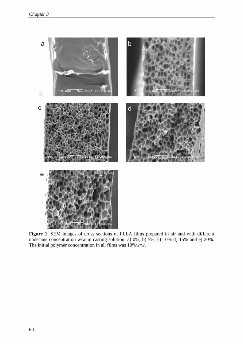
Chapter 3
60
Figure 1: SEM images of cross sections of PLLA films prepared in air and with different dodecane concentration w/w in casting solution: a) 0%, b) 5%, c) 10% d) 15% and e) 20%. The initial polymer concentration in all films was 10%w/w.

Mechanical properties and porosity
61
Figure 2: SEM images of cross sections of films prepared from 10:05:85 w/w PLLA:dodecane:DCM with different non-solvents: a) methanol, b) methanol, surface, c) 60:40 w/w methanol:water, d) 60:40 w/w methanol:water, surface, e) water, f) water, surface.
Film porosity and density
Figure 3 shows the porosity and density of films prepared in air as a function of
dodecane concentration. The results were in line with the SEM observations; the
higher the dodecane concentration the higher the porosity in the films. With
increasing porosity, obviously the bulk density of the films decreases (see Figure

Chapter 3
62
3a). Figure 3a shows that the theoretical model always overestimates the measured
porosity, which points to partial collapse of the structure. This effect is, perhaps
surprisingly, more pronounced at low dodecane concentrations (see Figure 3b). It is
expected to be caused by the time scale of solidification in these films which was
slow compared to high dodecane concentrations which solidify fast [32]. This is
expected to lead to better preservation of the structure. Although part of the
porosity that is induced by dodecane collapses, it is important to note that the
largest part of this porosity remains, and more so at higher concentrations of
dodecane (Figure 3b). This is true not only for air-cast films but also for films
prepared by immersion precipitation [32]. This indicates an additional way to
induce porosity in a film away from pores generated by regular phase separation.
Whether this also leads to mechanically more interesting structures is discussed in
the next section.
Figure 3: Influence of dodecane concentration on porosity and bulk density of PLLA films prepared in air, theoretical porosity and bulk density of the films were calculated using equations 1 and 2. The initial polymer concentration in all films was 10%w/w.
Mechanical properties
The ultimate tensile strength, elasticity modulus and the elongation at break were
measured for all films described in the materials and methods section. The effects
of dodecane, preparation method, and type of PLA on the mechanical properties of
the films were investigated.

Mechanical properties and porosity
63
Ultimate tensile strength and elastic modulus
In Figures 4, the effect of dodecane on the ultimate tensile strength (Figure 4a) and
elasticity modulus (Figure 4 b) of PLLA films prepared by either air casting or
immersion precipitation with methanol or water is shown. The film without
dodecane prepared by air casting shows a maximum tensile strength of
approximately 70 Mpa and an elasticity modulus of approxmately 2.4 Gpa, which
are, in fact, similar to those reported elsewhere for neat PLLA [5, 37]. When the
same film was prepared by immersion into methanol, the tensile modulus and
strength drop to around half of the original values (see Figure 4), and we expect
that the presence of pores (less than if dodecane were added) has caused these
effects. This conclusion is in line with the fact, that dodecane induced porosity
leads to significant decreases in ultimate strength and elasticity modulus. Please
note that these latter porosities can be as high as 80% (Figure 3).
When comparing films with dodecane prepared in air and methanol, we found it
remarkable that the films show approximately the same ultimate strength, whereas
the elasticity moduli of films prepared in air were higher than those in methanol
(Figure 4b). When comparing with films prepared in water, it is obvious that both
the ultimate strength and elasticity were considerably less than for methanol and air.
This was investigated in more detail; with increasing water concentration in
water/methanol mixtures, the films became gradually more fragile and weak
(Figure 4c). This can be attributed to the formation of large pores, that are known to
occur in films with dodecane cast in water (see Figures 2e and f), and which are
generally considered weak spots within the structure [35]. In air-cast films, and
films submerged in methanol, these large pores are not present (see Figures 1 b-e,
and Figures 2 a, and b).
The results for crystalline PLLA are compared to those for amorphous PDLLA and
1:1 mixtures of both polymers. As expected, the higher the PLLA concentration in
the films the higher the strength and modulus (see Figure 5). This is ascribed to the
high load-bearing capacity of the crystalline domains in PLLA [27, 38].

Chapter 3
64
Figure 4: Influence of dodecane concentration and preparation method (air, methanol, and water) of 10% polymer films on: a) ultimate strength, b) elastic modules, of PLLA films., c) ultimate strength of 10:20:70 PLLA: dodecane:DCM films as a function of water concentration in the nonsolvent.

Mechanical properties and porosity
65
Figure 5: Influence of PLA type (PLLA and PDLLA) on: a) ultimate strength, b) elastic modules, of PLA films prepared in methanol and with different dodecane concentrations. The initial polymer concentration in all films was 10%w/w.
On the bases of these results, one can conclude that the mechanical properties of
the films are dependent not only on the polymer type but even more so on their
structure; the tensile strength and elasticity modulus decrease with increasing
porosity, pore size, and interconnectivity. This is reasonable, if we keep in mind
that fracture in films originates from local concentrations of stress at flaws,
scratches or notches [39] and propagates by nucleation and growth of cracks or
crazes inside structures. These cracks can easily propagate and grow in regions
containing voids, and they become even easier to grow from larger pores or
macrovoids within films. Furthermore, it is expected that the cross sectional area of
the load-bearing polymer decreases with increasing porosity in the film which will
reduce the tensile modulus and ultimate strength [11]. A first attempt to quantify
these effects is given in the next section.
The reduction in strength and elasticity modulus with increasing porosity was also
observed in other studies [35, 36, 40], and for highly porous materials, such as
foams, a power-law relation between porosity and elasticity modulus was given [36,
40]:
E = Eo (1 − ε) n (3)

Chapter 3
66
Where Eo is the modulus of the nonporous film, ε is the porosity, and n is a
constant. For Eo, the E-value of a film prepared without dodecane was taken, and
the model was fitted to our experimental data for films prepared in air using the
least sum of squares. For a value for n of 1.326, the data points are described
adequately (see Figure 6). The theoretical value of n for completely open cell
foams was reported to be 2, whereas for closed cells this value is around one [40].
Our value is between these extremes; indicating partially open cells (see Figure 1).
The same model was explored to describe the ultimate tensile strength of the films
as function of porosity (ε) and ultimate strength of the film prepared without
dodecane (σo):
σ = σo (1 − ε) n (4)
Again the data were fitted using the least sum of squares method and for n = 2.596
the fit is reasonable (see Figure 7).
Figure 6: Influence of porosity on elastic modules of PLLA films prepared in air with different dodecane concentrations. The initial polymer concentration in all films was 10%w/w.

Mechanical properties and porosity
67
Figure 7: Influence of porosity on ultimate strength of PLLA films prepared in air with different dodecane concentrations. The initial polymer concentration in all films was 10%w/w.
Elongation at break
Besides the elasticity modulus and the ultimate strength, the elongation at break is
also an important feature of a film. In Figure 8, results are shown for films prepared
by air casting and immersion precipitation (with or without added dodecane). When
no dodecane was added, the film formed in air showed typical stiff behavior of neat
PLLA with a total elongation of 8%, which is close to values stated in other studies
[26] . Remarkably, with methanol the flexibility of the film was significantly
enhanced and an elongation at break of up to 35% was recorded. This may be due
to the higher degree of deformation that a regular cell structure permits during
elongation.
The effect of dodecane depends on the preparation method used. A remarkable
improvement in ductility was observed for the films prepared in air (although
strength and modulus decreased considerably), (see Figure 8). For example, the
elongation of the film with 5% w/w dodecane was threefold that of a neat PLLA
film; that of a film with 10% w/w dodecane was eightfold that of a neat PLLA film.
This can be related to the structure of the films; in air-cast films with dodecane we
observed the formation of the fairly large but uniform pores (Figure 1), which can
deform during elongation and hence allow for a much larger maximum elongation

Chapter 3
68
at break. This was investigated in more detail with DSC, and the results are shown
in that section.
Figure 8: Influence of dodecane concentration and preparation method (air, methanol, and water) on elongation at break of PLLA films. The initial polymer concentration in all films was 10%w/w.
The effect of dodecane on the ductility of films prepared with methanol was
different from those prepared with air. At 5% w/w dodecane, the elongation at
break of the films was slightly enhanced in comparison with neat PLLA, but when
higher concentrations of dodecane were used, a progressive reduction in the
elongation at break was observed, as shown in Figure 8. At 5% the films still
contained many small pores (Figure 2a), but at higher dodecane concentrations
these films have many large pores with thin, fragile lamellae between them [32],
and are therefore, more susceptible to break at less elongation.
For films produced by precipitation in water, a low ductility was found; dodecane
did not show any influence on the elongation of the films (see Figure 8). These
findings are in line with the effects on strength and elasticity modulus; films
produced in water remained weak and we expect this to be caused by the small
contact areas between crystalline spherulites or domains (Figures 2e and f). When
water/methanol mixtures were used as non-solvent, the elongation at break of these

Mechanical properties and porosity
69
films was close to that of water (5.5 % elongation for 60:40 methanol:water and
3.9% elongation for 30:70 methanol/water).
The elongation at break for the PDLLA films is less than 2%, which is very low
compared to PLLA films (32%). Also addition of dodecane does not increase the
ductility (~ 2.2%). This is due to the low flexibility and deformability of the
amorphous domains in the PDLLA structure compared to the crystalline ones in
PLLA. Elongation of the PDLLA films was significantly enhanced through
blending with PLLA, for a 1:1 mixture the elongation at break is approximately
18%.
In the previous sections, we have shown structures in relation to observed
mechanical behaviour. In summary, porous films with controlled pore size and
distribution (i.e. PLLA films with dodecane prepared in air and those of methanol
with a low dodecane concentration) are more flexible under tension than nonporous
films (neat PLLA films prepared in air).
These effects are visualised in the morphology of fracture surfaces of the films.
SEM images of cross sections of PLLA films prepared in air and with different
dodecane concentration, taken directly below the fracture surface are shown in
Figure 9. Typical brittle fracture was observed for the films without dodecane (see
Figure 9 a), which is in line with findings in other studies [29]. The micrographs of
the films prepared with dodecane (see Figure 9 b-d) show a large amount of
plastically deformed materials. Figures 9 b-d show typical morphology of crazed
material consisting of two phases; the continuous phase consists of fibrils, and the
disperse phase consists of micro-voids. During deformation, the material can
absorb more energy because of the extension of the fibrils across the micro-voids;
dissipation of the energy can occur by deformation or friction of the fibrils; as they
pull out from the bulk of the material [39, 41]. In literature [29], similar fracture
structures have been reported in plasticized PLLA, which is more deformable than
neat PLLA.

Chapter 3
70
Figure 9: SEM images of cross sections of fracture surfaces of films prepared in air and with different dodecane concentration in casting solution: a) 0%, b) 5%, c) 10% and d) 15%. The initial polymer concentration in all films was 10%w/w.
DSC
Besides the structure itself, also the aggregation state (crystalline or amorphous)
will influence the mechanical behaviour. Especially the effect of dodecane needs
further elucidation (Figure 8); therefore, the thermal characteristics of PLLA films
prepared in air were investigated using DSC. Figure 10 shows DSC thermographs
of PLLA samples prepared with different dodecane concentrations. The results
indicate glass transition temperatures (Tg) of approximately 53 oC and melting
temperatures (Tm) from 172-177 oC, which did not differ significantly between the
samples. A significant decrease in the crystallization temperature of up to 10 oC
(from 93 to 83 oC) was observed when neat PLLA films and films with dodecane
were compared independently of the dodecane concentration used. For plasticized
PLLA, a reduction in the crystallization temperature was reported by other

Mechanical properties and porosity
71
researchers [26, 29] and they attributed this effect to an increase in chain mobility.
When relating these finding to our DSC results, one may conclude that the
dodecane phenomenologically slightly acts as a plasticizer.
Figure 10: DSC curves (first scan) of PLLA films prepared in air with different dodecane concentrations in casting solution: a): a) 0%, b) 5%, c) 10% and d) 15%. The initial polymer concentration in all films was 10%w/w.
Options for biomedical applications
For application of PLA products in the biomedical field, specific requirements
depending on the application will come into play and these will always be a
combination of structural and mechanical properties. For instance; in some
applications such as guided tissue regeneration, isolation of the wounded area
during the healing process is required. Therefore devices with dense structures are
preferred over porous ones, as the polymer implant will function as a barrier to
allow the growth of specific tissue and to obstruct the migration of other tissues
that disturb the healing process [9, 14]. For some other applications, devices with
porous structures are preferred. One may think here of for example cell scaffolding;
for transport of nutrients and oxygen to the cells, pores are required [9].

Chapter 3
72
The findings in the study show how certain structures with specific properties can
be produced, using the process conditions (e.g. air casting versus immersion
precipitation, choice of polymer, addition of dodecane and nonsolvent) as variables.
In principle this enables us to taylor for various biomedical applications by
adjusting the aforementioned process conditions. In Table 1, a summary of our
films is presented, with some suggestions for potential applications in the
biomedical field based on their strength, ductility and porosity.

Mechanical properties and porosity
73
Table 1: strength, ductility and porosity of Polylactide films prepared by immersion precipitation and film casting and with different dodecane concentration for different possible applications in biomedical field.
Films specification Parameter 1)
• PLLA • No dodecane • Film casting
1) • PLLA • No dodecane • precipitation in
methanol 2) • PLLA • low dodecane • precipitation in
methanol and film casting
3) • 1:1 PLLA: PDLLA • Low dodecane • Precipitation in
methanol and film casting
1) • PLLA • high dodecane • Film casting
1) • PDLLA • Precipitation in
methanol and Film casting
1) • PLLA • With dodecane • precipitation in
water
Strength (+ strong) +++ (70 Mpa) ++ (20-30 Mpa) + (4-10 Mpa) + (10-15 Mpa) - (<4 Mpa) Ductility (+ductile) - (7% elongation) + (20-40% elongation) ++ (40-70% elongation) -- (<4% elongation) -- (<4% elongation) Porosity(+porous) -- (nonporous) + (< 30%) ++ ( >60%) - (<10%) ++ (>60%) Potential application
• barriers against soft tissue invasion (guided tissue regeneration) [9]
• Sutures [1] • Buttress for
prevention of air leaks after stapled pulmonary resection [10]
• fracture fixation plates and rods
• Tissue engineering [16]
• drug delivery devices [13]
• cell culture [9] • Nerve regeneration
• Tissue engineering and repair,[16]
• drug delivery devices ,[13]
• heart tissue engineering [16]
• cartilage tissue engineering [15]
• bone tissue regeneration
• cell scaffolding and culture [9]
• guided tissue regeneration [9]
• Sutures [1]
• drug delivery devices [13]
• ultrasound contrast agents

Chapter 3
74
Conclusions
A variety of PLA films was prepared using film casting and immersion
precipitation methods. The results show that the mechanical properties,
morphology, and porosity of PLA films can be fine-tuned by preparation procedure,
nonsolvent quality, dodecane concentration, and PLA crystallinity.
Neat PLLA films prepared by air casting show the hard and brittle nature of the
polymer, whereas films prepared by immersion precipitation in methanol show less
hardness and improved ductility. The addition of dodecane was found to be an
effective and straightforward method to generate porous films with a highly regular
pore structure. The addition of dodecane decreased the tensile strength, and elastic
modulus of films, but remarkably improved the ductility of the films prepared in air.
The films prepared in water were fragile and much weaker than those prepared in
methanol and air. PDLLA films had less tensile strength, lower modulus, and
considerably lower ductility than PLLA films.
The diversity in the properties of the films obtained in this study opens the way for
a wide range of potential applications in the field of i.e. biomaterials.
Acknowledgements
The research described in this paper is part of the BURST project (IS042035).
Financial support by SENTER is kindly acknowledged. The authors would like to
thank their project partners from Philips Research in Eindhoven, Erasmus Medical
Centre in Rotterdam, and the Physics of Fluids group from Twente University in
Enschede for fruitful discussions.
The authors would further like to thank dr. Leonard Sagis, Food Physics Group,
Wageningen University, for enlightening discussions, and ing. Herman de
Beukelaer, A&F-BP Sustainable Chemistry and Technology, Wageningen
University, for his help in the DSC analysis, which is very much appreciated.

Mechanical properties and porosity
75
References 1. Heino, A., Naukkarinen, A., Kulju, T., Törmälä, P., Pohjonen, T., Mäkelä,
E.A., Characteristics of poly(L-)lactic acid suture applied to fascial closure in rats. Journal of biomedical materials research, 1996. 30(2): p. 187.
2. Auras, R., Harte, B., Selke, S., An overview of polylactides as packaging
materials. Macromolecular bioscience, 2004. 4(9): p. 835. 3. Urayama, H., Kanamori, T., Kimura, Y., Properties and biodegradability of
polymer blends of poly(L-lactide)s with different optical purity of the lactate units. Macromolecular materials and engineering, 2002. 287(2): p. 116.
4. Urayama, H., Ma, C., Kimura, Y., Mechanical and thermal properties of
poly(L-lactide) incorporating various inorganic fillers with particle and whisker shapes. Macromolecular materials and engineering, 2003. 288(7): p. 562.
5. Garlotta, D., A literature review of poly(lactic acid). Journal of polymers and
the environment, 2001. 9(2): p. 63. 6. Suyatma, N. E., Copinet, A., Tighzert, L., Coma, V., Mechanical and barrier
properties of biodegradable films made from chitosan and poly (lactic acid) blends. Journal of polymers and the environment, 2004. 12(1): p. 1.
7. Ouchi, T., Ichimura, S., Ohya, Y., Synthesis of branched poly(lactide) using
polyglycidol and thermal, mechanical properties of its solution-cast film. Polymer, 2006. 47(1): p. 429.
8. Auras, R. A., Harte, B., Selke, S., Hernandez, R., Mechanical, physical, and
barrier properties of poly(lactide) films. Journal of plastic film & sheeting, 2003. 19(2): p. 123.
9. Luciano, R. M., Zavaglia, C. A. C., Duek, E. A. R., Alberto-Rincon, M. C..,
Synthesis and characterization of poly(L-lactic acid) membranes: Studies in vivo and in vitro. Journal of materials science. Materials in medicine, 2003. 14(1): p. 87.
10. Itoh, E., Matsuda, S., Yamauchi, K., Oka, T., Iwata, H., Yamaoka, Y., Ikada, Y.,
Synthetic absorbable film for prevention of air leaks after stapled pulmonary resection. Journal of biomedical materials research, 2000. 53(6): p. 640.
11. Bleach, N. C., Nazhat, S. N., Tanner, K. E., Kellomäki, M., Törmälä, P., Effect
of filler content on mechanical and dynamic mechanical properties of particulate biphasic calcium phosphate - Polylactide composites. Biomaterials, 2002. 23(7): p. 1579.

Chapter 3
76
12. Furukawa, T., Matsusue, Y., Yasunaga, T., Shikinami, Y., Okuno, M.,
Nakamura, T., Biodegradation behavior of ultra-high-strength hydroxyapatite/poly (L-lactide) composite rods for internal fixation of bone fractures. Biomaterials, 2000. 21(9): p. 889.
13. Park, YJ., Nam, K. H., Ha, S. J., Pai, C.M., Chung, C. P., Lee, S. J., Porous poly(L-lactide) membranes for guided tissue regeneration and controlled drug delivery: Membrane fabrication and characterization. Journal of controlled release, 1997. 43(2-3): p. 151.
14. Giardino, R., Fini, M., Aldini, N. N., Giavaresi, G., Rocca, M., Polylactide
bioabsorbable polymers for guided tissue regeneration. Journal of trauma injury infection and critical care, 1999. 47(2): p. 303.
15. Lee, S. H., Kim, B. S., Kim, S. H., Kang, S. W., Kim, Y. H., Thermally
produced biodegradable scaffolds for cartilage tissue engineering. Macromolecular bioscience, 2004. 4(8): p. 802.
16. Pego, A. P., Siebum, B., Van Luyn, M. J. A., Gallego y Van Seijen, X. J., Poot,
A. A., Grijpma, D.W., Feijen, J., Preparation of Degradable Porous Structures Based on 1,3-Trimethylene Carbonate and D,L-Lactide (Co)polymers for Heart Tissue Engineering. Tissue engineering, 2003. 9(5): p. 981.
17. Babensee, J.E., Anderson, J. M., McIntire, L. V., Mikos, A. G., Host response
to tissue engineered devices. Advanced Drug Delivery Reviews, 1998. 33(1-2): p. 111.
18. Lu, L., Peter, S. J., Lyman, M. D., Lai, H. L., Leite, S. M. A., Tamada, J.,
Vacanti, J. P., Langer, R., Mikos, A.G., In vitro degradation of porous poly(L-lactic acid) foams. Biomaterials, 2000. 21(15): p. 1595.
19. Griffith, L. G., Polymeric biomaterials. Acta Materialia, 2000. 48(1): p. 263. 20. Gopferich, A., Mechanisms of polymer degradation and erosion. Biomaterials,
1996. 17(2): p. 103. 21. Li, H., Chang, J., pH-compensation effect of bioactive inorganic fillers on the
degradation of PLGA. Composites Science and Technology, 2005. 65(14): p. 2226.
22. Bleach, N. C., Tanner, K. E., Kellomäki, M., Törmälä, P., Effect of filler type
on the mechanical properties of self-reinforced polylactide-calcium phosphate composites. Journal of Materials Science: Materials in Medicine, 2001. 12(10-12): p. 911.

Mechanical properties and porosity
77
23. Pluta, M., Paul, M. A., Alexandre, M., Dubois, P., Plasticized polylactide/clay nanocomposites. II. The effect of aging on structure and properties in relation to the filler content and the nature of its organo-modification. Journal of Polymer Science Part B: Polymer Physics, 2006. 44(2): p. 312.
24. Ljungberg, N., Wesslén, B., Preparation and properties of plasticized
poly(lactic acid) films. Biomacromolecules, 2005. 6(3): p. 1789. 25. Jacobsen, S., Fritz, H. G., Plasticizing polylactide - the effect of different
plasticizers on the mechanical properties. Polymer engineering and science, 1999. 39(7): p. 1303.
26. Martin, O., Avérous, L., Poly(lactic acid): Plasticization and properties of
biodegradable multiphase systems. Polymer, 2001. 42(14): p. 6209. 27. Perego, G., Cella, G. D., Bastioli, C., Effect of molecular weight and
crystallinity on poly(lactic acid) mechanical properties. Journal of applied polymer science, 1996. 59(1): p. 37.
28. Ogata, N., Jimenez, G., Kawai, H., Ogihara, T., Structure and
thermal/mechanical properties of poly(/-lactide)-clay blend. Journal of polymer science. Part B, Polymer physics, 1997. 35(2): p. 389.
29. Kulinski, Z., Piorkowska, E., Crystallization, structure and properties of
plasticized poly(L-lactide). Polymer, 2005. 46(23): p. 10290. 30. Zhang, K., Wang, Y., Hillmyer, M. A., Francis, L. F., Processing and
properties of porous poly(L-lactide)/bioactive glass composites. Biomaterials, 2004. 25(13): p. 2489.
31. Van De Witte, P., Esselbrugge, H., Dijkstra, P. J., Van Den Berg, J. W. A.,
Feijen, J., Phase transitions during membrane formation of polylactides. I. A morphological study of membranes obtained from the system polylactide-chloroform-methanol. Journal of membrane science, 1996. 113(2): p. 223.
32. Sawalha, H., Schroën, K., Boom, R., Polylactide films formed by immersion
precipitation: Effects of additives, nonsolvent, and temperature. Journal of Applied Polymer Science, 2007. 104(2): p. 959.
33. Ma, G., Nagai, M., Omi, S., Preparation of uniform poly(lactide) microspheres
by employing the Shirasu Porous Glass (SPG) emulsification technique. Colloids and surfaces. A, Physicochemical and engineering aspects, 1999. 153(1-3): p. 383.
34. Hu, Y., Grainger, D. W., Winn, S. R., Hollinger, J. O., Fabrication of poly(α-
hydroxy acid) foam scaffolds using multiple solvent systems. Journal of biomedical materials research, 2002. 59(3): p. 563.

Chapter 3
78
35. Tasselli, F., Jansen, J. C., Sidari, F., Drioli, E., Morphology and transport
property control of modified poly(ether ether ketone) (PEEKWC) hollow fiber membranes prepared from PEEKWC/PVP blends: Influence of the relative humidity in the air gap. Journal of membrane science, 2005. 255(1-2): p. 13.
36. Zhang, K., Ma, Y., Francis, L. F., Porous polymer/bioactive glass composites
for soft-to-hard tissue interfaces. Journal of biomedical materials research, 2002. 61(4): p. 551.
37. Jiang, L., Wolcott, M. P., Zhang, J., Study of biodegradable
polylactide/poly(butylene adipate-co-terephthalate) blends. Biomacromolecules, 2006. 7(1): p. 199.
38. Hiljanen-Vainio, M., Karjalainen, T., Seppälä, J., Biodegradable lactone
copolymers. I. Characterization and mechanical behavior of ε-caprolactone and lactide copolymers. Journal of applied polymer science, 1996. 59(8): p. 1281.
39. Kinloch, A. J., Young, R. J., Fracture behaviour of polymers. Applied science
publisheres: London and Ney York, 1983. 40. Gibson, J. G., Michael, F. A., Cellular solids : structure and properties.
Pergamon Press: Oxford, 1988. 41. Liu, W., Shen, J., Wang, Z., Lu, F., Xu, M., The deformation mechanism of
polyphenylquinoxaline films. Polymer, 2001. 42(17): p. 7461.

Chapter 4
Polylactide microspheres prepared by pre-mix membrane
emulsification - effects of solvent removal rate*
*This chapter has been published as: Hassan Sawalha, Nanik Purwanti, Arjen Rinzema, Karin Schroën and Remko Boom, Polylactide microspheres prepared by premix membrane emulsification-Effects of solvent removal rate. Journal of membrane science, 2008. 310(1-2): p. 484-493.

Chapter 4
80
Abstract
Polylactide microspheres were prepared by pre-mix membrane emulsification and
subsequent extraction of solvent in a coagulation bath, and ultimately to the gas
phase. The polymer was dissolved in dichloromethane and emulsified with water or
water-methanol mixtures by repeated passage through a glass membrane. During
and after emulsification, the droplets are exposed to a bath consisting of a mixture
of water and methanol. Transfer of dichloromethane takes place into the bath and
(subsequently) to the gas phase. Compared to water, the solubility of
dichloromethane is increased when using water-methanol mixtures; the continuous
phase can quickly dissolve a significant amount of the solvent, while transfer to the
gas phase is strongly enhanced as well. This was observed experimentally and by
computer simulation, using a combined model based on the Maxwell-Stefan theory
for non-ideal, multi-component mass transfer.
With increasing methanol concentration, the size and span of the microspheres
became smaller, and was approximately 1 µm at 30% methanol. The surface
morphology of these particles was solid and smooth, whereas holes were observed
in those prepared in pure water. At methanol concentrations higher than 30%, the
size of the microspheres increased again. This is probably due to the swelling of the
particles because of the high in-diffusion of methanol which increases the porosity
of the particles. Our main conclusion is that particles of defined size and size
distribution can be produced by simply adjusting the non-solvent composition of
the pre-mix.

Polylactide microspheres by premix membrane emulsification
81
Introduction
In the last few decades, microspheres have been extensively prepared and used for
different purposes including, chromatography column packings, sensors, coatings,
and controlled drug delivery systems [1]. Besides as solid microspheres, also
hollow microspheres such as ultrasound contrast agents (UCAs) are of interest. In
this paper, the preparation of solid microspheres is investigated as a model system
for UCAs. UCAs, used in ultrasound imaging, are gas microbubbles stabilized by a
thin (biodegradable) polymer or protein shell [2], that can resonate with ultrasound
and reflect the signal, thus yielding much better resolution. The size of UCAs is
typically a few micrometers (1-7 µm), comparable to the size of the red blood cells,
which allows passage through the fine capillaries and veins in the body [3]. The
efficiency of the UCAs is expected to be dependent on their size, size distribution,
shell strength and elasticity, in vivo persistence, and colloidal stability [4-6].
Amongst others, the conditions of preparation are expected to influence these
properties, and how they do so is investigated in this paper. Using non-hollow
microspheres as model system allows us to distinguish effects of the primary
preparation process from effects caused by post-processing to form the cavity
inside the spheres.
Both UCAs and microspheres are produced by emulsification of a solution that
contains the biodegradable polymer (e.g., polylactide), in a nonsolvent phase that,
optionally, contains a stabilizer. The emulsification is currently done using standard
techniques such as sonicators, high-pressure homogenizers, and colloid mills[7, 8];
a few studies report on the use of newer techniques as membrane emulsification [9-
11]. In contrast to the standard techniques, membrane emulsification gives much
better control over the primary droplet size and hence over the microsphere size [11,
12]. After emulsification, the solvent, which is usually poorly miscible with the
nonsolvent, diffuses slowly from the droplets to the nonsolvent, and subsequently,
to the nonsolvent bath surface where it evaporates into a gas phase (usually air).
Due to the resulting loss of solvent, the polymer solution becomes more
concentrated and solidifies into a polymeric particle.

Chapter 4
82
The particle formation process is complicated by coalescence and other
disproportionation processes, such as Ostwald ripening. Thus, for control of the
size and size distribution of microspheres, factors such as solvent removal time
need to be controlled, because it influences the precipitation process of the polymer
[13-15]. This is also expected to be a key factor for the properties of UCAs,
together with thickness, and mechanical properties of the polymer shell.
In this study, we aim to produce polymer particles with better uniformity in size
and shape, by using one of the membrane emulsification techniques, called pre-mix
membrane emulsification. The model system investigated here is poly(L)lactic acid,
dissolved in dichloromethane (DCM), which is emulsified into a nonsolvent bath,
containing a mixture of water and optionally methanol. By varying the ratio
between water and methanol, one can vary the extraction rate of DCM, and the
microsphere formation time. The relevant time scales during microsphere
processing are investigated and optimized in relation to the size and morphology of
the microspheres.
Theory (Modelling)
Static system
The solvent extraction rates are important for understanding the particle formation
process. We therefore use a model for the mass transfer processes taking place.
Although the system comprises four components, we simplify this system to one
with three components; DCM, water, and methanol because the initial polymer
concentration is very low (around 1% w/w) and its effect on DCM removal is
expected to be negligible in the important initial stages of the process.
The process is assumed to be isothermal at 25 °C and takes place through three
phases (see Figure 1):, the organic phase (modelled as pure DCM), the nonsolvent
phase (water or water-methanol mixture), and the gas phase. DCM and methanol
are assumed to be the only components that are transported and evaporated during
the removal process. Water has a high boiling point compared to the other
components, DCM (40 °C) and methanol (64.7 °C). In addition, it is present in
excess, which means that its molar velocity (m·s-1) is very low, even when some

Polylactide microspheres by premix membrane emulsification
83
water would evaporate. Its velocity will have no influence on the other velocities in
the system and can thus be neglected. The DCM concentration at the organic-
nonsolvent interface is assumed equivalent to its saturation concentration in the
respective nonsolvent. Further, it is assumed that the diffusion coefficients are
independent of the concentration, and the activity coefficients are assumed constant
(but not equal to one) during the removal process.
The Maxwell-Stefan (MS) approach is used to describe the diffusive mass transfer
during the solvent removal process. The general form of the MS diffusion equation
is as follows:
( ) ( ) lnj k ii j i k
ij ik
x x d au u u u
D D dz− + − = − (1)
where xi is the mole fraction of component i (mol·mol-1), Dij is the Maxwell-Stefan
diffusion coefficient between components i and j (m2·s-1), u is the velocity (ms-1), a
is the activity of the component (-), and z is the relevant spatial coordinate (m).
Figure 1: Schematic layout of the model of the solvent removal process. The system is divided into three phases: organic, nonsolvent, and gas phase. Index i represents the number of the layer in the nonsolvent phase (from i=1 at the organic-nonsolvent interface to i=K at the nonsolvent-gas interface). Index n represents the number of the layer in the gas phase (n=1 at the gas-nonsolvent interface and n=2 is ambient air).
i=K i+1 i i-1
i=1
Aaq
Ni+dNi
∆Z
n=1
Ni
n=2
Ga
s p
ha
se
No
nso
lven
t p
ha
se
Org
an
ic
ph
ase

Chapter 4
84
Equation 1 can be rewritten such that the mass transfer is expressed in terms of
fluxes instead of velocities of the components, as shown in the following equations:
( ) ( )13 1 21 23 1 12 3 23 1
tc D dx dxN D x D x D x
A dz dz = − + + ⋅
(2)
( )23 1 22 13 2 13 2 12 3
tc D dx dxN D x D x D x
A dz dz = − + +
(3)
with 21 3 23 2 3 13 3 12A x x D x x D x D= + +
with N1 and N2 the fluxes of components 1 (DCM) and 2 (methanol) in the
nonsolvent phase (mol·m2 ·s-1), and ct the initial total concentration of nonsolvent
phase (mol·m-3) (the detailed derivation of the equations can be found in the
appendix). The diffusion equations of the components in the gas phase were
derived using the same procedure followed for the nonsolvent phase.
Mass balances
Two mass balance equations were derived for the system; equation 4 is the mass
balance for the total organic phase, while equation 5 is the balance for the
nonsolvent phase.
,,
o io i o
dMN A
dt= − (4)
, ,i
o i o i evp aq
dMN A N A
dt= − (5)
In this equation, Mo,i is the total amount of component i in the organic phase (M0)
(mol), and Mi is the total amount of component i in the nonsolvent phase (Mi) in
time (mol·s-1). Ni,evp is the molar flux of i, evaporating from the nonsolvent phase
(mol·m-2·s-1) into the air, No,i is the molar flux of component i from the organic to
the nonsolvent phase (mol·m-2·s-1), Ao is the surface area of the organic phase (m2),
and Aaq is the surface area of the nonsolvent phase (m2).
The system is non-stationary, and as the nonsolvent phase is not mixed, there are
gradients in the concentrations of DCM and methanol. Thus, we need to solve the
continuity equations in space and time:

Polylactide microspheres by premix membrane emulsification
85
i it
x Nc
t z
∂ ∂= −∂ ∂
(6)
A standard forward-in-time / centred-in-space (FTCS) differencing scheme is used
for solving the partial differential equations. The boundary conditions can be found
in the appendix.
Dynamic stirred system
In order to evaluate the mass transfer phenomena in more detail than the static
system allows, also a dynamic system is set-up. The system described here consists
of DCM droplets of uniform size that are homogenously distributed in a well-
mixed nonsolvent, which closely resembles the system during production of the
microspheres. Here, we only focus on the extraction of the solvent from the
droplets into the nonsolvent bulk, as it is the most important stage in the process
that determines the solidification of the polymer. The general mass transfer and
mass balance equations that govern this system are the same as the ones described
for the static system, and only two parameters should be adjusted. The first one is
the exchange area available for mass transfer; i.e. the interfacial area of the droplets,
while the second one is the thickness of the boundary layer around the droplets in
the well-mixed bulk. The boundary layer can be calculated as follows:
δ = d/Sh (7)
where δ is the thickness of the boundary layer around the droplet (m) d is the
diameter of the droplet (m) and Sh is the Sherwood number (-), which is in this case
equal to 2 (for small droplets having negligible velocity relative to the surrounding
fluid) [16].
Parameters
The parameters required for the model, such as diffusion coefficients, saturation
concentrations (maximum solubility), and activity coefficients of the components
were taken or calculated from literature, or measured experimentally. All the
diffusion coefficients are summarized in Table 1.

Chapter 4
86
Table 1: Reported and calculated diffusion coefficients of components in the system (m2·s-1)
Diffusivity (m2·s-1) DCM in water methanol in
water DCM in methanol
Literature 1.57·10-9 [17]
a
(1.62-2.1) ·10-9
[18] b ND
Calculated at 25oC,
Eq (8) 1.285·10-9 1.887·10-9 2.1·10-9
DCM in air methanol in air DCM in methanol
vapour
Literature 1.04·10-5 [19] 1.67·10-5 [19] ND
Calculated at 25oC,
Eq (10) 1.227·10-5 1.73·10-5 1.045·10-5
[a] reported at 30 ° C, [b] reported at the range of 30-40 0C.
Diffusion coefficients
To calculate the binary Maxwell-Stefan diffusivity in a multi-component mixture,
the following equation from Taylor and Krishna was used [20]:
1 1
0 02 2j i i jx x x x
ij ij jiD D D+ − + −
= (8)
where Dij is the Maxwell-Stefan diffusivity (m2·s-1) and Doij is the diffusivity at
infinite dilution (m2·s-1), which can be calculated with the Stokes-Einstein equation:
0
6b
ijj j
k TD
rπµ= (9)
where kb is Boltzmann’s constant (J·K-1), T is the temperature (K), µj is the
viscosity of the nonsolvent (Pa·s), and r i is the radius of the diffusing component
(m). The radii of the diffusing components were estimated by using a reverse
calculation of known diffusivity values using equation (9).
For diffusion coefficients of components in the gas phase, the equation from
Wesselingh and Krishna was used [21]:
Polylactide microspheres by premix membrane emulsification
87
( )2/1
23/13/1
75.1 11
+
+⋅=
jiji
xij MMP
TKD
νν (10)
Kx is a constant with a value of (3.1610-8 K-1.75kg1.5
m3s-3
mol-7/6), T is the
temperature (K), P is the pressure (Pa), ν is the molar (diffusion) volume of a
component in the gas phase (m3·mol-1), and M is the molecular weight of the
component (kg·mol-1).
Activity coefficients
For the calculation of the activity coefficients of components in our system, the
following ternary Bonham equations were used [22]:
20.5
2ln( )
i ij ki
k
i ij
x A BT
x Aγ
=
∑∑
with 2
112 b
bA = ,
1
221 b
bA = (11)
−=
2
2
1
1112 b
c
b
c
R
bB ,
−=
1
1
2
2221 b
c
b
c
R
bB (12)
2 227
64c
c
R Tc
P= ,
8c
c
RTb
P= (13)
where k indicates the investigated component, index i represents components 1 to n,
j is the reference component, A & B are derived from van der Waals constants, Tc is
the critical temperature (K), and Pc is the critical pressure of the corresponding
component (Pa).
Saturation concentration
The data available in the literature about the saturation concentration (maximum
solubility) of DCM in water-methanol mixtures is very limited; therefore theses
values were measured experimentally using Gas Chromatography (GC: see
materials and methods section).

Chapter 4
88
Experimental
Materials
In this study, poly (L-lactide) (PLLA) with an intrinsic viscosity of 1.21 dl·g-1 from
PURAC (Biochem B.V., Gorinchem, the Netherlands) was used. As a solvent for
the polymer, Dichloromethane (DCM), (HPLC, gradient grade) from Merck was
taken. As a nonsolvent, Milli-Q water with Methanol (HPLC, gradient grade,
≥99.9%) (Aldrich) was applied. As a stabiliser, poly-(vinylalcohol) (PVA 23/88)
from Ter Hell, Hamburg, Germany was used. All chemicals were used as received.
Methods
Gas Chromatography (GC)
To measure the concentration of DCM in different water-methanol mixtures, a
Hewlett Packard 6890 Series gas chromatograph equipped with a 30-m x 0.32-mm
x 1 µm capillary column, Chrompac 8762 CP Sil 19CB, with back injector (split
ratio of 200:1) and flame ionization detector was used. The temperature program
was initially set to 100 °C and increased linearly to 160 °C in seven min. Helium
was used as carrier gas at a flow rate of 367.6 mL·min-1. To obtain the actual
concentration of the components, calibration curves were used.
Saturation concentration (solubility) measurement
The saturation concentration of DCM (solvent) was measured using the liquid-
liquid equilibrium method, adapted from the method described by Peschke et al
[23]. Mixtures of DCM and nonsolvent phase, which consists of different water-
methanol mixtures, were prepared in equilibration cells. The DCM-nonsolvent
phase mixtures were shaken well, and then placed in a water bath that was
maintained at a temperature of 25 °C for about 24 hours. Samples from the
nonsolvent phase (top), and the DCM phase (bottom) were collected and analysed
by GC. The DCM-nonsolvent ratio was kept constant at 1:1 (v/v) at a total volume
of 15 ml. The following water-methanol mixtures were used: 1:0, 2:1, 1:1, and, 1:2
(v/v).
Validation experiment
For validation of the model, mixtures of DCM-nonsolvent phase (at constant ratio
of 1:1 (v/v)) were prepared in 10 mL (± 0.1 mL) graduated cylinders. As

Polylactide microspheres by premix membrane emulsification
89
nonsolvent phase, water, and 1:1 (v/v) water-methanol were used (in triplicate).
The cylinders were placed in a water bath at constant temperature of 25 °C. The
total weight and volume of each sample were measured at different times and the
concentration of the components in each phase was measured by GC. To validate
the assumptions used in the model, weight and volume of control samples that
consisted only of the continuous phase i.e. pure water, pure methanol, and water-
methanol mixtures were also measured.
Preparation of PLA microspheres
PLLA was first dissolved in DCM to prepare a stock solution of 10% (w/w). An
aqueous solution of PVA (1%) was used as another stock solution. To prepare a
pre-emulsion, 0.5 g of the polymer solution, and 1.15 g DCM were added to 11 g of
nonsolvent that consisted of 8 g of different methanol-water mixtures and 3 g of the
PVA solution. In the nonsolvent phase, the following concentrations of methanol
were used: 10%, 20%, 30%, 40%, 45%, and 50% w/w (nonsolvent). The polymer
and nonsolvent solutions were mixed for about 1 min in a closed vessel, using a
magnetic stirrer at approximately 1000 rpm. This emulsion will be called the
premix from now on. This premix was homogenized (in an open system) by
passing it through a 1-µm glass filter (Acrodisc GF syringe filter, glass fibre,
Sigma-Aldrich) manually. This process was repeated 11 times to ensure droplet
monodispersity as much as possible. The emulsion with the microspheres was then
left for about 1 h under gentle stirring in order to evaporate DCM, after which
PLLA microspheres were obtained. The microspheres were subsequently
repeatedly washed and centrifuged three times at a centrifugation speed of 3000
RCF. Each of the three centrifugation-steps was for 20 min; in between, the spheres
were washed with water in order to remove PVA. After that, the microspheres were
freeze-dried using a Christ Epsilon 2-6D freeze dryer (Salm and Kipp, the
Netherlands). The freeze drying program was initially set at -20 °C and 1.03 mbar
for about 4hr and then 9 hr at -5°C and the final drying step was conducted at 20°C
and 0.001 mbar for 12 hr.
Characterization of PLA microspheres
The size and size distribution of the microspheres were measured after evaporation
of the solvent using laser light scattering (Malvern Mastersizer 2000, Malvern

Chapter 4
90
Instruments Ltd., Worcestershire, United Kingdom) which allows detection of
particles in a diameter range of 0.01-1000 µm. The average particle size was
expressed as the volume-weighted mean diameter, d4,3. The morphology of the
freeze-dried microspheres was visualized by scanning electron microscopy (SEM)
(JEOL 6300F, Tokyo, Japan).
Results and discussion
Solubility of DCM
The solubility of DCM in the nonsolvent bath is an important parameter, since it
determines the initial rate of extraction of DCM. Its values in the methanol-water
mixtures are unfortunately unknown. Therefore, the solubility was measured by
GC; the results are shown in Figure 2a. Concentrations of methanol > 70% were
not considered because it is known from experimental studies that no microspheres
can be obtained at these concentrations.
The solubility of DCM increases with increasing methanol concentration. DCM is
only slightly miscible with pure water, and this capacity increases rapidly by
addition of methanol to the water, as DCM is fully miscible with methanol. From
Figure 2b, it is clear that the concentration of methanol in the aqueous phase was
always higher than that in the DCM phase.
Validation of the model
Evaporation of water was not included in the model; therefore, this effect had to be
compensated for. Evaporation fluxes from water and water methanol mixtures were
measured experimentally and calculated with a simple model (see appendix). The
calculated water fluxes agreed well with the measured values (5.1·10-7g·s-1 for the
simulation and 5.7·10-7g·s-1 for the experiment). The actual model was validated by
measuring the total mass reduction in systems consisting of DCM and water or
DCM and a 1:1 (v/v) methanol water mixture, and comparing those values with the
model predictions. Without taking the water evaporation flux into account the
model underestimates the measured values, however with the measured water
evaporation flux, the model predictions are adequate (see Figures 3a and b).

Polylactide microspheres by premix membrane emulsification
91
0.0
0.1
0.2
0.3
0.4
0 20 40 60 80
Methanol concentration in the nonsolvent (%v/v)
Mol
ar fr
actio
n of
met
hano
l (m
ol/m
ol)
in nonsolvent
in DCM phase
Figure 2a: Saturation concentration of DCM in different water-methanol mixtures.
Figure 2b: Equilibrium concentration of methanol in the nonsolvent and DCM phases for different methanol-water mixtures.
0.00
0.03
0.06
0.09
0.12
0.15
0 20 40 60 80
Methanol concentration in the nonsolvent (%v/v)
satu
ratio
n co
ncen
trat
ion
of D
CM
(m
ol/m
ol)

Chapter 4
92
Figure 3: Total mass of components removed using: a) water, b) 1:1 (v:v) methanol:water as nonsolvent. The initial volume of the components in the experiments and simulations was: 3.1 ml DCM and 3 ml of different methanol-water mixtures.
0
0.2
0.4
0.6
0 50 100 150 200 250time (hours)
tota
l mas
s re
mov
ed (
gram
)experiment
model
water flux added to the model
0
0.5
1
1.5
2
0 50 100 150 200 250
time (hour)
tota
l mas
s re
mov
ed (
gram
)
experimentmodelwater flux added to the model
(a)
(b)

Polylactide microspheres by premix membrane emulsification
93
Simulation results-static system
To investigate the overall removal rate of DCM, simulations were performed with
the static model system for different water-methanol mixtures. Both the total
amount of DCM extracted to the nonsolvent (amount lost from the DCM phase),
and the amount evaporated to the gas phase were calculated for different methanol
compositions in the nonsolvent; the results are shown in figure 4. When water was
used as nonsolvent, only small amounts of DCM were extracted, whereas these
amounts strongly increased with increasing methanol concentration in the
nonsolvent: the total rate of removal of DCM is increased by a factor of about 15,
when going from 0 to 67 % methanol. This is ascribed to the solubility increase of
DCM in water methanol mixtures as described previously in Figure 2ab. As a result
of the higher DCM concentrations in water methanol mixtures, the driving force for
evaporation of DCM to the gas phase also increases as is reflected in the amount of
evaporated DCM after 194 hours (see Figure 4; the time is the same as in the
validation experiment).
Figure 4: Total DCM weight dissolved in the nonsolvent phase (lost from DCM phase) and total amount evaporated in 194 hours using various methanol concentrations. The initial volume of the components in the simulation was: 3.1 ml DCM and 3 ml of different methanol-water mixtures.
0
2
4
0 33 50 67 methanol concentration in the nonsolvent
(%v/v)
wei
ght D
CM
(gr
am)
total DCM evaporated
total DCM removed

Chapter 4
94
To investigate the DCM removal process in more detail, concentration profiles of
DCM as a function of the position in the nonsolvent phase were evaluated. Figure 5
shows that the highest concentration, which was equivalent to the saturation
concentration in the nonsolvent used, was obviously found in the DCM-continuous
interface (layer 1 in Figure 1) and the concentration gradually decreases with
increasing distance away from the DCM interface to reach a minimum at the
nonsolvent bath – gas phase interface (layer k). These differences in concentration
indicate that the evaporation of DCM to the gas phase is considerably faster than its
diffusion across the nonsolvent.
Figure 5: Concentration profiles of DCM at different times as a function of layer position across the nonsolvent phase (simulation data of 1:1 (v:v) methanol:water system). The nonsolvent was divided into 51 layers, where layer 1 is the DCM-continuous phase interface (layer 1 in Figure 1), and layer 51 is the continuous-gas phase interface (layer k in Figure 1).
Mass transfer resistance analysis
The mass transfer resistances in the system were analysed. In Table 2, a
comparison of mass transfer resistances in the nonsolvent and the gas phase are
presented for two nonsolvents: pure water and 1:1 (v/v) methanol-water. The
transfer resistance in the nonsolvent phase is the dominant resistance in the system;
it is inversely proportional to the diffusion coefficient of the component which is in
general lower in liquids than in gases (see Table 1). The results show also that the
0
0.01
0.02
0.03
0 10 20 30 40 50 60
layer number
DC
M m
olar
frac
tion
in th
e no
nsol
vnet
40 s
1 hr
10 hr
50 hr
100 hr
194 hr

Polylactide microspheres by premix membrane emulsification
95
resistance in the liquid phase was similar for both nonsolvent baths, whereas in the
gas phase, the resistance for 1:1 (v/v) methanol-water is markedly higher than for
water. This can be explained as follows. The transfer resistance in the gas phase is
inversely proportional to the partition coefficient (ratio of concentration in gas and
in liquid), and since the DCM concentration in 1:1 (v/v) methanol-water is higher
than in water, the partition coefficient of DCM in water methanol mixture is lower
than in water, and consequently the transfer resistance is higher.
Table 2: Transfer resistance values (s·m-1) in the nonsolvent and gas phase for water and 1:1 (v/v) methanol:water
Transfer resistance (s.m-1) Liquid Gas
water 2.3·107 0.1·106
1:1 (v/v) methanol-water 2.3·107 3.8·106
From the previous analysis, it is clear that improving mass transfer in the liquid
phase is the main issue when faster production of microspheres is required. In
practice, this can be achieved by inducing convection (i.e., stirring), which was
investigated with experiments and simulations.
Simulation results-stirred system
The extraction rate of DCM from uniform DCM droplets in a well-mixed
nonsolvent bulk was investigated (without including evaporation to the gas phase in
the analysis). Figure 6 shows the simulation results of the total mass of DCM
extracted to the different nonsolvent baths as a function of time, and it is obvious
that the extraction of DCM proceeds very fast compared to the system without
stirring; saturation was reached in a few seconds. This can be attributed to the huge
interfacial area between the droplets and the well-mixed nonsolvent bulk. In
addition to that, the boundary layer around droplets is very small compared to the
system without stirring, which also leads to faster extraction. The high extraction
rate of the solvent in this system compared to the static system might suggest that
the mass transfer resistance at the droplets nonsolvent bulk interface is not the

Chapter 4
96
dominant resistance in the system, and the resistance at the gas-liquid interface
could become the rate limiting one. The removal profile is therewith changed into a
very fast primary extraction, and slower secondary evaporation. The evaporation is
much faster than without stirring, since the rate-limiting step (diffusion through the
nonsolvent bath) has been removed.
From the previous sections we can conclude that addition of methanol can facilitate
the initial removal of DCM from the droplets, and this could be an interesting lead
for the production of particles. The production process can be speeded up
considerably, and perhaps more importantly, the particles are expected to become
firmer earlier in the process, therewith making them less susceptible to deformation,
coalescence, and other disproportionation processes later.
Figure 6: Total DCM weight extracted from DCM droplets into the nonsolvent phase (without evaporation) as a function of time for different methanol-water mixtures (v/v) as nonsolvent. The initial volume of the components in the simulation was: 150 ml DCM and 300 ml of different methanol-water mixtures.
Effect of nonsolvent on properties of PLA microspheres
The effect of the nonsolvent composition on the properties of the final PLLA
microspheres (i.e. size, size distribution, and morphology) was investigated. The
microspheres were prepared in different methanol/water mixtures; the obtained size
distributions are shown in Figure 7. The type of nonsolvent used has a strong effect
0
50
100
150
0 10 20 30time (s)
Tot
al D
CM
mas
s (g
) ex
trac
ted
into
non
solv
ent
water30% methanol50% methanol66.6% methanol

Polylactide microspheres by premix membrane emulsification
97
on the size and size distribution of the microspheres. With increasing methanol
concentration, the size of the microspheres decreases and the size distribution gets
sharper. However, at 30% methanol, a minimum of 1 µm is reached. Increasing the
methanol concentration further (i.e. > 30%) leads to an increase in the size of the
microspheres. At a methanol concentration of 50% and higher, no microspheres
could be formed anymore.
These results are explained as follows: the presence of methanol lowers the
interfacial tension and, consequently, the droplets are more easily broken down to
smaller ones [24, 25] as is demonstrated for particles prepared with 5-30%
methanol. Together with this, the presence of methanol leads to faster precipitation
of the polymer, which preserves the size and size distribution of droplets better. In
water, the precipitation process takes longer, since the DCM evaporation takes a
considerable time, therewith possibly allowing undesired effects, such as Ostwald
ripening and coalescence of the droplets. However, when the methanol
concentration exceeds a certain limit (i.e. > 30%), the droplets become bigger again.
Possibly, at these concentrations, a significant amount of methanol diffuses into the
spheres, swelling them, and perhaps leading to particles with higher porosity and
thus bigger size. For methanol concentrations >50%, a film-like material is formed
instead of microspheres. It might be that the methanol/water/DCM becomes
completely miscible, and PLLA simply precipitates out, being incompatible with
the new single-phase bath. Another explanation could be that the interfacial tension
of the systems becomes very low at a certain methanol concentration, therewith
preventing PVA stabilisation of the particles.
SEM images of the microspheres prepared in 30% methanol and water are shown
in Figure 8. The SEM images confirmed the size distribution results; microspheres
prepared in water were bigger and had a broader size distribution compared to
those prepared with 30% methanol. Further, the surface morphology of the
microspheres was influenced by the nonsolvent. A solid and smooth surface was
obtained when the microspheres were prepared in 30% methanol (Figure 8a),
whereas holes were observed in the surface of the microsphere prepared in water
(Figure 8b). The optimum concentration of methanol seems to be 30%. The

Chapter 4
98
interfacial tension is low enough to allow production of small and monodisperse
particles, while DCM removal is fast enough to conserve the shape of the particles.
Figure 7a: Particle size distribution of PLLA microspheres prepared with water-methanol mixtures as nonsolvents.
Figure 7b: Average particle size and span of PLLA microspheres prepared with different water-methanol mixtures as nonsolvents.
0.01 0.26 0.51 1.02 2.04 4.07 8.13 16.22
w ater
10% methanol20% methanol
30% methanol40% methanol
45% methanol
0
5
10
15
20
25
diameter (µm)
volu
me(
%)
(a)
0
0.5
1
1.5
2
0 0.1 0.2 0.3 0.4 0.5methanol concentration (w/w)
D[4
,3] v
olum
e w
eigh
ted
mea
n (µ
m)
0
0.5
1
1.5
2
span
average sizespan
(b)

Polylactide microspheres by premix membrane emulsification
99
Figure 8: SEM images of PLLA microspheres prepared in different nonsolvents: a) 30% (w/w) methanol, b) water.
Conclusions
Poly(L)lactic acid (PLLA) microspheres were successfully prepared by premix
membrane emulsification of a PLA/dichloromethane solution in baths with varying
ratios of water and methanol. Addition of methanol to the bath gave smaller, more
monodisperse microspheres; at 30% methanol the microsphere size was around 1
µm with a span of 0.7. At higher methanol concentrations, the microsphere size
increased again.
This effect is due to an acceleration of solvent removal, by much stronger
extraction into the nonsolvent bath when methanol is present, as could be illustrated
with the help of a model for multi-component mass transfer. In addition,
emulsification was easier (less force was needed to push the emulsion through the
membrane) at intermediate methanol concentrations due to lower interfacial tension.
When the methanol concentration is too high, the in-diffusion of methanol into the
spheres increases, which probably increases the swelling and the porosity of the
spheres; thus the particle size increased for methanol concentrations > 30%.
Our main conclusion is that small particles of defined size and size distribution can
be produced by adjusting the nonsolvent bath composition of the pre-mix used for
membrane emulsification. This finding is relevant for the preparation of solid
microspheres and of ultrasound contrast agents (hollow microspheres).
(a) (b)

Chapter 4
100
Acknowledgements
The research described in this paper is part of the BURST project (IS042035).
Financial support by SENTER is kindly acknowledged. We wish to thank our
project partners from Philips Research in Eindhoven, Erasmus Medical Centre in
Rotterdam, and the Physics of Fluids group from Twente University in Enschede
for helpful discussions. We also would like to thank Adriaan Van Aelst for
preparing the SEM images.
List of Symbols
x : mole fraction [mol·mol-1]
D : Maxwell-Stefan diffusion coefficient [m2·s-1]
u : velocity [ms-1]
a : activity [-]
z : relevant spatial coordinate [m]
N : molar flux [mol·m2 ·s-1]
ct : initial total concentration of nonsolvent phase [mol·m-3]
Mo : total amount of organic phase [mol]
Mi : total amount of nonsolvent phase in time [mol·s-1]
Ao : surface area [m2]
δ : thickness of the boundary layer around the droplet [m]
d : diameter of the droplet [m]
Sh : Sherwood number [-]
Do : diffusivity at infinite dilution [m2·s-1]
kb : Boltzmann’s constant [J·K-1]
T : temperature [K]
µ : viscosity [Pa·s-1]
r : radius of diffusing component [m]
Kx : constant [3.1610-8 K-1.75kg1.5
m3s-3
mol-7/6]
P : pressure [Pa]

Polylactide microspheres by premix membrane emulsification
101
ν : molar (diffusion) volume of a component in the gas phase [m3·mol-1]
M : molecular weight of the component [kg·mol-1]
A : constant
B : constant
Tc : critical temperature [K]
Pc : critical pressure [Pa].

Chapter 4
102
References
1. Ravi Kumar, M. N., Nano and microparticles as controlled drug delivery
devices. Journal of pharmacy & pharmaceutical sciences, 2000. 3(2): p. 234. 2. Dayton, P.A., Chomas, J.E., Lum, A.F.H., Allen, J.S., Lindner, J.R., Simon, S.I.,
Ferrara, K.W., Optical and acoustical dynamics of microbubble contrast agents inside neutrophils. Biophysical journal, 2001. 80(3): p. 1547.
3. Schutt, E.G., Klein, D.H., Mattrey, R.M., Riess, J.G., Injectable microbubbles
as contrast agents for diagnostic ultrasound imaging: The key role of perfluorochemicals. Angewandte Chemie (International ed.), 2003. 42(28): p. 3218.
4. Bloch, S.H., Short, R.E., Ferrara, K.W., Wisner, E.R., The effect of size on the
acoustic response of polymer-shelled contrast agents. Ultrasound in medicine & biology, 2005. 31(3): p. 439.
5. De Jong, N., Bouakaz, A., Frinking, P., Basic acoustic properties of
microbubbles. Echocardiography, 2002. 19(3): p. 229. 6. Wang, S.H., Chang, P.H., Shung, K.K., Levene, H.B., Some considerations on
the measurements of mean frequency shift and integrated backscatter following administration of Albunex®. Ultrasound in medicine & biology, 1996. 22(4): p. 441.
7. Feinstein, S.B., Keller, M.W., Kerber, R.E., Vandenberg, B., Hoyte, J., Kutruff,
C., Bingle, J., Welsh, A.H., Sonicated echocardiographic contrast agents: reproducibility studies. Journal of the American Society of Echocardiography, 1989. 2(2): p. 125.
8. Winkelmann, J.W., Kenner, M.D., Dave, R., Chandwaney, R.H., Feinstein, S.B.,
Contrast echocardiography. Ultrasound in Medicine & Biology, 1994. 20(6): p. 507.
9. Ma, G., Nagai, M., Omi, S., Preparation of uniform poly(lactide) microspheres
by employing the Shirasu Porous Glass (SPG) emulsification technique. Colloids and surfaces. A, Physicochemical and engineering aspects, 1999. 153(1-3): p. 383.
10. Sabhapondit, A., Sarmah, A., Borthakur, A., Haque, I., Study of the preparation
and mechanism of formation of hollow monodisperse polystyrene microspheres by SPG (Shirasu Porous Glass) emulsification technique. Journal of applied polymer science, 2002. 85(7): p. 1530.

Polylactide microspheres by premix membrane emulsification
103
11. Vladisavljević, G.T., Williams, R.A., Recent developments in manufacturing emulsions and particulate products using membranes. Advances in colloid and interface science, 2005. 113(1): p. 1.
12. Liu, R., Ma, G., Meng, F.-T., Su, Z.-G., Preparation of uniform-sized PLA
microcapsules by combining Shirasu Porous Glass membrane emulsification technique and multiple emulsion-solvent evaporation method. Journal of Controlled Release, 2005. 103(1): p. 31.
13. Van de Witte, P., Dijkstra, P. J., Van den Berg, J. W. A., Feijen, J., Phase separation processes in polymer solutions in relation to membrane formation. Journal of Membrane Science, 1996. 117(1-2): p. 1.
14. Sawalha, H., Schroën, K., Boom, R., Polylactide films formed by immersion
precipitation: Effects of additives, nonsolvent, and temperature. Journal of Applied Polymer Science, 2007. 104(2): p. 959.
15. Sawalha, H., Schroën, K., Boom, R., mechanical properties and porosity of
polylactide for biomedical applications. Journal of applied polymer science, 2008. 107(1): p. 82.
16. Perry, R.H., Green, D.W. Perry's chemical engineers' handbook, McGraw-Hill
1999, New York, USA 1997 pp. 5-65. 17. Hartmans, S., Tramper, J., Dichloromethane removal from waste gases with a
trickle-bed bioreactor. Bioprocess engineering, 1991. 6(3): p. 83. 18. Lee, Y.E., Li, S.F.Y., Binary diffusion coefficients of the methanol/water system
in the temperature range 30-40 °C. Journal of chemical and engineering data, 1991. 36(2): p. 240.
19. Reid, R.C., Prausnitz J.M., Poling B.E., The properties of gases and liquids,
McGraw-Hill, Inc., New York, 1987. 20. Taylor, R., Khrisna,R. Multicomponent Mass Transfer, John Wiley & Sons, Inc.,
New York, 1993. 21. Wesselingh, J.A., Khrisna, R., Mass Transfer in Multicomponent Mixtures,
Delft University Press, Delft, 2000. 22. Van Winkle, M.Y.E., Distillation, McGraw-Hill, Inc., New York, 1967. 23. Peschke, N., Sandler, S.I., Liquid-liquid equilibria of fuel oxygenate + water +
hydrocarbon mixtures. 1. Journal of chemical and engineering data, 1995. 40(1): p. 315.

Chapter 4
104
24. Vladisavljević, G.T., Shimizu, M., Nakashima, T. , Production of multiple emulsions for drug delivery systems by repeated SPG membrane homogenization: Influence of mean pore size, interfacial tension and continuous phase viscosity. Journal of membrane science, 2006. 284(1-2): p. 373.
25. Van Der Graaf, S., Steegmans, M.L.J., Van Der Sman, R.G.M., Schroen,
C.G.P.H., Boom, R.M., Droplet formation in a T-shaped microchannel junction: A model system for membrane emulsification. Colloids and Surfaces A: Physicochemical and Engineering Aspects, 2005. 266(1-3): p. 106.

Polylactide microspheres by premix membrane emulsification
105
Appendices
Derivation of the diffusion equations using Maxwell Stefan relationships
The general Maxwell Stefan equations for DCM (1) and Methanol (2) relative to
water (3) are given by:
( ) ( )
( ) ( )
2 3 11 2 1 3
12 13
1 3 22 1 2 3
12 23
ln
ln
x x d au u u u
D D dz
x x d au u u u
D D dz
− + − = − − + − = −
(1a)
By taking γi constant, one can see that
ln ln 1 1i i i i i i
i i i
d a d x d x dx
dz dz x dz x dz
γ γγ
= = = (2a)
The bootstrap relation used here is u3 = 0, enabling us to obtain the velocities
compared to the laboratory fixed frame of reference. Equations (1a) and (2a) can be
combined in:
2 3 2 11 2
12 13 12 1
1 3 1 22 1
12 23 12 2
1
1
x x x dxu u
D D D x dz
x x x dxu u
D D D x dz
+ − = −
+ − = −
or
2 3 1 2 11 1 2
12 13 12
1 3 1 2 22 2 1
12 23 12
x x x x dxx u u
D D D dz
x x x x dxx u u
D D D dz
+ − = −
+ − = −
(3a)
This can be expressed as
B u X= with
1 2 1 3 1 2 1
12 13 12 1
21 2 1 2 2 3 2
12 12 23
; ;
x x x x x x dxD D D u dzB u X
ux x x x x x dx
D D D dz
+ − − = = = − + −
(4a)
By inversion of matrix B we can obtain the relations that are explicit in the
velocities:
1u L X B X−= = with ( )
( )
13 131 23 3 12 2 23
1 11
23 231 23 2 13 3 12
2 2
1
D Dx D x D x D
x xL B
D DAx D x D x D
x x
−
+ = = +
(5a)

Chapter 4
106
With A = x2x3D13+x1x3D23+x32D12. This ultimately yields as equations for the fluxes
Ni (with Ni = xi ct ui):
( ) ( )13 1 21 23 1 12 3 23 1
tc D dx dxN D x D x D x
A dz dz = − + + ⋅
(2, model)
( )23 1 22 13 2 13 2 12 3
tc D dx dxN D x D x D x
A dz dz = − + +
(3, model)
The equations are solved using a simple forward time / centered space (FTCS)
scheme.
Boundary conditions
The concentrations of the components are assumed to be constant in time at the
organic-nonsolvent interface. To calculate the driving force for DCM at this
boundary, the difference in molar fraction between interface and two layers above
is approximated by Taylor series approach with second order accuracy as follows:
4 ( , 1) ( , 1) 3 ( , )
2i i i idx x t i x t i x t i
dz z
+ − + −=∆
(6a)
At the nonsolvent-gas phase boundary, the fluxes of the components in the liquid
phase are equal to those in the gas phase at the interface (NiL = NiG). The driving
forces for fluxes of the component in the liquid and gas phase through this
boundary are approximated by Taylor series approach with second order accuracy
using an additional gridline (the concentrations of DCM and methanol in the bulk
of the air are assumed to be zero):
( , 1) ( , 1)
2( )
i i i
i i
G
dx x t k x t k
dz zdy y t
dz δ
+ − −=∆
= (7a)
where yi(t) is the composition at the gas-liquid interface, given by:
( ) ( ) ( ),i i sat ii
T
x t p Ty t
p
γ= (8a)
Psat,i is the saturation pressure of i at the interface, and PT is the total pressure of the
components.

Polylactide microspheres by premix membrane emulsification
107
Calculation of the evaporation flux of pure water
The evaporation flux of pure water to the ambient air was calculated using the
following expression:
( ) , ,sat w b wwGw
G
p pDN t
RT RTδ = −
(9a)
where Nw is the evaporation flux of water, DwG is the diffusion coefficient of water
vapor in air (calculated using equation 10), δG is the thickness of the gas phase
boundary layer, psat,w is the saturation concentration of water and pb,w is the partial
pressure of water in the air bulk phase:
,w b wp RH p= (10a)
RH is the relative humidity and pw is the vapour pressure of water, calculated using
the Antoine equation:
log ww w
w
Bp A
T C= −
+ (11a)
Aw(10.06), Bw(1650.27), and Cw(-46.81) are the Antoine constants for water.

Chapter 4
108

Chapter 5
Preparation of hollow polylactide microcapsules through
premix membrane emulsification - effects of nonsolvent
properties*
*This chapter has been published as: Hassan Sawalha, Yuxuan Fan, Karin Schroën and Remko Boom, Preparation of hollow polylactide microcapsules through premix membrane emulsification-Effects of nonsolvent properties. Journal of membrane science, 2008. 325(2): p. 665-671.

Chapter 5
110
Abstract
Hollow polylactide microcapsules that can be used as ultrasound contrast agents
were prepared using premix membrane emulsification.
Polylactide/dichloromethane and dodecane solutions were emulsified together with
a nonsolvent phase (water or a water-alcohol mixture) by repeated passage through
a glass fibre membrane. The solvent, dichloromethane, diffuses out of the droplets
and the polylactide solidifies around a droplet of dodecane. To investigate the
effect of the nonsolvent properties on the size and span of the microcapsules,
different methanol-water, ethanol-water and 2-propanol-water mixtures were used
as nonsolvents.
The alcohol lowers the interfacial tension and increases the viscosity of the
nonsolvent, and therewith it decreases the size and the span of the microcapsules. It
was remarkable that 2-propanol yields the smallest size (0.35 µm) followed by
ethanol (0.8 µm) and methanol (1.5µm). In contrast, the smallest span was obtained
with methanol (0.7), whereas 2-propanol gave the largest span (1.5). The results
further show that the size and the span of the microcapsules decreases with
increasing number of emulsification passes and transmembrane flux. The presence
of alcohol in the nonsolvent phase increases the efficiency of the emulsification
process and decreases the optimum number of passes required to obtain the
minimum average size of the droplets. A three-parameter correlation was defined
that could quantitatively describe the effects of all the aforementioned parameters
on the size of the microcapsules.

Preparation of hollow polylactide microcapsules
111
Introduction
Microcapsules or microspheres have become increasingly important because of
their widespread application in various fields ranging form cosmetics, coatings,
inks, pesticides, electronic photocopying, catalysis, chromatography column
packings, and calibration standards for biomedical and pharmaceutical products [1-
6]. Microcapsules prepared out of biodegradable polymer such as polylactide,
poly(glycolide), poly(ε-caprolactone), poly(saccharides), or albumin have been
frequently used to deliver several types of drugs in the body (e.g. antibiotics,
anticancer, antimicrobial drugs, vaccines, and proteins) [7-11]. Another important
application for biodegradable microspheres is hollow microcapsules as ultrasound
contrast agents (UCA’s), which are gas microbubbles encapsulated in a thin
polymer or protein shell [12]. The gas core of these microcapsules enables them to
oscillate in an acoustic field and effectively reflect the ultrasound signal [13].
The size of the microcapsules is important for in-vivo applications [14-17]. Small
particles can easily pass through the fine capillary blood vessels and the lymphatic
endothelium, therefore, they have longer circulation times in the blood before being
drained to the liver [18, 19]. Further, they have higher binding capability and
accumulation at the target sites, and give less inflammatory and immune response
from the tissues and cells of the body than big particles [18, 20].
Apart from the average size, a narrow size distribution gives better control over the
dose and release behavior of the encapsulated drug, yields higher drug
encapsulation efficiency, and better biocompatibility with cells and tissues of the
body than polydisperse particles [18, 21]. Specifically, monodisperse UCA’s are
expected to lead to a more specific and uniform acoustic response. Therefore,
preparation of microcapsules or UCA’s of small and uniform size is of a great
importance. However, preparation of these capsules with controlled size and size
distribution is still a challenge.
Biodegradable polymeric microcapsules and UCA’s are usually prepared by
emulsification. A homogenous solution that consists of polymer, solvent, and a
poor solvent is emulsified in a nonsolvent phase (continuous phase) that also may
contain a proper surfactant. The solvent is slightly miscible with the nonsolvent,
and therefore diffuses slowly from the droplets towards the nonsolvent-air surface,

Chapter 5
112
and evaporates there. Due to the resulting loss of solvent, the droplets become
smaller. The poor solvent inside the droplets (which is insoluble in the nonsolvent
bath) becomes supersaturated and forms a droplet in the core of the emulsion
droplet. The polymeric solution surrounding the droplet becomes more and more
concentrated and solidifies into a polymeric shell. Subsequent removal of the
particles from the solution and freeze-drying to remove the alkane, results in the
required microbubbles [22].
Conventional emulsification techniques such as mechanical stirring,
homogenization, or ultrasonication are frequently used to prepare hollow and solid
microcapsules [23, 24]. However, none of these techniques gives a good control
over the size and size distribution of the microcapsules. Relatively newer
emulsification techniques such as membrane emulsification were proposed in
literature for preparation of monodisperse emulsions and microspheres [25-27].
Several types of membrane emulsification can be distinguished such as cross-flow
membrane emulsification, microchannel emulsification, and premix membrane
emulsification [27]. The method of choice in this paper is premix emulsification,
which starts with coarse polydisperse droplets that are pushed through a membrane.
Passing the emulsion through the pores of the membrane breaks the coarse droplets
up into smaller ones, and repeating this process results in an emulsion with small
droplets with a more uniform size distribution than the original emulsion. In earlier
work, we showed that premix membrane emulsification could lead to very
narrowly dispersed emulsions, however these emulsions were used to prepare solid
polylactide particles [28].
Based on the aforementioned considerations, the objective of this study was to
prepare hollow microcapsules of controlled size and size distribution (ultimately
aimed at use as UCA’s) by premix membrane emulsification. For preparation of the
microcapsules, polylactide is dissolved in dichloromethane (the solvent). This
solution together with dodecane (the poor solvent) was then emulsified with a
nonsolvent containing a mixture of water and different alcohols. Dichloromethane
is better soluble in a nonsolvent that contains an alcohol, and therefore addition of
alcohol will speed up the solidification process. At the same time, addition of an
alcohol increases the viscosity of the nonsolvent phase and it will reduce the
interfacial tension of the droplets. It was expected that smaller droplets could be

Preparation of hollow polylactide microcapsules
113
obtained at higher viscosity of the continuous phase, and at a lower interfacial
tension. Therefore, the nonsolvent phase was systematically varied. The effects on
the microcapsules were evaluated in terms of size, monodispersity, and
morphology. A quantitative correlation was developed to relate the particle size
with the interfacial tension and viscosity of the nonsolvent, the transmembrane
flux, and the number of emulsification cycles.
Materials and methods
Materials
Poly(L-lactide) (PLLA), (intrinsic viscosity of 1.21 dl·g-1) was provided by
PURAC Biochem B.V. (Gorinchem, the Netherlands). Dichloromethane (DCM)
(HPLC, gradient grade) was supplied by Merck (Amsterdam, the Netherlands) and
used as a solvent for the polymer. Dodecane (≥99%) was obtained from Sigma-
Aldrich (Zwijndrecht, the Netherlands) and used as poor solvent for the polymer.
Milli-Q water with Methanol, 2-propanol (HPLC, gradient grade, ≥99.9%) from
Aldrich (Zwijndrecht, the Netherlands), and ethanol (HPLC, gradient grade) from
Merck (Amsterdam, the Netherlands) were applied as nonsolvents. Poly-
(vinylalcohol) (PVA 23/88) provided by Ter Hell (Hamburg, Germany) was used
as a stabilizer.
Methods
Preparation of microcapsules
In this study, premix membrane emulsification was used to prepare microcapsules.
Certain amounts of PLLA were dissolved in DCM to prepare a 2% (w/w) stock
solution. To 0.5 g of this solution, 1 g DCM and 0.15 g dodecane were added, and
this mixture was added to 11 g of nonsolvent solution. The nonsolvent consisted of
3 g of 1% (w/w) PVA/water solution and 8 g of water-alcohol mixture. All
ingredients were mixed for 1 min with a magnetic stirrer at approximately 900 rpm
to form the coarse premix emulsion. To prevent solidification of the polymer,
especially in the presence of alcohols, the alcohols were added to the mixture
immediately after the start of premixing. The premix emulsion was then manually

Chapter 5
114
passed through a 1-µm glass fiber syringe membrane (Acrodisc GF syringe filter,
Pall) between 1 and 15 times using the same membrane, to form the polymer
emulsion with smaller droplets. DCM was subsequently removed by stirring at 900
rpm on a magnetic stirrer for 1 hr, leaving behind oil-filled microcapsules. These
microcapsules were collected by centrifugation at 3000 RCF for 20min, and
washed with Milli-Q water to remove the PVA. The same collection/washing step
was repeated three times. The oil core of the microcapsules was removed by freeze
drying using a Christ Epsilon 2-6D freeze dryer (Osterode, Germany) to obtain the
hollow microcapsules. The freeze drying step was conducted under the following
conditions: -20 °C and 103 Pa for about 4hr, and then for 9 hr at -5°C and the final
drying stage was at gradually decreased pressure from 103 to 0.1 Pa at a constant
temperature of 20°C for 12 hr.
Control of transmembrane flux and reproducibility
A certain volume of the premix emulsion was manually pushed through the
membrane within a certain time. With increasing experience of the person who
carried out the experiments the syringe could be emptied during the time that was
needed for specific conditions, and reproducible experiments could be carried out.
We tried to control the applied manual force to keep passing time as constant as
possible through all emulsification passes.
Size and size distribution measurements
To determine the average size and size distribution profiles, laser light scattering
(Malvern Mastersizer 2000, Malvern Instruments Ltd., Worcestershire, United
Kingdom) was used. The same sample was measured in triplicate and each size
distribution was calculated by the instrument software. From this an average size
distribution was constructed which was subsequently used to calculate the average
volume median diameter d50. This procedure was used for all samples prepared
with alcohols. For water samples, the first measurement was taken since solvent
removal was taking place, therewith influencing the droplet size during
measurement.

Preparation of hollow polylactide microcapsules
115
Scanning electron microscope
The morphology of the microcapsules was observed by scanning electron
microscopy (SEM) (JEOL 6300F, Tokyo, Japan). The samples were prepared as
follows: a suspension of oil-filled microcapsules was filtered with a filter paper to
remove PVA solution. After that, the capsules are dried and visualized together
with the filter in the CryoSEM; the background structure is the filter. The
CryoSEM was operated at the freezing temperature of liquid nitrogen (-196 °C); to
prevent evaporation of dodecane.
In order to show the hollow core of the microcapsules, the microcapsules were
fractured before viewing with SEM. To do that, a droplet of hollow microcapsules
suspension was trapped in between two glass plates coated with a thin layer of
polylysine and dried in air. Microcapsules were then fractured by splitting the glass
plates.
Interfacial tension measurements
The interfacial tension between DCM and different nonsolvents was measured
using dynamic drop shape tensiometery. A drop of DCM was generated at the tip of
a needle that was submerged in a nonsolvent bath and the interfacial tension
between the DCM-nonsolvent interfaces was then measured in time. Based on the
shape of the DCM drop, the interfacial tension was calculated by the tensiometer
software using the Laplace equation.
Viscosity and density measurements
The density of different nonsolvents was determined from the mass/volume ratio
using a graduated cylinder. For the viscosity measurements of the nonsolvents, a
Ubbelohde viscometer was used. Before samples were measured, the instrument
was carefully cleaned and calibrated by determining the flow time of de-ionized
water. All the measurements were carried out in triplicate at a constant temperature
of (approximately) 25 °C.

Chapter 5
116
Results and discussion
Effect of nonsolvent properties on size and size distribution
To investigate the effect of the nonsolvent on the size and size distribution, a
PLLA-DCM and dodecane solution was emulsified with different alcohol-water
mixtures. Figures 1a-c show the average size, and span of the microcapsules
obtained after emulsification with methanol-water (Figure 1a), ethanol-water
(Figure 1b), and 2-propanol-water (Figure 1c), respectively. Both size and span
decrease with increasing alcohol concentration in the nonsolvent, up to a certain
alcohol concentration at which a minimum size is obtained (30% for methanol and
ethanol, and 25% for 2-propanol). At higher alcohol concentrations, the size and
span increase again, and above a certain concentration (50% for methanol, 45% for
ethanol and 35% (w/w) for 2-propanol), no droplets were formed anymore.
2-Propanol gives the smallest average size (0.4 µm) followed by ethanol (0.8 µm)
and methanol (1.5µm) (see Figure 2 for size distributions); however the smallest
span was obtained with methanol (0.7), while 2-propanol had the largest span of
(1.5). These effects can be attributed to the combined effects of alcohols on
interfacial tension and viscosity of the nonsolvent, and on the removal rate of the
solvent. Several researchers have reported that the size of the droplets obtained
with premix membrane emulsification is directly proportional to the interfacial
tension and inversely proportional to the viscosity of the nonsolvent [29, 30].

Preparation of hollow polylactide microcapsules
117
Figure 1: Average size and span of UCA’s particles prepared with different water-alcohols mixtures as nonsolvents: methanol-water (a), ethanol-water (b), 2-propanol-water (c). The size distribution was measured after evaporation of the solvent. The measurements were done at a transmembrane flux of 97 m3 m-2 h-1.
0
2
4
6
8
0 0.1 0.2 0.3 0.4 0.5ethanol concentration (w/w)
diam
eter
(d5
0) [µ
m]
0
3
6
span
[-]
sizespan
0
2
4
6
8
0 0.1 0.2 0.3 0.4 0.5
methanol concentration (w/w)
diam
eter
(d5
0) [µ
m]
0
3
6
span
[-]
sizespan
(a)
(b)
0
2
4
6
8
0 0.1 0.2 0.3 0.4 0.52-propanol concentration (w/w)
diam
eter
(d5
0) [µ
m]
0
3
6
span
[-]
sizespan
(c)

Chapter 5
118
Figure 2: Particle size distribution of PLLA microcapsules prepared with different alcohol-water mixtures as nonsolvents. The size distribution was measured after evaporation of the solvent. The measurements were done at a transmembrane flux of 97 m3 m-2 h-1.
Table 1: Interfacial tension and dynamic viscosity of water alcohols mixtures.
Nonsolvent Interfacial tension
(σ) [mN.m-1]
Viscosity (η)
[mPa.s]
Water 28.2 0.92
30% (w/w) methanol-water 8.4 1.54
30% (w/w) ethanol-water 2.4 2.17
25% (w/w) 2-propanol-water below measuring
limit
2.27
Addition of alcohol to the nonsolvent not only lowers the interfacial tension but
also remarkably increases the viscosity of the nonsolvent (Table 1), and it was
found indeed that the droplet sizes were influenced. The fact that the span is largest
with the use of 2-propanol might be related to the stability: a very low interfacial
0
5
10
15
20
0.01 0.1 1 10 100 1000diameter [µm]
volu
me
(%)
w ater30% methanol30% ethanol25% 2-propanol

Preparation of hollow polylactide microcapsules
119
tension and a relatively high solubility of DCM in the nonsolvent phase could well
result in coalescence and significant Oswalt ripening.
The presence of alcohols in the nonsolvent increases the solvent removal rate from
the droplets, as DCM is fully miscible with the pure alcohols [28]. As a result, the
polymer solidifies relatively quickly (compared with water), and the droplet size
and size distribution are better preserved. With water, DCM removal is very slow,
because DCM is only slightly miscible with water, therefore the solidification of
the polymer takes much longer, which allows the still liquid droplets to aggregate,
ripen and coalesce, leading to larger droplets and wider distributions [28]. High
alcohol concentrations in the nonsolvent phase not only increase the out-diffusion
rate of the solvent from the droplets, but also increase the in-diffusion of the
alcohol into the droplets. This may cause the particles to swell, and may result in an
increase of the porosity of the final microcapsules. At even higher alcohol
concentrations (>50% for methanol, >45% for ethanol, or >35% for 2-propanol), it
is expected that DCM/dodecane will dissolve in the alcohol water mixture, forming
a single phase. PLLA becomes supersaturated in this phase, and precipitates in the
form of much smaller (solid) spheres, or a film on the bottom. This phenomenon
occurs for all alcohols, albeit at different concentrations, which indicates that the
interaction of nonsolvent solution with PLLA and DCM depends on the alcohol
used.
SEM micrographs of the prepared microcapsules are shown in Figures 3 and 4.
Figure 3 shows the non-freeze dried microcapsules and the capsules are filled with
dodecane; Figure 4 shows the hollow microcapsules obtained after freeze-drying.
The majority of the particles are spherical and have a smooth surface. The sizes in
the micrographs are in agreement with the values in Figures 1 and 2.
Effect of nonsolvent properties on emulsification efficiency
Figure 5 shows the effect of the number of emulsification cycles on the size and
size distribution of emulsion droplets prepared with water and 30% (w/w) methanol
as nonsolvent. With 30% methanol, the size of the droplets decreases already after
a single pass, and does not change anymore after three passes. With water, the
homogenization process is less effective. The average size decreases gradually, and
a minimum size was only approached after about 10 passes. Figure 5b shows that

Chapter 5
120
while the use of methanol always results in narrow size distributions, this is not the
case with water, even after 11 passes (figure 5c). In addition, the droplets made
with methanol are more stable.
Figure 3: SEM micrographs of dodecane-filled PLLA microcapsules prepared with different alcohol-water mixtures as nonsolvents: 30% methanol (a), magnification of a (b), 30% ethanol (c), 25% 2-propanol (d), magnification of d (e).
(a)
(c)
(b)
(e)
(d)

Preparation of hollow polylactide microcapsules
121
Figure 4: SEM micrographs of hollow PLLA microcapsules prepared with different nonsolvents: 30% methanol (a), magnification of a (b), water (c), magnification of c (d).
Hunter and Frisken suggested that during passage through the hydrophilic pores of
the membrane, the emulsion droplet deforms into a cylinder. The oil droplets are
separated from the wall of the pore by a lubrication layer of nonsolvent [31]. At
low interfacial tension and/or high viscosity of the nonsolvent, the thickness of the
lubrication layer increases, and the radius of the oil cylinders decreases, resulting in
smaller droplets [29, 31]. Besides this, solidification proceeds faster with 30%
methanol, and therefore there is less chance of coalescence or ripening.
(a)
(c)
(b)
(d)

Chapter 5
122
0
20
40
60
80
100
0 3 6 9 12 15number of passes [-]
diam
eter
(d5
0) [µ
m]
water
30% methanol
0
5
10
15
20
0.01 0.1 1 10 100 1000diameter [µm]
volu
me
(%)
(b)
11 passespremix
5 passes
3 passes
0
5
10
15
20
0.01 0.1 1 10 100 1000diameter [µm]
volu
me
(%)
(b)
11 passespremix
5 passes
3 passes
0
5
10
15
20
0.01 0.1 1 10 100 1000diameter [µm]
volu
me
(%)
(c)
5 passes
premix
3 passes
11 passes
0
5
10
15
20
0.01 0.1 1 10 100 1000diameter [µm]
volu
me
(%)
(c)
5 passes
premix
3 passes
11 passes
(a)
Figure 5 : Effect of number of number of emulsification passes on: average diameter of microcapsules droplets prepared with water and 30 % methanol-water mixture as nonsolvents (a), size distribution profiles of microcapsules droplets prepared with 30% methanol (b) and water (c). The size was measured immediately after emulsification.

Preparation of hollow polylactide microcapsules
123
Effect of transmembrane flux
The transmembrane flux J [m3.m-2.s-1] through the membrane is defined as:
ε⋅=
A
QJ v (1)
In which Qv is the volumetric flow rate [m3.h-1], A is the cross sectional area of the
membrane [m2] and ε is the porosity of the membrane [-]. The transmembrane flux
defined in this way is equal to the average velocity of the emulsion in the
membrane. Figure 6 shows that high transmembrane fluxes result in smaller
droplets. This is in line with our expectations: the shear stress inside the pores of
the membrane increases (as was the case at high viscosity), and that facilitates
droplet break-up [29].
Figure 6: Effect of transmembrane flux on average size and span of microcapsules particles prepared with 30 % methanol-water mixture as nonsolvent. The size distribution was measured after evaporation of the solvent.
Development of a correlation
We expect that the droplet size, relative to the pore size of the membrane, depends
on the viscosity of the nonsolvent and the interfacial tension via the capillary
number [29, 32]:
0
3
6
0 40 80 120transmembrane flux (m 3 m -2 h-1)
diam
eter
(d5
0) [µ
m]
0.0
0.5
1.0
1.5sp
an [-
]sizespan

Chapter 5
124
With J
Ca⋅
=η
σ (2)
in which σ is the interfacial tension of the nonsolvent [mN.m-1] and η is the
dynamic viscosity of the nonsolvent [mPa.s]. In addition, the obtained droplet size
will depend on the number of passes N. This results in a correlation of the form:
50
pore
dCa N
dβ γα= ⋅ ⋅ (3)
Where, d50 is the average diameter of the droplets, and dpore is the average diameter
of the pores in the membrane.
Because the droplets shrink after emulsification, due to extraction of the solvent,
the original droplet size was calculated from the measured average diameter of the
final solidified droplets, assuming that the total reduction in volume of the
microcapsules is equivalent to the total volume of solvent lost.
Equation 3 was fitted to all data with alcohols using the least sum of squares
method, which led to an acceptable fit (see Figure 7), and parameter values α =
0.481, β = 0.422 and γ = -0.249. The standard deviation of these parameters (α, β,
γ) was typically less than 10%, which indicates a good reliability for the
investigated system. The correlation could not adequately describe the data
obtained with only water. This may be caused by the much slower removal rates of
the solvent that possibly also allows the liquid droplets to coalesce. In that case, it
is also not expected that the correlation would cover these data points.

Preparation of hollow polylactide microcapsules
125
Figure 7: Measured values of the median diameter (d50) (squares) fitted to equation 3 (solid line).
Conclusions
Narrowly-dispersed polylactide hollow microcapsules with sizes 0.35 – 5 µm were
successfully prepared by premix membrane emulsification of a
polylactide/dichloromethane/ dodecane solution in alcohol-water mixtures. We
found that the size and the span of the microcapsules could be precisely controlled
by choosing the appropriate type and concentration of alcohol in the nonsolvent.
Addition of alcohol to the nonsolvent strongly decreases the size of the
microcapsules: the alcohol lowers the interfacial tension and increases the viscosity
of the nonsolvent resulting in more effective emulsification. The minimum size of
about 0.35 µm was obtained with 25% 2-propanol-water as nonsolvent followed by
0.8 µm for 30% ethanol-water and 1.4 µm for 30% methanol-water. The size
distribution of the particles obtained with 30% methanol was sharper than the ones
obtained with 30% ethanol-water, and 25%propanol-water. The composition of the
nonsolvent is an important parameter, which can be used to adjust both the average
size and the span of the microcapsules, while at the same time speeding up the
preparation process through faster solidification.
0
2
4
6
8
10
0 2 4 6 8 10d50
d50
(Ca,
N)

Chapter 5
126
Acknowledgements
The research described in this paper is part of the BURST project (IS042035).
Financial support by SENTER is kindly acknowledged. The authors wish to thank
Adriaan Van Aelst and Jacqueline Donkers for preparing the SEM images.

Preparation of hollow polylactide microcapsules
127
References
1. Perugini, P., Genta, I., Pavanetto, F., Conti, B., Scalia, S., Baruffini, A., Study
on glycolic acid delivery by liposomes and microspheres. International Journal of Pharmaceutics, 2000. 196(1): p. 51.
2. Guo, H. L., Zhao, X. P., Preparation of a kind of red encapsulated
electrophoretic ink. Optical Materials, 2004. 26(3): p. 297. 3. Shukla, P. G., Kalidhass, B., Shah, A., Palaskar, D.V., Preparation and
characterization of microcapsules of water-soluble pesticide monocrotophos using polyurethane as carrier material. Journal of microencapsulation, 2002. 19(3): p. 293.
4. Wu, H., Liu, Z., Wang, X., Zhao, B., Zhang, J., Li, C., Preparation of hollow
capsule-stabilized gold nanoparticles through the encapsulation of the dendrimer. Journal of Colloid and Interface Science, 2006. 302(1): p. 142.
5. Maciejewska, M. M., Osypiuk, J., Gawdzik, B., Preparation and
characterization of the chromatographic properties of ethylene glycol dimethacrylate/divinylbenzene polymeric microspheres. Journal of polymer science. Part A, Polymer chemistry, 2005. 43(14): p. 3049.
6. Freiberg, S., Zhu, X. X., Polymer microspheres for controlled drug release.
International Journal of Pharmaceutics, 2004. 282(1-2): p. 1. 7. Prior, S., Gamazo, C., Irache, J. M., Merkle, H. P., Gander, B., Gentamicin
encapsulation in PLA/PLGA microspheres in view of treating Brucella infections. International Journal of Pharmaceutics, 2000. 196(1): p. 115.
8. Chen, H., Langer, R., Oral particulate delivery: status and future trends.
Advanced Drug Delivery Reviews, 1998. 34(2-3): p. 339-350. 9. Hanes, J., Cleland, J. L., Langer, R., New advances in microsphere-based
single-dose vaccines. Advanced Drug Delivery Reviews, 1997. 28(1): p. 97. 10. Perez, M. H., Zinutti, C., Lamprecht, A., Ubrich, N., Astier, A., Hoffman, M.,
Bodmeier, R., Maincent, P., The preparation and evaluation of poly([epsilon]-caprolactone) microparticles containing both a lipophilic and a hydrophilic drug. Journal of Controlled Release, 2000. 65(3): p. 429.
11. Chen, Y., McCulloch, R. K., Gray, B. N., Synthesis of albumin-dextran sulfate
microspheres possessing favourable loading and release characteristics for the anticancer drug doxorubicin. Journal of Controlled Release, 1994. 31(1): p. 49.

Chapter 5
128
12. Lathia, J. D., Leodore, L., Wheatley, M. A., Polymeric contrast agent with targeting potential. Ultrasonics, 2004. 42(1-9): p. 763.
13. Dayton, P. A., Chomas, J. E., Lum, A. F. H., Allen, J. S., Lindner, J. R., Simon,
S. I., Ferrara, K. W., Optical and acoustical dynamics of microbubble contrast agents inside neutrophils. Biophysical journal, 2001. 80(3): p. 1547.
14. De Jong, N., Bouakaz, A., Frinking, P., Basic acoustic properties of microbubbles. Echocardiography, 2002. 19(3): p. 229.
15. Bloch, S. H., Short, R. E., Ferrara, K. W., Wisner, E. R., The effect of size on
the acoustic response of polymer-shelled contrast agents. Ultrasound in medicine & biology, 2005. 31(3): p. 439.
16. Grizzi, I., Garreau, H., Li, S., Vert, M., Hydrolytic degradation of devices based
on poly(-lactic acid) size-dependence. Biomaterials, 1995. 16(4): p. 305. 17. Wang, S. H., Chang, P. H., Shung, K. K., Levene, H. B., Some considerations
on the measurements of mean frequency shift and integrated backscatter following administration of Albunex®. Ultrasound in medicine & biology, 1996. 22(4): p. 441.
18. Chu, L. Y., Park, S. H., Yamaguchi, T., Nakao, S. I., Preparation of Micron-
Sized Monodispersed Thermoresponsive Core-Shell Microcapsules. Langmuir, 2002. 18(5): p. 1856.
19. Gref, R. R., Minamitake, Y., Peracchia, M. T., Trubetskoy,V., Torchilin,V.,
Langer, R., Biodegradable long-circulating polymeric nanospheres. Science, 1994. 263(5153): p. 1600.
20. Anderson, J. M., Shive, M. S., Biodegradation and biocompatibility of PLA and
PLGA microspheres. Advanced Drug Delivery Reviews, 1997. 28(1): p. 5. 21. Liu, R., Huang, S.-S., Wan, Y.-H., Ma, G.-H., Su, Z.-G., Preparation of
insulin-loaded PLA/PLGA microcapsules by a novel membrane emulsification method and its release in vitro. Colloids and Surfaces B: Biointerfaces, 2006. 51(1): p. 30.
22. Böhmer, M. R., Schroeders, R., Steenbakkers, J. A. M., de Winter, S. H. P. M.,
Duineveld, P. A., Lub, J., Nijssen, W. P. M., Pikkemaat, J. A., Stapert, H. R., Preparation of monodisperse polymer particles and capsules by ink-jet printing. Colloids and surfaces. A, Physicochemical and engineering aspects, 2006. 289(1-3): p. 96.
23. Feinstein, S. B., Keller, M. W., Kerber, R. E., Vandenberg, B., Hoyte, J.,
Kutruff, C., Bingle, J., Welsh, A. H., Sonicated echocardiographic contrast

Preparation of hollow polylactide microcapsules
129
agents: reproducibility studies. Journal of the American Society of Echocardiography, 1989. 2(2): p. 125.
24. Winkelmann, J. W., Kenner, M. D., Dave, R., Chandwaney, R. H., Feinstein, S.
B., Contrast echocardiography. Ultrasound in Medicine & Biology, 1994. 20(6): p. 507.
25. Liu, R., Ma, G., Meng, F.-T., Su, Z.-G., Preparation of uniform-sized PLA
microcapsules by combining Shirasu Porous Glass membrane emulsification technique and multiple emulsion-solvent evaporation method. Journal of Controlled Release, 2005. 103(1): p. 31.
26. Ma, G. H., Omi, S., Dimonie, V. L., Sudol, E. D., El-Aasser M. S., Study of the
preparation and mechanism of formation of hollow monodisperse polystyrene microspheres by SPG (Shirasu Porous Glass) emulsification technique. Journal of applied polymer science, 2002. 85(7): p. 1530.
27. Vladisavljević, G. T., Williams, R. A., Recent developments in manufacturing
emulsions and particulate products using membranes. Advances in colloid and interface science, 2005. 113(1): p. 1.
28. Sawalha, H., Purwanti, N., Rinzema, A., Schroën, K., Boom, R., Polylactide
microspheres prepared by premix membrane emulsification-Effects of solvent removal rate. Journal of membrane science, 2008. 310(1-2): p. 484.
29. Vladisavljević, G. T., Shimizu, M., Nakashima, T., Production of multiple
emulsions for drug delivery systems by repeated SPG membrane homogenization: Influence of mean pore size, interfacial tension and continuous phase viscosity. Journal of membrane science, 2006. 284(1-2): p. 373.
30. Vladisavljević, G. T., Surh, J., McClements, J. D., Effect of emulsifier type on
droplet disruption in repeated Shirasu porous glass membrane homogenization. Langmuir, 2006. 22(10): p. 4526.
31. Hunter, D. G., Frisken, B. J., Effect of extrusion pressure and lipid properties
on the size and polydispersity of lipid vesicles. Biophysical journal, 1998. 74(6): p. 2996.
32. Xu, J. H., Luo, G. S., Chen, G. G., Wang, J. D., Experimental and theoretical
approaches on droplet formation from a micrometer screen hole. Journal of membrane science, 2005. 266(1-2): p. 121.

Chapter 5
130

Chapter 6
Addition of oils to polylactide casting solutions as a tool to
tune film morphology and mechanical properties*
* This chapter has been submitted for publication as: Hassan Sawalha, Karin Schroën and Remko Boom, Addition of oils to polylactide casting solutions as a tool to tune film morphology and mechanical properties.

Chapter 6
132
Abstract
PLLA films exhibit toughening by addition of oils to the polymer casting. This was
investigated by casting films from solution and evaporation in air; the investigated
oils were linear alkanes, cyclic alkanes, and two terpenes (limonene and eugenol).
The addition of the oils greatly influenced the morphology and thermal and
mechanical properties of the films. Most oils rendered porous films; a variety of
morphologies was obtained. Films prepared with hexane and eugenol showed a
solid, nonporous structure similar to neat PLLA films, with similar mechanical
properties. The thermal transition temperatures of the films decreased through
addition of oil, depending on the oil used, the decrease could be up to 30 °C (glass
transition), 45 °C (cold crystallization), and 15 °C (melting temperature). The films
prepared without oil were stiff and brittle. Upon addition of most of the oils, the
maximum strength and elastic moduli decreased, but their ductility improved
considerably. Limonene, the most extreme case, gave a very ductile film with an
elongation at break up to 200%. The main conclusion of this study is that various
oils can be used to tune and improve the properties of PLLA films.

Addition of oils to polylactide casting solutions
133
Introduction
Polylactide (PLA) is a biodegradable polymer that is produced through
polymerization of lactide, the cyclic dimers of lactic acid, which can be derived by
fermentation of renewable resources such as corn or sugarcane [1]. Lactide is
generally available in two different isomers, (L-lactide) and (D-lactide) with
different properties. Optically pure poly(L-lactide) (PLLA) and poly(D-lactide)
(PDLA) are semicrystalline polymers, while poly(DL-lactide) (PDLLA), a random
copolymer that can be prepared from a racemic mixture of D and L isomers, is
completely amorphous [1, 2].
PLA is well biodegradable, biocompatible, and has high mechanical strength [3],
which makes it an attractive polymer for biomaterials such as scaffolds [4], films
[5], and microparticles [6-8]. However, the inherent brittleness and low toughness
of PLA remain limitations for large-scale application in medical devices and
packaging materials [1, 9, 10].
Much effort has been put into improving the flexibility of PLA through several
approaches including copolymerization, blending, and plasticization. Various types
of polymers have been used for toughening PLA by blending, such as:
polycaprolactone [11], poly(ethylene succinate) [12], poly[(butylenes-adipate)-co-
terephthalate] [13], poly(propylene glycol) [14], poly(ethylene glycol) [15], and
starch [16]. The miscibility and compatibility between PLA and these polymers are
key factors for successful toughening of the blends [17, 18]. Poor miscibility or
compatibility can induce phase separation in the blend, resulting in different
mechanical properties [18-20].
Low molecular weight plasticizers like glucose monoester [21], citrate esters [22],
partial fatty acid ester [21], ethylene oxide [23], tributyl citrate [24] and oligomeric
lactic acid [10] have also been reported to increase the flexibility of PLA. Addition
of these plasticizers improved the ductility to (up to 200% elongation at break) and
efficiently reduced the glass transition temperature of the polymer from around 60
°C to 30 °C [10, 24]. However, migration of the plasticizers out of the bulk of the
polymer may weaken the stability of the blend [24, 25].

Chapter 6
134
Toughening PLA materials is an important research objective that has attracted
much attention in the last decade. In a previous study [26], PLLA films were
prepared through the film casting method; dodecane was added to the casting
solution to induce extra porosity in the films. The dodecane did not only increase
the porosity but it also increased the elongation at break and reduced the
crystallization temperature of the films. This has motivated us to investigate the
effects of other alkanes and oils on the properties PLLA films.
We report here on the use of alkanes (hexane, decane, dodecane, and hexadecane),
cyclic alkanes (cyclohexane and cyclodecane), and two terpenes (limonene and
eugenol). PLLA films are prepared through film casting in air, and the resulting
films were evaluated on their thermal properties (glass transition temperature, cold
crystallization temperature, melting temperature, and enthalpies of crystallization
and melting), and mechanical properties (maximum strength, and elongation at
break). Besides, the morphology of the films was observed with SEM and related to
the observed thermal and mechanical properties.
Materials and Methods
Materials
The PLLA used in this study was purchased from PURAC (Biochem B.V.,
Gorinchem, the Netherlands) with an intrinsic viscosity of 1.21 dl·g-1. The solvent
dichloromethane (DCM) (HPLC, gradient grade) was obtained from Merck. The
oils added to the polymer casting solution were hexane (HPLC, gradient grade,
(≥99.9%)) from Aldrich, dodecane (≥99%) from Sigma-Aldrich, hexadecane
(>99%), and cyclohexane (≥99.5%) from Merck, and decane (95%), cyclodecane
(95%), eugenol, and limonene (≥96%) from Fluka.
Methods
Preparation of the PLLA films
Specific amounts of PLLA were dissolved in DCM. The oil was then added to the
polymer solution to prepare casting solutions with concentrations of 10:90 (w/w)
PLLA/DCM and 10/10/80 (w/w) PLLA/oil/DCM. The casting solutions were
stirred for 1-2 days. To prepare the films, the film casting method was used. First,

Addition of oils to polylactide casting solutions
135
the casting solution was cast on a mould with an initial layer thickness of 100 µm,
and was then left in a fume-cupboard under ambient conditions to evaporate the
DCM. After evaporation, the films were collected and freeze-dried to remove the
residual oil using a Christ Epsilon 2-6D freeze dryer (Salm and Kipp, the
Netherlands). The freeze dryer was initially run for about 4 hr at -20 °C and 1.03
mbar and then for 9 hr at -5°C and 1.03 mbar, and finally at 20°C and 0.001 mbar
for about 12 hr. After freeze-drying, the films were ready for characterization.
Characterization of the films
Thermal properties
The thermal properties of the films were studied using differential scanning
calorimetry (DSC). Pieces of the films were cut, placed in stainless steel pans and
then placed in Perkin Elmer Diamond DSC (Perkin-Elmer Co., Norwalk, CT). Two
samples of each film were heated in the DSC from 0°C to 200 °C at heating rate of
10°C /min. The glass transition temperature, cold crystallization temperature,
melting temperature, and enthalpies of cold crystallization, pre-melt crystallization,
and melting of the samples were then determined from the DSC curves.
Mechanical properties
The tensile strength, the elongation at break, and Young’s modulus of the films
were measured using the Texture Analyzer T2 (Stable Micro Systems Ltd., Surrey,
UK). Samples were cut from the films in a dog-bone shape with a total length of 37
mm, gauge length of about 15 mm (±1), and width of 13 mm at the top and 7.2 mm
(narrowest) at the middle to induce the fracture in the middle of the sample. The
tensile tests were performed with three to seven samples of each film at constant
crosshead speed of 0.1 mm/second until break.
Morphology
The morphology of the films was visually observed with scanning electron
microscope (SEM) (JEOL, JSM-5600 LV). Cross-sections of the films were
fractured in liquid nitrogen and then coated with a thin platinum layer (~5 nm)
using a sputter-coater (JEOL, JFC-1300) before viewing with SEM.

Chapter 6
136
Results and discussion
Morphology
The effect of the oil on the morphology of PLLA films was investigated using
SEM. Figure 1 shows the SEM images of PLLA films prepared with different oils
in the casting solution. For the neat PLLA film prepared from a solution of (10:90
PLLA:DCM), a solid, dense and nonporous structure was obtained (see Figure 1a).
As soon as the film is exposed to the air after casting, evaporation of the DCM
takes place and consequently, the polymer solidifies. It is expected here that the
polymer solidified into a dense and nonporous film; however, the thickness of the
film is around 22 µm, which is about a factor 2 higher than would be expected on
the basis of the fraction of the polymer in the solution (10% in a film with 100 µm
initial thickness). The film therefore seems to contain some mesoscopic porosity.
The structure of the films is different with oil added to the casting solution. For
linear alkanes, either a solid or a porous film was obtained, depending on the
alkane used. With hexane, a solid and nonporous structure similar to that of neat
PLLA was obtained (see Figure 1 b), most probably because the hexane evaporated
before it could have any strong effect on the structure (even though the film seems
to be somewhat thinner). With higher linear alkanes, porous structures were
formed. With decane, an asymmetric morphology consisting of a thin solid top
layer and a fairly uniform porous sub-layer was observed (Figure 1c), whereas with
dodecane and hexadecane, the top solid layer was not present anymore. The
structure became more open and showed bigger pores (Figures 1d and 1e). The
asymmetricity in the film made with decane may be due to some evaporation of the
decane in conjunction with DCM, leading to a lower concentration of decane near
the surface, and hence a lower porosity in the film near the surface. As dodecane
and higher alkanes are less volatile, they will not evaporate to the same extent
during film formation, and the asymmetric structure will not be created. Clearly,
there is a relation between the type of alkane, and the film structure obtained.

Addition of oils to polylactide casting solutions
137
Figure 1: SEM images of cross sections of PLLA films prepared with different alkanes in the casting solution: a) neat PLLA (no oil), b) hexane, c) decane, d) dodecane, e) hexadecane and f) thickness of the films as a function of alkane C-atoms. The initial polymer and oil concentrations in all films were 10%w/w.
To investigate this further, cyclic alkanes were used. Comparison of cyclohexane
(figure 2b) and cyclodecane (figure 2a) yields the same trend as with linear alkanes:
the higher the alkane, the thicker and more symmetric the film. Comparison of the
film prepared with cyclodecane (Figure 2a) to that with linear decane (Figure 1c)
shows a symmetric morphology with more open structure and bigger pores. With
a b
c d
e f
0
10
20
30
40
50
0 5 10 15 20
Alkane (C-atoms)
Film
thic
knes
s (µ
m)

Chapter 6
138
cyclohexane, some semicircular closed cells were formed in the film prepared with
cyclohexane (Figure 2b), which is differed from the nonporous film obtained with
its linear counterpart. Other oils (limonene and eugenol) yielded different
morphologies. With limonene, an asymmetric, open cellular morphology with
relatively small pores was obtained (Figure 2c), whereas eugenol gave a rough,
dense, and nonporous structure (see Figure 2d), with some evidence of a non-brittle
fracture in the sample preparation (even though carried out in liquid nitrogen). This
indicates that the interaction between the eugenol and the polymer is sufficiently
strong to avoid phase separation between the oil and the polymer during
evaporation of the solvent. In other words: in contrast to limonene, eugenol seems
to be almost a good solvent for the polymer.
Figure 2: SEM images of cross sections of PLLA films prepared with different oils in the casting solution: a) cyclodecane, b) cyclohexane, c) limonene, and d) eugenol. The initial polymer and oil concentrations in all films were 10%w/w.
a b
c d

Addition of oils to polylactide casting solutions
139
The difference in the physicochemical properties of the oils i.e. boiling point,
chemical structure, and interaction with the polymer and solvent are expected to
cause the differences in film morphology. During formation of the film, DCM
evaporates, which slowly increases the concentrations of the polymer and oil in the
film until a certain limit is reached at which the solution in the film will de-mix into
a polymer rich matrix phase and a dispersed polymer poor, oil rich phase [26].
Upon further removal of DCM, the polymer around the oil droplets solidifies
forming the wall of the pores, and the trapped oil droplets are the precursors of the
pores observed in the structure [27].
Besides DCM, also evaporation or out-diffusion of the oil can take place during
film formation depending on the oil used. This reduces the oil concentration in the
film, and subsequently, the porosity. When the boiling point of the oil is
sufficiently low (as with hexane), large amounts of the oil evaporate from the film
before a significant amount of the solvent has evaporated. No porosity will be
formed, and the film will be dense. All other oils have higher boiling points,
therefore no evaporation occurs and porous films were obtained. For eugenol, a
nonporous film was obtained in spite of the high boiling point of the oil (> 250 °C).
Most probably, the polymer is more compatible with eugenol than the other oils
due to its hydroxyl group. Thus after the solvent was evaporated, the polymer was
highly swollen with the remaining eugenol and probably still somewhat fluid.
Therefore, slow out-diffusion of the eugenol after the film was supposed to have
formed, ultimately led to the formation of a dense film.
One can therefore conclude that porosity in PLA films can be induced and tailored
through the type of the oil. In the following section, we will discuss the effect of
the oils on the thermal characteristics of the polymer such as glass transition (Tg),
cold crystallization (Tc), and melting temperature (Tm).
Thermal properties
Figure 3 shows some examples of DSC heat-up thermographs of the films. As
illustrated in the figure, the neat PLLA film exhibited a glass transition at 59 °C,
two exothermic peaks for cold crystallization at 103 °C and pre-melt crystallization
at 161 °C, respectively, and an endothermic melting peak at 179 °C. The thermal
characteristics of all films are summarized in Table 1.

Chapter 6
140
Neat PLLA
Dodecane
Hexadecane
Decane
Hexane
TmTcTg Tc2
Neat PLLA
Dodecane
Hexadecane
Decane
Hexane
TmTcTg Tc2
The addition of most of the oils decreased the thermal transition temperatures (Tg,
Tc, and Tm) of the films. Depending on the oil used, the decrease can be as much as
30 °C in Tg, 45 °C in Tc and 15 °C in Tm (see Table 1). The films prepared with
cyclic alkanes showed lower Tg, Tc and Tm than those prepared with linear alkanes,
and therefore the interaction between cyclic alkanes and PLA seems to be stronger
than the interaction with linear alkanes. With limonene, the lowest thermal
transition temperatures were observed (Tg = 30 °C, Tc = 58 °C and Tm = 163 °C),
which supports the previous conclusion of strong interaction between this oil and
PLA.
Figure 3: DSC curves of PLLA films prepared with different oils in the casting solution. First line (left), glass transition temperature, second line cold crystallisation temperature, third line pre-melt crystallisation, fourth line, melting temperature.
The degree of crystallinity (Xc) of the films was derived from the heat of fusion of
the film (∆Hf) and heat of fusion of 100% crystalline PLLA (∆Hf0) as shown in
equation 1a. The heat of fusion of the films was calculated by subtracting the
melting enthalpy (∆Hm) from the enthalpies of cold crystallization (∆Hc) and pre-

Addition of oils to polylactide casting solutions
141
melt crystallization (∆Hc2) (equation 1b) [24]. The heat of fusion of 100%
crystalline PLLA was taken from literature (93 J/g) [24].
%100*0f
fc H
HX
∆∆
= (1a)
With ∆Hf = ∆Hm-∆Hc- ∆Hc2 (1b)
Table 1: Thermal characteristics of PLLA films prepared with different oils.
The results show that the crystallinity of the films strongly depends on the oil used.
The maximum degree of crystallinity (48%) was found in the film prepared with
eugenol (see Table 1). In general, the films prepared with linear alkanes were more
crystalline than those prepared with cyclic alkanes. The lowest Xc (14%) was found
for the film prepared with limonene (see Table 1).
The variation in the thermal behavior is in line with the observed structures
(Figures 1 and 2). The thermodynamic interactions largely determine the
solidification mechanism of the polymer during the preparation process, which
affects the morphology and mobility of the created structures. The effects of the
Oil Tg
°C
Tc
°C
Tm
°C
∆Hc
J/g
∆Hc2
J/g
∆Hm
J/g
Xc
%
Neat PLLA 59 103 179 28 8 56 22
Hexadecane 59 90 178 21 7 58 32
Dodecane 56 87 175 19 8 55 30
Decane 54 85 176 23 9 56 26
Hexane 48 85 178 19 8 56 31
Cyclodecan
e 54 79 164 17 6 38 16
Cyclohexan
e 39 67 168 22 4 43 18
Eugenol 34 87 170 2 1 48 48
Limonene 30 58 163 26 1 40 14

Chapter 6
142
0
20
40
60
0 0.5 1 1.5 2 2.5Strain
Stre
ss (
Mpa
)
neat PLLA
eugenol
decane
limonene
cyclodecane
oils on the thermal behavior of the film are similar to those reported for plasticized
PLLA, which may suggest that the addition of oils increased the segmental
mobility of the PLLA chains in the structure [14, 24]. This is manifested in the
decrease in Tg and Tc, which allows the polymer to crystallize at lower
temperatures. Therefore, it is expected that this will influence the mechanical
properties of the films as described in the next section.
The high crystallinity obtained with eugenol can be explained by the film formation
process: the eugenol remained inside the film after evaporation of the solvent, and
remains there for some time (while slowly diffusing outwards). The high mobility
in the film, due to the high swelling then gives ample time crystallization to take
place. Thus, the final film will have higher crystallinity than those films in which
the chain mobility was reduced more quickly.
Mechanical properties
Figure 4 shows typical stress-strain dependency curves of the films. The maximum
strength, elastic moduli, and elongation values at break are shown in figures 5, 6
and 7, respectively. Neat PLLA film were brittle with a high maximum strength of
approximately 60 MPa, elastic modulus of around 2 GPa, and very low elongation
at break of < 8%.
Figure 4: Stress-strain diagrams of PLLA films prepared with different oils in the casting solution. The initial polymer and oil concentrations in all films were 10%w/w.

Addition of oils to polylactide casting solutions
143
Figure 5: Ultimate strength of PLLA films prepared with different oils in the casting solution. The initial polymer and oil concentrations in all films were 10%w/w.
Figure 6: Elasticity modulus of PLLA films prepared with different oils in the casting solution. The initial polymer and oil concentrations in all films were 10%w/w.
Figure 7: Elongation at break of PLLA films prepared with different oils in the casting solution. The initial polymer and oil concentrations in all films were 10%w/w.
0
50
100
150
200
250
Neat P
LLA
Hexad
ecane
Dodec
ane
Decan
e
Hexan
e
Cyclode
cane
Cyclohe
xane
Limon
ene
Eugen
ol
Elo
ngat
ion
at b
reak
%
0
1
2
3
Neat P
LLA
Hexad
ecane
Dodec
ane
Decan
e
Hexan
e
Cyclo
deca
ne
Cyclo
hexa
ne
Limon
ene
Eugen
ol
E (
Gpa
)
0
20
40
60
80
Neat P
LLA
Hexad
ecane
Dodec
ane
Decan
e
Hexan
e
Cyclo
deca
ne
Cyclo
hexa
ne
Limon
ene
Eugen
ol
Ulti
mat
e st
rteng
th (
MP
a)

Chapter 6
144
As was the case for the film morphology and their thermal properties, the
mechanical properties were highly dependent on the oil used as well (see Figures 4-
7). In general, the addition of oils led to reduced tensile strength and elastic moduli
whereas the ductility was enhanced. The films prepared with hexane, cyclohexane
and eugenol showed similar behavior to neat PLLA film with a relatively high
maximum strength and elasticity, and a low elongation at break. With higher linear
alkanes (decane, dodecane, and hexadecane), the films were weaker, but the
ductility of the films was considerably improved and their elongation at break
could be up to 90%. The most remarkable effect was found for limonene, which
yielded a very ductile film with an elongation at break of more than 200%. This
high ductility is similar to that reported for plasticized PLLA and can be attributed
to the fact that the film prepared with limonene has the lowest thermal transitions
temperatures, evidence of high segmental mobility of the polymer chains in the
film [10, 24]. The increase in the segmental mobility was obvious from the strain
hardening of the film (stretching behaviour) during tensile testing as shown in
Figure 4.
Summarizing, for porous films, addition of the oils reduces the thermal transitions
temperatures of the films and accordingly increases their flexibility and decreases
their stiffness. For the solid films formed with hexane and eugenol, we see different
behaviour: hexane has very limited influence, while eugenol shows evidence of
long-term plasticization, slow formation of a dense film and hence high
crystallinity.
Porous films show lower strength and elasticity but higher plastic deformability
compared to nonporous films. The presence of the pores in the structure allows
dissipation of the fracture energy through the interfaces, which increases the ability
of material to absorb more energy during deformation, thus final rupture of the
films is delayed [26, 28].
The nature of the porous structure, i.e. the size and size distribution of the pores,
seems to influence the mechanical properties. Films with large pores and open
structures (i.e cyclodecane or hexadecane) were weaker compared to other films
with less open structures (e.g. decane). During stretching of the film, fracture can
propagate easily and grow through the porous areas, and this growth is easer and

Addition of oils to polylactide casting solutions
145
faster in large pores, which reduces the maximum stress that the film can bear
during tension [26, 28].
From these results, it is obvious that addition of oils to PLLA films gives a variety
of effects on their properties. PLLA films can be given a range of properties, and
therewith, the field of application for PLLA may be extended considerably due to
the flexibility of the preparation method.
Conclusions
The properties of PLLA films prepared through air casting can be influenced
through addition of oil to the casting solution. The morphology, thermal and
mechanical properties of films could be tailored.
In general, addition of oils leads to porosity and lowers the glass transition
temperature, therewith allowing the polymer to crystallize at lower temperatures.
As a result, the toughness the films improves, and the stiffness is reduced.
Linear alkanes show higher porosities and more symmetric films with increasing
alkane chain length, which is due to their volatility. Cyclic alkanes show the same
trend, but show higher porosity. This shows that interaction with solvent or
polymer is important as well.
Eugenol yielded a dense film with high crystallinity, due to its strong interaction
with the polymer; the eugenol remains in the film after evaporation of the solvent
and plasticizes the matrix, facilitating crystallization.
The main conclusion of this study is that PLLA films with more desirable
properties can be obtained through addition of oils to the casting solution and
subsequent removal by freeze-drying. The technique that was used is flexible, and
the differences in properties are very pronounced, e.g. up to 200% deformation at
break.
Acknowledgements
The research described in this paper is part of the BURST project (IS042035).
Financial support by SENTER is kindly acknowledged. The authors would like to
thank ing. H.A.Teunis, Membrane Technology Group, University of Twente for his

Chapter 6
146
for preparing the SEM images, and ing. Herman de Beukelaer, A&F-BP
Sustainable Chemistry and Technology, Wageningen University, for his help in the
DSC analysis.
Addition of oils to polylactide casting solutions
147
References
1. Jacobsen, S., Degée, Ph., Fritz, H.G., Dubois, Ph., Jérôme, R., Polylactide
(PLA) - a new way of production. Polymer engineering and science, 1999. 39(7): p. 1311.
2. Maillard, D., Prud'homme, R.E., Chirality Information Transfer in
Polylactides: From Main-Chain Chirality to Lamella Curvature. Macromolecules, 2006. 39(13): p. 4272.
3. Drumright, R.E., Gruber, P.R., Henton, D.E., Polylactic acid technology.
Advanced materials, 2000. 12(23): p. 1841. 4. Karageorgiou, V., Kaplan, D., Porosity of 3D biomaterial scaffolds and
osteogenesis. Biomaterials, 2005. 26(27): p. 5474. 5. Luciano, R. M., Zavaglia, C. A. C., Duek, E. A. R., Alberto-Rincon, M. C.,
Synthesis and characterization of poly(L-lactic acid) membranes: Studies in vivo and in vitro. Journal of materials science. Materials in medicine, 2003. 14(1): p. 87.
6. Mohamed, F., Van Der Walle, C.F., Engineering biodegradable polyester
particles with specific drug targeting and drug release properties. Journal of Pharmaceutical Sciences, 2008. 97(1): p. 71.
7. Sawalha, H., Purwanti, N., Rinzema, A., Schroën, K., Boom, R., Polylactide
microspheres prepared by premix membrane emulsification-Effects of solvent removal rate. Journal of membrane science, 2008. 310(1-2): p. 484.
8. Sawalha, H., Fan, Y., Schroën, K., Boom, R.,Preparation of hollow polylactide
microcapsules through premix membrane emulsification-Effects of nonsolvent properties. Journal of membrane science, 2008. 325(2): p. 665.
9. Ljungberg, N., Wesslén, B., The effects of plasticizers on the dynamic
mechanical and thermal properties of poly(lactic acid). Journal of Applied Polymer Science, 2002. 86(5): p. 1227.
10. Martin, O., Avérous, L., Poly(lactic acid): plasticization and properties of
biodegradable multiphase systems. Polymer, 2001. 42(14): p. 6209. 11. Brostrom, J., Boss, A., Chronakis, I.S., Biodegradable films of partly branched
poly(L-lactide)-co-poly(ε-caprolactone) copolymer: Modulation of phase morphology, plasticization properties and thermal depolymerization. Biomacromolecules, 2004. 5(3): p. 1124.

Chapter 6
148
12. Lu, J., Qiu, Z., Yang, W., Fully biodegradable blends of poly(l-lactide) and poly(ethylene succinate): Miscibility, crystallization, and mechanical properties. Polymer, 2007. 48(14): p. 4196.
13. Jiang, L., Wolcott, M.P., Zhang, J., Study of biodegradable polylactide/poly(butylene adipate-co-terephthalate) blends. Biomacromolecules, 2006. 7(1): p. 199.
14. Kulinski, Z., Piorkowska, E., Gadzinowska, K., Stasiak, M., Plasticization of
poly(L-lactide) with poly(propylene glycol). Biomacromolecules, 2006. 7(7): p. 2128.
15. Sheth, M., Kumar, R.A., Davé, V., Gross, R.A., Mccarthy, S.P., Biodegradable
polymer blends of poly(lactic acid) and poly(ethylene glycol). Journal of applied polymer science, 1997. 66(8): p. 1495.
16. Ke, T., Sun, S.X., Seib, P., Blending of poly(lactic acid) and starches
containing varying amylose content. Journal of applied polymer science, 2003. 89(13): p. 3639.
17. Younes, H., Cohn, D., Phase separation in poly(ethylene glycol)/poly(lactic
acid) blends. European polymer journal, 1988. 24(8): p. 765. 18. Pillin, I., Montrelay, N., Grohens, Y., Thermo-mechanical characterization of
plasticized PLA: Is the miscibility the only significant factor? Polymer, 2006. 47(13): p. 4676.
19. Hu, Y., Hu, Y.S., Topolkaraev, V., Hiltner, A., Baer, E., Aging of
poly(lactide)/poly(ethylene glycol) blends. Part 2. Poly(lactide) with high stereoregularity. Polymer, 2003. 44(19): p. 5711.
20. Park, J.W., Im, S.S., Phase behavior and morphology in blends of poly(L-lactic
acid) and poly(butylene succinate). Journal of Applied Polymer Science, 2002. 86(3): p. 647.
21. Jacobsen, S., Fritz, H.G., Plasticizing polylactide - the effect of different
plasticizers on the mechanical properties. Polymer engineering and science, 1999. 39(7): p. 1303.
22. Labrecque, L.V., Kumar, R.A., Davé, V., Gross, R.A., Mccarthy, S.P., Citrate
esters as plasticizers for poly(lactic acid). Journal of applied polymer science, 1997. 66(8): p. 1507.
23. Chen, C.-C., Chueh, J.-Y., Tseng, H., Huang, H.-M., Lee, S.-Y., Preparation
and characterization of biodegradable PLA polymeric blends. Biomaterials, 2003. 24(7): p. 1167.

Addition of oils to polylactide casting solutions
149
24. Ljungberg, N., Wesslén, B., Preparation and properties of plasticized poly(lactic acid) films. Biomacromolecules, 2005. 6(3): p. 1789.
25. Shibata, M., Teramoto, N., Inoue, Y., Mechanical properties, morphologies,
and crystallization behavior of plasticized poly(l-lactide)/poly(butylene succinate-co-l-lactate) blends. Polymer, 2007. 48(9): p. 2768.
26. Sawalha, H., Schroën, K., Boom, R., mechanical properties and porosity of polylactide for biomedical applications. Journal of applied polymer science, 2008. 107(1): p. 82.
27. Sawalha, H., Schroën, K., Boom, R., Polylactide films formed by immersion
precipitation: Effects of additives, nonsolvent, and temperature. Journal of Applied Polymer Science, 2007. 104(2): p. 959.
28. Kinloch, A. J., Young, R. J., Fracture Behaviour of Polymers. Applied science
publisheres: London and Ney York, 1983.

Chapter 6
150

Chapter 7
Hollow polylactide microcapsules with controlled
morphology and thermal and mechanical properties*
*This chapter has been submitted for publication as: Hassan Sawalha, Karin Schroën and Remko Boom, Hollow polylactide microcapsules with controlled morphology and thermal and mechanical properties.

Chapter 7
152
Abstract
Hollow polylactide microcapsules were prepared by multistage premix membrane
emulsification of polylactide/dichloromethane/oil solutions in water (nonsolvent).
Through extraction and evaporation of dichloromethane, a polymer shell was
formed around the oil droplet, which was subsequently removed by freeze drying to
obtain hollow microcapsules.
The effects of the different oils on the morphology, thermal and mechanical
properties of the hollow microcapsules were investigated. All oils resulted in
hollow microcapsules with controlled shell thickness of ~ 50 nm except for
eugenol, in which irregular, massive capsules were obtained. The properties of the
microcapsules were strongly dependent on the oil used, e.g. the thermal transition
temperatures found for hollow capsules were lower than for solid particles prepared
without any oil. The crystallinity and transition temperatures of the capsules
prepared with linear alkanes were higher than for cyclic alkanes; terpenes gave the
lowest transition temperatures. The shell stiffness, measured with AFM, was highly
dependent on the oil used. Capsules prepared with dodecane showed higher
stiffness (3.3 Nm−1) than with limonene (2 Nm−1) or cyclohexane (1.4 Nm−1).

Hollow polylactide microcapsules with controlled properties
153
Introduction
Biopolymer polylactide (PLA) is used for biomedical materials because it is
nontoxic, biocompatible, and biodegradable in the human body and has a high
mechanical strength [1, 2]. Various types of biomaterials have been prepared from
PLA, e.g. scaffolds for tissue engineering, implants, sutures, and films [3-5]. PLA
is also used in drug delivery systems [6-8]. Encapsulation of drugs and other
bioactive compounds within microcapsules may control the release of the
encapsulated compounds or may protect them from fast degradation in the body
[7]. Hollow PLA microcapsules can serve as ultrasound contrast agents (UCA’s)
[9-11]. During imaging of the body with ultrasound, the gas core of these hollow
capsules allows them to resonate in the acoustic field, which reflects the ultrasound
and enhances the image contrast [10]. Moreover, hybrid UCA’s that also
encapsulate drugs have been developed. In this case the ultrasound can be used for
imaging and to trigger the release of the encapsulated drug [9].
PLA microcapsules or UCA’s are usually prepared through emulsification [11, 12].
The polymer is dissolved in a proper solvent (e.g. dichloromethane) plus a poor
solvent (often called oil). This solution is then emulsified in a continuous phase
which consists of a nonsolvent (i.e. water) and a proper stabilizer. After
emulsification, the solvent diffuses through the nonsolvent bath and evaporates at
the surface of the bath. The droplets slowly become more and more concentrated in
both polymer and oil, and at a certain moment these two become incompatible, and
phase separation inside the droplet takes place. An internal droplet of mainly oil is
created, surrounded by a polymeric solution, which solidifies and forms a skin
around the oil droplet. To obtain hollow microcapsules, the oil is removed by
freeze-drying [13].
The properties of the polymer shells of microcapsules and UCA’s are important for
the final application. Factors like crystallinity, glass transition temperature (Tg),
morphology, porosity, mechanical strength, and elasticity of the polymer shell will
influence the drug release properties and acoustic activity of the microcapsules [14-
18]. Some studies have reported that the release rate of a drug can be fine-tuned by
controlling the Tg and crystallinity degree of particles [14]; drug release increases

Chapter 7
154
with decreasing Tg and degree of crystallinity [14]. Moreover, the biodegradation
behaviour and biocompatibility of the microcapsules in the human body are
influenced by the thermo-mechanical state of the particles. Crystalline regions
within the polymer matrix degrade at a slower rate compared to amorphous ones
[19]. Good control may therefore improve and broaden the field of application of
hollow PLA capsules.
In a previous work, we have shown that the thermal and mechanical properties of
PLA films prepared through film casting can be optimised by addition of different
oils to PLA casting solutions [20]. The objective of this study is to investigate these
effects for PLA microcapsules. For this purpose, semi-crystalline poly(L-lactide)
(PLLA) was used to produce microcapsules by premix membrane emulsification.
Different types of oils were used to prepare the microcapsules and the morphology
and thermal properties of the capsules were determined and compared. In addition,
the mechanical properties of the capsules were probed with atomic force
microscopy technique and compared to those obtained for films.
Materials and methods
Materials
PLLA with an intrinsic viscosity of 1.21 dl·g-1 was obtained from PURAC
(Biochem B.V., Gorinchem, the Netherlands). Dichloromethane (DCM) (HPLC,
gradient grade) was purchased from Merck and used as solvent. The oils used for
preparation of the capsules were decane (95%), cyclodecane (95%), eugenol, and
limonene (≥96%) from Fluka, hexadecane (>99%), and cyclohexane (≥99.5%)
from Merck, hexane (HPLC, gradient grade, (≥99.9%)) from Aldrich and dodecane
(≥99%) from Sigma-Aldrich. As nonsolvent, Milli-Q water was used with poly-
(vinylalcohol) (PVA 23/88) from Ter Hell (Hamburg, Germany) as a stabilizer.
Methods
Preparation of PLLA microcapsules
The nonsolvent used consisted of 3 g of a 1% w/w PVA aqueous solution and 8 g
of pure water. The polymer solution was 2% w/w PLLA in DCM. To 11 g of
nonsolvent, 0.5 g of polymer solution, 1 g DCM and 0.15 g oil were added. The

Hollow polylactide microcapsules with controlled properties
155
premix emulsion was prepared by mixing these solutions in a closed vessel for 1
min at 900 rpm using a magnetic stirrer. The formed emulsion was then
homogenized by pushing the emulsion manually through a 1 µm glass-fiber syringe
membrane (Acrodisc GF syringe filter, Pall). To evaporate the solvent, the
homogenized emulsion was then stirred in an open vessel for 1 h by a magnetic
stirrer. Due to out-diffusion and evaporation of the solvent, the polymer solidified
around the oil droplets and oil-filled PLLA microcapsules were formed. The
microcapsules were then collected by centrifugation, washed with pure water, and
subsequently centrifuged again to remove the PVA. After centrifugation, the
microcapsules were freeze-dried with Christ Epsilon 2-6D freeze-dryer (Osterode,
Germany) to remove the oil core and form hollow capsules. The freeze-drying step
was conducted using the same program as was earlier applied for the films [13, 20].
Differential scanning calorimetry (DSC)
The thermal characteristics of the freeze-dried microcapsules including glass
transition temperature (Tg), cold crystallization temperature (Tc), melting
temperature (Tm) and enthalpies of cold crystallization (∆Hc), pre-melt
crystallization (∆Hc2) and melting (∆Hm) were measured using DSC. A specific
amount of capsules was placed in stainless steel pans and then scanned using
Perkin Elmer Diamond DSC (Perkin-Elmer Co., Norwalk, CT) from -60°C to
200°C at a heating rate of 10°C /min.
Mostly, no peaks for the oils were found, but in some preparations, the
microcapsules contained traces of oil.
Scanning electron microscope (SEM)
The morphology of the microcapsules was visualized using SEM (JEOL, JSM-
5600 LV). A droplet of freeze-dried microcapsules, re-suspended in water was
dried on a glass plate and then coated with 10 nm platinum in a dedicated
preparation chamber (CT 1500 HF, Oxford Instruments, Oxford UK) before
viewing with SEM. To view the inner core of the capsules and to estimate the shell
thickness, some of the capsules were fractured before observation with SEM. To
break microcapsules, the powder was put on the sticky side of transparent
“household” single side sticky tape. The capsules were pushed firmly on the sticky
layer with pressed airflow. The transparent tape with the microcapsules was then

Chapter 7
156
put onto double-sided sticky carbon tape (EMS, Washington, U.S.A.). The
transparent tape was peeled off from the carbon tape, to fracture the capsules.
Atomic force microscopy (AFM)
Force-distance curves of PLLA microcapsules were obtained using a NanoScope
IIIa multimode scanning probe microscope (SPM) with a PicoForce extension
(Veeco Instruments Inc, Plainview, NY) equipped with piezoelectric scanner "E"
(x, y range 12.5 µm × 12.5 µm). The force measurements were carried out using
standard V-shaped contact mode cantilever (Veeco Instruments Inc, Plainview,
NY) with a nominal spring constant of 0.57 Nm−1 and tip radius of ~10 nm. The
deflection sensitivity of the photo-detector of the AFM was calibrated using a hard
substrate (silica chip, which was cleaned in plasma oven).
To immobilize the microcapsules, a droplet of the capsule suspension was dripped
onto a silica chip after which the sample was frozen at -80 °C for 1h. The frozen
silica chip with microcapsules was subsequently freeze-dried to firmly immobilize
the hollow capsules on the chip. To measure the force curves, the chip with
microcapsules was located onto the piezoelectric scanner. The sample was then
placed under the AFM-tip and the immobilized capsule was compressed between
the AFM-tip and silica chip. The vertical position of the tip and the deflection of
the cantilever were recorded by instrument software and converted later into force-
distance curve. All measurements were performed under ambient conditions (~20
°C and 40% relative humidity) on at least five randomly selected microcapsules per
sample. Topographic images of the microcapsules before and after measurements
were taken by AFM.
Results and discussion
Effects of oil on thermal properties
Figure 1 shows the DSC thermographs of PLLA microcapsules prepared with
different oils. The thermal transition temperatures (Tg, Tc and Tm) and enthalpies
(∆Hm, ∆Hc and ∆Hc2) are presented in Table 1. The oils have a big effect on the
thermal characteristics of the capsules. Neat PLLA particles (solid particles
prepared without oil) showed Tg at 58 °C, Tc at 90 °C and Tm at 177 °C. The
thermal transition temperatures of the hollow capsules prepared with oils were

Hollow polylactide microcapsules with controlled properties
157
lower than those of solid particles made out of neat PLLA. Table 1 illustrates that
depending on the type of oil used, the Tg of the capsules could be reduced up to 30
°C. The same trend was found for Tc and Tm, in which a maximum reduction of
about 20 °C, and 15 °C was observed, respectively. The Tg, Tc and Tm of the
capsules papered with cyclic alkanes were lower than those prepared with normal
alkanes (see Table 1). The terpenes showed the biggest effect on the thermal
behaviour of the capsules in which eugenol gave the lowest thermal transition
temperatures (Tg = 30 °C, Tc = 71 °C and Tm = 163 °C).
The type of oil did not only influence the thermal transition temperatures, but also
the crystallinity of the capsules. The degree of crystallinity is defined as the ratio
between the heat of fusion of the capsules and the heat of fusion of 100%
crystalline PLLA as described in the following equations.
%100*0f
fc H
HX
∆∆
= (1a)
with 2f m c cH H H H∆ = ∆ − ∆ − ∆ (1b)
Where Xc is the degree of crystallinity, ∆Hf is the heat of fusion of capsules, ∆Hf0 is
the heat of fusion of 100% crystalline PLLA which was given in the literature as 93
J/g [22], and ∆Hc ∆Hc2, and ∆Hm are enthalpies of cold crystallization, pre-melt
crystallization, and melting respectively.
As can be seen in Table 1, the neat PLLA particles had an Xc of approximately
20%. The Xc of the capsules prepared with oils varied with the type of oil used from
around 0% to 45%. The minimum Xc of 0% was obtained with cyclodecane,
whereas limonene gave the maximum Xc of 44%. Cyclic alkanes produced capsules
with lower crystallinity as compared to the normal alkanes (see Table 1).
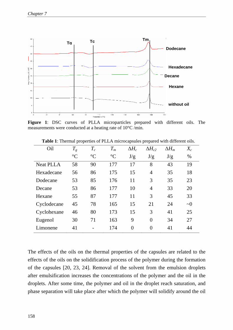
Chapter 7
158
Figure 1: DSC curves of PLLA microparticles prepared with different oils. The measurements were conducted at a heating rate of 10°C /min.
Table 1: Thermal properties of PLLA microcapsules prepared with different oils.
The effects of the oils on the thermal properties of the capsules are related to the
effects of the oils on the solidification process of the polymer during the formation
of the capsules [20, 23, 24]. Removal of the solvent from the emulsion droplets
after emulsification increases the concentrations of the polymer and the oil in the
droplets. After some time, the polymer and oil in the droplet reach saturation, and
phase separation will take place after which the polymer will solidify around the oil
Oil Tg
°C
Tc
°C
Tm
°C
∆Hc
J/g
∆Hc2
J/g
∆Hm
J/g
Xc
%
Neat PLLA 58 90 177 17 8 43 19
Hexadecane 56 86 175 15 4 35 18
Dodecane 53 85 176 11 3 35 23
Decane 53 86 177 10 4 33 20
Hexane 55 87 177 11 3 45 33
Cyclodecane 45 78 165 15 21 24 ~0
Cyclohexane 46 80 173 15 3 41 25
Eugenol 30 71 163 9 0 34 27
Limonene 41 - 174 0 0 41 44
without oil
Hexadecane
Hexane
Dodecane
Decane
Tm Tc Tg

Hollow polylactide microcapsules with controlled properties
159
droplets. The solidification process of the polymer greatly determines the eventual
properties of the resulting capsules. The low thermal transition temperatures of the
capsules prepared with eugenol suggests that PLLA has better interaction with
eugenol than with other oils, which makes the polymer chains more mobile during
capsule formation. Correspondingly, the high crystallinity of the capsules prepared
with limonene indicates strong interaction of PLLA with limonene which slows
down the phase separation and gives mobility and time for crystallization to occur
before complete solidification of the polymer. The polymer seems to be less
compatible with alkanes as compared to the other oils, consequently, the phase
separation takes place more quickly while the mobility of the polymer is reduced
faster. Therefore, the thermal transition temperatures with alkanes are higher than
for other oils.
When comparing the thermal behaviour of the capsules with that of air-cast films,
one can see that most of oils give similar trends in which the thermal transition
temperatures were the highest with normal alkanes and the lowest with terpenes,
although it should be noted that the preparation method of films is different [20].
The films were prepared by casting and evaporation to the air, while capsules were
submersed in a nonsolvent. This indicates that the interactions between the polymer
and most of the oils are rather important for the solidification of the polymer.
Besides this, solvent interactions are expected to play are role, since the
crystallinity in the films was different than in capsules. For example, the
crystallinity of the films prepared with limonene was lowest (~15%) [20], whereas
it was highest for the capsules with limonene (~ 45%). This may be ascribed to the
slower removal rate of the DCM from the capsules into the nonsolvent (DCM and
water are poorly miscible) which gives more time for crystallization to take place.
The results obtained so far clearly demonstrate that the oils can be effectively used
to influence the thermal properties of the capsules. The next section focuses on the
effects of the oils on the morphology of the capsules.
Effects of oil on morphology
Figures 2 and 3 show SEM micrographs of hollow PLLA microcapsules prepared
with different oils. The majority of the capsules are spherical and have average
sizes between 1-2 µm. With all oils hollow capsules could be produced except for

Chapter 7
160
eugenol, in which irregular,, massive particles were formed (see Figure 3d). The
formation of hollow capsules is thus influenced by the physical properties of the oil
(i.e. mutual solubility and interaction with polymer, solubility in the nonsolvent and
boiling point). The oil must be insoluble in the polymer and nonsolvent (otherwise
it will not form a separate droplet inside the droplet of polymer solution), and its
boiling point should be high enough to stay inside the polymer shell during out-
diffusion and evaporation of the solvent. For instance, when the capsules were
prepared with volatile oils (i.e. hexane and cyclohexane), some of the oil
evaporated during capsules formation which yielded solid particles. The fact that
with eugenol no hollow particles were formed may be ascribed to the good
interaction of this oil with PLLA which implies that the oil did not create a single
droplet of oil and leaves the polymer highly swollen with the oil. The solidification
of the polymer slows down, therewith allowing enough time for the oil to diffuse
out to the external nonsolvent phase (the solubility in water is higher than for other
oils).
Figure 2: SEM micrographs of PLLA microparticles prepared with different alkanes: (a) and (b) hexadecane, (c) decane, and (d) hexane.
a b
d c

Hollow polylactide microcapsules with controlled properties
161
The effects of the oils on the morphology of the air-cast films and capsules
prepared in this study are comparable. The oils that produced hollow capsules were
the same ones that produced porous films. With eugenol, a dense and nonporous
film was obtained which supports the conclusion of strong interaction of the oil
with the polymer such that phase separation was avoided and the structure
collapsed into a dense film [20].
The shell thickness is another important property of the capsules. Figures 2 and 3
show that most of the oils yielded capsules with relatively homogeneous and
controlled shell thicknesses of approximately 50-70 nm. The concentration of the
oil is one of the factors that is expected to influence the thickness and morphology
of the shell. Figure 4 shows SEM pictures of PLLA capsules prepared from a 10%
w/w polymer stock solution and with two different dodecane concentrations. The
pictures showed that with increasing the oil concentration from 9% (normal recipe)
(Figure 4a) up to 24% w/w (Figure 4b), the shell thickness was reduced and more
defects appeared in the shell. In addition, the capsules buckled and crumbled when
more oil was used; and this indicates that the shell became much weaker, and could
not withstand the generated capillary forces during removal of dodecane. This
illustrates that the polymer/oil ratio is an important parameter that can be used to
fine-tune the shell thickness and consequently the strength of the capsules.
To investigate the mechanical shell properties in detail, AFM was used, as is
described in the next section.
Effects of oil on mechanical properties of capsules
The force-distance curves of hollow PLLA microcapsules prepared with different
oils were obtained through AFM. The AFM images of all measured capsules
showed that the sizes of these capsules were comparable (between 1-2 µm). In
addition, no changes in the topography of capsules before and after experiments
were observed, which indicates that the applied load did not cause any irreversible
deformation in the capsules, and that the measurements were performed in the
elastic regime. Figure 5 shows the typical cantilever deflection (d) versus the piezo

Chapter 7
162
Figure 3: SEM micrographs of PLLA microparticles prepared with different oils: (a) cyclodecane, (b) and (c) limonene, and (d) eugenol.
Figure 4: SEM micrographs of PLLA microparticles prepared with different dodecane concentrations: (a) 9% w/w, and (b) 24% w/w. The polymer concentration was 10% w/w PLLA/DCM.
a
d c
b
a b

Hollow polylactide microcapsules with controlled properties
163
displacement (z) curve together with a schematic description of the experiment.
The solid curve in the figure corresponds to the approach phase of the tip towards
the surface of the capsule. This curve can be divided into three stages. In stage I the
tip is at a large distance from the capsule and no force is noted. Once the capsule
contacts the tip, due to the attractive forces (van der Waals or electrostatic) at stage
II , the cantilever deflects and upon increasing the applied load, the cantilever
continues deflecting until the maximum load is reached (stage III ). After that, the
retraction phase starts in which the piezo movement is reversed and the capsule
starts to retract from the tip (dashed curve in Figure 5). Because of adhesion forces,
the capsule will stay in contact with the tip, even though no force is applied, until
the maximum adhesion force is overcome at point IV, after which the tip and the
capsule separate and the cantilever returns to its starting point at stage I. To convert
the deflection-distance curve shown in Figure 5 into a force-distance curve, the
applied force (F) [nN] can be estimated from the deflection [nm] using Hooke’s
law as shown in equation 2:
dkF c ⋅−= (2)
Where kc is the cantilever spring constant [nN/nm or Nm−1], and d is the deflection
[nm].
Figure 6 shows typical force-distance curves (approach) measurements performed
on clean silica chip and hollow PLLA microcapsules prepared with dodecane,
limonene and cyclohexane. The stiffness of the shell of the capsules can be
calculated from the slope of the force-distance curve as described by Sboros and
others [25]. According to Sboros, the deflection of a cantilever applied on
elastically deformable material like a microcapsule combines two springs in series
as shown in equation 3.
sctotal kkk
111 += (3)
Where ks is the stiffness of the shell of the capsule (effective spring constant of the
shell) [Nm−1], kc is the cantilever spring constant [Nm−1], and ktotal is the overall

Chapter 7
164
effective spring constant which is equivalent to the slope of the force-distance
curve of the capsule shown in Figure 6.
Figure 5: Typical cantilever deflection (d) vs piezo displacement (z) curve measured by AFM (left). On The right, a schematic representation of the positions of the tip and the surface of the sample at different stages during the measurements as indicated by the Roman numerals. The actual curves were measured for microparticle prepared with dodecane.
Figure 6: Force-distance curves (approach) measured for clean silica chip and PLLA microparticles prepared with dodecane, limonene and cyclohexane as oils.
-10
40
90
140
190
500 750 1000
Z [nm]
For
ce [n
N]
silica chipdodecanelimonenecyclohexane
-50
50
150
250
350
500 750 1000
Z [nm]
defle
ctio
n (d
) [n
m]
approachretraction
A B
C
D
I
II
III
-50
50
150
250
350
500 750 1000
Z [nm]
defle
ctio
n (d
) [n
m]
approachretraction
I II
III
IV

Hollow polylactide microcapsules with controlled properties
165
The slope of the force-distance curve measured for a clean silica chip was
equivalent to the spring constant of the cantilever (~0.57 Nm−1). The curves for the
microcapsules showed lower average slopes than the hard silica surface (0.48 ±
0.02 Nm−1 for dodecane, 0.44 ± 0.01 Nm−1 for limonene and 0.41 ± 0.01 Nm−1
for cyclodecane). In Figure 7, the calculated average shell stiffness of these
microcapsules is shown. Capsules prepared with dodecane showed significantly
higher stiffness (3.3 Nm−1) than with limonene (2 Nm−1) and cyclohexane (1.4
Nm−1). The high stiffness of the capsules prepared with dodecane can be related to
the high thermal transition temperatures of these capsules compared to those
prepared with limonene and cyclohexane (see Table 1). The shells of the capsules
prepared with limonene and cyclohexane were more flexible, which correlates with
strong interaction between oil and polymer and higher chain mobility of the shell as
was deduced from the DSC results.
Figure 7: Average shell stiffness of PLLA microparticles prepared with dodecane, limonene and cyclohexane as oils. The average stiffness shown in the figure was measured from 5 particles of each sample. 3 force-distance curves of each particle were analyzed and the slopes were very much similar.
Comparison of the stiffness of the capsules and air-cast films shows major
differences. The films prepared with cyclohexane were stiffer than those prepared
with dodecane and limonene and the films prepared with limonene were the most
0
1
2
3
4
5
dodecane limonene cyclohexane
shel
l stif
fnes
s (N
m-1
)

Chapter 7
166
flexible among other films. This could be ascribed to the difference in the structure
of the films and capsules. The films prepared with limonene and dodecane have
much higher porosity compared to those prepared with cyclohexane, which delayed
the final rupture of the films. From this, it is clear that mechanical properties of
films are not good indicators for microcapsule properties.
Conclusions
Hollow PLLA microcapsules were prepared using a solution of PLLA in a mixture
of good and a poor solvent (oil), emulsified into water, being nonsolvent for the
polymer that is immiscible with the two solvents.
The thermal and mechanical properties of hollow PLLA microcapsules depend on
the type of oil used. The presence of the oil during formation of the capsules
enhanced the mobility during the formation of the shell and reduced the glass
transition temperature; allowing crystallization to take place to a greater extent. The
crystallinity degree of the capsules could be varied from 0% to 45% by using
different oils. Hollow capsules with well-defined shell thicknesses (~ 50 nm) were
obtained with all oils, except for eugenol. The stiffness of the shell of the capsules,
measured with AFM, was not related to the stiffness of the PLLA films, but was
highly dependent on the type of oil. Capsules prepared with dodecane were stiffer
than those prepared with limonene and cyclohexane.
Acknowledgements
The research described in this paper is part of the BURST project (IS042035).
Financial support by SENTER is kindly acknowledged. The authors wish to thank
Francisco Rossier and Dr. Mieke Kleijn for helping with AFM measurements,
Adriaan Van Aelst for preparing the SEM images, and Herman de Beukelaer for
his help with the DSC experiments.

Hollow polylactide microcapsules with controlled properties
167
References 1. Drumright, R.E., Gruber, P.R., Henton, D.E., Polylactic acid technology.
Advanced materials, 2000. 12(23): p. 1841. 2. Lim, L.-T., Auras, R., Rubino, M., Processing technologies for poly(lactic
acid). Progress in polymer science, 2008. 33(8): p. 820. 3. Lam, K.H., Nijenhuis, A.J., Bartels, H., Postema, A.R., Jonkman, M.F.,
Pennings, A.J., Nieuwenhuis, P., Reinforced poly(L-lactic acid) fibres as suture material. Journal of applied biomaterials, 1995. 6(3): p. 191.
4. Luciano, R. M., Zavaglia, C. A. C., Duek, E. A. R., Alberto-Rincon, M. C.,
Synthesis and characterization of poly(L-lactic acid) membranes: Studies in vivo and in vitro. Journal of materials science. Materials in medicine, 2003. 14(1): p. 87.
5. Lee, S. H., Kim, B. S., Kim, S. H., Kang, S. W., Kim, Y. H., Thermally
produced biodegradable scaffolds for cartilage tissue engineering. Macromolecular bioscience, 2004. 4(8): p. 802.
6. Mohamed, F., Van Der Walle, C.F., Engineering biodegradable polyester
particles with specific drug targeting and drug release properties. Journal of Pharmaceutical Sciences, 2008. 97(1): p. 71.
7. Freiberg, S., Zhu, X. X., Polymer microspheres for controlled drug release.
International Journal of Pharmaceutics, 2004. 282(1-2): p. 1. 8. Liu, R., Ma, G., Meng, F.-T., Su, Z.-G., Preparation of uniform-sized PLA
microcapsules by combining Shirasu Porous Glass membrane emulsification technique and multiple emulsion-solvent evaporation method. Journal of Controlled Release, 2005. 103(1): p. 31.
9. Kooiman, K., Böhmer, M.R., Emmer, M., Vos, H.J., Chlon, C., Shi, W.T., Hall,
C.S., de Winter, S.H.P.M., Schroën, K., Versluis, M., de Jong, N., van Wamel, A., Oil-filled polymer microcapsules for ultrasound-mediated delivery of lipophilic drugs. Journal of Controlled Release. In Press, Corrected Proof.
10. Lathia, J. D., Leodore, L., Wheatley, M. A., Polymeric contrast agent with
targeting potential. Ultrasonics, 2004. 42(1-9): p. 763. 11. Böhmer, M. R., Schroeders, R., Steenbakkers, J. A. M., de Winter, S. H. P. M.,
Duineveld, P. A., Lub, J., Nijssen, W. P. M., Pikkemaat, J. A., Stapert, H. R., Preparation of monodisperse polymer particles and capsules by ink-jet printing. Colloids and surfaces. A, Physicochemical and engineering aspects, 2006. 289(1-3): p. 96.

Chapter 7
168
12. Vladisavljević, G.T., Williams, R.A., Recent developments in manufacturing
emulsions and particulate products using membranes. Advances in colloid and interface science, 2005. 113(1): p. 1.
13. Sawalha, H., Fan, Y., Schroën, K., Boom, R.,Preparation of hollow polylactide microcapsules through premix membrane emulsification-Effects of nonsolvent properties. Journal of membrane science, 2008. 325(2): p. 665.
14. Norris, D.A., Puri, N., Sinko, P.J., The effect of physical barriers and properties
on the oral absorption of particulates. Advanced Drug Delivery Reviews, 1998. 34(2-3): p. 135.
15. Kim, H.K., Park, T.G., Comparative study on sustained release of human
growth hormone from semi-crystalline poly(-lactic acid) and amorphous poly(,-lactic-co-glycolic acid) microspheres: morphological effect on protein release. Journal of Controlled Release, 2004. 98(1): p. 115.
16. Bouakaz, A., Versluis, M., de Jong, N., High-speed optical observations of
contrast agent destruction. Ultrasound in Medicine & Biology, 2005. 31(3): p. 391-399.
17. Grayburn, P.A., Current and Future Contrast Agents. Echocardiography, 2002.
19(3): p. 259. 18. Raisinghani, A., DeMaria, A.N., Physical principles of microbubble ultrasound
contrast agents. The American Journal of Cardiology, 2002. 90(10, Supplement 1): p. 3.
19. Wuisman, P.I.J.M., Smit, T.H., Bioresorbable polymers: Heading for a new
generation of spinal cages. European spine journal, 2006. 15(2): p. 133. 20. Sawalha, H., Schroën, K., Boom, R., Addition of oils to polylactide casting
solutions as a tool to tune film morphology and mechanical properties. Submitted, 2008.
21. Ljungberg, N., Wesslén, B., Preparation and properties of plasticized
poly(lactic acid) films. Biomacromolecules, 2005. 6(3): p. 1789. 22. Sawalha, H., Schroën, K., Boom, R., mechanical properties and porosity of
polylactide for biomedical applications. Journal of applied polymer science, 2008. 107(1): p. 82.
23. Sawalha, H., Schroën, K., Boom, R., Polylactide films formed by immersion
precipitation: Effects of additives, nonsolvent, and temperature. Journal of Applied Polymer Science, 2007. 104(2): p. 959.

Hollow polylactide microcapsules with controlled properties
169
24. Sboros, V., Glynos, E., Pye, S.D., Moran, C.M., Butler, M., Ross, J., Short, R., McDicken, W.N., Koutsos, V., Nanointerrogation of ultrasonic contrast agent microbubbles using atomic force microscopy. Ultrasound in Medicine & Biology, 2006. 32(4): p. 579.

Chapter 7
170

Chapter 8
Polylactide microcapsule formation*
* This chapter is to be submitted for publication as: Hassan Sawalha, Karin Schroën and Remko Boom, Polylactide microcapsules formation.

Chapter 8
172
Abstract
Polymeric microcapsules are used for different applications. They are commonly
produced through emulsion solvent-evaporation / extraction method. In this
technique, the polymer (mainly polylactide) is dissolved in a good solvent and
together with a poor solvent (oil) is emulsified into a nonsolvent phase. After
emulsification, the solvent is removed to the nonsolvent and evaporated, which
results in solidification of the polymer around the oil droplets, which is removed
later when hollow capsules are required.
This paper discusses the fundamental aspects of the formation process of hollow
polylactide microcapsules and its effects on the physical and chemical properties of
the capsules, with emphasis on the solidification process of the polymer and the
resulting properties of the shell. The scope for improvement and adaptation of the
current process, including new emulsification techniques, is also discussed.
The main conclusion of this work is that the properties of the capsules can be fine-
tuned through the solidification process of the polymer which can be highly
influenced by the choice of the nonsolvent and oil. Since this field is hardly
investigated in literature, there is room for improvement, especially if the capsules
can be produced with the newest emulsification technologies that are becoming
available.
Polylactide microcapsules formation
173
Introduction
Biopolymers have been used extensively for various applications including
packaging, membranes, and biomaterials [1-9]. For each product, the required
properties of the materials are different from those of the pure polymer. Different
routes have been found to alter the properties, especially by using different
production techniques, and by using additives. Alternatively, also chemical
modification, grafting, cross linking, and interpenetrating polymer networks were
suggested, but these options are not considered in this paper.
Polymeric products are generally produced by melt or solution processing [3, 10-
12]. During melt processing, the polymer is heated above its melting point, shaped,
and then solidified by cooling [10, 12]. Different techniques have been proposed in
the literature and much effort has been put into optimizing the processing
conditions [10, 12]. For solution processing, the polymer is dissolved in a solvent,
shaped into the desired product, and then solidified by evaporation of the solvent to
air or extraction to an external nonsolvent [3, 11], which induces phase separation.
In both methods, additives may be used to improve the physical and chemical
properties of the resulting materials, including other polymers, plasticizers, and
fillers [13-20].
We will here discuss the production of polymeric microcapsules; they can contain a
liquid droplet, or a gas bubble (hollow microcapsules). These microcapsules are
typically between 0.1 and 100 µm, and prepared by solvent evaporation or phase
separation, as it allows lower viscosities during processing, which is important
when moulding the material in very small dimensions. As the preparation process
usually is a complex interplay of interactions between the polymer, and several
solvents and additives, prediction of the resulting encapsulate properties is a
challenge.
This paper reports on the use of ‘minor’ variations in the formulation, and process
conditions, which can result in large differences in microcapsule properties. We
will attempt to link basic phase behaviour to product properties, and these findings

Chapter 8
174
are used in the outlook to give suggestions for improvement of the production
method.
Polymer microcapsules – preparation and properties
Polymeric microcapsules have attracted a great deal of interest because of their
application in different fields such as medicine, catalysis, cosmetics, and foods [4,
11, 21-24]. In the biomedical field, microcapsules prepared from biodegradable
polymers have been frequently used to encapsulate drugs for controlled and
sustained release [4, 11]. Biodegradable hollow microcapsules prepared from
polylactide (PLA) were successfully used as ultrasound contrast agents (UCA’s) to
increase the backscatter signal of the ultrasound field and enhance the image
resolution [5, 25]. It was even recently reported that microcapsules can be loaded
with a drug, and at the same time be acoustically active [25]. The idea behind these
capsules is that, when injected in the body, these microcapsules can be ruptured by
the ultrasound field, and locally induce high dosage of the drug.
Each application of the microcapsules has specific demands regarding the
properties of the capsules, such as their size, size distribution, shell thickness,
mechanical characteristics, and in-vivo stability. For example, the acoustic activity
of capsules, and therewith related the drug release, is expected to be influenced by
the mechanical strength, thickness, and crystallinity of the shell [26-30]. In
addition, size and size distribution of the capsules can highly influence their
biocompatibility and circulation time within the body [31, 32]. Therefore, tuning
the properties of the capsules is a challenge in every sense.
Although different techniques have been used for preparation of microcapsules,
such as complex coacervation, spray drying, layer-by-layer assembly, and emulsion
solvent-extraction / evaporation, we will focus on the last method, which is used
most [11, 33, 34]. In this paper, we will focus further on the biodegradable
polymer, polylactide (PLA), which is used often in microcapsule production, and
present the effects reported for this polymer as an illustrative example for which
effects can be induced in polymers through relatively small changes in the
formulation and the formation process.

Polylactide microcapsules formation
175
Phase behaviour of solvent, nonsolvent, and polymer
For the emulsion solvent extraction / evaporation technique, the polymer (i.e.
polylactide) is dissolved in a mixture of a good solvent (e.g., dichloromethane,
DCM) and a poor solvent (from now on called oil, since it is a higher alkane). The
polymer / solvent / oil mixture is then emulsified into a non-solvent phase (water)
which usually contains a stabilizer (i.e. polyvinyl alcohol, PVA). Since the solvent
is in general hardly soluble in the non-solvent phase, the droplets are initially
stable. However, the solvent slowly diffuses to the continuous non-solvent phase
and evaporates at the surface. Hence, the droplets slowly become more
concentrated.
The demixing process inside the droplets during out-diffusion of solvent is
illustrated in more detail in the phase diagram shown in Figure 1. The shaded area
is the miscibility gap (oil is a poor solvent for PLA), which is bordered by the
binodal. Any composition in the shaded area is not stable, and will demix into a
polymer poor and a polymer rich phase, which are connected by tie lines in the
phase diagram (interrupted lines). For an initial solution of PLA in DCM and oil
represented by point a, the solvent is slowly extracted to the external nonsolvent,
and the composition of the droplet changes along the arrow towards the PLA-oil
axis. The solution becomes richer in PLA and oil: both oil and PLA are strongly
incompatible with water, and are not volatile, therefore they do not diffuse out to
the nonsolvent and stay in the droplet.
As the droplet becomes more and more enriched in PLA and oil, the composition
enters the miscibility gap. Since that will happen at a PLA concentration that is
higher than that of the critical point, one will see nucleation of a polymer poor and
oil rich phase. From this point, the droplet has two phases; each phase is located on
one arm of the bimodal (the direction of the arrow in Figure 1 only represents the
overall composition). Since the concentration of solvent is lowest at the interface
and highest in the middle of the droplet, the nucleation of the PLA poor (oil rich)
phase will be in the middle of the droplet while the polymer rich phase will be in

Chapter 8
176
between the internal oil droplet and the outside nonsolvent bath. The polymer
around the oil droplet will ultimately from a (solid) shell. The total volume
reduction of the primary emulsion droplet is given by the amount of solvent
removed, and therewith the size of the microcapsules is a priori known. The
volume of the internal droplet is determined by the amount of oil present [35, 36],
and therewith, the shell thickness can be tuned.
Figure 1: Schematic phase diagram of the PLA-DCM-oil system.
The ultimate solidification of the shell is for an amorphous polymer by glass
transition, vitrification, or gelation [37, 38], but for a semi-crystalline polymer as
PLA by crystallization [39-41]. The actual structure and properties of the shell is
influenced by the rate of solvent removal and the interaction between PLA and oil
(as is discussed in the next sections) [3, 39], and therewith it is clear that the mutual
thermodynamic interactions between PLA, DCM, oil, and water greatly determine
the ultimate properties of the microcapsule [3, 42].
PLA
Solvent Oil a
binodal
-
Critical point

Polylactide microcapsules formation
177
Solvent removal
a. Process options
The miscibility of DCM with the nonsolvent determines its removal rate from the
droplets. Water is practically immiscible with DCM, the removal rate is low, and
the total formation process is slow as well, therewith allowing ample time for
crystallization of PLA [11, 35, 43]. Besides, other effects, such as Ostwald ripening
and coalescence, can occur, therewith increasing the polydispersity of the capsules.
These drawbacks, can be considerably reduced when the solvent is removed faster.
Several attempts have been made to speed up the removal of the solvent e.g. by
increasing the temperature (and hence increasing diffusion rates, DCM dissolution,
and increasing its vapour pressure) or decreasing the pressure in emulsion
preparation vessel (thereby increasing its evaporation rate) [43]. It was found that
increasing the temperature led to larger capsules and lower encapsulation efficiency
(i.e., the oil remaining in the shell was lower) [11, 43], and this could be due to less
effective solidification at higher temperatures. Decreasing the pressure can enhance
the evaporation of DCM to air; however, it will not speed up the transport from
droplets towards the water surface, which is the rate-limiting step in the formation
process, evaporation is orders of magnitude faster. Some researchers reported that
the encapsulation efficiency increased with decreasing pressure whereas others
reported a decrease [11, 43]. This may be due to differences in vapour pressure of
the oil used; generally, the effects of pressure on encapsulation efficiency are
minor, as could be expected for a system that is not limited by evaporation.
Another route is to increase the compatibility between DCM and the nonsolvent
phase. Mostly a two-step process is used in literature [11, 44]; the PLA solution is
first emulsified into a small amount of water, which facilitates emulsion handling,
after which extra water is added. This is done in a single step or in many
consecutive steps [44, 45]. Obviously, more water means that more DCM can be
extracted. However, since the maximum concentration of DCM in water is quite
low, this method requires large amounts of water which complicates harvesting of
the capsules, and further, rather large quantities of water contaminated with DCM
are produced.

Chapter 8
178
b. Co-solvents
Some studies report the use of co-solvent systems to speed up the total removal of
solvent from the spheres [33, 46, 47]. The polymer was here dissolved in a mixture
of DCM and another solvent that is miscible with water, e.g. an alcohol. After
emulsification, the alcohol is extracted to the water phase, followed by much
slower extraction of the DCM. Even though the addition of alcohol will initially
increase the total rate of solvent removal, the rate-limiting step in particle
formation, which is the out-diffusion of DCM, is not alleviated.
c. Nonsolvent
To overcome the disadvantages of slow removal rate of the solvent, which are
inherent to the use of immiscible nonsolvents such as water, it is logical to use a
nonsolvent that is better miscible with the solvent. In previous papers, we reported
the effects of the nonsolvent quality on the formation of PLA microcapsules and
films [35, 36]. To improve the compatibility between DCM and the nonsolvent
phase, a lower alcohol (e.g. methanol), which is completely miscible with DCM,
was added to water. The solubility of DCM strongly increased with increasing
alcohol concentration, therewith speeding up the formation process, as is shown
below.
The effect of enhanced removal rate on the structure was evaluated for thin films
prepared through immersion precipitation, as model system for capsule preparation
[35, 39]. Thin films of PLLA/DCM/dodecane solutions were cast on a glass plate
and submerged in a bath filled with different water-methanol mixtures, and phase
separation was followed in time. For pure water, demixing starts after ~ 6 min,
whereas the demixing time dramatically decreases with increasing methanol
concentration; with pure methanol demixing starts after only 2 seconds [39]. As a
result, major differences in the structure of the films were observed. With
methanol, an asymmetric structure was found consisting of a solid dense top layer
and a porous sub-layer (see Figure 2a) [39, 48]. With water, the morphology was
completely different: symmetric spherulitic (crystalline) structures with large and
irregular interstitial pores were obtained (Figure 2b). The formation of the
spherulites (crystals) is ascribed to the slow out-diffusion of DCM which gives
ample time for crystallization to take place in the film; the regular porous structure

Polylactide microcapsules formation
179
of the film made with methanol indicates liquid-liquid demixing before
crystallization could set in. The nonsolvent composition did not only influence the
morphology but also the mechanical properties of the films. The films prepared
with water were weak and fragile, while with increasing methanol concentration,
the strength and ductility of the films were significantly improved [48].
It is clear, that the nonsolvent influences the rate of the demixing process strongly,
and consequently, the properties of the films. Other researchers have reported that
the solvent removal rate can influence the crystallinity of the capsules [49]; faster
removal of the solvent can decrease the crystallinity of the capsules, as there is not
enough time for crystallization to proceed. Similar effects were reported for
different oils that also influence the solvent removal rate, as is discussed
extensively in the following sections.
Figure 2: Typical SEM images PLLA films prepared from 10:05:85 w/w PLLA:dodecane:DCM with different non-solvents: a) methanol, b) water [48].
In our own work we found that with increasing methanol concentration, the
microcapsules indeed formed faster, and as a desired side effect, their size
decreased [35]. Addition of methanol up to 30% facilitated the initial removal of
DCM from the droplets, which resulted in faster solidification, which preserved the
original shape and size of the capsules (no coalescence was observed). The smaller
size of the capsules may be caused by two effects. First, addition of methanol
significantly reduces the interfacial tension of the nonsolvent, which will yield
smaller droplets during emulsification; ultimately leading to smaller capsules.
Second, the addition of alcohol to the nonsolvent bath will also increase the
(a) (b)

Chapter 8
180
compatibility of the oil with the nonsolvent. Consequently, some of the oil may be
lost to the nonsolvent bath, and the resulting capsules will be smaller. For the
combination of non-solvent and oil used in this study, the effect is expected to be
only of minor influence, if any, but it may become relevant for other combinations.
At concentrations over 30% w/w, the size of the capsules became bigger, so there is
a clear optimum in the alcohol concentration that can be applied. Most likely, the
diffusion of methanol into the capsules increased at high methanol concentration;
the capsules swell, become bigger and more porous. At methanol concentrations >
50 %, fragments of polymer aggregates were formed instead of microcapsules.
Probably, the solution now enters the demixing region near the critical point, or
even below it, due to much higher alcohol concentrations in the droplets, and no
capsules can be formed.
Besides methanol, also ethanol and 2-propanol were used [36]. The size and the
span of the microcapsules were highly dependent on the type of alcohol used. For
example, use of 25% (w/w) 2-propanol in the water bath produced capsules with
average size of 0.4 µm, compared to 0.8 µm with 30% ethanol, 1.4 µm with 30%
methanol, and 2.2 µm with water. In contrast, the size distribution of the capsules
formed with 2-propanol was broader than for ethanol and methanol (see Figure 3).
The fact that the sizes of the microcapsules were different can be related to various
quantities that play a role during emulsification, i.e. interfacial tension and
viscosity, but also to different interactions between the polymer, solvent, and oil.
How all these parameters are exactly linked is currently unknown.
The main conclusion that can be drawn on the use of different non-solvents is that
the properties of the microcapsules (i.e. size, size distribution, and morphology) can
be fine-tuned by adjusting the quality of the nonsolvent. An added benefit of
enhanced DCM removal through appropriate choice of non-solvent is that the
production process can become considerably faster, and a smaller volume of
nonsolvent mixture is needed. Besides that, there is also a clear link to the
production of smaller capsules, albeit not fully understood.

Polylactide microcapsules formation
181
0
5
10
15
20
0.01 0.1 1 10 100 1000diameter [µm ]
volu
me
(%)
water
30% methanol
30% ethanol
25% 2-propanol
0
5
10
15
20
0.01 0.1 1 10 100 1000diameter [µm ][µm ]
volu
me
(%)
water
30% methanol
30% ethanol
25% 2-propanol
Figure 3: Typical particle size distribution of PLLA microcapsules prepared with different alcohol-water mixtures as nonsolvent [36].
Other options to influence material properties
Blending and annealing
Modification of material properties is mostly achieved through blending/mixing
with other polymers or plasticizers, such as poly (ethylene glycol) PEG,
polycaprolactone (PCL), poly(1,5-dioxepan) (PDOX), and chitosan. These
components were used to influence (bio) degradation, and thermal behaviour of the
microcapsules [33, 50-52]. Park and co-workers, showed that capsules prepared
from PLLA-PEG copolymer were more hydrophilic and have lower glass transition
temperature compared with those prepared with PLLA only [50]. In addition, the
presence of the PEG hydrophilic group within the PLLA matrix was reported to
increase the drug release rates. In another study, Edlund and Albertsson blended
PLLA with PDOX and found that the crystallinity of the microcapsules decreases
with increasing PDOX content in the blend [52]. Even though blending therefore
seems a successful method, blending polymers has limited practicality, because of
limited number of polymers that are compatible; inter-polymer phase separation
during microcapsule formation can be expected, and this will not lead to integral
microcapsules [53].

Chapter 8
182
Sosnowsk reported that the degree of crystallinity of PLLA capsules could be
controlled (0% to 60%) through annealing of the capsules [54]; however, exposing
the capsules to high temperatures is not trivial, because it can also lead to major
changes in the structure or even complete degradation [54].
Effects of oil
As mentioned previously, removal rate of the solvent plays an important role in the
microcapsule formation process. Besides the non-solvent phase, also the oil phase
is expected to be of influence on the removal rate, as is clear from Figure 1. To
estimate the effects of oil on microcapsule formation, films were used as a model.
Films with different dodecane concentrations were immersed in methanol baths,
and the delay time of demixing (i.e., the time after which significant demixing sets
in after immersion) was shorter for films with higher dodecane concentrations.
Demixing starts after ~16 seconds in films prepared without dodecane, whereas
demixing is almost instantaneous with 10% dodecane [39]. The presence of the oil
in the PLA solution brought the initial composition much closer to the demixing
gap. Thus, only a small out-flux of DCM was sufficient to induce demixing, and
consequently, the morphology and mechanical properties of the films changed [39].
The presence of the oil induces porosity in the films which reduces their strength,
but sometimes increases their ductility [39, 48]. Demixing in these films takes
place by nucleation and growth of dodecane-rich droplets entrapped within the
polymer rich phase. These droplets were the precursors of most of the pores
observed. The oil can therefore play an important role in the formation process and
properties of the resulting film. Consequently, it was expected that this would be
similar for microcapsules, prepared by premix membrane emulsification [36, 55].
Various oils were tested including alkanes, cyclic alkanes and terpenes (limonene
and eugenol), and as expected, the morphology and thermal and mechanical
properties of the capsules strongly depended on the oil [55]. With all oils, hollow
microcapsules with controlled shell thickness of approximately 50 nm could be
formed, except with eugenol, which led to irregular, non-hollow microcapsules.
The thermal transition temperatures of the hollow capsules were lower than of solid
microcapsules prepared without oil. Depending on the type of oil used, a reduction

Polylactide microcapsules formation
183
of about 30 °C in the glass transition temperature, 20 °C in the cold crystallization
temperature, and 15 °C in the melting temperature was recorded [55]. Besides,
crystallinity of the microcapsules was highly dependent on the type of oil: the
degree of crystallinity varied from 0% to 45 % depending on the oil used.
Furthermore, the type of oil influenced the mechanical stiffness of the shell as
measured with AFM: microcapsules prepared with dodecane were stiffer than when
prepared with limonene and cyclohexane. Therefore, just as with the films, the
mechanical properties of the very thin microcapsule shells were influenced by the
type of oil used, albeit differently than for films, as will be described later [55].
The differences in the thermal and mechanical properties of the capsules can be
attributed to different interactions between polymer, solvent, nonsolvent, and oil,
which affect the demixing and solidification process. For example, the low thermal
transition temperature of the capsules prepared with terpenes is due to the better
interaction of the polymer with these oils, which delays the demixing during
microcapsule formation, and gives extra mobility to the polymer chains before
complete solidification of the shell (and this can lead to crystallization that is more
extensive). Eugenol seems to have such a strong interaction with PLLA that the oil
and the polymer did no phase separate. No separate oil droplet was formed in the
middle of the capsule, and a swollen, non-hollow particle resulted.
From the above, it is clear that interactions between oil and polymer can be
essential for microcapsule formation; however, also the non-solvent can have an
influence as discussed in previous sections. Although indirectly, influence of
nonsolvent in-diffusion on the solidification process could be estimated from a
comparison of results for microcapsules and air cast films [55, 56]. Most of the oils
induced porosity and reduced the thermal transition temperatures, and the
maximum strength of the films, while the ductility improved [56]. As for capsules,
the thermal transitions of the films were lowest with terpenes, and highest with
alkanes, in spite of the absence of the nonsolvent. This implies that the interaction
between the polymer and most of the oils is a dominant effect during solidification.
The crystallinity of the films did not show the same trend as for microcapsules. For
instance, capsules prepared with limonene and water as nonsolvent were much
more crystalline (~ 45%) than air-cast films prepared with the same oil (~ 15%).

Chapter 8
184
This may well be due to the nonsolvent; the removal of DCM from the capsules
through the external water phase is much slower than for air-cast films (direct
evaporation of DCM), which allows more time for crystallization in the capsules.
Further, a major difference in the stiffness of the capsules and films was observed.
In contrast to the microcapsules, films prepared with limonene and dodecane were
much more ductile than those prepared with cyclohexane. This can be attributed to
the porosity of the first mentioned films, which allows more dissipation of fracture
energy during extension [56].
The effects of the oils on the thermal and mechanical properties of the capsules and
films indicate a plasticizing effect of the oil on the polymer chains; the effects are
quite similar to those reported in the literature for plasticizers. This is interesting
since it was thought that the main purpose of the oil in the current process was to
form a template for the shell, resulting in control over the shell thickness, whereas
from the effects described earlier it is clear that it is just as important for tuning the
thermal and mechanical properties.
In summary, the major effects in phase behaviour seem to be caused by the
nonsolvent and oil; properties of PLA microcapsules can be tuned, and this may
extend their range of application.
Emulsification methods
Next to the mechanical properties of the microcapsule walls, also size and size
distribution are important. Various existing options for production of the primary
emulsions have been reported, which will be discussed below. In addition, some
newer technologies will be mentioned in the outlook section, and their potential for
capsule formation will be discussed.
Existing methods
a. Pre-mix emulsification
Currently, pre-mix membrane emulsification seems to be the method of choice for
microcapsule production, because of its simplicity, versatility, and productivity.

Polylactide microcapsules formation
185
The method involves mixing of the ingredients and repeated passage through a
membrane. The microcapsules that are produced have a relatively sharp
distribution, because of repeated passage through the membrane. However, there is
a limit to the rate of phase separation, since the droplet should remain fluid during
repeated passage through the membrane. As mentioned previously, proper choice
of the non-solvent may help in optimising this.
Pre-mix emulsification also allows production of microcapsules that have
combined functionalities. For example, microcapsules can be produced that are
only partially filled with oil, which carries a lipophilic drug, and shows acoustic
activity, as demonstrated by Kooiman and co-workers [25]. In this way, the
capsules can be used for diagnostics and therapy. The emulsification step does not
have to be adjusted; only the PLA solution should contain a mixture of oils, of
which one is not removed by freeze-drying.
An issue that still needs to be resolved is how pre-mix emulsification can be scaled
up. For a niche product like microcapsules, the current method is sufficient, but if
larger volumes are to be produced, obviously, the critical times for the various
processes to take place need to be adjusted, to allow handling of the larger
volumes, and this is still considered a challenge.
b. Cross-flow emulsification
Although the capsule size distribution obtained for pre-mix membrane
emulsification is relatively narrow, further improvement would be useful. In
literature, cross flow emulsification is used for regular oil-in-water and water-in-oil
emulsions, particles, and capsule formation, and with this technology, good results
could be obtained [21, 57-60]. For cross-flow emulsification, the oil is pressed
through the membrane, and the oil droplets which emerge from the topside of the
membrane, are carried away by the cross-flowing continuous phase. In general, low
interfacial tensions (as reported for alcohol / water mixtures) facilitate production
of smaller droplets by cross-flow emulsification. The membrane of choice is
Shirazu porous glass, which works well in various applications. A critical point for
this method is that phase separation should not take place inside the membrane,
since this would lead to blockage of pores and loss of productivity. Besides, the

Chapter 8
186
membrane area needed for larger production rates could be a drawback, as was the
case for pre-mix emulsification.
c. Ink jetting method
A different method is ink jetting as described by Böhmer and co-workers [34].
Very monodisperse particles and capsules could be obtained, as is illustrated in
Figure 4. The productivity, of the technique is quite high; up to 600,000 capsules
per second could be made with one nozzle. The size of the capsules is uniform, and
could be slightly tuned through the choice of the emulsification nozzle. In general,
the generated droplets are large and relatively low polymer concentrations have to
be used to allow sufficient reduction in size through removal of the solvent.
Although the productivity of one nozzle is high, scale-up of this technology is a
major challenge, as is the reduction of the amount of solvent that is currently
needed.
Figure 4: SEM images of products prepared through ink jetting. Solid particles are shown on the left, on the right hollow particles are shown that were obtained by freeze drying of oil filled particles [34].

Polylactide microcapsules formation
187
Outlook
Microcapsules
For each application, the PLA microcapsules will need to meet specific
requirements. The size of UCA’s, together with the ductility, and mechanical
strength of the shell are expected to highly influence acoustic behaviour. For
instance, a ductile shell is expected to allow the bubble to resonate better and
longer in the acoustic field than stiffer shells before bursting completely, which
would be important for imaging purposes. The mechanical strength of the shell is
expected to determine the maximum acoustic pressure needed to burst the
microbubble. Its strength should be sufficient but not too high, since the bubble
should burst at medically safe pressures. In drug delivery, a fast and controlled
release of the drug is preferred in some cases, whereas in other cases slow release is
required. The various factors discussed in the previous sections showed that control
of structure, crystallinity, glass transition, and mechanical strength, are within
reach, and therewith, optimising the properties for specific application may become
feasible.
One of the main problems that occurred during preparation of drug-loaded capsules
was that the drug encapsulation efficiency was insufficient, due to out-diffusion of
the encapsulated drug during microcapsule formation. A faster solidification of the
polymer through faster removal is expected to improve encapsulation efficiency
considerably. Use of alcohol-water mixtures as non-solvent system seems to be a
promising road for that.
The properties of the capsules strongly depend on the demixing and solidification
process of the polymer, which can be controlled by the choice of nonsolvent and
oil. A window of operation that links the properties of the capsules with the
solidification process of the polymer can lead to a straightforward route for
optimization of the properties of the microcapsules. To establish that, the phase
separation behaviour of the polymer with different oils, solvents, and nonsolvent
systems needs to be studied quantitatively. Although this sounds like a very
straightforward way to go, construction of phase diagrams, and investigation of
microcapsule properties, is a time consuming task. However, given the possibilities
that it opens, it may be well worth the (time) investment.

Chapter 8
188
Although ideally, all interactions are taken into account as described above, some
general rules of thumb are obvious from the previous sections. The solvent–
nonsolvent interaction is expected to dominate the initial stages of the phase
separation, and therewith the size and the morphology of the microcapsules.
Whereas, the oil-polymer interaction is expected to be most relevant for the
solidification processes (i.e. crystallization, vitrification or glassification), which
will determine the mechanical and thermal properties of the shell.
Although this paper focuses on PLA, it is expected that similar effects will play a
role for microcapsules from other polymers. The nonsolvent-solvent interactions
will determine the solvent removal rate, and therewith the size and morphology.
The solidification process and consequently the properties of the capsules or films
will mostly depend on the polymer and its interactions with oil.
Emulsification methods
Within the field of microtechnology, various new emulsification technologies have
been proposed, and some of these seem to be well suited for the preparation of
monodisperse capsules, albeit that they have not been tested for this specific
purpose. Here we give a small overview of those methods that would allow up- or
outscaling to realistic productivities (see Table 1).
Table 1: Comparison of two of micro technological devices for capsule production. Technique Formation
mechanism
Productivity Scalable Issues Feasibility
T-junctions Shear forces
detach
droplets
High Yes Surface
interactions
+
Micro
channels
Spontaneous
droplet
formation
Medium Yes Surface
interactions
+

Polylactide microcapsules formation
189
Microsieves and microchannels with T-shaped (see Figure 5) or Y-shaped
junctions, use the shear force by a cross-flowing phase to detach droplets, as was
the case for cross-flow membrane emulsification [61]. The main advantage of the
micro technological device is that the shear forces can be precisely controlled, and
therefore, uniform droplets are formed. The productivity per junction is high, up to
1000’s of droplets per second, depending on the choice of the components used.
Outscaled systems (i.e., systems using many microchannels working in parallel)
have been reported in literature. Nisisako and Torii [62], developed a system with
256 parallel junctions, while Van der Graaf, and Abrahamse used microsieves
featuring many pores actively simultaneously [63, 64].
Figure 5: Scalable micro technological methods for the production of droplets. A: T-junction where the oil is pushed from the small channel into the larger channel that hosts the cross-flowing continuous phase [61]. B. Microchannel, where the oil is pushed onto a terrace where it forms a disk. Once the disk reaches the end of the terrace, it can form a spherical droplet, which subsequently detaches [65].
In microchannel emulsification, first presented by Sigiura and co-workers [65], and
scaled-up by Kobayashi c.s., oil is first pushed onto a so-called terrace, where it
assumes a disk-like shape. This disk grows through the continuous oil supply, and
eventually reaches the end of the terrace, where it can assume a spherical shape.
Because of the Laplace pressure differences that are involved in this change in
shape, the droplet will snap off spontaneously; at low throughputs, the size of the
droplets is determined by the design of the terrace only. Very monodisperse
emulsions can be produced with these systems. Although the productivity is not as
high as for shear- based systems, the method is scalable and does have the
advantage that only the oil phase needs to be controlled. Out-scaled versions of
microchannels have been developed and were discussed in literature [62].
(a) (b)

Chapter 8
190
Although various other microtechnological methods are known from literature,
only microchannel emulsification, cross-flow emulsification with microsieves, and
to a lesser degree, T- and Y-shaped microchannel systems can be outscaled. As all
these systems inherently feature a very high surface-to-volume ratio, interaction of
the polymer solutions with the surface of the device is a matter of concern. Surfaces
that are not sufficiently hydrophilic or that would change their properties during
processing will lead to plugged microchannels and process failure. Various
modification methods are known from literature, and the surfaces of glass and Si
(typical materials used for micro devices) can be modified seemingly at will [66-
69]. However, reliable application of these methods inside a microstructure is still a
challenge.
Conclusion: The ‘ideal’ process
In an ideal production system for polylactide microcapsules, small, and relatively
uniform capsules can be produced. Further, it is possible to control the thermal and
mechanical properties of the shell. And even more ideally, the process is flexible
and has a high throughput.
Premix membrane emulsification combines many of these aspects; it has high
throughput and acceptable control over the size and size distribution. Through
adjustment of the nonsolvent composition, solidification of the polymer can be
controlled, the original size and morphology of the capsules can be preserved
better, and it is expected that also encapsulation efficiency can be enhanced. The
properties of the microcapsules can even be fine-tuned through the choice of the
added ‘oil’. And therewith, pre-mix emulsification has many benefits.
In the future, it is expected that premix emulsification can be replaced by other
micro-technological devices such i.e. micro-channel or micro-sieve systems, which
give sharper size distributions. For cross-flow systems, the size of the particles can
even be reduced further when using a non-solvent with a low interfacial tension
(given that there is sufficient time before solidification sets in). In microchannel

Polylactide microcapsules formation
191
systems, a non-solvent with a high interfacial tension (i.e. water) needs to be used
to produce small droplets. For further tuning of the microcapsule properties,
different stages including other non-solvent baths, could be an option, albeit that
this would lead to more complex microcapsule harvesting as indicated in previous
sections. For both technologies, it is clear that out-scaling and surface modification
of these systems to make them more compatible with polymer solutions is still a
major challenge, but once these hurdles are taken, a new generation of truly
monodisperse microcapsules will become available.

Chapter 8
192
References
1. Mohamed, F., Van Der Walle, C.F., Engineering biodegradable polyester particles with specific drug targeting and drug release properties. Journal of Pharmaceutical Sciences, 2008. 97(1): p. 71.
2. Auras, R., Harte, B., Selke, S., An overview of polylactides as packaging
materials. Macromolecular bioscience, 2004. 4(9): p. 835. 3. Van de Witte, P., Dijkstra, P. J., Van den Berg, J. W. A., Feijen, J., Phase
separation processes in polymer solutions in relation to membrane formation. Journal of Membrane Science, 1996. 117(1-2): p. 1.
4. Freiberg, S., Zhu, X. X., Polymer microspheres for controlled drug release.
International Journal of Pharmaceutics, 2004. 282(1-2): p. 1. 5. Lathia, J. D., Leodore, L., Wheatley, M. A., Polymeric contrast agent with
targeting potential. Ultrasonics, 2004. 42(1-9): p. 763. 6. Karageorgiou, V., Kaplan, D., Porosity of 3D biomaterial scaffolds and
osteogenesis. Biomaterials, 2005. 26(27): p. 5474. 7. Lam, K.H., Nijenhuis, A.J., Bartels, H., Postema, A.R., Jonkman, M.F.,
Pennings, A.J., Nieuwenhuis, P., Reinforced poly(L-lactic acid) fibres as suture material. Journal of applied biomaterials, 1995. 6(3): p. [d]191.
8. Luciano, R. M., Zavaglia, C. A. C., Duek, E. A. R., Alberto-Rincon, M. C.,
Synthesis and characterization of poly(L-lactic acid) membranes: Studies in vivo and in vitro. Journal of materials science. Materials in medicine, 2003. 14(1): p. 87.
9. Lee, S. H., Kim, B. S., Kim, S. H., Kang, S. W., Kim, Y. H., Thermally
produced biodegradable scaffolds for cartilage tissue engineering. Macromolecular bioscience, 2004. 4(8): p. 802.
10. Rhim, J.-W., Mohanty, A.K., Singh, S.P., Ng, P.K.W., Effect of the processing
methods on the performance of polylactide films: Thermocompression versus solvent casting. Journal of applied polymer science, 2006. 101(6): p. 3736.
11. Freitas, S., Merkle, H.P., Gander, B., Microencapsulation by solvent
extraction/evaporation: Reviewing the state of the art of microsphere preparation process technology. Journal of Controlled Release, 2005. 102(2): p. 313

Polylactide microcapsules formation
193
12. Lim, L.-T., Auras, R., Rubino, M., Processing technologies for poly(lactic acid). Progress in polymer science, 2008. 33(8): p. 820.
13. Broström, J., Boss, A., Chronakis, I.S., Biodegradable films of partly branched
poly(L-lactide)-co-poly(ε-caprolactone) copolymer: Modulation of phase morphology, plasticization properties and thermal depolymerization. Biomacromolecules, 2004. 5(3): p. 1124.
14. Sheth, M., Kumar, R.A., Davé, V., Gross, R.A., Mccarthy, S.P., Biodegradable
polymer blends of poly(lactic acid) and poly(ethylene glycol). Journal of applied polymer science, 1997. 66(8): p. 1495.
15. Ke, T., Sun, S.X., Seib, P., Blending of poly(lactic acid) and starches
containing varying amylose content. Journal of applied polymer science, 2003. 89(13): p. 3639.
16. Labrecque, L.V., Kumar, R.A., Davé, V., Gross, R.A., Mccarthy, S.P. , Citrate
esters as plasticizers for poly(lactic acid). Journal of applied polymer science, 1997. 66(8): p. 1507.
17. Jacobsen, S., Fritz, H.G., Plasticizing polylactide - the effect of different
plasticizers on the mechanical properties. Polymer engineering and science, 1999. 39(7): p. 1303.
18. Martin, O., Avérous, L., Poly(lactic acid): plasticization and properties of
biodegradable multiphase systems. Polymer, 2001. 42(14): p. 6209. 19. Ljungberg, N., Wesslén, B., Preparation and properties of plasticized
poly(lactic acid) films. Biomacromolecules, 2005. 6(3): p. 1789. 20. Leszczyńska, A., Njuguna, J., Pielichowski, K., Banerjee, J.R.,
Polymer/montmorillonite nanocomposites with improved thermal properties: Part I. Factors influencing thermal stability and mechanisms of thermal stability improvement. Thermochimica Acta, 2007. 453(2): p. 75.
21. Vladisavljević, G. T., Williams, R. A., Recent developments in manufacturing
emulsions and particulate products using membranes. Advances in colloid and interface science, 2005. 113(1): p. 1.
22. Johnston, A.P.R., Cortez, C., Angelatos, A.S., Caruso, F., Layer-by-layer
engineered capsules and their applications. Current Opinion in Colloid & Interface Science, 2006. 11(4): p. 203.
23. Buonomenna, M.G., Figoli, A., Spezzano, I., Davoli, M., Drioli, E., New PVDF
microcapsules for application in catalysis. Applied Catalysis B: Environmental, 2008. 80(3-4): p. 185.

Chapter 8
194
24. Morganti, P., Ruocco, E., Wolf, R., Ruocco, V., Percutaneous absorption and
delivery systems. Clinics in Dermatology, 2001. 19(4): p. 489. 25. Kooiman, K., Böhmer, M.R., Emmer, M., Vos, H.J., Chlon, C., Shi, W.T., Hall,
C.S., de Winter, S.H.P.M., Schroën, K., Versluis, M., de Jong, N., van Wamel, A., Oil-filled polymer microcapsules for ultrasound-mediated delivery of lipophilic drugs. Journal of Controlled Release. In Press, Corrected Proof.
26. Kim, H.K., Park, T.G., Comparative study on sustained release of human growth hormone from semi-crystalline poly(-lactic acid) and amorphous poly(,-lactic-co-glycolic acid) microspheres: morphological effect on protein release. Journal of Controlled Release, 2004. 98(1): p. 115.
27. Grayburn, P.A., Current and Future Contrast Agents. Echocardiography, 2002.
19(3): p. 259. 28. Norris, D.A., Puri, N., Sinko, P.J., The effect of physical barriers and properties
on the oral absorption of particulates. Advanced Drug Delivery Reviews, 1998. 34(2-3): p. 135.
29. Bouakaz, A., Versluis, M., de Jong, N., High-speed optical observations of
contrast agent destruction. Ultrasound in Medicine & Biology, 2005. 31(3): p. 391.
30. Raisinghani, A., DeMaria, A.N., Physical principles of microbubble ultrasound
contrast agents. The American Journal of Cardiology, 2002. 90(10, Supplement 1): p. 3.
31. Gref, R. R., Minamitake, Y., Peracchia, M. T., Trubetskoy,V., Torchilin,V.,
Langer, R., Biodegradable long-circulating polymeric nanospheres. Science, 1994. 263(5153): p. 1600.
32. Chu, L. Y., Park, S. H., Yamaguchi, T., Nakao, S. I., Preparation of Micron-
Sized Monodispersed Thermoresponsive Core-Shell Microcapsules. Langmuir, 2002. 18(5): p. 1856.
33. Lassalle, V., Ferreira, M.L., PLA Nano- and Microparticles for Drug Delivery:
An Overview of the Methods of Preparation. Macromolecular Bioscience, 2007. 7(6): p. 767.
34. Böhmer, M. R., Schroeders, R., Steenbakkers, J. A. M., de Winter, S. H. P. M.,
Duineveld, P. A., Lub, J., Nijssen, W. P. M., Pikkemaat, J. A., Stapert, H. R., Preparation of monodisperse polymer particles and capsules by ink-jet printing. Colloids and surfaces. A, Physicochemical and engineering aspects, 2006. 289(1-3): p. 96.

Polylactide microcapsules formation
195
35. Sawalha, H., Purwanti, N., Rinzema, A., Schroën, K., Boom, R., Polylactide microspheres prepared by premix membrane emulsification-Effects of solvent removal rate. Journal of membrane science, 2008. 310(1-2): p. 484.
36. Sawalha, H., Fan, Y., Schroën, K., Boom, R.,Preparation of hollow polylactide
microcapsules through premix membrane emulsification-Effects of nonsolvent properties. Journal of membrane science, 2008. 325(2): p. 665.
37. Wienk, I. M., Boom, R. M., Beerlage, M. A. M., Bulte, A. M. W., Smolders, C.
A., Strathmann, H., Recent advances in the formation of phase inversion membranes made from amorphous or semi-crystalline polymers. Journal of Membrane Science, 1996. 113(2): p. 361.
38. Young, T. H., Cheng, L. P., Lin, D. J., Fane, L., Chuang, W. Y., Mechanisms of PVDF membrane formation by immersion-precipitation in soft (1-octanol) and harsh (water) nonsolvents. Polymer, 1999. 40(19): p. 5315.
39. Sawalha, H., Schroën, K., Boom, R., Polylactide films formed by immersion
precipitation: Effects of additives, nonsolvent, and temperature. Journal of Applied Polymer Science, 2007. 104(2): p. 959.
40. Bulte, A. M. W., Mulder, M. H. V., Smolders, C. A., Strathmann, H., Diffusion
induced phase separation with crystallizable nylons. II. Relation to final membrane morphology. Journal of Membrane Science, 1996. 121(1): p. 51.
41. Cheng, L. P., Young, T. H., You, W. M., Formation of crystalline EVAL
membranes by controlled mass transfer process in water-DMSO-EVAL copolymer systems. Journal of Membrane Science, 1998. 145(1): p. 77.
42. Van De Witte, P., Dijkstra, P.J., Van Den Berg, J.W.A., Feijen, J., Phase
behavior of polylactides in solvent-nonsolvent mixtures. Journal of Polymer Science Part B: Polymer Physics, 1996. 34(15): p. 2553.
43. Li, M., Rouaud, O., Poncelet, D., Microencapsulation by solvent evaporation:
State of the art for process engineering approaches. International Journal of Pharmaceutics, 2008. 363(1-2): p. 26.
44. Jeyanthi, R., Thanoo, B.C., Metha, R.C., DeLuca, P.P., Effect of solvent
removal technique on the matrix characteristics of polylactide/glycolide microspheres for peptide delivery. Journal of Controlled Release, 1996. 38(2-3): p. 235.
45. Yang, Y.-Y., Chung, T.-S., Bai, X.-L., Chan, W.-K., Effect of preparation
conditions on morphology and release profiles of biodegradable polymeric microspheres containing protein fabricated by double-emulsion method. Chemical Engineering Science, 2000. 55(12): p. 2223.

Chapter 8
196
46. Maia, J.L., Santana, M.H.A., Ré, M.I., The effect of some processing conditions on the characteristics of biodegradable microspheres obtained by an emulsion solvent evaporation process. Brazilian Journal of Chemical Engineering, 2004. 21: p. 01.
47. Peltonen, L., Koistinen, P., Karjalainen, M., Hakkinen, A., Hirvonen, J., The
effect of cosolvents on the formulation of nanoparticles from low-molecular-weight poly(I)lactide. AAPS PharmSciTech, 2002. 3(4): p. 52.
48. Sawalha, H., Schroën, K., Boom, R., mechanical properties and porosity of
polylactide for biomedical applications. Journal of applied polymer science, 2008. 107(1): p. 82.
49. Izumikawa, S., Yoshioka, S., Aso, Y., Takeda, Y., Preparation of poly(l-
lactide) microspheres of different crystalline morphology and effect of crystalline morphology on drug release rate. Journal of Controlled Release, 1991. 15(2): p. 133.
50. Park, S.-J., Kim, S.-H., Preparation and characterization of biodegradable
poly(l-lactide)/poly(ethylene glycol) microcapsules containing erythromycin by emulsion solvent evaporation technique. Journal of Colloid and Interface Science, 2004. 271(2): p. 336.
51. Garkhal, K., Verma, S., Jonnalagadda, S., Kumar, N. , Fast degradable poly(L-
lactide-co-ε-caprolactone) microspheres for tissue engineering: Synthesis, characterization, and degradation behavior . Journal of Polymer Science Part A: Polymer Chemistry, 2007. 45(13): p. 2755.
52. Edlund, U., Albertsson, A.-C., Morphology engineering of a novel poly(L
lactide)/poly(1,5-dioxepan-2-one) microsphere system for controlled drug delivery. Journal of Polymer Science Part A: Polymer Chemistry, 2000. 38(5): p. 786.
53. Hu, Y., Hu, Y.S., Topolkaraev, V., Hiltner, A., Baer, E., Aging of
poly(lactide)/poly(ethylene glycol) blends. Part 2. Poly(lactide) with high stereoregularity. Polymer, 2003. 44(19): p. 5711.
54. Sosnowski, S., Poly(-lactide) microspheres with controlled crystallinity.
Polymer, 2001. 42(2): p. 637. 55. Sawalha, H., Schroën, K., Boom, R., Hollow polylactide microcapsules with
controlled morphology and thermal and mechanical properties. Submitted, 2008.

Polylactide microcapsules formation
197
56. Sawalha, H., Schroën, K., Boom, R., Addition of oils to polylactide casting solutions as a tool to tune film morphology and mechanical properties. Submitted, 2008.
57. Vladisavljevic, G.T., Schubert, H., Preparation and analysis of oil-in-water
emulsions with a narrow droplet size distribution using Shirasu-porous-glass (SPG) membranes. Desalination, 2002. 144(1-3): p. 167.
58. Ma, G. H., Omi, S., Dimonie, V. L., Sudol, E. D., El-Aasser M. S., Study of the
preparation and mechanism of formation of hollow monodisperse polystyrene microspheres by SPG (Shirasu Porous Glass) emulsification technique. Journal of applied polymer science, 2002. 85(7): p. 1530.
59. Ma, G., Nagai, M., Omi, S., Preparation of uniform poly(lactide) microspheres
by employing the Shirasu Porous Glass (SPG) emulsification technique. Colloids and surfaces. A, Physicochemical and engineering aspects, 1999. 153(1-3): p. 383.
60. Liu, R., Ma, G., Meng, F.-T., Su, Z.-G., Preparation of uniform-sized PLA
microcapsules by combining Shirasu Porous Glass membrane emulsification technique and multiple emulsion-solvent evaporation method. Journal of Controlled Release, 2005. 103(1): p. 31.
61. Van Der Graaf, S., Steegmans, M.L.J., Van Der Sman, R.G.M., Schroën,
C.G.P.H., Boom, R.M., Droplet formation in a T-shaped microchannel junction: A model system for membrane emulsification. Colloids and Surfaces A: Physicochemical and Engineering Aspects, 2005. 266(1-3): p. 106.
62. Nisisako, T., Torii, T., Microfluidic large-scale integration on a chip for mass
production of monodisperse droplets and particles. Lab on a Chip - Miniaturisation for Chemistry and Biology, 2008. 8 (2): p. 287.
63. Van Der Graaf, S., Schroën, C.G.P.H., Van Der Sman, R.G.M., Boom, R.M.,
Influence of dynamic interfacial tension on droplet formation during membrane emulsification. Journal of Colloid and Interface Science, 2004. 277(2): p. 456.
64. Gijsbertsen-Abrahamse, A.J., Boom, R.M., Van Der Padt, A., Why liquid
displacement methods are sometimes wrong in estimating the pore-size distribution. AIChE Journal, 2004. 50(7): p. 1364.
65. Sugiura, S., Nakajima, M., Tong, J., Nabetani, H., Seki, M., Preparation of
Monodispersed Solid Lipid Microspheres Using a Microchannel Emulsification Technique. Journal of Colloid and Interface Science, 2000. 227(1): p. 95.
Chapter 8
198
66. Sieval, A.B., Linke, R., Heij, G., Meijer, G., Zuilhof, H., Sudholter, E.J.R., Amino-Terminated Organic Monolayers on Hydrogen-Terminated Silicon Surfaces. Langmuir, 2001. 17(24): p. 7554.
67. De Smet, L.C.P.M., Stork, G.A., Hurenkamp, G.H.F., Sun, Q.-Y., Topal, H.,
Vronen, P.J.E., Sieval, A.B., Wright, A., Visser, G.M., Zuilhof, H.,Sudholter, E.J.R., Covalently Attached Saccharides on Silicon Surfaces. Journal of the American Chemical Society, 2003. 125(46): p. 13916.
68. Arafat, A., Schroën, K., De Smet, L.C.P.M., Sudholter, E.J.R., Zuilhof, H.,
Tailor-Made Functionalization of Silicon Nitride Surfaces. Journal of the American Chemical Society, 2004. 126(28): p. 8600.
69. Rosso, M., Arafat, A., Schroën, K., Giesbers, M., Roper, C.S., Maboudian, R.,
Zuilhof, H., Covalent Attachment of Organic Monolayers to Silicon Carbide Surfaces. Langmuir, 2008. 24(8): p. 4007.

Summary

Summary
200
Summary
Polylactide (PLA) is a biodegradable, biocompatible, and nontoxic polyester,
which has various applications i.e. in the biomedical, and pharmaceutical field. In
the biomedical field, PLA is used to prepare different types of biomaterials e.g.
sutures, bone screws, scaffolds, films for tissue engineering, and microcapsules for
controlled drug delivery systems. Besides, hollow PLA microcapsules can be used
as ultrasound contrast agent (UCA). Imaging of the body with ultrasound can be
significantly improved when UCA’s are used because these capsules can resonate
in the acoustic field which increases the backscatter signal of the ultrasound.
Loading the UCA’s with drugs gives extra benefits as the drug can be released at
the desired location by bursting the capsules with the ultrasound. Successful
application of these capsules in the body requires control over various properties
including size, size distribution, structure, and thermal and mechanical properties;
therefore, these aspects will be discussed extensively in the following chapters. The
overall aim of the thesis is to produce hollow microcapsules with tuneable
properties.
Various emulsification techniques can be used to prepare PLA microcapsules such
as sonication, high-pressure homogenizers, and (pre-mix) membrane
emulsification. Since pre-mix membrane emulsification offers the best combination
of control over size and size distribution of the primary emulsion droplets and high
throughput, this method was used to prepare the microcapsules. For microcapsule
preparation, a solution containing PLA, dichloromethane (solvent for the polymer),
and an oil (poor solvent for the polymer) is used. This solution is mixed with a non-
solvent phase (water) to give the coarse pre-mix. The pre-mix, is repeatedly pushed
through a membrane, and upon passage of the membrane, the large droplets are
broken into smaller ones. After emulsification, the solvent is slowly extracted from
the droplets to the water phase, and the solution becomes unstable and phase
separates. Because the oil is poorly compatible with water, it will from a droplet
inside the original droplet; the polymer between the internal oil droplet and the
external water phase will eventually solidify, forming a shell around the oil droplet.
Removal of the oil by freeze-drying eventually leads to formation of hollow
microcapsules.

Summary
201
Although the emulsification process determines the size and size distribution of the
emulsion that is initially formed, the solidification process determines whether size
and size distribution can be conserved. Therefore, the effects of nonsolvent and oil
on the solidification process of the polymer are first studied in a model system for
microcapsules: PLA films prepared by immersion precipitation (chapter 2).
PLA/DCM/dodecane solutions are cast on a glass plate and submersed in different
water-methanol mixtures that are used as nonsolvent. With water, the solidification
of the polymer occurs very slowly, whereas the phase separation rate significantly
increases with increasing methanol concentration in the nonsolvent. This results in
different morphologies of the films, and as shown in chapter 3, in major differences
in the mechanical properties of the films. Weak and fragile films are obtained with
water, while the strength and ductility of the films considerably increase with
increasing methanol concentration in the nonsolvent. The insights obtained in
chapters 2 and 3, are applied to microcapsules in the following two chapters.
In chapter 4 and 5, results are shown for PLA microcapsules prepared with premix
membrane emulsification using different water-alcohol mixtures as nonsolvent. The
solvent removal process is characterised experimentally and by computer
simulation based on a Maxwell-Stefan model for non-ideal, multi-component mass
transfer. It was shown that addition of alcohol speeds up the formation process of
the capsules through faster extraction of DCM from the droplets to the nonsolvent
because of increased solubility of DCM in the nonsolvent phase (DCM is fully
miscible with the pure alcohol). This leads to faster solidification and preservation
of the droplet size; the particles prepared with alcohol have a smoother surface than
those prepared with water. As an added benefit, the alcohol lowers the interfacial
tension, which decreases the size and span of the capsules, and enhances the
emulsification efficiency.
Besides the effect of nonsolvent, also, the effects of various oils on morphology
and other properties are investigated. In chapter 6, results for air-cast films are
presented, and in chapter 7, results for microcapsules are shown. The tested oils are
linear alkanes, cyclic alkanes, and two terpenes (limonene and eugenol). Addition
of most of the oils induces extra porosity in the films, and results in hollow

Summary
202
microcapsules. The thermal and mechanical properties of the films and particles are
strongly dependent on the oil; the glass transition temperatures of the films and
hollow capsules are lower when prepared with oil. The oil induces extra mobility in
the structure, which allows crystallization to start at lower temperatures, and
therewith crystallinity of the films and capsules is influenced. As a result, the
mechanical strength and elastic modules of the films are lower, whereas ductility is
improved. The stiffness of some of the capsules is measured with AFM, and again
a relation with the oil used is found, albeit that the relation is slightly different from
the one found for films.
In chapter 8, various factors that are discussed in the previous chapters are brought
together. Fundamental aspects of the formation process of hollow PLA
microcapsules through emulsion solvent/evaporation are discussed, in relation to
both phase separation phenomena and emulsion preparation. The main effects of
the nonsolvent and oil on the solidification process of the polymer and on the
physical and chemical properties of the capsules are summarised and compared
with literature. It is expected that the solvent/non-solvent interactions determine the
size of the microcapsules and the polymer/oil interactions the mechanical
properties. Besides, different emulsification methods for preparation of
microcapsules are presented and evaluated, and finally, improvements and
adaptations for the current process are presented which lead to the proposed ‘ideal’
production system for the microcapsules.

Samenvatting

Samenvatting
204
Samenvatting
Polymelkzuur, dat ook bekend staat onder de naam polylactide (PLA), is een
biologisch afbreekbare, biocompatibele, niet toxische polyester die verschillende
toepassingen kent in het biomedische en farmaceutische veld. In het biomedische
veld wordt PLA gebruikt om verschillende biomaterialen te maken, zoals
hechtingen, schroeven die in botten gebruikt kunnen worden, dragermateriaal om
cellen op te laten groeien, films voor ‘cel engineering’, en microcapsules voor de
gecontroleerde vrijgave van medicijnen. Los daarvan kunnen holle PLA
microcapsules gebruikt worden als ultrageluid-contrastmiddel; de beeldkwaliteit
van een echograaf kan aanzienlijk worden verbeterd als deze contrastmiddelen
gebruikt worden omdat de capsules resoneren in het akoestische veld waardoor de
signalen versterkt worden. Als de contrastmiddelen beladen worden met
medicijnen kan dit extra voordelen geven omdat de medicijnen lokaal kunnen
worden ‘afgeleverd’ door de capsules open te laten barsten met het ultrageluid. Om
tot een succesvolle toepassing van deze capsules in het lichaam te komen, moeten
verschillende eigenschappen van de capsules goed beheerst kunnen worden, zoals
de grootte, de grootteverdeling, de structuur, en de thermische and mechanische
eigenschappen, vandaar dat deze aspecten uitvoerig worden besproken in de
verschillende hoofdstukken van dit proefschrift. Het uiteindelijke doel van dit
onderzoek was om holle microcapsules te produceren waarvan de eigenschappen
naar wens kunnen worden ingesteld.
Verschillende emulgeertechnieken kunnen worden gebruikt om microcapsules te
bereiden zoals sonificeren, homogeniseren bij hoge druk, en (premix)
membraanemulgeren. Aangezien premix membraanemulgeren de beste combinatie
geeft van controle over de grootte en grootteverdeling van de primaire emulsie, en
tevens een hoge productiviteit heeft, is deze methode gebruikt om de capsules te
vervaardigen. Voor de bereiding van de microcapsules wordt een oplossing van
PLA in di-chloro-methaan (DCM: goed oplosmiddel voor het polymeer) gemaakt,
en daaraan wordt een olie toegevoegd (de olie is een slecht oplosmiddel voor het
polymeer). Deze oplossing wordt gemengd met een non-solvent fase (water) zodat
een grove premix emulsie ontstaat. De premix wordt vervolgens herhaaldelijk door
een membraan geperst, en tijdens het passeren van het membraan worden de grote

Samenvatting
205
emulsiedruppels opgebroken in kleinere. Nadat het emulgeren is voltooid, zal het
oplosmiddel langzaam uit de druppels naar de waterfase worden geëxtraheerd; de
oplossing wordt instabiel en fasescheiding treedt op. Omdat de olie slechts weinig
compatibel is met water, zal zich een oliedruppel in de emulsiedruppel vormen,
waarbij het polymeer dat zich tussen de interne oliedruppel en de externe waterfase
bevindt vast zal worden en zo een schil vormt rond de oliedruppel. De olie kan
verwijderd worden door vriesdrogen waarna holle microcapsules ontstaan.
Het emulgeerproces bepaalt de grootte en grootteverdeling van de emulsie die
initieel gevormd wordt, maar het fasescheidingsproces bepaalt of deze ook
behouden blijven. Daarom zijn eerst de effecten van nonsolvent en olie op het
solidificatieproces van het polymeer onderzocht aan de hand van een modelsysteem
voor microcapsules: polymeerfilms die bereid zijn via immersieprecipitatie
(hoofdstuk 2). Van een PLA/DCM/dodecane oplossing wordt een film gemaakt op
een glasplaat die vervolgens wordt ondergedompeld in een water/methanol mengsel
dat gebruikt wordt als nonsolvent. Met alleen water voltrekt de solidificatie zich erg
traag, terwijl de fasescheiding aanzienlijk sneller wordt met toenemende
hoeveelheid methanol in het nonsolvent. Hierdoor worden films met verschillende
morfologie gevormd, en, zoals in hoofdstuk 3 wordt geïllustreerd, ook films met
hele verschillende mechanische eigenschappen. Zwakke en fragiele films worden
verkregen met water als nonsolvent terwijl de sterkte en rekbaarheid van films
aanzienlijk toeneemt met toenemende hoeveelheid methanol in het nonsolvent. De
inzichten verkregen in hoofdstukken 2 en 3 worden toegepast op microcapsules in
de volgende twee hoofdstukken.
In hoofdstukken 4 en 5 worden resultaten gepresenteerd voor PLA microcapsules
die bereid zijn via premix membraanemulgeren waarbij verschillende water/alcohol
mengsels als nonsolvent zijn gebruikt. De verwijdering van het oplosmiddel DCM
is gekarakteriseerd, zowel experimenteel als modelmatig door computersimulaties
gebaseerd of een Maxwell-Stefan model voor niet-ideaal, multi-component massa
transport. Toevoeging van alcohol versnelt het vormingsproces van de capsules
door sneller extractie van DCM naar de nonsolvent-fase waarin DCM beter oplost
(DCM is volledig mengbaar met pure alcohol). Dit leidt tot een snellere
solidificatie en behoud van de druppelgrootte; capsules die bereid zijn met alcohol

Samenvatting
206
hebben een gladder oppervlak dan capsules die met water bereid zijn. Als
toegevoegd voordeel verlaagt alcohol de grensvlakspanning, waardoor kleinere
capsules gemaakt kunnen worden en hun grootteverdeling scherper wordt: met
andere woorden, de emulgeerefficiency wordt hoger.
Behalve het effect van nonsolvent is ook het effect van de olie onderzocht op onder
andere de morfologie. In hoofdstuk 6 worden resultaten gepresenteerd voor films
die aan de lucht gemaakt zijn, en in hoofdstuk 7 worden resultaten weergegeven
voor microcapsules. De getest oliën zijn lineaire alkanen, cyclische alkanen, en
twee terpenen (limoneen en eugenol). Toevoeging van de meeste oliën leidt tot
extra porositeit in de films, en resulteert in holle microcapsules. De thermische en
mechanische eigenschappen van de films en deeltjes zijn sterk afhankelijk van de
gebruikte olie. De glasovergang van de films en holle capsules gebeurt bij lagere
temperaturen als olie gebruikt is bij de bereiding. De olie zorgt voor extra
mobiliteit in de structuur waardoor kristallisatie al bij lagere temperatuur kan
beginnen en de kristalliniteit van de films en capsules wordt beïnvloed. Het
resultaat is dat de mechanische sterkte en elasticiteitsmodulus van de films lager
wordt terwijl de rekbaarheid groter wordt. De stijfheid van sommige capsules is
gemeten met AFM, en opnieuw is een relatie gevonden met de gebruikte olie, al is
deze relatie anders dan die voor films.
In hoofdstuk 8 worden verschillende factoren die in de eerdere hoofdstukken zijn
gepresenteerd bediscussieerd en samengebracht. Fundamentele aspecten van het
vormingsproces van holle PLA microcapsules door emulsie solvent/verdamping
worden besproken in relatie tot fasescheiding en emulsiebereiding. De
hoofdeffecten van nonsolvent en olie op het solidificatieproces van het polymeer,
en op de fysische en chemische eigenschappen van de capsules worden samengevat
en vergeleken met de literatuur. De verwachting is dat de solvent/non-solvent
interacties de grootte van de capsule bepalen en de polymeer/olie interacties de
mechanische eigenschappen. Verder worden verschillende emulgeermethoden
gepresenteerd en geëvalueerd, en uiteindelijk worden verbeteringen en
aanpassingen voor het huidige proces gepresenteerd die moeten leiden tot het
‘ideale’ productiesysteem voor microcapsules.

Acknowledgments
207
Acknowledgments
Alhamdulillah, doing a PhD was a dream for me since my childhood and I worked
hard to achieve that. Achieving this dream is a great grace of almighty (Allah) who
provided me with the power, health, and patience to accomplish my PhD. All
praises and thanks goes to you Allah for everything and I pray day and night to
accept this work from me.
I am profoundly grateful to my mother and father for their never ending support
and encouragement all through my life. Without your prayers, it would have not
been possible to accomplish this work.
I would like to thank all of my brothers and sisters. I am especially indebted to my
brothers Abd Al-Salam, Hafez, Fareed and late brother Mahmoud. This
achievement is yours as it is mine, you contributed to it since I was a little child by
making everything possible to continue my education, only Allah can reward you
for all that you have done for me, thanks a lot my brothers.
My dearest and lovely wife Amal, you are a major contributor to this success. You
took all the troubles and difficulties to let me concentrate only on my PhD. You
took good care of our house and little daughters (Amra and Salsabeel) and in the
mean time you were busy with your master degree, so you had to fight at many
fronts in the same time, but you were successful in all of them, Alhamdulillah. I am
very grateful to you and to Amra and Salsabeel, and at the same time I am very
proud of you.
I like also to thank Allah that he brought me to Wageningen University to do my
PhD with such kind people like Karin and Remko. My dear daily supervisor, Karin,
I don’t know if these few words would be enough to express my deep gratitude for
all what you have done for me and family during our stay in the Netherlands.
Generally, the paper work concerning visa application is a hassle for most of the
overseas students but for a student coming from Palestine is an absolute challenge,
as Palestine is not yet considered a state in the Netherlands. However, you didn’t
give up and have tried your best to convince the immigration office (IND) to grant
us the visa and finally we could come to the Netherlands. In the mean time, you
arranged the accommodation and every thing that can make our life easy and
comfortable and that allows me actually to smoothly start my work in the group.
During my daily work, I got all the help, support, and encouragement from you

Acknowledgments
208
which were a real drive for success. I learned many things from you, especially
how to write in a concise and scientific manner. Thanks a lot Karin!
I like to abundantly thank my promoter Remko. Dear Remko, your smart ideas and
valuable comments have given the work extra value and the enthusiasm you
showed in my work has motivated me to do my best in the project. I have learned a
lot about polymers form you and I highly appreciate your help with the
constructions of the phase diagrams. Finally, it was an honor for me to be part of
your research team.
To my colleagues at the process engineering group in the 6th floor, Biotechnion, I
deeply thank all of you for the joyful and ‘gezellige’ atmosphere. Being with nice
colleagues like you has left in mind a lot of nice memories which I will never
forget, especially form the lovely ‘labuitjes’. A special thank goes to Francisco
Rossier, for the kind help with the cover and with the AFM measurements. I wish
also to especially thank my roommates (room 621, Biotechnion), Mohammed,
Sayam, Petra, and Lieke for the friendly and pleasant environment in our office.
I like to thank all the BSc and MSc students who helped me during my PhD. I
express my thanks here to Nanik Purwanti, Zheng Ke, Yuxuan Fan, and Shuang
Liu for their valuable contributions to my thesis.
I would like to express my sincere thankfulness to the BURST team, Marcel,
Ceciel, Klazina, Nico, Annemieke, and Michel for the fruitful discussions, brilliant
ideas, and kind help over the period of the project.
The laboratory and technical support is very essential for any PhD student; I wish
here to thank Jos Sewalt for his help with the equipment in the lab. Thanks are also
expressed to Herman Teunis, Addrian van Aelst and Jacqueline Donkers for their
help with SEM images and Herman de Beukelaer for his help with the DSC
experiments.
Back home in my lovely country Palestine, I would like to thank all of my relatives,
uncles, aunts, sisters and brothers in-law, mother in-law, nephews, nieces (Shireen,
Sharihan, Boshra, Maraam, …), teachers, and friends, for their encouragement and
support.
My deep and warm thanks and gratitude to my best friends in Wageningen and the
Netherlands, with whom my family and I spent the most joyful and pleasant times
in the Netherlands: The families of Abu Osama, Rafat, Ahmad Altatari, Sami,
Mohammed Tayem, Abu Ibrahim, Mustafa, Shahrul, Feras and Mohammad. Also

Acknowledgments
209
to my dear brothers at the Mosque of Wageningen: Jamal, Mohammed Yaqoub,
Ala, Saleh, Shehab Aldeen, Hamada, Jasper, Nizar, Mateen, Hussien, AbdulAziz,
Ibrahim, Mohsin, Djeni, Hadyanto, Zaydi, Nasir, Abdulrahman, and Chia. To
brothers in the Wageningen Muslim student association (WMSA): Joni, Nadim,
Nazir, Abid, Kashif, Akmal, Abbas and others.
For all of you I must say that your presence in our life gave it extra flavor, thanks
for everything.

Publication list
210
Publication list
1. Hassan Sawalha, Yuxuan Fan, Karin Schroën and Remko Boom, Preparation of hollow polylactide microcapsules through premix membrane emulsification-Effects of nonsolvent properties. Journal of membrane science, 2008. 325(2): p. 665-671.
2. Hassan Sawalha, Nanik Purwanti, Arjen Rinzema, Karin Schroën and
Remko Boom, Polylactide microspheres prepared by premix membrane emulsification-Effects of solvent removal rate. Journal of membrane science, 2008. 310(1-2): p. 484-493.
3. Hassan Sawalha, Karin Schroën and Remko Boom, Mechanical
properties and porosity of polylactide for biomedical applications. Journal of applied polymer science, 2008. 107(1): p. 82-93.
4. Hassan Sawalha, Karin Schroën and Remko Boom, Polylactide films
formed by immersion precipitation: Effects of additives, nonsolvent, and temperature. Journal of applied polymer science, 2007. 104(2): p. 959-971.
5. Hassan Sawalha, Karin Schroën and Remko Boom, Addition of oils to
polylactide casting solutions as a tool to tune film morphology and mechanical properties. Submitted for publication
6. Hassan Sawalha, Karin Schroën and Remko Boom, Hollow
polylactide microcapsules with controlled morphology and thermal and mechanical properties. Submitted for publication.
7. Hassan Sawalha, Karin Schroën and Remko Boom, Polylactide
microcapsules formation. To be submitted for publication
Conferences
1. Hassan Sawalha, Karin Schroën and Remko Boom, Preparation of Polylactide microspheres for use as ultrasound contrast agents, 8th World Biomaterials Congress, 28 May - 1 June, 2008, Amsterdam, The Netherlands.
2. Hassan Sawalha, Karin Schroën and Remko Boom, Polylactide particles for ultrasound imaging, advanced nanomaterials conference, 21-25 June, 2008, Aveiro, Portugal.

Overview of completed training activities
211
Overview of completed training activities
Discipline specific activities
Year
ECTS
Courses
Lattice Boltzmann simulation for particulate systems, Wageningen
university
2005 1.4
Bionanotechnology, VLAG 2006 1.5
Unified approach to mass transfer, OSPT/VLAG 2007 1.4
Meetings
Netherlands Process Technology Symposium, the Netherlands 2005/2007 2.2
11th NMG Posterday, Ede, the Netherlands 2005 0.8
Dies symposium on Bionanotechnology, Wageningen, 2006 0.3
8th World Biomaterials Congress, Amsterdam 2008 2.0
Advanced nanomaterials conference, Aveiro, Portugal 2008 2.0
Microbubbles for Ultrasound Imaging and Drug Delivery
workshop, Eindhoven, the Netherlands
2008 0.3
General courses
Techniques for writing and presenting a scientific paper,
Wageningen University
2007 1.2
Supervising student thesis, Wageningen University 2007 0.7
Optionals
Preparation PhD research proposal 6.0
Group theme meetings 3.0
Project theme meetings 3.0
PhD trip Denmark and Sweden 2006 1.7
PhD trip Japan 2008 2.5
Brian storm week 2005 1.4
Total 31.4

About the author
212
About the author Hassan Sawalha was born in Bani Naim, Hebron, Palestine, on August 5th, 1979. In
1997, he graduated from secondary school (Bani Naim secondary school) and in
the same year, he joined An-Najah National University, Nablus, Palestine, to study
Chemical Engineering. In 2002, he obtained his BSc. degree in Chemical
Engineering. Shortly after that, Hassan was employed as research assistance at
Hebron University, Palestine. In August 2003, he received a scholarship from the
Swedish Institute to do his master degree at Chalmers University of Technology,
Gothenburg, Sweden in the international master program of ‘Environmentally
Sustainable Process Technology’. Hassan obtained his MSc. degree from Chalmers
University in December 2004. In April 2005, he started his PhD. Project, entitled
‘Bubbles for Ultrasound and Therapy (BURST)’, at the Food and Bioprocess
Engineering Group of Wageningen University, the Netherlands. The results of this
PhD. research are presented in this thesis. Hassan can be reached through his email
address: [email protected].

Arabic summary ا ا
ات اآ و ا,ن ا(دس و ا() ن &%$ #ع ا! اف إ ل ا
3 ا! ت . ا0#0 و اار 0 ت و /.- ا< ه>; ا6را7 ، 98 76ام أ#اع
) و آ>A B ا! ت ا@ Cد اE.8 أن 8 ا! &%$ آ ). ا6 0#ت و ا6 0#ت ا
F و آ ت أ#اع ا! ت ا(6 أدت إ &0ن /.- (F وأن 8 ./ت ا
و اار L 3,ل # ) اCة و او# (ا6را7 آ>B أ#J 30 اG >0H I0ل G ات ا0#0
. ا! اف N !Oف ا< ا,Lإ ا !ي اLP,ف G &%$ ه>; ا! ت /ت ا
.AR اB8 ) ه>; ا! ت
) اآ! 0H< أآ C)ال ا G Sا/< إ I& ا E.8ا ا< ا3T 3 ه>; ا6را7
&%$ #ع F ا> U و ا! ا O&8ت ا0 ت ا/ . < اN !O ) ا و &%$;
E.8ا 8 0H< أ77 B8 0 ت AR اVذ# & &I 6C& I ا9اح S8ا G
77 S>; ا0 تYات ا G 6اV 0& G& و .ا &I ا/< إG S ه>; ا7
. أ

Arabic summary ا ا
C78 F ا@ و S6اا7 G $[ . I& T 6R C \ 6ام7 >9 BG 8 ه>; ا6را7
O&8ا0 ت ا IOR G I069را آا 3 ا G& ا وL[ SاLا . I C @ا ;<S ا0 ت &
ل ا: ا8 ا ^L 6 ل 3 E&8ا ^ ء 76ام ا,ط اC# 0#0م ا
ور 9@ات ا . ذو Gت /bة و (و G اIO #(" 8<"ا و اء داHF >Lء (
(و I داL< ه>; ات ]دي إ &0( اC@ات ا0ة إ 9@ات أ/b و ل أORم
.& ا^ 6ة ات L,ل Gت اHbء ا(
3 U <,ص اآ7 ، CR,ت ا ا و 30 اC \ ^CG 6& P إن /ت آ ت ا
< G ه>ا اd & درا7 اا. ا0 ت و &U7 ا 30 أن &]$ &%$ا آا G /ت ه>; ا0 ت
.ا]$ة L. ا0 ت Ie8 >0H و &
U7& F ا> U و &آ! ا! # $%& ا< اT# و اdT 3 ه>; ا6را7
. ا& I ا B SH وفf & ه& I& /.- 3 ا & ه>; ا6را7
SG ا0 ت . ه>; ا.- &h \ 3 I دهV 3!ء 3 ل AR اB8 اي ز &
3 ل &اآ! < F > G S)F I& ج و اV!3 ا @9 ا6 0ن
3 ل ا أدى إ ) $# آر 6 اTن(ا7,ص ا> U . اT#ل G اء &U7 ا
C9د / >0k .,7ا اT#ل إ اء أدى إ ز دة 7Glأن إ E.8ا 7ع ص8 U <ا
&U7 ا 0H< آ و ه>ا أدى إ إ6Rاث &b آ G ات اآ و ا0#0 G
-. . F G ه& I& ا.- ا J#3 أR G ، # F و Hه آ# ^CG ءا > U ي
S#ء زادت 9ة ه>; ا.- و زادت وا G ل#T. ! دة &آ! ا أن &آ! ا! E.8ا lأو B<آ
&U7 ا G S دورا Uل ا أدى إ ز دة . >Lا6 0ن دا ز دة &آ! ز )
8)& S#و 30 و اE.8 ا &I ال S 3 ا.- &I . ا.- أS&9 3 nl ا0#0
ا0 ت آ وl- ذG B ا< اا- و اo 3 ه>; ا7 SC@&.
7 ا7,ص ا> U 3 ا0 U <ا F ت/ $%& G 3 ا6ا &I درا7Oل ا,L 3 ت
اء و ا0ل و آ>e# B 76ام #ذج < F > 6ة G ا0 ت & h \
وl اE.8 أن ا76ام ا0ل ) اء G < .ر 0H 8 l< أ77 9#ن ا#Cل ادة
) $# آر 6 اTن( ا> U 3 ا0 ت و ذU) B أن ا> F U ا> U أدى إ ا7pاع G إ7,ص
6Vا7 إ7,ص ا> U ه>; أدت إ . ذا.0H U< آ< ) ا0ل 3R G أن ذا.G J اء 9
3)& ا7pاع U7& G ا أدى إ &0ن ا0 ت 0H< أ7ع و ا>ي 67 6ور;
ا0ل إ F ا> U أدى إ &0 3 آ ت ذات أORم . ه>; ا0 ت/تGlن إG ،Bإ ذ Glp
C78& Tة و أآb/ .

Arabic summary ا ا
ا ا
ا,ج G < و ا7S@ات اي ض 3 اY -V8ا Hا . اrأن ا Pإ
H& ف وHآP بز اSOأو ا اYاض &0H 6< أ77 وVد &0k G b< أو IOR ا
G SGH6 3 اآP V# V, اضYو ,ج ه>; ا >Rا Bض &0ن 9< ذ CCا و 30 ا6ا
S اYوRاض < . اY3 ا nH0 S6اا7 I و I& ق 6دة\ B8ق هG kY ا Sأه
kYه 3 اL ه %6اد آة وG& و )# S8$ Lر dR 3 S !ات) و ا & ا&
0 &اJV اY\ء 68 . ارة Cر# ) \ق ا اLYىH أن ه8ك P!ات، إ3 ه>; ا IFا
، ذB أن kYا ;<S I)Oا H& حlو 0H< آف ]$ kYه>; ا o0 (@) P ا6م
ه>; >B . ارة UbI I)O3 اCR G 6 ! 6V >0H kYه>; ا o0 مC& اد (6ة
C9د H& اضYا H& G Iح ارة و (هlو . ه>; ااد ه رة 3 آ ت 3 ا
SOR اءآ ت ا6م ا IO >$)اء) 0و٥ -١Sا 3 ا6اL< و F . دVو
و (3 kY! 6 3 ا#0س ا ل ا& Oا G ه!ازP3 ا S80 ه>; ا0 ت >Lاء داSا
ت اH، 30 &@ .اH 0H< آ G S6اإ ا7 Glإ S8CR I d ه>; ا0 ت
ا,ج و G 3 6! 30 إGاغ ه>ا ا6واء G ا, و اSVY!ة اkY6ام ا6واء و 7
6واء #Oر ا$r3 ا >C . و S0kو أ SOR% 6Oا I0ا Uإن ا7P6ام ا@ ال S>; ا0 ت @
0# و اار /&S ا0.
B8ذات " ا,آP ،BآB أ67"إن ا6Sف اS 77Y>; ا7 ه &I آ ت 3 AR ا
S>ا اbض 98 76ام . أORم /bة (و #( و&) ت &آ و 0#0 و Rار 6دة
ه>; ا0 B8ت و ذ BJ ت !ة أهY Jن 0ن 3 أآT اات AR ا
< اي داL< اI)O آ أ#J (ح . ا76ا G اPOت ا@SG>ا ا F 7م و 9<
اOوح و\L طL >T آ>G B 76اI& dR ،\ J ا7G J &8) اT0 3 ااد ا@
& V,ت ا . ا.- و ا0 ت ا(6 G ا
آ ت&& AR ا6V U < G B8 ا6أ " $# آر 6 اTن" ذاGlإ I I$
< 3 ا! Cا">C$ ل" د 0ن. إ ه>ا ا ( 3 ل ا 8 ^ آL I B6 ذ L| ل
U <ا F ) ء و ا>يل . دة 0ن ا ا7,ص ا> U 3 9@ات ل ا ا7@
U <ا F)ءا ( N و &; CRP ]دي إ &U7 ا Rل 9@ات ا! #0 آ ت
آ!ي $I (ج ا! 3 داL< ه>; ا0 ت 6 ذV I B) ه>; ا0 ت 76ام SVز ا@د ا. !
SاءN ل ا . ا7@ SVز ا 6 اnO ]دي إ &0ن آات ^L إن
T< ا,ط ا0#0 و ا S8 E8 F 9@ات ذات أORم 6C& ة!SV6ام أدة 7 I& ء

هداءأإل
. . . الحنان و العطاءوعينبإلى
أمي و أبي
إلى روح الحبيب محمود
. . . و التفاني الوفاءنبعإلى
عبد السالم و حافظ و فريد
إلى اخوتي و أخواتي
إلى زوجتي الحبيبة أمل
إلى عمرة و سلسبيل
إلى جميع األهل و األصدقاء
هذا العمل المتواضعإليكم جميعا اهدي

Arabic summary ا ا
ا ا ا
:::: مبلمر حامض اللبنيك مبلمر حامض اللبنيك مبلمر حامض اللبنيك مبلمر حامض اللبنيك وصفائح وصفائح وصفائح وصفائحكرياتكرياتكرياتكريات
التحضير و الخصائصالتحضير و الخصائصالتحضير و الخصائصالتحضير و الخصائص
را ر"! دآ
حسن اسماعيل محمود صوالحة
# + ه(" ا)'اء و ا !ت ا
ا/((.ڤ,!
ه(ا–ا/((. ڤ
٢٠٠٩/ ، اا;: أذار ١٤٣٠/ ر3 ا2ول

In the name of Allah the most gracious most merciful
))))وقل رب زدني علماوقل رب زدني علماوقل رب زدني علماوقل رب زدني علما((((
"O my Lord! advance me in knowledge."

The research described in this PhD. thesis is part of the ‘Bubbles for Ultrasound and
Therapy (BURST)’ project (IS042035) which was financially supported by SENTER.
Cover: modified scanning electron microscope images of polylactide microspheres
(see Figure 8a, chapter 4) and film (see Figure 2d, chapter 3). The images were
embedded within Arabic architecture.
Printed by Ponsen & Looijen B.V., Ede.
Copyright © 2022 FDOKUMEN