Physicochemical and biological properties of hydrogel/gelatin/hydroxyapatite PAA/G/HAp/AgNPs...
10
Delivered by Publishing Technology to: Main CID is 80004805 (JPP) IP: 89.78.180.27 On: Sun, 23 Jun 2013 02:39:25 Copyright: American Scientific Publishers RESEARCH ARTICLE Copyright © 2012 American Scientific Publishers All rights reserved Printed in the United States of America Journal of Nanoscience and Nanotechnology Vol. 12, 9302–9311, 2012 Physicochemical and Biological Properties of Hydrogel/Gelatin/Hydroxyapatite PAA/G/HAp/AgNPs Composites Modified with Silver Nanoparticles Agnieszka Sobczak-Kupiec 1 ∗ , Dagmara Malina 1 , Marek Pi¸ atkowski 2 , Kinga Krupa- ˙ Zuczek 1 , Zbigniew Wzorek 1 , and Bo ˙ zena Tyliszczak 3 1 Institute of Inorganic Chemistry and Technology, Cracow University of Technology, Warszawska 24 Street, 31-155 Cracow, Poland 2 Department of Chemistry and Technology of Polymers, Cracow University of Technology, Warszawska 24 Street, 31-155 Cracow, Poland 3 Chair of Biotechnology and Renewable Materials, Cracow University of Technology, Warszawska 24 Street, 31-155 Cracow, Poland Composites comprising biodegradable polymer matrix, bioactive ceramic fillers and metallic nanoparticles can be applied in the substitution of bone tissue and many others medical and den- tal applications. Recently, fully resorbable composite materials applicable as bone substitutes are the subject of intensive studies in surgical reconstruction and bone tissue engineering. Biologi- cal composites, such as bone and teeth, contain hydroxyapatite (HAp), a mineral component with composition Ca 10 (PO 4 6 (OH) 2 . Silver nanoparticles or silver ions have long been known to have strong inhibitory and bactericidal effects as well as a broad spectrum of antimicrobial activities. In this study we applied natural origin hydroxyapatite obtained from pork bone sludge. As polymer matrix gelatin and poly(acrylic acid) were used. Composite materials were obtained with the use of microwave irradiation. The influence of metallic nanoparticles presence on the degradation process of composite materials was investigated by pH and conductivity analyses of water filtrates. In vitro tests in simulated body fluid (SBF) and artificial saliva confirmed that it is possible to produce hydroxyapatite/polymer composites doped with silver nanoparticles for medical applications. Tests proved that content of silver nanoparticles in composites had influence on degradation behaviour of HA/Polymer/AgNPs in artificial media such as simulated body fluid and saliva. Keywords: Hydroxyapatite, Hydrogel Matrix, Silver Nanoparticles, Composites, In Vitro. 1. INTRODUCTION For many years the main goal of tissue engineering was production of bioactive materials for the repair and recon- structions of cartilage and bones. In a living organism bone tissue is formed through inorganic nanophase crystalliza- tion on organic medium, which means that the natural bone is a composite. This inorganic–organic interaction encourages many researchers to construct various compos- ites helpful in regeneration of osseous defects. Obtained composite materials comprise at least two different mate- rial components or phases which can be selected from vari- ous materials such as metals, ceramics or polymers, but the combination of the same material class is also possible. 1–3 ∗ Author to whom correspondence should be addressed. The most frequently inorganic phase used as bone tissue scaffold is hydroxyapatite (HAp), which represents 65–70 wt% of mineral component in natural bone. HAp is biocompatible, osteoconductive, nontoxic, noninflam- matory agent and has ability to form strong bonds with the human hard tissue. 4–7 However, the main disadvan- tages of this material are low mechanical properties (e.g., fragility, mechanical strength), what makes HAp unsuit- able for load-bearing implants. 8–9 It was observed that the addition of other materials to hydroxyapatite significantly improves these properties of composite. In recent years, trends in implantology indicate a grow- ing importance of composite materials based on polymers. Many natural and synthetic biodegradable polymers, which are flexible in comparison with HAp, have been physi- cally combined to create polymer/HAp composite scaffold 9302 J. Nanosci. Nanotechnol. 2012, Vol. 12, No. 12 1533-4880/2012/12/9302/010 doi:10.1166/jnn.2012.6756
Transcript of Physicochemical and biological properties of hydrogel/gelatin/hydroxyapatite PAA/G/HAp/AgNPs...

Delivered by Publishing Technology to: Main CID is 80004805 (JPP)IP: 89.78.180.27 On: Sun, 23 Jun 2013 02:39:25
Copyright: American Scientific Publishers
RESEARCH
ARTIC
LE
Copyright © 2012 American Scientific PublishersAll rights reservedPrinted in the United States of America
Journal ofNanoscience and Nanotechnology
Vol. 12, 9302–9311, 2012
Physicochemical and Biological Properties ofHydrogel/Gelatin/Hydroxyapatite PAA/G/HAp/AgNPs
Composites Modified with Silver Nanoparticles
Agnieszka Sobczak-Kupiec1�∗, Dagmara Malina1, Marek Piatkowski2,Kinga Krupa-Zuczek1, Zbigniew Wzorek1, and Bozena Tyliszczak31Institute of Inorganic Chemistry and Technology, Cracow University of Technology,
Warszawska 24 Street, 31-155 Cracow, Poland2Department of Chemistry and Technology of Polymers, Cracow University of Technology,
Warszawska 24 Street, 31-155 Cracow, Poland3Chair of Biotechnology and Renewable Materials, Cracow University of Technology,
Warszawska 24 Street, 31-155 Cracow, Poland
Composites comprising biodegradable polymer matrix, bioactive ceramic fillers and metallicnanoparticles can be applied in the substitution of bone tissue and many others medical and den-tal applications. Recently, fully resorbable composite materials applicable as bone substitutes arethe subject of intensive studies in surgical reconstruction and bone tissue engineering. Biologi-cal composites, such as bone and teeth, contain hydroxyapatite (HAp), a mineral component withcomposition Ca10(PO4�6(OH)2. Silver nanoparticles or silver ions have long been known to havestrong inhibitory and bactericidal effects as well as a broad spectrum of antimicrobial activities.In this study we applied natural origin hydroxyapatite obtained from pork bone sludge. As polymermatrix gelatin and poly(acrylic acid) were used. Composite materials were obtained with the use ofmicrowave irradiation. The influence of metallic nanoparticles presence on the degradation processof composite materials was investigated by pH and conductivity analyses of water filtrates. In vitrotests in simulated body fluid (SBF) and artificial saliva confirmed that it is possible to producehydroxyapatite/polymer composites doped with silver nanoparticles for medical applications. Testsproved that content of silver nanoparticles in composites had influence on degradation behaviourof HA/Polymer/AgNPs in artificial media such as simulated body fluid and saliva.
Keywords: Hydroxyapatite, Hydrogel Matrix, Silver Nanoparticles, Composites, In Vitro.
1. INTRODUCTION
For many years the main goal of tissue engineering wasproduction of bioactive materials for the repair and recon-structions of cartilage and bones. In a living organism bonetissue is formed through inorganic nanophase crystalliza-tion on organic medium, which means that the naturalbone is a composite. This inorganic–organic interactionencourages many researchers to construct various compos-ites helpful in regeneration of osseous defects. Obtainedcomposite materials comprise at least two different mate-rial components or phases which can be selected from vari-ous materials such as metals, ceramics or polymers, but thecombination of the same material class is also possible.1–3
∗Author to whom correspondence should be addressed.
The most frequently inorganic phase used as bonetissue scaffold is hydroxyapatite (HAp), which represents65–70 wt% of mineral component in natural bone. HApis biocompatible, osteoconductive, nontoxic, noninflam-matory agent and has ability to form strong bonds withthe human hard tissue.4–7 However, the main disadvan-tages of this material are low mechanical properties (e.g.,fragility, mechanical strength), what makes HAp unsuit-able for load-bearing implants.8–9 It was observed that theaddition of other materials to hydroxyapatite significantlyimproves these properties of composite.In recent years, trends in implantology indicate a grow-
ing importance of composite materials based on polymers.Many natural and synthetic biodegradable polymers, whichare flexible in comparison with HAp, have been physi-cally combined to create polymer/HAp composite scaffold
9302 J. Nanosci. Nanotechnol. 2012, Vol. 12, No. 12 1533-4880/2012/12/9302/010 doi:10.1166/jnn.2012.6756

Delivered by Publishing Technology to: Main CID is 80004805 (JPP)IP: 89.78.180.27 On: Sun, 23 Jun 2013 02:39:25
Copyright: American Scientific Publishers
RESEARCH
ARTIC
LE
Sobczak-Kupiec et al. Physicochemical and Biological Properties of PAA/G/HAp/AgNPs Composites
materials. Moreover, such implants with polymer phasecan be used in computer tomography (CT) or magneticresonance imaging (MRI) without being hindered by arti-facts due to its radiolucency.1
The first bioactive polymer/ceramic composite(hydroxyapatite–reinforced polyethylene) was used in1995 by Smith and Nephev for successful constructionof middle ear implant with properties similar to naturalbone.10 Since then, there have been several in vitro studieswith the use of artificial body fluids and in vivo with theanimal models reported in the literature. The biocom-patibility and biodegradation of hydroxyapatite (HAp)/poly(L-lactide) (PLLA) composite bone implant rods werestudied by Hasegawa et al. Samples implanted into themedullary canal of rabbit tibias showed no osteolytic orosteoarthritic changes, even in a period of 7 years, andthe polymer is gradually absorbed into the body with nolate aseptic reactions.11 Also Wang and co-workers suc-cessfully fabricated composite scaffolds of PLLA/Hap.8
The resulting scaffolds had better structure stability thanpure PLLA scaffold and less indolent inflammatory reac-tion during the cultivation of cell and tissue. Compositescontaining HAp and PLLA were also examined for theuse in plastic and reconstructive surgery as bioresorbableinternal bone fixation devices with improved mechanicalproperties, biocompatible and shape memory.12–13 Anotherpolymer demonstrating osteoconductivity, biodegrada-tion and sufficient mechanical strength, so widely usedin production of composites with HAp is gelatin.6�14–16
Improved cellular responses of the gelatin/HAp porousnanocomposites have high potential for use as a hard tissuescaffold.6�13�15 Moreover, these ceramic/polymer structuresmay be designed as effective devices for the controlledrelease of antibiotic agents.14 Chitosan—biopolymer, con-sidered as promising candidate for scaffold material forcartilage, intervertebral disc and bone tissue engineering,17
is also used as a component of discussed composites.18
Studies of Sailaja et al. have shown that composites con-taining both HAp and chitosan, but also other componentslike poly(acrylic acid) (PAA) are highly biocompatible andexhibit better viability and cell adhesion compared withcomposites without these components.19 Another researchhave demonstrated that the presence of chitosan facilitatesthe creation of structures with controllable shape, porosityand expected homogeneity.20–21 Next worth mentioningbiopolymer mixed with HAp is the protein existing inmatrix material of bones, teeth and connective tissues—collagen. The hydroxyapatite/collagen composites wereexamined inter alia by implantation into tibia of Japanesewhite rabbits and the back subcutaneous tissue of Wistarrats.22–24 Results have demonstrated osteoclastic resorptionof the composite followed by new bone formation byosteoblasts, which is very similar to the reaction of a trans-planted autogenous bone. Other studies have confirmedthe excellent biocompatibility of these composites in the
bone remodeling metabolism.25–27 According to anotherpublished research, many other polymers are predicted asexcellent candidates for ceramic/polymer composites usedfor bone graft scaffolds.28–31
The aim of this work is receiving and characteriza-tion of hydrogel/gelatin/hydroxyapatite composites as apotential materials useful in regeneration of cartilage.Resultant composite was additionally modified with silvernanoparticles showing anti-microbial properties, what canprevent infection and inflammation after implantation ofcomposite.
2. EXPERIMENTAL PART
2.1. Methods
SEM image was recorded on a JEOL JSM 7500F micro-scope with BSE detector (Back Scaterred Electrons).A drop of the sample solution was left to dry on a copperholder coated with chromium film.Particles size distribution was analyzed with a DLS
measurement technique. Dynamic light-scattering mea-surements were performed on a Malvern Zetasizer NanoZS apparatus (Malvern Instruments) at 25 �C and started2 min after the cuvette was placed in the DLS apparatus toallow the temperature to equilibrate. Measurements werecarried out 24 h after the preparation of the suspension.The concentration of silver was determined by the AAS
method with an Analyst 300 Perkin Elmer Spectrometer.The FT-IR investigations were carried out with the use
of a Scimitar Series FTS 2000 Digilab spectrophotometerin the range of 4000–400 cm−1, by the KBr pellet tech-nique. 16 scans and the resolution of 4 cm−1 characterizedthese measurements.Electrical conductivity and the pH measurements were
determined with an Elmetron CX-741 device equippedwith a conductometer and silver–silver chloride electrode.The phase composition of the samples was analysed
with the use of X-ray diffraction on Philips X’Pert diffrac-tometer equipped with a graphite monochromator PW1752/00, Cu K� 1.54 nm, Ni filter (40 kV, 30 mA).The weight and number average molecular weight of
solution ingredients after degradation was measured withGPC method at 35 �C relative to poly(ethylene glycol)/poly(ethylene oxide) standards on POLYSEP-GFC-P4000column (Phenomenex) and a refractive index detector. Thewater with a flow rate of 0.5 ml/min was used as theeluent.
2.2. Materials
The silver nitrate (99.9% AgNO3� and sodium borohy-dride (98% NaBH4� were purchased from POCh ChemicalCompany and poly(vinylpirrolidone) (PVP Mw = 8000)from Acros Organics. Gelatin, acrylic acid, ammoniumpersulfate and potassium hydroxide were purchased from
J. Nanosci. Nanotechnol. 12, 9302–9311, 2012 9303

Delivered by Publishing Technology to: Main CID is 80004805 (JPP)IP: 89.78.180.27 On: Sun, 23 Jun 2013 02:39:25
Copyright: American Scientific Publishers
RESEARCH
ARTIC
LE
Physicochemical and Biological Properties of PAA/G/HAp/AgNPs Composites Sobczak-Kupiec et al.
Fig. 1. XRD pattern of hydroxyapatite applied in research.
POCh (Poland). Diacrylate ethylene glycol, M = 525 ascrosslinking agent, was purchased from Sigma Aldrich.All the chemicals were used without further purification.Double distilled water was used for the preparation of allsamples.
2.2.1. Hydroxyapatite
In our research animal origin hydroxyapatite was applied.Raw bone material was previously defatted and depro-teinized by hydrolysis in lactic acid, which guaranteesnon-aggressive conditions and considerable decrease inquantity and quality of generated by-products. Afterhydrolysis bone material was calcined in two-step processin chamber oven at temperatures 600 and 750 �C and expo-sure time 180 minutes.32
2.2.2. Silver Nanoparticles (AgNPs)
Silver nanoparticles were prepared by the chemicalreduction process, that is, by the reduction of silvernitrate with sodium borohydrate in the presence ofpoly(vinylpyrrolidone) PVP.33 Concentration of silveramounted 750 ppm.
2.2.3. Preparation of Composites
Preparation process of hydrogel/gelatin/hydroxyapatitecomposites modified with silver nanoparticles consist of
Fig. 2. SEM images of hydroxyapatite with different magnifications.
following stages: neutralization of acrylic acid by 40%potassium hydroxide solution, preparation of HAp (0–5%)dispersion in gelatin (dispersing agent), polymerizationand finally crosslinking of the product. During neutraliza-tion, which was exothermic reaction, the rapid increase oftemperature was observed. Then the mixture was cooledand previously prepared HAp dispersion was added. Next,the proper amount (5%) of silver nanoparticles suspension(AgNPs) with concentration of 750 ppm, initiator andcrosslinking agent were added. After homogenization themixture was microwave irradiated for 3 minutes to enhancepolymerization and crosslinking reactions.
3. RESULTS AND DISCUSSION
3.1. Characterization of Hydroxyapatite
Figure 1 presents an X-ray diffraction pattern of hydrox-yapatite obtained at 750 �C. The X-ray analyses demon-strated that hydroxyapatite was the only crystalline phaseobserved in the calcined product.Figure 2 shows representative SEM picture of hydrox-
yapatite (HAp). The HAp received in two-step calciningprocess was characterized with a similar size of grains.However HAp exhibited a regular shape of grains resem-bling a prism, with a diameter of approximately 0.5 �m(Figs. 2(B) and (C)), it could be noticed that particulargrains were sintered (Fig. 2(A)).
3.2. Characterization of Silver Nanoparticles AgNPs
In the process of silver nanoparticles preparation by thechemical reduction method, poly(vinylpyrrolidone) as anexcellent stabilizer and NaBH4 as a reducer have beenused. The content of stabilizer in nanoparticles suspensionamounted to 3%. The molar ratio of AgNO3:NaBH4 wasamounted to 10:1.Figure 3 shows the electron microscopy images of the
silver nanoparticles obtained from the reduction of silvernitrate by sodium borohydrate. The considerable share inparticles of the shape of a prism with a hexagonal basewas observed for used AgNPs suspension. However theparticles were flat and flake-like shapes.
9304 J. Nanosci. Nanotechnol. 12, 9302–9311, 2012

Delivered by Publishing Technology to: Main CID is 80004805 (JPP)IP: 89.78.180.27 On: Sun, 23 Jun 2013 02:39:25
Copyright: American Scientific Publishers
RESEARCH
ARTIC
LE
Sobczak-Kupiec et al. Physicochemical and Biological Properties of PAA/G/HAp/AgNPs Composites
Fig. 3. SEM-BSE images of silver nanoparticles with different magnifications.
Figure 4 presents the silver particles size distributiondetermined with the DLS technique. The calculatedparticle size distribution by volume was included in therange of 0.88–4.57 nm (99.9% of AgNPs).
3.3. Characterization of Hydrogel/Gelatin/Hydroxyapatite/Ag Composites
3.3.1. SEM Investigations
Figure 5 shows SEM images of composite withoutHAp and with maximum content of HAp. The addi-tion of hydroxyapatite results in changes in morphol-ogy. The surface of sample without HAp was distinctly
0
10
20
30
40
1 10 100 1000 10000
Vol
ume
(%)
Size (r.nm)
Statistics Graph (5 measurements)
Mean with Max-Min error bar
Fig. 4. Size distribution of AgNPs.
corrugated in contradistinction to sample which containshydroxyapatite.Figure 6 presents single point EDS spectrum and liner
scan of sample containing 5% hydroxyapatite. It was foundthat particles of hydroxyapatite were under the surface ofpolymer in form of agglomerates. EDS spectrum demon-strates peaks corresponded to polymer matrix as well ashydroxyapatite—calcium and phosphorus.
3.3.2. FT-IR
Spectral analyses were conducted for all ceramic-polymercomposites doped with silver nanoparticles. Figure 7demonstrates the FT-IR spectra of composite samples withdifferent content of HAp. On the basis of these investi-gations it was found that vibration characteristic for func-tional groups present in polymer matrix, were observed.Spectra for all samples were similar. However, the increaseof HAp concentration decreased intensity of severalabsorption bands at: 2916, 1690, 1553 and 1396 cm−1.
3.4. In Vitro Tests
3.4.1. Simulated Body Fluid
The immersion study was carried out in a solution of sim-ulated body fluid prepared according to Kokubo et al.34
J. Nanosci. Nanotechnol. 12, 9302–9311, 2012 9305

Delivered by Publishing Technology to: Main CID is 80004805 (JPP)IP: 89.78.180.27 On: Sun, 23 Jun 2013 02:39:25
Copyright: American Scientific Publishers
RESEARCH
ARTIC
LE
Physicochemical and Biological Properties of PAA/G/HAp/AgNPs Composites Sobczak-Kupiec et al.
Fig. 5. SEM images of composite samples (A)–(C) 0%HAp, (D)–(F) 5%HAp with different magnifications.
The assessment of in vitro bioactivity was carried out bysoaking the composite samples in a plastic container in50 ml of SBF with pH 7.34 and maintained at 37 �C. TheSBF solution had a composition and concentration similarto that of human plasma prepared according to Kokuboet al.35
Fig. 6. EDS spectrum and linear scan of sample containing 5%HAp.
The total time of soaking was 14 days. After soaking,the specimens were removed from the fluid, washed withdistilled water and dried to a constant weight.For all samples the increase of pH value in total
period of incubation in simulated body fluid were observed(Fig. 8). During the first 24 h, a significant increase in pHvalue caused a rapid release of calcium ions to the solu-tion in samples containing HAp. For composite samplewithout HAp insignificant increase of pH value from 7.34to 7.43 was noticed after first day of incubation. Furtheranalysed extracts showed negligible decreasing tendencyof pH value with incubation time.Highest pH value in comparison with fresh SBF could
be caused selective desorption of calcium ions from
Fig. 7. FT-IR spectra of composite samples.
9306 J. Nanosci. Nanotechnol. 12, 9302–9311, 2012

Delivered by Publishing Technology to: Main CID is 80004805 (JPP)IP: 89.78.180.27 On: Sun, 23 Jun 2013 02:39:25
Copyright: American Scientific Publishers
RESEARCH
ARTIC
LE
Sobczak-Kupiec et al. Physicochemical and Biological Properties of PAA/G/HAp/AgNPs Composites
Fig. 8. Changes of pH value of SBF during degradation.
hydroxyapatite into solution during incubation. For allsamples immersed in simulated body fluid changes of pHvalue were similar, however content of Hap in polymermatrix influenced in vitro behaviour of composites. Theincrease of HAp content caused highest pH value.Spectral analyses were conducted for all ceramic-
polymer composites doped with silver nanoparticles.Figure 9 demonstrates the FT-IR spectra of obtained mate-rials after incubation in simulated body fluid. On thebasis of these investigations it was found that vibrationcharacteristic for functional groups present in polymermatrix, were observed. The broad absorption band at therange of 3500–3000 cm−1 corresponded to stretch vibra-tions of hydroxyl groups, broadened because of watermolecules absorbed by polymer matrix. In the rangeof 2970–2930 cm−1 valence vibrations as well as tor-sional vibrations in the range of 1150–350 cm−1 of CH2
groups were observed. The absorption band at 1640 cm−1
was corresponded to tensile vibrations of C O group.The band in the range of 1580–1475 cm−1 was aresult of deforming vibrations of –NH and C–N groups.However bands from CH2–CO group were visible inthe range of 1435–1390 cm−1. The bands in the range
Fig. 9. FT-IR spectra of composite samples after incubation in SBF.
Table I. Results of gel permeation chromatography analyses (GPC) ofSBF after incubation.
Mw Weight Mn number P molecularSample average molecular average molecular weight[%HAp] weight weight distribution
0 241 249 0.971 264 272 0.972 278 287 0.973 278 287 0.974 293 301 0.975 301 309 0.97
of 1100–1200 cm−1 was corresponded to valence vibra-tions of –C–N– group. The most intensive absorptionbands from hydroxyapatite were observed. The band at1200–1000 cm−1 was corresponded to asymmetric stretchvibrations of P O bond. Content of HAp in compos-ite material had strong influence on in vitro behaviourand FT-IR spectra of samples after incubation period. TheFT-IR spectra were distinct i.e., presence of absorptionbands and also relative height of particular peaks.In samples with SBF used as the degradation medium
only oligomeric fractions were found (Table I, Fig. 10).Therefore polymeric materials based on gelatine andhydroxyapatite are considered to be unstable in simulatedbody fluid.
3.4.2. Artificial Saliva
The stability of tooth substance with respect to the mediumof immersion is a critical issue for the laboratory testing ofvarious restorative dental materials. In a study of the corro-sion of PAA/G/HAp/AgNPs composites an artificial salivawas required for in vitro tests that would provide practi-cal simulation of the chemical conditions existing insidemouth. The composition of artificial saliva was presentedin Table II. Immersion period was amounted 14 days at37 �C.
Fig. 10. Representative chromatogram of SBF sample after immersion.
J. Nanosci. Nanotechnol. 12, 9302–9311, 2012 9307

Delivered by Publishing Technology to: Main CID is 80004805 (JPP)IP: 89.78.180.27 On: Sun, 23 Jun 2013 02:39:25
Copyright: American Scientific Publishers
RESEARCH
ARTIC
LE
Physicochemical and Biological Properties of PAA/G/HAp/AgNPs Composites Sobczak-Kupiec et al.
Table II. Composition of artificial saliva.36
Component Amount [g/dm3]
NaCl 0.400KCl 0.400CaCl2 ·H2O 0.795NaH2PO4 ·H2O 0.780Na2S ·H2O 0.005Urea 1. 000
Samples immersed in artificial saliva demonstrated dis-tinct behaviour in comparison with samples incubated inSBF (Fig. 11). The pH value negligibly increased dur-ing the first four days of in vitro test from 5.15 up to∼ 5.39 for sample containing 5% HAp. After this periodthe rapid increased of pH value was noticed for all sam-ples. In the second period of incubation test the pHincreased and remained at this level ∼ 5.5–5.72 up to endof investigations. The pH value of pure artificial salivawas 5.15. For the rapid increase of the value calcium ions
Fig. 11. Changes of pH value of artificial saliva during degradation.
Fig. 12. FT-IR spectra of PAA/G/HAp/AgNPs composites after soakingin artificial saliva.
Table III. Results of gel permeation chromatography analyses (GPC)of artificial saliva after incubation.
Mw Weight Mn number P molecularSample average molecular average molecular weight[%HAp] weight weight distribution
0 2100 2000 1.0533 37 0.90
1 2600 2500 1.0630 33 0.89
2 1900 1800 1.0429 33 0.89
3 1800 1700 1.0430 33 0.89
4 1600 1500 1.0430 33 0.89
5 1500 1400 1.0330 34 0.89
from hydroxyapatite dissolved in acidic solution could beresponsible. It was confirmed that all PAA/G/Hap/AgNPscomposite materials influenced the immersion behaviourin artificial saliva regardless of HAp content.FT-IR analyses were conducted after incubation in artifi-
cial saliva for all composites samples (Fig. 12). All spectrawere similar and contained only absorption bands corre-sponded to polymer matrix (PAA and gelatin). No absorp-tion bands from hydroxyapatite were observed because ofhigh solubility of hydroxyapatite in solution with low pHvalue.In samples with artificial saliva as the degradation
medium only oligomeric and low molecular fractions ofsaliva ingredients can be observed (Table III and Fig. 13).The increase of hydroxyapatite amount in solution affectsmedium pH, what is a reason of retention time changes andmolecular weights correlated directly with it. Polymericmaterials based on gelatine are also stable in artificialsaliva solution.
Fig. 13. Representative chromatogram of saliva sample afterimmersion.
9308 J. Nanosci. Nanotechnol. 12, 9302–9311, 2012
Delivered by Publishing Technology to: Main CID is 80004805 (JPP)IP: 89.78.180.27 On: Sun, 23 Jun 2013 02:39:25
Copyright: American Scientific Publishers
RESEARCH
ARTIC
LE
Sobczak-Kupiec et al. Physicochemical and Biological Properties of PAA/G/HAp/AgNPs Composites
Fig. 14. Changes of pH value of double distilled water duringdegradation.
3.4.3. Degradation in Water Environment
Double distilled water was applied in order to evaluatethe degradation process of composite samples. Chemicalstability was evaluated on the basis of pH value and elec-trical conductivity changes of distilled water. The time ofsoaking was 14 days at 37 �C.
Figure 14 demonstrates changes of pH value of redis-tilled water in the function of degradation time of ceramic-polymer composite modified with silver nanoparticles. Theinitial pH value of redistilled water at temperature 25 �Cwas amounted to 6.95. In case of PAA/G/HA/AgNPs com-posites the rapid growth of pH value in the first period ofdegradation test was observed. The quantity of hydroxya-patite have no significant effect on pH value of water fil-trates. For composite without hydroxyapatite insignificantincrease of pH value of water filtrates during 14 days ofdegradation was observed. However in the second periodof degradation for all composites the decrease of pH valuewas noticed and then remained at constant level.Figure 15 demonstrates changes of electric conduc-
tivity of distilled water in function of immersion time
Fig. 15. Changes of electric conductivity of distilled water duringdegradation.
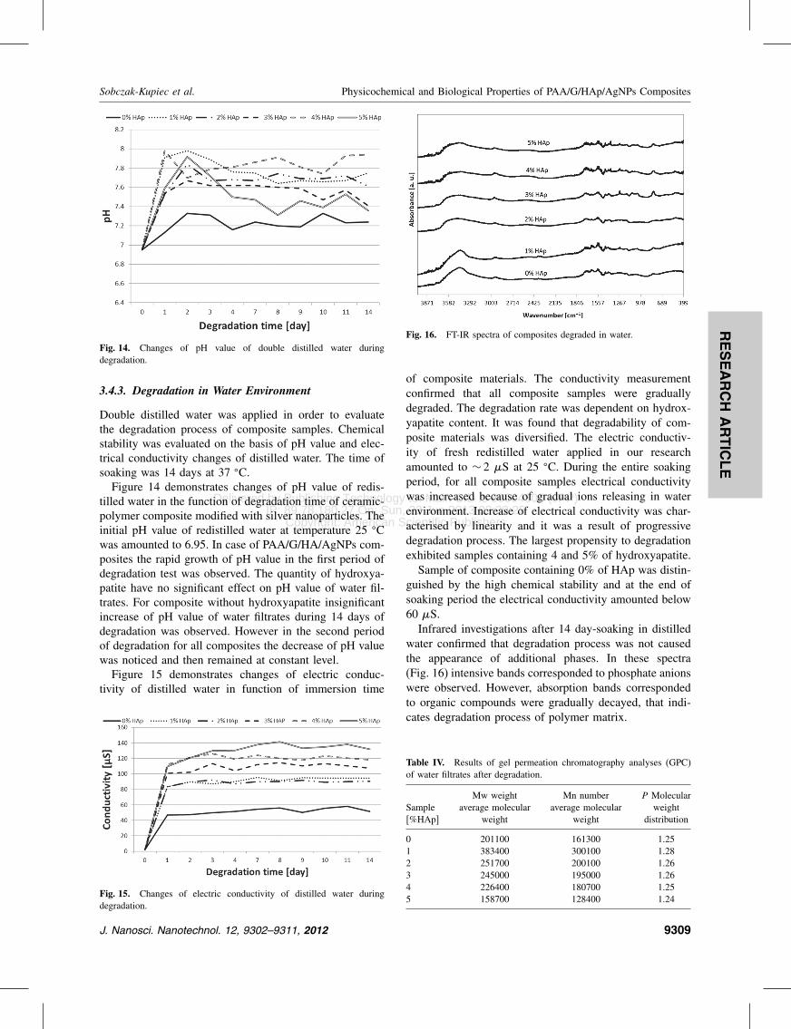
Fig. 16. FT-IR spectra of composites degraded in water.
of composite materials. The conductivity measurementconfirmed that all composite samples were graduallydegraded. The degradation rate was dependent on hydrox-yapatite content. It was found that degradability of com-posite materials was diversified. The electric conductiv-ity of fresh redistilled water applied in our researchamounted to ∼ 2 �S at 25 �C. During the entire soakingperiod, for all composite samples electrical conductivitywas increased because of gradual ions releasing in waterenvironment. Increase of electrical conductivity was char-acterised by linearity and it was a result of progressivedegradation process. The largest propensity to degradationexhibited samples containing 4 and 5% of hydroxyapatite.Sample of composite containing 0% of HAp was distin-
guished by the high chemical stability and at the end ofsoaking period the electrical conductivity amounted below60 �S.Infrared investigations after 14 day-soaking in distilled
water confirmed that degradation process was not causedthe appearance of additional phases. In these spectra(Fig. 16) intensive bands corresponded to phosphate anionswere observed. However, absorption bands correspondedto organic compounds were gradually decayed, that indi-cates degradation process of polymer matrix.
Table IV. Results of gel permeation chromatography analyses (GPC)of water filtrates after degradation.
Mw weight Mn number P MolecularSample average molecular average molecular weight[%HAp] weight weight distribution
0 201100 161300 1.251 383400 300100 1.282 251700 200100 1.263 245000 195000 1.264 226400 180700 1.255 158700 128400 1.24
J. Nanosci. Nanotechnol. 12, 9302–9311, 2012 9309

Delivered by Publishing Technology to: Main CID is 80004805 (JPP)IP: 89.78.180.27 On: Sun, 23 Jun 2013 02:39:25
Copyright: American Scientific Publishers
RESEARCH
ARTIC
LE
Physicochemical and Biological Properties of PAA/G/HAp/AgNPs Composites Sobczak-Kupiec et al.
Fig. 17. Representative chromatogram of water filtrate.
In samples with water used as the degradation mediumone polymer fraction is observed with Mw in the rangebetween 380000 and 158000 (Table IV and Fig. 17). How-ever gelatine concentration was relatively low, the increaseof hydroxyapatite amount in solution affects polymerdegradation.
4. CONCLUSION
This study was undertaken in order to find a new and sim-ple way of synthesis biomedical composites by introducingthe mineral and nanometal phase into the polymer matrix.Hydroxyapatite and gelatin, resembling the inorganic
and organic components of bone, can assure bioaffinityand bioactivity, and supply useful ingredients for new boneformation during resorption.Nanosilver, an antibacterial agent, which could be gen-
erally used in the infected bone healing, was incorporatedinto the composites. Ceramic phase in composites—natural HAp could improve biocompability and bioac-tivity of composite and nanosilver as an antibacterialagent incorporated in composites could prevent infec-tions after implantation. From presented data it can beinferred that hydrogel/gelatin/hydroxyapatite/AgNPs com-posite could find potential application in the bone repairand regeneration.Composites for bone and dental applications based on
hydroxyapatite/gelatin may be designed as effectivedevices for the controlled release of antimicrobial agentssuch as nanosilver. PAA/G/HAp/AgNPs composites wereprepared by the polymerization under microwave irradia-tion. The crystal structure and characteristic groups presentin the PAA/G/HAp/AgNPs composite is evident from theXRD and FT-IR data.Obtained results confirmed the presence of all phases
of composites after synthesis by the polymerization inmicrowave irradiation, however in vitro studies showeddegradation of some of composites phase dependent onenvironment of immersion medium.
Summarizing, from presented data it can be inferred thathydrogel/gelatin/hydroxyapatite/AgNPs composite afterslight modification in stability could find potential uses inthe bone repair and regeneration.
Acknowledgments: The research was supported byThe National Centre for Science, Poland.
References and Notes
1. B. Gasser, Injury, Int. J. Care Injured 31, S-D48 (2000).2. M. Swetha, K. Sahithi, A. Moorthi, N. Srinivasan, K. Ramasamy,
and N. Selvamurugan, Int. J. Biol. Macromol. 47, 1 (2010).3. P. S. Gils, D. Rayb, and P. Kumar Sahoo, Int. J. Biol. Macromol.
46, 237 (2010).4. I. Sopyana, M. Melb, S. Rameshc, and K. A. Khalidd, Sci. Tech.
Adv. Mater. 8, 116 (2007).5. M. H. Fathia, A. Hanifia, and V. Mortazavi, J. Mater. Process Tech.
202, 536 (2008).6. J. B. Lee, S. E. Kim, D. N. Heo, and I. K. Kwon, Macromol. Res.
18, 1195 (2010).7. M. Zhang, Ch. Liu, J. Sun, and X. Zhang, Ceram. Int. 37, 2025
(2011).8. X. Wang, G. Song, and T. Lou, J. Mater. Sci.: Mater. Med. 21, 183
(2010).9. N. Tudorachi and A. P. Chiriac, J. Polym. Environ. 19, 546 (2011).10. P. Rosółand J. Chłopek, Composites 1, 39 (2006).11. S. Hasegawa, S. Ishii, J. Tamura, T. Furukawa, M. Neo, Y. Matsusue,
Y. Shikinami, M. Okuno, and T. Nakamura, Biomaterials 27, 1327(2006).
12. Y. Shikinami and M. Okuno, Biomaterials 20, 859 (1999).13. X. Zheng, S. Zhou, X. Li, and J. Weng, Biomaterials 27, 4288
(2006).14. M. Sivakumar and K. Panduranga Rao, Biomaterials 23, 3175
(2002).15. H.-W. Kim, H.-E. Kim, and V. Salih, Biomaterials 26, 5221 (2005).16. H.-W. Kim, J.-H. Song, and H.-E. Kim, Adv. Funct. Mater. 15, 1988
(2005).17. A. Di Martino, M. Sittinger, and M. V. Risbud, Biomaterials 26,
5983 (2005).18. S. Saravanan, S. Nethala, S. Pattnaik, A. Tripathi, A. Moorthi, and
N. Selvamurugan, Int. J. Biol. Macromol. 49, 188 (2011).19. G. S. Sailaja, P. Ramesh, T. V. Kumary, and H. K. Varma, Acta
Biomater. 2, 651 (2006).20. R. Murugan and S. Ramakrishna, Biomaterials 25, 3829 (2004).21. V. M. Rusu, C.-H. Ng, M. Wilke, B. Tiersch, P. Fratzl, and M. G.
Peter, Biomaterials 26, 5414 (2005).22. M. Kikuchi, T. Ikoma, S. Itoh, H. N. Matsumoto, Y. Koyama,
K. Takakuda, K. Shinomiya, and J. Tanaka, Compos. Sci. Technol.64, 819 (2004).
23. M. Kikuchi, H. N. Matsumoto, T. Yamad, J. Koyam, K. Takakuda,and J. Tanaka, Biomaterials 25, 63 (2004).
24. A. Sionkowska and J. Kozłowska, Int. J. Biol. Macromol. 47, 483(2010).
25. D. Bakos, M. Soldán, and I. Hernández-Fuentes, Biomaterials20, 191 (1999).
26. M. Wang, Biomaterials 24, 2133 (2003).27. A. Sionkowska and J. Kozłowska, Int. J. Biol. Macromol. 47, 483
(2010).28. H. Kobayashi, M. Kato, T. Taguchi, T. Ikoma, H. Miyashita,
S. Shimmura, K. Tsubota, and J. Tanaka, Mater. Sci. Eng. C 24, 729(2004).
29. K. Furuichi, Y. Oaki, H. Ichimiya, J. Komotori, and H. Imai, Sci.Tech. Adv. Mater. 7, 219 (2006).
9310 J. Nanosci. Nanotechnol. 12, 9302–9311, 2012

Delivered by Publishing Technology to: Main CID is 80004805 (JPP)IP: 89.78.180.27 On: Sun, 23 Jun 2013 02:39:25
Copyright: American Scientific Publishers
RESEARCH
ARTIC
LE
Sobczak-Kupiec et al. Physicochemical and Biological Properties of PAA/G/HAp/AgNPs Composites
30. S. Velayudhan, T. V. Anilkumar, T. V. Kumary, P. V. Mohanan,A. C. Fernandez, H. K. Varma, and P. Ramesh, Acta Biomater. 1, 20(2005).
31. X. Shen, H. Tong, Z. Zhu, P. Wan, and J. Hu, Mater. Lett. 61, 629(2007).
32. X. Xiao, R. Liu, Q. Huang, and X. Ding, J. Mater. Sci.: Mater. Med.20, 2375 (2009).
33. A. Sobczak-Kupiec and Z. Wzorek, Ceram. Int. 38, 641(2012).
34. A. Sobczak-Kupiec, D. Malina, Z. Wzorek, and M. Zimowska,Micro. Nano Lett. 6, 656 (2011).
35. T. Kokubo and H. Takadama, Biomaterials 27, 2907 (2006).36. ISO 10271:2011 Dental metallic materials-Corrosion test methods
(2011), pp. 1–26.
Received: 1 January 2012. Accepted: 15 June 2012.
J. Nanosci. Nanotechnol. 12, 9302–9311, 2012 9311