Phototransduction by Vertebrate Ultraviolet Visual Pigments: Protonation of the Retinylidene Schiff...
10
Phototransduction by Vertebrate Ultraviolet Visual Pigments: Protonation of the Retinylidene Schiff Base following Photobleaching ² Abhiram Dukkipati, ‡,§,|,⊥ Anakarin Kusnetzow, ‡,⊥,# Kunnel R. Babu, § Lavoisier Ramos, ‡,3 Deepak Singh, ‡,1 Barry E. Knox,* ,§ and Robert R. Birge* ,‡,3 Departments of Chemistry and Biology, Syracuse UniVersity, 111 College Place, Syracuse, New York 13244-4100, Department of Biochemistry and Molecular Biology, SUNY Upstate Medical UniVersity, 750 East Adams Street, Syracuse, New York 13210, and Department of Chemistry, UniVersity of Connecticut, Storrs, Connecticut 06269 ReceiVed March 27, 2002; ReVised Manuscript ReceiVed May 29, 2002 ABSTRACT: The photochemical and subsequent thermal reactions of the mouse short-wavelength visual pigment (MUV) were studied by using cryogenic UV-visible and FTIR difference spectroscopy. Upon illumination at 75 K, MUV forms a batho intermediate (λ max ∼380 nm). The batho intermediate thermally decays to the lumi intermediate (λ max ∼440 nm) via a slightly blue-shifted intermediate not observed in other photobleaching pathways, BL (λ max ∼375 nm), at temperatures greater than 180 K. The lumi intermediate has a significantly red-shifted absorption maximum at 440 nm, suggesting that the retinylidene Schiff base in this intermediate is protonated. The lumi intermediate decays to an even more red-shifted meta I intermediate (λ max ∼480 nm) which in turn decays to meta II (λ max ∼380 nm) at 248 K and above. Differential FTIR analysis of the 1100-1500 cm -1 region reveals an integral absorptivity that is more than 3 times smaller than observed in rhodopsin and VCOP. These results are consistent with an unprotonated Schiff base chromophore. We conclude that the MUV-visual pigment possesses an unprotonated retinylidene Schiff base in the dark state, and undergoes a protonation event during the photobleaching cascade. SWS1 1 pigments mediate visual sensitivity in the short- wavelength region of the spectrum, exhibiting absorption maxima (λ max ) between 350 and 450 nm, the widest energy range in the opsin classes (1). Moreover, SWS1 pigments are the only opsins with absorption maxima blue-shifted compared to a protonated retinylidene Schiff base in aqueous solution, λ max ) 440 nm (2). The pigments in the SWS1 group are divided into two subgroups based on the absorption maxima: those that absorb in the 400-440 nm range (blue/ violet) and those that absorb in the 350-390 nm range (UV). One explanation for the variation in λ max hypothesizes that SWS1 pigments with λ max in the UV (<400 nm) have an unprotonated Schiff base while those with λ max in the blue- violet region (λ max >400 nm) have a protonated Schiff base (3). However, the molecular mechanisms of spectral tuning in SWS1 pigments may also include contributions from the relative position of the counterion to the Schiff base, and multiple dispersive interactions distributed among numerous amino acids in the retinal binding pocket (4). Studies on the phototransduction properties of Xenopus (5), human (6), and salamander (7) SWS1 pigments have shown reduced catalytic efficiency and shorter lifetime of the light-activated conformation compared to bovine rhodop- sin. These differences in signal transduction may play an important role in shaping the dynamics of light response in cones (8, 9). To understand the molecular basis for the unique functional properties of SWS1 pigments, detailed spectro- scopic, biochemical, and site-directed mutagenesis studies are required, in pigments that absorb both in the UV as well as in the violet region. We have previously studied spectral tuning and photo- transduction in the Xenopus SWS1 opsin (VCOP, λ max ∼427 nm). FTIR difference spectroscopy used to examine the meta II intermediate (10) and site-directed mutagenesis studies (11) have shown that the retinylidene Schiff base linkage is protonated in the dark state. Furthermore, the counterion (D108) to the protonated Schiff base was found to be at the ² This work was supported in part by NIH Grants GM-34548 (to R.R.B.) and EY-11256 and EY-12975 (to B.E.K.), the W. M. Keck Center for Molecular Electronics at Syracuse University, and a grant from the Research to Prevent Blindness Foundation. * Correspondence should be addressed to either B.E.K.: tel (315) 464-8719, fax (315) 464-8750, email [email protected]; or R.R.B.: tel (860) 486-6720, fax (860) 486-2981, email: [email protected]. ‡ Syracuse University. § SUNY Upstate Medical University. | Present address: Department of Microbiology and Immunology, 299 Campus Dr., Stanford School of Medicine, Stanford, CA 94305. ⊥ These authors contributed equally to this work. # Present address: Jules Stein Eye Institute, UCLA School of Medicine, 100 Stein Plaza, BH-973, Los Angeles, CA 90095. 3 University of Connecticut. 1 Present address: GeneFormatics Inc., 5830 Oberlin Dr., Suite 200, San Diego, CA 92121. 1 Abbreviations: SWS1, short-wavelength-sensitive; MUV, mouse SWS1 opsin; VCOP, Xenopus laeVis SWS1 opsin; RH, rhodopsin(s); M/LWS, medium/long-wavelength-sensitive; TM#, transmembrane helical domain #; SB, Schiff base; nm, nanometers; PSS(xyz), photo- stationary state generated by illumination at wavelength xyz in nanometers; DM, n-dodecyl--D-maltoside; HEPES, N-(2-hydroxyeth- yl)piperazine-N′-2-ethanesulfonic acid; batho, lumi, meta I, and meta II, discrete thermal intermediates of the visual opsin bleaching pathway; BL, photobleaching intermediate formed between batho and lumi; PSS, photostationary state. 9842 Biochemistry 2002, 41, 9842-9851 10.1021/bi025883g CCC: $22.00 © 2002 American Chemical Society Published on Web 07/11/2002
Transcript of Phototransduction by Vertebrate Ultraviolet Visual Pigments: Protonation of the Retinylidene Schiff...
Phototransduction by Vertebrate Ultraviolet Visual Pigments: Protonation of theRetinylidene Schiff Base following Photobleaching†
Abhiram Dukkipati,‡,§,|,⊥ Anakarin Kusnetzow,‡,⊥,# Kunnel R. Babu,§ Lavoisier Ramos,‡,3 Deepak Singh,‡,1
Barry E. Knox,*,§ and Robert R. Birge*,‡,3
Departments of Chemistry and Biology, Syracuse UniVersity, 111 College Place, Syracuse, New York 13244-4100,Department of Biochemistry and Molecular Biology, SUNY Upstate Medical UniVersity, 750 East Adams Street,Syracuse, New York 13210, and Department of Chemistry, UniVersity of Connecticut, Storrs, Connecticut 06269
ReceiVed March 27, 2002; ReVised Manuscript ReceiVed May 29, 2002
ABSTRACT: The photochemical and subsequent thermal reactions of the mouse short-wavelength visualpigment (MUV) were studied by using cryogenic UV-visible and FTIR difference spectroscopy. Uponillumination at 75 K, MUV forms abathointermediate (λmax ∼380 nm). Thebathointermediate thermallydecays to thelumi intermediate (λmax ∼440 nm) via a slightly blue-shifted intermediate not observed inother photobleaching pathways,BL (λmax ∼375 nm), at temperatures greater than 180 K. Thelumiintermediate has a significantly red-shifted absorption maximum at 440 nm, suggesting that the retinylideneSchiff base in this intermediate is protonated. Thelumi intermediate decays to an even more red-shiftedmeta Iintermediate (λmax ∼480 nm) which in turn decays tometa II(λmax ∼380 nm) at 248 K and above.Differential FTIR analysis of the 1100-1500 cm-1 region reveals an integral absorptivity that is morethan 3 times smaller than observed in rhodopsin and VCOP. These results are consistent with anunprotonated Schiff base chromophore. We conclude that the MUV-visual pigment possesses anunprotonated retinylidene Schiff base in the dark state, and undergoes a protonation event during thephotobleaching cascade.
SWS11 pigments mediate visual sensitivity in the short-wavelength region of the spectrum, exhibiting absorptionmaxima (λmax) between 350 and 450 nm, the widest energyrange in the opsin classes (1). Moreover, SWS1 pigmentsare the only opsins with absorption maxima blue-shiftedcompared to a protonated retinylidene Schiff base in aqueoussolution, λmax ) 440 nm (2). The pigments in the SWS1
group are divided into two subgroups based on the absorptionmaxima: those that absorb in the 400-440 nm range (blue/violet) and those that absorb in the 350-390 nm range (UV).One explanation for the variation inλmax hypothesizes thatSWS1 pigments withλmax in the UV (<400 nm) have anunprotonated Schiff base while those withλmax in the blue-violet region (λmax >400 nm) have a protonated Schiff base(3). However, the molecular mechanisms of spectral tuningin SWS1 pigments may also include contributions from therelative position of the counterion to the Schiff base, andmultiple dispersive interactions distributed among numerousamino acids in the retinal binding pocket (4).
Studies on the phototransduction properties ofXenopus(5), human (6), and salamander (7) SWS1 pigments haveshown reduced catalytic efficiency and shorter lifetime ofthe light-activated conformation compared to bovine rhodop-sin. These differences in signal transduction may play animportant role in shaping the dynamics of light response incones (8, 9). To understand the molecular basis for the uniquefunctional properties of SWS1 pigments, detailed spectro-scopic, biochemical, and site-directed mutagenesis studiesare required, in pigments that absorb both in the UV as wellas in the violet region.
We have previously studied spectral tuning and photo-transduction in theXenopusSWS1 opsin (VCOP,λmax ∼427nm). FTIR difference spectroscopy used to examine themetaII intermediate (10) and site-directed mutagenesis studies (11)have shown that the retinylidene Schiff base linkage isprotonated in the dark state. Furthermore, the counterion(D108) to the protonated Schiff base was found to be at the
† This work was supported in part by NIH Grants GM-34548 (toR.R.B.) and EY-11256 and EY-12975 (to B.E.K.), the W. M. KeckCenter for Molecular Electronics at Syracuse University, and a grantfrom the Research to Prevent Blindness Foundation.
* Correspondence should be addressed to either B.E.K.: tel (315)464-8719, fax (315) 464-8750, email [email protected]; or R.R.B.:tel (860) 486-6720, fax (860) 486-2981, email: [email protected].
‡ Syracuse University.§ SUNY Upstate Medical University.| Present address: Department of Microbiology and Immunology,
299 Campus Dr., Stanford School of Medicine, Stanford, CA 94305.⊥ These authors contributed equally to this work.# Present address: Jules Stein Eye Institute, UCLA School of
Medicine, 100 Stein Plaza, BH-973, Los Angeles, CA 90095.3 University of Connecticut.1 Present address: GeneFormatics Inc., 5830 Oberlin Dr., Suite 200,
San Diego, CA 92121.1 Abbreviations: SWS1, short-wavelength-sensitive; MUV, mouse
SWS1 opsin; VCOP,Xenopus laeVis SWS1 opsin; RH, rhodopsin(s);M/LWS, medium/long-wavelength-sensitive; TM#, transmembranehelical domain #; SB, Schiff base; nm, nanometers; PSS(xyz), photo-stationary state generated by illumination at wavelengthxyz innanometers; DM,n-dodecyl-â-D-maltoside; HEPES,N-(2-hydroxyeth-yl)piperazine-N′-2-ethanesulfonic acid;batho, lumi, meta I, andmetaII , discrete thermal intermediates of the visual opsin bleaching pathway;BL, photobleaching intermediate formed betweenbathoandlumi; PSS,photostationary state.
9842 Biochemistry2002,41, 9842-9851
10.1021/bi025883g CCC: $22.00 © 2002 American Chemical SocietyPublished on Web 07/11/2002
same position in TM3 as in bovine rhodopsin (11). Duringphotobleaching, VCOP undergoes four spectrally discreteconformational changes analogous to the intermediates statedescribed for rhodopsin:batho, lumi, meta I, andmeta II(10). We have also suggested that the counterion and residuesthat interact with the counterion (e.g., S85 in TM2) play animportant role in regulating the photobleaching pathway andability to activate transducin (4, 11). These studies have ledus to propose the extended counterion model for the SWS1pigments (4).
Although the mouse SWS1 (MUV) contains 11-cis-retinalcovalently attached via a Schiff base linkage to the protein(11, 12), the λmax ∼357 nm suggests the Schiff base isunprotonated. The strongest supporting evidence comes froma site-specific mutant at the putative counterion position,E108Q (11, 13). Unlike bovine rhodopsin, in which themutation of the counterion (E113Q) causes a dramatic changein the Schiff base pKa and an accompanying shift in theλmax
to 380 nm at neutral pH (14-16), no significant change wasobserved in the MUV counterion mutant (11, 13).
In rhodopsin, Schiff base deprotonation accompanies theformation ofmeta II, the active intermediate state (17, 18).Site-specific mutants (e.g., E113Q, G90D, H211C) that alterthe pKa of the Schiff base, either in the dark or afterisomerization of the chromophore, dramatically affect thephotobleaching properties of rhodopsin (14, 19, 20). E113,the counterion, is the acceptor of the Schiff base proton, andmutation of the counterion to a neutral residue slows thedecay of the later photointermediates (21). Similar distur-bances in the photobleaching pathway of VCOP, which alsocontains a protonated Schiff base, were observed in coun-terion mutants (11). In addition, previous data reported fromcryogenic experiments are consistent with an unprotonatedSchiff base (3). If the UV-sensitive SWS1 pigments havean unprotonated Schiff base in the dark, does a protonationevent occur upon light activation, and, if so, what is the roleof the protonation of the Schiff base in phototransductionof these pigments?
To address this question, we have carried out a detailedspectroscopic and biochemical study of MUV, characterizingthe complete photobleaching pathway of this pigment. Weobserve the formation of abatho intermediate, indicatingnormalcis to trans isomerization, as well as the formationof the later intermediates,lumi, meta I, andmeta II. Mostsignificantly, the Schiff base undergoes a protonation fol-lowing photobleaching, most likely during the transition frombatho to lumi.
MATERIALS AND METHODS
Expression, Isolation, and Purification of the VisualPigments. The MUV pigments contained the first 328 codonsof the MUV coding region followed by the last 14 codonsof bovine rhodopsin as an epitope tag (3). Transfection intoCOS1 cells and preparation of MUV pigments were per-formed as previously described (3, 10, 11). Typically, thirty150 mm plates were transfected. The pigment was purifiedand concentrated to∼300µL. MUV samples had an averageA360 between 0.3 and 0.4, with anA280/A360 of ∼4.0. Allpigments were stored in the dark at-80 °C. Experimentswere repeated with pigment purified from at least twoseparate transfections.
Cryogenic Electronic Spectroscopy. Low-temperature spec-troscopy was performed as previously described (3, 10).Samples for cryogenic studies were prepared in 67% glycerol,0.05% n-dodecyl-â-D-maltoside (DM, Anatrace, Maumee,OH) in buffer A (buffer A) 50 mM HEPES, 140 mM NaCl,3 mM MgCl2, pH 6.6) with a finalA360 ∼ 0.1. Samples forcryogenic studies at 45-200 K were placed in an AirProducts Displex helium-refrigerated cryostat coupled to aShimadzu 3010 UV-vis-NIR spectrophotometer as de-scribed previously (3, 10). Samples for studies at 240-273K were placed in a Varian Cary 3E modified for accuratetemperature control as described previously (10). The pho-tostationary states (PSS) were generated by successiveilluminations at 305 nm until no further spectral changescould be detected (∼1 h, unless specified otherwise).
The spectra shown in the figures are the averages of fourseparate scans at each temperature. Theλmax absorbancevalues of the initial dark sample were used to normalize thespectra from different experiments. Each of the differencespectra was calculated by subtracting the appropriate pho-tostationary state from the corresponding dark or previousphotostationary state using SigmaPlot (Jandel, Chicago, IL).
FTIR Difference Spectroscopy.Glycerol and HEPES wereremoved from the protein sample (10). The buffer of thesample was changed to buffer B [10 mM potassiumphosphate buffer (pH 6.8) and 0.02% DM], using CentriconC-30 filters, and excess peptide (1D4) in the sample wasalso removed using prepacked Sephadex G-50 columns(Pharmacia, Piscataway, NJ) (10). The sample was recon-centrated to∼100 µL. A thin film of the protein (∼100 µgin 100 µL) was prepared by drying the sample in 10µLaliquots onto a cool CaF2 IR window under a continuousstream of nitrogen. This was repeated until successive layerswere formed. The film was sealed with another CaF2
window. The window was mounted on a Nicolet Magna IR750, series II, instrument (Nicolet Instrument Corp., Madison,WI). The sample was kept equilibrated at 263 K for 1 h.The dark-state spectra were taken by averaging 512 scansat 2 cm-1 resolution. To generatemeta II, the sample wasgenerated by illumination with light from a filtered (305 nmcutoff filter, Melles Griot 03FCG121) 250 W quartz halogenlamp (illumination time) 5 min) at 263 K. The sample wasagain illuminated for an additional 5 min to guaranteecomplete formation of themeta II state, and spectra werecollected. Data collection was performed using OMNIC 3.1(Nicolet Instrument Corp). The amide I peak (1654 cm-1)of the dark-state andmeta II state spectra was normalized.The dark-state spectrum was subtracted from themeta IIstatespectrum to generate the difference spectrum. The resultingdifference spectrum of the second illumination was the sameas with the first illumination, indicating complete formationof the photoproduct at 263 K.
HPLC Analysis. After illumination, visual pigment sampleswere warmed to 0°C, and hydroxylamine was added to afinal concentration of 500 mM, pH 7. Retinal oximes of thePSS305 generated at 243 K were extracted and analyzed aspreviously reported (3), and these values were used for allsubsequent analysis.
Homology Models. The crystal structure of bovine rhodop-sin, PDB accession no. 1F88 (22), was used to constructhomology models for VCOP and MUV. The sequence ofbovine rhodopsin was aligned with the sequences for VCOP
Photobleaching Pathway of Ultraviolet Visual Pigments Biochemistry, Vol. 41, No. 31, 20029843

and MUV using CLUSTALW (23). These alignments wereused to construct models for VCOP and MUV using theprogram WHATIF (24). Hydrogens were added in CHARMM(version 27b2) (25). The models were minimized by 4000steps of the adopted-basis Newton Raphson (ABNR) algo-rithm using the charmm22 force field (26). The environmentwas represented by a distant-dependence (1/r) dielectricfunction. The initial chromophore geometry was retainedfrom the X-ray structure. The retinylidene chromophore wasrepresented by a potential adapted from the CHARMM-optimized all-trans-retinyl-protonated Schiff base (27). Allcalculations were performed with harmonic constraints onthe protein backbone.
RESULTS
Formation of the Early Intermediates.Improvements inthe yield of MUV from transfected COS1 cells (11) havepermitted cryogenic studies of this pigment at higherconcentrations than in our previous reports, eliminating thelight-scattering component and permitting an analysis of thelow-temperature intermediates without correction of thespectra. Upon cooling to 75 K, the MUV spectrum undergoesa small 2 nm red shift (λmax ∼359 nm, Figure 1A, inset) andexhibits a shoulder at∼380 nm. Illumination of the darksample at 305 nm caused a red shift in the chromophorepeak, which was complete within 1 h ofillumination (Figure1B,D). There also was a decrease in the protein extinctioncoefficient that accompanied the batho shift in the chro-mophore peak, and continued to decrease after the chro-mophore peak had reached a stationary state (Figure 1C, alsosee below). This photostationary state (PSS305) consistedmainly of thebathointermediate (all-trans-retinal bound to
opsin) and a minor component of the original (11-cis-retinalbound to opsin) dark state (11-cis-retinal bound to opsin).There was very little isopigment (9-cis-retinal) formed underthese conditions. The isomeric composition of the PSS305generated at higher temperatures (243 K) had 11% 11-cis,83% all-trans, and 5% 9-cis,(2%.
The difference spectrum between PSS305 and the darkstate has an absorption maximum at 404 nm and exhibitsvibronic fine structure atλmax ∼380 nm (Figure 1C),consistent with our earlier report (3). Thus, MUV forms ared-shifted primary photoproduct like another SWS1 pig-ment, VCOP (which has a protonated Schiff base linkage),and other visual pigments (3, 28).
To trap the additional intermediates in the bleachingpathway, the batho-shifted PSS305 was gradually warmedfrom 80 to 220 K. After equilibrating the sample at aspecified temperature (i.e., 180 K), the sample was graduallycooled back to 75 K where all spectra were collected to avoidartifacts arising from temperature-dependent baseline shifts.Small changes in the absorption spectrum of PSS305 between120 and 180 K were observed (Figure 2A), primarily adecrease in the fine structure at 380 nm. Multiple changesin the absorption spectrum occurred at temperatures above180 K: an increase in absorbance at wavelengths from 420to 540 nm, a decrease in absorption of the 404 nmcomponent, and an increase in absorbance at 280 nm (Figure2). These changes are consistent with the thermal transitionto another intermediate in the photobleaching pathway,presumablylumi. However, the absence of a clear isosbesticpoint during thebathoto lumi transition suggests the presenceof an additional intermediate during the transition, whoseabsorption spectrum overlaps significantly with thebatho
FIGURE 1: Photochemistry of the mouse SWS1 at cryogenic temperatures. Mouse SWS1 opsin (dark) was illuminated at 75 K with 305 nmlight to generate the primary PSS305. (A and B) Spectra after successively illuminating the dark state (in minutes). The inset in panel Ashows the spectra of MUV at room temperature (RT) and at 75 K. (C) Light- dark difference spectra generated by subtracting the darkstate from the PSS305 after 15, 60, and 150 min of illumination. (D) Plot of the absorbance changes monitored at 280 and 400 nm duringthe successive illuminations. Lines are single-exponential fits to the data.
9844 Biochemistry, Vol. 41, No. 31, 2002 Dukkipati et al.

FIGURE 2: Thermal decay of the mouse SWS1 photostationary state formed at 75 K. The PSS305 formed at 75 K (spectrum labeled 75 K)was gradually warmed in 20 K increments, equilibrated for 1 h, and cooled back to 75 K before recording the absorption spectra. (A andB) Spectra of PSS305 after equilibration at 75, 160, 180, 200, and 220 K, respectively. The inset in (A) shows, on an expanded scale, onlya minor change in the main chromophore peak in the sample warmed to 180 K compared to the initial PSS305 at 75 K. (C) Light- darkdifference spectra generated by subtracting the dark state at 75 K from the PSS305 warmed to 160, 180, 200, and 220 K, respectively. (D)Plot of the absorbance changes monitored at 280 and 450 nm during the temperature ramping experiments in (A).
FIGURE 3: Photochemistry of the mouse SWS1 at 243 K. The mouse SWS1 (dark) was illuminated at 243 K with 305 nm light successively.(A and B) Spectra of the dark state and after 60, 120, 240, and 340 min of illumination. The inset in (A) shows a plot of the absorbancechanges monitored at 360 and 475 nm during the illumination procedure. (C) Light- dark difference spectra obtained by subtracting thedark state from the PSS305 after 180 and 340 min of illumination at 243 K. (D) Difference spectrum of the PSS305 after 340 min ofillumination and the PSS305 after 180 min of illumination. The red shift in the PSS305 is apparent at this temperature. The line is asmoothed fit (third-order polynomial regression, SigmaPlot, Jandel Scientific) to the difference spectrum.
Photobleaching Pathway of Ultraviolet Visual Pigments Biochemistry, Vol. 41, No. 31, 20029845

spectrum. The presence of such an intermediate was alsosuggested during deconvolution of the spectra (see Discus-sion). The increase in absorbance withλmax∼440 nm stronglysuggests that the MUVlumi intermediate has a protonatedSchiff base linkage like other visual pigments. The proteinabsorbance recovers to that of the dark state, suggesting thatconformational strains that were induced in the formationof bathoare relaxed during the formation oflumi.
Formation of the Later Intermediates.To study the laterphotointermediates, we investigated the formation of themetaI andmeta IIstates of MUV at temperatures>240 K, abovethe temperature required to formlumi. Illumination of thedark sample (λmax ∼358 nm) at 305 nm, 243 K produced aphotostationary state with a broad absorption band within 1h (Figure 3A). However, after the initial decrease at 358 nm(within 1 h), no additional decrease at 358 nm was observed,even under extended illumination conditions (∼6 h). Il-lumination of the sample produced a spectrum with a broadabsorbance centered at∼470 nm and a decrease in theabsorption at 358 nm (Figure 3A,B), which followed similartime courses (Figure 3A, inset). Prolonged illuminationcaused a further broadening of the red-shifted absorptionband and a small reduction of the remaining dark state, withsignificant intensity beyond 600 nm (Figure 3C,D). Theremaining absorbance at 358 nm was due to 11-cis-retinalbound to the protein, presumably arising from photoconver-sion of later intermediates back to the 11-cis form. Uponwarming the illuminated sample, the absorption band at 358nm progressively lost intensity while the band centered at
470 nm gained intensity with an isosbestic point at∼422nm (Figure 4). Changes in the spectrum were essentiallycompleted upon warming and equilibrating at 258 K. Despiterepeated attempts, we were unable to acid-trap the protein-chromophore Schiff base linkage of the final species (datanot shown). This is similar to the observation made withVCOP (10) and may represent an inherent property of allSWS1 pigments.
Changes in MUV Protein Absorbance during the Forma-tion of Photointermediates.We observed a decrease inabsorbance in the UV portion of the spectrum, 260-320 nm,upon the formation of PSS305 at 75 K (Figure 1). The dropin extinction was approximately 13%, which is equivalentto a ∆ε of ∼8500 M-1 cm-1 assuming an extinctioncoefficient at 280 nm of 65 000 M-1 cm-1 as for bovinerhodopsin.2 The difference between PSS305 at 75 K and thedark spectrum was a broad peak centered around 290 nm,with bands at 281, 289, and 299 nm (Figure 5A). This dropin extinction recovered during the transition frombatho tolumi, with the difference spectrum of PSS305 at 220 K andthe dark spectrum exhibiting a number of small bands onthe broad difference peak (Figure 5B). The protein absor-bance changes that occurred in the transition frommeta Itometa IIwere much smaller, with two distinct bands, at 293
2 Estimated fromε500 ) 40 600 M-1 cm-1 (29) andA280/A500 ) 1.6for purified rhodopsin (30). The aromatic amino acid composition ofMUV (32 Phe/16 Tyr/7 Trp) is similar to bovine opsin (31 Phe/16Tyr/5 Trp).
FIGURE 4: Thermal decay of the mouse SWS1 photostationary state formed at 243 K. The PSS305 formed at 243 K was gradually increasedin 5 K increments. (A and B) Spectra of the PSS305 at 243 K and states formed after equilibration at 248, 253, 258, and 263 K. (C)Difference spectra between the PSS305 at various temperatures and the initial PSS305 formed at 243 K. (D) Plot of the absorbance changesmonitored at 385 and 480 nm during the warming experiments.
9846 Biochemistry, Vol. 41, No. 31, 2002 Dukkipati et al.
and 301 nm (Figure 5C). These changes are consistent withalterations in the environment of one or more of the aromaticresidues, possibly tryptophan, since there are significantchanges>290 nm.
FTIR Spectroscopic Studies of the Protonation State ofthe Chromophore.We carried out FTIR integral analysis ofthe 1100-1500 cm-1 region of themeta IIminus dark MUVvibrational spectrum using methodology outlined for theVCOP pigment (10). Our difference spectrum showed bandsassociated with the dark state of MUV at 1164, 1220, 1249,and 1270 cm-1. A band at∼1220 cm-1 [see (10) for adiscussion of this band, termed p2] is normally associated
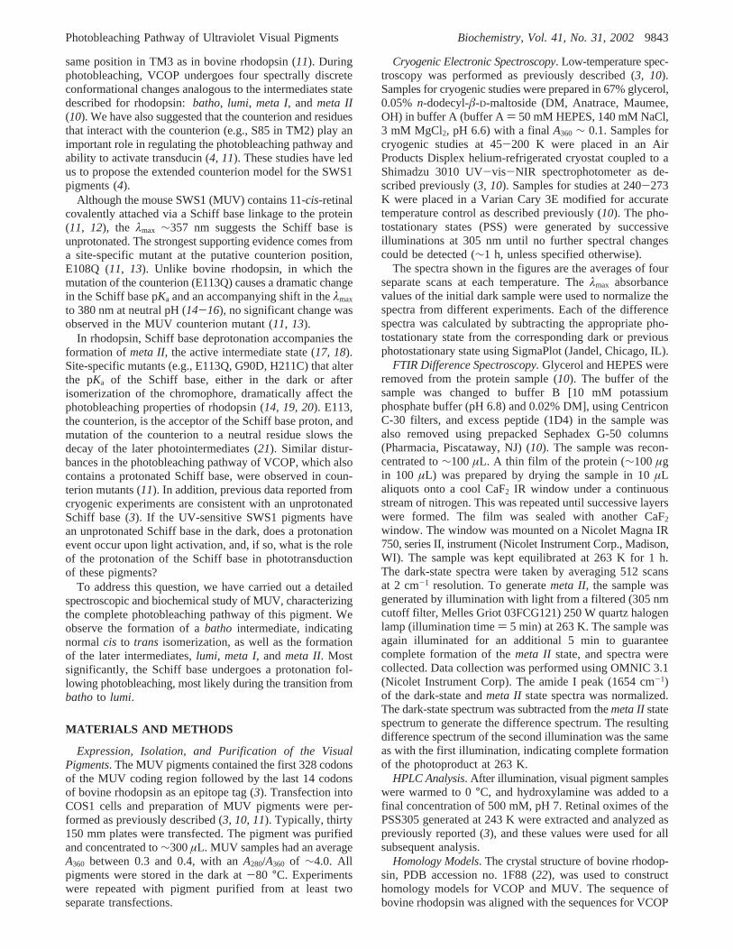
with a protonated chromophore. The integral value of thedifference spectrum in the region between 1100 and 1500cm-1 of this band was compared to theoretical simulations(Table 1). It is clear from this analysis that the intensity ofthis region in the MUV is greatly reduced compared torhodopsin and VCOP, and similar to the integral found inthe bovine rhodopsin E113Q mutant, which has an unpro-tonated Schiff base at pH 6.8 (31). We conclude that thechromophore in the 11-cis dark state of MUV is unproto-nated. The theoretical simulations of the MUV binding site(Figure 8) using mixed density functional and semiempiricalmethods theoretically predict a 1227 cm-1 band that includesa mixing of the chromophore modes with nearby protonatedglutamic acid modes. This provides an explanation for theobserved intensity of the 1220 cm-1 band.
DeriVation of the Spectra of MUV Intermediates.Spectraof the pure intermediates were calculated by deconvolutionusing the HPLC results to assign the isomeric compositionof PSS305 and assuming that the amount of the dark (11-cis) and light (11-cis, all-trans, and 9-cis) states remainedconstant during the temperature experiments described above(Figure 6). In contrast to other visual pigments, thelumi andmeta I intermediates are significantly red-shifted comparedto thebatho intermediate. Theλmax of the lumi (λmax ∼440nm) intermediate of MUV is the same as that of protonatedSchiff base model compounds in solution, thus indicatingthat the Schiff base in the MUV pigment undergoes aprotonation during photobleaching. The blue shift of theλmax
to 385 nm upon formation ofmeta II indicates that theformation of this intermediate is accompanied by the
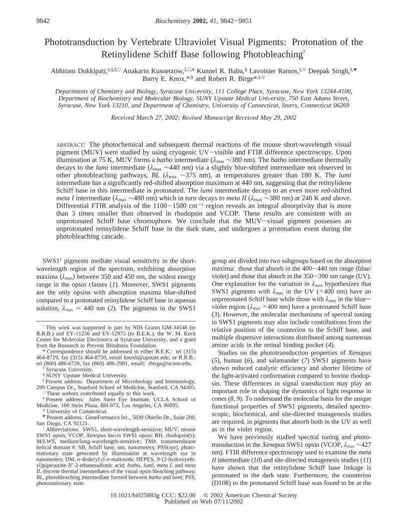
FIGURE 5: Photobleaching difference spectra of MUV between 260and 320 nm. (A) Spectrum formed by illuminating MUV with 305nm light at 75 K for the indicated duration (in minutes) wassubtracted from the spectrum of the unilluminated sample. (B)Difference spectra were generated by subtracting the PSS305 spectraat the indicated temperatures from the dark spectrum at 75 K. (C)Difference spectra were generated by subtracting the PSS305 spectraat the indicated temperatures from the dark spectrum at-30 °C.Reference lines in each panel are given in nanometers.
FIGURE 6: Calculated spectra of the photobleaching intermediates.Electronic absorption spectra of MUV and its photobleachingintermediates generated via decomposition of the temperature-trapped photostationary state spectra. Each spectrum is measuredat the temperature indicated in the legend.
Photobleaching Pathway of Ultraviolet Visual Pigments Biochemistry, Vol. 41, No. 31, 20029847

deprotonation of the Schiff base, as observed in other visualpigments (28).
DISCUSSION
The photobleaching pathway of the MUV opsin isschematically represented in Figure 7. Upon illumination at75 K, MUV forms a batho state, λmax ∼375 nm, withsignificant fine structure, as expected for an unprotonatedSchiff base (32). In our previous study of MUV (3), wecompared the difference spectra of the bathochromic pho-toproduct photostationary state with other visual pigmentsand found that MUV did not fit the trend that the other visualpigments followed, i.e., a linear correlation of the PSS-integrated spectrum with theλmax of PSS difference spectra[see (3) for discussion]. However, due to the lower proteinconcentration in that study, there was considerable lightscattering in the absorbance spectra. The light scatteringcomponent was subtracted, and then all spectra were normal-ized at 280 nm for the analysis presented in (3). The lightscattering obscured the changes in absorbance at∼280 nmthat occur during the formation ofbatho(Figures 1 and 5).With normalization, integration of the difference spectrumgave integral values of 2.1 and-1.9 (3). With the uncor-rected absorbance (Figure 1), integral values are 1.89 and-1.36. These new values are well removed from the trendlines observed for the other pigments, and support the ideathat the Schiff base is unprotonated in MUV.
At temperatures above 180 K, the MUVbathostate startsforming a red-shiftedlumi intermediate with aλmax ∼440nm. Spectral deconvolution using previously describedmethods (10) indicates that another intermediate, slightlyblue-shifted compared tobatho, is formed during thebathoto lumi transition. Henceforth, we refer to this intermediateasBL (λmax ∼370 nm), and is presumably analogous to theBL intermediate formed in the chicken blue opsin pathway(33). The formation of theBL intermediate is not obviousfrom the raw spectra during the temperature rampingexperiments due to overlapping spectral features and finestructure (Figure 7).
Table 1: Comparison of Observed and Calculated Integrals for themeta II Minus Dark FTIR Difference Spectraa
Experimental Measurementsprotein pH Schiff base integral value
solubilized rhodopsin 6.8 protonated -0.149( 0.037E113Q rhodopsin 8.2 unprotonated -0.034( 0.042VCOP 6.8 protonated -0.146( 0.051mouse UV (MUV) 6.8 (see Discussion) -0.041( 0.048
Theoretical Simulations11-cis chromophore methodb p2 band (int.)c integral value
MUV binding sited (SB) B3LYP, 6-31G 1227.3 (216) -0.072MUV binding sited (PSB) B3LYP, 6-31G 1230.2 (424) -1.461unprotonated Schiff base (SB) B3LYP, 6-31G 1221.1 (55.5) -0.014SB B3LYP, 6-31G(d) 1203.8 (63.2) 0.006protonated Schiff base (PSB) B3LYP, 6-31G 1247.2 (1579.5) -0.816PSB+ perchlorate B3LYP, 6-31G 1233.6 (413.9) -0.262PSB+ perchlorate+ 2 waters HF, 6-31G(d) 1218.9 (443.0) -0.180
a All the experimental integral values are formeta II minus dark state and were measured from 1100 to 1500 cm-1 in units of cm-1 with thevalues normalized by adjusting the amide I band absorptivity to 1. The theoretical values are calculated relative to the all-trans unprotonated Schiffbase (e.g.,meta II-like) chromophore (B3LYP, 6-31G) with the integrals normalized to the central amide I band of polyglycine multiplied by thetotal number of residues in the protein [see ref (10)]. b Calculations were carried out usingGaussian 98(51). c Location in wavenumber of theintense mode, labeled p2 in the isolated protonated Schiff base, which corresponds to the observed mode at∼1238 cm-1 in rhodopsin and, byanalogy, at 1232 cm-1 in VCOP [see ref (10)]. The intensity is in KM/mol.d The calculations were carried out using the ONIUM method with asemiempirical basis set for the surrounding binding site (PM3) and density functional methods (B3LYP, 6-31G) to handle the chromophore, thewater molecule near the Schiff base, and the nearby E108 counterion.
FIGURE 7: Photobleaching pathway of the mouse,XenopusSWS1,and rhodopsin. The top portion of the figure shows the blue shiftassociated with incorporation of a hypothetical all-trans protonatedSchiff base chromophore (λmax ∼600 nm) into the binding site forthe protonated intermediates. The lower half shows a comparisonof the photobleaching pathways of the mouse SWS1, theXenopusSWS1, and rhodopsin. Thebatho intermediate (λmax ∼380 nm) isformed upon illumination at 75 K.Bathodecays toBL (λmax ∼375nm) at 160 K, which decays tolumi at 200 K. The lumi intermediatedecays to meta I (λmax ∼490 nm) and then to meta II (λmax ∼385nm) at 243 and 248 K, respectively.
9848 Biochemistry, Vol. 41, No. 31, 2002 Dukkipati et al.
The large spectral red shift coinciding with the formationof the MUV lumi intermediate raises two possibilities: theSchiff base remains unprotonated in thebatho and BLintermediates and undergoes a protonation event duringformation of thelumi intermediate. Alternatively, the Schiffbase may undergo a protonation event during the formationof the batho intermediate, but have such a strained chro-mophore conformation that theλmax does not red shift to∼440 nm until the protein undergoes a large rearrangementduring the transition tolumi, thus relieving the strain on thechromophore. Results from the photobleaching pathway ofthe MUV E108Q counterion mutant support the first alterna-tive (in preparation).
The absorption maximum of the MUVmeta Iintermediateis very similar to that of themeta I intermediate formed inrhodopsin, although the MUV spectrum is much broader.This is surprising when compared to the VCOP meta Iintermediate, which has an absorption maximum that is at420 nm. Apart from the chicken blue opsin (33), MUV isthe only opsin that has ameta I intermediate that is red-shifted compared tolumi. The MUV meta I intermediatedecays tometa IIat 248 K, and the transition is complete at253 K (Figure 4B).
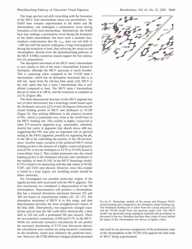
The three-dimensional structure of the MUV pigment hasnot yet been determined, but a homology model based uponthe rhodopsin structure (22) reveals divergence between theretinal binding pocket of MUV and rhodopsin or VCOP(Figure 8). One striking difference is the putative locationof F81, which is positioned very close to the Schiff base inthe MUV binding site. This residue is highly conserved inother UV-sensitive pigments (e.g., salamander, zebrafish,Anolis) but varies in pigments that absorb above 400 nm,suggesting that F81 may play an important role in spectraltuning in the SWS1 pigments, possibly by regulating the pKa
of the SB or by controlling the polarity of the SB environ-ment. Another major variation in the predicted MUV retinalbinding pocket is the absence of a highly conserved glutamicacid (E181 in bovine rhodopsin or E176 in VCOP) found inextracellular loop 2. This residue penetrates into the retinalbinding pocket in the rhodopsin structure and contributes tothe stability ofmeta II (34). In the MUV homology model,E176 is found to be interacting with the side chains of W186,Y187, and Y263 (not shown). However, since this residueis found in a loop region, our modeling results should betaken cautiously.
We investigated two possible molecular origins of thesignificant blue shift associated with the MUV pigment. Thefirst mechanism we considered is deprotonation of the SBchromophore. Deprotonation will produce a chromophorethat has a nominal absorption maximum between 355 and380 nm based on experimental studies (32, 35-37). Theabsorption maximum of MUV is in this range, and thusdeprotonation provides the most straightforward source ofthe blue shift. Alternatively, two negatively charged coun-terions placed near the SB could affect theoretically a blueshift to 310 nm with a protonated SB (not shown). Thereare two potential counterions, E108 and E176, in the MUV.While our molecular dynamics calculations do not predictthe necessary positioning of E176 (see discussion above),the calculations were carried out using harmonic constraintson the backbone, which may influence the predicted struc-ture. However, the FTIR difference integral method presented
and used in our previous assignment of the protonation stateof the chromophore in the VCOP (10) supports the dark stateof MUV being unprotonated.
FIGURE 8: Homology models of the mouse andXenopusSWS1retinal binding sites compared to the rhodopsin retinal binding site.The rhodopsin binding site is taken from the crystal structure (22)and the VCOP model from our previous study (10). The MUVmodel was generated using analogous methods and procedures asdiscussed in the text. Residues that have their center of mass behindthe plane of the chromophore are shown in lighter colors.
Photobleaching Pathway of Ultraviolet Visual Pigments Biochemistry, Vol. 41, No. 31, 20029849
What determines the protonation state of SWS1 pigments?In the case of VCOP, the principal counterion is D108, andthis counterion is stabilized via hydrogen bonding interactionswith S85 (4). In MUV, D108 is replaced with E108, andthis counterion would nominally be positioned closer to thechromophore. Density functional (B3LYP, 6-31G) molecularorbital simulations (not shown) indicate that when a glutamicacid counterion approaches to within∼3 Å of the SB proton,the SB hydrogen spontaneously migrates to the E108 residue,yielding an unprotonated chromophore. To have a protonatedSB, either a relatively large stabilizing field within thebinding site is necessary, which the calculations suggest doesnot exist in either MUV or VCOP, or the position of thecounterion is stabilized by additional interactions, such asprovided by S85 in VCOP (4). Both interactions appear tobe absent in MUV.
In bovine rhodopsin, a number of structural changes occurin going from the dark to themeta IIstate, most notably therigid-body motion of TM6 relative to the other helices anddeprotonation of the Schiff base, leading to the activeconformation (38). Deprotonation occurs during the transitionfrom meta I to meta II, but it is not clear at what point inthe photobleaching pathway the movement of TM6 occurs.Isomerization of the chromophore drives it into a highlystrained all-trans conformation (39), with no large changesin protein conformation taking place during formation of thebathointermediate (39, 40). Photoaffinity cross-linking withretinal analogues has shown that theâ-ionone ring interactswith W265 in TM6 in the dark andbatho conformations,and interacts with A169 in TM4 inlumi, meta I, andmeta II(41). Thebathoto lumi transition in bovine rhodopsin is alsoassociated with changes in the protein backbone (42-45),although these changes have not been well characterized.UV absorbance spectroscopy (46-48), linear dichroism (49),and site-specific mutagenesis (50) have shown that theenvironments of two tryptophan residues, W126 and W265,are perturbed during the formation of the active state.
The results presented here show significant protein con-formational changes in the formation ofbathoand lumi inMUV. These changes are directly reflected in the proteinabsorbance of aromatic residues, as well as the large changein the SB pKa upon formation of lumi, with apparentstoichiometric protonation. The formation ofmeta II alsoinvolves the deprotonation of the all-trans-retinal SB inmetaI. Although the MUV and rhodopsin pigments have widelydifferent absorbance properties in the dark, there appear tobe conserved changes in both pigments following isomer-ization of the chromophore, including major protein confor-mational changes to formlumi with a protonated SB. Thechanges most likely involve the retinylidene SB environment,possibly alterations of the interactions with the counterion,E108, or F81, to permit electrostatic stabilization of thepositive charge on the SB.
NOTE ADDED IN PROOF
The importance of the F81 residue to spectral tuning inMUV and bovine blue has been recently demonstrated (52).
REFERENCES
1. Ebrey, T., and Koutalos, Y. (2001)Prog. Retinal Eye Res. 20,49-94.
2. Pitt, G. A., Collins, F. D., Morton, R. A., and Stok, P. (1955)Biochem. J. 59, 122-127.
3. Vought, B. W., Dukkipati, A., Max, M., Knox, B. E., and Birge,R. R. (1999)Biochemistry 38, 11287-11297.
4. Dukkipati, A., Vought, B. W., Birge, R. R., and Knox, B. E. (2001)Biochemistry 40, 15098-15108.
5. Starace, D. M., and Knox, B. E. (1997)J. Biol. Chem. 272, 1095-1100.
6. Fasick, J., Lee, N., and Oprian, D. (1999)Biochemistry 38, 11593-11596.
7. Ma, J. X., Kono, M., Xu, L., Das, J., Ryan, J. C., Hazard, E. S.,Oprain, D. D., and Crouch, R. K. (2001)Vis. Neurosci. 18, 393-399.
8. Schneeweis, D. M., and Schnapf, J. L. (1999)J. Neurosci. 19,1203-1216.
9. Perry, R., and McNaughton, P. (1991)J. Physiol. 433, 561-87.10. Kusnetzow, A., Dukkipati, A., Babu, K. R., Singh, D., Vought,
B. W., Knox, B. E., and Birge, R. R. (2001)Biochemistry 40,7832-7844.
11. Babu, K. R., Dukkipati, A., Birge, R. R., and Knox, B. E. (2001)Biochemistry 40, 13760-13766.
12. Yokoyama, S., Radlwimmer, F. B., and Kawamura, S. (1998)FEBS Lett. 423, 155-158.
13. Shi, Y., Radlwimmer, F. B., and Yokoyama, S. (2001)Proc. Natl.Acad. Sci. U.S.A. 98, 11731-11736.
14. Sakmar, T. P., Franke, R. R., and Khorana, H. G. (1989)Proc.Natl. Acad. Sci. U.S.A. 86, 8309-8313.
15. Nathans, J. (1990)Biochemistry 29, 9746-9752.16. Zhukovsky, E. A., and Oprian, D. D. (1989)Science 246, 928-
931.17. Doukas, A. G., Aton, B., Callender, H., and Ebrey, T. G. (1978)
Biochemistry 17, 2430-2435.18. Longstaff, C., Seckler, B., Calhoon, R. D., and Rando, R. R. (1987)
in Biophysical studies of retinal proteins(Ebrey, T. G., Frauen-felder, H., Honig, B., and Nakanishi, K., Eds.) pp 64-70,University of Illinois Press, Champaign, IL.
19. Weitz, C. J., and Nathans, J. (1993)Biochemistry 32, 14176-14182.
20. Rao, V. R., Cohen, G. B., and Oprian, D. D. (1994)Nature 367,639-642.
21. Lewis, J., Szundi, I., Fu, W., Sakmar, T., and Kliger, D. (2000)Biochemistry 39, 599-606.
22. Palczewski, K., Kumasaka, T., Hori, T., Behnke, C. A., Mo-toshima, H., Fox, B. A., Le Trong, I., Teller, D. C., Okada, T.,Stenkamp, R. E., Yamamoto, M., and Miyano, M. (2000)Science289, 739-745.
23. Thompson, J. D., Higgins, D. G., and Gibson, T. J. (1994)NucleicAcids Res. 22, 4673-4680.
24. Vriend, G. (1990)J. Mol. Graph. 8, 52-56.25. Brooks, B., Bruccoleri, R. E., Olafson, B. D., States, D. J.,
Swaminathan, S., and Karplus, M. (1983)J. Comput. Chem. 4,187-217.
26. MacKerell, A. D., Jr., Bashford, D., Bellott, M., Dunbrack, R. L.,Evanseck, J. D., Field, M. J., Fischer, S., Gao, J., Guo, H., Ha,S., Joseph-McCarthy, D., Kuchnir, L., Kuczera, K., Lau, F. T.K., Mattos, C., Michnick, S., Ngo, T., Nguyen, D. T., Prodhom,B., Reiher, W. E., III, Roux, B., Schlenkrich, M., Smith, J. C.,Stote, R., Straub, J., Watanabe, M., Wiorkiewicz-Kuczera, J., Yin,D., and Karplus, M. (1998)J. Phys. Chem. B 102, 3586-3616.
27. Hermone, A., and Kuczera, K. (1998)Biochemistry 37, 2843-2853.
28. Shichida, Y., and Imai, H. (1998)Cell Mol. Life Sci. 54, 1299-315.29. Wald, G., and Brown, P. K. (1953)J. Gen. Physiol. 37, 189-200.30. Ridge, K. D., Lu, Z., Liu, X., and Khorana, H. G. (1995)
Biochemistry 34, 3261-3267.31. Sakmar, T. P. (1998)Prog. Nucleic Acid Res. 59, 1-34.32. Murray, L. P., and Birge, R. R. (1985)Can. J. Chem. 63, 1967-
1971.33. Imai, H., Terakita, A., Tachibanaki, S., Imamoto, Y., Yoshizawa,
T., and Shichida, Y. (1997)Biochemistry 36, 12773-12779.34. Yan, E. C., Kazmi, M. A., De, S., Chang, B. S., Seibert, C., Marin,
E. P., Mathies, R. A., and Sakmar, T. P. (2002)Biochemistry 41,3620-3627.
35. Waddell, W. H., Schaffer, A. M., and Becker, R. S. (1977)J. Am.Chem. Soc. 99, 8456-8460.
36. Harosi, F. I., Favrot, J., Leclercq, Vocelle, D., and Sandorfy, C.(1978)ReV. Can. Biol. 37, 257-271.
37. Sandorfy, C., Lussier, L. S., Thanh, H. L., and Vocelle, D. (1987)in Biophysical studies of retinal proteins(Ebrey, T. G., Frauen-
9850 Biochemistry, Vol. 41, No. 31, 2002 Dukkipati et al.
felder, H., Honig, B., and Nakanishi, K., Eds.) pp 247-251,University of Illinois Press, Champaign, IL.
38. Farrens, D. L., Altenbach, C., Yang, K., Hubbell, W. L., andKhorana, H. G. (1996)Science 274, 768-770.
39. Kandori, H., Shichida, Y., and Yoshizawa, T. (2001)Biochemistry(Moscow) 66, 1197-1209.
40. Kandori, H., and Maeda, A. (1995)Biochemistry 34, 14220-14229.
41. Borhan, B., Souto, M., Imai, H., Shichida, Y., and Nakanishi, K.(2000)Science 288, 2209-2212.
42. Ujj, L., Jager, F., and Atkinson, G. H. (1998)Biophys. J. 74, 1492-1501.
43. Ganter, U. M., Ga¨rtner, W., and Siebert, F. (1988)Biochemistry27, 7480-7488.
44. Maeda, A., Ohkita, Y. J., Sasaki, J., Shichida, Y., and Yoshizawa,T. (1993)Biochemistry 32, 12033-12038.
45. Imai, H., Mizukami, T., Imamoto, Y., and Shichida, Y. (1994)Biochemistry 33, 14351-14358.
46. Rafferty, C. (1979)Photochem. Photobiol. 29, 109-120.47. Rafferty, C., Mullenberg, C., and Shichi, H. (1980)Biochemistry
19, 2145-2151.48. Lewis, J., Jager, S., and Kliger, D. (1997)Photochem. Photobiol.
66, 741-746.
49. Chabre, M., and Breton, J. (1979)Photochem. Photobiol. 30, 295-299.
50. Lin, S. W., and Sakmar, T. P. (1996)Biochemistry 35, 11149-11159.
51. Frisch, M. J., Trucks, G. W., Schlegel, H. B., Scuseria, G. E.,Robb, M. A., Cheeseman, J. R., Zakrzewski, V. G., Montgomery,J. A., Stratmann, R. E., Burant, J. C., Dapprich, S., Millam, J.M., Daniels, A. D., Kudin, K. N., Strain, M. C., Farkas, O.,Tomasi, J., Barone, V., Cossi, M., Cammi, R., Mennucci, B.,Pomelli, C., Adamo, C., Clifford, S., Ochterski, J., Petersson, G.A., Ayala, P. Y., Cui, Q., Morokuma, K., Malick, D. K., Rabuck,A. D., Raghavachari, K., Foresman, J. B., Cioslowski, J., Ortiz,J. V., Stefanov, B. B., Liu, G., Liashenko, A., Piskorz, P.,Komaromi, I., Gomperts, R., Martin, R. L., Fox, D. J., Keith, T.,Al-Laham, M. A., Peng, C. Y., Nanayakkara, A., Gonzalez, C.,Challacombe, M., Gill, P. M. W., Johnson, B. G., Chen, W.,Wong, M. W., Andres, J. L., Head-Gordon, M., Replogle, E. S.,and Pople, J. A. (1998) Gaussian Inc., Pittsburgh, PA.
52. Fasick, J. I., Applebury, M. L., and Oprian, D. D. (2002)Biochemistry 41, 6860-6865.
BI025883G
Photobleaching Pathway of Ultraviolet Visual Pigments Biochemistry, Vol. 41, No. 31, 20029851