Dynamics of proteins in Golgi membranes: comparisons between mammalian and plant cells highlighted...
14
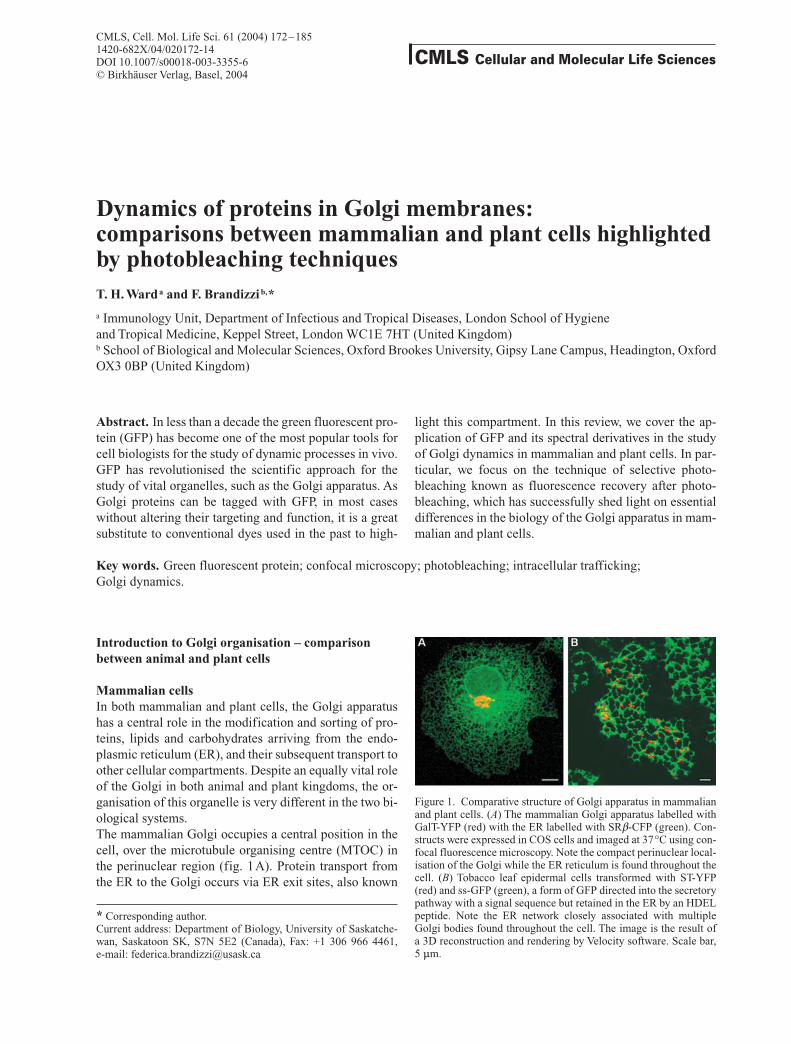
Dynamics of proteins in Golgi membranes: comparisons between mammalian and plant cells highlighted by photobleaching techniques T. H. Ward a and F. Brandizzi b, * a Immunology Unit, Department of Infectious and Tropical Diseases, London School of Hygiene and Tropical Medicine, Keppel Street, London WC1E 7HT (United Kingdom) b School of Biological and Molecular Sciences, Oxford Brookes University, Gipsy Lane Campus, Headington, Oxford OX3 0BP (United Kingdom) Abstract. In less than a decade the green fluorescent pro- tein (GFP) has become one of the most popular tools for cell biologists for the study of dynamic processes in vivo. GFP has revolutionised the scientific approach for the study of vital organelles, such as the Golgi apparatus. As Golgi proteins can be tagged with GFP, in most cases without altering their targeting and function, it is a great substitute to conventional dyes used in the past to high- CMLS, Cell. Mol. Life Sci. 61 (2004) 172 – 185 1420-682X/04/020172-14 DOI 10.1007/s00018-003-3355-6 © Birkhäuser Verlag, Basel, 2004 CMLS Cellular and Molecular Life Sciences light this compartment. In this review, we cover the ap- plication of GFP and its spectral derivatives in the study of Golgi dynamics in mammalian and plant cells. In par- ticular, we focus on the technique of selective photo- bleaching known as fluorescence recovery after photo- bleaching, which has successfully shed light on essential differences in the biology of the Golgi apparatus in mam- malian and plant cells. Key words. Green fluorescent protein; confocal microscopy; photobleaching; intracellular trafficking; Golgi dynamics. Introduction to Golgi organisation – comparison between animal and plant cells Mammalian cells In both mammalian and plant cells, the Golgi apparatus has a central role in the modification and sorting of pro- teins, lipids and carbohydrates arriving from the endo- plasmic reticulum (ER), and their subsequent transport to other cellular compartments. Despite an equally vital role of the Golgi in both animal and plant kingdoms, the or- ganisation of this organelle is very different in the two bi- ological systems. The mammalian Golgi occupies a central position in the cell, over the microtubule organising centre (MTOC) in the perinuclear region (fig. 1 A). Protein transport from the ER to the Golgi occurs via ER exit sites, also known * Corresponding author. Current address: Department of Biology, University of Saskatche- wan, Saskatoon SK, S7N 5E2 (Canada), Fax: +1 306 966 4461, e-mail: [email protected] Figure 1. Comparative structure of Golgi apparatus in mammalian and plant cells. (A) The mammalian Golgi apparatus labelled with GalT-YFP (red) with the ER labelled with SRb-CFP (green). Con- structs were expressed in COS cells and imaged at 37 °C using con- focal fluorescence microscopy. Note the compact perinuclear local- isation of the Golgi while the ER reticulum is found throughout the cell. (B) Tobacco leaf epidermal cells transformed with ST-YFP (red) and ss-GFP (green), a form of GFP directed into the secretory pathway with a signal sequence but retained in the ER by an HDEL peptide. Note the ER network closely associated with multiple Golgi bodies found throughout the cell. The image is the result of a 3D reconstruction and rendering by Velocity software. Scale bar, 5 mm.
Transcript of Dynamics of proteins in Golgi membranes: comparisons between mammalian and plant cells highlighted...
Dynamics of proteins in Golgi membranes:comparisons between mammalian and plant cells highlightedby photobleaching techniquesT. H. Warda and F. Brandizzib,*
a Immunology Unit, Department of Infectious and Tropical Diseases, London School of Hygiene and Tropical Medicine, Keppel Street, London WC1E 7HT (United Kingdom)b School of Biological and Molecular Sciences, Oxford Brookes University, Gipsy Lane Campus, Headington, OxfordOX3 0BP (United Kingdom)
Abstract. In less than a decade the green fluorescent pro-tein (GFP) has become one of the most popular tools forcell biologists for the study of dynamic processes in vivo.GFP has revolutionised the scientific approach for thestudy of vital organelles, such as the Golgi apparatus. AsGolgi proteins can be tagged with GFP, in most caseswithout altering their targeting and function, it is a greatsubstitute to conventional dyes used in the past to high-
CMLS, Cell. Mol. Life Sci. 61 (2004) 172–1851420-682X/04/020172-14DOI 10.1007/s00018-003-3355-6© Birkhäuser Verlag, Basel, 2004
CMLS Cellular and Molecular Life Sciences
light this compartment. In this review, we cover the ap-plication of GFP and its spectral derivatives in the studyof Golgi dynamics in mammalian and plant cells. In par-ticular, we focus on the technique of selective photo-bleaching known as fluorescence recovery after photo-bleaching, which has successfully shed light on essentialdifferences in the biology of the Golgi apparatus in mam-malian and plant cells.
Key words. Green fluorescent protein; confocal microscopy; photobleaching; intracellular trafficking; Golgi dynamics.
Introduction to Golgi organisation – comparisonbetween animal and plant cells
Mammalian cellsIn both mammalian and plant cells, the Golgi apparatushas a central role in the modification and sorting of pro-teins, lipids and carbohydrates arriving from the endo-plasmic reticulum (ER), and their subsequent transport toother cellular compartments. Despite an equally vital roleof the Golgi in both animal and plant kingdoms, the or-ganisation of this organelle is very different in the two bi-ological systems.The mammalian Golgi occupies a central position in thecell, over the microtubule organising centre (MTOC) inthe perinuclear region (fig. 1A). Protein transport fromthe ER to the Golgi occurs via ER exit sites, also known
* Corresponding author.Current address: Department of Biology, University of Saskatche-wan, Saskatoon SK, S7N 5E2 (Canada), Fax: +1 306 966 4461, e-mail: [email protected]
Figure 1. Comparative structure of Golgi apparatus in mammalianand plant cells. (A) The mammalian Golgi apparatus labelled withGalT-YFP (red) with the ER labelled with SRb-CFP (green). Con-structs were expressed in COS cells and imaged at 37°C using con-focal fluorescence microscopy. Note the compact perinuclear local-isation of the Golgi while the ER reticulum is found throughout thecell. (B) Tobacco leaf epidermal cells transformed with ST-YFP(red) and ss-GFP (green), a form of GFP directed into the secretorypathway with a signal sequence but retained in the ER by an HDELpeptide. Note the ER network closely associated with multipleGolgi bodies found throughout the cell. The image is the result of a 3D reconstruction and rendering by Velocity software. Scale bar,5 mm.

CMLS, Cell. Mol. Life Sci. Vol. 61, 2004 Multi-author Review Article 173
as transitional ER (tER) [1]. These are relatively immo-bile domains of 1–2 mm in diameter closely apposed totubular clusters, also termed pre-Golgi intermediates,vesicular-tubular clusters (VTCs), or ERGIC (ER-Golgiintermediate compartment) [2–5].Initial transport of cargo out of the ER is mediated by theactivity of the small GTPase Sar1 and the cytosolic coatcomplex known as COPII [6–9]. COPII components in-teract sequentially: an integral ER membrane protein,Sec12 [10], acts as a specific guanine nucleotide ex-change factor (GEF) for the activation of Sar1, a smallGTP-binding protein [11–13]; Sar1 is recruited to the ERmembrane by Sec12 [10, 14]; the exchange of GDP forGTP on Sar1 determines its activation; the Sec23-Sec24heterodimer then binds to membrane-anchored Sar1-GTP [11, 15]; coat and vesicle formation is initiated bythe binding of a Sec13-Sec31 complex [15, 16], whichacts as scaffolding for the newly forming vesicle; GTPhydrolysis on Sar1 dissociates the GTPase from the ER[11], rendering the COPII components labile and easilydisplaced from a completed vesicle [17].A second coat complex known as COPI [18–21] whoseassembly is regulated by the GTPase Arf1 [22, 23] is re-quired for maturation of pre-Golgi intermediates[24–26]. Arf1 is activated by a GEF sensitive to the fun-gal metabolite brefeldin A (BFA) [27, 28], a tool oftenused to block COPI assembly and thus to disrupt traf-ficking pathways in the early secretory pathway. COPI isalso involved in regulating intra-Golgi trafficking, al-though the nature of this role is unclear [29–35].The connection between tER and VTCs is not clearly un-derstood. COPII-coated vesicles containing protein cargomay bud on the surface of tER and then fuse with VTCs[3, 8]. Alternatively, tER may mature directly by tubulefusion and fission into VTCs that translocate to the Golgi,and ER-to-Golgi protein transport may then take placewithout vesicle carriers [32, 36, 37].Pre-Golgi intermediates are transient discrete entities thatmove through the action of dynein-driven motors on themicrotubule cytoskeleton [38, 39], to fuse with the cis-face of the Golgi apparatus and deliver protein cargothrough Rab1-dependent tethering and fusion factors [40,41]. The pre-Golgi compartment may also be the first siteof retrieval and recycling of proteins back to the ER [24,42, 43]. Retrieval of escaped ER proteins as well as ret-rograde transport of itinerant proteins such as the KDELreceptor is also a function of the cis-Golgi [5, 29, 44, 45].Once arrived at the Golgi, the modality of intra-Golgitransport of secretory cargo is still a matter of debate (re-viewed in [46–49]). According to the cisternal matura-tion model, the Golgi apparatus exists as a polarised or-ganelle where cisternae progress through the Golgi, suchthat the cis-face is newly formed upon fusion of the pre-Golgi compartments and the trans-face would representthe oldest portion of the Golgi [1, 49–54]. Alternatively,
intra-Golgi trafficking may exploit vesicles as cargo car-riers to accomplish movement and sorting of cargo in dif-ferent compartments of the Golgi apparatus, as postu-lated in the vesicle carrier model [31, 33, 55–57]. A fur-ther model incorporates aspects of both hypotheses suchthat intra-Golgi transport occurs both upon cisternal mat-uration and with some movement of vesicle carriers [47,54, 58]. Another model suggests that cargo movesthrough permanent or transient continuities (i.e. transporttubules) that connect successive compartments, therebycreating a continuous compartment through which cargocan flow [46, 51, 59, 60].Transport from the Golgi apparatus to the plasma mem-brane occurs by post-Golgi carriers [61, 62]. These initi-ate as tubular extensions from the TGN, which extend outfrom Golgi membranes and are severed at the farthest endby the action of a dynamin-2 [63]. Post-Golgi transportmay require an intact actin cytoskeleton to be efficient, asdepolymerisation of actin with cytochalasin B signifi-cantly slows export of cargo from the Golgi, while mi-crotubule depolymerisation with nocodazole has no ef-fect on cargo export [61].
PlantsStudies on the dynamics of the plant Golgi are relativelypreliminary in comparison with the mammalian counter-part. Plant endomembranes do not label with vitallipophilic dyes that specifically stain the membranes oftargeted organelles in mammalian cells [64]. Studies onplant Golgi dynamics using GFP have also suffered a de-lay with respect to other biological systems due to thepresence of a cryptic intron in the wild-type GFP thatcaused aberrant splicing of the GFP coding sequencesuch that the mature protein was unable to fluoresce [65,66]. This phenomenon restricted the use of GFP to RNAviral vectors where expression is not controlled by splic-ing [67]. It was not until 1997 that Haseloff et al. pro-duced a GFP with altered codon usage, which eliminatedthe occurrence of aberrant splicing [66]. This opened upthe possibility of GFP technology to the study of plant or-ganelle dynamics [68, 69]. The subsequent availability ofnew GFP spectral variants lacking the cryptic intron hasfurther accelerated the study of plant cell dynamics. An-other reason for the comparable delay in the characterisa-tion of plant Golgi dynamics is due to the scarcity of gen-uine plant Golgi enzymes to be used as markers in vivo.In fact, it was not until GFP was fused to the putative Arabidopsis H/KDEL receptor Erd2 (a protein found inmammalian cells to cycle in the ER-to-Golgi pathwaywhile recycling soluble proteins tagged with KDEL backto the ER [44, 70]) and also to the transmembrane domainof a rat sialyl-transferase (a Golgi glycosylation enzyme)[71], and that both chimaeras were found to localise toGolgi stacks by electron microscopy (EM), that the plant

Golgi could be investigated in vivo for the first time [72].Subsequent identification of genuine plant Golgi en-zymes and their fusion to GFP has opened up further thecharacterisation of the dynamics of the Golgi apparatus indifferent plant cell systems [73–76].The organisation of plant Golgi apparatus at the lightlevel appears rather different to the mammalian Golgi(fig. 1B). The plant Golgi apparatus is scattered in thecell as small cisternal stacks in the cortical cytoplasm andwithin trans-vacuolar strands of cytoplasm [72–75]. Inleaves, using fluorescent protein constructs, it appearsthat the individual cisternal stacks of the Golgi apparatusare closely associated with the cortical ER network [72,77]. These stacks move in an actin-myosin dependentmanner over the tubules of the ER [72, 75], in contrast tomammalian cells where microtubules are required forER-to-Golgi trafficking and Golgi positioning [38, 61].Movie sequences of the ER-Golgi continuum indicatethat movement of the plant Golgi may in part be relatedto modelling of the underlying ER network through con-trol of ER tubule growth (see movies http://www.brookes.ac.uk/schools/bms/research/molcell/hawes/gfp/gfp.html) [78]. This indicates that the ER and Golgi maybehave as one dynamic system, either through directmembrane continuities or through continuous vesicle ortubule formation/fusion reactions [78].This dynamic organisation of the plant secretory pathwayopens up questions about the modality of ER-to-Golgitransport and the distribution of ER export sites. Onemodel originated from observations gathered in BY-2cells expressing a mannosidase II (a Golgi-resident gly-cosylation enzyme [79])-GFP chimaera. As the Golgistacks move, arrest and regain movement after time inter-vals, it was postulated the discontinuity in Golgi move-ment is linked to ER-to-Golgi protein transport. Thiswould occur during Golgi arrest, possibly after transientdetachment from actin (‘stop-and-go model’) [75]. Alter-natively, Golgi movement in plant cells would allow theGolgi to continually collect vesicles budding from the ER(‘vacuum cleaner model’) [72].In yeast and mammalian cells, most of the genes involvedin ER-to-Golgi transport have been characterised (seeabove). The cytosolic coat protein complexes COPI andCOPII shuttle proteins between the ER and Golgi. Grow-ing evidence in the plant literature [80–83] indicates thatcomponents of the COP systems also operate in proteintrafficking in plant cells. Homologues of Sar1, Rab1 andArf1 have been cloned, and their GTPase-deficient mu-tants act as trans dominant negatives for protein transport[82, 84–86]. Takeuchi et al. established that an Arf1-GFPchimaera localises at organelles, which they considerGolgi [87]. These findings are in accordance with im-munolocalisation of COPI components [81, 83] to smallvesicles surrounding or budding from cisternae at the cisface of the Golgi apparatus in plant cells [81, 83].
The modality of transport of proteins through the plantGolgi is still a matter of debate. Ritzenthaler et al. postu-lated that intra-Golgi transport in BY-2 cells occurs bycisternal maturation [83]. This was based on results obtained with the fungal metabolite BFA, which col-lapses Golgi membranes into the ER [88]. A less inva-sive approach is still needed to establish the nature of the real mechanisms underlying intra-Golgi transport inplants.
Fluorescence recovery after photobleaching
GFP fluorescence has proved invaluable as a visual toolfor determining protein location and transport pathwayswithin cells, for investigating the biogenesis and dynam-ics of organelles, as well as for direct observation of thetrafficking of proteins within, and between, living cells.However, a more sophisticated application of GFP for the study of cellular dynamics is based on the ability to irreversibly photobleach the GFP fluorophore and to then measure the recovery of fluorescence by exchangeof bleached for unbleached GFP molecules, a processknown as fluorescence recovery after photobleaching(FRAP). This technique has been used to assess the struc-ture and connectivity of artificial [89] and biologicalmembranes [90].A general downside of a FRAP protocol is the develop-ment of free radicals, generated by the high-energy laserirradiation, capable of causing damage to the cell. How-ever, GFP is a particularly well-suited fluorescent tag forFRAP experiments. Upon bleaching, the GFP barrel thatsurrounds the core fluorophore constrains such free radi-cals from escaping and damaging cellular components,such as proteins and lipids.The key fundamentals of FRAP are (i) the fluorescence ofthe fluorochrome (such as GFP) is irreversibly eliminatedwith an intense photobleaching pulse of laser light; (ii) inthe absence of physical constraints, a protein is free todiffuse, i.e. it is able to move randomly by Brownian mo-tion in its environment and (iii) recovery of fluorescenceinto the bleached area is the result of exchange due to dif-fusion of unbleached molecules with bleached ones in thesame area [90].There are two types of FRAP protocols, quantitative andqualitative. Qualitative FRAP allows protein movementto be followed in vivo, within a defined area. This willproduce high-quality images that show a global fluores-cence recovery within the bleached area but no data use-ful for quantifying protein mobility. The quantitative pro-tocol, on the other hand, allows estimation of a diffusioncoefficient, calculation of the mobile fraction and thetime of recovery of fluorescence after bleaching of a re-gion of interest (ROI). The mobile fraction is the percent-age of fluorescent protein that diffuses into a bleached
174 T. H. Ward and F. Brandizzi Mammalian and plant Golgi dynamics

area, and a diffusion coefficient is a measure of move-ment of the mobile pool of fluorescent proteins over time.In FRAP experiments, the half-time of recovery indicatesthe time required for the fluorescence to reach 50% of aplateau of intensity.With the advent of two-colour GFP imaging [91], anotherapplication of FRAP is to use photobleaching to visualiseprotein trafficking or flux through a two-colour bleachingprotocol [78, 92, 93]. Proteins or signals to be visualisedshould be tagged with either the cyan- (CFP) or yellow-(YFP) fluorescent protein and coexpressed in the samecells [91]. The two fluorochromes may be targeted to thesame organelle or even two chimaeras of the same pro-tein. In this FRAP protocol, YFP and CFP molecules haveto be excited together, using a multi-tracking mode of aconfocal microscope with a 514-nm laser line to exciteYFP and a 458-nm (or 413-nm) laser line to excite CFP.YFP is selectively photobleached using the 514-nm laserline at maximum power, leaving CFP fluorescence unal-tered. This technique is a great advance of the FRAP tech-nology as it exploits CFP as a visual reference to followthe dynamics of a targeted organelle, and simultaneouslyto follow and quantify YFP fluorescence recovery. In ad-dition, when YFP and CFP chimaeras of the same proteinare coexpressed, fluorescence recovery may be tracked byimage differencing by subtracting the image of thebleached fluorochrome from that of the unbleached fluo-rochrome, a technique called fluorescence localisationafter photobleaching (FLAP) [93].Another application of photobleaching is to reduce fluo-rescence from background noise to reveal faint popula-tions of fluorescent proteins [34]. Alternatively, photo-bleaching of a large pool of protein, for example on theplasma membrane, allows visualisation of organelle dy-namics inside the cell usually masked by the plasmamembrane pool. For example, a protein that cycles be-tween the Golgi and plasma membrane might then be vi-sualised in translocating intermediates [94].Finally, fluorescence loss in photobleaching (FLIP) in-vestigates fluorophore mobility and continuity betweenfluorescent organelles [95]. FLIP is similar to FRAP inthat a region of interest is photobleached with a high-power laser; however, unlike FRAP, this is repeatedlybleached over time to deplete the entire fluorescent pool.With this technique it is possible to assess the spatial con-tinuity between two organelles if photobleaching of onedepletes the entire fluorescence of the other or even to de-termine the free diffusion of a protein within a subcellu-lar compartment.
FRAP applied to mammalian Golgi dynamics
FRAP as a tool to answer biological questions has foundapplications in both animal and plant cells. However, this
technology has uncovered noticeable differences betweenthe dynamics of the Golgi in the two systems.In mammalian cells, FRAP has been applied to investi-gate diffusion, exchange, kinetics of proteins passingthrough the Golgi apparatus.
Diffusion of proteins in the GolgiInitial visualisation of the mammalian Golgi in vivo wasbased on the application of fluorescent lipid derivatives,such as NBD ceramide, to label the membrane as therewas no way to label proteins while maintaining cell via-bility [96]. The usefulness of the vital stain NBD ce-ramide was restricted by its metabolism into sphin-golipids that were then trafficked to the plasma mem-brane, making it only a transient marker for the Golgi[97]. However, it allowed visualisation of tubulovesicularprocesses emerging from the trans-Golgi network(TGN), which either formed transport intermediates orcould fuse with adjacent trans-Golgi elements in a mi-crotubule-dependent manner, forming a reticular network[98]. By using FRAP, Cooper et al. were able to show thatthese tubulovesicular processes formed pathways allow-ing diffusion of membrane lipids between joined trans-Golgi elements, although not between obviously sepa-rate, discrete elements [98]. This study therefore demon-strated for the first time that the dynamic processes at theTGN are not only required for post-Golgi transport butalso for connectivity within the Golgi itself. Storrie et al.used indirect labelling to visualise the movement of acargo protein through the secretory pathway in vivo [99].Cells infected with the ts045 mutant of vesicular stoma-titis virus (VSV-ts045) were held at 40°C to accumu-late the virus glycoprotein (ts045-G or VSVG) in theER. Microinjection of rhodamine-conjugated Fab frag-
ments of a monoclonal antibody against the ts-045-G cytoplasmic tail enabled tracking of the protein throughthe cell. FRAP of a thin strip across the Golgi in cells la-belled with rhodamine label revealed that VSVG was ableto diffuse in Golgi and trans-Golgi membranes at ratesfaster than those found for proteins on the plasma mem-brane, i.e. it is not constrained by protein complexes [99,100].A real boost to FRAP application was given by the adventof GFP technology. In 1996, Cole et al. published a sem-inal paper not only using GFP as a marker for membranetrafficking in mammalian cells for the first time, but alsodemonstrating the biophysical potential of GFP for use inlive cell imaging and photobleaching techniques [95].This enabled analysis of the behaviour of cellular proteinsnot possible by previous, more invasive, means such asbiochemistry and EM. The authors created a number ofchimaeric Golgi membrane proteins, including Erd2 andmannosidase II (Man2 [101]), by fusing the protein cod-
CMLS, Cell. Mol. Life Sci. Vol. 61, 2004 Multi-author Review Article 175

ing sequence to GFP-encoding DNA and then expressingthe constructs in mammalian cell lines. These chimaericproteins were found to correctly localise to the Golgi.Furthermore, a chimaera comprising just the transmem-brane domain of galactosyltransferase fused to GFP wasalso found to localise to the Golgi, showing that targetingsignals present in that domain alone are responsible forGolgi localisation [95, 102, 103]. In fact it appears thatthe length of the transmembrane domain of a protein, andnot its amino acid content, is sufficient to determineGolgi localisation [102, 103].At this time, the vesicular trafficking model of intra-Golgi transport predicted that Golgi-localised enzymeswere found in the cisternae of the stack, while transitingcargo was ferried between cisternae by vesicles [31, 104].A dilemma was how the enzymes were prevented fromentering the vesicles. One hypothesis was that ‘kin recog-nition’ between fellow enzymes would cause them to in-teract and cluster together, creating large heterooligomersunable to fit into the small translocating vesicles [105].These protein complexes might even be further tetheredto an underlying Golgi matrix holding the cisternae to-gether in a stacked formation [105]. This proposal wasbased on the observation that overexpression of a Golgienzyme misdirected to the ER caused relocalisation ofother enzymes to the ER [101]. A FRAP approach onGolgi proteins tagged with GFP elegantly provided thefirst evidence for disproving the kin-recognition model[95]. When Cole et al. analysed the dynamics of the GFP-tagged Golgi membrane proteins in living cells, theybleached small strips of fluorescence across the Golgistack and looked for recovery [95]. The kin recognitionmodel predicted that the membrane proteins would beheld in a stable conformation. However, the GFP chi-maeras were found to rapidly recover the fluorescence tothe bleached region of interest with very high mobilefraction (85–98%) and diffusion coefficient. This indi-cated that the GFP chimaeras were freely diffusible in thelipid bilayer within the Golgi cisternae and that they werenot aggregating or being slowed by interactions withother proteins. Chimaeric GFP proteins of the p24 familyalso exhibit fast diffusion through Golgi membranes fol-lowing photobleaching of a small region [106]. Alu-minium fluoride (AIF) is an activator of heterotrimeric Gproteins [107] and causes irreversible binding of periph-eral protein complexes to the Golgi membrane [23, 108,109]. In FRAP experiments on cells pretreated with AlFfor 10 min, virtually complete immobilization of all ofthe GFP chimaeras in Golgi membranes was observed[95, 106]. This showed that only when peripheral proteinsare unable to release from membranes are Golgi proteinsimmobilized.To investigate the extent to which Golgi membranes arecontinuous, the same study [95] used a FLIP protocol forfirst time. By repeatedly photobleaching a strip across the
Golgi, the authors could entirely deplete Golgi fluores-cence extending outside the strip. Different regions of theGolgi lost fluorescence at different rates, possibly due todifferences in connectivity, but overall extensive lateraldiffusion occurred between Golgi stacks. Thus, the chi-maeras could diffuse rapidly and freely in Golgi mem-branes, suggesting that Golgi targeting and retention ofthese molecules does not depend on protein immobiliza-tion.Measuring diffusion of Golgi components was also usedto determine the fate of the Golgi membranes during mi-tosis [110]. The tightly packed pericentriolar localisationof the Golgi of interphase cells becomes dispersed duringmitosis and then recoalesces as cells enter interphaseonce more. The nature of these dispersed membranes hasbeen much disputed [111, 112]. One model argues thatthe Golgi becomes absorbed into the ER, taking advan-tage of preexisting cycling pathways [110, 111], while theother proposes that the Golgi is dispersed into vesiclesdistributed throughout the cell [112]. Zaal et al. usedFRAP, in the first instance, of a Golgi membrane proteingalactosyl transferase (GalT) tagged with GFP to investi-gate its mobility in mitotic cells [110]. They found thatthe rate of diffusion of the membrane protein in the dis-persed mitotic membranes was equivalent to that foundfor the same chimaera when expressed in the ER in inter-phase cells, i.e. in a continuous membrane system. Sincethis does not exclude the possibility that vesicles wouldbe able to diffuse very fast through the cytosol of the mi-totic cell, the authors then compared this mobility to thatof Golgi lipids in mitotic cells. If the Golgi redistributesinto vesicles in a mitotic cell, then the fluorescence ofGolgi-resident proteins and lipids would be expected torecover at identical rates into a bleached region since theywould be on the same carrier, rather than moving as sep-arate components of a continuous membrane system.However, a fluorescent lipid analogue BODIPY-ceramidewas found by FRAP to have a diffusional mobility muchgreater (10-fold faster) than the GFP-tagged Golgi pro-tein, consistent with the different mobility expected be-tween a protein and a lipid in an interconnected mem-brane system [110]. BODIPY-ceramide dynamics in amitotic cell also matched those found for the dye when re-distributed to the ER, upon treatment with BFA, in inter-phase cells. These data are therefore not consistent with avesicle model for the Golgi in mitosis. Furthermore,when GalT-GFP was repetitively photobleached using theFLIP protocol in mitotic cells, the diffuse label was com-pletely depleted, demonstrating that it is capable of dif-fusing throughout the membranes in metaphase cells, andtherefore indicative of localisation to the ER [110]. In mi-totic sea urchin embryo cells, FLIP of Golgi markers in-cluding GalT-GFP and a KDEL-receptor-GFP (Erd2)also revealed their localisation to a continuous membranesystem [113]. This suggests that relocalisation of the
176 T. H. Ward and F. Brandizzi Mammalian and plant Golgi dynamics

CMLS, Cell. Mol. Life Sci. Vol. 61, 2004 Multi-author Review Article 177
Golgi membranes to the ER may represent a commonmechanism for the division of the Golgi between daugh-ter cells in eukaryotes.
ExchangeFRAP has been pivotal in defining the mammalian Golgias a dynamic organelle in the secretory pathway throughanalysis of the behaviour of the Golgi’s constituent parts.There are many different types of proteins associatedwith the Golgi that are required to accomplish its manyfunctions. Membrane-bound proteins include the glyco-sylation enzymes, required for the maturation of secre-tory cargo, as well as putative cargo receptors such as ER-GIC-53, the p24 family and Erd2 (KDEL receptor) thatcycle between the ER and the Golgi. Then there are theperipherally associated proteins, such as coat proteins(COPI and the clathrin/adaptor complex), the scaffoldproteins thought to hold the Golgi in its characteristicstacked formation (e.g., GM130, GRASP65 [114, 115])and signalling molecules such as PLD1, PKA, MEKK2and PI-3K [116–120].While Golgi membrane proteins had been found to befreely diffusible within the Golgi apparatus [95] and itin-erant proteins such as ERGIC-53 and Erd2 had beenfound to cycle constitutively between the ER and theGolgi [5, 44], the Golgi glycosylation enzymes were stillconsidered held within the boundaries of the Golgi as awhole. It had been observed that upon treatment of cellswith nocodazole (which causes microtubules to depoly-merise), Golgi membrane proteins became localised tosmall punctate structures [121, 122]. It was proposed thatthis was caused by fragmentation and dispersal of theGolgi [122, 123]. These nocodazole-induced structureswere found to resemble mini-Golgi bodies by EM [124].When cells expressing GFP chimaeras of Golgi glycosy-lation enzymes were treated with nocodazole, FRAP ofthese mini-stacks showed that these membrane proteinswere not constrained to the individual structures since thefluorescence recovered, suggesting that resident proteinswere able to exchange between stacks, potentially via theER, although exchange by trafficking of small vesiclesfrom other mini-stacks could not be excluded [125].However, when the whole Golgi was photobleached im-mediately after addition of nocodazole, fluorescence wasstill found to accumulate into mini-stacks in the absenceof new protein synthesis [110].In these experiments, recovery of fluorescence indicatedthe existence of another cellular pool of the Golgi en-zymes, which was thought to be the ER. Moreover,FRAP experiments indicated that an exchange of thephotobleached molecules with an unbleached pool en-abled the fluorescence recovery by cycling pathways be-tween the ER and the Golgi. That Golgi enzymes cyclethrough the ER was confirmed by microinjection of a
dominant GTP-bound Sar1 mutant, which blocks proteinER exit. Blockage of exit from the ER causes Golgi en-zymes to gradually accumulate in the ER as they recyclefrom the Golgi [110, 125]. To show that Golgi enzymesstill cycle constitutively in the absence of cellular pertur-bants, the Golgi pool of GalT-GFP was selectively pho-tobleached in untreated cells (fig. 2A). Fluorescencewas found to recover to the area within 20 min, indicat-ing exchange with the ER pool. Conversely, if the totalcellular fluorescence outside of the Golgi (i.e. the ERpool) was photobleached, it was found to recover withcomparable decrease in total Golgi fluorescence [110].The purpose of this cycling pathway in interphase cellsis not clear but may underlie maintenance of Golgi struc-ture [32, 110].In contrast to the Golgi enzymes, other membrane pro-teins associated with the Golgi, such as KDEL receptor,are required to cycle between the ER and the Golgi in or-der to fulfil their proposed functions, i.e. to retrieve es-caped soluble KDEL-labelled ER proteins back from theGolgi [44]. Other proteins such as ERGIC-53 and the p24family of proteins are also thought to be cargo receptorsand cycle between the ER and the Golgi [5, 126]. Unsur-prisingly, therefore, FRAP of the Golgi fluorescence incells expressing GFP chimaeras of these proteins showsthat recovery is much faster (within 4 min) than that ofthe glycosylation enzymes, indicating faster cyclingtimes within the early secretory pathway [26, 127].FRAP of membrane-associated proteins has also led tothe identification of novel trafficking pathways [94]. GFPtargeted to membranes through a glycosyl phosphatidyli-nositol (GPI) anchor, or another endogenous GPI-linkedprotein tagged with GFP, CD59-GFP, are both found pre-dominantly on the plasma membrane, but there is also asignificant Golgi pool. FRAP of the Golgi pool in the ab-sence of protein synthesis showed that the photobleachedGFP chimaeras were exchanging with the plasma mem-brane pool, enabling recovery of fluorescence to theGolgi. Nichols et al. then showed that this recovery wasindependent of clathrin-mediated endocytosis, therebyindicating that there is a route from plasma membrane tothe Golgi that bypasses the endocytic pathway [94].The behaviour of the peripheral Golgi coat proteins isvery different from the membrane-associated Golgi pro-teins. COPI is a cytosolic protein complex that is re-cruited onto the membrane by the small GTPase Arf1,where it then facilitates membrane budding and fission oftransport intermediates. FRAP of Golgi membranes la-belled with either Arf1-GFP or eCOP-GFP (a componentof COPI) shows that there is rapid exchange between cy-tosolic and membrane-bound pools of the proteins [34,128]. Cycling is dependent on the guanine nucleotidebinding cycle of Arf1: when the cells additionally expressa dominant positive mutant of Arf1, i.e. it is held in theGTP-bound state, both Arf1 and COPI become irre-

178 T. H. Ward and F. Brandizzi Mammalian and plant Golgi dynamics
versibly bound to Golgi membranes and no recovery isseen when the fluorescence is photobleached [34, 128].The scaffold, or matrix, proteins (including GM130 andGRASP65) are peripherally associated Golgi proteinsthat are thought to hold the Golgi in its stacked con-formation [114, 115]. Expression of a full-lengthGRASP65-GFP chimaera labelled the Golgi [129]. Whenthis was selectively photobleached, recovery to the mem-branes was very rapid with significant fluorescence visi-ble within 1 min, similar to the recovery seen for the coatproteins [26]. To determine whether the fast recovery af-ter photobleaching was due to its rapid cycling throughER-Golgi membrane pathways or to its binding and re-leasing from Golgi membranes, Ward et al. examined theeffect of microtubule disruption on the recovery kinetics[26]. As microtubules serve as tracks for the ER-to-Golgitransport of membrane-bound intermediates [38], micro-tubule disruption would only be expected to inhibitGRASP65-GFP dynamics if the protein were rapidly cy-cling to and from the Golgi via membrane traffickingpathways, but would have no effect if GRASP65-GFPwere binding and releasing from Golgi membranes.When the Golgi fluorescence was photobleached imme-diately after depolymerisation of microtubules (i.e. be-fore Golgi fragmentation), recovery of GRASP65-GFPwas unaffected. Under the same conditions, similar re-
sults were found for eCOP-GFP, which does not requiremembrane cycling pathways to associate with the Golgi[26, 34], but recovery of a membrane-bound protein, p58-GFP, was significantly impaired. This therefore showedthat the scaffold proteins do not form a stable matrix butinstead associate dynamically with Golgi membranes in amanner reminiscent of the coat proteins.The cargo protein VSVG tagged with GFP has proved avery important marker for membrane traffic. It has en-abled characterisation for the first time not only of ER-to-Golgi transport intermediates [38] but also post-Golgi in-termediates [61]. In both cases, rather than being smallvesicular carriers, VSVG-GFP showed that the cell useslarge pleiomorphic structures to translocate materialthrough the cytosol. In order to show that the VSVG-GFP-containing tubular structures budding off the ER aretruly destined for the Golgi, fluorescence in the Golgi wasselectively photobleached [38]. Trafficking intermediateswere then visualised translocating into the Golgi region,and concomitant recovery of fluorescence to the Golgiwas found [38].White et al. have used the development of two-colourGFP imaging [91] to label the Golgi with T2-CFP (thestalk region of N-acetylgalactosaminyltransferase-2tagged with cyan fluorescent protein [125]), while visu-alising translocating cargo dynamics with VSVG tagged
Figure 2. Demonstration of FRAP in living cells. (A) NRK cells stably expressing GalT-YFP to label the mammalian Golgi. The Golgi re-gion was photobleached with high-intensity laser, and recovery was monitored over time at low laser intensity. (B) NRK cells stably ex-pressing Sec13-YFP to label mammalian ER exit sites. A region of interest encompassing a number of ER exit sites was photobleached,and recovery was monitored over time. (C) Photobleaching of Erd2-GFP fluorescence localised in the Golgi (arrows) of tobacco leaf epi-dermal cells. The cells were treated as described in [78], with actin and microtubule depolymerising agents Fluorescence recovery indi-cates that movement along the actin/microtubule cytoskeleton is not required for ER-to-Golgi membrane protein transport. Scale bar, 5 mm.(A) and (B) reproduced from The Journal of Cell Biology (2001) 155: 557–570 by copyright permission of The Rockefeller UniversityPress.

with yellow fluorescent protein [92]. FRAP of VSVG-YFP, while simultaneously imaging unbleached T2-CFPto follow the morphology of Golgi membranes, showedthat at the resolution offered by a confocal microscope,Golgi membranes do not appear to change while VSVGtraffics through, and exits from, the organelle [92].The debate over the mechanism of Golgi replication inmitotic mammalian cells as discussed above has led totwo models to explain the reformation of the Golgi in thedaughter cells. Merging of the Golgi with the ER duringmitosis would enable a new Golgi to grow out of the ERby de novo synthesis [26, 110], whereas the alternativemodel suggests that preexisting Golgi in the form of themitotic vesicles and fragmented membranes would fuseback together, i.e. that the Golgi is an autonomouslyreplicating organelle [130–134]. Many approaches havebeen used to address this question. These have centeredmainly on finding whether non-mitotic cells are able toregrow a Golgi. The fungal metabolite BFA causes re-versible absorption of Golgi membranes by the ER [88].However, some components do not fuse with the ER; forexample the matrix proteins and cycling membrane pro-teins, such as ERGIC-53, label small punctate structuresscattered through the cell [114, 135, 136]. Similarly,when mammalian cells express the activated Sar1 mutantSar1[H79G], the glycosylation enzymes are retained inthe ER, while matrix and cycling proteins such as ER-GIC-53 and KDEL receptor are found in separate struc-tures [26]. The nature of these structures has been thesubject of much discussion [26, 131]. They have beentermed ‘Golgi remnants’ that are left behind as residualGolgi membranes that would then seed the growth of newGolgi upon release of the transport block [131, 137].However, Ward et al. showed that upon treatment withBFA, these structures were not originating from the Golgibut appear in the cytosol without tracking out from theGolgi and therefore must form de novo [26]. They colo-calise with ER exit sites as defined by COPII label [26].Subunits of the COPII coat complex required for ER exitexhibit fast membrane-cytosol exchange as visualised byFRAP of ER exit sites expressing GFP chimaeras (fig.2B) [25, 26], in a manner similar to COPI [26, 34, 128].Upon treatment with BFA, FRAP experiments of COPIIcomponents proved that these are still cycling at ER exitsites [26]. The subsequent finding, by FRAP, that ER-GIC-53 chimaeras, as well as matrix proteins, are still cy-cling either between the structures and the ER, or ex-changing with a cytosolic pool, confirmed that the BFA-induced ‘Golgi remnants’are at ER exit sites, and are ableto recruit only certain classes of proteins [26]. Release ofthe COPI block (after removal of BFA by washout) allowsthese membranes, which closely resemble VTCs [26,138], to mature into pre-Golgi intermediates and reformthe Golgi in the perinuclear region. Expression of thedominant negative Sar1[T39N] (GDP-bound Sar1 mu-
tant) [26] or very high levels of Sar1[H79G] (GTP-boundSar1 mutant) [139] prevent formation of COPII-labelledER exit sites, and all Golgi components become redis-tributed to the ER.Thus, FRAP analysis had led to unique understanding ofthe dynamics of recycling and resident Golgi membraneproteins, coatomers and matrix proteins. It appears thatall these components of the Golgi are dynamically asso-ciated with this organelle. These data and the observationthat when the protein cycling pathways are disrupted,Golgi integrity breaks down, and Golgi components areredistributed to the site of the transport block, suggestthat when conditions for a Golgi reformation apply, theGolgi may then reform de novo.FRAP has also been used to investigate processes under-lying cellular differentiation. During skeletal muscle celldifferentiation from myoblasts to myotubes, the Golgi un-dergoes a dramatic reorganization, such that instead of acompact perinuclear compartment, it is dispersed into el-ements that form a fenestrated belt around the myotubenuclei. FRAP analysis of myoblasts expressing mannosi-dase II labeled with GFP after Golgi reorganizationdemonstrated that Golgi-localised proteins are still cy-cling between the Golgi elements and the ER [140]. Themechanism for Golgi dispersal may follow a Golgi-ERcycling route similar to that found in undifferentiatedcells on treatment with nocodazole [140].
KineticsQuantitative time-lapse imaging data of single cells ex-pressing GFP-tagged molecules have been used to ad-dress the kinetic properties of secretory transport, includ-ing how long a protein resides in a particular compart-ment and the rate of transfer of a protein betweencompartments. This approach was first used to investi-gate the behaviour of VSVG-GFP as it translocates the se-cretory pathway. Hirschberg et al. were able to determinethe rates of VSVG-GFP influx into, and efflux from, dif-ferent secretory compartments [61]. They found that evenat very high concentration of cargo (e.g. when VSVG isfirst released from the 40°C temperature block and be-gins to exit the ER), efflux from the ER occurs at a con-sistent rate, indicating that the machinery is not saturated.The kinetic analysis also indicated that there would be nolag as VSVG translocated the Golgi, contrary to the pre-dictions of the cisternal maturation model [141]. Thesequantitation experiments required data sets obtained sim-ply by visualization of VSVG-GFP as it is transportedthrough the cell. However, FRAP of the Golgi pool ofGFP chimaeric proteins has enabled quantitation of theircompartmental exchange. For example, kinetic modelingof FRAP experiments revealed that an average GalT-GFPmolecule cycles between Golgi and ER every 85 min, re-siding in the Golgi for approximately 58 min and in the
CMLS, Cell. Mol. Life Sci. Vol. 61, 2004 Multi-author Review Article 179

ER for 27 min [110]. In contrast, an itinerant Golgi pro-tein related to ERGIC-53, VIP36-GFP, was found to havea half-life for residency in the ER of 113 min but only1.67 min in the Golgi [127], accounting for the chi-maera’s steady-state distribution of 13% Golgi/87% ER.In the Golgi-to-plasma membrane cycling pathway dis-covered for the GPI-anchored proteins, it was found thaton average GPI-GFP molecules reside in the plasmamembrane for ~200 min and in the Golgi complex for ~ 9 min [94].Kinetic analysis of the lifetime of residency of the coatproteins eCOP-GFP and Arf1-GFP on Golgi membranes,as measured by both FRAP and release upon addition ofBFA, has given important information about the en-zyme’s kinetics and the behaviour of the COPI coat andits interactions with the Golgi membrane [34]. While ac-tivated Arf1 is required to bring COPI to the membrane,COPI remains bound once associated with the membraneeven after Arf1 has hydrolysed its GTP and dissociated.This uncoupling of COPI from Arf1, and the ability forthese proteins to exchange at 4°C, below the temperaturefor vesicle budding, suggests more than the simple for-mation of a vesicle coat, and it seems possible that Arf1and COPI are together required for generating membranedomains that might then become transport intermediates[34].
Plant Golgi dynamics analysed by FRAP
FRAP, as with many other sophisticated applications ofGFP and its fluorescent derivatives, is a relatively noveltechnique for the study of the endomembrane system inplant cells. Brandizzi et al. reported for the first time in2002 on the movement of membrane proteins in vivofrom the ER to the Golgi in tobacco leaves [78]. In thisstudy, EM showed that Golgi apparatus has an apparentcontinuity with the ER. Such a vicinity raised questionson the nature of protein movement from the ER to theGolgi. It is possible, on the basis of the EM observation,that protein transport to the Golgi could occur by diffu-sion. To solve this issue, FRAP experiments were per-formed on leaf tissues expressing GFP fused to eitherErd2 (H/KDEL receptor) or the membrane domain of arat sialyl transferase (fig. 2C). These experiments showedthat recovery of fluorescence to the bleached Golgi is en-ergy dependent, and therefore ER-to-Golgi transport inplant cells could not be a process of passive diffusion[78].In tobacco plant cells, Golgi are distributed on the ER andalternate motile and arrested phases [72, 77, 85]. Themovement of Golgi bodies over the ER is driven by actin-myosin motors [72, 77, 78]. To investigate whether anactin or microtubule cytoskeleton participates in proteintransport towards the Golgi, leaf tissues were treated with
cytoskeleton depolymerising agents. In the absence of anintact actin cytoskeleton, Golgi bodies stopped moving,and when a FRAP experiment was carried out in theseconditions, Golgi stacks recovered their fluorescencewithin 5 min of the bleaching event [78]. This shows thatactin is clearly not required for ER-to-Golgi traffickingeven though it is required for Golgi movement.To investigate the involvement of microtubules in themovement of membranes towards the Golgi, leaf tissueswere treated with colchicine to depolymerise micro-tubules. In the absence of microtubules, Golgi bodieswere still mobile. Therefore, the concomitant use of anactin depolymerising agent was still required in order toperform FRAP on immobilised Golgi. Again, Golgi bod-ies regained fluorescence within 5 min of the bleachingevents [78].FRAP experiments performed in the absence of intact mi-crotubules and/or actin on membrane proteins like Erd2-GFP and a transmembrane domain of a rat sialyl-trans-ferase fused to GFP (ST-GFP) [72], which locate all overthe Golgi and towards the trans areas of the Golgi, re-spectively [72], did not highlight substantial differencesin the time of recovery upon photobleaching, suggestingthat Golgi components that behave differently in mam-malian cells are able to exchange between the plant ERand Golgi with similar kinetics. The recovery time ofthese two proteins also did not differ from the recoverytime of a glycosylated form of ST-GFP [78, 85].These results highlight differences between the plant andmammalian Golgi. In mammalian cells, microtubules areessential for translocation of pre-Golgi intermediates tothe Golgi apparatus, but in the absence of microtubules(by treatment with nocodazole) the emergence of mini-Golgi stacks at ER exit sites reestablishes secretory flowfrom the ER to the plasma membrane [32, 124]. In plants,the cytoskeleton does not seem to be essential for proteintransport to the Golgi. However, there are still two openquestions. It is still unknown in plant cells whether pro-tein transport to the Golgi would be faster in the presenceof an intact cytoskeleton and whether the protein trans-port to the Golgi occurs during movement or only whenGolgi are immobile on the ER. It is possible in fact thatupon use of actin-depolymerising agents, Golgi stackshalt on ER export sites. At present we cannot rule out thatprotein transport to the Golgi may also occur during themotion phases of the stop-and-go pattern as well as thearrest phase over the ER.The results from Brandizzi et al. are in accordance withthose of Saint-Jore et al. [77, 78]. In the latter case BFAwas used to investigate the involvement of the cytoskele-ton in transport to and from the Golgi with some of theGolgi markers used by Brandizzi et al. [78]. In mam-malian cells BFA induces redistribution of the Golgimembrane proteins to the ER [88]. After treatment withBFA, fluorescent Golgi markers expressed in tobacco
180 T. H. Ward and F. Brandizzi Mammalian and plant Golgi dynamics

that dictate Golgi morphology and physiology. It was de-veloped to bridge the gap between visualisation of a dy-namic process and its quantification, and without thistechnology many processes could not have been other-wise verified and investigated. In this respect, FRAP hasproved an invaluable tool to answer fundamental biologi-cal questions regarding the dynamics of mammalian andplant Golgi. The application of FRAP to Golgi proteins inboth animal and plant cells has been used to demonstratenot only that Golgi proteins are not static/restrainedwithin the organelle, but also the connectivity betweenorganelles within the secretory pathway, i.e. the extent towhich the Golgi is in flux with both the ER and plasmamembrane. In mammalian cells, FRAP has also beenused to elucidate the distribution of Golgi components inboth mitosis and interphase and in the presence of cellu-lar perturbants. It has enabled identification of new path-ways and has revealed that the Golgi has no stable scaf-fold. In addition, it has added insight into the mechanismsof coat protein function.FRAP technology is in continuous development. With theproduction of GFP spectral variants such as CFP and YFP,FRAP has been adapted to answer new biological ques-tions. In the future, it is likely that two-colour FRAP willbe applied to cells more frequently. In particular, this willbe particularly useful to carry out FRAP experiments onmotile organelles. For example, FRAP of yeast or plantGolgi may now be performed in a more natural environ-ment. It will no longer be necessary to stop Golgi move-ment with cellular perturbants to perform a single-colourFRAP, as double labelling of Golgi with YFP and CFPwill enable moving Golgi to be followed by imaging CFPfluorescence, while quantifying YFP fluorescence afterphotobleaching at the same time. Another applicationwill be to label a resident protein, for example a Golgi enzyme, and then analyse the behaviour of cargo as it traverses the Golgi to identify exit and entry domains [60, 92].Finally, the recent development of a photoactivatableGFP [145] will provide a complementary approach todissecting organelle dynamics. This spectral variant ofGFP (PA-GFP) is activated by high-level 413-nm laser ir-radiation, rather than photobleached, such that it can thenbe visualised with 488-nm excitation. This enables acti-vation of a specific pool of GFP-tagged proteins, whichcan then be tracked through the cell, in effect a real-timepulse-chase mechanism. Use of PA-GFP avoids the prob-lem of a potential pool of unfolded GFP that might createlater fluorescence [110, 132], as only the activated mole-cules will fluoresce. In addition, once activated, the poolof fluorescent protein is then stable for long periods oftime. This allows monitoring of longer-term cellularchanges not possible through standard FRAP applica-tions, which require protein translation inhibitors to pre-vent production of new GFP-tagged molecules.
CMLS, Cell. Mol. Life Sci. Vol. 61, 2004 Multi-author Review Article 181
epidermal leaves and BY-2 cells are redistributed in theER [72, 77, 83]. This effect is reversible upon BFAwashout [77, 142, 143]. Saint-Jore et al. proved that in theabsence of an intact actin and microtubule cytoskeleton,the BFA effect and BFA washout effect were still takingplace [77], indicating that movement of proteins to andfrom the Golgi are cytoskeleton independent.The effect of BFA on ER/Golgi trafficking in plant cellsin vivo was further analysed by FRAP of Golgi double-labelled with YFP and CFP chimaeras of the same protein[78]. YFP was selectively bleached and the integrity ofthe Golgi stacks, within the limited resolution offered bya confocal microscope, was simultaneously monitoredwith CFP fluorescence. When either AtErd2-YFP or ST-YFP was bleached in the presence of BFA, but before re-distribution of fluorescence into the ER, no recovery offluorescence was observed in 50% of ST-YFP and 40%of Erd2-YFP labelled stacks. During the recovery period,the CFP signal indicated that photobleaching does not ap-pear to damage the Golgi. Moreover, the double expres-sion results indicate that BFA can inhibit forward trans-port of proteins to the Golgi before any morphologicalchange in the Golgi, detectable within the resolution ofthe light microscope, and Golgi membrane reabsorptioninto the ER, can occur. An explanation for these resultsmay reside in the rapid loss of the COPI coat from Golgimembranes due to BFA inhibition of the Arf1 guanine nu-cleotide exchange factor [27, 28, 83, 144]. In mammaliancells, BFA treatment has been shown to prevent Golgi en-zymes and some cargo from being recruited to ER exitsites, even though the Sar1-COPII system is still opera-tional at these sites [26, 135]. This finding has led to theproposal that the sequential activity of the Sar1-COPIIand Arf1-coatomer systems jointly serve to form andmaintain Golgi structures [24, 25], whose componentscontinuously circulate through the ER [26]. In this view,Arf1-coatomer is required for forward trafficking out ofthe ER due to its role in differentiating ER exit domainsformed by the Sar1-COPII system. Without the joint ac-tivities of both Sar1-COPII and Arf1-coatomer, forwardtrafficking into the Golgi cannot occur. BFA block on for-ward transport of AtErd2-GFP and ST-GFP to the Golgiprovides important support for this model.
FRAP: where to in the future?
FRAP technology is a key tool to answer questions aboutthe dynamics of cellular organelles. FRAP opens up new and exciting perspectives for investigating dynamicsof vital organelles and addresses questions about pro-tein mobility, membrane connectivity and movement in vivo.GFP-based FRAP is less than a decade old, but it has rev-olutionised the study of the Golgi in vivo and the proteins
FRAP is a state-of-the-art technology in continuous de-velopment, and microscopes and software are becomingexponentially more powerful. It may not be long beforewe will witness the development of more sophisticatedimaging facilities able to provide a strong technologicalplatform to apply different combinations of FRAP condi-tions and to answer simultaneously multiple biologicalquestions inside just a single cell.
1 Bannykh S. I. and Balch W. E. (1997) Membrane dynamics atthe endoplasmic reticulum-Golgi interface. J. Cell Biol. 138:1–4
2 Saraste J. and Kuismanen E. (1992) Pathways of protein sort-ing and membrane traffic between the rough endoplasmicreticulum and the Golgi complex. Semin. Cell Biol. 3: 343–355
3 Bannykh S. I., Rowe T. and Balch W. E. (1996) The organiza-tion of endoplasmic reticulum export complexes. J. Cell Biol.135: 19–35
4 Hammond A. T. and Glick B. S. (2000) Dynamics of transi-tional endoplasmic reticulum sites in vertebrate cells. Mol.Biol. Cell 11: 3013–3030
5 Hauri H.-P., Kappeler F., Andersson H. and Appenzeller C.(2000) ERGIC-53 and traffic in the secretory pathway. J. CellSci. 113: 587–596
6 Novick P., Ferro S. and Schekman R. (1981) Order of eventsin the yeast secretory pathway. Cell 25: 461–469
7 Kaiser C. A. and Schekman R. (1990) Distinct sets of SECgenes govern transport vesicle formation and fusion early inthe secretory pathway. Cell 61: 723–733
8 Schekman R. and Orci L. (1996) Coat proteins and vesiclebudding. Science 271: 1526–1533
9 Kuehn M. J. and Schekman R. (1997) COPII and secretorycargo capture into transport vesicles. Curr. Opin. Cell Biol. 9:477–483
10 Barlowe C. and Schekman R. (1993) SEC12 encodes a gua-nine-nucleotide-exchange factor essential for transport vesi-cle budding from the ER. Nature 365: 347–349
11 Yoshihisa T., Barlowe C. and Schekman R. (1993) Require-ment for a GTPase-activating protein in vesicle budding fromthe endoplasmic reticulum. Science 259: 1466–1468
12 Barlowe C., d’Enfert C. and Schekman R. (1993) Purificationand characterization of SAR1p, a small GTP-binding proteinrequired for transport vesicle formation from the endoplasmicreticulum. J. Biol. Chem. 268: 873–879
13 Kuge O., Dascher C., Orci L., Rowe T., Amherdt M., PlutnerH. et al. (1994) Sar1 promotes vesicle budding from the en-doplasmic reticulum but not Golgi compartments. J. Cell Biol.125: 51–65
14 Weissman J. T., Plutner H. and Balch W. E. (2001) The mam-malian guanine nucleotide exchange factor mSec12 is essen-tial for activation of the Sar1 GTPase directing endoplasmicreticulum export. Traffic 2: 465–475
15 Salama N. R., Yeung T. and Schekman R. W. (1993) The Sec13pcomplex and reconstitution of vesicle budding from the ERwith purified cytosolic proteins. EMBO J. 12: 4073–4082
16 Salama N. R., Chuang J. S. and Schekman R. W. (1997)SEC31 encodes an essential component of the COPII coat re-quired for transport vesicle budding from the endoplasmicreticulum. Mol. Biol. Cell 8: 205–217
17 Barlowe C., Orci L., Yeung T., Hosobuchi M., Hamamoto S.,Salama N. et al. (1994) COPII: a membrane coat formed bySec proteins that drive vesicle budding from the endoplasmicreticulum. Cell 77: 895–907
18 Duden R., Griffiths G., Frank R., Argos P. and Kreis T. E.(1991) b-COP, a 110 kd protein associated with non-clathrin-
coated vesicles and the Golgi complex, shows homology to b-adaptin. Cell 64: 649–665
19 Serafini T., Stenbeck G., Brecht A., Lottspeich F., Orci L.,Rothman J. E. et al. (1991) A coat subunit of Golgi-derivednon-clathrin-coated vesicles with homology to the clathrin-coated vesicle coat protein b-adaptin. Nature 349: 215–220
20 Waters M. G., Serafini T. and Rothman J. E. (1991) ‘Coatomer’:a cytosolic protein complex containing subunits of non-clathrin-coated Golgi transport vesicles. Nature 349: 248–251
21 Waters M. G., Griff I. C. and Rothman J. E. (1991) Proteins in-volved in vesicular transport and membrane fusion. Curr.Opin. Cell Biol. 3: 615–620
22 Serafini T., Orci L., Amherdt M., Brunner M., Kahn R. A. andRothman J. E. (1991) ADP-ribosylation factor is a subunit ofthe coat of Golgi-derived COP-coated vesicles: a novel rolefor a GTP-binding protein. Cell 67: 239–253
23 Donaldson J. G., Cassel D., Kahn R. A. and Klausner R. D.(1992) ADP-ribosylation factor, a small GTP-binding protein,is required for binding of the coatomer protein b-COP toGolgi membranes. Proc. Natl. Acad. Sci. USA 89: 6408–6412
24 Aridor M., Bannykh S. I., Rowe T. and Balch W. E. (1995) Se-quential coupling between CopII and CopI vesicle coats in en-doplasmic reticulum to Golgi transport. J. Cell Biol. 131:875–893
25 Stephens D. J., Lin-Marq N., Pagano A., Pepperkok R. andPaccaud J. P. (2000) COPI-coated ER to Golgi transport com-plexes segregate from COPII in close proximity to ER exitsites. J. Cell Sci. 113: 2177–2185
26 Ward T. H., Polishchuk R. S., Caplan S., Hirschberg K. andLippincott-Schwartz J. (2001) Maintenance of Golgi structureand function depends on the integrity of ER export. J. CellBiol. 155: 557–570
27 Donaldson J. G., Finazzi D. and Klausner R. D. (1992)Brefeldin A inhibits Golgi membrane-catalysed exchange ofguanine nucleotide onto ARF protein. Nature 360: 350–352
28 Peyroche A., Antonny B., Robineau S., Acker J., Cherfils J.and Jackson C. L. (1999) Brefeldin A acts to stabilize anabortive ARF-GDP-Sec7 domain protein complex: involve-ment of specific residues of the Sec7 domain. Mol. Cell 3:275–285
29 Letourneur F., Gaynor E. C., Hennecke S., Demollière C., Du-den R., Emr S. D. et al. (1994) Coatomer is essential for re-trieval of dilysine-tagged proteins to the endoplasmic reticu-lum. Cell 79: 1199–1207
30 Pelham H. R. B. (1994) About turn for the COPs? Cell 79:1125–1127
31 Rothman J. E. and Wieland F. T. (1996) Protein sorting bytransport vesicles. Science 272: 227–234
32 Lippincott-Schwartz J., Roberts T. H. and Hirschberg K.(2000) Secretory protein trafficking and organelle dynamicsin living cells. Annu. Rev. Cell Dev. Biol. 16: 557–589
33 Orci L., Amherdt M., Ravazzola M., Perrelet A. and RothmanJ. E. (2000) Exclusion of Golgi residents from transport vesi-cles budding from Golgi cisternae in intact cells. J. Cell Biol.150: 1263–1270
34 Presley J. F., Ward T. H., Pfeifer A. C., Siggia E. D., Phair R.D. and Lippincott-Schwartz J. (2002) Dissection of COPI andArf1 dynamics in vivo and role in Golgi membrane transport.Nature 417: 187–193
35 Lowe M. and Kreis T. E. (1998) Regulation of membrane traf-fic in animal cells by COPI. Biochim. Biophys. Acta 1404:53–66
36 Clermont Y., Rambourg A. and Hermo L. (1994) Connectionsbetween the various elements of the cis- and mid-compart-ments of the Golgi apparatus of early rat spermatids. Anat.Rec. 240: 469–480
37 Krijnse-Locker J., Ericsson M., Rottier P. J. and Griffiths G.(1994) Characterization of the budding compartment of
182 T. H. Ward and F. Brandizzi Mammalian and plant Golgi dynamics

mouse hepatitis virus: evidence that transport from the RER tothe Golgi complex requires only one vesicular transport step.J. Cell Biol. 124: 55–70
38 Presley J. F., Cole N. B., Schroer T. A., Hirschberg K., Zaal K.J. M. and Lippincott-Schwartz J. (1997) ER-to-Golgi transportvisualized in living cells. Nature 389: 81–85
39 Scales S. J., Pepperkok R. and Kreis T. E. (1997) Visualizationof ER-to-Golgi transport in living cells reveals a sequentialmode of action for COPII and COPI. Cell 90: 1137–1148
40 Allan B. B., Moyer B. D. and Balch W. E. (2000) Rab1 re-cruitment of p115 into a cis-SNARE complex: programmingbudding COPII vesicles for fusion. Science 289: 444–448
41 Moyer B. D., Allan B. B. and Balch W. E. (2001) Rab1 inter-action with a GM130 effector complex regulates COPII vesi-cle cis-Golgi tethering. Traffic 2: 268–276
42 Tang B. L., Low S. H., Hauri H.-P. and Hong W. (1995) Seg-regation of ERGIC53 and the mammalian KDEL receptorupon exit from the 15 degrees C compartment. Eur. J. CellBiol. 68: 398–410
43 Klumperman J., Schweizer A., Clausen H., Tang B. L., HongW., Oorschot V. et al. (1998) The recycling pathway of proteinERGIC-53 and dynamics of the ER-Golgi intermediate com-partment. J. Cell Sci. 111: 3411–3425
44 Lewis M. J. and Pelham H. R. B. (1992) Ligand-induced re-distribution of a human KDEL receptor from the Golgi com-plex to the endoplasmic reticulum. Cell 68: 353–364
45 Sciaky N., Presley J., Smith C., Zaal K. J. M., Cole N., Mor-eira J. E. et al. (1997) Golgi tubule traffic and the effects ofbrefeldin A visualized in living cells. J. Cell Biol. 139: 1137–1155
46 Mironov A. Jr, Luini A. and Mironov A. (1998) A syntheticmodel of intra-Golgi traffic. FASEB J. 12: 249–252
47 Pelham H. R. B. (2001) Traffic through the Golgi apparatus. J.Cell Biol. 155: 1099–1101
48 Beznoussenko G. V. and Mironov A. A. (2002) Models of in-tracellular transport and evolution of the Golgi complex. Anat.Rec. 268: 226–238
49 Glick B. S., Elston T. and Oster G. (1997) A cisternal matura-tion mechanism can explain the asymmetry of the Golgi stack.FEBS Lett. 414: 177–181
50 Clermont Y., Xia L., Rambourg A., Turner J. D. and Hermo L.(1993) Transport of casein submicelles and formation of se-cretion granules in the Golgi apparatus of epithelial cells ofthe lactating mammary gland of the rat. Anat. Rec. 235: 363–373
51 Mironov A. A., Weidman P. and Luini A. (1997) Variations onthe intracellular transport theme: maturing cisternae and traf-ficking tubules. J. Cell Biol. 138: 481–484
52 Glick B. S. and Malhotra V. (1998) The curious status of theGolgi apparatus. Cell 95: 883–889
53 Lanoix J., Ouwendijk J., Lin C.-C., Stark A., Love H. D., Os-termann J. et al. (1999) GTP hydrolysis by arf-1 mediates sort-ing and concentration of Golgi resident enzymes into func-tional COP I vesicles. EMBO J. 18: 4935–4948
54 Mironov A. A., Beznoussenko G. V., Nicoziani P., Martella O.,Trucco A., Kweon H. S. et al. (2001) Small cargo proteins andlarge aggregates can traverse the Golgi by a common mecha-nism without leaving the lumen of cisternae. J. Cell Biol. 155:1225–1238
55 Rothman J. E. (1994) Mechanisms of intracellular proteintransport. Nature 372: 55–63
56 Pelham H. R. B. and Rothman J. E. (2000) The debate abouttransport in the Golgi – two sides of the same coin? Cell 102:713–719
57 Cosson P., Amherdt M., Rothman J. E. and Orci L. (2002) Aresident Golgi protein is excluded from peri-Golgi vesicles inNRK cells. Proc. Natl. Acad. Sci. USA 99: 12831–12834
58 Martínez-Menárguez J. A., Prekeris R., Oorschot V. M.,Scheller R., Slot J. W., Geuze H. J. et al. (2001) Peri-Golgi
vesicles contain retrograde but not anterograde proteins con-sistent with the cisternal progression model of intra-Golgitransport. J. Cell Biol. 155: 1213–1224
59 Weidman P. (1995) Anterograde transport through the Golgicomplex: do Golgi tubules hold the key? Trends Cell Biol. 5:302–305
60 Hirschberg K., Phair R. D., Polishchuk R. S., Gerlich D., Pat-terson G. H. and Lippincott-Schwartz J. (2002) Secretorytransport from the Golgi is a mass action process and involvescargo-based membrane partitioning. Mol. Biol. Cell 13(S):1510
61 Hirschberg K., Miller C. M., Ellenberg J., Presley J. F., SiggiaE. D., Phair R. D. et al. (1998) Kinetic analysis of secretoryprotein traffic and characterization of Golgi to plasma mem-brane transport intermediates in living cells. J. Cell Biol. 143:1485–1503
62 Toomre D., Keller P., White J., Olivo J. C. and Simons K.(1999) Dual-color visualization of trans-Golgi network toplasma membrane traffic along microtubules in living cells. J.Cell Sci. 112: 21–33
63 Jones S. M., Howell K. E., Henley J. R., Cao H. and McNivenM. A. (1998) Role of dynamin in the formation of transportvesicles from the trans-Golgi network. Science 279: 573– 577
64 Fricker M., Parson A., Tlalka M., Blancaflor E., Gilroy S.,Meyer A. et al. (2001) Fluorescent probes for living cells. In:Plant Cell Biology, pp. 35–84, Hawes C. and Satiat-Jeune-maitre B. (eds), Oxford University Press, Oxford
65 Sheen J., Hwang S., Niwa Y., Kobayashi H. and Galbraith D.W. (1995) Green-fluorescent protein as a new vital marker inplant cells. Plant J. 8: 777–784
66 Haseloff J., Siemering K. R., Prasher D. C. and Hodge S.(1997) Removal of a cryptic intron and subcellular localiza-tion of green fluorescent protein are required to mark trans-genic Arabidopsis plants brightly. Proc. Natl. Acad. Sci. USA94: 2122–2127
67 Cruz S. S., Chapman S., Roberts A. G., Roberts I. M., Prior D.A. and Oparka K. J. (1996) Assembly and movement of a plantvirus carrying a green fluorescent protein overcoat. Proc.Natl. Acad. Sci. USA 93: 6286–6290
68 Köhler R. H. (1998) GFP for in vivo imaging of subcellularstructures in plant cells. Trends Plant Sci. 3: 317–320
69 Haseloff J. and Siemering K. R. (1998) The uses of GFP inplants. In: Green Fluorescent Protein: Strategies, Applicationsand Protocols, pp. 191–220, Chalfie M. and Kain S. (eds),Wiley, New York
70 Pelham H. R. B. (1990) The retention signal for soluble pro-teins of the endoplasmic reticulum. Trends Biochem. Sci. 15:483–486
71 Munro S. (1991) Sequences within and adjacent to the trans-membrane segment of a-2,6-sialyltransferase specify Golgiretention. EMBO J. 10: 3577–3588
72 Boevink P., Oparka K., Santa Cruz S., Martin B., Betteridge A.and Hawes C. (1998) Stacks on tracks: the plant Golgi appa-ratus traffics on an actin/ER network. Plant J. 15: 441–447
73 Wee E. G.-T., Sherrier D. J., Prime T. A. and Dupree P. (1998)Targeting of active sialyltransferase to the plant Golgi appara-tus. Plant Cell 10: 1759–1768
74 Essl D., Dirnberger D., Gomord V., Strasser R., Faye L., GlösslJ. et al. (1999) The N-terminal 77 amino acids from tobaccoN-acetylglucosaminyltransferase I are sufficient to retain a re-porter protein in the Golgi apparatus of Nicotiana benthami-ana cells. FEBS Lett. 453: 169–173
75 Nebenführ A., Gallagher L. A., Dunahay T. G., Frohlick J. A.,Mazurkiewicz A. M., Meehl J. B. et al. (1999) Stop-and-gomovements of plant Golgi stacks are mediated by the acto-myosin system. Plant Physiol. 121: 1127–1141
76 Mitsuda N., Enami K., Nakata M., Takeyasu K. and Sato M.H. (2001) Novel type Arabidopsis thaliana H+-PPase is local-ized to the Golgi apparatus. FEBS Lett. 488: 29–33
CMLS, Cell. Mol. Life Sci. Vol. 61, 2004 Multi-author Review Article 183

77 Saint-Jore C. M., Evins J., Batoko H., Brandizzi F., Moore I.and Hawes C. (2002) Redistribution of membrane proteins be-tween the Golgi apparatus and endoplasmic reticulum inplants is reversible and not dependent on cytoskeletal net-works. Plant J. 29: 661–678
78 Brandizzi F., Snapp E. L., Roberts A. G., Lippincott-SchwartzJ. and Hawes C. (2002) Membrane protein transport betweenthe endoplasmic reticulum and the Golgi in tobacco leaves isenergy dependent but cytoskeleton independent: evidencefrom selective photobleaching. Plant Cell 14: 1293–1309
79 Nilsson T. and Warren G. (1994) Retention and retrieval in theendoplasmic reticulum and the Golgi apparatus. Curr. Opin.Cell Biol. 6: 517–521
80 Andreeva A. V., Kutuzov M. A., Evans D. E. and Hawes C. R.(1997) Rab-GDP dissociation inhibitor isoforms in Arabidop-sis thaliana. J. Exp. Bot. 48: 2109–2110
81 Pimpl P., Movafeghi A., Coughlan S., Denecke J., Hillmer S.and Robinson D. G. (2000) In situ localization and in vitro in-duction of plant COPI-coated vesicles. Plant Cell 12: 2219–2236
82 Phillipson B. A., Pimpl P., daSilva L. L., Crofts A. J., Taylor J.P., Movafeghi A. et al. (2001) Secretory bulk flow of solubleproteins is efficient and COPII dependent. Plant Cell 13:2005–2020
83 Ritzenthaler C., Nebenführ A., Movafeghi A., Stussi-GaraudC., Behnia L., Pimpl P. et al. (2002) Reevaluation of the effectsof Brefeldin A on plant cells using tobacco Bright Yellow 2cells expressing Golgi-targeted green fluorescent protein andCOPI antisera. Plant Cell 14: 237–261
84 Andreeva A. V., Zheng H., Saint-Jore C. M., Kutuzov M. A.,Evans D. E. and Hawes C. R. (2000) Organization of transportfrom endoplasmic reticulum to Golgi in higher plants.Biochem. Soc. Trans. 28: 505–512
85 Batoko H., Zheng H.-Q., Hawes C. and Moore I. (2000) ARab1 GTPase is required for transport between the endoplas-mic reticulum and Golgi apparatus and for normal Golgimovement in plants. Plant Cell 12: 2201–2218
86 Takeuchi M., Ueda T., Sato K., Abe H., Nagata T. and NakanoA. (2000) A dominant negative mutant of Sar1 GTPase in-hibits protein transport from the endoplasmic reticulum to theGolgi apparatus in tobacco and Arabidopsis cultured cells.Plant J. 23: 517–525
87 Takeuchi M., Ueda T., Yahara N. and Nakano A. (2002) Arf1GTPase plays roles in the protein traffic between the endo-plasmic reticulum and the Golgi apparatus in tobacco and Ara-bidopsis cultured cells. Plant J. 31: 499–515
88 Lippincott-Schwartz J., Yuan L. C., Bonifacino J. S. andKlausner R. D. (1989) Rapid redistribution of Golgi proteinsinto the ER in cells treated with brefeldin A: evidence formembrane cycling from Golgi to ER. Cell 56: 801–813
89 Ladha S., Mackie A. R., Harvey L. J., Clark D. C., Lea E. J.,Brullemans M. et al. (1996) Lateral diffusion in planar lipidbilayers: a fluorescence recovery after photobleaching investi-gation of its modulation by lipid composition, cholesterol, oralamethicin content and divalent cations. Biophys. J. 71:1364–1373
90 Lippincott-Schwartz J., Snapp E. and Kenworthy A. (2001)Studying protein dynamics in living cells. Nat. Rev. Mol. CellBiol. 2: 444–456
91 Ellenberg J., Lippincott-Schwartz J. and Presley J. F. (1998)Two-color green fluorescent protein time-lapse imaging.Biotechniques 25: 838–846
92 White J., Keller P. and Stelzer E. H. K. (2001) Spatial parti-tioning of secretory cargo from Golgi resident proteins in livecells. BMC Cell Biol. 2: 19
93 Dunn G. A., Dobbie I. M., Monypenny J., Holt M. R. andZicha D. (2002) Fluorescence localization after photobleach-ing (FLAP): a new method for studying protein dynamics inliving cells. J. Microsc. 205: 109–112
94 Nichols B. J., Kenworthy A. K., Polishchuk R. S., Lodge R.,Roberts T. H., Hirschberg K. et al. (2001) Rapid cycling oflipid raft markers between the cell surface and Golgi complex.J. Cell Biol. 153: 529–541
95 Cole N. B., Smith C. L., Sciaky N., Terasaki M., Edidin M. andLippincott-Schwartz J. (1996) Diffusional mobility of Golgiproteins in membranes of living cells. Science 273: 797–801
96 Lipsky N. G. and Pagano R. E. (1985) A vital stain for theGolgi apparatus. Science 228: 745–747
97 van Meer G., Stelzer E. H. K., Wijnaendts-van-Resandt R. W.and Simons K. (1987) Sorting of sphingolipids in epithelial(Madin-Darby canine kidney) cells. J. Cell Biol. 105: 1623–1635
98 Cooper M. S., Cornell-Bell A. H., Chernjavsky A., Dani J. W.and Smith S. J. (1990) Tubulovesicular processes emerge fromtrans-Golgi cisternae, extend along microtubules and interlinkadjacent trans-Golgi elements into a reticulum. Cell 61:135–145
99 Storrie B., Pepperkok R., Stelzer E. H. K. and Kreis T. E.(1994) The intracellular mobility of a viral membrane glyco-protein measured by confocal microscope fluorescence recov-ery after photobleaching. J. Cell Sci. 107: 1309–1319
100 Wier M. L. and Edidin M. (1986) Effects of cell density andextracellular matrix on the lateral diffusion of major histo-compatibility antigens in cultured fibroblasts. J. Cell Biol.103: 215–222
101 Nilsson T., Hoe M. H., Slusarewicz P., Rabouille C., WatsonR., Hunte F. et al. (1994) Kin recognition between medialGolgi enzymes in HeLa cells. EMBO J. 13: 562–574
102 Munro S. (1995) An investigation of the role of transmem-brane domains in Golgi protein retention. EMBO J. 14: 4695–4704
103 Cole N. B., Ellenberg J., Song J., DiEuliis D. and Lippincott-Schwartz J. (1998) Retrograde transport of Golgi-localizedproteins to the ER. J. Cell Biol. 140: 1–15
104 Dunphy W. G. and Rothman J. E. (1985) Compartmental or-ganization of the Golgi stack. Cell 42: 13–21
105 Nilsson T., Slusarewicz P., Hoe M. H. and Warren G. (1993)Kin recognition. A model for the retention of Golgi enzymes.FEBS Lett. 330: 1–4
106 Blum R., Pfeiffer F., Feick P., Nastainczyk W., Kohler B.,Schäfer K. H. et al. (1999) Intracellular localization and invivo trafficking of p24A and p23. J. Cell Sci. 112: 537–548
107 Helms J. B. (1995) Role of heterotrimeric GTP binding pro-teins in vesicular protein transport: indications for both classi-cal and alternative G protein cycles. FEBS Lett. 369: 84–88
108 Melancon P., Glick B. S., Malhotra V., Weidman P. J., SerafiniT., Gleason M. L. et al. (1987) Involvement of GTP-binding‘G’ proteins in transport through the Golgi stack. Cell 51:1053–1062
109 Robinson M. S. and Kreis T. E. (1992) Recruitment of coatproteins onto Golgi membranes in intact and permeabilizedcells: effects of brefeldin A and G protein activators. Cell 69:129–138
110 Zaal K. J. M., Smith C. L., Polishchuk R. S., Altan N., Cole N.B., Ellenberg J. et al. (1999) Golgi membranes are absorbedinto and reemerge from the ER during mitosis. Cell 99: 589–601
111 Lippincott-Schwartz J. and Zaal K. J. M. (2000) Cell cyclemaintenance and biogenesis of the Golgi complex. Histo-chem. Cell Biol. 114: 93–103
112 Shorter J. and Warren G. (2002) Golgi architecture and inher-itance. Annu. Rev. Cell Dev. Biol. 18: 379–420
113 Terasaki M. (2000) Dynamics of the endoplasmic reticulumand Golgi apparatus during early sea urchin development.Mol. Biol. Cell 11: 897–914
114 Nakamura N., Rabouille C., Watson R., Nilsson T., Hui N.,Slusarewicz P. et al. (1995) Characterization of a cis-Golgimatrix protein, GM130. J. Cell Biol. 131: 1715–1726
184 T. H. Ward and F. Brandizzi Mammalian and plant Golgi dynamics

131 Seemann J., Jokitalo E., Pypaert M. and Warren G. (2000)Matrix proteins can generate the higher order architecture ofthe Golgi apparatus. Nature 407: 1022–1026
132 Jokitalo E., Cabrera-Poch N., Warren G. and Shima D. T.(2001) Golgi clusters and vesicles mediate mitotic inheritanceindependently of the endoplasmic reticulum. J. Cell Biol. 154:317–330
133 Prescott A. R., Farmaki T., Thomson C., James J., Paccaud J.-P., Tang B.-L. et al. (2001) Evidence for prebudding arrestof ER export in animal cell mitosis and its role in generatingGolgi partitioning intermediates. Traffic 2: 321–335
134 Pelletier L., Stern C. A., Pypaert M., Sheff D., Ngô H. M.,Roper N. et al. (2002) Golgi biogenesis in Toxoplasma gondii.Nature 418: 548–552
135 Lippincott-Schwartz J., Donaldson J. G., Schweizer A., BergerE. G., Hauri H. P., Yuan L. C. et al. (1990) Microtubule-de-pendent retrograde transport of proteins into the ER in thepresence of brefeldin A suggests an ER recycling pathway.Cell 60: 821–836
136 Tang B. L., Low S. H. and Hong W. (1995) Differential re-sponse of resident proteins and cycling proteins of the Golgito brefeldin A. Eur. J. Cell Biol. 68: 199–205
137 Hendricks L. C., McClanahan S. L., McCaffery M., Palade G.E. and Farquhar M. G. (1992) Golgi proteins persist in thetubulovesicular remnants found in Brefeldin A-treated pan-creatic acinar cells. Eur. J. Cell Biol. 58: 202–213
138 Orci L., Perrelet A., Ravazzola M., Wieland F. T., SchekmanR. and Rothman J. E. (1993) ‘BFA bodies’: a subcompartmentof the endoplasmic reticulum. Proc. Natl. Acad. Sci. USA 90:11089–11093
139 Miles S., McManus H., Forsten K. E. and Storrie B. (2001)Evidence that the entire Golgi apparatus cycles in interphaseHeLa cells: sensitivity of Golgi matrix proteins to an ER exitblock. J. Cell Biol. 155: 543–555
140 Lu Z., Joseph D., Bugnard E., Zaal K. J. M. and Ralston E.(2001) Golgi complex reorganization during muscle differen-tiation: visualization in living cells and mechanism. Mol. Biol.Cell 12: 795–808
141 Bonfanti L., Mironov A. A. Jr, Martínez-Menárguez J. A.,Martella O., Fusella A., Baldassarre M. et al. (1998) Procolla-gen traverses the Golgi stack without leaving the lumen of cis-ternae: evidence for cisternal maturation. Cell 95: 993–1003
142 Satiat-Jeunemaitre B. and Hawes C. (1994) G.A.T.T. (A Gen-eral Agreement on Traffic and Transport) and Brefeldin A inplant cells. Plant Cell 6: 463–467
143 Satiat-Jeunemaitre B., Cole L., Bourett T., Howard R. andHawes C. (1996) Brefeldin A effects in plant and fungal cells:something new about vesicle trafficking? J. Microsc. 181:162–177
144 Robineau S., Chabre M. and Antonny B. (2000) Binding siteof brefeldin A at the interface between the small G proteinADP-ribosylation factor 1 (ARF1) and the nucleotide-ex-change factor Sec7 domain. Proc. Natl. Acad. Sci. USA 97:9913–9918
145 Patterson G. H. and Lippincott-Schwartz J. (2002) A photoac-tivatable GFP for selective photolabeling of proteins and cells.Science 297: 1873–1877
CMLS, Cell. Mol. Life Sci. Vol. 61, 2004 Multi-author Review Article 185
115 Barr F. A., Puype M., Vandekerckhove J. and Warren G.(1997) GRASP65, a protein involved in the stacking of Golgicisternae. Cell 91: 253–262
116 Bomsel M. and Mostov K. (1992) Role of heterotrimeric Gproteins in membrane traffic. Mol. Biol. Cell 3: 1317–1328
117 Brown W. J., DeWald D. B., Emr S. D., Plutner H. and BalchW. E. (1995) Role for phosphatidylinositol 3-kinase in thesorting and transport of newly synthesized lysosomal en-zymes in mammalian cells. J. Cell Biol. 130: 781–96
118 Muñiz M., Alonso M., Hidalgo J. and Velasco A. (1996) A reg-ulatory role for cAMP-dependent protein kinase in proteintraffic along the exocytic route. J. Biol. Chem. 271: 30935–30941
119 Fanger G. R., Johnson N. L. and Johnson G. L. (1997) MEKkinases are regulated by EGF and selectively interact withRac/Cdc42. EMBO J. 16: 4961–4972
120 Freyberg Z., Sweeney D., Siddhanta A., Bourgoin S., FrohmanM. and Shields D. (2001) Intracellular localization of phospho-lipase D1 in mammalian cells. Mol. Biol. Cell 12: 943–955
121 Robbins E. and Gonatas N. K. (1964) Histochemical and ul-trastructural studies on HeLa cell cultures exposed to spindleinhibitors with special reference to the interphase cell. J. His-tochem. Cytochem. 12: 704–711
122 Turner J. R. and Tartakoff A. M. (1989) The response of theGolgi complex to microtubule alterations: the roles of meta-bolic energy and membrane traffic in Golgi complex organi-zation. J. Cell Biol. 109: 2081–2088
123 Thyberg J. and Moskalewski S. (1985) Microtubules and theorganization of the Golgi complex. Exp. Cell Res. 159: 1–16
124 Cole N. B., Sciaky N., Marotta A., Song J. and Lippincott-Schwartz J. (1996) Golgi dispersal during microtubule disrup-tion: regeneration of Golgi stacks at peripheral endoplasmicreticulum exit sites. Mol. Biol. Cell 7: 631–650
125 Storrie B., White J., Röttger S., Stelzer E. H. K., Suganuma T.and Nilsson T. (1998) Recycling of Golgi-resident glycosyl-transferases through the ER reveals a novel pathway and pro-vides an explanation for nocodazole-induced Golgi scattering.J. Cell Biol. 143: 1505–1521
126 Muñiz M., Nuoffer C., Hauri H.-P. and Riezman H. (2000)The Emp24 complex recruits a specific cargo molecule intoendoplasmic reticulum-derived vesicles. J. Cell Biol. 148:925–930
127 Dahm T., White J., Grill S., Füllekrug J. and Stelzer E. H. K.(2001) Quantitative ER ´ Golgi transport kinetics and pro-tein separation upon Golgi exit revealed by vesicular integralmembrane protein 36 dynamics in live cells. Mol. Biol. Cell12: 1481–1498
128 Vasudevan C., Han W., Tan Y., Nie Y., Li D., Shome K. et al.(1998) The distribution and translocation of the G proteinADP-ribosylation factor 1 in live cells is determined by itsGTPase activity. J. Cell Sci. 111: 1277–1285
129 Barr F. A., Nakamura N. and Warren G. (1998) Mapping theinteraction between GRASP65 and GM130, components of aprotein complex involved in the stacking of Golgi cisternae.EMBO J. 17: 3258–3268
130 Pelletier L., Jokitalo E. and Warren G. (2000) The effect of Golgidepletion on exocytic transport. Nat. Cell Biol. 2: 840–846