PCR Analysis of Egyptian Respiratory Adenovirus Isolates, Including Identification of Species,...
10
JOURNAL OF CLINICAL MICROBIOLOGY, Nov. 2005, p. 5743–5752 Vol. 43, No. 11 0095-1137/05/$08.000 doi:10.1128/JCM.43.11.5743–5752.2005 Copyright © 2005, American Society for Microbiology. All Rights Reserved. PCR Analysis of Egyptian Respiratory Adenovirus Isolates, Including Identification of Species, Serotypes, and Coinfections David Metzgar, 1 * Miguel Osuna, 1 Samuel Yingst, 2 Magda Rakha, 3 Kenneth Earhart, 2 Diaa Elyan, 2 Hala Esmat, 3 Magdi D. Saad, 2 Adriana Kajon, 4 Jianguo Wu, 1 Gregory C. Gray, 5 Margaret A. K. Ryan, 1 and Kevin L. Russell 1 Department of Defense Center for Deployment Health Research, Naval Health Research Center, San Diego, California 1 ; U.S. Naval Medical Research Unit No. 3, Cairo, Egypt 2 ; Ministry of Health, Arab Republic of Egypt, Cairo, Egypt 3 ; Lovelace Respiratory Research Institute, Albuquerque, New Mexico 4 ; and College of Public Health, University of Iowa, Iowa City, Iowa 5 Received 1 March 2005/Returned for modification 18 April 2005/Accepted 9 August 2005 Eighty-eight adenovirus (Ad) isolates and associated clinical data were collected from walk-in patients with influenza-like illness in Egypt during routine influenza surveillance from 1999 through 2002. Respiratory Ad distributions are geographically variable, and serotype prevalence has not been previously characterized in this region. Serotype identity is clinically relevant because it predicts vaccine efficacy and correlates strongly with both clinical presentation and epidemiological pattern. Species and serotype identities were determined using several well-validated multiplex PCR protocols culled from the literature and supplemented with a few novel primer sets designed to identify rare types. The isolates included common species B1 serotypes (Ad3 and Ad7), common species C serotypes (Ad1, Ad2, and Ad5), the less common species B2 serotype Ad11, and three isolates of the rare species B1 serotype Ad16. Two isolates that appear to be variant Ad16 were also identified. Fifteen coinfections of multiple adenoviral types, primarily AdB/AdC and Ad3/Ad7 dual infections, were detected. The majority of these were verified using redundant PCR tests targeted at multiple genes. PCR is able to resolve coinfections, in contrast to traditional serum neutralization tests. PCR is also comparatively rapid and requires very little equipment. Application of the method allowed an inclusive determination of the serotypes found in the Egyptian respiratory sample set and demonstrated that coinfections are common and may play a previously unrecognized role in adenovirus pathogenesis, evolution, and epidemiology. In partic- ular, coinfections may influence adenoviral evolution, as interserotypic recombination has been identified as a source of emerging strains. Adenoviruses (Ads) are a diverse group of double-stranded DNA viruses responsible for a wide variety of human ailments (28). Ad are categorized by species (AdA, AdB1, AdB2, AdC, AdD, AdE, and AdF), further by serotype (Ad1 to Ad51), and even further by genome type. These distinctions correlate strongly with antigenicity, clinical presentation, and epidemio- logical character (30), and the same groups are similarly clus- tered on the basis of genetic homology (11). Under most cir- cumstances, the clinically important Ad species are those associated with febrile respiratory disease (AdB1, AdC, and AdE) since these can cause widespread outbreaks with severe clinical presentations, including viral pneumonia and death (17, 20, 25, 29). Ad3 and Ad7 (both species B1 serotypes) can cause large disseminated outbreaks that affect both adults and children and can cocirculate in a given geographic area (18, 29). These two serotypes are often responsible for the most severe respi- ratory symptomologies associated with adenoviral infection (12, 16). Outbreaks of Ad3 and Ad7 occur worldwide and have been documented in Japan (20), Korea (13), the United King- dom (7), the United States (4, 9), South America (17), China (21), and elsewhere. AdB also includes the less common sero- types 11, 14, 16, 21, 34, and 35, all of which have been, at least occasionally, associated with respiratory disease (4, 9, 28). Military-recruit populations in the United States regularly suffer from high rates of febrile respiratory disease caused by the sole AdE serotype, Ad4 (a serotype that is relatively rare in civilian populations), and AdB serotypes 3, 7, 14, and 21. In these populations, outbreaks can encompass the majority of the population (28, 29). The AdC species includes serotypes 1, 2, 5, and 6. These serotypes are commonly associated with febrile respiratory ill- ness in children and are noted to be endemic in certain regions (28), a pattern sometimes shared with Ad3 (9). AdC serotypes are also associated with a wide variety of illnesses in immuno- compromised patients (1) and, though more rarely, in healthy adults (7, 31). Serosurveys suggest that virtually all people are exposed to these Ads during childhood (15, 26). They can be retained in an asymptomatic carrier state until at least young adulthood (10) and may be actively shed long after symptom- atic infection (9). These patterns strongly suggest a reason for the endemic nature of these Ads and also point to the prob- lems that may be associated with assigning causality based upon viral detection alone. One controlled study of 18,000 children and infants found that many healthy children in non- epidemic situations tested positive for AdC by culture, though sick children were significantly more likely to test positive (3). Clearly, serotype information is medically relevant. Serotype correlates with the severity and symptomology of disease, as * Corresponding author. Mailing address: Naval Health Research Center, P.O. Box 85122, San Diego, CA 92186-5122. Phone: (619) 553-9106. Fax: (619) 553-0935. E-mail: [email protected]. 5743 on August 15, 2015 by guest http://jcm.asm.org/ Downloaded from
-
Upload
independent -
Category
Documents
-
view
6 -
download
0
Transcript of PCR Analysis of Egyptian Respiratory Adenovirus Isolates, Including Identification of Species,...
JOURNAL OF CLINICAL MICROBIOLOGY, Nov. 2005, p. 5743–5752 Vol. 43, No. 110095-1137/05/$08.00�0 doi:10.1128/JCM.43.11.5743–5752.2005Copyright © 2005, American Society for Microbiology. All Rights Reserved.
PCR Analysis of Egyptian Respiratory Adenovirus Isolates, IncludingIdentification of Species, Serotypes, and Coinfections
David Metzgar,1* Miguel Osuna,1 Samuel Yingst,2 Magda Rakha,3 Kenneth Earhart,2 Diaa Elyan,2Hala Esmat,3 Magdi D. Saad,2 Adriana Kajon,4 Jianguo Wu,1 Gregory C. Gray,5
Margaret A. K. Ryan,1 and Kevin L. Russell1
Department of Defense Center for Deployment Health Research, Naval Health Research Center, San Diego, California1;U.S. Naval Medical Research Unit No. 3, Cairo, Egypt2; Ministry of Health, Arab Republic of Egypt, Cairo, Egypt3;
Lovelace Respiratory Research Institute, Albuquerque, New Mexico4; and College of Public Health,University of Iowa, Iowa City, Iowa5
Received 1 March 2005/Returned for modification 18 April 2005/Accepted 9 August 2005
Eighty-eight adenovirus (Ad) isolates and associated clinical data were collected from walk-in patients withinfluenza-like illness in Egypt during routine influenza surveillance from 1999 through 2002. Respiratory Addistributions are geographically variable, and serotype prevalence has not been previously characterized in thisregion. Serotype identity is clinically relevant because it predicts vaccine efficacy and correlates strongly withboth clinical presentation and epidemiological pattern. Species and serotype identities were determined usingseveral well-validated multiplex PCR protocols culled from the literature and supplemented with a few novelprimer sets designed to identify rare types. The isolates included common species B1 serotypes (Ad3 and Ad7),common species C serotypes (Ad1, Ad2, and Ad5), the less common species B2 serotype Ad11, and threeisolates of the rare species B1 serotype Ad16. Two isolates that appear to be variant Ad16 were also identified.Fifteen coinfections of multiple adenoviral types, primarily AdB/AdC and Ad3/Ad7 dual infections, weredetected. The majority of these were verified using redundant PCR tests targeted at multiple genes. PCR is ableto resolve coinfections, in contrast to traditional serum neutralization tests. PCR is also comparatively rapidand requires very little equipment. Application of the method allowed an inclusive determination of theserotypes found in the Egyptian respiratory sample set and demonstrated that coinfections are common andmay play a previously unrecognized role in adenovirus pathogenesis, evolution, and epidemiology. In partic-ular, coinfections may influence adenoviral evolution, as interserotypic recombination has been identified as asource of emerging strains.
Adenoviruses (Ads) are a diverse group of double-strandedDNA viruses responsible for a wide variety of human ailments(28). Ad are categorized by species (AdA, AdB1, AdB2, AdC,AdD, AdE, and AdF), further by serotype (Ad1 to Ad51), andeven further by genome type. These distinctions correlatestrongly with antigenicity, clinical presentation, and epidemio-logical character (30), and the same groups are similarly clus-tered on the basis of genetic homology (11). Under most cir-cumstances, the clinically important Ad species are thoseassociated with febrile respiratory disease (AdB1, AdC, andAdE) since these can cause widespread outbreaks with severeclinical presentations, including viral pneumonia and death(17, 20, 25, 29).
Ad3 and Ad7 (both species B1 serotypes) can cause largedisseminated outbreaks that affect both adults and childrenand can cocirculate in a given geographic area (18, 29). Thesetwo serotypes are often responsible for the most severe respi-ratory symptomologies associated with adenoviral infection(12, 16). Outbreaks of Ad3 and Ad7 occur worldwide and havebeen documented in Japan (20), Korea (13), the United King-dom (7), the United States (4, 9), South America (17), China(21), and elsewhere. AdB also includes the less common sero-
types 11, 14, 16, 21, 34, and 35, all of which have been, at leastoccasionally, associated with respiratory disease (4, 9, 28).
Military-recruit populations in the United States regularlysuffer from high rates of febrile respiratory disease caused bythe sole AdE serotype, Ad4 (a serotype that is relatively rare incivilian populations), and AdB serotypes 3, 7, 14, and 21. Inthese populations, outbreaks can encompass the majority ofthe population (28, 29).
The AdC species includes serotypes 1, 2, 5, and 6. Theseserotypes are commonly associated with febrile respiratory ill-ness in children and are noted to be endemic in certain regions(28), a pattern sometimes shared with Ad3 (9). AdC serotypesare also associated with a wide variety of illnesses in immuno-compromised patients (1) and, though more rarely, in healthyadults (7, 31). Serosurveys suggest that virtually all people areexposed to these Ads during childhood (15, 26). They can beretained in an asymptomatic carrier state until at least youngadulthood (10) and may be actively shed long after symptom-atic infection (9). These patterns strongly suggest a reason forthe endemic nature of these Ads and also point to the prob-lems that may be associated with assigning causality basedupon viral detection alone. One controlled study of 18,000children and infants found that many healthy children in non-epidemic situations tested positive for AdC by culture, thoughsick children were significantly more likely to test positive (3).
Clearly, serotype information is medically relevant. Serotypecorrelates with the severity and symptomology of disease, as
* Corresponding author. Mailing address: Naval Health ResearchCenter, P.O. Box 85122, San Diego, CA 92186-5122. Phone: (619)553-9106. Fax: (619) 553-0935. E-mail: [email protected].
5743
on August 15, 2015 by guest
http://jcm.asm
.org/D
ownloaded from

well as with epidemiological characteristics (12, 28, 31). Knowl-edge of circulating serotypes can also predict the potentialusefulness of available vaccines. The worst epidemics, oftencaused by newly emerging genome types (as defined by restric-tion enzyme analysis), may occur in areas where the causalserotype was rare in recent times (25).
Adenovirus serotype (and, indirectly, species) is traditionallydetermined through neutralization tests, in which antibodiesraised against specific serotypes are used to suppress cyto-pathic effect in tissue culture assays. These tests are technicallydemanding, lengthy (2 weeks), and require expensive and dif-ficult-to-obtain antiserum. They also require active viral cul-ture and associated biosafety measures, which are not availablein most clinical settings (1).
Not surprisingly, serotype correlates very well with sequencepolymorphisms in the genes coding for the primary antigenicdeterminants, including both the hexon coat protein and thereceptor-binding fiber protein. This correlation allowed thedevelopment and validation of several multiplex PCR assays(1, 34, 35) which together are capable of discriminating all ofthe common respiratory Ad serotypes. This method of testingis far less demanding than traditional neutralization and relieson commonly available and inexpensive equipment and re-agents.
PCR is also capable of revealing coinfections of multiple Adserotypes. This has been shown analytically with artificial mix-tures of Ad types (1, 35) and in at least one clinical isolatecontaining a dual infection of two species C Ad (1). In contrast,neutralization, by nature of its design, can only reveal onedominant serotype. Our own recent work with PCR-basedidentification has shown that coinfections may be common inspecific populations or environments (G. Vora and D. Metz-gar, submitted for publication). The clinical implications ofthese coinfections are not yet clear, but the availability andincreasing popularity of molecular tests capable of recognizingmultiple coinfecting serotypes should permit elucidation oftheir impact.
In this study, a series of PCR tests was used to identify theserotypes of Ad associated with influenza-like illness (ILI) inEgypt, a country with previously poor documentation of circu-lating respiratory Ad strains. Where coinfections were appar-ent from multiple PCR signals, secondary PCRs targeted atdifferent genetic loci were used to verify the initial results.
MATERIALS AND METHODS
Sample collection and processing. Throat swab samples (n � 88) were col-lected as part of routine influenza surveillance from outpatients with ILI atMonira General Hospital (Cairo), Kitchener General Hospital (Cairo), andAlexandria Fever Hospital (Alexandria), all in Egypt. Patients who voluntarilydonated samples were anonymous but were assumed to be representative of thegeneral civilian Egyptian population. Thirteen samples were collected in Cairoand 75 in Alexandria, during the period from January 1999 to June 2002. Patientswere recruited if their symptoms fit the World Health Organization definition ofILI or if, in the judgment of the physician, the patient was suffering from an ILI.All but two had fevers exceeding 38°C. Cough, headache, muscle ache, runnynose, fatigue, sore throat, and chills were all common (�50%). Some patients(�30%) also experienced vomiting. Isolates were obtained from patients aged 0to 55 years, with about two-thirds of the isolates coming from infants, children,and adolescents (�18 years). Samples were tested for influenza and Ad at theNaval Medical Research Unit 3 facility in Cairo, Egypt. All samples used in thisstudy tested negative for influenza. Ad was initially identified by immunofluo-rescence after growth on MRC-5 or H292 tissue culture cell lines.
Infected tissue culture fluid (ITCF) was collected and frozen at Naval MedicalResearch Unit 3 and sent frozen on dry ice to the Naval Health Research Center(NHRC) for further testing. Two-hundred-microliter aliquots of ITCF sampleswere extracted using an Epicenter MasterPure complete DNA and RNA puri-fication kit (Epicenter, Madison, Wis.) per the directions for saliva. Extracts werestored frozen at �20°C, and remaining ITCF was refrozen and stored at �80°Cfor further analysis. While the strains identified herein were tested as tissueculture amplified isolates, the same techniques are fully applicable to originalpatient specimens. The NHRC laboratory routinely uses PCR on original spec-imens as a means of identifying and serotyping Ads collected from military-recruit populations, and this method has been thoroughly tested and validatedusing culture and microneutralization as the gold standard. It should be notedthat many coinfections, which are often quite biased in titer, may yield ampliconsfor only one strain in original samples. It is recommended that ITCF (grownvirus) be diluted 1:100 in water before extraction and PCR with these methodsto minimize the risks of cross-contamination and cross-reaction.
Most of the primers used here were taken from the existing literature, andthese were exhaustively tested against neutralization results in previous papers.Primers from existing papers included the Ad universal primers (35), the primersfor the split species-specific multiplex testing for species set B and E and speciesset A, C, D, and F (35); the species B serotype-specific multiplex primers testingfor Ad3, Ad7, and Ad21 (34); the species C serotype-specific multiplex primerstesting for Ad1, Ad2, Ad5, and Ad6 (1); and the Ad4-specific primers (14). Novelprimers were developed to distinguish rare members of species B not covered bythe existing tests (the results of these were only accepted when confirmed by asecond method; see Results and Discussion). For purposes of further discussion,the reactions using these primers will be termed “novel monoplex PCRs.”
Secondary tests were also used to verify apparent coinfections. For apparentmultispecies (AdB/AdC) coinfections, initially identified by the hexon-targetedspecies-specific PCR (35), verification was performed with another publishedspecies-specific primer set targeted at the fiber (27), using the AdB-specific andAdC-specific primer pairs for monoplex PCRs. For verification of Ad3/Ad7coinfections, initially identified by the hexon-targeted Ad3/Ad7/Ad21 multiplexPCR (34), a new multiplex was developed for Ad3, Ad7, and Ad21 targeted tothe fiber gene. This PCR is not precisely paired to the initial hexon multiplexbecause Ad7h (a currently common Ad7 genome type) carries a recombined Ad3fiber that replaces the normal Ad7 fiber (19). This aspect of the test and theimplications as related to the data are described in more detail in Results andDiscussion. All primers used in this paper are shown in Table 1, and all cyclingconditions are included in Table 2.
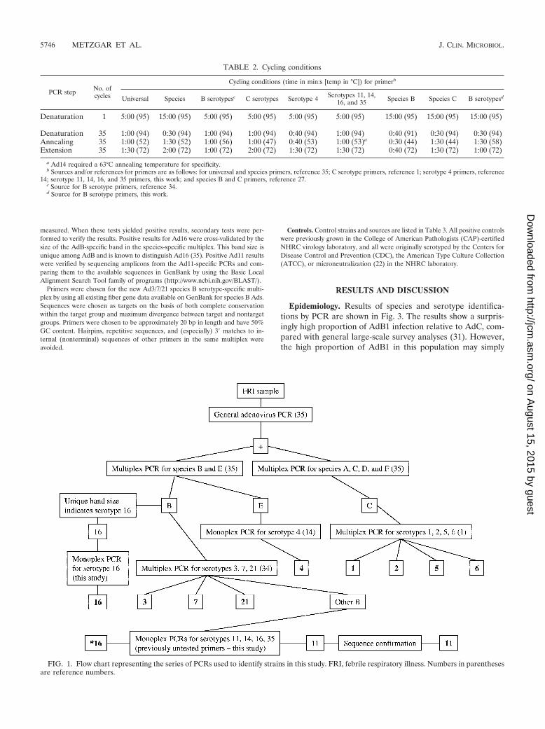
PCRs were carried out as shown in the flow chart (Fig. 1). The species-specificmultiplex (35) was divided into two parts, testing separately for set B and E andset A, C, D, and F. All PCR tests were done for all controls and all samples tomake sure there were no cross-reactions. Reactions were performed as follows.
Multiplex PCRs, including both the initial and the verifying species-specificPCRs, the initial and verifying Ad3/Ad7/Ad21 species B serotype-specific PCRs,and the AdC serotype-specific PCRs, were performed using a QIAGEN multi-plex kit (QIAGEN, Valencia, Calif.). Reaction mixtures contained 0.5� Q-Solution, 1� Multiplex buffer, 0.2 �M concentration of each primer (IntegratedDNA Technologies, Coralville, Iowa), and 2 �l of extracted sample in 25-�laqueous reactions.
Reaction mixtures for monoplex PCRs contained 1� Q-Solution (QIAGEN),1� PCR buffer (Promega, Madison, Wis.), 0.6 �M concentration of each primer(Integrated DNA Technologies), 0.8 mM concentration of each deoxynucleosidetriphosphate (Promega), 3 mM concentration of MgCl2 (Promega), 1.25 U ofTaq polymerase (Promega), and 2 �l of extracted sample in 25-�l aqueousreactions.
Cycling conditions included a 10-min final extension at 72°C and a final holdat 4°C. In regards to other parameters, reactions were cycled as shown in Table2.
All products were mixed 5:1 with loading dye (Sigma Aldrich, St. Louis, Mo.)and run for 90 min at 125 V on 1.5% agarose (Sigma Aldrich) gels with ethidiumbromide (Sigma Aldrich). Standard 100-bp DNA ladders were used as a refer-ence (New England Biolabs, Beverly, Mass.). “Product ladders” were also usedfor the multiplexes, generated by balanced combination of all Ad types targetedby the multiplex. Gels were visualized and recorded by photography on a UVlight box.
Due to the occasional appearance of faint gray bands resulting from misprim-ing between closely related species B serotypes, products were recorded aspositive when a band of “normal” intensity was observed in the expected sizerange. Normal intensity was defined as being within the range observed amongexpected bands amplified from positive-control strains (Fig. 2; see also Resultsand Discussion, “Methodology”).
5744 METZGAR ET AL. J. CLIN. MICROBIOL.
on August 15, 2015 by guest
http://jcm.asm
.org/D
ownloaded from
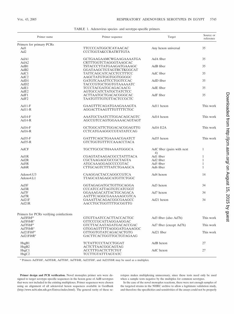
Primer design and PCR verification. Novel monoplex primer sets were de-signed to target serotype-specific sequences in the hexon gene of AdB serotypesthat were not included in the existing multiplexes. Primer sequences were chosenusing an alignment of all adenoviral hexon sequences available in GenBank(http://www.ncbi.nlm.nih.gov/Entrez/index.html). The general rarity of these se-
rotypes makes multiplexing unnecessary, since these tests need only be usedwhen a sample tests negative by the multiplex for common serotypes.
In the case of the novel monoplex reactions, there were not enough samples ofthe targeted strains in the NHRC archive to allow a legitimate validation study,and therefore the specificities and sensitivities of the assays could not be properly
TABLE 1. Adenovirus species- and serotype-specific primers
Primer name Primer sequence Target Source orreference
Primers for primary PCRsAd1 TTCCCCATGGCICAYAACAC Any hexon universal 35Ad2 CCCTGGTAKCCRATRTTGTA
AdA1 GCTGAAGAAMCWGAAGAAAATGA AdA fiber 35AdA2 CRTTTGGTCTAGGGTAAGCACAdB1 TSTACCCYTATGAAGATGAAAGC AdB fiber 35AdB2 GGATAAGCTGTAGTRCTKGGCATAdC1 TATTCAGCATCACCTCCTTTCC AdC fiber 35AdC2 AAGCTATGTGGTGGTGGGGCAdD1 GATGTCAAATTCCTGGTCCAC AdD fiber 35AdD2 TACCCGTGCTGGTGTAAAAATCAdE1 TCCCTACGATGCAGACAACG AdE fiber 35AdE2 AGTGCCATCTATGCTATCTCCAdF1 ACTTAATGCTGACACGGGCAC AdF fiber 35AdF2 TAATGTTTGTGTTACTCCGCTC
Ad11-F GAAGTTTCAGATGAAGAAAGTA Ad11 hexon This workAd11-R AGGACTTAAGTTTGTTTTCTGC
Ad14-F AAATGCTAATCTTGGACAGCAGTC Ad14 hexon This workAd14-R AGCCGTCCAGTGGAAAACAGTAGT
Ad16-F GCTGGCATTCTGGACACGGAGTTG Ad16 E2A This workAd16-R CCTCATGAAGGCCGTATATCCAG
Ad35-F GATTTCAGCTGAAAACGAATCT Ad35 hexon This workAd35-R GTCTGGTGTTTCCAAACCTACA
AdCF TGCTTGCGCTHAAAATGGGCA AdC fiber (pairs with next4)
1
Ad1R CGAGTATAAGACGCCTATTTACA Ad1 fiber 1Ad2R CGCTAAGAGCGCCGCTAGTA Ad2 fiber 1Ad5R ATGCAAAGGAGCCCCGTAC Ad5 fiber 1Ad6R CTTGCAGTCTTTATCTGAAGCA Ad6 fiber 1
Adeno4.U3 CAAGGACTACCAGGCCGTCA Ad4 hexon 14Adeno4.L1 TTAGCATAGAGCATGTTCTGGC
Ad3F GGTAGAGATGCTGTTGCAGGA Ad3 hexon 34Ad3R CCCATCCATTAGTGTCATCGGTAd7F GGAAAGACATTACTGCAGACA Ad7 hexon 34Ad7R AATTTCAGGCGAAAAAGCGTCAAd21F GAAATTACAGACGGCGAAGCC Ad21 hexon 34Ad21R AACCTGCTGGTTTTGCGGTTG
Primers for PCRs verifying coinfectionsAd3FibFa GTGTTAATCCACTTACCACTGC Ad3 fiber (also Ad7h) This workAd3FibRa GTTCCCGCATTAGGAAGGACAd7FibFa GTCTTACAATAGATGACACCGAC Ad7 fiber (except Ad7h) This workAd7FibRa GTGGAGTTTTTAGGGATGAAAGGCAd21FibFa GTTGGTGTATCAGACACTGTG Ad21 fiber This workAd21FibRa GACTTCACTGGTTGCTGTAGAAG
HsgB1 TCTATTCCCTACCTGGAT AdB hexon 27HsgB2 ACTCTTAACGGCAGTAGHsgC1 ACCTTTGACTCTTCTGT AdC hexon 27HsgC2 TCCTTGTATTTAGTATC
a Primers Ad3FibF, Ad3FibR, Ad7FibF, Ad7FibR, Ad21FibF, and Ad21FibR may be used as a multiplex.
VOL. 43, 2005 RESPIRATORY ADENOVIRUS SEROTYPES IN EGYPT 5745
on August 15, 2015 by guest
http://jcm.asm
.org/D
ownloaded from
measured. When these tests yielded positive results, secondary tests were per-formed to verify the results. Positive results for Ad16 were cross-validated by thesize of the AdB-specific band in the species-specific multiplex. This band size isunique among AdB and is known to distinguish Ad16 (35). Positive Ad11 resultswere verified by sequencing amplicons from the Ad11-specific PCRs and com-paring them to the available sequences in GenBank by using the Basic LocalAlignment Search Tool family of programs (http://www.ncbi.nih.gov/BLAST/).
Primers were chosen for the new Ad3/7/21 species B serotype-specific multi-plex by using all existing fiber gene data available on GenBank for species B Ads.Sequences were chosen as targets on the basis of both complete conservationwithin the target group and maximum divergence between target and nontargetgroups. Primers were chosen to be approximately 20 bp in length and have 50%GC content. Hairpins, repetitive sequences, and (especially) 3� matches to in-ternal (nonterminal) sequences of other primers in the same multiplex wereavoided.
Controls. Control strains and sources are listed in Table 3. All positive controlswere previously grown in the College of American Pathologists (CAP)-certifiedNHRC virology laboratory, and all were originally serotyped by the Centers forDisease Control and Prevention (CDC), the American Type Culture Collection(ATCC), or microneutralization (22) in the NHRC laboratory.
RESULTS AND DISCUSSION
Epidemiology. Results of species and serotype identifica-tions by PCR are shown in Fig. 3. The results show a surpris-ingly high proportion of AdB1 infection relative to AdC, com-pared with general large-scale survey analyses (31). However,the high proportion of AdB1 in this population may simply
FIG. 1. Flow chart representing the series of PCRs used to identify strains in this study. FRI, febrile respiratory illness. Numbers in parenthesesare reference numbers.
TABLE 2. Cycling conditions
PCR step No. ofcycles
Cycling conditions (time in min:s [temp in °C]) for primerb
Universal Species B serotypesc C serotypes Serotype 4 Serotypes 11, 14,16, and 35 Species B Species C B serotypesd
Denaturation 1 5:00 (95) 15:00 (95) 5:00 (95) 5:00 (95) 5:00 (95) 5:00 (95) 15:00 (95) 15:00 (95) 15:00 (95)
Denaturation 35 1:00 (94) 0:30 (94) 1:00 (94) 1:00 (94) 0:40 (94) 1:00 (94) 0:40 (91) 0:30 (94) 0:30 (94)Annealing 35 1:00 (52) 1:30 (52) 1:00 (56) 1:00 (47) 0:40 (53) 1:00 (53)a 0:30 (44) 1:30 (44) 1:30 (58)Extension 35 1:30 (72) 2:00 (72) 1:00 (72) 2:00 (72) 1:30 (72) 1:30 (72) 0:40 (72) 1:30 (72) 1:00 (72)
a Ad14 required a 63°C annealing temperature for specificity.b Sources and/or references for primers are as follows: for universal and species primers, reference 35; C serotype primers, reference 1; serotype 4 primers, reference
14; serotype 11, 14, 16, and 35 primers, this work; and species B and C primers, reference 27.c Source for B serotype primers, reference 34.d Source for B serotype primers, this work.
5746 METZGAR ET AL. J. CLIN. MICROBIOL.
on August 15, 2015 by guest
http://jcm.asm
.org/D
ownloaded from
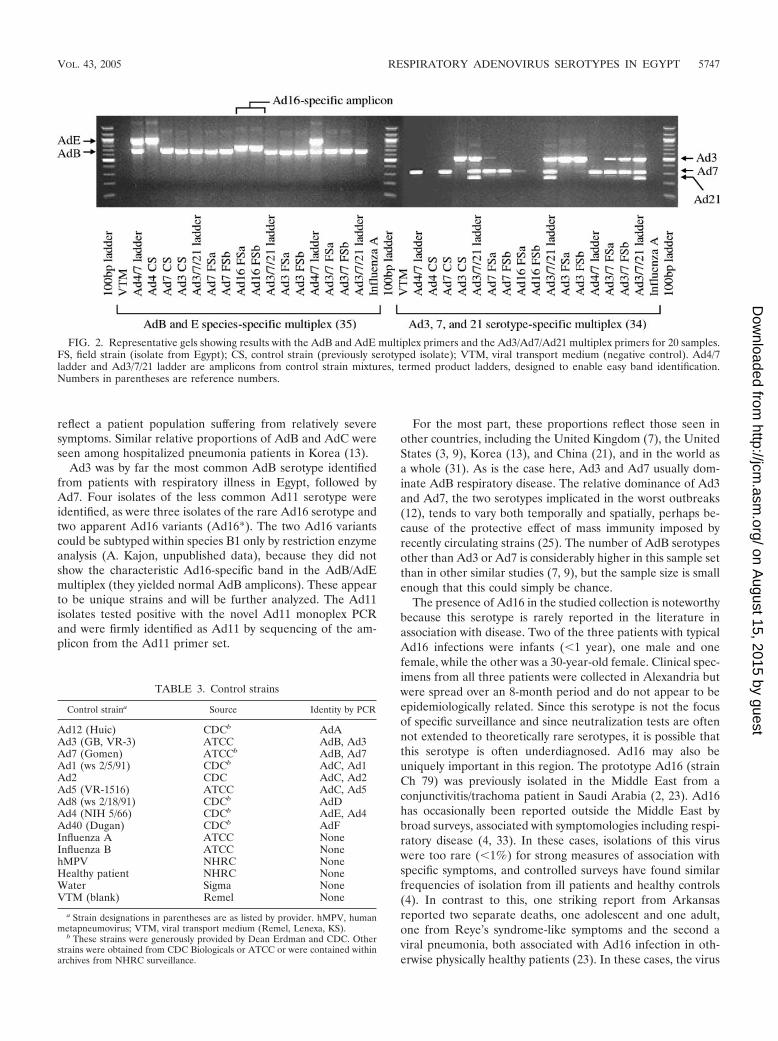
reflect a patient population suffering from relatively severesymptoms. Similar relative proportions of AdB and AdC wereseen among hospitalized pneumonia patients in Korea (13).
Ad3 was by far the most common AdB serotype identifiedfrom patients with respiratory illness in Egypt, followed byAd7. Four isolates of the less common Ad11 serotype wereidentified, as were three isolates of the rare Ad16 serotype andtwo apparent Ad16 variants (Ad16*). The two Ad16 variantscould be subtyped within species B1 only by restriction enzymeanalysis (A. Kajon, unpublished data), because they did notshow the characteristic Ad16-specific band in the AdB/AdEmultiplex (they yielded normal AdB amplicons). These appearto be unique strains and will be further analyzed. The Ad11isolates tested positive with the novel Ad11 monoplex PCRand were firmly identified as Ad11 by sequencing of the am-plicon from the Ad11 primer set.
For the most part, these proportions reflect those seen inother countries, including the United Kingdom (7), the UnitedStates (3, 9), Korea (13), and China (21), and in the world asa whole (31). As is the case here, Ad3 and Ad7 usually dom-inate AdB respiratory disease. The relative dominance of Ad3and Ad7, the two serotypes implicated in the worst outbreaks(12), tends to vary both temporally and spatially, perhaps be-cause of the protective effect of mass immunity imposed byrecently circulating strains (25). The number of AdB serotypesother than Ad3 or Ad7 is considerably higher in this sample setthan in other similar studies (7, 9), but the sample size is smallenough that this could simply be chance.
The presence of Ad16 in the studied collection is noteworthybecause this serotype is rarely reported in the literature inassociation with disease. Two of the three patients with typicalAd16 infections were infants (�1 year), one male and onefemale, while the other was a 30-year-old female. Clinical spec-imens from all three patients were collected in Alexandria butwere spread over an 8-month period and do not appear to beepidemiologically related. Since this serotype is not the focusof specific surveillance and since neutralization tests are oftennot extended to theoretically rare serotypes, it is possible thatthis serotype is often underdiagnosed. Ad16 may also beuniquely important in this region. The prototype Ad16 (strainCh 79) was previously isolated in the Middle East from aconjunctivitis/trachoma patient in Saudi Arabia (2, 23). Ad16has occasionally been reported outside the Middle East bybroad surveys, associated with symptomologies including respi-ratory disease (4, 33). In these cases, isolations of this viruswere too rare (�1%) for strong measures of association withspecific symptoms, and controlled surveys have found similarfrequencies of isolation from ill patients and healthy controls(4). In contrast to this, one striking report from Arkansasreported two separate deaths, one adolescent and one adult,one from Reye’s syndrome-like symptoms and the second aviral pneumonia, both associated with Ad16 infection in oth-erwise physically healthy patients (23). In these cases, the virus
FIG. 2. Representative gels showing results with the AdB and AdE multiplex primers and the Ad3/Ad7/Ad21 multiplex primers for 20 samples.FS, field strain (isolate from Egypt); CS, control strain (previously serotyped isolate); VTM, viral transport medium (negative control). Ad4/7ladder and Ad3/7/21 ladder are amplicons from control strain mixtures, termed product ladders, designed to enable easy band identification.Numbers in parentheses are reference numbers.
TABLE 3. Control strains
Control straina Source Identity by PCR
Ad12 (Huic) CDCb AdAAd3 (GB, VR-3) ATCC AdB, Ad3Ad7 (Gomen) ATCCb AdB, Ad7Ad1 (ws 2/5/91) CDCb AdC, Ad1Ad2 CDC AdC, Ad2Ad5 (VR-1516) ATCC AdC, Ad5Ad8 (ws 2/18/91) CDCb AdDAd4 (NIH 5/66) CDCb AdE, Ad4Ad40 (Dugan) CDCb AdFInfluenza A ATCC NoneInfluenza B ATCC NonehMPV NHRC NoneHealthy patient NHRC NoneWater Sigma NoneVTM (blank) Remel None
a Strain designations in parentheses are as listed by provider. hMPV, humanmetapneumovirus; VTM, viral transport medium (Remel, Lenexa, KS).
b These strains were generously provided by Dean Erdman and CDC. Otherstrains were obtained from CDC Biologicals or ATCC or were contained withinarchives from NHRC surveillance.
VOL. 43, 2005 RESPIRATORY ADENOVIRUS SEROTYPES IN EGYPT 5747
on August 15, 2015 by guest
http://jcm.asm
.org/D
ownloaded from
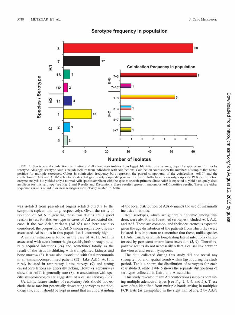
was isolated from parenteral organs related directly to thesymptoms (spleen and lung, respectively). Given the rarity ofisolation of Ad16 in general, these two deaths are a goodreason to test for this serotype in cases of Ad-associated dis-ease. If the two Ad16 variants (Ad16*) seen here are alsoconsidered, the proportion of Ad16 among respiratory disease-associated Ad isolates in this population is extremely high.
A similar situation is found in the case of Ad11. Ad11 isassociated with acute hemorrhagic cystitis, both through natu-rally acquired infections (24) and, sometimes fatally, as theresult of the virus hitchhiking with transplanted kidneys andbone marrow (6). It was also associated with fatal pneumoniain an immunocompromised patient (32). Like Ad16, Ad11 israrely isolated in respiratory illness surveys (9) and strongcausal correlations are generally lacking. However, serosurveysshow that Ad11 is generally rare (8), so associations with spe-cific symptomologies are suggestive of a causal etiology (33).
Certainly, future studies of respiratory Ads should not ex-clude these rare but potentially devastating serotypes method-ologically, and it should be kept in mind that an understanding
of the local distribution of Ads demands the use of maximallyinclusive methods.
AdC serotypes, which are generally endemic among chil-dren, were also found. Identified serotypes included Ad1, Ad2,and Ad5. These are common, and their occurrence is expectedgiven the age distribution of the patients from which they wereisolated. It is important to remember that these, unlike speciesB1 Ads, usually establish long-lasting latent infections charac-terized by persistent intermittent excretion (3, 9). Therefore,positive results do not necessarily reflect a causal link betweenthe viruses and recent symptoms.
The data collected during this study did not reveal anystrong temporal or spatial trends within Egypt during the studyperiod. Table 4 shows the distribution of serotypes for eachyear studied, while Table 5 shows the separate distributions ofserotypes collected in Cairo and Alexandria.
This study revealed many Ad coinfections (samples contain-ing multiple adenoviral types [see Fig. 2, 3, 4, and 5]). Thesewere often identified from multiple bands arising in multiplexPCR tests (as exemplified in the right half of Fig. 2 by Ad3/7
FIG. 3. Serotype and coinfection distributions of 88 adenovirus isolates from Egypt. Identified strains are grouped by species and further byserotype. All single-serotype counts include isolates from individuals with coinfections. Coinfection counts show the numbers of samples that testedpositive for multiple serotypes. Colors in coinfection frequency bars represent the paired components of the coinfections. Ad16* and thecoinfection of Ad7 and Ad16* refer to isolates that gave serotype-specific positive results for Ad16 by either serotype-specific PCR or restrictionenzyme analysis but yielded only a normal AdB species amplicon with the species-specific primers. Since Ad16 is expected to yield a uniquely sizedamplicon for this serotype (see Fig. 2 and Results and Discussion), these results represent ambiguous Ad16 positive results. These are eithersequence variants of Ad16 or new serotypes most closely related to Ad16.
5748 METZGAR ET AL. J. CLIN. MICROBIOL.
on August 15, 2015 by guest
http://jcm.asm
.org/D
ownloaded from

FSa and FSb). Many coinfections involved serotypes from bothspecies AdB and species AdC, while others involved multipleserotypes of AdB.
All of the AdB/AdC coinfections were partially verified bysecondary identification of two different serotypes correspond-ing to the two initially identified species. However, the sero-type-specific tests and species-specific tests overlapped interms of the targeted loci. The original species-specific test(35) and the species C serotype-specific test (1) both target thefiber gene, while the species B serotype-specific test (34) tar-gets the hexon. Hence, this initial verification could not obviatethe possibility of a single cross-reactive or recombined locusthat might yield a double signal as an artifact in both tests.
This shortcoming was addressed through further verificationof the coinfections by using redundant tests targeting indepen-dent loci. AdB/AdC coinfections originally identified using thefiber gene (35) were retested using secondary AdB- and AdC-specific primers targeting the hexon gene (27). Of eight initiallyidentified AdB/AdC coinfections, seven were positive for both
AdB and AdC by the secondary test and were hence verified ascoinfections (Fig. 4 shows examples of these data). Seventy-two of the 80 samples that initially appeared to be monoinfec-tions of just one species also appeared to be monoinfections ofthe same species by the secondary test. The remaining eightsamples that were initially identified as monoinfections (ofAdB) were all verified as AdB but also yielded weak AdC-positive results by the second test (Ad7 FSa in Fig. 4 providesan example), suggesting that there may be more coinfectionsthan originally recognized and also that AdC may often befound at a lower titer than AdB in AdB/AdC coinfections.Probability tree calculations addressing the overall level ofcorrelation between the two coinfection assays yielded a Pvalue of 0.000013 (representing the probability of both testsagreeing on 79 out of 88 samples by chance; data and calcu-lations not shown). These results strongly validate the idea thatthe apparent coinfections are real and that they involve wholegenomes as opposed to recombined parts or individually cross-reacting intermediate sequences.
Ad3/Ad7 coinfections presented a more complex problem in
FIG. 4. Representative PCR results for the secondary test (27) used to verify AdB/AdC coinfections. The gel includes amplicons (generatedseparately for AdB and AdC) from eight samples initially identified as AdB/AdC coinfections, three samples initially identified as AdBmonoinfections, and three samples initially identified as AdC monoinfections, along with various controls. Initial identifications are indicated bythe strain names. See the text for details.
TABLE 4. Serotype distribution by year of isolation
Yr ofisolation
No. of isolates of serotype
Ad3 Ad7 Ad16 Ad16* Ad11 Ad1 Ad2 Ad5
1999 4 3 0 0 0 2 0 12000 17 3 1 1 3 3 2 02001 34 9 2 1 0 7 1 12002 5 2 0 0 1 1 0 0
Total 60 17 3 2 4 13 3 2
TABLE 5. Serotype distribution by city of isolation
City ofisolation
No. of isolates of serotype
Ad3 Ad7 Ad16 Ad16* Ad11 Ad1 Ad2 Ad5
Cairo 10 5 0 0 0 4 0 1Alexandria 50 12 3 2 4 9 3 1
Total 60 17 3 2 4 13 3 2
VOL. 43, 2005 RESPIRATORY ADENOVIRUS SEROTYPES IN EGYPT 5749
on August 15, 2015 by guest
http://jcm.asm
.org/D
ownloaded from
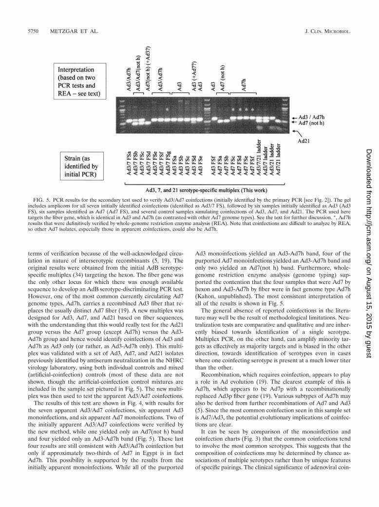
terms of verification because of the well-acknowledged circu-lation in nature of interserotypic recombinants (5, 19). Theoriginal results were obtained from the initial AdB serotype-specific multiplex (34) targeting the hexon. The fiber gene wasthe only other locus for which there was enough availablesequence to develop an AdB serotype-discriminating PCR test.However, one of the most common currently circulating Ad7genome types, Ad7h, carries a recombined Ad3 fiber that re-places the usually distinct Ad7 fiber (19). A new multiplex wasdesigned for Ad3, Ad7, and Ad21 based on fiber sequences,with the understanding that this would really test for the Ad21group versus the Ad7 group (except Ad7h) versus the Ad3-Ad7h group and hence would identify coinfections of Ad3 andAd7h as Ad3 only (or rather, as Ad3-Ad7h only). This multi-plex was validated with a set of Ad3, Ad7, and Ad21 isolatespreviously identified by antiserum neutralization in the NHRCvirology laboratory, using both individual controls and mixed(artificial-coinfection) controls (most of these data are notshown, though the artificial-coinfection control mixtures areincluded in the sample set pictured in Fig. 5). The new multi-plex was then used to test the apparent Ad3/Ad7 coinfections.
The results of this test are shown in Fig. 4, with results forthe seven apparent Ad3/Ad7 coinfections, six apparent Ad3monoinfections, and six apparent Ad7 monoinfections. Two ofthe initially apparent Ad3/Ad7 coinfections were verified bythe new method, while one yielded only an Ad7(not h) bandand four yielded only an Ad3-Ad7h band (Fig. 5). These lastfour results are still consistent with Ad3/Ad7h coinfection butonly if approximately two-thirds of Ad7 in Egypt is in factAd7h. This possibility is supported by the results from theinitially apparent monoinfections. While all of the purported
Ad3 monoinfections yielded an Ad3-Ad7h band, four of thepurported Ad7 monoinfections yielded an Ad3-Ad7h band andonly two yielded an Ad7(not h) band. Furthermore, whole-genome restriction enzyme analysis (genome typing) sup-ported the contention that the four samples that were Ad7 byhexon and Ad3-Ad7h by fiber were in fact genome type Ad7h(Kahon, unpublished). The most consistent interpretation ofall of the results is shown in Fig. 5.
The general absence of reported coinfections in the litera-ture may well be the result of methodological limitations. Neu-tralization tests are comparative and qualitative and are inher-ently biased towards identification of a single serotype.Multiplex PCR, on the other hand, can amplify minority tar-gets as effectively as majority targets and is biased in the otherdirection, towards identification of serotypes even in caseswhere one coinfecting serotype is present at a much lower titerthan the other.
Recombination, which requires coinfection, appears to playa role in Ad evolution (19). The clearest example of this isAd7h, which appears to be Ad7p with a recombinationallyreplaced Ad3p fiber gene (19). Various subtypes of Ad7h mayalso be derived from further recombinations of Ad7 and Ad3(5). Since the most common coinfection seen in this sample setis Ad7/Ad3, the potential evolutionary implications of coinfec-tions are clear.
It can be seen by comparison of the monoinfection andcoinfection charts (Fig. 3) that the common coinfections tendto involve the most common serotypes. This suggests that thecomposition of coinfections may be determined by chance as-sociations of multiple serotypes rather than by unique featuresof specific pairings. The clinical significance of adenoviral coin-
FIG. 5. PCR results for the secondary test used to verify Ad3/Ad7 coinfections (initially identified by the primary PCR [see Fig. 2]). The gelincludes amplicons for all seven initially identified coinfections (identified as Ad3/7 FS), followed by six samples initially identified as Ad3 (Ad3FS), six samples identified as Ad7 (Ad7 FS), and several control samples simulating coinfections of Ad3, Ad7, and Ad21. The PCR used heretargets the fiber gene, which is identical in Ad3 and Ad7h (as contrasted with other Ad7 genome types). See the text for further discussion. *, Ad7hresults that were definitively verified by whole-genome restriction enzyme analysis (REA). Note that coinfections are difficult to analyze by REA,so other Ad7 isolates, especially those in apparent coinfections, could also be Ad7h.
5750 METZGAR ET AL. J. CLIN. MICROBIOL.
on August 15, 2015 by guest
http://jcm.asm
.org/D
ownloaded from
fections, like the significance of Ad11 and Ad16, can only beunderstood through further surveillance and analysis.
Methodology. PCR provides a rapid and inclusive method ofserotype identification. Of 88 strains, the method successfullyidentified a species in all cases and a serotype in 86 cases. Thetwo remaining untypeable isolates have now been studied fur-ther through restriction enzyme analysis and appear to bevariants of Ad16 (Ad16*) (Kahon, unpublished). In all casesexcept one (a possible coinfection identified as Ad7 plusAd16*), positive serotype identifications were supported byspecies results, including cases for which multiple serotypes(coinfections) were observed.
The ability of PCR to identify multiple coinfecting Ad typeswas strongly supported by the results. Coinfections were iden-tified at both species and serotype levels, and multiple testsdesigned to cross-validate apparent coinfection signals by usingseparate genetic loci as targets correlated well with one an-other.
Results in general were clear and conclusive. All controlstrains and negative controls yielded results consistent withexpectations, strengthening the already intensive validations ofthe literature-derived multiplex PCRs used here. The novelmonoplex primers developed and used to determine the sero-types of otherwise unidentifiable isolates also served their pur-pose. Identifications of Ad11 were easily verified with sequenc-ing, which is available to any PCR-enabled laboratory at lowcost through commercial sequencing services. Identificationsof three of the Ad16 isolates found here were readily verifiedwith reference to Ad16-specific signatures from the species-specific (AdB and AdE) multiplex. The other two apparentAd16s (Ad16*) matched some known Ad16 molecular signa-tures but not others. This may well be because so little molec-ular data exist for Ad16 and hence the range of molecularsignatures found among Ad16 is not yet well defined. Therelative ease of PCR test design and implementation allowedall of the infections to be diagnosed with enough resolution toreveal the impact of potentially underestimated serotypes.
The AdB serotype-specific test (34) relies on rare polymor-phisms in the hexon gene to distinguish these very closelyrelated serotypes. It was found that the primers used in this testare capable of producing weak (gray) “shadow bands” forclosely related serotypes when used to test some high-titerITCF isolates. This can be seen in two of the cases shown in theAd3, 7, and 21 portion of Fig. 2 (Ad7 FSb and Ad16 FSa). TheNHRC laboratory recently saw similar bands with control sam-ples used in other studies. In both of the cases noted in Fig. 2,independent monoplex tests for the serotypes suggested by theshadow bands gave negative results. Therefore, the data col-lected in this study were analyzed under a criterion for positiveresults that demands a clear band, within the intensity rangeseen among correct bands for positive-control samples. Fur-ther efforts toward clinical validation of these methods sug-gested that the observed shadow bands can be avoided by 10-or 100-fold dilution of ITCF samples prior to extraction. Thehigh-titer results are shown here for illustration of the poten-tially ambiguous results. In any case, these shadow bands arereadily distinguishable from the much brighter bands resultingfrom specific amplification.
The inclusiveness of this method, extending to both rareserotypes and coinfections of multiple serotypes, makes it an
attractive and accessible alternative to classic culture and neu-tralization methods. This method serves to identify not onlyserotype but also presence/absence and species and in thatsense offers a complete method of analysis that can be per-formed on either grown virus or original patient specimens.
Application of the PCR typing method to a unique anddiverse sample set from Egypt allowed nearly complete de-scription of the species and serotype identities of the collectedadenovirus strains. Furthermore, the strengths of such molec-ular analyses in identifying coinfections became clear, offeringa powerful tool for investigating the dynamics of genetic evo-lution and recombination events in this important humanpathogen.
ACKNOWLEDGMENTS
This work represents NHRC report no. 04-37, supported by theDepartment of Defense Global Emerging Infections Surveillance andResponse System (GEIS) under research work unit 60501.
The views expressed in this article are those of the authors and donot reflect the official policy or position of the Department of the Navy,Department of Defense (DoD), or the U.S. government.
This research has been conducted in compliance with all applicablefederal and international regulations governing the protection of hu-man subjects in research (DoD protocol no. 30983). Protocols werealso reviewed and accepted by the Minister of Health and Populationof the Arab Republic of Egypt.
We declare that no financial conflict of interest exists.Thanks to Elizabeth Metzgar, Krista Hensinger, and Erin McDon-
ough for thoughtful editing of the content and writing and to KevinGratwick for help with the figures.
REFERENCES
1. Adhikary, A. K., T. Inada, U. Banik, J. Numaga, and N. Okabe. 2004.Identification of subgenus C adenoviruses by fiber-based multiplex PCR.J. Clin. Microbiol. 42:670–673.
2. Bell, S. D., D. E. McComb, E. S. Murray, R. S. Chang, and J. C. Snyder.1959. Adenoviruses isolated from Saudi Arabia. I. Epidemiologic features.Am. J. Trop. Med. Hyg. 8:492–500.
3. Brandt, C. D., H. W. Kim, B. C. Jeffries, G. Pyles, E. E. Christmas, J. L. Reid,R. M. Chanock, and R. H. Parrott. 1972. Infections in 18,000 infants andchildren in a controlled study of respiratory tract disease. II. Variation inadenovirus infections by year and season. Am. J. Epidemiol. 95:218–227.
4. Brandt, C. D., H. W. Kim, A. J. Vargosko, B. C. Jeffries, J. O. Arrobio, B.Rindge, R. H. Parrott, and R. M. Chanock. 1969. Infections in 18,000 infantsand children in a controlled study of respiratory tract disease. I. Adenoviruspathogenicity in relation to serologic type and illness syndrome. Am. J.Epidemiol. 90:484–500.
5. Bruzzone, M., M. Barro, and E. Spencer. 2001. Identification of adenovirus7h heterogeneity in the E3 region. Biol. Res. 34:75–82.
6. Carrigan, D. R. 1997. Adenovirus infections in immunocompromised pa-tients. Am. J. Med. 102:71–74.
7. Cooper, R. J., R. Hallett, A. B. Tullo, and P. E. Klapper. 2000. The epide-miology of adenovirus infections in Greater Manchester, UK 1982–96. Epi-demiol. Infect. 125:333–345.
8. D’Ambrosio, E., N. Del Grosso, A. Chicca, and M. Midulla. 1982. Neutral-izing antibodies against 33 human adenoviruses in normal children in Rome.J. Hyg. (London) 89:155–161.
9. Fox, J. P., C. E. Hall, and M. K. Cooney. 1977. The Seattle virus watch. VII.Observations of adenovirus infections. Am. J. Epidemiol. 105:362–386.
10. Garnett, C. T., D. Erdman, W. Xu, and L. R. Gooding. 2002. Prevalence andquantitation of species C adenovirus DNA in human mucosal lymphocytes.J. Virol. 76:10608–10616.
11. Green, M., J. K. Mackay, W. S. M. Wold, and P. Rigden. 1979. Thirty-onehuman adenovirus serotypes (Ad1-Ad31) form five groups (A-E) based uponDNA genome homologies. Virology 93:481–492.
12. Hierholzer, J. 1995. Adenoviruses, p. 169–188. In E. H. Lennette, D. A.Lennette, and E. T. Lennette (ed.), Diagnostic procedures for viral, rickett-sial and chlamydial infections, 7th ed. American Public Health Association,Washington, D.C.
13. Hong, J. Y., H. J. Lee, P. A. Piedra, E. H. Choi, K. H. Park, Y. Y. Koh, andW. S. Kim. 2001. Lower respiratory tract infections due to adenovirus inhospitalized Korean children: epidemiology, clinical features, and prognosis.Clin. Infect. Dis. 32:1423–1429.
14. Houng, H. S., S. Liang, C. M. Chen, J. Keith, M. Echavarria, J. L. Sanchez,
VOL. 43, 2005 RESPIRATORY ADENOVIRUS SEROTYPES IN EGYPT 5751
on August 15, 2015 by guest
http://jcm.asm
.org/D
ownloaded from
S. A. Kolavic, D. W. Vaughn, and L. N. Binn. 2002. Rapid type-specificdiagnosis of adenovirus type 4 infection using a hexon-based quantitativefluorogenic PCR. Diagn. Microbiol. Infect. Dis. 42:227–236.
15. Jordan, W. S., Jr. 1958. Occurrence of adenovirus infections in civilianpopulations. Arch. Intern. Med. 101:54–59.
16. Kajon, A., and G. Wadell. 1994. Genome analysis of South American ade-novirus strains of serotype 7 collected over a 7-year period. J. Clin. Micro-biol. 32:2321–2323.
17. Kajon, A. E., S. A. R. Portes, W. A. de Mello, J. P. Nascimento, and M. M.Siqueira. 1999. Genome type analysis of Brazilian adenovirus strains ofserotypes 1, 2, 3, 5, and 7 collected between 1976 and 1995. J. Med. Virol.58:408–412.
18. Kajon, A. E., A. S. Mistchenko, C. Videla, M. Hortal, G. Wadell, and L. F.Avendano. 1996. Molecular epidemiology of adenovirus acute lower respi-ratory infections of children in the south cone of South America (1991–1994). J. Med. Virol. 48:151–156.
19. Kajon, A. E., and G. Wadell. 1996. Sequence analysis of the E3 region andfiber gene of human adenovirus genome type 7h. Virology 215:190–196.
20. Kim, Y.-J., J.-Y. Hong, H.-J. Lee, S.-H. Shin, Y.-K. Kim, T. Inada, M.Hashido, and P. A. Piedra. 2003. Genome type analysis of adenovirus types3 and 7 isolated during successive outbreaks of lower respiratory tract infec-tions in children. J. Clin. Microbiol. 41:4594–4599.
21. Li, Q. G., Q. J. Zheng, Y. H. Liu, and G. Wadell. 1996. Molecular epidemi-ology of adenovirus types 3 and 7 isolated from children with pneumonia inBeijing. J. Med. Virol. 49:170–177.
22. Malasig, M. D., P. R. Goswami, L. K. Crawford-Miksza, D. P. Schnurr, andG. C. Gray. 2001. Simplified microneutralization test for serotyping adeno-virus isolates. J. Clin. Microbiol. 39:2984–2986.
23. Morgan, P. N., E. B. Moses, E. P. Fody, and A. L. Barron. 1984. Associationof adenovirus type 16 with Reye’s-syndrome-like illness and pneumonia.South. Med. J. 77:827–830.
24. Mufson, M. A., and R. B. Belshe. 1976. A review of adenoviruses in theetiology of acute hemorrhagic cystitis. J. Urol. 115:191–194.
25. Noda, M., T. Yoshida, T. Sakeguchi, Y. Ikeda, K. Yamaoka, and T. Ogino.2002. Molecular and epidemiological analyses of human adenovirus type 7strains isolated from the 1995 nationwide outbreak in Japan. J. Clin. Micro-biol. 40:140–145.
26. Potter, C. W., and W. I. H. Shedden. 1963. The distribution of adenovirusantibodies in normal children. J. Hyg. (London) 61:155–160.
27. Pring-Åkerblom, P., F. E. J. Trijssenaar, T. Adrian, and H. Hoyer. 1999.Multiplex polymerase chain reaction for subgenus-specific detection of hu-man adenoviruses in clinical samples. J. Med. Virol. 58:87–92.
28. Rubin, B. A. 1993. Clinical picture and epidemiology of adenovirus infec-tions. Acta Microbiol. Hung. 40:303–323.
29. Ryan, M. A., G. C. Gray, B. Smith, J. A. McKeehan, A. W. Hawksworth, andM. D. Malasig. 2002. Large epidemic of respiratory illness due to adenovirustypes 7 and 3 in healthy young adults. Clin. Infect. Dis. 34:577–582.
30. Sambrook, J., M. Sleigh, J. A. Engler, and T. R. Broker. 1980. The evolutionof the adenoviral genome. Ann. N. Y. Acad. Sci. 354:426–452.
31. Schmitz, H., R. Wigand, and W. Heinrich. 1983. Worldwide epidemiology ofhuman adenovirus infections. Am. J. Epidemiol. 117:455–466.
32. Siegal, F. P., S. H. Dikman, R. B. Arayata, and E. J. Bottone. 1981. Fataldisseminated adenovirus type 11 pneumonia in an agammaglobulinemicpatient. Am. J. Med. 71:1062–1067.
33. Wold, W. S., J. K. Mackey, P. Rigden, and M. Green. 1979. Analysis ofhuman cancer DNAs for DNA sequences of human adenovirus serotypes 3,7, 11, 14, 16, and 21 in group B1. Cancer Res. 39:3479–3484.
34. Xu, W., and D. D. Erdman. 2001. Type-specific identification of humanadenovirus 3, 7, and 21 by a multiplex PCR assay. J. Med. Virol. 64:537–542.
35. Xu, W., M. C. McDonough, and D. D. Erdman. 2000. Species-specific iden-tification of human adenoviruses by a multiplex PCR assay. J. Clin. Micro-biol. 38:4114–4120.
5752 METZGAR ET AL. J. CLIN. MICROBIOL.
on August 15, 2015 by guest
http://jcm.asm
.org/D
ownloaded from