Characterization and epidemiological relationships of Spanish Brachyspira hyodysenteriae field...
10
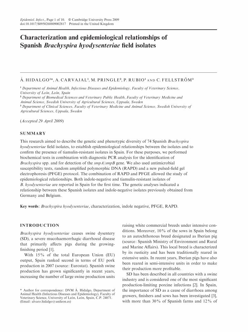
Characterization and epidemiological relationships of Spanish Brachyspira hyodysenteriae field isolates A ´ . HIDALGO 1 *, A. CARVAJAL 1 , M. PRINGLE 2 , P. RUBIO 1 AND C. FELLSTRO ¨ M 3 1 Department of Animal Health, Infectious Diseases and Epidemiology, Faculty of Veterinary Science, University of Leo ´n, Leo ´n, Spain 2 Department of Biomedical Sciences and Veterinary Public Health, Faculty of Veterinary Medicine and Animal Science, Swedish University of Agricultural Sciences, Uppsala, Sweden 3 Department of Clinical Sciences, Faculty of Veterinary Medicine and Animal Science, Swedish University of Agricultural Sciences, Uppsala, Sweden (Accepted 29 April 2009) SUMMARY This research aimed to describe the genetic and phenotypic diversity of 74 Spanish Brachyspira hyodysenteriae field isolates, to establish epidemiological relationships between the isolates and to confirm the presence of tiamulin-resistant isolates in Spain. For these purposes, we performed biochemical tests in combination with diagnostic PCR analysis for the identification of Brachyspira spp. and for detection of the smpA/smpB gene. We also used antimicrobial susceptibility tests, random amplified polymorphic DNA (RAPD) and a new pulsed-field gel electrophoresis (PFGE) protocol. The combination of RAPD and PFGE allowed the study of epidemiological relationships. Both indole-negative and tiamulin-resistant isolates of B. hyodysenteriae are reported in Spain for the first time. The genetic analyses indicated a relationship between these Spanish isolates and indole-negative isolates previously obtained from Germany and Belgium. Key words : Brachyspira hyodysenteriae, characterization, indole negative, PFGE, RAPD. INTRODUCTION Brachyspira hyodysenteriae causes swine dysentery (SD), a severe mucohaemorrhagic diarrhoeal disease that primarily affects pigs during the growing- finishing period [1]. With 15% of the total European Union (EU) output, Spain ranked second in terms of EU pork production in 2007 (source: Eurostat). Spanish swine production has grown significantly in recent years, increasing the number of large swine production units raising white commercial breeds under intensive con- ditions. Moreover, 10% of the sows in Spain belong to an autochthonous breed designated as Iberian pig (source: Spanish Ministry of Environment and Rural and Marine Affairs). This local breed is characterized by its rusticity and has been traditionally reared in extensive units. In recent years, Iberian pigs have also been reared in semi-intensive units in order to make their production more profitable. SD has been described in all countries with a swine industry and is considered one of the most significant production-limiting porcine infections [2]. In Spain, the importance of SD as a cause of diarrhoea among growers, finishers and sows has been investigated [3], with more than 30% of Spanish farms and 12% of * Author for correspondence : DVM A ´ . Hidalgo, Department of Animal Health (Infectious Diseases and Epidemiology), Faculty of Veterinary Science, University of Leo´ n, Leo´ n, Spain, C.P. 24071. (Email : [email protected]) Epidemiol. Infect., Page 1 of 10. f Cambridge University Press 2009 doi:10.1017/S0950268809002817 Printed in the United Kingdom
Transcript of Characterization and epidemiological relationships of Spanish Brachyspira hyodysenteriae field...
Characterization and epidemiological relationships of
Spanish Brachyspira hyodysenteriae field isolates
A. HIDALGO 1*, A. CARVAJAL1, M. PRINGLE 2, P. RUBIO 1AND C. FELLSTROM3
1 Department of Animal Health, Infectious Diseases and Epidemiology, Faculty of Veterinary Science,University of Leon, Leon, Spain2 Department of Biomedical Sciences and Veterinary Public Health, Faculty of Veterinary Medicine and
Animal Science, Swedish University of Agricultural Sciences, Uppsala, Sweden3 Department of Clinical Sciences, Faculty of Veterinary Medicine and Animal Science, Swedish University ofAgricultural Sciences, Uppsala, Sweden
(Accepted 29 April 2009)
SUMMARY
This research aimed to describe the genetic and phenotypic diversity of 74 Spanish Brachyspira
hyodysenteriae field isolates, to establish epidemiological relationships between the isolates and to
confirm the presence of tiamulin-resistant isolates in Spain. For these purposes, we performed
biochemical tests in combination with diagnostic PCR analysis for the identification of
Brachyspira spp. and for detection of the smpA/smpB gene. We also used antimicrobial
susceptibility tests, random amplified polymorphic DNA (RAPD) and a new pulsed-field gel
electrophoresis (PFGE) protocol. The combination of RAPD and PFGE allowed the study of
epidemiological relationships. Both indole-negative and tiamulin-resistant isolates of
B. hyodysenteriae are reported in Spain for the first time. The genetic analyses indicated a
relationship between these Spanish isolates and indole-negative isolates previously obtained from
Germany and Belgium.
Key words : Brachyspira hyodysenteriae, characterization, indole negative, PFGE, RAPD.
INTRODUCTION
Brachyspira hyodysenteriae causes swine dysentery
(SD), a severe mucohaemorrhagic diarrhoeal disease
that primarily affects pigs during the growing-
finishing period [1].
With 15% of the total European Union (EU)
output, Spain ranked second in terms of EU pork
production in 2007 (source: Eurostat). Spanish swine
production has grown significantly in recent years,
increasing the number of large swine production units
raising white commercial breeds under intensive con-
ditions. Moreover, 10% of the sows in Spain belong
to an autochthonous breed designated as Iberian pig
(source: Spanish Ministry of Environment and Rural
and Marine Affairs). This local breed is characterized
by its rusticity and has been traditionally reared in
extensive units. In recent years, Iberian pigs have also
been reared in semi-intensive units in order to make
their production more profitable.
SD has been described in all countries with a swine
industry and is considered one of the most significant
production-limiting porcine infections [2]. In Spain,
the importance of SD as a cause of diarrhoea among
growers, finishers and sows has been investigated [3],
with more than 30% of Spanish farms and 12% of
* Author for correspondence : DVM A. Hidalgo, Department ofAnimal Health (Infectious Diseases and Epidemiology), Faculty ofVeterinary Science, University of Leon, Leon, Spain, C.P. 24071.(Email : [email protected])
Epidemiol. Infect., Page 1 of 10. f Cambridge University Press 2009
doi:10.1017/S0950268809002817 Printed in the United Kingdom
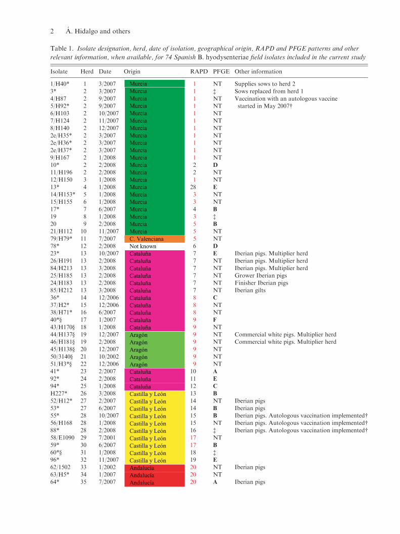
Table 1. Isolate designation, herd, date of isolation, geographical origin, RAPD and PFGE patterns and other
relevant information, when available, for 74 Spanish B. hyodysenteriae field isolates included in the current study
Isolate Herd Date Origin RAPD PFGE Other information
1/H40* 1 3/2007 MurciaMurciaMurciaMurciaMurciaMurciaMurciaMurciaMurciaMurciaMurciaMurciaMurciaMurciaMurciaMurciaMurciaMurciaMurciaMurciaMurciaC. ValencianaNot knownCataluñaCataluñaCataluñaCataluñaCataluñaCataluñaCataluñaCataluñaCataluñaCataluñaCataluñaAragónAragónAragónAragónAragónCataluñaCataluñaCataluñaCastilla y LeónCastilla y LeónCastilla y LeónCastilla y LeónCastilla y LeónCastilla y LeónCastilla y LeónCastilla y LeónCastilla y LeónCastilla y LeónAndalucíaAndalucíaAndalucía
1 NT Supplies sows to herd 2
3* 2 3/2007 1 $ Sows replaced from herd 14/H87 2 9/2007 1 NT Vaccination with an autologous vaccine5/H92* 2 9/2007 1 NT started in May 2007#
6/H103 2 10/2007 1 NT7/H124 2 11/2007 1 NT8/H140 2 12/2007 1 NT
2e/H35* 2 3/2007 1 NT2e/H36* 2 3/2007 1 NT2e/H37* 2 3/2007 1 NT
9/H167 2 1/2008 1 NT10* 2 2/2008 2 D
11/H196 2 2/2008 2 NT12/H150 3 1/2008 1 NT
13* 4 1/2008 28 E
14/H153* 5 1/2008 3 NT15/H155 6 1/2008 3 NT
17* 7 6/2007 4 B
19 8 1/2008 3 $
20 9 2/2008 5 B
21/H112 10 11/2007 5 NT79/H79* 11 7/2007 5 NT78* 12 2/2008 6 D
23* 13 10/2007 7 E Iberian pigs. Multiplier herd
26/H191 13 2/2008 7 NT Iberian pigs. Multiplier herd84/H213 13 3/2008 7 NT Iberian pigs. Multiplier herd25/H185 13 2/2008 7 NT Grower Iberian pigs
24/H183 13 2/2008 7 NT Finisher Iberian pigs85/H212 13 3/2008 7 NT Iberian gilts36* 14 12/2006 8 C
37/H2* 15 12/2006 8 NT38/H71* 16 6/2007 8 NT40*· 17 1/2007 9 F
43/H170· 18 1/2008 9 NT44/H137· 19 12/2007 9 NT Commercial white pigs. Multiplier herd46/H181· 19 2/2008 9 NT Commercial white pigs. Multiplier herd45/H138· 20 12/2007 9 NT
50/3140· 21 10/2002 9 NT51/H3*· 22 12/2006 9 NT41* 23 2/2007 10 A
92* 24 2/2008 11 E
94* 25 1/2008 12 C
H227* 26 3/2008 13 B
52/H12* 27 2/2007 14 NT Iberian pigs53* 27 6/2007 14 B Iberian pigs55* 28 10/2007 15 B Iberian pigs. Autologous vaccination implemented#56/H168 28 1/2008 15 NT Iberian pigs. Autologous vaccination implemented#
88* 28 2/2008 16 $ Iberian pigs. Autologous vaccination implemented#58/E1090 29 7/2001 17 NT59* 30 6/2007 17 B
60*· 31 1/2008 18 $
96* 32 11/2007 19 E
62/1502 33 1/2002 20 NT Iberian pigs
63/H5* 34 1/2007 20 NT64* 35 7/2007 20 A Iberian pigs
2 A. Hidalgo and others
faecal specimens testing positive for B. hyodysenter-
iae. Moreover, decreased susceptibility to the main
antimicrobials used in the treatment of SD has been
detected in Spanish B. hyodysenteriae isolates [4].
Diverse methodologies, such as serotyping [5], re-
striction endonuclease analysis (REA) [6], multilocus
enzyme electrophoresis (MLEE) [7], pulsed-field gel
electrophoresis (PFGE) [8], random amplified poly-
morphic DNA (RAPD) [9], biochemical character-
ization [10], DNA restriction fragment polymorphism
analysis [11] and multilocus sequence typing (MLST)
[12], have been used to characterize and analyse the
diversity of Brachyspira spp. isolates.
The research reported herein was performed to
describe the genetic and phenotypic diversity of
Spanish B. hyodysenteriae field isolates and to inves-
tigate epidemiological relationships between them.
Moreover, we attempted to confirm the presence
of tiamulin-resistant isolates and to investigate their
common or independent origin.
METHODS
Bacterial strains and growth conditions
A set of 74 Spanish isolates of strongly b-haemolytic
intestinal spirochaetes recovered from pigs and class-
ified as B. hyodysenteriae according to species-specific
PCR [13] was used in the current study. Isolates were
selected in order to include samples representing the
most important pig production regions of the coun-
try. All isolates were obtained from faecal samples
from growers, finishers or sows submitted for routine
diagnostics to the Laboratory of Infectious Diseases
in the Veterinary Faculty at the University of
Leon, Spain, and stored in liquid nitrogen. A list
depicting isolate designation, herd, date of isolation,
geographical origin and other relevant information, if
available, is presented in Table 1. The isolates were
sent in Amies medium to the National Veterinary
Institute (SVA), Uppsala, Sweden, where they were
tested using duplex PCR [14], based on the tlyA and
Table 1 (cont.)
Isolate Herd Date Origin RAPD PFGE Other information
65/H173 36 2/2008 AndalucíaAndalucíaAndalucíaExtremaduraExtremaduraExtremaduraCastilla-La ManchaCastilla-La ManchaAndalucíaNot knownAragónAragónCataluñaCataluñaCataluñaMurciaExtremaduraExtremaduraExtremadura
20 NT Iberian pigs66/H57* 37 5/2007 20 NT Iberian pigs
97/H88 41 9/2007 20 NT Iberian pigs69/H13* 38 2/2007 20 NT Iberian pigs70/H21* 38 2/2007 20 NT Iberian pigs
95/H141 40 12/2007 20 NT Iberian pigs71/H44* 39 4/2007 20 NT73* 42 10/2007 21 A
89/H203 43 2/2008 21 NT ½Iberian pigsr½Duroc81* 44 10/2001 22 A
93* 45 1/2008 23 C
98* 46 4/2007 24 E
H9*· 47 1/2007 25 F
H19* 48 2/2007 26 $
H72* 49 6/2007 27 C
87/H208 51 2/2008 NT NT B. hyodysenteriae and B. innocens mixed culture90/H197 50 2/2008 NT NT B. hyodysenteriae and B. murdochii mixed culture67/E1697 52 2/2002 NT NT B. hyodysenteriae and B. pilosicoli mixed culture
68/H23 53 2/2007 NT NT B. hyodysenteriae and B. pilosicoli mixed culture
Date : Month and year of isolation ; Origin : administrative region, coloured according tothe map (right), where the farm was located; RAPD : pattern assigned in the RAPD study(RAPD patterns in red type are shared by isolates from different herds) ; PFGE : pulsed-
field gel electrophoresis cluster for MluI, according to groups (A–F) established inFigure 2.NT, Not tested.
* Isolates tested with smpA/smpB PCR.# Autologous B. hyodysenteriae vaccination programme consisting of a whole-herdvaccination repeated each 4 months.
$ PFGE tested and not clustered with an 80% cut-off value.· Indol-negative Spanish B. hyodysenteriae field isolates.
B. hyodysenteriae characterization 3
the 16S rRNA genes, for detection of B. hyodysen-
teriae and B. pilosicoli, respectively.
We also investigated two German indole-negative
B. hyodysenteriae isolates, designated 5677/96 and
T4 [12] from the Swedish collection at SVA and the
B. hyodysenteriae reference strain B204 (ATCC 31212),
B. hyodysenteriae type strain B78T (ATCC 27164T),
and B. pilosicoli type strain P43/6/78T (ATCC 51139)
were used as controls for PCR and biochemical
characterization.
Bacteria were grown on fastidious anaerobe agar
(FAA, SVA, Sweden) at 42 xC in anaerobic jars
[GENbox (bioMerieux, France) with AnaeroGen
sachets (Oxoid, UK)].
Biochemical tests and b-haemolysis
Biochemical characterization was performed as pre-
viously described by Fellstrom & Gunnarsson [15].
In brief, 3-day-old cultures were tested for weak
or strong b-haemolysis on trypticase soy agar
supplemented with 5% ovine blood. Indole pro-
duction was investigated using the spot indole test ;
a-galactosidase activity was determined using diag-
nostic tablets (Rosco Diagnostica, Denmark) and
hippurate hydrolysis as described by Rubsamen &
Rubsamen [16].
Testing antimicrobial susceptibility
Eleven Spanish B. hyodysenteriae isolates (for ref-
erence see Table 2), selected on the basis of
their reduced susceptibility to tiamulin (o2 mg/ml)
determined in a previous investigation [4], were tested
for antimicrobial susceptibility using VetMICTM
Brachy QCR high panels (SVA, Sweden) according
to the manufacturer’s protocol. The antimicrobial
agents tested were tiamulin, valnemulin, doxycycline,
lincomycin, tylosin, and tylvalosin. The minimum
inhibitory concentration (MIC) was determined as the
lowest concentration of antimicrobial agent that pre-
vented visible growth. Absence of contamination was
checked by phase contrast microscopy.
RAPD
Seventy B. hyodysenteriae isolates confirmed by
duplex PCR [14] and biochemical tests [15] as well as
the reference and type strains of B. hyodysenteriae
(B204 and B78T) were typed by RAPD following the
technique described by Quednau et al. [17], slightly
modified. DNA samples were prepared from 3-day-
old pure cultures grown on FAA. Two filled 1-ml
loops of the bacteria were washed twice in phosphate
buffered saline (pH 7.3), boiled in nuclease-free water
(Sigma-Aldrich, USA) and centrifuged. The super-
natant was transferred to a sterile microtube. Extrac-
ted DNA samples were adjusted to a concentration
of 20 ng/ml. RAPD fingerprints were generated with
primer P73 (5k-ACGCGCCCT-3k) and primer P1254
(5k-CCGCAGCCAA-3k), resulting in two different
pattern sets that were visually analysed. Results were
interpreted with strict criteria and isolates which dif-
fered in at least one fragment (including weak, barely
visible and broad bands) were assigned to different
RAPD types. In order to ensure reproducibility, this
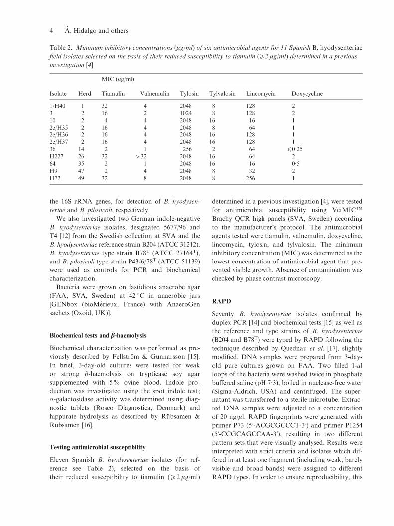
Table 2. Minimum inhibitory concentrations (mg/ml) of six antimicrobial agents for 11 Spanish B. hyodysenteriae
field isolates selected on the basis of their reduced susceptibility to tiamulin (o2 mg/ml) determined in a previous
investigation [4]
Isolate Herd
MIC (mg/ml)
Tiamulin Valnemulin Tylosin Tylvalosin Lincomycin Doxycycline
1/H40 1 32 4 2048 8 128 23 2 16 2 1024 8 128 210 2 4 4 2048 16 16 12e/H35 2 16 4 2048 8 64 1
2e/H36 2 16 4 2048 16 128 12e/H37 2 16 4 2048 16 128 136 14 2 1 256 2 64 f0.25
H227 26 32 >32 2048 16 64 264 35 2 1 2048 16 16 0.5H9 47 2 4 2048 8 32 2
H72 49 32 8 2048 8 256 1
4 A. Hidalgo and others
technique was repeated at least three times for each
isolate.
PFGE
Thirty-one B. hyodysenteriae isolates were typed by
PFGE, including 28 Spanish field isolates represent-
ing the different RAPD patterns (see Table 1), the
reference and type strains of B. hyodysenteriae (B204
and B78T), and one German indole-negative isolate
(5677/96).
The DNA preparation procedure for PFGE was
adapted from a previous protocol described for
Treponema spp. [18]. For each isolate, bacterial cells
from two FAA plates were harvested, suspended in
10 ml TE buffer (10 mM Tris, 1 mM EDTA) and
washed three times in 5 ml TE buffer. The cells were
then suspended in 1.5 ml Pett IV buffer (10 mM Tris–
HCl, 1 M NaCl), adjusted to an optical density of
0.800 at 405 nm and mixed 1:1 (v:v) with 1.5%
low melting temperature agarose (NA agarose, GE
Healthcare, UK). The agarose plugs were incubated
in ESP (0.5 M EDTA, 1% N-lauroyl sarcosine, 0.2%
pronase E) at 50 xC for 24 h, restoring the liquid
after 1.5 h. Gel plugs were then washed six times in
TE buffer. Digestion with restriction enzymes MluI
(5k-A›CGCGT) and SalI (5k-G›TCGAC) and pulsed-
field electrophoresis were performed as described by
Fellstrom et al. [10], using a CHEF-DR1 III pulsed
field electrophoresis system (Bio-Rad Laboratories
AB, Sweden) at 6 V/cm2 with an included angle of
120x. Initial and final switch times were 5 s and 70 s,
respectively. The gels were run for 24 h in 0.5rTBE
buffer (44.5 mM Tris, 44.5 mM boric acid, 1 mM
EDTA) at 14 xC and subsequently stained with
ethidium bromide. A lambda marker (New England,
Biolabs, USA) was included to normalize the PFGE
banding patterns that were used for producing den-
drograms, following calculation of the Dice coef-
ficient and analysis with the unweighted pair-group
method by arithmetic averages (UPGMA) clustering
fusion strategy, performed with the GelCompar pro-
gram (Applied Maths, Belgium).
SmpA/smpB-specific PCR
A PCR assay for specific detection of smpA or smpB
genes was performed on 42 Spanish B. hyodysenteriae
field isolates (listed in Table 1) as described by Holden
et al. [19]. Genomic DNA was prepared by the CTAB
extraction method and at least one isolate per RAPD
pattern was included. Reference strain B204 was in-
cluded as smpA-positive control.
RESULTS
PCR identification and biochemical characterization
Duplex PCR analysis for the detection of B. hyodys-
enteriae and B. pilosicoli, resulted in the tlyA gene
fragment being amplified for all 74 isolates ; the 16S
rRNA gene fragment specific for B. pilosicoli was
amplified for two isolates (67/E1697 and 68/H28).
These latter two isolates were considered to be
B. hyodysenteriae and B. pilosicoli mixed cultures.
In addition, the biochemical tests placed 70 of the
72 presumptive B. hyodysenteriae isolates (according
to the duplex PCR) in group I (B. hyodysenteriae)
[20]. However, isolates 90/H197 and 87/H208 were
classified as group III, B. innocens and B. murdochii,
respectively, and considered as mixed cultures. Sixty-
one group I isolates (87.1%) were recorded as indole
positive in the spot indole test, while nine group
I isolates (12.9%; 40, 43/H170, 44/H137, 46/H181,
45/H138, 50/3140, 51/H3, 60 and H9), were indole
negative.
MIC determinations
The MICs of the six antimicrobial agents studied for
the 11 selected Spanish B. hyodysenteriae isolates are
shown in Table 2.
RAPD analysis
Twenty-eight dissimilar RAPD patterns were ob-
tained for the 70 Spanish B. hyodysenteriae field iso-
lates. A different figure was given for each RAPD
pattern (Table 1). German indole-negative isolates,
5677/96 and T4, were assigned to RAPD pattern
number 9. Reference and type strains B204 and B78T
did not share any RAPD pattern with the studied field
isolates.
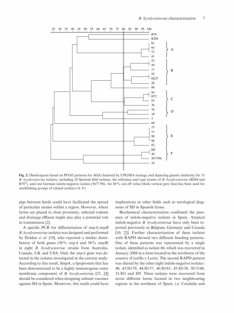
PFGE
Digestion of B. hyodysenteriae DNA produced 7–18
and 4–9 fragments for MluI and SalI, respectively.
The quality of the gels obtained was high, with clearly
defined bands (Fig. 1). All tested isolates yielded
a PFGE pattern with at least one of the enzymes,
although isolate H19 did not generate any visible
B. hyodysenteriae characterization 5
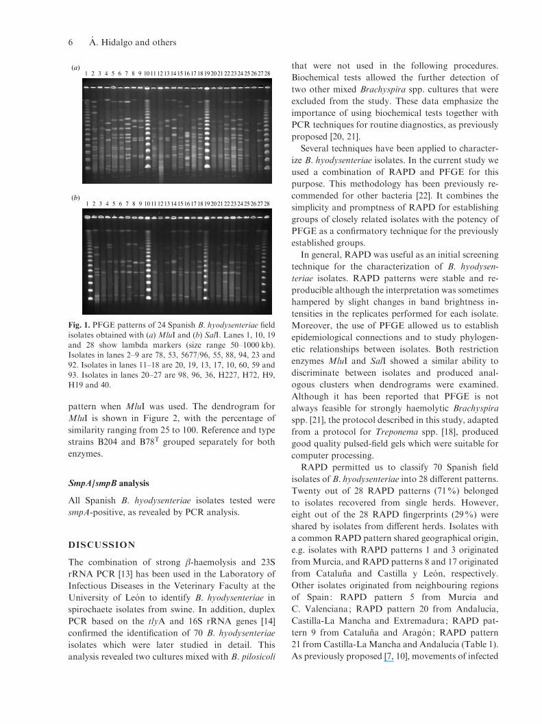
pattern when MluI was used. The dendrogram for
MluI is shown in Figure 2, with the percentage of
similarity ranging from 25 to 100. Reference and type
strains B204 and B78T grouped separately for both
enzymes.
SmpA/smpB analysis
All Spanish B. hyodysenteriae isolates tested were
smpA-positive, as revealed by PCR analysis.
DISCUSSION
The combination of strong b-haemolysis and 23S
rRNA PCR [13] has been used in the Laboratory of
Infectious Diseases in the Veterinary Faculty at the
University of Leon to identify B. hyodysenteriae in
spirochaete isolates from swine. In addition, duplex
PCR based on the tlyA and 16S rRNA genes [14]
confirmed the identification of 70 B. hyodysenteriae
isolates which were later studied in detail. This
analysis revealed two cultures mixed with B. pilosicoli
that were not used in the following procedures.
Biochemical tests allowed the further detection of
two other mixed Brachyspira spp. cultures that were
excluded from the study. These data emphasize the
importance of using biochemical tests together with
PCR techniques for routine diagnostics, as previously
proposed [20, 21].
Several techniques have been applied to character-
ize B. hyodysenteriae isolates. In the current study we
used a combination of RAPD and PFGE for this
purpose. This methodology has been previously re-
commended for other bacteria [22]. It combines the
simplicity and promptness of RAPD for establishing
groups of closely related isolates with the potency of
PFGE as a confirmatory technique for the previously
established groups.
In general, RAPD was useful as an initial screening
technique for the characterization of B. hyodysen-
teriae isolates. RAPD patterns were stable and re-
producible although the interpretation was sometimes
hampered by slight changes in band brightness in-
tensities in the replicates performed for each isolate.
Moreover, the use of PFGE allowed us to establish
epidemiological connections and to study phylogen-
etic relationships between isolates. Both restriction
enzymes MluI and SalI showed a similar ability to
discriminate between isolates and produced anal-
ogous clusters when dendrograms were examined.
Although it has been reported that PFGE is not
always feasible for strongly haemolytic Brachyspira
spp. [21], the protocol described in this study, adapted
from a protocol for Treponema spp. [18], produced
good quality pulsed-field gels which were suitable for
computer processing.
RAPD permitted us to classify 70 Spanish field
isolates of B. hyodysenteriae into 28 different patterns.
Twenty out of 28 RAPD patterns (71%) belonged
to isolates recovered from single herds. However,
eight out of the 28 RAPD fingerprints (29%) were
shared by isolates from different herds. Isolates with
a common RAPD pattern shared geographical origin,
e.g. isolates with RAPD patterns 1 and 3 originated
fromMurcia, and RAPD patterns 8 and 17 originated
from Cataluna and Castilla y Leon, respectively.
Other isolates originated from neighbouring regions
of Spain: RAPD pattern 5 from Murcia and
C. Valenciana; RAPD pattern 20 from Andalucıa,
Castilla-La Mancha and Extremadura; RAPD pat-
tern 9 from Cataluna and Aragon; RAPD pattern
21 from Castilla-LaMancha and Andalucıa (Table 1).
As previously proposed [7, 10], movements of infected
1 2 3 4 5 6 7 8 9 10 11 12 13 14 15 16 17 18 19 20 21 22 23 24 25 26 27 28
1 2 3 4 5 6 7 8 9 10 11 12 13 14 15 16 17 18 19 20 21 22 23 24 25 26 27 28
(a)
(b)
Fig. 1. PFGE patterns of 24 Spanish B. hyodysenteriae field
isolates obtained with (a) MluI and (b) SalI. Lanes 1, 10, 19and 28 show lambda markers (size range 50–1000 kb).Isolates in lanes 2–9 are 78, 53, 5677/96, 55, 88, 94, 23 and
92. Isolates in lanes 11–18 are 20, 19, 13, 17, 10, 60, 59 and93. Isolates in lanes 20–27 are 98, 96, 36, H227, H72, H9,H19 and 40.
6 A. Hidalgo and others
pigs between herds could have facilitated the spread
of particular strains within a region. However, where
farms are placed in close proximity, infected rodents
and drainage effluent might also play a potential role
in transmission [2].
A specific PCR for differentiation of smpA/smpB
B. hyodysenteriae isolates was designed and performed
by Holden et al. [19], who reported a similar distri-
bution of both genes (50% smpA and 50% smpB)
in eight B. hyodysenteriae strains from Australia,
Canada, UK and USA. Only the smpA gene was de-
tected in the isolates investigated in the current study.
According to this result, SmpA, a lipoprotein that has
been demonstrated to be a highly immunogenic outer
membrane component of B. hyodysenteriae [23, 24]
should be considered when designing subunit vaccines
against SD in Spain. Moreover, this result could have
implications in other fields such as serological diag-
nosis of SD in Spanish farms.
Biochemical characterization confirmed the pres-
ence of indole-negative isolates in Spain. Atypical
indole-negative B. hyodysenteriae have only been re-
ported previously in Belgium, Germany and Canada
[10, 25]. Further characterization of these isolates
with RAPD showed two different banding patterns.
One of these patterns was represented by a single
isolate, identified as isolate 60, which was recovered in
January 2008 in a farm located in the northwest of the
country (Castilla y Leon). The second RAPD pattern
was shared by the other eight indole-negative isolates :
40, 43/H170, 44/H137, 46/H181, 45/H138, 50/3140,
51/H3 and H9. These isolates were recovered from
seven different farms located in two neighbouring
regions in the northeast of Spain, i.e. Cataluna and
25 30 35 40 45 50 55 60 65 70 75 80 85 90 95 100
B78B204
81
6473
41
53
59
17
55
H227
20
88
94H729336
78
103
6023
13
98
9692
H9405677/96
19
A
B
C
D
E
F
Fig. 2. Dendrogram based on PFGE patterns for MluI clustered by UPGMA strategy and depicting genetic similarity for 31B. hyodysenteriae isolates, including 28 Spanish field isolates, the reference and type strains of B. hyodysenteriae (B204 andB78T), and one German indole-negative isolate (5677/96). An 80% cut-off value (thick vertical grey line) has been used for
establishing groups of related isolates (A–F).
B. hyodysenteriae characterization 7
Aragon, between 2002 and 2008. Surprisingly, this
RAPD pattern was also shared by the two indole-
negative German isolates, T4 and 5677/96. For fur-
ther investigation of this relationship, the German
isolate 5677/96 together with two Spanish indole-
negative isolates, H9 and 40, were analysed by PFGE.
The three isolates grouped together markedly sep-
arated from other clusters, with a high percentage of
similarity (94% for MluI). Moreover, Belgian indole-
negative isolates have been previously shown to be
indistinguishable from isolate 5677/96 [10]. The rare
occurrence of indole-negative isolates combined with
the results of RAPD and PFGE procedures strongly
indicates an epidemiological relationship between
these isolates, although our epidemiological records
do not allow an absolute confirmation of this fact.
Nevertheless, the trade of pigs from these countries
to Spain supports this possibility, with more than
207 000 animals sold in 2000 and 135000 in 2001
(source: Spanish Ministry of Environment and Rural
and Marine Affairs). Migratory birds may also be
considered as a risk for transmission of Brachyspira
isolates between countries [21]. The national, seem-
ingly clonal, spread of this indole-negative strain
could have been the result of frequent movements
and trade of animals in the northeast area of Spain
and the presence of this RAPD type (isolates 44/H137
and 46/H181) in one Spanish multiplier herd (no. 19).
The RAPD fingerprints of 20 Spanish B. hyodys-
enteriae field isolates recovered from Iberian pigs were
divided into six different RAPD patterns, designated
as 7, 14, 15, 16, 20 and 21. Subsequent analysis by
PFGE grouped RAPD type 14 (isolate 53) together
with RAPD type 15 (isolate 55) and RAPD type 20
(isolate 64) together with RAPD type 21 (isolate 73).
The spread of RAPD type 20, detected in eight
Iberian pig units located in the southwest of Spain
(Andalucıa and Extremadura), is probably a conse-
quence of trade with carriers or diseased pigs. The
particular conditions of the Iberian pig market, which
is characterized by high demand for a limited number
of available pigs and entirely lacking or deficient
herd health programmes, could have facilitated this
fact.
The key role of carrier swine in within-herd spread
of infection [2] was evident in herd no. 13, a semi-
intensive Iberian pig unit where SD appeared and was
subsequently disseminated to four productive units
situated at different locations.
When more than one isolate per herd were analysed
by RAPD, we found identical isolates in four herds
(nos. 13, 19, 27 and 38). However, slight variations
among isolates were recorded in two other herds (nos.
2 and 28). These isolates were subsequently confirmed
by PFGE as closely related. Interestingly, vaccination
with an inactivated autologous vaccine of B. hyo-
dysenteriae had been implemented in both herds.
Herd no. 2 was analysed further, including 12 isolates
recovered from March 2007 to February 2008.
Vaccination started in May 2007. The RAPD pattern
was stable from February 2007 to January 2008,
but slight differences were recorded for two isolates
recovered in February 2008. This difference was
subsequently confirmed by PFGE. Moreover, the
antimicrobial susceptibility pattern also changed.
MIC values yielded by isolates from March 2007 (3,
2e/H35, 2e/H36, 2e/H37) were compared with those
displayed by one isolate from February 2008 (isolate
10). An increase in the sensitivity of two dilution steps
(from 16 mg/ml to 4 mg/ml) was observed for the MIC
of tiamulin and of three dilution steps (from 128 mg/
ml to 16 mg/ml) for the MIC of lincomycin. This new
closely related isolate had not been recovered on the
farm previously. One explanation for the isolation of
new variants of B. hyodysenteriae in the herd may be
the introduction of sows from herd no. 1 (Table 1).
However, the minor genetic differences recorded
could be the result of adaptive advantages, first, by the
selective pressure caused by vaccination or second, by
the changes in the antibiotic therapy protocols in the
farm subsequent to the success of the immunological
treatment. A similar result was reported by Atyeo
et al. [8] in Australian herds and the microevolution
theory was also proposed as the most plausible ex-
planation.
On the other hand, genetic stability over time for
four Spanish B. hyodysenteriae field isolates was also
demonstrated. Isolate 50/3140, an indole-negative
isolate, was recovered in October 2002 and yielded an
identical RAPD pattern to the indole-negative isolate
46/H181, from February 2008. Similar results were
obtained for isolates 58/E1090 and 59 recovered in
July 2001 and June 2007, respectively, and isolates
62/1502 and 65/H173, recovered from January 2002
and February 2008, respectively. Similarly, using
PFGE, isolate 81 from October 2001 was identical to
isolate 73, from October 2007. Hence, stability in
some Spanish field isolates of B. hyodysenteriae was
registered for up to 6 years, in agreement with a pre-
vious report on Swedish isolates [10].
According to Rønne & Szancer [26], B. hyodys-
enteriae isolates with MICs >4 mg/ml for tiamulin
8 A. Hidalgo and others
should be considered as resistant isolates. In the cur-
rent study, 7/11 B. hyodysenteriae isolates selected on
the basis of their reduced susceptibility to tiamulin [4]
were classified as resistant. The tiamulin-resistant
isolates 1/H40, 3, 2e/H35, 2e/H36 and 2e/H37 shared
the same RAPD pattern. The RAPD patterns for the
other two tiamulin-resistant isolates were unique;
thus three different RAPD and PFGE types of
tiamulin-resistant B. hyodysenteriae (MIC 32 mg/ml)
were confirmed. Valnemulin decreased susceptibility
was present in all tested isolates. Subsequent analysis
of the geographical distribution of the herds where the
resistant isolates had been collected showed that they
were from three different and distant areas of the
country: Murcia, Castilla y Leon and Cataluna. The
tiamulin-resistant isolates should be considered as a
risk to the swine industry.
In conclusion, the results from RAPD and PFGE
demonstrated the presence of diverse B. hyodysenter-
iae field isolates in Spain and allowed the investi-
gation of epidemiological relationships between these
isolates. Furthermore, this is the first report of
Spanish indole-negative B. hyodysenteriae isolates
and the clonal spread of one of these. Moreover, the
existence of tiamulin-resistant B. hyodysenteriae iso-
lates, which have emerged independently in Spain,
was also demonstrated.
ACKNOWLEDGEMENTS
The authors express their thanks to Marih Jonsson
and Gloria Fernandez Bayon for excellent technical
assistance. Alvaro Hidalgo is supported by a grant
from Consejerıa de Educacion of the Junta de Castilla
y Leon and the European Social Fund. This work was
funded by the Ministerio de Educacion y Ciencia
(Spanish Ministry of Education and Science) and
co-financed by the European Regional Development
Funds (ERDF) as Project AGL2005-01976/GAN
(January 2006).
DECLARATION OF INTEREST
None.
REFERENCES
1. Hampson DJ, Fellstrom C, Thomson JR. Swine dys-
entery. In : Straw BE, Zimmerman JJ, D’Allaire S,Taylor DJ, eds.Diseases of Swine, 9th edn. Ames, Iowa:Blackwell Publishing Professional, 2006, pp. 785–805.
2. Hampson DJ, Atyeo RF, Combs BG. Swine dysentery.In : Hampson DJ, Stanton TB, eds. Intestinal Spiro-
chaetes in Domestic Animals and Humans. Wallingford,England: CAB International, 1997, pp. 175–209.
3. Carvajal A, et al. Prevalence of Brachyspira species in
pigs with diarrhoea in Spain. Veterinary Record 2006;158 : 700–701.
4. Hidalgo A, et al. Antimicrobial susceptibility testing ofSpanish field isolates of Brachyspira hyodysenteriae.
Research in Veterinary Science 2009; 87 : 7–12.5. Hampson DJ, et al. Proposed revisions to the sero-
logical typing system for Treponema hyodysenteriae.
Epidemiology and Infection 1989; 102 : 75–84.6. Combs BG, Hampson DJ, Harders SJ. Typing of
Australian isolates of Treponema hyodysenteriae by
serology and by DNA restriction endonuclease analysis.Veterinary Microbiology 1992; 31 : 273–285.
7. Trott DJ, Oxberry SL, Hampson DJ. Evidence for
Serpulina hyodysenteriae being recombinant, with anepidemic population structure.Microbiology 1997; 143 :3357–3365.
8. Atyeo RF, Oxberry SL, Hampson DJ. Analysis of
Serpulina hyodysenteriae strain variation and its mol-ecular epidemiology using pulsed-field gel electrophor-esis. Epidemiology and Infection 1999; 123 : 133–138.
9. Dugourd D, et al. Characterization of Serpulina hyo-dysenteriae isolates of serotypes 8 and 9 by randomamplification of polymorphic DNA analysis. Veterinary
Microbiology 1996; 48 : 305–314.10. Fellstrom C, et al. Emended descriptions of indole-
negative and indole positive isolates of Brachyspira
(Serpulina) hyodysenteriae. Veterinary Microbiology1999; 70 : 225–238.
11. Jensen NS, Casey TA, Stanton TB. Characterization ofSerpulina (Treponema) hyodysenteriae and related in-
testinal spirochetes by ribosomal RNA gene restrictionpatterns. FEMSMicrobiology Letters 1992; 72 : 235–241.
12. Rasback T, et al.Development of a multilocus sequence
typing scheme for intestinal spirochaetes within thegenus Brachyspira.Microbiology 2007; 153 : 4074–4087.
13. Leser TD, et al. Specific detection of Serpulina hyo-
dysenteriae and potentially pathogenic weakly beta-haemolytic porcine intestinal spirochetes by polymerasechain reaction targeting 23S rDNA. Molecular andCellular Probes 1997; 11 : 363–372.
14. Rasback T, et al. Comparison of culture and bio-chemical tests with PCR for detection of Brachyspirahyodysenteriae and Brachyspira pilosicoli. Journal of
Microbiological Methods 2006; 66 : 347–353.15. Fellstrom C, Gunnarsson A. Phenotypical characteris-
ation of intestinal spirochaetes isolated from pigs. Re-
search in Veterinary Science 1995; 59 : 1–4.16. Rubsamen S, Rubsamen S. Hippurate hydrolysis : a ra-
pid test for differentiation of Treponema hyodysenteriae
and Treponema innocens [in German]. TierarztlicheUmschau 1986; 41 : 673–677.
17. Quednau M, et al. Identification of clinically importantspecies of Enterococcus within 1 day with randomly
amplified polymorphic DNA (RAPD). Current Micro-biology 1998; 36 : 332–336.
B. hyodysenteriae characterization 9
18. Pringle M, et al. Isolation and characterization ofTreponema phagedenis-like spirochetes from digital
dermatitis lesions in Swedish dairy cattle. Acta Veter-inaria Scandinavica 2008; 50, 40.
19. Holden J, et al. SmpB: a novel outer membrane protein
present in some Brachyspira hyodysenteriae strains.Veterinary Microbiology 2006; 113 : 109–116.
20. Fellstrom C, et al. Identification and genetic finger-printing of Brachyspira species. Journal of Micro-
biological Methods 2008; 72 : 133–140.21. Rasback T, et al. A novel enteropathogenic, strongly
haemolytic spirochaete isolated from pig and mallard,
provisionally designated ‘Brachyspira suanatina ’ sp.nov. Environmental Microbiology 2007; 9 : 983–991.
22. Gori A, et al. Comparison of pulsed-field gel electro-
phoresis and randomly amplified DNA polymorphismanalysis for typing extended-spectrum-beta-lactamase-producing Klebsiella pneumoniae. Journal of Clinical
Microbiology 1996; 34 : 2448–2453.
23. Sellwood R, et al. Antibodies to a common outerenvelope antigen of Treponema hyodysenteriae with
antibacterial activity. Journal of General Microbiology1989; 135 : 2249–2257.
24. Thomas W, Sellwood R. Monoclonal antibodies to a
16-kDa antigen of Serpulina (Treponema) hyodysen-teriae. Journal of Medical Microbiology 1992; 37 : 214–220.
25. Hommez J, et al. Identification of porcine Serpulina
strains in routine diagnostic bacteriology. VeterinaryMicrobiology 1998; 62 : 163–169.
26. Rønne H, Szancer J. In vitro susceptibility of Danish
field isolates of Treponema hyodysenteriae to chemo-therapeutics in swine dysentery (SD) therapy. Inter-pretation of MIC results based on the pharmacokinetic
properties of the antibacterial agents. In : Proceedings ofthe 11th International Pig Veterinary Society Congress.Lausanne, Switzerland: International Pig Veterinary
Society, 1990, pp. 126.
10 A. Hidalgo and others