Parathyroid Hormone-Related Protein(1-34) Regulates Phex Expression in Osteoblasts through the...
10
Parathyroid Hormone-Related Protein(1–34) Regulates Phex Expression in Osteoblasts through the Protein Kinase A Pathway MIGUEL A ´ NGEL VARGAS, MATHIEU ST-LOUIS, LUC DESGROSEILLERS, JEAN-LOUIS CHARLI, AND GUY BOILEAU De ´partement de Biochimie (M.A ´ .V., M.S.L., L.D., G.B.), Faculte ´ de Me ´decine, Universite ´ de Montre ´al, Montre ´al, Que ´bec, Canada H3C 3J7; and Departamento de Gene ´tica del Desarrollo y Fisiologı ´a Molecular (M.A.V., J.-L.C.), Instituto de Biotecnologı´a, Universidad Nacional Auto ´noma de Me ´xico, Cuernavaca, Morelos 62271, Me ´xico Phex (a phosphate-regulating gene with homologies to en- dopeptidases on the X chromosome) is expressed predomi- nantly in bone in which it has been implicated in the miner- alization process. Multiple factors and hormones, including PTHrP, regulate formation, development, and/or homeostasis of bone. The purpose of the present study was to determine whether PTHrP(1–34) regulates Phex expression and identify the signaling pathway used. Phex mRNA and protein levels were analyzed by RT-PCR and immunoblotting, respectively. In UMR-106 cells, PTHrP(1–34) caused a time- and concentra- tion-dependent decrease in Phex expression. Forskolin, an adenylate cyclase activator, had the same effect. Dibutiryl cAMP also decreased Phex expression, and its effect was blocked by H89, a protein kinase A (PKA) inhibitor. In con- trast, 12-O-tetradecanoyl phorbol-13-acetate, a protein kinase C (PKC) activator, increased Phex expression in a time- and dose-dependent manner. This effect was reversed by bisin- dolylmaleimide , a PKC inhibitor. Bovine PTH(3–34), which activates PKC but not PKA, had no effect. On the contrary, human PTH(1–31), which activates PKA but not PKC, de- creased Phex expression. H89 but not bisindolylmaleimide blocked the effect of PTHrP(1–34). PTHrP(1–34) also de- creased Phex expression in cultures of fetal rat calvaria cells at d 7 of culture but not at later stages. These data demonstrate that PTHrP(1–34), through PKA, down-regulates Phex expres- sion in osteoblasts. (Endocrinology 144: 4876 – 4885, 2003) P HEX (FORMERLY PEX; a phosphate-regulating gene with homologies to endopeptidases on the X chromo- some) was identified by positional cloning as the gene re- sponsible for X-linked hypophosphatemia (XLH) in humans (1). XLH is a mendelian disorder of phosphate homeostasis characterized by growth retardation, renal defects in vitamin D metabolism, hypophosphatemia resulting from decreased phosphate reabsorption in the renal proximal tubules, and defective mineralization of teeth and bones causing rachitic and osteomalacic bone disease (2). Much of our actual un- derstanding of the pathophysiology of XLH has been ob- tained using a murine model for human XLH, the Hyp mouse, which presents a large deletion in the 3 region of the Phex gene (PHEX and Phex refer to the human and mouse genes, respectively) (3) and phenotypic features similar to those of XLH patients (4). The defective renal phosphate transport in Hyp mice has been attributed to decreased renal proximal tubule expression of the type and sodium- dependent phosphate cotransporter genes and proteins (5, 6). Parabiosis experiments and cross-kidney transplantation between Hyp and normal mice (7, 8) suggested the presence of a circulatory phosphaturic hormone, designated phospha- tonin (9), responsible for renal phosphate wasting. In accor- dance with this hypothesis, two groups have reported the presence of a renal phosphate transport inhibitory activity in conditioned medium of cultured osteoblasts isolated from Hyp mice (10, 11). Although the exact nature of phosphatonin remains to be determined, several candidates have been pro- posed (for a review see Ref. 12) including FGF 23 (13), matrix extracellular phosphoglycoprotein (14), and Frizzled-related protein-4 (15). Human and mouse PHEX/Phex cDNAs have been cloned and sequenced (3, 16 –20). Amino acid sequence comparisons have demonstrated homologies between PHEX/Phex pro- tein and members of the M13 zinc metalloendopeptidase family. The peptidases of this family are type II integral membrane glycoproteins with a relatively short N-terminal intracellular region, a single-transmembrane domain, and a large extracellular domain, which contains the active site of the enzyme (21). Peptidases of the M13 family have been shown to regulate the activity of biologically active peptides either by degradation of active peptides [neprilysin (NEP), neutral endopeptidase 24.11] (22) or in processing inactive peptide precursors into active forms endothelin-converting enzymes 1 and 2 (23). Several studies support a role of PHEX/Phex in mineral- ization. PHEX/Phex mRNA and protein are localized pre- dominantly in bone and teeth, specifically in osteoblasts, osteocytes, and odontoblasts but not in preosteoblasts (3, 16, 19, 24 –26). In developing bones in vivo or during osteoblast differentiation in vitro, Phex mRNA and protein expression Abbreviations: AP1, Activator protein 1; bPTH, bovine PTH; (Bu) 2 cAMP, N 6 ,2-O-dibutiryladenosine-3,5-cyclic monophosphate; FBS, fetal bovine serum; G3PDH, glyceraldehyde-3-phosphate dehy- drogenase; H89, N-[2-(p-bromocinnamylamino)ethyl]-5-isoquinoline- sulfonamide dihydrochloride; NEP, neprilysin; 4-PDD, 4-phorbol 12,13-didecanoate; Phex, a phosphate-regulating gene with homologies to endopeptidases on the X chromosome; PKA, protein kinase A; PKC, protein kinase C; TPA, 12-O-tetradecanoyl phorbol-13-acetate, XLH, X-linked hypophosphatemia. 0013-7227/03/$15.00/0 Endocrinology 144(11):4876 – 4885 Printed in U.S.A. Copyright © 2003 by The Endocrine Society doi: 10.1210/en.2003-0253 4876
-
Upload
independent -
Category
Documents
-
view
0 -
download
0
Transcript of Parathyroid Hormone-Related Protein(1-34) Regulates Phex Expression in Osteoblasts through the...

Parathyroid Hormone-Related Protein(1–34) RegulatesPhex Expression in Osteoblasts through the ProteinKinase A Pathway
MIGUEL ANGEL VARGAS, MATHIEU ST-LOUIS, LUC DESGROSEILLERS, JEAN-LOUIS CHARLI,AND GUY BOILEAU
Departement de Biochimie (M.A.V., M.S.L., L.D., G.B.), Faculte de Medecine, Universite de Montreal, Montreal, Quebec,Canada H3C 3J7; and Departamento de Genetica del Desarrollo y Fisiologıa Molecular (M.A.V., J.-L.C.), Instituto deBiotecnologıa, Universidad Nacional Autonoma de Mexico, Cuernavaca, Morelos 62271, Mexico
Phex (a phosphate-regulating gene with homologies to en-dopeptidases on the X chromosome) is expressed predomi-nantly in bone in which it has been implicated in the miner-alization process. Multiple factors and hormones, includingPTHrP, regulate formation, development, and/or homeostasisof bone. The purpose of the present study was to determinewhether PTHrP(1–34) regulates Phex expression and identifythe signaling pathway used. Phex mRNA and protein levelswere analyzed by RT-PCR and immunoblotting, respectively.In UMR-106 cells, PTHrP(1–34) caused a time- and concentra-tion-dependent decrease in Phex expression. Forskolin, anadenylate cyclase activator, had the same effect. DibutirylcAMP also decreased Phex expression, and its effect was
blocked by H89, a protein kinase A (PKA) inhibitor. In con-trast, 12-O-tetradecanoyl phorbol-13-acetate, a protein kinaseC (PKC) activator, increased Phex expression in a time- anddose-dependent manner. This effect was reversed by bisin-dolylmaleimide �, a PKC inhibitor. Bovine PTH(3–34), whichactivates PKC but not PKA, had no effect. On the contrary,human PTH(1–31), which activates PKA but not PKC, de-creased Phex expression. H89 but not bisindolylmaleimide �blocked the effect of PTHrP(1–34). PTHrP(1–34) also de-creased Phex expression in cultures of fetal rat calvaria cellsat d 7 of culture but not at later stages. These data demonstratethat PTHrP(1–34), through PKA, down-regulates Phex expres-sion in osteoblasts. (Endocrinology 144: 4876–4885, 2003)
PHEX (FORMERLY PEX; a phosphate-regulating genewith homologies to endopeptidases on the X chromo-
some) was identified by positional cloning as the gene re-sponsible for X-linked hypophosphatemia (XLH) in humans(1). XLH is a mendelian disorder of phosphate homeostasischaracterized by growth retardation, renal defects in vitaminD metabolism, hypophosphatemia resulting from decreasedphosphate reabsorption in the renal proximal tubules, anddefective mineralization of teeth and bones causing rachiticand osteomalacic bone disease (2). Much of our actual un-derstanding of the pathophysiology of XLH has been ob-tained using a murine model for human XLH, the Hypmouse, which presents a large deletion in the 3� region of thePhex gene (PHEX and Phex refer to the human and mousegenes, respectively) (3) and phenotypic features similar tothose of XLH patients (4). The defective renal phosphatetransport in Hyp mice has been attributed to decreased renalproximal tubule expression of the type � and �� sodium-dependent phosphate cotransporter genes and proteins (5,6). Parabiosis experiments and cross-kidney transplantationbetween Hyp and normal mice (7, 8) suggested the presence
of a circulatory phosphaturic hormone, designated phospha-tonin (9), responsible for renal phosphate wasting. In accor-dance with this hypothesis, two groups have reported thepresence of a renal phosphate transport inhibitory activity inconditioned medium of cultured osteoblasts isolated fromHyp mice (10, 11). Although the exact nature of phosphatoninremains to be determined, several candidates have been pro-posed (for a review see Ref. 12) including FGF 23 (13), matrixextracellular phosphoglycoprotein (14), and Frizzled-relatedprotein-4 (15).
Human and mouse PHEX/Phex cDNAs have been clonedand sequenced (3, 16–20). Amino acid sequence comparisonshave demonstrated homologies between PHEX/Phex pro-tein and members of the M13 zinc metalloendopeptidasefamily. The peptidases of this family are type II integralmembrane glycoproteins with a relatively short N-terminalintracellular region, a single-transmembrane domain, and alarge extracellular domain, which contains the active site ofthe enzyme (21). Peptidases of the M13 family have beenshown to regulate the activity of biologically active peptideseither by degradation of active peptides [neprilysin (NEP),neutral endopeptidase 24.11] (22) or in processing inactivepeptide precursors into active forms endothelin-convertingenzymes 1 and 2 (23).
Several studies support a role of PHEX/Phex in mineral-ization. PHEX/Phex mRNA and protein are localized pre-dominantly in bone and teeth, specifically in osteoblasts,osteocytes, and odontoblasts but not in preosteoblasts (3, 16,19, 24–26). In developing bones in vivo or during osteoblastdifferentiation in vitro, Phex mRNA and protein expression
Abbreviations: AP1, Activator protein 1; bPTH, bovine PTH;(Bu)2cAMP, N6,2�-O-dibutiryladenosine-3�, 5�-cyclic monophosphate;FBS, fetal bovine serum; G3PDH, glyceraldehyde-3-phosphate dehy-drogenase; H89, N-[2-(p-bromocinnamylamino)ethyl]-5-isoquinoline-sulfonamide dihydrochloride; NEP, neprilysin; 4�-PDD, 4�-phorbol12,13-didecanoate; Phex, a phosphate-regulating gene with homologiesto endopeptidases on the X chromosome; PKA, protein kinase A; PKC,protein kinase C; TPA, 12-O-tetradecanoyl phorbol-13-acetate, XLH,X-linked hypophosphatemia.
0013-7227/03/$15.00/0 Endocrinology 144(11):4876–4885Printed in U.S.A. Copyright © 2003 by The Endocrine Society
doi: 10.1210/en.2003-0253
4876
is temporally associated with the mineralization process (19,24–27). Finally, studies in vivo and in vitro have shown thatthe loss of Phex function in osteoblasts causes an intrinsicdefect in the mineralization process (28–31). However, themechanism by which mutations in the PHEX/Phex genecause renal phosphate wasting and impaired bone mineral-ization in XLH patients and Hyp mice is not known. Basedon the sequence homology of PHEX/Phex to members of theM13 family, it has been postulated that PHEX/Phex inacti-vates phosphatonin.
Although it is well documented that PHEX/Phex is in-volved in regulating phosphate homeostasis and bone min-eralization, little information is available concerning theregulation of PHEX/Phex expression. Recently, it has beenreported that Phex expression is down-regulated in vitro byvitamin D3 in primary osteoblasts derived from calvaria andMC3T3 cells and up-regulated by glucocorticoids in UMR-106 cells (27, 32). In vivo, Phex mRNA levels are up-regulatedby glucocorticoids in calvaria and IGF-I and GH in calvariaand lungs (32–34). The present study was undertaken todetermine whether PTHrP(1–34) can regulate Phex expres-sion and identify elements of the signaling pathway used bythis hormone in UMR-106 (35) cells and fetal rat calvaria cells.PTHrP(1–34) is a hormone synthesized by osteoblasts, actingauto- or paracrinally and involved in the development ofbones (36, 37). Using semiquantitative RT-PCR and immu-noblotting to determine Phex mRNA and protein levels, re-spectively, we show that PTHrP(1–34), through the proteinkinase A (PKA) pathway, down-regulates Phex expression.
Materials and MethodsReagents
Bisindolylmaleimide �, 12-O-tetradecanoyl phorbol-13-acetate (TPA),4�-phorbol 12,13-didecanoate (4�-PDD), forskolin, N6,2�-O-dibutirylad-enosine-3�, 5�-cyclic monophosphate [(Bu)2cAMP], N-[2-(p-bromocin-namylamino)ethyl]-5-isoquinolinesulfonamide dihydrochloride (H89),and 1,9-dideoxyforskolin were obtained from Sigma (St. Louis, MO).PTHrP(1–34) and PTHrP(107–111), rat PTH, bovine PTH(3–34) (bPTH),and human PTH(1–31) were purchased from Peninsula Laboratories(Belmont, CA) or Bachem (Torrance, CA).
Culture of UMR-106 cells and treatments
Stock cultures of UMR-106 cells (American Type Culture Collection,Manassas, VA) were maintained in DMEM (Life Technologies, Burl-ington, Ontario, Canada) supplemented with 10% fetal bovine serum(FBS), glucose (4.5 g/liter), penicillin (50 U/ml), streptomycin (50 �g/ml), fungizone (125 ng/ml), glutamine (2 mm), and sodium pyruvate (1mm) in a water-saturated atmosphere of 95% O2 and 5% CO2 at 37 C.Cells were passaged every 3–4 d when reaching confluence and were notused beyond passage 17.
To study the regulation of Phex expression, 2 � 105 cells were platedin 60-mm tissue culture dishes in 2 ml DMEM supplemented as indi-cated above; the medium was changed every 2 d. At d 4 of culture, whencells reached confluence, medium was replaced with DMEM (phenol redfree) supplemented as indicated above except that FBS was substitutedfor 10% stripped FBS. After 24 h, the medium was replaced by a freshone, and cells were incubated with agents at indicated doses and times.Finally, cells were washed twice with PBS and kept at �80 C or imme-diately processed for RNA or protein extraction.
TPA (10�2 m), 4�-PDD (10�2 m), bisindolylmaleimide I (10�3 m),forskolin (10�2 m), 1,9-dideoxyforskolin (10�2 m), and H89 (10�2 m) weredissolved in dimethylsulfoxide; (Bu)2cAMP (10�1 m), hormones, andpeptides (10�4 m) in water. Stock solutions of drugs were diluted withculture medium to appropriate concentration just before addition to
cultures. Dimethylsulfoxide had no effect on Phex mRNA levels. In-hibitors were added 1 h before agents.
Culture of fetal rat calvaria cells and treatments
This protocol was approved by Le Comite de deontologie en exper-imentation animale de l’Universite de Montreal. Cells were enzymati-cally isolated from the calvaria of 21-d Wistar rat fetuses as described byBonnelye et al. (38). Isolated cells were plated on 12-well plates at adensity of 104 cells/well. After 24-h incubation, the medium waschanged and supplemented with 50 �g/ml ascorbic acid, 10 mm sodium�-glycerophosphate, and 10�8 m dexamethasone. Medium was changedevery 2 or 3 d, and the cells were maintained in a water-saturatedatmosphere of 95% O2 and 5% CO2 at 37 C. Cells were treated withPTHrP(1–34) (10�7 m), forskolin (10�6 m), or TPA (10�8 m), or vehiclealone at d 7, 12, 16, 19, and 22. Medium was changed before drugtreatments, and cells were then incubated with drugs for 16 h. Cells werethen washed twice with PBS and processed for RNA extraction.
RNA extraction
Total RNA was isolated using the RNeasy kit (QIAGEN, Mississauga,Ontario, Canada) according to the manufacturer’s instructions. RNAwas digested with ribonuclease-free deoxyribonuclease (QIAGEN) toremove possible contaminating genomic DNA. RNA concentration wasdetermined by absorbance at 260 nm; RNA yields were similar amongtreatments and reproducible (80–100 �g total RNA per dish). Only RNAsamples with a 260/280 nm absorbance ratio greater than 1.6 were usedin RT-PCR.
Phex and NEP mRNA semiquantification by RT-PCR
Changes in Phex or NEP mRNA levels were determined by semi-quantitative RT-PCR. Total RNA (0.26 �g) was reverse transcribed andcDNA amplified by PCR using the One-Step RT-PCR kit (QIAGEN) asrecommended by the manufacturer, in presence of a recombinant ribo-nuclease inhibitor (Applied Biosystems, Foster City, CA). The house-keeping genes �-actin or glyceraldehyde-3-phosphate dehydrogenase(G3PDH) were used as internal controls. For cDNA amplification weused the following sets of specific primers (custom synthesized by LifeTechnologies): 5�-CCATGTACGTAGCCATCCAG-3� and 5�-GCAC-GATTTCCCTCTCAGCTGT-3� sense/antisense strands, amplifyingbases 2186–2422 of the rat �-actin cDNA (accession no. V01217); 5�-AAACCCATCACCATCTTCCAGG-3� and 5�-ACCACCCTGTTGCTG-TAGCC-3� sense/antisense strands, amplifying bases 238–998 of the ratG3PDH cDNA (accession no. M17701); 5�-GGATTGGCTATCCTGAT-GAC-3� and 5�-CCTAGTGTGTTAATTCCA-3� sense/antisense strands,amplifying bases 1514–2015 of the rat NEP cDNA (accession no.M15944); 5�-CCTGAAGGGGTTTGGTCAGAGAGA-3� and 5�-GTC-CCCTGGATTACCCTTGAGAA-3� sense/antisense strands, amplifyingbases 1007–1668 of the rat Phex cDNA (accession no. AJ001637).
Because initial experiments showed that the linear amplificationrange for each product (�-actin, G3PDH, NEP, and Phex) occurred indifferent cycle ranges, cDNAs were amplified at the same time in dif-ferent tubes. After initial denaturation at 95 C for 15 min, 20 or 27 cycleswere carried out to amplify �-actin/G3PDH or Phex/NEP cDNAs,respectively. Each cycle consisted of denaturation, annealing, and ex-tension steps of 1 min at 94 C, 1 min at 58 C, and 1 min at 72 C,respectively, followed by a final extension step at 72 C for 10 min.RT-PCR was performed in a Peltier thermal cycler (MJ Research Inc.,Boston, MA). The PCR products were separated by electrophoresis on1% agarose gels and stained with ethidium bromide to visualize andquantify the bands by computerized densitometric scanning using theChemicDoc system and the Quantity One software (Bio-Rad Laborato-ries, Hercules, CA). As expected, PCR amplification yielded productswith sizes consistent with 237-, 761-, 502-, and 662-bp products for�-actin, G3PDH, NEP, and Phex, respectively. Phex or NEP RT-PCRproduct densitometric values were normalized against those corre-sponding to �-actin or G3PDH products. The intensity of �-actin orG3PDH PCR products per microgram total RNA did not change sig-nificantly between the treatments. Two negative controls and sampleswithout RT reaction or without RNA were usually included; in these
Vargas et al. • Regulation of Phex Expression by PTHrP(1–34) Endocrinology, November 2003, 144(11):4876–4885 4877
conditions no products were visualized, eliminating genomic DNA orlaboratory contaminations.
Phex protein extraction and immunoblotting
After treatments, UMR-106 cells were washed twice with PBS andresuspended by scraping in 0.5 ml PBS containing 1% n-octylglucoside.The cell suspension was incubated at 4 C for 4 h on a rotating wheel andfinally centrifuged at 13,000 rpm for 30 min at 4 C. The supernatant wasconcentrated 15 times using Microcon centrifugal devices (Millipore,Bedford, MA). Quantitation was made using the DC protein assay kit(Bio-Rad, Mississauga, Ontario, Canada). For protein analysis, 50 �gprotein were resolved by SDS-PAGE on a 7.5% gel, transferred to anitrocellulose membrane, and immunodetected with the 13B12 anti-PHEX monoclonal antibody (25) (BioMep, Montreal, Quebec, Canada).The protein/antibody complex was revealed by chemilumines-cence using the Western lightning kit (Perkin-Elmer Life Sciences,Boston, MA).
cAMP accumulation assays
UMR-106 cells were grown in DMEM in 100-mm petri dishes. Beforetreatments, the cells were starved in serum-free medium and labeledovernight (16 h) with 1 �Ci/ml [3H]adenine (Perkin-Elmer Life Science;24.2 Ci/mmol). Radioactive medium was then replaced with freshDMEM, and the cells were mechanically detached and thoroughlywashed (three times) with PBS (4 C). Viability was assessed using trypanblue. Then 5 � 105 cells were resuspended in 300 �l assay mixturecontaining PBS and the different drugs at the indicated concentrationsand incubated for 20 min at 37 C. The assay was terminated by theaddition of 600 �l of an ice-cold solution containing 5% trichloroaceticacid, 5 mm ATP, and 5 mm cAMP. [3H]ATP and [3H]cAMP were sep-arated by sequential chromatography on Dowex exchange resin andaluminum oxide (39). Results were expressed as the ratio of[3H]cAMP:[3H]ATP�[3H]cAMP.
Protein kinase C (PKC) activation
Cultures of UMR-106 cells were starved in serum-free medium for20 h and incubated in the absence or presence of 10�7 m PTHrP(1–34)or 10�8 m TPA for 30 min in the same medium containing 0.1% BSA.Subcellular fractions were prepared as previously described with minormodifications (40). Cells were washed with cold PBS and scraped fromthe plates on ice in 750 �l buffer A [50 mm Tris-HCl (pH 7.4), 2 mm EDTA,2 mm EGTA, 1 mm dithiothreitol, 10 �g/ml leupeptin, 10 �g/ml apro-tinin, 10 �g/ml trypsin inhibitor, 500 �m 4-(2-aminoethyl)benzene sul-fonyl fluoride, 1 mm NaF, and 1 mm sodium ortho vanadate]. Cells wereallowed to swell on ice for 1 h and homogenized by passage several timesthrough an 18-gauge needle. Homogenates were centrifuged at 600 � gfor 20 min at 4 C to pellet the crude nuclear fraction. The supernatantwas further centrifuged at 100,000 � g for 30 min at 4 C. Membranes werecollected in the pellet and solubilized in buffer A containing 1% TritonX-100. The cytosol was recovered in the supernatant. The crude nuclearfraction was resuspended in buffer A containing 0.1% Triton X-100,layered over 30% sucrose (wt/vol in buffer A), and centrifuged at 5000 �g for 30 min at 4 C. The pellet containing the nuclear fraction wasresuspended in buffer A containing 1% Triton X-100 and sonicated onice for 15 sec.
Quantitation of proteins in the cytosolic, membrane, and nuclearfractions was made using the DC protein assay kit (Bio-Rad Laborato-ries). For protein analysis, 50 �g of proteins were resolved by SDS-PAGEon a 7.5% gel, transferred to a nitrocellulose membrane, and immuno-detected with monoclonal antibodies specific to PKC-� and PKC-� (BDBioscience Pharmalgen, San Diego, CA). The protein/antibody complexwas revealed by chemiluminescence using the Western lightning kit(Perkin-Elmer Life Sciences).
Statistical analyses
Data generally represent the mean � sem values from two indepen-dent cultures (each performed in triplicate), except where indicated.One-way ANOVA followed by Duncan multiple-range test was used to
determine statistical significance between individual means. Differenceswere considered significant at P � 0.05.
ResultsPTHrP(1–34) down-regulates Phex expression inUMR-106 cells
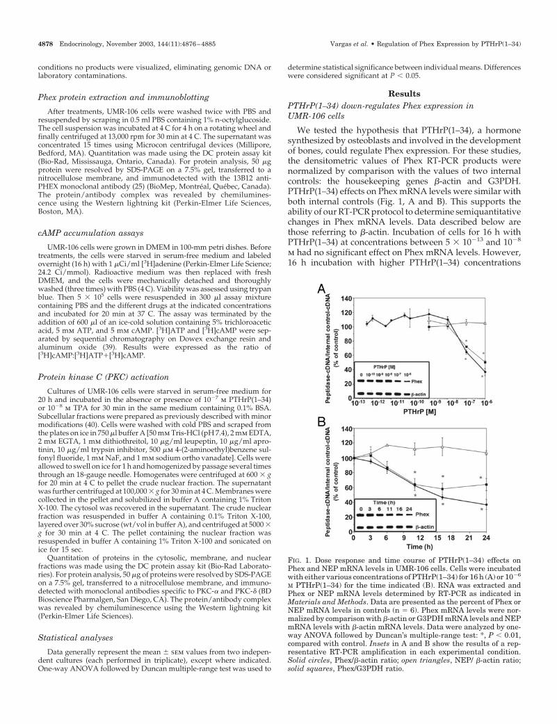
We tested the hypothesis that PTHrP(1–34), a hormonesynthesized by osteoblasts and involved in the developmentof bones, could regulate Phex expression. For these studies,the densitometric values of Phex RT-PCR products werenormalized by comparison with the values of two internalcontrols: the housekeeping genes �-actin and G3PDH.PTHrP(1–34) effects on Phex mRNA levels were similar withboth internal controls (Fig. 1, A and B). This supports theability of our RT-PCR protocol to determine semiquantitativechanges in Phex mRNA levels. Data described below arethose referring to �-actin. Incubation of cells for 16 h withPTHrP(1–34) at concentrations between 5 � 10�13 and 10�8
m had no significant effect on Phex mRNA levels. However,16 h incubation with higher PTHrP(1–34) concentrations
FIG. 1. Dose response and time course of PTHrP(1–34) effects onPhex and NEP mRNA levels in UMR-106 cells. Cells were incubatedwith either various concentrations of PTHrP(1–34) for 16 h (A) or 10�6
M PTHrP(1–34) for the time indicated (B). RNA was extracted andPhex or NEP mRNA levels determined by RT-PCR as indicated inMaterials and Methods. Data are presented as the percent of Phex orNEP mRNA levels in controls (n � 6). Phex mRNA levels were nor-malized by comparison with �-actin or G3PDH mRNA levels and NEPmRNA levels with �-actin mRNA levels. Data were analyzed by one-way ANOVA followed by Duncan’s multiple-range test: *, P � 0.01,compared with control. Insets in A and B show the results of a rep-resentative RT-PCR amplification in each experimental condition.Solid circles, Phex/�-actin ratio; open triangles, NEP/ �-actin ratio;solid squares, Phex/G3PDH ratio.
4878 Endocrinology, November 2003, 144(11):4876–4885 Vargas et al. • Regulation of Phex Expression by PTHrP(1–34)

(10�7 and 10�6 m) significantly decreased Phex mRNA levelsin a dose-dependent way (65 � 3% and 37 � 5% of control,respectively; n � 6) (Fig. 1A). The effect of PTHrP(1–34) (10�6
m) was time dependent, showing statistically significant de-crease at 11 h (63 � 3%, n � 6) and a maximal effect at 16–24h (47 � 3% and 37 � 7%, respectively, n � 6) (Fig. 1B). Underthe same treatment conditions, mRNA levels of NEP, anothermember of the M13 family of peptidases present in osteo-blasts (41), were not affected significantly, suggesting a spe-cific effect of the hormone on Phex mRNA (Fig. 1, A and B).
PTHrP(1–34) and PTH(1–34) effects on osteoblasts are me-diated by the common type-1 PTH/PTHrP receptor (42–44),suggesting that PTH(1–34) could also reduce Phex expres-sion. As expected, treatment of UMR-106 cells with rPTH(1–34), bPTH(1–34), or human PTH(1–34) (10�7 m) for 24 hsignificantly reduced Phex mRNA levels (Table 1).
To determine whether Phex protein levels were also af-fected by treatment of cells with PTHrP(1–34), UMR-106 cellstreated for 24 h with vehicle alone or with 10�7 m PTHrP(1–34) were extracted and the cell extracts analyzed by immu-noblotting. Consistent with the decrease in mRNA levels, thepresence of the hormone in the culture medium decreasedthe levels of Phex protein (Fig. 2).
Because PTHrP(1–34) and PTH(1–34) are known to acti-vate the PKC and PKA signaling pathways through the com-
mon type-1 PTH/PTHrP receptor (42–44), we next deter-mined the effects of activating independently either pathwayon Phex expression.
The PKC pathway up-regulates Phex expression in UMR-106 cells
To determine whether PKC regulates Phex mRNA levels,cells were incubated for 16 h with increasing concentrationsof TPA (from 10�10 to 10�6 m), a PKC activator. TPA induceda concentration-dependent increase in Phex mRNA levels,maximum (218 � 17%, n � 6) at 10�8 m (Fig. 3A). TPA (10�8
m) caused a time-dependent increase of Phex expression (Fig.3B). The maximal effect occurred at 16 h (238 � 18%, n � 6);after 24 h of TPA exposure, mRNA levels returned towardcontrol values (125 � 22%, n � 6). Incubation of cells for 16 hwith 4�-PDD (10�8 m), an inactive analog of TPA, had nosignificant effect on Phex mRNA levels (Table 2; experimentA). Treatment with TPA (10�8 m) for 16 h had no significanteffect on NEP mRNA levels (not shown). As for PTHrP(1–34),the effect of TPA added to the culture medium at a concen-tration of 10�8 m for 24 h was also observed at the proteinlevel (Fig. 2).
To demonstrate that PKC is involved in Phex mRNA up-regulation by TPA, cells were incubated for 16 h with the
TABLE 1. Effects of PTHrP(1–34), PTHrP(107–111), PTH(1–34),and PTH fragments on Phex mRNA levels in UMR-106 cells
Hormones Phex-cDNA/�-actin-cDNA (% of control)
Control 100 � 4PTHrP(1–34) 64 � 2a
rPTH(1–34) 59 � 2a
bPTH(1–34) 57 � 3a,b
bPTH(3–34) 90 � 8hPTH(1–34) 47 � 2a
hPTH(1–31) 37 � 4a
PTHrP(107–111) 98 � 9
Phex mRNA levels were determined by RT-PCR in cells incubatedfor 24 h in the absence of hormone (control) or in the presence of thehormones at concentration of 10�7 M. Data are presented as thepercentage of Phex mRNA levels in control (n � 6). Data were ana-lyzed by one-way ANOVA followed by Duncan’s multiple range test.
a P � 0.01 with respect to control; b P � 0.01 with respect tobPTH(3–34).
FIG. 2. PTHrP(1–34), forskolin, and TPA regulate Phex protein lev-els in UMR-106 cells. Cells were incubated for 24 h with eitherPTHrP(1–34) (10�7 M), TPA (10�8 M), forskolin (10�6 M), or vehiclealone. Proteins were extracted as described in Material and Methodsand 50 �g protein were resolved by SDS-PAGE on a 7.5% polyacryl-amide gel. Phex protein was revealed using an anti-PHEX antibody.
FIG. 3. Dose response and time course of TPA effects on Phex mRNAlevels in UMR-106 cells. Cells were incubated with either variousconcentrations of TPA for 16 h (A) or 10�8 M TPA for the time indicated(B). Phex mRNA level determination and data analysis were as de-scribed in Fig. 1 (n � 3). *, P � 0.01, compared with control. Insets inA and B show the results of a representative RT-PCR amplificationin each experimental condition.
Vargas et al. • Regulation of Phex Expression by PTHrP(1–34) Endocrinology, November 2003, 144(11):4876–4885 4879

PKC inhibitor bisindolylmaleimide � (10�6 m), alone or incombination with 10�8 m TPA. Bisindolylmaleimide � did notmodify significantly the basal Phex mRNA levels but inter-fered with the TPA-induced up-regulation (Table 2; exper-iment B). These data demonstrate that PKC activation up-regulates Phex expression.
The cAMP pathway down-regulates Phex expression inUMR-106 cells
The involvement of the cAMP pathway in Phex expressionwas evaluated with forskolin, an adenylate cyclase activator.Forskolin present for 16 h down-regulated Phex mRNA lev-els in a dose-dependent manner, with a maximal effect (47 �6%, n � 6) at 10�6 m (Fig. 4A). A time-dependent decreaseof Phex mRNA was observed when cells were incubatedwith 10�6 m forskolin (Fig. 4B). A significant decrease (75 �2%, n � 6) occurred at 6 h, and a maximal effect was observedat 16–24 h (32 � 5% and 20 � 5%, respectively, n � 6) (Fig.4B). Treatment of cells for 16 h with an inactive analog offorskolin, 1,9-dideoxyforskolin (10�6 m), did not change sig-nificantly Phex mRNA levels (Table 3; experiment A). For-skolin at 10�6 m had no significant effect on NEP mRNAlevels (not shown). Forskolin present at a concentration of10�6 m for 24 h also had an inhibitory effect at the proteinlevel (Fig. 2).
Adenylate cyclase activation augments intracellular cAMPlevels. Incubation of cells for 16 h with the permeable cAMPanalog (Bu)2cAMP (10�3 m) resulted in a decrease in PhexmRNA levels (Table 3; experiment B). PKA is one of the majortargets of cAMP. To determine whether (Bu)2cAMP regu-lated Phex mRNA levels through PKA activation, cells weretreated for 16 h with the PKA inhibitor H89, alone or incombination with (Bu)2cAMP. H89 (10�8 m) had no signif-icant effect on basal Phex mRNA levels but blocked the effectof 10�3 m (Bu)2cAMP (Table 3; experiment B), demonstrating
that increased cAMP levels down-regulate Phex expressionby PKA activation.
Interaction between the PKC and PKA pathways in theregulation of Phex mRNA levels in UMR-106 cells
Because diverse transduction pathways may interact atmany levels, we determined whether the PKC and PKApathways interact to regulate Phex mRNA levels. Cotreat-ment for 16 h with maximal effective doses of TPA (10�8 m)and increasing concentrations of forskolin (10�10 to 10�6 m)caused a dose-dependent decrease in Phex mRNA levels.Compared with TPA (10�8 m), significant effects of forskolinwere observed at 10�8 m (60 � 3%), 10�7 m (35 � 2%), and10�6 m (27 � 8%). These values are very similar to thoseobtained with forskolin alone (Fig. 5). This suggests that PKAactivation efficiently inhibits the effect of PKC on PhexmRNA levels.
PTHrP(1–34) does not regulate Phex mRNA levels throughPKC activation in UMR-106 cells
PTHrP(1–34) causes two peaks of PKC activation in ratosteosarcoma cells: one peak is observed at 10�12 to 10�11 mand the other at 10�9 to 10�8 m; a peak of adenylate cyclaseactivation is obtained at 10�7 m (42). Because a significant
TABLE 2. Effects of PKC pathway on Phex mRNA levels inUMR-106 cells
Treatments Phex-cDNA/�-actin-cDNA(% of control)
Experiment AControl 100 � 5 (6)TPA 200 � 13 (6)a
4�-PDD 104 � 7 (6)Experiment B
Control 100 � 8 (3)TPA 253 � 9 (3)a
Bisindolylmaleimide I 91 � 2 (3)TPA � bisindolylmaleimide I 140 � 8 (3)b
Experiment CControl 100 � 2 (3)PTHrP(1–34) 60 � 7 (3)a
Bisindolylmaleimide I 104 � 3 (3)PTHrP(1–34) �
bisindolylmaleimide I70 � 3 (3)a
Phex mRNA levels were determined by RT-PCR in cells incubatedfor 16 h in the absence of drug (control) or in the presence of 10�8 MTPA or 4�-PDD (experiment A), 10�6 M bisindolylmaleimide I aloneor in combination with TPA (experiment B), 10�7 M PTHrP(1–34), or10�6 M bisindolylmaleimide I alone or in combination with PTHrP(1–34) (experiment C). Data are presented and analyzed as in Table 1.
a P � 0.01 compared with their respective controls; b P � 0.01compared with TPA group.
FIG. 4. Dose response and time course of forskolin effects on PhexmRNA levels in UMR-106 cells. Cells were incubated with eithervarious concentrations of forskolin for 16 h (A) or 10�6 M forskolin forthe time indicated (B). Phex mRNA level determination and dataanalysis were as described in Fig. 1 (n � 3). *, P � 0.01, compared withcontrol. Insets in A and B show the results of a representative RT-PCRamplification in each experimental condition.
4880 Endocrinology, November 2003, 144(11):4876–4885 Vargas et al. • Regulation of Phex Expression by PTHrP(1–34)

effect of PTHrP(1–34) was not detected in the pico- or nano-molar range and its effect in the micromolar range was op-posite to that of PKC activation, the data suggested that PKCactivation by PTHrP(1–34) did not regulate Phex expression.This result may be explained by an interference of the PKApathway with the PKC pathway and/or by the fact that theisozyme(s) activated by TPA to regulate Phex expressionmay differ from that used by PTHrP(1–34). To test the pos-sibility that activation of the cAMP pathway may havemasked PKC activation, cells were treated with bPTH (3–34),an agonist of the type-1 PTH/PTHrP receptor that activatesPKC but not PKA (45–47). Incubation of cells for 16 h withbPTH (3–34) at concentrations between 10�10 and 10�7 m(data not shown) or for 24 h with 10�7 m bPTH (3–34) (Table
1) did not significantly alter Phex mRNA levels. This obser-vation suggests that PTHrP(1–34) (or PTH1–34) does notactivate the isozyme of PKC that increases Phex mRNA lev-els. UMR-106 cells are known to express several PKCisozymes (48), but only the PKC-� was reported activated byPTH(1–34) (49).
To test the effects of TPA and PTHrP(1–34) on activationof PKC-� and PKC-�, recruitment of these isozymes to theplasma membrane and nucleus, respectively, was deter-mined by immunoblotting of fractionated extracts from con-trol UMR-106 cells or cells treated with 10�8 m TPA or 10�7
m PTHrP(1–34). TPA but not PTHrP(1–34) provoked a dis-placement of PKC-� to the plasma membrane (Fig. 6A). Asreported previously (49), TPA provoked a decrease in PKC-�protein levels but no significant recruitment of this isozymeto the nucleus. In contrast, PTHrP(1–34) activated PKC-�isozyme (Fig. 6B). These results using PTHrP(1–34) as agonistof the type-1 PTH/PTHrP receptor are consistent with aprevious report by Erclic and Mitchell (49) showing activa-tion of PKC-� but not PKC-� by PTH(1–34), and clearly showthat PTHrP(1–34) and TPA do not activate the same PKCisozymes.
In osteoblasts and osteoblastic cell lines, including UMR-106 cells, PTHrP(107–111) activates PKC through a receptordifferent from the type-1 PTH/PTHrP receptor (50, 51).PTHrP(107–111) (10�7 m) did not significantly modify PhexmRNA levels at 24 h (Table 1). These results indicate thatactivation of an adequate PKC isozyme is necessary to up-regulate Phex mRNA levels. As expected, treatment of cellswith bisindolylmaleimide � (10�6 m) before incubation with10�7 m PTHrP(1–34) for 16 h did not prevent down-regulation of Phex mRNA levels (Table 2; experiment C).
TABLE 3. Effects of cAMP pathway on Phex mRNA levels inUMR-106 cells
Treatments Phex-cDNA/�-actin-cDNA(% of control)
Experiment AControl 100 � 5 (6)Forskolin 30 � 2 (6)a
1,9-dideoxyforskolin 87 � 5 (6)Experiment B
Control 100 � 12 (3)(Bu)2cAMP 70 � 4 (3)a
H89 100 � 6 (3)(Bu)2cAMP � H89 115 � 11 (3)b
Experiment CControl 100 � 2 (3)PTHrP(1–34) 60 � 7 (3)a
H89 109 � 4 (3)PTHrP(1–34) � H89 92 � 6 (3)c
Phex mRNA levels were determined by RT-PCR in cells incubatedfor 16 h in the absence of drug (control) or in the presence of 10�6 Mforskolin or 1,9-dideoxyforskolin (experiment A), 10�3 M (Bu)2cAMPor 10�8 M H89 alone or in combination with (Bu)2cAMP (experimentB), or with 10�7 M PTHrP(1– 34) or 10�8 M H89 alone or in combi-nation with PTHrP(1–34) (experiment C). Data are presented andanalyzed as in Table 1.
a P � 0.01 compared with respective controls; b P � 0.01 comparedwith (Bu)2cAMP group; c P � 0.01 compared with PTHrP(1–34) group.
FIG. 5. Forskolin blocks TPA effect on Phex mRNA levels in UMR-106 cells. Cells were treated with increasing concentrations of fors-kolin alone (open circles) or in combination with 10�8 M TPA (solidcircles) for 16 h. Phex mRNA level determination and data analysiswere as described in Fig. 1. Dashed line shows the levels of Phexexpression in cells treated with 10�8 M TPA. Numbers in parenthesesindicate the number of determinations for each point (solid circles).*, P � 0.01 with respect to 10�8 M TPA alone.
FIG. 6. Activation of PKC isozymes in UMR-106 cells by TPA andPTHrP(1–34). UMR-106 control cells or cells treated with 10�7 MPTHrP(1–34) or 10�8 M TPA were fractionated and subcellular frac-tions analyzed by immunoblotting with an anti-PKC-� antibody (A)or an anti-PKC-� antibody (B).
Vargas et al. • Regulation of Phex Expression by PTHrP(1–34) Endocrinology, November 2003, 144(11):4876–4885 4881
PTHrP(1–34) down-regulates Phex mRNA levels throughthe PKA pathway in UMR-106 cells
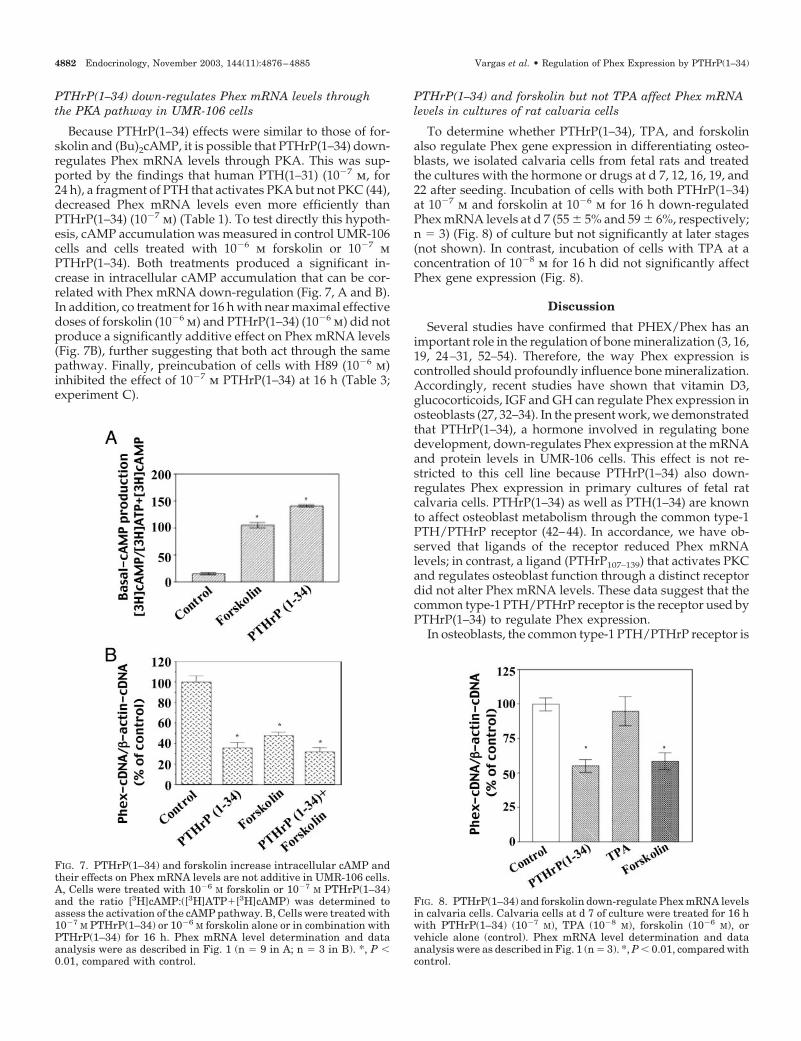
Because PTHrP(1–34) effects were similar to those of for-skolin and (Bu)2cAMP, it is possible that PTHrP(1–34) down-regulates Phex mRNA levels through PKA. This was sup-ported by the findings that human PTH(1–31) (10�7 m, for24 h), a fragment of PTH that activates PKA but not PKC (44),decreased Phex mRNA levels even more efficiently thanPTHrP(1–34) (10�7 m) (Table 1). To test directly this hypoth-esis, cAMP accumulation was measured in control UMR-106cells and cells treated with 10�6 m forskolin or 10�7 mPTHrP(1–34). Both treatments produced a significant in-crease in intracellular cAMP accumulation that can be cor-related with Phex mRNA down-regulation (Fig. 7, A and B).In addition, co treatment for 16 h with near maximal effectivedoses of forskolin (10�6 m) and PTHrP(1–34) (10�6 m) did notproduce a significantly additive effect on Phex mRNA levels(Fig. 7B), further suggesting that both act through the samepathway. Finally, preincubation of cells with H89 (10�6 m)inhibited the effect of 10�7 m PTHrP(1–34) at 16 h (Table 3;experiment C).
PTHrP(1–34) and forskolin but not TPA affect Phex mRNAlevels in cultures of rat calvaria cells
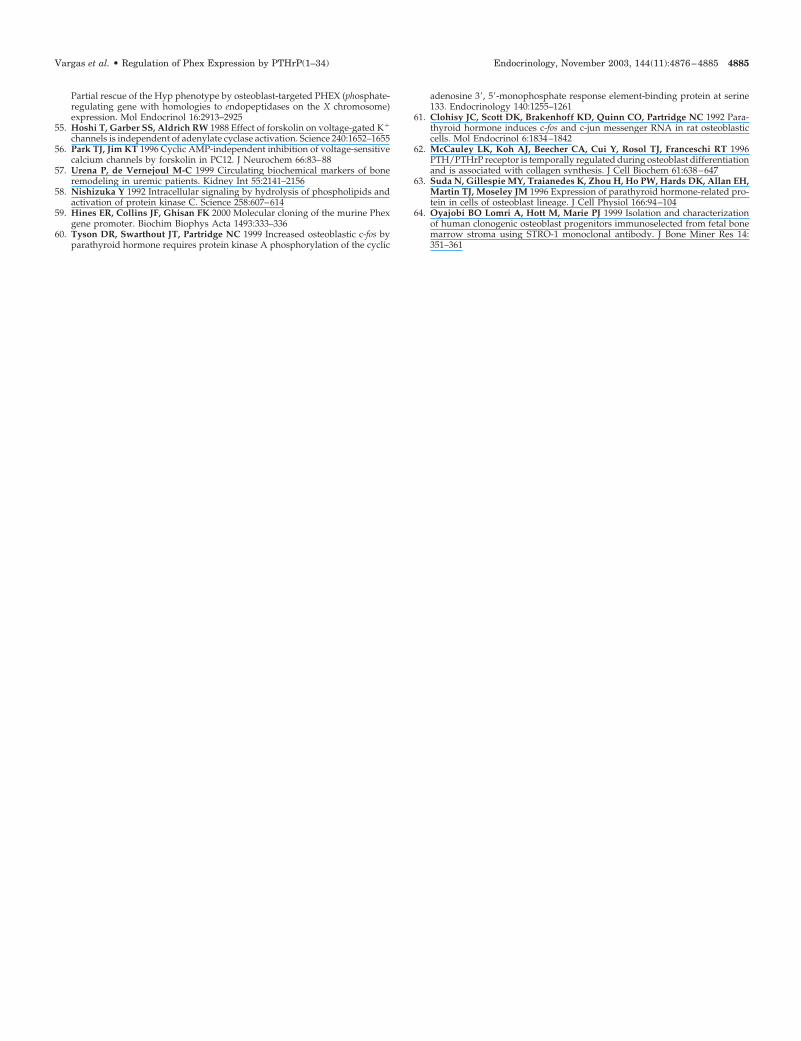
To determine whether PTHrP(1–34), TPA, and forskolinalso regulate Phex gene expression in differentiating osteo-blasts, we isolated calvaria cells from fetal rats and treatedthe cultures with the hormone or drugs at d 7, 12, 16, 19, and22 after seeding. Incubation of cells with both PTHrP(1–34)at 10�7 m and forskolin at 10�6 m for 16 h down-regulatedPhex mRNA levels at d 7 (55 � 5% and 59 � 6%, respectively;n � 3) (Fig. 8) of culture but not significantly at later stages(not shown). In contrast, incubation of cells with TPA at aconcentration of 10�8 m for 16 h did not significantly affectPhex gene expression (Fig. 8).
Discussion
Several studies have confirmed that PHEX/Phex has animportant role in the regulation of bone mineralization (3, 16,19, 24–31, 52–54). Therefore, the way Phex expression iscontrolled should profoundly influence bone mineralization.Accordingly, recent studies have shown that vitamin D3,glucocorticoids, IGF and GH can regulate Phex expression inosteoblasts (27, 32–34). In the present work, we demonstratedthat PTHrP(1–34), a hormone involved in regulating bonedevelopment, down-regulates Phex expression at the mRNAand protein levels in UMR-106 cells. This effect is not re-stricted to this cell line because PTHrP(1–34) also down-regulates Phex expression in primary cultures of fetal ratcalvaria cells. PTHrP(1–34) as well as PTH(1–34) are knownto affect osteoblast metabolism through the common type-1PTH/PTHrP receptor (42–44). In accordance, we have ob-served that ligands of the receptor reduced Phex mRNAlevels; in contrast, a ligand (PTHrP107–139) that activates PKCand regulates osteoblast function through a distinct receptordid not alter Phex mRNA levels. These data suggest that thecommon type-1 PTH/PTHrP receptor is the receptor used byPTHrP(1–34) to regulate Phex expression.
In osteoblasts, the common type-1 PTH/PTHrP receptor is
FIG. 7. PTHrP(1–34) and forskolin increase intracellular cAMP andtheir effects on Phex mRNA levels are not additive in UMR-106 cells.A, Cells were treated with 10�6 M forskolin or 10�7 M PTHrP(1–34)and the ratio [3H]cAMP:([3H]ATP�[3H]cAMP) was determined toassess the activation of the cAMP pathway. B, Cells were treated with10�7 M PTHrP(1–34) or 10�6 M forskolin alone or in combination withPTHrP(1–34) for 16 h. Phex mRNA level determination and dataanalysis were as described in Fig. 1 (n � 9 in A; n � 3 in B). *, P �0.01, compared with control.
FIG. 8. PTHrP(1–34) and forskolin down-regulate Phex mRNA levelsin calvaria cells. Calvaria cells at d 7 of culture were treated for 16 hwith PTHrP(1–34) (10�7 M), TPA (10�8 M), forskolin (10�6 M), orvehicle alone (control). Phex mRNA level determination and dataanalysis were as described in Fig. 1 (n � 3). *, P � 0.01, compared withcontrol.
4882 Endocrinology, November 2003, 144(11):4876–4885 Vargas et al. • Regulation of Phex Expression by PTHrP(1–34)
coupled to Gs and Gq proteins that activate the PKA and PKCtransduction pathways, respectively. Activation of PKC ac-tivity presents two peaks: the first is reached at 10�12 to 10�11
m of ligand and the second at 10�9 to 10�8 m (42–43). Incontrast, 10�7 m of ligand is needed to observe a maximalincrease in intracellular cAMP levels and PKA activation(42–43). The following evidences suggest that PTHrP(1–34)activates PKA: 1) PTHrP(1–34) down-regulated Phex expres-sion only in the 10�7 to 10�6 m range; 2) human PTH(1–31),a ligand of the common type-1 PTH/PTHrP receptor thatactivates PKA but not PKC, also decreased Phex mRNAlevels; 3) forskolin and (Bu)2cAMP mimicked PTHrP(1–34)effects on Phex regulation but 1–9-dideoxyforskolin, an an-alog that mimics the adenylate-cyclase-independent effectsof forskolin (55, 56), had no effect on Phex expression; 4) Phexdown-regulation correlated with increased intracellularcAMP accumulation provoked by PTHrP(1–34) and forsko-lin; 5) cotreatment with near maximal effective doses of for-skolin and PTHrP(1–34) did not produce an additive effecton Phex mRNA levels, indicating that both factors may actby the same pathway; and 6) finally, the target of increasedintracellular cAMP accumulation is probably PKA because aspecific inhibitor of PKA, H89, blocked the effects ofPTHrP(1–34) and (Bu)2cAMP. All these data indicate thatPTHrP(1–34) uses the cAMP pathway to down-regulate Phexexpression through PKA activation.
We observed down-regulation of Phex expression only athigh PTHrP(1–34) concentrations (10�7 and 10�6 m). Thissuggests that PTHrP(1–34), produced locally by osteoblasts,acts in a paracrine/autocrine pathway. It probably also ex-cludes a role for PTH(1–34), produced by the parathyroidglands, in regulating Phex gene expression, because this hor-mone is found circulating at much lower levels (approxi-mately 10�12 m) (57).
We also observed that incubation of UMR-106 cells with aphorbol ester that activates PKC (TPA) increased Phex ex-pression. However, when PTHrP(1–34) was used in the con-centration range (5 � 10�13 to 10�9 m) that activates PKC butnot the cAMP pathway, Phex mRNA levels were not mod-ified. Similar results were obtained when cells were incu-bated with bPTH(3–34) that binds the common type-1 PTH/PTHrP receptor and activates the PKC but not the cAMPpathway. One possibility to explain this discrepancy is topostulate that, because PKC is a superfamily of isozymeswith different regulatory properties (58) and because normalosteoblasts and many osteoblastic cell lines, including UMR-106 cells, express several isozymes (48), the PKC isozyme(s)that PTHrP(1–34) or PTH(1–34) activates in UMR-106 cells isdistinct from the isozyme(s) induced by TPA that regulatesPhex expression. Our results indicate that in UMR-106 cellsTPA and PTHrP(1–34) do indeed activate different PKCisozymes: the � and � isozymes, respectively. These findingssuggest that PKC-� can regulate Phex gene expression,whereas PKC-�, which is coupled to the type-1 PTH/PTHrPreceptor, has no effect on Phex expression.
In osteoblasts, downstream signaling from PKA and PKCinvolves transcription factors that regulate gene expression.The murine Phex gene promoter has several potential cis-acting elements including binding sites for cAMP responseelement-binding protein, activator protein 1 (AP1), glucocor-
ticoid receptor, and estrogen receptor (59). Transactivation ofcAMP response element-binding protein induced by PTH(1–34) is mediated by PKA but not PKC or calcium in osteo-blastic cells (60, 61). PTH(1–34) also regulates the expressionof c-fos and c-jun, components of the AP1 transcription factor(60). Our results indicate that cAMP response and/or AP1elements may mediate the effects on Phex expression ofPTHrP(1–34) and TPA, respectively.
Interestingly, the effects of PTHrP(1–34) on primary cul-tures of rat calvaria cells were restricted to an early stage ofculture: Inhibition of Phex mRNA levels was observed at d7 but not later. This observation is best explained by a down-regulation of the type-1 PTH/PTHrP receptor as cellsprogress toward final differentiation. This phenomenon hasbeen well documented in primary cultures of rat calvariacells (62). In addition, higher levels of PTHrP expression arefound in less differentiated cells (63, 64). In contrast to theexpression patterns of PTHrP and its receptor, we observedthat Phex expression in rat calvaria cells is weak at d 7, startsto increase around d 10, and reaches a maximum at d 15,corresponding with mineralization (results not shown).Thus, there is an inverse correlation between the expressionof Phex and that of PTHrP(1–34) and its receptor. We suggestthat PTHrP(1–34) acting in an autocrine or paracrine waydown-regulates Phex expression in osteoprogenitor cells andpreosteoblasts.
We see no effect of TPA on Phex expression in primarycultures of rat calvaria cells. This can be explained by theabsence in these cells of the isozyme(s) responsible for Phexgene activation. Alternatively, the presence in these culturesof autocrine/paracrine factor(s) activating the PKA pathwaymay mask the effect of TPA, as we observed in this studywhen increasing concentrations of forskolin were added toTPA-stimulated cells.
In conclusion, the work presented in this paper demon-strates that PTHrP(1–34) by binding to the common type-1PTH/PTHrP receptor down-regulates Phex expressionthrough activation of the PKA transduction pathway. Thisrole of PTHrP(1–34) may be important for the timing of Phexexpression in differentiating osteoprogenitor cells. It wouldbe interesting to determine whether other hormones thatactivate PKA or PKC and regulate the mineralization of bonealso regulate the expression of Phex.
Acknowledgments
We are grateful to Demian Barbas for his help in measuring cAMPlevels.
Received February 25, 2003. Accepted July 28, 2003.Address all correspondence and requests for reprints to: Guy Boileau,
Departement de Biochimie, Faculte de Medecine, Universite de Mon-treal, C.P. 6128, Succ. Centre-Ville Montreal, Quebec, Canada H3C 3J7.E-mail: [email protected].
This work was supported by Grants NRF13052 from the CanadianInstitutes of Health Research and ER-73358 from Fonds pour la Forma-tion des Chercheurs et l’Aide a la Recherche (FCAR).
References
1. The HYP Consortium 1995 A gene (PEX) with homologies to endopeptidasesis mutated in patients with X-linked hypophosphatemic rickets. Nat Genet11:130–136
2. Tenenhouse HS, Econs MJ 2001 Mendelian hypophosphatemias. In: Scriver
Vargas et al. • Regulation of Phex Expression by PTHrP(1–34) Endocrinology, November 2003, 144(11):4876–4885 4883
CR, Beaudet AL, Sly WS, Valle D, eds. The metabolic and molecular bases ofinherited disease. Chap 197. New York: McGraw Hill Book Co.; 5039–5067
3. Beck L, Soumounou Y, Martel J, Krishamurthy G, Gauthier C, Goodyer CGTenenhouse HS 1997 Pex/PEX tissue distribution and evidence for a deletionin the 3� region of the Pex gene in X-linked hypophosphatemic mice. J ClinInvest 99:1200–1209
4. Eicher EM, Southard JL, Scriver CR, Glorieux FH 1976 Hypophosphatemia:mouse model for human familial hypophosphatemic (vitamin D resistant)rickets. Proc Natl Acad Sci USA 73:4667–4671
5. Tenenhouse HS, Werner A, Biber J, Ma S, Martel J, Roy S, Murer H 1994Renal Na�-phosphate cotransporter in murine X-linked hyphophosphatemicrickets. Molecular characterization. J Clin Invest 93:671–676
6. Tenenhouse HS, Roy S, Martel J, Gauthier C 1998 Differential expression,abundance and regulation of Na�-phosphate cotransporter genes in murinekidney. Am J Physiol 268:1062–1069
7. Meyer Jr RA, Meyer MH, Gray RW 1989 Parabiosis suggests a humoral factoris involved in X-linked hypophosphatemia in mice. J Bone Miner Res 4:493–500
8. Nesbitt T, Coffman T, Griffiths R, Drezner M 1992 Cross-transplantation ofkidneys in normal and Hyp mice: evidence that the Hyp mouse phenotype isunrelated to an intrinsic renal defect. J Clin Invest 89:1453–1459
9. Econs MJ, Drezner MK 1994 Tumor-induced osteomalacia: unveiling a newhormone. N Engl J Med 330:1679–1681
10. Lajeunesse D, Meyer Jr RA, Hamel L 1996 Direct demonstration of a hu-morally mediated inhibition of renal phosphate transport in the Hyp mouse.Kidney Int 50:1531–1538
11. Nesbitt T, Fujiwara I, Thomas R, Xiao Z-S, Quarles LD, Drezner MK 1999Coordinated maturational regulation of PHEX and renal phosphate transportinhibitory activity: evidence for the pathophysiology role of PHEX in X-linkedhypophosphatemia. J Bone Miner Res 14:2027–2035
12. Schiavi SC, Moe OW 2002 Phosphatonins: a new class of phosphate-regu-lating proteins. Curr Opin Nephrol Hypertens 11:423–430
13. ShimadaT, Mizutani S, Muto T, Yoneya T, Hino R, Takeda S, Takeuchi Y,Fujita T, Fukumoto S, Yamashita T 2001 Cloning and characterization ofFGF23 as a causative factor of tumor induced osteomalacia. Proc Natl Acad SciUSA 98:6500–6505
14. Rowe PNS, De Zoysa PA, Dong R, Wang HR, White KE, Econs MJ, OudetCL 2000 MEPE, a new gene expressed in bone marrow and tumors causingosteomalacia. Genomics 67:54–68
15. Jan de Beur SM, Finnigan RB, Vassiliadis J, Cook B, Barberio D, Estes S,Manavalan P, Petroziello J, Madden SL, Cho JY, Kumar R, Levine MA,Schiavi SC 2002 Tumors associated with oncogenic osteomalacia expressgenes important in bone and mineral metabolism. J Bone Miner Res 17:1102–1110
16. Du L, Desbarats M, Viel J, Glorieux FH, Cawthorn C, Ecarot B 1996 cDNAcloning of the murine Pex gene implicated in X-linked hypophosphatemia andevidence for expression in bone. Genomics 36:22–28
17. Strom TM, Francis F, Lorenz B, Boddrich A, Econs MJ, Lehrach H, MeitingerT 1997 Pex gene deletions in Gy and Hyp mice provide mouse models forX-linked hypophosphatemia. Hum Mol Genet 6:165–171
18. Grieff M, Mumm S, Waeltz P, Mazarella R, Whyte MP, Thakker RV, Schless-inger D 1997 Expression and cloning of the human X-linked hypophos-phatemia gene cDNA. Biochem Biophys Res Commun 231:635–639
19. Guo R, Quarles LD 1997 Cloning and sequencing of human PEX from a bonecDNA library: evidence for its developmental stage-specific regulation inosteoblasts. J Bone Miner Res 12:1009–1017
20. Lipman ML, Panda D, Bennety HPJ, Henderson JE, Shane E, Shen Y, Goltz-man D, Karaplis AC 1998 Cloning of human Pex cDNA. Expression, subcel-lular localization, and endopeptidase activity. J Biol Chem 273:13729–13737
21. Barret JA, Rawlings ND, Woessner JF 2000 Handbook of proteolytic enzymes.London: Academic Press
22. Roques BP, Noble F, Dauge V, Fournie-Zaluski M-C, Beaumont A 1993Neutral endopeptidase 24.11: structure, inhibition, and experimental and clin-ical pharmacology. Pharmacol Rev 45:87–146
23. Turner AJ 1997 Endothelin converting enzyme. In: Kenny AJ, Boustead CM,eds. Cell-surface peptidases in health and disease. Oxford, UK: BIOS ScientificPublishers Ltd.; 137–153
24. Ruchon AF, Marcinkiewicz M, Siegfried G, Tenenhouse HS, DesGroseillersL, Crine P, Boileau G 1998 Pex mRNA is localized in developing mouseosteoblasts and odontoblasts. J Histochem Cytochem 46:459–468
25. Ruchon AF, Tenenhouse HS, Marcinkiewicz M, Siegfried G, Aubin JE,DesGroseillers L, Crine P, Boileau G 2000 Developmental expression andtissue distribution of Phex protein: effect of the hyp mutation and relationshipto bone markers. J Bone Miner Res 15:1440–1450
26. Thompson DL, Sabbagh Y, Tenenhouse HS, Roche PC, Drezner MK, Salis-bury JL, Grande JP, Poeschla EM, Kumar R 2002 Ontogeny of Phex/PHEXprotein expression in mouse embryo and subcellular localization in osteo-blasts. J Bone Miner Res 17:311–320
27. Ecarot B, Desbrats M 1999 1, 25-(OH)2D3 down-regulates expression of Phex,a marker of the mature osteoblast. Endocrinology 140:1192–1199
28. Ecarot-Charrier B, Desbarats F, Travers R, Desbarats M, Bouchard F, HinekA 1988 Defective bone formation by transplanted Hyp mouse bone cells intonormal mice. Endocrinology 123:768–773
29. Ecarot B, Glorieux F, Desbarats M, Travers R, Labelle L 1992 Defect boneformation by Hyp mouse bone cells transplanted into normal mice: evidencein a favor of an intrinsic osteoblast defect. J Bone Miner Res 7:215–220
30. Xiao ZS, Crenshaw M, Guo R, Nesbitt T, Drezner MK, Quarles LD 1998Intrinsic mineralization defect in Hyp mouse osteoblasts. Am J Physiol En-docrinol Metab 275:E700–E709
31. Miao D, Bai X, Panda D, McKee M, Karaplis A, Goltzman D 2001 Osteo-malacia in Hyp mice is associated with abnormal Phex expression and withaltered bone matrix protein expression and deposition. Endocrinology 142:926–939
32. Hines ER, Collins JF, Jones M, Serey SH, Ghishan FK 2002 Glucocorticoidregulation of the murine PHEX gene. Am J Physiol Renal Physiol 283:F356–F363
33. Zoidis E, Gosteli-Peter M, Ghirlanda-Keller C, Meinel L, Zapf J, Schmid C2002 IGF-I and GH stimulate Phex mRNA expression in lungs and bones and1, 25-dihydroxyvitamin D3 production in hypophysectomized rats. Eur J En-docrinol 146:97–105
34. Zoidis E, Zapf J, Schmid C 2000 Phex cDNA cloning from rat bone and studieson Phex mRNA expression: tissue specificity, age dependency, and regulationby insulin-like growth factor (IGF) in vivo. Mol Cell Endocrinol 168:41–55
35. Partridge NC, Alcorn D, Michelangeli VP, Ryan G, Martin TJ 1983 Mor-phological and biochemical characterization of four clonal osteogenic sarcomacell lines of rat origin. Cancer Res 43:4308–4314
36. Inoue D, Matsumoto T 2000 Parathyroid hormone-related peptide and bone:pathological and physiological aspects. Biomed Pharmacother 54(Suppl 1):32–41
37. Karaplis AC 2001 PTHrP: novel roles in skeletal biology. Curr Pharm Des7:655–670
38. Bonnelye E, Merdad L, Kung V, Aubin JE 2001 The orphan nuclear estrogenreceptor-related receptor � (ERR�) is expressed throughout osteoblast differ-entiation and regulates bone formation in vitro. J Cell Biol 153:971–983
39. Pineyro G, Azzi M, De Lean A, Schiller P, Bouvier M 2001 Short-terminverse-agonist treatment induces reciprocal changes in �-opioid agonist andinverse-agonist binding capacity. Mol Pharmacol 60:816–827
40. Hsu SL, Chou YH, Yin SC, Liu JY 1998 Differential effects of phorbol ester ongrowth and protein kinase C isozymes regulation in human hepatoma Hep3Bcells. Biochem J 333:57–64
41. Ruchon AF, Marcinkiewicz M, Ellefsen K, Aubin JE, Crine P, Boileau G 2000Cellular localization of neprilysin in mouse bone tissue and putative role inhydrolysis of osteogenic peptides. J Bone Miner Res 15:1266–1273
42. Gagnon L, Jouishomme H, Whitfield JF, Durkin JP, Maclean S, NeugebauerW, Willick G, Rixon RH, Chakravarthy B 1993 Protein kinase C-activatingdomains of parathyroid hormone-related protein. J Bone Miner Res 3:497–503
43. Jouishomme H, Whitfield JF, Chakravarthy B, Durkin JP, Gagnon L, IsaacsRJ, MacLean S, Neugebauer W, Willick G, Rixon RH 1992 The proteinkinase-C activation domain of the parathyroid hormone. Endocrinology 130:53–60
44. Blind E, Raue F, Knappe V, Schroth J, Ziegler R 1993 Cyclic AMP formationin rat bone and kidney cells is stimulated equally by parathyroid hormone-related protein (PTHrP) 1–34 and PTH 1–34. Exp Clin Endocrinol 101:150–155
45. Chakravarthy BR, Durkin JP, Rixon RH, Whitfield JF 1990 Parathyroidhormone fragment (3–34) stimulates protein kinase C (PKC) activity in ratosteosarcoma and murine T-lymphoma cells. Biochem Biophys Res Commun171:1105–1110
46. Fujimore A, Cheng SL, Avioli LV, Civitelli R 1992 Structure-function rela-tionship of parathyroid hormone: activation of phospholipase C, protein ki-nase A and C in osteosarcoma cells. Endocrinology 130:29–36
47. Jouishomme H, Whitfield JF, Gagnon L, Maclean S, Isaacs RJ, ChakravarthyB, Durkin JP, Neugebauer W, Willick G, Rixon RH 1994 Further definitionof the protein kinase C activation domain of the parathyroid hormone. J BoneMiner Res 9:943–949
48. Sanders JL, Stern PH 1996 Expression and phorbol ester-induced down-regulation of protein kinase C isozymes in osteoblasts. J Bone Miner Res11:1862–1872
49. Erclik MS, Mitchell J 2002 The role of protein kinase C-� in PTH stimulationof IGF-binding protein-5 mRNA in UMR-106–01 cells. Am J Physiol Endo-crinol Metab 282:E534–E541
50. Valin A, Garcia-Ocana A, de Miguel F, Sarasa J. L, Esbrit P 1997 Antipro-liferative effect of the C-terminal fragments of parathyroid hormone-relatedprotein, PTHrP-(107–111) and (107–139), on osteoblastic osteosarcoma cells.J Cell Physiol 170:209–215
51. Valin A, de Miguel F, Garcia-Ocana A, Esbrit P 1999 Parathyroid hormone-related protein (107–111) decreased alkaline phosphatase in osteoblastic os-teosarcoma cells UMR 106 by a protein kinase C-dependent pathway. CalcifTissue Int 65:148–151
52. Shih NR, Jo OD, Yanagawa N 2002 Effects of PHEX antisense in humanosteoblast cells. J Am Soc Nephrol 171:394–399
53. Liu S, Guo R, Tu Q, Quarles LD 2001 Overexpression of Phex in osteoblastsfails to rescue the Hyp mouse phenotype. J Biol Chem 277:3686–3697
54. Bai X, Miao D, Panda D, Grady S, McKee MD, Goltzman D, Karaplis AC 2002
4884 Endocrinology, November 2003, 144(11):4876–4885 Vargas et al. • Regulation of Phex Expression by PTHrP(1–34)
Partial rescue of the Hyp phenotype by osteoblast-targeted PHEX (phosphate-regulating gene with homologies to endopeptidases on the X chromosome)expression. Mol Endocrinol 16:2913–2925
55. Hoshi T, Garber SS, Aldrich RW 1988 Effect of forskolin on voltage-gated K�
channels is independent of adenylate cyclase activation. Science 240:1652–165556. Park TJ, Jim KT 1996 Cyclic AMP-independent inhibition of voltage-sensitive
calcium channels by forskolin in PC12. J Neurochem 66:83–8857. Urena P, de Vernejoul M-C 1999 Circulating biochemical markers of bone
remodeling in uremic patients. Kidney Int 55:2141–215658. Nishizuka Y 1992 Intracellular signaling by hydrolysis of phospholipids and
activation of protein kinase C. Science 258:607–61459. Hines ER, Collins JF, Ghisan FK 2000 Molecular cloning of the murine Phex
gene promoter. Biochim Biophys Acta 1493:333–33660. Tyson DR, Swarthout JT, Partridge NC 1999 Increased osteoblastic c-fos by
parathyroid hormone requires protein kinase A phosphorylation of the cyclic
adenosine 3�, 5�-monophosphate response element-binding protein at serine133. Endocrinology 140:1255–1261
61. Clohisy JC, Scott DK, Brakenhoff KD, Quinn CO, Partridge NC 1992 Para-thyroid hormone induces c-fos and c-jun messenger RNA in rat osteoblasticcells. Mol Endocrinol 6:1834–1842
62. McCauley LK, Koh AJ, Beecher CA, Cui Y, Rosol TJ, Franceschi RT 1996PTH/PTHrP receptor is temporally regulated during osteoblast differentiationand is associated with collagen synthesis. J Cell Biochem 61:638–647
63. Suda N, Gillespie MY, Traianedes K, Zhou H, Ho PW, Hards DK, Allan EH,Martin TJ, Moseley JM 1996 Expression of parathyroid hormone-related pro-tein in cells of osteoblast lineage. J Cell Physiol 166:94–104
64. Oyajobi BO Lomri A, Hott M, Marie PJ 1999 Isolation and characterizationof human clonogenic osteoblast progenitors immunoselected from fetal bonemarrow stroma using STRO-1 monoclonal antibody. J Bone Miner Res 14:351–361
Vargas et al. • Regulation of Phex Expression by PTHrP(1–34) Endocrinology, November 2003, 144(11):4876–4885 4885