Immunohistochemical study of nuclear changes associated with male germ cell death and spermiogenesis
Life Science Journal 2013;10(4) http://www.lifesciencesite.com
2496
Nutritional Impact of Cauliflower and Broccoli against Development of Early Vascular Lesions Induced by Animal Fat Diet (Biochemical and immunohistochemical studies)
Amany, A. Salem1, Faten. F. Mohammed2 and Samiha, A. Alloush1
1Food Technology Research Institute, Agricultural Research Center, Ministry of Agriculture, Giza, Egypt 2Department of Pathology, Faculty of Veterinary Medicine, Cairo University, Giza, Egypt
[email protected], [email protected]
Abstract: This study was performed to evaluate the nutritional effect of cauliflower and broccoli against development vascular histological alterations and related changes in biochemical parameters induced by feeding of rats on animal fat diet. The 24 adult male rats (105±5g) were randomly divided into 4 groups, negative control, animal fat diet (positive control), animal fat diet supplemented either with 5% fiber of cauliflower or broccoli for third and fourth group respectively. Estimation of serum glucose, lipid profile, liver and kidney function were performed and histopathological and immunohistochemical studies on blood vessels, liver and kidney were carried out. Results revealed that cauliflower improves the lipid profile than broccoli. Both plants significantly lowering of serum total lipid, total cholesterol, HDL-C, LDL-C and VLDL-C compared with positive control, improvement of liver and kidney function were achieved in the cauliflower and broccoli supplemented dietary groups. Histopathological examination showed alleviation of vascular histopathological alterations that were observed in animal fat diet fed group including focal intimal thickening and vacuolation of tunica media with strong positive expression of vascular endothelial growth factor (VEGF) in smooth muscle cells comprising vascular tunica media these alterations were markedly ameliorated in cauliflower then broccoli group. Conclusion: ameliorative effect of cauliflower and broccoli against development of changes in lipid profile and vascular pathology were attributed in partly to lowering the serum level of total lipid, total cholesterol, HDL-C, LDL-C and VLDL-C and decrease the expression of VEGF in smooth muscle cells of blood vessel wall. [Amany, A. Salem, Faten. F. Mohammed and Samiha, A. Alloush. Nutritional Impact of Cauliflower and Broccoli against Development of Early Vascular Lesions Induced by Animal Fat Diet (Biochemical and immunohistochemical studies). Life Sci J 2013; 10(4): 2496-2509]. (ISSN: 1097-8135). http://www.lifesciencesite.com. 334 Key words: Broccoli, cauliflower, fiber, lipid profile, vascular histopathology, vascular endothelial growth factor expression (VEGF). 1-Introduction
Broccoli sprouts are rich source of health promoting compounds including glucosinalates and isothiocyanates (Jeffery and Araya, 2009). Animal studies have reported that young broccoli sprout improved lipid profiles (Lee et al., 2009). A key bioactive component in broccoli sprout is sulforaphane (1- isothiocyanate-4-methylsulfinylbutane) which is the most potent inducer of the endogenous antioxidant defense (Riedl et al., 2009). Recently a phase 1 study revealed that consuming fresh broccoli sprouts for one week increased HDL-C, and decreased total cholesterol, LDL-C and some biomarkers of oxidative stress (Murashima et al., 2004).
Cauliflower belongs to the most valuable vegetables because it has high nutritive value, is tasty and easy to prepare. Low calorie value is it’s another advantage. Cauliflower is considered as one of the highest antioxidative activity among plants (Grajek, 2003). A number of experiments indicate that vegetables added to laboratory animals’ diet had positive effects on lipid profile. Antioxidants from
vegetables were shown to lower atherogenic cholesterol fraction (LDL +VLDL) concomitantly increasing HDL fraction, which is believed to be beneficial for the prevention of cardiovascular diseases (Gorinstein et al., 2006). Rasmussen et al. (2005) suggested that polyphenols are slowing down metabolism of cholesterol in the liver, reducing excretion of VLDL to blood, which leads to lower cholesterol accumulation in the aorta.
Vegetables are a good source of dietary fibers, phytonutrients, provitamins, antioxidants, polyphenols and minerals. Kahlon et al. (2007b) concluded that, relative to cholestyramine, the in vitro bile acid binding on an equal dry mater (DM) and total dietary fiber (TDF) basis were for cauliflower (1% and 2%) and broccoli 4% as reported by Kahlon et al. (2007a). Relative bile acid binding on DM basis may indicate their relative health-promoting potential. The differences in bile acid binding between the fresh colorful vegetables tested may relate to the variability in their phytonutrients (antioxidants, hydroxycinnamic acid, glucosinolates, isothiocyanates, flavonoids, polyphenols, and micro-elements), non-protein
Life Science Journal 2013;10(4) http://www.lifesciencesite.com
2497
composition and structure, hydrophobicity of undigested fractions, anionic or cationic nature of the metabolites produced during digestion or their interaction with active binding sites.
Western-style diets which are low in dietary fiber and high in saturated fatty acids, are associated with increased risk of cardiovascular disease, diabetes and obesity (Hosomi et al., 2009). Several studies have demonstrated the hypocholesterolemic and hypotriglyceridemic effects of polyunsaturated fatty acids (PUFAs) (Russo, 2009) and soluble dietary fiber (Rideout et al., 2008). There are increasing efforts to identify functional foods or individual dietary components that may prevent or alleviate chronic diseases. As functional foods and individual dietary components are consumed as part of a mixed diet, and not in isolation, it is necessary to understand the combined effects of different food components, within diets on health outcomes.
Consumption of dietary fiber from cereals and fruits was inversely associated with risk of coronary heart disease. For every 10g/day increase in total fiber, there was a reduction of 14% in coronary events and 27 % in coronary deaths. For every 10g/day increment of fruit fiber, there was 16 % decrease in coronary events and 30 percent in deaths. Food and Drug Administration (FDA) authorized that foods meeting specific compositional requirements and containing 0.75 g to 1.7g of soluble fiber per serving can reduce the risk of coronary heart disease (Position of the American Dietetic Association, 2008). Broccoli fiber acts via its physical properties to reduce enterohepatic bile acid recycling and intestinal lipid absorption and increase luminal binding of bile acids, resulting in increased fecal bile acid excretion (Mandimika et al., 2012).
Accordingly, this study was a trial to evaluate the nutritional effect of broccoli and cauliflower on blood glucose, lipid profiles, liver and kidney function in association of feeding on animal fat diet as well as studying the possible ameliorative effect of broccoli and cauliflower against development of early histopathological vascular alterations and related hepatic and renal induced pathology. 2-Material and methods 2-.1.Plant materials preparation:-
Broccoli (Brassica olearaceassp.) and cauliflower (Brassica oleracea L. ssp. botrytis) bunches were obtained from a local market in Giza, Egypt. The bunch was separated into florets and stems. The floret and stem were chopped with a knife before cooking. Broccoli and cauliflower floret (200g) were added to boiling tap water in a covered stainless-steel pot (1:5 food/water) and cooked on a moderate flame. Then, samples were drained off for 30 seconds.
Cooking conditions were optimized by preliminary experiments carried out for each broccoli and cauliflower. For all cooking treatments, the minimum cooking time was used to reach a similar tenderness for an adequate palatability and taste, according to the Egyptian food habits. 2.2. Determination of fiber fraction:-
Dietary fiber was determined according to Van Soest and Wine (1968). 2.3. Animals:-
Twenty four adult male Sprague-Dawley rats 4 weeks old (105±5 g) were purchased from the Laboratory Animal Department, Research Institute of Ophthalmology, Giza, Egypt. The animals were housed in plastic cages and fed on basal diet (Reeves et al., 1993), and provided water ad libitum for one week as an adaptation period. The animal room was maintained at 21˚C ± 2˚C with timed lighting 12h and relative air humidity of 40% to 60%. 2.3. Experimental design:-
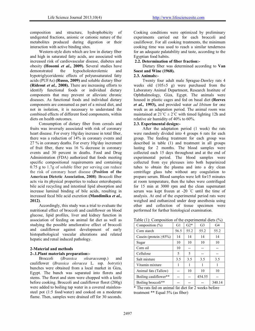
After the adaptation period (1 week) the rats were randomly divided into 4 groups 6 rats for each group. The feeding treatment for each group was described in table (1) and treatment in all groups lasting for 2 months. The blood samples were collected each 15 days throughout and at the end of experimental period. The blood samples were collected from eye plexuses into both heparinized tubes to obtain the plasma and into a dry clean centrifuge glass tube without any coagulation to prepare serum. Blood samples were left for15 minutes at room temperature, then the tubes were centrifuged for 15 min at 3000 rpm and the clean supernatant serum was kept frozen at -20 ºC until the time of analysis. At end of the experimental period rats were weighed and euthanized under deep anesthesia using ether and collection of tissue specimen were performed for further histological examination. Table (1): Composition of the experimental diets (%)
Composition (%) G1 G2* G3 G4
Corn starch 56.5 55.2 55.2 55.2
Casein (protein ≥85%) 14 14 14 14
Sugar 10 10 10 10
Corn oil 10 -- -- --
Cellulose 5 5 -- --
Salt mixture 3.5 3.5 3.5 3.5
Vitamin mixture 1 1 1 1
Animal fats (Tallow) -- 10 10 10
Boiling cauliflower** -- -- 454.55 --
Boiling broccoli** -- -- -- 340.14
* The rats fed on animal fat diet for 2 weeks before treatment ** Equal 5% (as fiber)
Life Science Journal 2013;10(4) http://www.lifesciencesite.com
2498
2.4. Biochemical analysis:- Serum glucose, total cholesterol, total lipid, Total
triglycerides and high-density lipoproteins (HDL), urea, creatinine, aspartate aminotransferase (AST) and Alanine aminotransferase (ALT) were estimated using Kits ( obtained from Randox Laboratories Ltd., Diamond Road, Crumlin,Co., Antrim, United Kingdom, BT. 294QY) and estimatedaccording to Barham and Trinder(1972), Rifai et al. (1999), Frings and Dunn(1970), Assmann (1979), Tomas (1998a), Tomas (1998b) and Moss and Henderson (1999), respectively. Low-density lipoproteins (LDL) and very low-density lipoproteins (VLDL) were calculated as according to Essam El-Din (2012). 2.5. Histopathological and immunohistochemical examination:-
Tissue specimens were collected from aorta and carotid blood vessels, heart, liver and kidney and preserved in 10% neutral buffered formalin, dehydrated in different grades of alcohol,cleared in xylene, embedding in paraffin, sectioned with microtome at 5µ thickness and finally stained with hematoxylin and eosin (H&E) and Masson`s trichrome (MTC) according to (Bancroft et al.,1996) on other hand tissue section of blood vessels were dewaxed in xylene, rehydrated and pretreated with 3 % hydrogen peroxide for blocking the activity of endogenous peroxidase.microwave as sited antigen retrieval was done for 20 minutes and sections were incubated overnight at 40 C with primary antibody for vascular endothelial growth factor (VEGF) (catalog MA1-166629,Thermo Scientific Co., UK) was dilutes with phosphate buffer saline (PBS) (1:50) then washed with PBS and incubated with biotinylated mouse secondary antibody (Cat No.32230,ThermoScientific Co., UK) and finally conjugated with streptavidin-peroxidase.sections were washed with PBS and incubated with diaminobenzidid (DAB) for 5 minutes. Statistical analysis:
Statistical analyses were carried out by SPSS (1998), SPSS10 program. Data were expressed as means ± SEM and the Statistical analysis was performed using one-way analysis of variance followed by Duncan’s tests. 3. Results:- 3.1. Crude fiber and fiber fraction for cauliflower and broccoli:-
The results in table (2) demonstrated the crude fiber content and fiber fraction for cauliflower and broccoli. Cauliflower had higher content of hemicellulose and neutral detergent fiber (NDF) than broccoli (5.28 vs. 1.23% and 19.85 vs. 18.37%, respectively). While, crude fiber, cellulose and acid detergent fiber (ADF) content were higher in broccoli
compared to cauliflower (14.7 vs. 11.03, 16.05% vs. 13.48% and 17.13 vs. 14.56%, respectively). 3.2. Effect of cauliflower and broccoli diet on food intake, body weight gain and organ somatic index:-
The results showed that, the rats fed on animal fat diet with cauliflower had the lowest food intake compared to other groups. The rats fed on animal fat diet with broccoli had higher than of food intake compared with rats in control (-), but lower than rats in control (+) as shown in (Table 3). The beginning of experimental, there was no significant differences in rats between groups. While, there were high significant differences in final body weight between groups (P≥0.05). The rats fed on animal fat diet with 5% cauliflower showed decrease of body weight gain compared with control (-). Rats fed on animal fat diet with 5% broccoli were highest level of body weight gain (128.83g). On other hand, results showed that liver somatic and kidney somatic index and absolute organ weight were lower in rats fed on animal fat diet with cauliflower compared with control (+) as shown in (Table 4). There were significant differences in liver and kidney weight between groups (P≥0.05). 3.3. Effect of cauliflower and broccoli on biochemical profiles:-
Lipid profile in different animal treated groups was demonstrated in table (5). Results revealed that, the rats fed on animal fat diet with (5% as fiber) from cauliflower or broccoli had a low level of total lipids, cholesterol levels compared to control (+). Moreover, the rats fed on animal fat diet with cauliflower had the lowest level of triglycerides (153.09mg/dl). While, the rats fed on animal fat diet with broccoli had an increased levels of triglycerides (246.17mg/dl). There are no significant differences between groups for HDL-C. On the other hand, the results showed that rats fed on animal fat diet with cauliflower had the lowest level of VLDL and a little rise of LDL-cholesterol. While, therats fed on broccoli had a low level of LDL-cholesterol and a high level of VLDL. This result may be due to increase in triglycerides level for rats fed on animal fat diet with 5% broccoli. Generally, the present study showed that cauliflower decrease levels of lipids profile more than broccoli. Also, the rats fed on cauliflower had lower risk ratio (T.C/HDL) 2.20% than broccoli 2.30%, respectively compared with positive control group (2.87%).
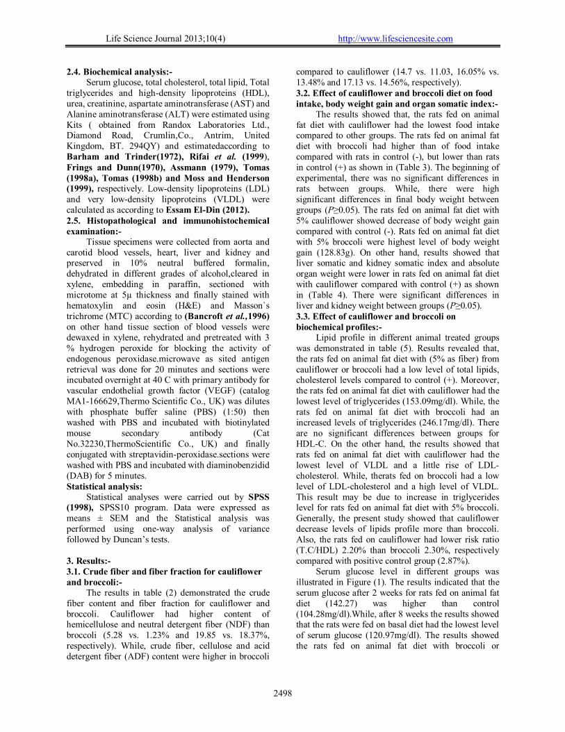
Serum glucose level in different groups was illustrated in Figure (1). The results indicated that the serum glucose after 2 weeks for rats fed on animal fat diet (142.27) was higher than control (104.28mg/dl).While, after 8 weeks the results showed that the rats were fed on basal diet had the lowest level of serum glucose (120.97mg/dl). The results showed the rats fed on animal fat diet with broccoli or
Life Science Journal 2013;10(4) http://www.lifesciencesite.com
2499
cauliflower had high serum glucose compared to control group.
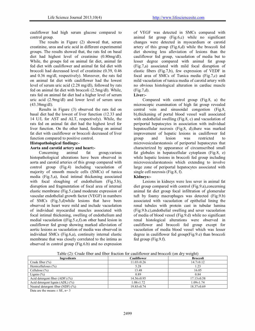
The results in Figure (2) showed that, serum creatinine, urea and uric acid in different experimental groups. The results showed that, the rats fed on basal diet had highest level of creatinine (0.80mg/dl). While, the groups fed on animal fat diet, animal fat fed diet with cauliflower and animal fat fed diet with broccoli had decreased level of creatinine (0.39, 0.46 and 0.36 mg/dl, respectively). Moreover, the rats fed on animal fat diet with cauliflower had the lowest level of serum uric acid (2.28 mg/dl), followed by rats fed on animal fat diet with broccoli (2.5mg/dl). While, rats fed on animal fat diet had a higher level of serum uric acid (2.9mg/dl) and lower level of serum urea (43.38mg/dl).
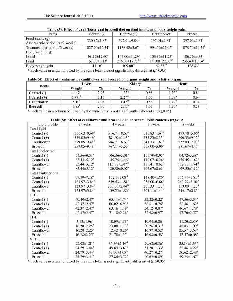
Results in Figure (3) observed the rats fed on basal diet had the lowest of liver function (12.33 and 14 U/L for AST and ALT, respectively). While, the rats fed on animal fat diet had the highest level for liver function. On the other hand, feeding on animal fat diet with cauliflower or broccoli decreased of liver function compared to positive control. Histopathological findings:- Aorta and carotid artery and heart:-
Concerning animal fat group,various histopathological alterations have been observed in aorta and carotid arteries of this group compared with control group (Fig.4) including vacuolation of majority of smooth muscle cells (SMCs) of tunica media (Fig.5,a), focal intimal thickening associated with focal sloughing of endothelium (Fig.5.b), disruption and fragmentation of focal area of internal elastic membrane (Fig.5.c)and moderate expression of vascular endothelial growth factor (VEGF) in numbers of SMCs (Fig.5,d)while lesions that have been observed in heart were mild and include vacuolation of individual myocardial muscles associated with focal intimal thickening, swelling of endothelium and medial vacuolation ((Fig.5.e,f).on other hand lesion in cauliflower fed group showing marked alleviation of aortic lesions as vacuolation of media was observed in individual SMCs (Fig.6,a), continuity internal elastic membrane that was closely correlated to the intima as observed in control group (Fig.6.b) and no expression
of VEGF was detected in SMCs compared with animal fat group (Fig.6,c) while no significant changes were detected in myocardium or carotid artery of this group (Fig.6,d) while the broccoli fed diet showing less alleviation of lesions than the cauliflower fed group, vacuolation of media but to lesser degree compared with animal fat group (Fig.7,a) associated with mild focal disruption of elastic fibers (Fig.7,b), few expression of VEDF in focal area of SMCs of Tunica media (Fig.7,c) and mild vacuolation of tunica media of carotid artery with no obvious histological alteration in cardiac muscle (Fig.7,d). Liver:-
Compared with control group (Fig.8, a) the microscopic examination of high fat group revealed central vein and sinusoidal congestion (Fig.8, b),thickening of portal blood vessel wall associated with endothelial swelling (Fig.8, c) and vacuolation of periportal hepatocytes in association with individual hepatocellular necrosis (Fig.8, d),there was marked improvement of hepatic lesions in cauliflower fed group and lesion was restricted to microvesicularsteatosis of periportal hepatocytes that characterized by appearance of circumscribed small fat globules in hepatocellular cytoplasm (Fig.8, e) while hepatic lesions in broccoli fed group including microvesicularsteatosis which extending to involve large zone of periportal hepatocytes associated with single cell necrosis (Fig.8, f). Kidneys:-
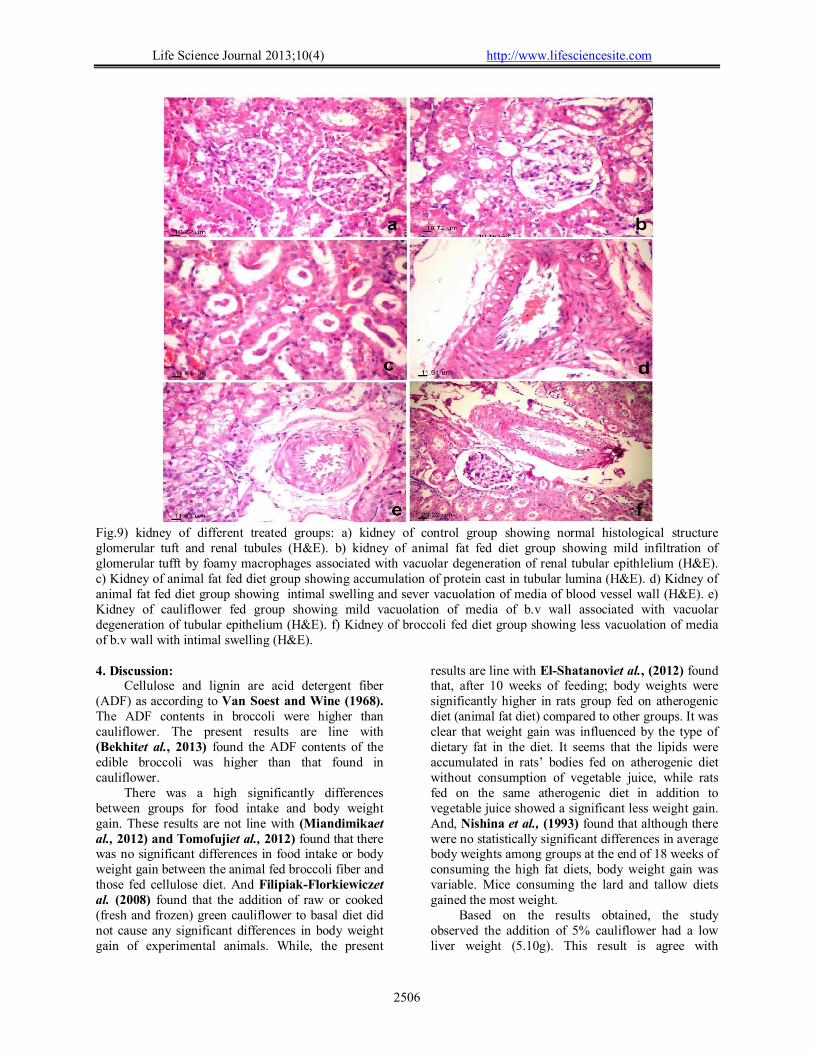
Lesions in kidneys were less sever in animal fat diet group compared with control (Fig.9.a),concerning animal fat diet group focal infiltration of glomerular tuft by foamy macrophages was detected (Fig.9.b) associated with vacuolation of epithelial lining the renal tubules with protein cast in tubular lumina (Fig.9.b.c),endothelial swelling and sever vacuolation of media of blood vessel (Fig.9.d) while no significant renal histological alterations were observed in cauliflower and broccoli fed group except for vacuolation of media blood vessel which was lesser degree in cauliflower fed group(Fig.9.e) than broccoli fed group (Fig.9.f).
Table (2): Crude fiber and fiber fraction for cauliflower and broccoli (on dry weight)
Ingredients Cauliflower Broccoli Crude fiber (%) 11.03±0.26 14.7±0.12 Hemicelluloses (%) 5.28 1.23 Cellulose (%) 13.48 16.05 Lignin (%) 0.89 0.84 Acid detergent fiber (ADF) (%) 14.56±0.95 17.13±0.58 Acid detergent lignin (ADL) (%) 1.08±1.72 1.09±1.74 Neutral detergent fiber (NDF) (%) 19.85±0.74 18.37±0.69 Data are the means ± SE, n= 3
Life Science Journal 2013;10(4) http://www.lifesciencesite.com
2500
Table (3): Effect of cauliflower and broccoli diet on food intake and body weight gain: Items Control (-) Control (+) Cauliflower Broccoli
Food intake (g): Atherogenic period (rat/2 weeks)
330.67±1.87a 397.01±9.84b 397.01±9.84b 397.01±9.84b
Treatment period (rat/6 weeks) 1027.00±16.54a 1138.48±3.67c 994.56±22.05a 1078.70±10.59b
Body weight (g): Initial
106.17±12.60a
107.00±11.29a
106.67±11.25a
106.50±9.33a
Final 151.33±9.13a 216.00±17.35bc 171.00±22.37ab 235.40±18.84c
Body weight gain 45.16a 109.00bc 64.33ab 128.83c
* Each value in a raw followed by the same letter are not significantly different at (p≥0.05) Table (4): Effect of treatment by cauliflower and broccoli on organs weight and relative organs
Items Liver Kidney Heart
Weight % Weight % Weight % Control (-) 4.47a 2.95 1.33a 0.88 1.23a 0.81
Control (+) 6.77±b 3.13 2.27ab 1.05 1.38a 0.64
Cauliflower 5.10a 2.98 1.47ab 0.86 1.27a 0.74
Broccoli 6.83b 2.90 2.47b 1.05 1.37a 0.58
* Each value in a column followed by the same letter is not significantly different at (p ≥0.05)
Table (5): Effect of cauliflower and broccoli diet on serum lipids contents (mg/dl): Lipid profile 2 weeks 4 weeks 6 weeks 8 weeks
Total lipid Control (-) Control (+) Cauliflower Broccoli
300.63±9.69a
559.05±9.48b
559.05±9.48b
559.05±9.48b
516.71±8.67a
581.92±3.43b
584.71±6.63b
767.11±3.55c
515.83±1.67a
735.83±8.33d
643.33±1.67b
665.00±5.00c
499.78±5.00a
800.33±9.52d
527.00±7.00b
581.67±4.41c
Total cholesterol Control (-) Control (+) Cauliflower Broccoli
74.56±0.51a
83.44±5.12a
83.44±5.12a 83.44±5.12a
106.36±3.01a
145.75±3.46c
115.58±5.07ab
120.80±0.07b
101.79±0.05a
140.07±0.26c
111.41±0.62b
109.67±0.66b
94.72±5.38a
150.45±1.62c
102.85±5.74ab
109.50±1.62b
Total triglycerides Control (-) Control (+) Cauliflower Broccoli
97.89±7.18a
123.97±3.84b
123.97±3.84b
123.97±3.84b
172.791.08ab
249.43±1.81c
200.00±2.04bc
139.23±1.86a
148.40±1.80a
256.00±6.66c
201.33±1.33b
203.11±1.44b
176.79±1.81ab
260.79±2.10bc
153.09±1.23a
246.17±0.83c
HDL Control (-)
Control (+) Cauliflower Broccoli
49.40±2.47a 42.37±2.47a
42.37±2.47a 42.37±2.47a
65.11±1.74a
86.02±6.93b
63.16±1.19a
71.18±2.28a
52.22±0.22a
58.61±0.76b 54.12±0.87a
52.98±0.97a
47.56±5.54a
52.46±1.62a
46.67±1.78a
47.70±2.57a
LDL Control (-) Control (+) Cauliflower Broccoli
3.13±1.96a
16.28±2.25b
16.28±2.25b
16.28±2.25b
10.09±1.55a
23.08±1.13b
12.42±0.20a
21.78±1.37b
19.94±0.06b
30.26±0.31c
16.97±0.52a
16.08±0.58a
11.80±2.80a
45.83±1.09c
25.57±3.69b 12.57±0.68a
VLDL Control (-) Control (+) Cauliflower Broccoli
22.02±1.01a
24.79±3.44b
24.79±3.44b
24.79±3.44b
34.56±2.16ab
49.89±3.63c
40.00±4.08bc
27.84±3.72a
29.68±0.36a
51.20±1.33c
40.27±0.27b
40.62±0.89b
35.34±3.63b
52.46±4.22c
30.62±2.48a
49.24±1.67c
* Each value in a raw followed by the same letter is not significantly different at (p ≥0.05)
Life Science Journal 2013;10(4) http://www.lifesciencesite.com
2501
Fig (1): Effect of cauliflower and broccoli diet on serum glucose for rats during nutritional period (mg/dl).
Fig (2): Effect of cauliflower and broccoli diet on serum creatinine, urea and uric acid for rats during
nutritional period (mg/dl).
Fig (3): Effect of cauliflower and broccoli diet on serum AST and ALT for rats during nutritional period
(U/L).
0
50
100
150
Control (-) Control (+) Cauliflower Broccoli
104.28a
142.27b 142.27b 142.27b
120.97a125.09b 134.19c 131.1c
Glucose serum (mg/100dl)
2weeks 8 weeks
0
20
40
60
Control (-) Control (+) Cauliflower Broccoli
0.80a 0.39b 0.46b 0.36b
43.62a 43.38a 45.93a 46.31a
2.58ab 2.9b 2.28a 2.5ab
Kidney function
Creatinine Urea Uric acid
0
10
20
30
Control (-) Control (+) Cauliflower Broccoli
12.33a15b 13ab 13ab14a
23.33c
18.43b21.35bc
Liver function
AST ALT
Life Science Journal 2013;10(4) http://www.lifesciencesite.com
2502
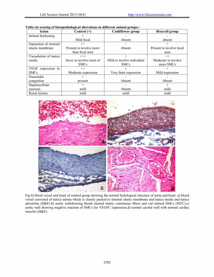
Table (6) scoring of histopathological alterations in different animal groups:- lesion Control (+) Cauliflower group Broccoli group
Intimal thickening + Mild focal
- Absent
- absent
Separation of internal elastic membrane
++ Present to involve more
than focal area
- Absent
+ Present to involve focal
area Vacuolation of tunica media
+++ Sever to involve most of
SMCs
+ Mild to involve individual
SMCs
++ Moderate to involve
more SMCs VEGF expression in SMCs
++ Moderate expression
± Very faint expression
+ Mild expression
Sinusoidal congestion
+ present
- Absent
- Absent
Hepatocellular necrosis
+ mild
- Absent
+ mild
Renal lesions mild mild mild
Fig.4) blood vessel and heart of control group showing the normal histological structure of aorta and heart: a) blood vessel consisted of tunica intima which is closely packed to internal elastic membrane and tunica media and tunica adventitia (H&E).b) aortic wallshowing bluish stained elastic continuous fibers and red stained SMCs (MTC).c) aortic wall showing negative reaction of SMCs for VEGFC expression.d) normal carotid wall with normal cardiac muscles (H&E).
Life Science Journal 2013;10(4) http://www.lifesciencesite.com
2503
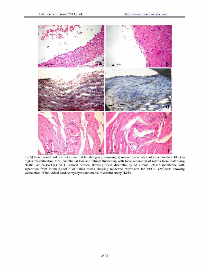
Fig.5) blood vessel and heart of animal fat fed diet group showing: a) marked vacuolation of tunica media (H&E) b) higher magnification focal endothelial loss and intimal thickening with focal separation of intima from underlying elastic lamina(H&E)c) MTC stained section showing focal discontinuity of internal elastic membrane with separation from intima.d)SMCS of tunica media showing moderate expression for VEGF. e&f)heart showing vacuolation of individual cardiac myocytes and media of carotid artery(H&E).
Life Science Journal 2013;10(4) http://www.lifesciencesite.com
2504
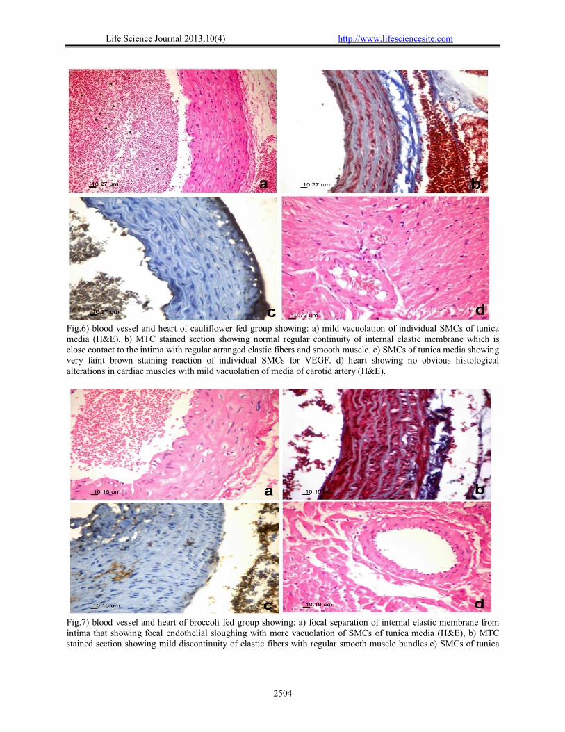
Fig.6) blood vessel and heart of cauliflower fed group showing: a) mild vacuolation of individual SMCs of tunica media (H&E), b) MTC stained section showing normal regular continuity of internal elastic membrane which is close contact to the intima with regular arranged elastic fibers and smooth muscle. c) SMCs of tunica media showing very faint brown staining reaction of individual SMCs for VEGF. d) heart showing no obvious histological alterations in cardiac muscles with mild vacuolation of media of carotid artery (H&E).
Fig.7) blood vessel and heart of broccoli fed group showing: a) focal separation of internal elastic membrane from intima that showing focal endothelial sloughing with more vacuolation of SMCs of tunica media (H&E), b) MTC stained section showing mild discontinuity of elastic fibers with regular smooth muscle bundles.c) SMCs of tunica
Life Science Journal 2013;10(4) http://www.lifesciencesite.com
2505
media showing brown staining reaction of SMCs for VEGF. d)heart showing no obvious histological alterations in cardiac muscles with very mild vacuolation of media of carotid artery(H&E).
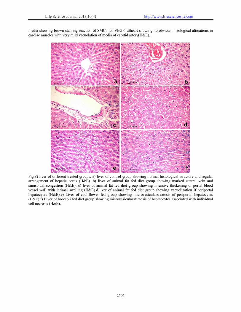
Fig.8) liver of different treated groups: a) liver of control group showing normal histological structure and regular arrangement of hepatic cords (H&E). b) liver of animal fat fed diet group showing marked central vein and sinusoidal congestion (H&E). c) liver of animal fat fed diet group showing intensive thickening of portal blood vessel wall with intimal swelling (H&E).d)liver of animal fat fed diet group showing vacuolization if periportal hepatocytes (H&E).e) Liver of cauliflower fed group showing microvesicularsteatosis of periportal hepatocytes (H&E).f) Liver of broccoli fed diet group showing microvesicularsteatosis of hepatocytes associated with individual cell necrosis (H&E).
Life Science Journal 2013;10(4) http://www.lifesciencesite.com
2506
Fig.9) kidney of different treated groups: a) kidney of control group showing normal histological structure glomerular tuft and renal tubules (H&E). b) kidney of animal fat fed diet group showing mild infiltration of glomerular tufft by foamy macrophages associated with vacuolar degeneration of renal tubular epithlelium (H&E). c) Kidney of animal fat fed diet group showing accumulation of protein cast in tubular lumina (H&E). d) Kidney of animal fat fed diet group showing intimal swelling and sever vacuolation of media of blood vessel wall (H&E). e) Kidney of cauliflower fed group showing mild vacuolation of media of b.v wall associated with vacuolar degeneration of tubular epithelium (H&E). f) Kidney of broccoli fed diet group showing less vacuolation of media of b.v wall with intimal swelling (H&E). 4. Discussion:
Cellulose and lignin are acid detergent fiber (ADF) as according to Van Soest and Wine (1968). The ADF contents in broccoli were higher than cauliflower. The present results are line with (Bekhitet al., 2013) found the ADF contents of the edible broccoli was higher than that found in cauliflower.
There was a high significantly differences between groups for food intake and body weight gain. These results are not line with (Miandimikaet al., 2012) and Tomofujiet al., 2012) found that there was no significant differences in food intake or body weight gain between the animal fed broccoli fiber and those fed cellulose diet. And Filipiak-Florkiewiczet al. (2008) found that the addition of raw or cooked (fresh and frozen) green cauliflower to basal diet did not cause any significant differences in body weight gain of experimental animals. While, the present
results are line with El-Shatanoviet al., (2012) found that, after 10 weeks of feeding; body weights were significantly higher in rats group fed on atherogenic diet (animal fat diet) compared to other groups. It was clear that weight gain was influenced by the type of dietary fat in the diet. It seems that the lipids were accumulated in rats’ bodies fed on atherogenic diet without consumption of vegetable juice, while rats fed on the same atherogenic diet in addition to vegetable juice showed a significant less weight gain. And, Nishina et al., (1993) found that although there were no statistically significant differences in average body weights among groups at the end of 18 weeks of consuming the high fat diets, body weight gain was variable. Mice consuming the lard and tallow diets gained the most weight.
Based on the results obtained, the study observed the addition of 5% cauliflower had a low liver weight (5.10g). This result is agree with
Life Science Journal 2013;10(4) http://www.lifesciencesite.com
2507
Filipiak-Florkiewiczet al. (2008) found that the addition of cauliflower to basal diet statistically significantly lowered liver weight in rats.
Results of this study showed that groups of rats fed on animal fat diet with cauliflower or broccoli at levels of intake (5%) had significant decreased in total cholesterol, triglycerides, HDL, LDL-C, VLDL-C. As well as serum level of creatinine and uric acid in addition a significant increase in liver function (AST and ALT) and urea concentrations. These changes may be related to the phenolic compounds in cauliflower or broccoli and its properties as antioxidant activity. These results were clarified by Maria et al. (2011) as reported that phenolic compounds was higher in Brassica vegetables in which flavonols were always the major compounds. In addition consumption of Brassica vegetables has been related to human health due to their phytochemicals, such as glucosinolates and phenolic compounds that induce a variety of physiological functions including antioxidant activity, enzymes regulation and apoptosis control and the cell cycle Maria et al. (2011).
These results are line with previously studies Nishina, et al., (1993) found that increased in T.C., LDL-C, VLDL-C and T.G. levels in rats consumed a high fat diet (plants or animals sources). The increase in total cholesterol levels could be attributed mainly to the combined VLDL plus LDL-C fraction. Filipiak-Florkiewiczet al. (2008), Bahadoranet al. (2012), Miandimikaet al. (2012), Tomofujiet al.(2012) and Farahmandi, et al. (2013) found that serum total cholesterol, LDL-C were significantly lower in group fed on broccoli than compared to positive control.
Broccoli fiber and corn oil are lowering serum cholesterol levels via different mechanisms. The hypocholesterolemic effects of broccoli supplementation in the diet may be mainly due to a reduction of the reabsorption of bile acids at the terminal ileum for enterohepatic recycling and an increase in catabolism of cholesterol to bile acids and not by the suppression of the rate of cholesterol synthesis (Ramjiganeshet al., 2000). Studies have shown that soluble fibers reduce serum cholesterol by altering the composition of enterohepatic bile acid pools and by increasing the faecal loss of total bile acids (Jones et al., 2008 and Rideoutet al., 2008). Broccoli fiber may also have a physical effect, resulting in reducing intestinal lipid absorption and/or increasing luminal binding of bile acids (Matheson et al., 1995). While the hypocholesterolemic effects of corn oil may be due to both its suppression of cholesterol/lipid synthesis and its effect on increasing the catabolism rate of cholesterol to bile acids. The results are agree with Filipiak-Florkiewiczet al.
(2008) observed decrease in T.C., LDL-C, VLDL-C and T.G after feeding rats on 10% of cauliflower (DM) which added to basal diet.
Focal intimal thickening,vacuolation of smooth muscle cells comprising the medial of the blood vessel wall and expression of VEGF in the smooth muscle cells were the most characteristic histological alterations that have been observed in the animal fat fed groups that was ameliorated in cauliflower and broccoli fed animals groups indicating the anti atherogenic potential of cauliflower and broccoli against development of early vascular changes induced by feeding on animal fat diet ;this potential effect could be related to decreasing the expression of VEGF in smooth muscle cells which have a role in progression of atherosclerosis that discussed by (Lemströmet al., 2002) as they clarify that the blockade of signaling downstream of VEGF receptor significantly reduced arteriosclerotic lesions also (Francesca et al.,2001) founded that vascular endothelial growth factor enhances atherosclerotic plaque progression. Cauliflower and broccoli decreasing the expression of VEGF in smooth muscle cells via decreasing nitric oxide level as they considered antioxidant (JózefDulaket al., 2000;) proved the role of nitric oxide in induction of synthesis of vascular endothelial growth factor by rat vascular smooth Muscle Cells. Lesions in liver and kidney was greatly alleviated in cauliflower and broccoli fed group that was related to partially to alleviation in vascular changes and improvement of biochemical profiles that observed in cauliflower and broccoli fed groups. References 1. Assmann, G. (1979). “Cholesterol
determination in high density lipoproteins separated by three different methods.”Internist. 20: 559-604.
2. Bahadoran, Z.; Mirmiran, P.; Hosseinpanah, F.; Rajab, A.; Asghari, G. and Azizi, F. (2012). “Broccoli sprouts powder could improve serum triglyceride and oxidized LDL/LDL-cholesterol ratio in type 2 diabetic patients: A randomized double-blind placebo-controlled clinical trial”. Diabetes Research and Clinical Practice 9 6: 348 – 354.
3. Banchroft, J.D., Stevens, A. and Turner, D.R. (1996). “Theory and practice Of Histological Techniques”. Fourth Ed. Churchil Livingstone, New York, London, San Francisco, Tokyo.
4. Barham, D. and Trinder, P. (1972).“An improved color reagent for the determination of blood glucose by the oxidase system” Analyst. Pp. 142-145.
Life Science Journal 2013;10(4) http://www.lifesciencesite.com
2508
5. Bekhit, A. E. D.; Lingming, K.; Mason, S.L.; Zhou, J. H. and Sedcole, J. R. (2013). “Upgrading the utilization of brassica wastes: physicochemical properties and sensory
6. evaluation of fermented brassica stalks”. Inter. Food Res. J. 20(4): 1961-1969.
7. El-Shatanovi, G.A.T.A.; Ashoush, I.S.; Ahmed, Enaam K. and Ali, Soad A. (2012).“Antiatherogenic properties of vegetable juice rich in antioxidants in cholesterol-fed rats”.Annals of Agricultural Science. 57(2), 167–173
8. Essam El-Din, Maha, M. (2012). “The protective effect of Turnip leaves against oxidative stress induced by high cholesterol diet in adult rats”. World Applied Sciences Journal 20 (1): 154 – 163.
9. Farahmandi, K.; Khazdoozy, S.; Barati, Sara and farahmandi, S. (2013).“The Effect of Hydro-Alcoholic Extract of Broccoli Leaves on Sugar and Lipids in Serum of Diabetic Rats”.Sumit Kumar.: Asian Journal of Biomedical and Pharmaceutical Sciences 3(16): 24-26.
10. Filipiak-Florkiewicz, A.; Cieślik, E. and Kostogrys, R.B. (2008).“Effect of addition of romanesco type green cauliflower to diet on serum lipid profile in rats”.Pol. J. Food Nutr. Sci. 58 (1): pp. 119-123.
11. Francesca, L. C.; Jacob, M. W.; Philippe, G. A.; Andrea B.; Paul, R. H. and Michael D. D. (2001). “Vascular endothelial growth factor (VEGF) can promote angiogenesis but may also exert certain effects to alter the rate of atherosclerotic plaque development”. Nature Medicine 7, 425 – 429.
12. Frings, C.S. and Dunn, R. (1970).“Colorimetric method for determination of total serum lipids based on the sulphosvanillin reaction” Am. J. Clin. Path., 53: 89-91.
13. Grajek, W. (2003). “Changes of antioxidative potential of plant material during processing and
intestine digestion”.Żywność, 4, 26 ‑ 35 (in Polish; English abstract).
14. Gorinstein, S.; Leontowicz, H.; Leontowicz, M.; Drzewiecki, J.; Najman, K.; Katrich, E.; Barasch, D.; Yamamoto, K. and Trakhtenberg S., (2006). “Raw and boiled garlic enhances plasma antioxidant activity and improves plasma lipid metabolism in cholesterol-fed rats”. Life Sci., 78, 655-663.
15. Hosomi, R., Fukunaga, K., Arai, H., Nishiyama, T., & Yoshida, M. (2009)“Effects ofdietary fish protein on serum and liver lipid concentrations in rats and theexpression of
hepatic genes involved in lipid metabolism” Journal of Agriculturaland Food Chemistry, 57, 9256–9262.
16. Jeffery E, and Araya M. (2009). “Physiological effects of broccoli consumption”. Phytochem Rev. 8:283–98.
17. Jones, B. V., Begley, M., Hill, C., Gahan, C. G. M., and Marchesi, J. R. (2008).“Functional and comparative metagenomic analysis of bile salt hydrolase activity in the human gut microbiome”. Proceedings of the National Academy of Sciences of the United States of America, 105, 13580–13585.
18. JózefDulak, AlicjaJózkowicz,AldonaDembinska-Kiec, Ibeth Guevara, Anna Zdzienicka, DanutaZmudzinska-Grochot, IzabelaFlorek, Anna Wójtowicz,AndrzejSzuba, John P. Cooke (2000).“Nitric Oxide Induces the Synthesis of Vascular Endothelial Growth Factor by Rat Vascular Smooth Muscle Cells”.Arteriosclerosis, Thrombosis, and Vascular Biology. 20: 659-666 doi: 10.1161/01.ATV.20.3.659.
19. Kahlon, T.S.; Chapman, M.H. and Smith, G.E. (2007a) “In vitrobinding of bile acids by okra, beets, asparagus, eggplant, turnips, green beans, carrots, and cauliflower” Food Chemistry 100: 1531–1536.
20. Kahlon, T.S.; Chapman, M.H. and Smith, G.E. (2007b) “In vitro binding of bile acids by okra, beets, asparagus, eggplant, turnips, green beans, carrots, and cauliflower” Food Chemistry 103: 676–680.
21. Lee, J.; Shin, H.; Lee, Y.; Kim, A., and Lee, M. (2009). “Effect of broccoli sprout on cholesterol-lowering and anti-obesity effects in rats fed high fat diet”. J Korean Soc Food SciNutr 38(3):309–18.
22. Lemström, K. B.; Rainer KrebsA. I.; Nykänen, J. M.; Tikkanen,R. K.; Sihvola, E. M. Aaltola, P. J.; Häyry, J.; Wood, Kari Alitalo; SeppoYlä-Herttuala, and Petri K. Koskinen, (2002).“Vascular Endothelial Growth Factor Enhances Cardiac Allograft Arteriosclerosis”.Circulation. 105: 2524-2530
23. Mandimika, T.; Paturi, G.; DeGuzman, C.E.; Butts, C.A.; Nones, K.; Monro, J.A.; Butler, R.C.; Joyce, N.I.; Mishra, S. and Ansell, J. (2012). “Effects of dietary broccoli fiber and corn oil on serum lipids, faecal bile acid excretion and hepatic gene expression in rats” Food Chemistry 131: 1272–1278.
24. Maria, E.C., F. Marta, S. Pilar and Pablo, V. (2011).“Phenolic Compounds in Brassica Vegetables”. Molecules, 16: 251-280.
Life Science Journal 2013;10(4) http://www.lifesciencesite.com
2509
25. Matheson, H. B., Colon, I. S., and Story, J. A. (1995). “Cholesterol 7{alpha}-hydroxylase activity is increased by dietary modification with psyllium hydrocolloid, pectin, cholesterol and cholestyramine in rats”. Journal of Nutrition, 125, 454–458.
26. Moss, D.W. and Henderson A.R. (1999).“Clinical enzymology. In: Burtis CA, Ashwood, E.R., editors. Tietz textbook of clinical chemistry. 3rd ed. Philadelphia: WB Saunders Company; p. 617-721.
27. Murashima, M.; Watanabe, S.; Zhuo, X.G.; Uehara, M. and Kurashige, A. (2004). “Phase 1 study of multiple biomarkers for metabolism and oxidative stress after one-week intake of broccoli sprouts”. Biofactors, 22(1–4):271–5.
28. Nishina, P.M.; Lowe, S.; Verstuyft, J.; Naggert, J.K.; Kuypers, F.A. and Paigen, B. (1993).“Effects of dietary fats from animal and plant sources on diet-induced fatty streak lesions in C57BL/6J mice”. J Lipid Res. 34: 1413-1422.
29. Position of the American Dietetic Association (2008): Health Implications of Dietary Fiber. J Am Diet Assoc. 108: 1716-1731.
30. Ramjiganesh, T., Roy, S., Nicolosi, R. J., Young, T. L., McIntyre, J. C., and Fernandez, M. L. (2000). “Corn husk oil lowers plasma LDL cholesterol concentrations by decreasing cholesterol absorption and altering hepatic cholesterol metabolism in guinea pigs”. Journal of Nutritional Biochemistry, 11, 358–366.
31. Rasmussen, S.E.; Frederiksen, H.; Struntze Krogholm, K. and Poulsen L. (2005). “Dietary proanthocyanidins: occurrence, dietary intake, bioavailability, and protection against cardiovascular disease”. Mol. Nutr. Res., 2005, 49, 159-174.
32. Reeves, P.G.; Nielsen, F.H. and Fahey, G.C. (1993) “AIN-93 purified diets for laboratory rodents: final report of the American Institute of Nutrition Ad HOC writing Committee on the reformulation of the AIN-76 a rodent diet.” J. Nutr., 123(12): 1939-1951.
33. Rideout, T., Harding, S., Jones, P., & Fan, M. (2008). “Guar gum and similar solublefibers in
the regulation of cholesterol metabolism: Current understandings andfuture research priorities” Vascular Health and Risk Management, 4, 1023–1033.
34. Riedl, M.A.; Saxon, A. and Diaz-Sanchez, D. (2009). “Oral sulforaphane increases phase II antioxidant enzymes in the human upper airway”. ClinImmunol 130(3):244–51.
35. Rifai, N.; Bacorik, P.S. and Albers, J.J. (1999). “Lipids, Lipoproteins and Apolipoproteins, In: Burtis CA, Ashwood, E.R., editors. Tietz “Textbook of Clinical Chemistry”. 3rd ed. Philadelphia: WB Saunders Company; p. 809-861.
36. Russo, G. L. (2009). “Dietary n-6 and n-3 polyunsaturated fatty acids: Frombiochemistry to clinical implications in cardiovascular prevention” Biochemical
37. Pharmacology, 77, 937–946. 38. SPSS (1998).Statistical Algorithm, SPSS Inc.,
Reports Library of Congress,US. 39. Tomas L. (1998a). “Clinical laboratory
diagnostics” 1 st ed. Frankfurt: TH – Books verlagsgesellschft; p. 208-214.
40. Tomas L. (1998b). “Clinical laboratory diagnostics” 1 st ed. Frankfurt: TH – Books verlagsgesellschft; p. 366-374.
41. Tomofuji, T.; Ekunia, D.; Azumaa, T.; Irie, K.; Endoa, Y.; Yamamotob, T.; Ishikado, A.; Satoc, T.; Haradac, K.; Suido, H. and Morita, M. (2012). “Supplementation of broccoli or Bifidobacteriumlongum–fermented broccoli suppresses serum lipid peroxidation and osteoclast differentiation on alveolar bone surface in rats fed a high-cholesterol diet”. Nutr. Res. 32: 301 – 307.
42. Trinder, P. (1969). “Determination of glucose in blood using glucose oxidase with an altemative oxygen acceptor.” Am. Clin. Biochem. 6:24-27.
43. Van Soest, P.J. and Wine, R.H. (1968). “Determination of Lignin and Cellulose in acid detergent fiber with Permanganate” J. of the Associ. Of Official Anal. Chem. Int. 52: 780-785.
11/12/2013
Copyright © 2022 FDOKUMEN