Immunohistochemical study of nuclear changes associated with male germ cell death and spermiogenesis
23
IMMUNOHISTOCHEMICAL STUDY OF NUCLEAR CHANGES ASSOCIATED WITH MALE GERM CELL DEATH AND SPERMIOGENESIS Leon M. McClusky 1* , Sean Patrick 2 , Irene E.J. Barnhoorn 2 , Jacobus C. van Dyk 3 , Christiaan de Jager 3 , Maria S. Bornman 2 Department of Health and Nursing Science, Narvik University College, Norway 1 , Department of Urology 2 and School of Health Systems and Public Health 3 , Faculty of Health Sciences, University of Pretoria, South Africa. Running title : caspase-3 labeling of dying germ cells Keywords : apoptosis, germ cells, nucleus, cleaved caspase-3, acrosome, GATA-4, immunohistochemistry *Corresponding author: Leon M. McClusky, Tel +47 76966239; Fax +47 76966813 Email: [email protected]
Transcript of Immunohistochemical study of nuclear changes associated with male germ cell death and spermiogenesis

IMMUNOHISTOCHEMICAL STUDY OF NUCLEAR CHANGES ASSOCIATED
WITH MALE GERM CELL DEATH AND SPERMIOGENESIS
Leon M. McClusky1*
, Sean Patrick2, Irene E.J. Barnhoorn
2, Jacobus C. van Dyk
3, Christiaan
de Jager3, Maria S. Bornman
2
Department of Health and Nursing Science, Narvik University College, Norway1, Department
of Urology2 and School of Health Systems and Public Health
3, Faculty of Health Sciences,
University of Pretoria, South Africa.
Running title: caspase-3 labeling of dying germ cells
Keywords: apoptosis, germ cells, nucleus, cleaved caspase-3, acrosome, GATA-4,
immunohistochemistry
*Corresponding author: Leon M. McClusky, Tel +47 76966239; Fax +47 76966813
Email: [email protected]

2
ABSTRACT
In a previous study on the effects of gestational and lactational exposure of para-nonylphenol
on male rats, we noted in both induced and uninduced rats, that variations in cleaved caspase-
3 immunostaining patterns were associated with distinct nuclear alterations in mainly basally
located germ cells (spermatogonia and preleptotene spermatocytes). These were re-analysed
and compared with cleaved caspase-3-labeled germ cells in the aging human and the
spermatogenically active catfish testis. In the rat testes, cytoplasmic immunostaining was
progressively associated with lateral compression of the nucleus, its break up into large pieces
which can contain immunostained marginated chromatin masses. The pale remnants of the
nucleus continued to shrink in size concomitant with the appearance of blue-purplish stained
regions in the cytoplasm similar in color to the condensed chromatin in spermatids, a
condition which was TUNEL-negative. These large clumps of chromatin also eventually
disappeared, giving rise to cells resembling cytoplasmic ghosts, a condition which was
TUNEL-positive. By contrast, the immunolabeled nuclei of human and catfish germ cells
condensed into a single mass, after which they lost immunoreactivity. To exclude the
possibility that these observations could reflect alterations in Sertoli nuclei, rat testicular
sections were probed with a mouse anti-human GATA-4 monoclonal (MHM) antibody. The
MHM was, however, the second of two GATA-4 antibodies tested, with a goat anti-mouse
polyclonal (GMP) initially used to label rat the Sertoli nuclei. GMP unexpectedly, but
distinctly labeled the complete development of the acrosome in the rat testis, a fortuitous
finding with utility for staging of the seminiferous epithelium.
3
INTRODUCTION
During spermatogenesis, primitive spermatogonia enter into complex developmental
pathway involving stage-related processes directed specifically towards the nuclei of their
progeny prior to and after meiosis. For example, differentiating spermatogonia undergoing
spontaneous physiological cell death (apoptosis) are dismantled in an orderly fashion, their
chromatin compacted and/or marginated and their DNA eventually fragmented as the nuclear
compartment continues to shrink (Allan et al. 1987; 1992). These deaths are considered vital
for tissue homeostasis as they represent a mechanism to rid the tissue of superfluous or
defective cells and has been estimated to result in a loss of 25-75% of the total number of
germ cells produced in the rat testis (Huckins 1978).
Whereas the mitotic peaks of differentiating spermatogonia are associated with significant
spontaneous spermatogonial death, characterized by the formation of large clumps of chromatin
in these cells (Huckins 1978), the mechanism of death in other rat germ cell stages is described
as complex due to the lack of classic apoptotic morphology (Russell and Clermont 1977; Allan
et al. 1987; Ghosh et al. 1991, Wang et al. 1998; Bartke 1995; Russell et al. 2002). It has even
been proposed that spermatocytes die via a non-apoptotic mechanism (Allan et al. 1987;
1992; Russell et al. 2002), even though the demise of these cells during the first wave of
spermatogenesis in pubertal rats was subsequently found to be caspase-mediated and TUNEL-
labeled, in spite of the absence of pyknotic chromatin (Jahnukainen et al. 2004; Moreno et al.
2006). Since spermatogonia were also unexpectedly never labeled with the various
immunofluorescent apoptotic markers used in the study of Moreno et al. (2006), these authors
questioned whether spermatogonial death involved a different subtype of apoptosis, i.e.
anoikis (Moreno et al. 2006). Furthermore, the identity of dying peripherally located germ
cells following hormone deprivation (Billig et al. 1995) was questioned by Bartke (1995),
who suggested that, based on earlier studies (Russell and Clermont 1977) on the effects of
hypophysectomy in male rats, the dying germ cells were probably not spermatocytes as
indicated by Billig et al. (1995), but most likely spermatogonia. Thus, our knowledge of the
morphological and mechanistic aspects of male germ cell death in general is incomplete.
In the second instance, the process of spermatid differentiation, which commences after
meiosis, is initiated by two major events which impact the round spermatid nucleus, i.e. the
development of an acrosome from the fusion of Golgi-derived vesicles, and the shaping and
maximal compaction of the chromatin in the future sperm head from a spherical configuration
into its final species-specific shape (For review, Toshimori and Ito 2003; Kierszenbaum and
Tres 2004). The leading edge of the expanding acrosome is a prominent marginal ring, the
acroplaxome, which anchors the acrosome to the spermatid nucleus. The acroplaxome is
thought to play an important role in the shaping of the spermatid head during acrosome
development, such that the spermatid head changes shape from round to oblong and
eventually a long thin structure, as in the case of the rat.
The commercial availability of a variety of antibodies to the host of factors constituting
the apoptosome facilitates immunohistochemical detection of some of the more upstream
events of apoptosis, while intracellular morphological detail is still largely retained even at the
light microscopic level. Among such upstream events, is the activation of cysteine aspartyl-
specific proteases (caspases), of which cleaved caspase-3 is a major executioner of apoptotic
morphology (Stadelmann and Lassmann 2000; Ramuz et al. 2003; Kamada et al. 2005).
In our previous report on the effects of gestational and lactational exposure of para-
nonylphenol (p-NP) on male rats, variations in the immunostaining patterns obtained with a
rabbit polyclonal antibody to cleaved caspase-3 were associated with distinct nuclear
alterations in mainly dying basally located germ cells in both induced and uninduced rats
(McClusky et al. 2007). Since a full analysis of the range of observed apoptotic morphologies

4
was outside the scope of the latter study, we now present these findings, several of which
might shed further light on aspects of male germ cell death. In this qualitative and
comparative study, we examined further the cleaved caspase-3 associated intracellular
changes in dying rat premeiotic germ cells, and comparisons are made with TUNEL-labeled
and caspase-labeled germ cells in two other vertebrate species, the aging human and the
spermatogenically active catfish. As a result of these analyses, we accidentally also found a
marker of acrosome development during spermiogenesis in the rat, which may have utility in
staging of the cycle of the seminiferous epithelium.

5
MATERIALS AND METHODS
Organisms and tissue sampling
The present study utilized archival Bouin’s fixed testicular tissues from an earlier study
on the effects of para-nonylphenol (p-NP) that used the modified Organization for Economic
Cooperation and Development (OECD) 415 one –generation test (de Jager et al. 1999). In
brief, in that study seven-day pregnant Sprague-Dawley rats received 100 mg/kg, or 250
mg/kg p-NP, or vehicle (cottonseed oil) only intragastrically. Dosing of all groups (n =
20/group) was daily, and continued through gestation, lactation, and afterwards, directly to all
male offspring until 10 weeks of age. Animals were housed, according to the Animal Use and
Care guidelines of the University of Pretoria, under routine laboratory conditions (12hr/12hr
light-dark cycle; 22 °C ± 3 room temperature and 30-70% relative humidity) at the Laboratory
Animal Facility of the Faculty of Veterinary Sciences, University of Pretoria and were given
access to standard rat chow and tap water ad libitum. On the day of sampling, animals were
decapitated and both testes removed. The testes were slit open after which they were
immersion-fixed in Bouin’s solution for 12 hours. Before further processing, testes were cut
in half and fixed for a further 2.5 days before being transferred to 70% ethanol. No tissues
were collected for molecular or protein analysis.
To limit the unnecessary use of animals, vehicle-treated animals served as uninduced
(normal) rats in the present study, consistent with Ghosh et al. (1991) who also found no
difference between sham-operated controls and normal untreated animals. For comparative
analysis, testes were obtained from four men, aged 57 - 70 years old, who underwent
orchidectomy as treatment for advanced prostate cancer but otherwise healthy with no known
endocrine disease, as described in McClusky et al. (2007), and ten laboratory-raised sexually
mature adult feral sharptooth catfish (Clarias gariepinus), sampled and processed as
described in McClusky et al. (2008). These human testicular tissues were specifically used
because they were the only readily available material. Fixed testes of all three species tissues
were thoroughly rinsed in 70% ethanol, before they were embedded in Tissue Tek III paraffin
wax (Sakura Finetek, CA, USA).
Immunohistochemistry
Paraffin sections (4 µm thick) from the embedded testes of all three species were floated
in a water bath (45 °C), collected on Superfrost Plus slides (Menzel-Glaser, Germany),
deparaffinized and rehydrated stepwise through an ethanol series. Deparaffinized and
rehydrated sections of all three species were subjected to an antigen retrieval procedure, i.e.
heating in a microwave oven (800W) in 10 mM sodium citrate buffer (pH 6.0) for 10 min.
After cooling for 20 min at RT, sections were rinsed in distilled water and treated in darkness
for 10 min with 3% hydrogen peroxide to quench endogenous peroxidase. Following a rinse
in distilled water, the sections were placed in 0.1% Tween 20 in phosphate buffered saline
(PBS-A, 137 mM NaCl, 29 mM NaH2PO4·H2O, 9 mM Na2HPO4, pH 7.4) for 5 min. Sections
were then incubated for 1 hour at RT with a blocking solution consisting either of 5% normal
goat serum, 5% normal horse serum, or 5% normal rabbit serum in 0.1% Tween 20-PBS-A.
Thereafter sections were incubated overnight at 4 °C with their respective primary antibodies,
which were: a rabbit polyclonal anti-cleaved caspase-3 (17/19 kDa fragment of activated
caspase-3, Cell Signaling Technology, Beverly, MA, USA) diluted 1:100, a mouse antihuman
GATA-4 monoclonal antibody (sc-25310, Santa Cruz Biotechnology, Santa Cruz, CA, USA)
diluted 1:200, and a goat antimouse GATA-4 polyclonal antibody (sc-1237, Santa Cruz
Biotechnology, USA) diluted 1:200 in each of their respective blocking solutions. (Only the
rat testicular sections were treated with the two GATA-4 antibodies). After three washes in
0.1% Tween 20/PBS-A, sections were incubated for 30 min at RT with either a biotinylated

6
antirabbit, antimouse or antigoat IgG, washed in PBS-A, and then incubated with the
Vectastain avidin-biotin-complex (Vectorlabs, Burlingame, CA, USA) for a further 30 min at
RT. Following three 5 min PBS-A washes, the antigen was finally detected by treating the
sections, according to the supplier's instructions (Vectorlabs), for 1-2 min with a 3,3’-
diaminobenzidine (DAB) substrate kit, after which positive immunoreactivity was revealed as
brown staining. Sections were counterstained with 0.5% crystal violet-containing methyl
green in 100 mM sodium acetate (pH 4), dehydrated in 100% butanol, cleared in xylene and
mounted with Entellan permanent mounting medium (Merck, Germany). Negative controls
were generated by substituting the primary antibodies with the appropriate normal serum and
by serial dilution of the primary antibody.
In situ end-labeling of DNA fragments (TUNEL)
One deparaffinized and rehydrated section from all three species was pretreated with 0.5%
Triton X-100 for 10 min at RT followed by treatment in 3% hydrogen peroxide for 7 min to
quench endogenous peroxidise and two washes in PBS-B (50 mM sodium phosphate, pH 7.4;
200 mM NaCl). Subsequent steps for TUNEL staining were carried out using the ApopTag-
Peroxidase Kit according to the supplier’s instructions (Chemicon, Temecula, CA, USA).
Following incubation of the sections with the TUNEL reaction mixture in a humidified
chamber at 37 °C for 1 hr, antidigoxigenin-peroxidase complex was added for 30 min at RT.
Sections were then treated with DAB for 1-2 min according to the supplier's instructions
(Vectorlabs, Burlingame, CA) such that positive cells stained brown. Sections were
counterstained with 0.5% crystal violet-containing methyl green in 100 mM sodium acetate
(pH 4.0), dehydrated in 100% butanol, cleared in xylene and mounted with Entellan (Merck,
Germany). To generate negative controls, TdT was replaced with sterile water, whereas for
positive controls, sections were pretreated with DNase I (10 µg/ml in distilled water) to
generate DNA fragments.
Microscopy and data analysis
Cross-sections were viewed and photographed with a Nikon DXM1200F Digital camera
fitted to a Nikon Optiphot microscope. As previously described in McClusky et al. (2007),
because of its metachromatic staining of condensed chromatin as found during spermiogenesis,
the crystal violet-containing methyl green counterstain had utility for the staging of the
spermatogenic cycle in rats. Basally located cleaved caspase-3 immunoreactive germ cells in
rat testes were thus regularly observed in all rat seminiferous tubules either lacking or only
with round spermatids, i.e. stages XII-XIV and I-III (~ 333 – 426 tubules per cross-section).
All stage XII-XIV and I-III cross-sectional tubular profiles in the three rat treatment groups (n
= 7-10/group), at least 200 and 100 profiles in the human and catfish testicular sections were
scanned, respectively. The number of caspase- and TUNEL-positive cells, as well as the type
of caspase-immunoreactivity within the cells noted for each tubule.

7
RESULTS
In both induced and uninduced rats, germ cells labeled with the rabbit polyclonal anti-
cleaved caspase-3 were found scattered around the periphery of the seminiferous epithelium
(Fig. 1). Based on the stages of the seminiferous epithelium cycle in which these cells were
found (McClusky et al. 2007), these were premeiotic germ cells (oblong and round shaped
spermatogonia and preleptotene spermatocytes) with their readily visible low ratio of
cytoplasmic volume:nuclear volume. Labeling was very distinct, and varied from discrete
staining of only the cytoplasm in normal-looking cells to intense staining of the cytoplasm
accompanied by changes in nuclear morphology. Although these differential immunostaining
patterns clearly reflect the sequential progression of apoptosis, the intermediate stages were
infrequent, which is indicative of the asynchronous nature of apoptosis in a highly
heterogeneous tissue such as the seminiferous epithelium.
Nevertheless, based on the changes affecting the nucleus and its contents, all caspase-
labeled rat germ cells could be grouped into several immunomorphological categories. In one
group, cleaved caspase-3 was immunolocalised to one side of the large round nucleus in the
scant cytoplasm of the normal-looking germ cells (Fig. 1a). In a second group, intense
cytoplasmic immunostaining was associated with what appeared to be lateral
compression/deformation of the unlabeled nucleus into an oblong shape (Fig. 1a). Higher
magnification of these cells revealed the unlabeled nucleus with its clear matrix to be
fragmented into at least two large pieces. Another though infrequent observation of such germ
cells was the intense caspase immunostaining of sharply circumscribed chromatin masses
(Fig. 1b). In a third group of labeled germ cells, the pale nuclear fragments became greatly
reduced size, with some cells also containing one or two large blue-purplish regions in the
cytoplasm adjacent to the shrunken pale nuclear fragments (Fig. 1c). In a fourth group of
cells, cleaved caspase-3 immunoreactivity was diminished, with only the blue-purplish
stained regions still visible (Fig. 1d). These blue-purplish regions, which had the same color
as that of the condensed chromatin in the spermatid nuclei (Fig. 1d), were small or absent in
other cells with diminished cleaved caspase-3 immunoreactivity (Fig. 1e).
For comparative analyses, the aging human testis and spermatogenically active catfish
testis were similarly processed and the testicular sections scanned for cleaved caspase-3
immunoreactive germ cells. In the human testis, immunolabeled germ cells with their large
ratio of cytoplasmic volume:nuclear volume, were located throughout the seminiferous
epithelium, but very often along the basement membrane, indicating that these cells were
mostly premeiotic germ cells, spermatocytes and occasionally round spermatids (Fig. 1f).
After being initially only immunolocalised to the cytoplasm, cleaved caspase-3 was strongly
immunolocalized to the nucleus, after which immunoreactivity in human and catfish germ cell
nuclei was lost and the nucleus acquired a deep blue color similar to that of the condensed
chromatin in the spermatid nuclei (Fig. 1f, g). Although a different germ cell stage was
immunolabeled in the catfish testis, i.e. those at secondary spermatocyte – spermatid
transition, the observed variations in caspase immunostaining patterns resembled that in the
human (Fig. 1g). Quantitative analysis of the main categories of caspase-labeled germ cells
highlighted the chromatin differences associated with caspase immunostaining in the
uninduced rat and aging human testes (Table 1).
To investigate whether these caspase-3-related nuclear changes had any relation to DNA
fragmentation in situ in apoptotic germ cells, testicular sections of all three species were
processed with a classic apoptotic marker which labels events downstream of cleaved
caspase-3, i.e. the TUNEL assay. Since fluorescent techniques are often preferred for dual
labelling studies, we opted instead for separate analysis with the TUNEL assay since overall
cellular morphology with brightfield microscopy was considered of greater importance. As

8
shown in Fig. 2, whereas rat germ cells containing large clumps of blue-purplish colored
nuclear material were consistently not TUNEL-labeled (Fig. 2a), germ cells (spermatogonia
and spermatocytes) with a rather non-descript internal morphology and diffuse nuclear
content resembling ghosts and/or lacking definitive nuclear content, were often TUNEL-
positive (Fig. 2a,b). By comparison, cellular morphology of TUNEL-positive spermatocytes
in both the human (Fig. 2c) and catfish testis (Fig. 2d) were less non-descript compared to the
rat germ cells, as the outlines of their nuclei were faintly distinguishable. In the catfish testis,
degenerate, shrunken cells with a single large clump of blue-purplish nuclear material were,
never TUNEL-labeled (Fig. 2d), even though neighbouring germ cells could be heavily
TUNEL-labeled (Fig. 2d).
One of the two types of Sertoli nuclei discovered in at least one other mammal have been
found to resemble spermatogonia at the light microscopic level (McCoard et al. 2001). To
exclude the possibility that these observations might involve such Sertoli nuclei which are
also well-known for their irregular, lobulated shape, testicular sections of all induced and
uninduced rats were probed with a protein marker for Sertoli nuclei to inform about its
morphology. For this purpose, a mouse anti-human GATA-4 monoclonal antibody (MHM)
was used. Fig. 3 shows that the MHM antibody distinctly labeled the basally located Sertoli
nuclei in all stages of the seminiferous epithelial cycle, though the intensity of the
immunolabeling varied somewhat among the stages. Weak labeling of Leydig cells were also
observed in some stages (not shown). Immunostaining also revealed changes in the position
of Sertoli nuclei. Whereas the nucleus was positioned with its long axis horizontally along the
basement membrane in most stages (Fig. 3b), in stages III-V and VIII it tended to be
positioned with its long axis perpendicular to the basement membrane (Fig. 3a). In addition,
many Sertoli nuclei were also located at an intermediate position in the seminiferous
epithelium in stage VIII tubules (not shown).
The immunostaining observations made with the mouse monoclonal antihuman GATA-4
(Fig. 3) were made after trial-and-error in the search for a suitable protein marker of Sertoli
nuclei. The MHM antibody was actually the second of two GATA-4 antibodies tested. In our
initial attempts to label the Sertoli nuclei, we used a goat anti-mouse GATA-4 polyclonal
(GMP), which gave unexpected results as shown in Fig. 4 and 5 and without there being any
differences in the immunostaining patterns between testes from induced and uninduced rats.
Staining with the GMP antibody distinctly labeled the complete development of the acrosome,
from its assembly from fused Golgi vesicles (Fig. 4a), to the lateral expansion of the
acroplaxome which eventually covers almost a third of the round spermatid nucleus (Fig. 4e).
This specific immunolabeling of the acrosome greatly facilitated the staging of the
seminiferous epithelial cycle, from stage I through stage XIV. The GMP antibody was most
intensely immunolocalised to the acrosome at the apex of particularly round spermatids, from
its early stages of formation (stages I-III, Fig. 4a, b), through to all the stages during which the
acroplaxome was also prominently immunostained (stages V-VIII, Fig. 4c-e) and until
acroplaxome immunoreactivity began to diminish in postspermiation stages (stages IX, Fig 4f,
5a). The GMP antibody also served as a marker of nuclear elongation in the developing
spermatids (stages X-XII, Fig. 5b, c) and until its completion in elongating spermatids in stage
XIII tubules (Fig. 5d). In addition, the GMP distinctly labeled tubules which had undergone
the second meiotic division to produce the new cohort of round spermatids (Fig. 5e). The
GMP antibody did, however, label Sertoli nuclei faintly in some stages, e.g. stages IV – XII
(Fig. 4f). To rule out possible batch to batch differences with commercially purchased
antibodies, findings were verified with a new batch of GMP antibody kindly provided by the
supplier, with identical results.

9
DISCUSSION
Here we report cleaved caspase-3-associated changes in the nuclei of basally located
dying male rat germ cells. These include distinct, strong cytoplasmic immunostaining
associated with lateral compression or deformation of the nucleus, and its break up into large
pieces which may contain marginated caspase-positive chromatin masses. Given the similar
morphological fate of the oocyte nucleus in atretic rat ovarian follicles (Devine et al. 2000;
Ortiz et al. 2006), these observations in dying male rat premeiotic germ cells may yet reflect
an hitherto lesser known mode of death common to germ cells which are well-known for their
complex mechanisms of death (Bartke 1995; Russell et al. 2002; Wang et al. 1998). The
dying spermatogonia described here refer to the same dying cells described three decades ago
by Huckins (1978). These may simply have been overlooked/ignored in previous analyses
because of their close similarity to the adjacent spermatocytes (Billig et al. 1995; Bartke
1995) and the focus on other germ cell stages with far higher rates of apoptosis than
spermatogonia (e.g. Russell and Clermont 1977). The progressive immunostaining patterns of
cleaved caspase-3 early on in normal-looking rat germ cells agree with similar findings made
in other tissues, and do not indicate necrosis, which is known to be negative for cleaved
caspase-3 (Tsutsumi and Kamoshida 2003; Stadelmann and Lassmann 2000).
As dismantling of the nucleus is a major focus in dying nucleated cells, it is possible that
alternate morphological routes in death which are not consistent with the classic
morphological criteria of apoptosis may reflect spatio-structural constraints and/or stage-
related differences in various cell types. For example, differences exist in the morphological
routes taken between cells with a high versus low ratio of cytoplasmic volume:nuclear volume
(Suciu 1983). It is worth noting that immature rat spermatogonia with their oblong nuclei,
have a low ratio of cytoplasmic volume:nuclear volume (Russell et al. 1990; De Rooij and
Russell 2000) compared to human and primate germ cells with their round nuclei (Nistal et al.
1987; Johnson et al. 2001; Marshall et al. 2005). Lastly, size differences between early-stage
and late-stage spermatogonia in the shark testis are also reflected in the way they die, which is
via pyknosis of the entire nucleus in apoptotic immature spermatogonia and multinucleate cell
formation among apoptotic mature spermatogonia (McClusky 2005; 2006). Furthermore,
break up of nuclei during death has been reported in various metazoan cells, including in
dying cells in a haemopoietic cell line (Dini et al. 1996), starving apoptotic cockroach midgut
cells (Park et al. 2009), guinea pig vallate taste cells (Huang and Lu 2001), denervated
skeletal muscle cells (Borisov and Carlson 2000) and caspase-3-mediated death of rat
hippocampal neurons (Chen et al. 1998). One other instance of lateral deformation/
compression of the nucleus during apoptosis is known, i.e. during erythrocyte apoptosis in
Xenopus larvae (Sakamoto et al. 2004).
Other findings of note in the present study include the shrinking of the initially large pale
nuclear fragments and the appearance of blue-purplish stained regions in the cytoplasm of
dying rat germ cells. Results indicate that protracted upstream cleaved capase-3 events
mediate these nuclear changes in the dying germ cells. Although caspase-3 does not contain a
nuclear-localising signal (Kihlmark et al. 2004), findings presented here nevertheless agree in
principle with the reported nuclear localisation of caspase-3 in apoptotic Jurkat cells (Ramuz
et al. 2003) and HepG2 cells (Kamada et al. 2005). Unlike with immunofluorescence-based
assays, this immunoperoxidase-based assay sheds light on a possible stepwise sequence in
cleaved caspase 3-associated germ cell apoptosis in male germ cells in these three species.
One explanation could be that following cleaved caspase-3’s accumulation in the cytoplasm,
it either immunolocalizes to the entire nucleus in the human and catfish, or, as in the rat germ
cell, it is associated with the break up of the nuclear compartment with subsequent formation
of several clumps of chromatin to which cleaved caspase-3 then immunolocalizes to.

10
Thereafter, the nucleus or the chromatin masses lose caspase-3 immunoreactivity and stain
intensely blue-purple, a color similar to that of the condensed chromatin in spermatid nuclei,
and is which proof that the chromatin in the apoptotic germ cell is indeed now condensed. All
of these stages of the death process are TUNEL-negative in all three species, as has been
previously reported (McClusky et al. 2007; 2008). By contrast, subsequent phases of the death
process, i.e. the non-descript-looking cells lacking definitive nuclear content, are TUNEL-
positive. These latter phases correspond to what Huckins (1978) referred to as cytoplasmic
ghosts which form as a result of the extrusion of the large chromatin masses out of the
spermatogonial nucleus, a conclusion which was also made about dying neurons more than a
century ago (Bataillon 1891; Collin 1906, in Clarke 1990). It is worth noting that the loss of
several nuclear matrix chromatin-binding proteins in condensed chromatin in Jurkat cells,
U937 and HeLa cells is followed by disruption of chromatin-nuclear matrix interactions such
that only a non-chromatin nuclear structure remains at the end of apoptosis (Gerner et al.,
2002). Taken together then, our TUNEL findings in all three species are therefore consistent
with the notion that chromatin condensation and large scale internucleosomal DNA
fragmentation are separate events, the latter occurring at the very end of apoptosis (Collins et
al. 1997; Robertson et al. 2000). It is proposed then that these distinctive phases of apoptotic
death underlie the discordances noted between cleaved caspase-3-labeling and TUNEL-labeling
in the testis of the pubertal rat (Moreno et al. 2006) and adult fish (McClusky et al. 2008),
ovarian follicles in buffalo and cattle (Feranil et al. 2005) and humans (Hurst et al. 2006).
Given the protracted upstream events mediated by this key executioner of apoptotic
morphology, findings presented here indicate that cleaved caspase-3 immunohistochemistry is
a superior method to detect apoptosis in situ.
An observed difference at the light microscope level between spermatocyte cell death in
the rat, and that in the human and catfish is the indistinguishable outlines of the nucleus in the
dying rat spermatocyte. Given the known complexities of spermatocyte death in rats (Allan et
al. 1987; Ghosh et al. 1991, Bartke 1995; Russell et al. 2002) and the discordances between
their caspase-3- and TUNEL-labeling (this study; Moreno et al. 2006), it might be that the time-
scales of the death process are different for unremarkable dying spermatocytes, a common
feature in the seminiferous epithelium of hormone-deprived rodents (Russell and Clermont
1977; Sharpe et al. 1990; Sinha Hikim et al. 1997) and for the pyknotic death of spermatogonia.
The diffuse chromatin in these dying rat spermatocytes are readily TUNEL-stained (Sinha
Hikim et al. 1997; Sinha Hikim and Swerdloff 1999). Interestingly, however, pyknotic death
induced experimentally in erythrocytes is slower than abrupt death induced in these cells
(Burgoyne 1999). Taken together, the available evidence suggests that the long drawn out
apoptotic death accompanied by pyknosis and chromatin compaction is more readily detected
by cleaved caspase-3 immunohistochemistry than with the TUNEL method.
As the only intratubular supporting somatic cell, Sertoli cells express a variety of growth
factors and proteins, including the zinc finger transcription factor GATA-4, which is often
used as a marker of Sertoli nuclei (McCoard et al. 2001; Sawhney et al. 2005). In our hands,
the Sertoli nuclei, and to a lesser extent the Leydig cells, in both induced and uninduced rats
were distinctly labeled with the mouse anti-human GATA-4 monoclonal. Except for its faint
labeling of Sertoli nuclei in particularly stages IV – XII, the goat polyclonal, however,
distinctly labeled acrosome development during spermiogenesis, an observation hitherto not
made in rats. Considering that polyclonal antibodies are well-known to cross-react with
several epitopes and are prone to batch to batch variability, the labeling of acrosome
development obtained here with the goat antimouse polyclonal antibody (Santa Cruz
Biotechnology) was replicated with another batch from the supplier, with the exact same
results. The only other reported incidence of acrosome labeling with the exact same antibody
from this supplier is in adult cisplatin-treated mice, though neither the antibody dilution used
11
nor length of incubation was specified in that study (Sawhney et al. 2005). This, together with
the faint labeling of Sertoli nuclei reported in certain stages of the seminiferous epithelium in
the present study, would suggest species-specific differences to antibody concentration and in
immunoreactivity. Any other alternative explanation is difficult, except that these findings to
a large extent resemble those of another study which also reported unexpected results and
species-specific differences, i.e. the detection of different structures in different species
(normal human, guinea pig, rat and mouse organs) with a battery of polyclonal anti-nitric
oxide synthase obtained from several commercial suppliers, including Santa Cruz
Biotechnology (Coers et al. 1998). For example, anti-human nitric oxide synthase from
Transduction Laboratories identified neuronal cells in human neuronal tissues, but the anti-
human nitric oxide synthase antibody from Santa Cruz Biotechnology failed to detect these
same cells, even at high concentrations, but instead reacted with smooth muscle cells in the
vessel wall (Coers et al. 1998). Moreover, differences in labeling with the goat anti-mouse
polyclonal antibody to GATA-4 from this particular supplier (Santa Cruz Biotechnology)
have been noted before. For example, whereas Sertoli nuclei in neonatal and postpubertal
boars (McCoard et al. 2001) and early postnatal mouse testis (Viger et al. 1998) were
distinctly labeled with the same goat polyclonal antibody from Santa Cruz Biotechnology,
Sertoli nuclei in pubertal and adult mice totally lacked labeling (Viger et al. 1998) or were
well-labeled (Sawhney et al. 2005) with this antibody from Santa Cruz Biotechnology,
suggesting that interstrain and/or species-specific differences in immunoreactivities may exist.
It must be noted that two other incidences of germ cell labeling with the same goat
polyclonal antibody from the same supplier are also known, namely labelling of fetal germ
cells and spermatogonia in the human fetal testis and prepubertal boys, respectively (Ketola et
al. 2000), and of germ cells in 23 day old mice (Viger et al. 1998). These findings, together
with the reported GATA-4 gene expression in neonatal mouse testis-derived multipotent
germline stem cells (Baba et al. 2007) support, in principle, our findings of germ cell GATA-
4 immunoexpression, the details of which remain to be elucidated. Though not excluding the
possibility of false positive staining, this fortuitous labeling of acrosome development with
the goat anti-mouse GATA-4 polyclonal provides another alternative to the use of peanut and
soybean agglutinin lectin histochemistry (Malmi and Söderström 1988) in the staging of the
cycle of the seminiferous epithelium in rats. Unlike the fixative- and pepsin-treatment-
dependent variations in the staining patterns of these two lectins in the testis (Malmi and
Söderström 1988), the method presented here is facile with fixatives and pretreatments
commonly used in immunohistochemistry.
In conclusion, findings presented here show that the rabbit polyclonal anti-cleaved
caspase-3 in combination with methyl green counterstaining has utility in highlighting the
differences in nuclear alterations between dying germ cells in rats, and those in human and
catfish, which have a readily visible greater ratio of cytoplasmic volume:nuclear volume than
rat germ cells. Additional immunohistochemical evidence is also presented which highlights
the crossreactivities for which polyclonal antibodies are renowned for, but which resulted in
the fortuitous labeling of acrosome development during spermiogenesis.
ACKNOWLEDGMENTS
The authors express their appreciation to the Department of Physiology, University of
Pretoria for laboratory facilities, Prof. J.H.J van Vuren of the Department Zoology, University
of Johannesburg for providing laboratory aquarium facilities for the rearing of the catfish, and
Dr EH Abdel Goad of the Department of Urology, Nelson Mandela School of Medicine,
University of KwaZulu Natal for supplying the human testicular tissues. Financial support
from the National Research Foundation of South Africa (Project no’s. NRF 9657, NRF 9657)

12
and the Water Research Commission of South Africa (K5/1505) is acknowledged. The
authors thank Mrs. Joey Breedt of the Dept. of Veterinary Pathology, Faculty of Veterinary
Sciences, University of Pretoria for tissue sectioning, Mr. Alan Hall of the Laboratory for
Microscopy and Microanalysis, University of Pretoria for assistance with digital
photomicrography, and Dr. Ross Wakelin for his comments on the manuscript.

13
REFERENCES
Allan DJ, Harmon BV, Kerr JFR (1987) Cell death in spermatogenesis. In: Potten CS (ed)
Perspectives on mammalian cell death. Oxford University Press, London, pp 229-258
Allan DJ, Harmon BV, Roberts SA (1992) Spermatogonial apoptosis has three
morphologically recognizable phases and shows no circadian rhythm during normal
spermatogenesis in the rat. Cell Prolif 25:241-250
Baba S, Heike T, Umeda K, Iwasa T, Kaichi S, Hiraumi Y, Doi H, Yoshimoto M, Kanatsu-
Shinohara M, Shinohara T, Nakahata T (2007) Generation of cardiac and endothelial cells
from neonatal mouse testis-derived multipotent germline stem cells. Stem Cells 25:1375-1383
Bartke A (1995) Editorial: Apoptosis of male germ cells, a generalized or a cell type-specific
phenomenon? Endocrinology 136:3-4
Billig H, Furuta I, Rivier C, Tapanainen J, Parvinen M, Hsueh AJW (1995) Apoptosis in testis
germ cells: developmental changes in gonadotropin dependence and localization to selective
tubule stages. Endocrinology 136:5-12
Borisov AB, Carlson BM (2000) Cell death in denervated skeletal muscle is distinct from
classical apoptosis. Anat Rec 258:305-318
Burgoyne LA (1999) The mechanisms of pyknosis: hypercondensation and death. Exp Cell
Res 248:214-222
Chen J, Nagayama T, Jin K, Stetler RA, Zhu RL, Graham SH, Simon RP (1998) Induction of
caspase-3-like protease may mediate delayed neuronal death in the hippocampus after
transient cerebral ischemia. J Neuroscience 18:4914-4928
Clarke PGH (1990) Developmental cell death: morphological diversity and multiple
mechanisms. Anat Embryol 181:195-213
Coers W, Timens W, Kempinga C, Klok PA, Moshage H (1998) Specificity of antibodies to
nitric oxide synthase isoforms in human, guinea pig, rat, and mouse tissues. J Histochem
Cytochem 46:1385-1391
Collins JA, Schandl CA, Young KK, Vesely J, Willingham MC (1997) Major DNA
fragmentation is a late event in apoptosis. J Histochem Cytochem 45:923-934
de Jager C, Bornman MS, Van der Horst G (1999) II. The effect of p-nonylphenol on the
fertility potential of male rats after gestational, lactational and direct exposure. Andrologia
31:107-113
De Rooij DG, Russell LD (2000) All you wanted to know about spermatogonia but were
afraid to ask. J Androl 21:776-798
Del Bino G, Darzynkiewicz Z, Degraef C, Mosselmans R, Fokan D, Galand P (1999)
Comparison of methods based on annexin-V binding, DNA content or TUNEL for evaluating
cell death in HL-60 and adherent MCF-7 cells. Cell Prolif 32:25-37

14
Devine PJ, Payne CM, McCuskey MK, Hoyer PB (2000) Ultrastructural evaluation of oocytes
during atresia in rat ovarian follicles. Biol Reprod 63:1245-1252
Dini L, Coppola S, Ruzittu MT, Ghibelli L (1996) Multiple pathways for apoptotic nuclear
fragmentation. Exp Cell Res 223:340-347
Feranil JB, Isobe N, Nakao T (2005) Apoptosis in the antral follicles of swamp buffalo and
cattle ovary: TUNEL and caspase-3 histochemistry. Reprod Dom Anim 40:111-116
Gerner C, Gotzmann J, Fröhwein U, Schamberger C, Ellinger A, Sauermann G (2002)
Proteome analysis of nuclear matrix proteins during apoptotic chromatin condensation. Cell
Death Differ 9:671-681
Ghosh S, Sinha-Hikim AP, Russell LD (1991) Further observations of stage-specific effects
seen after short-term hypophysectomy in the rat. Tiss Cell 23:613-630
Huang Y, Lu K (2001) TUNEL staining and electron microscopy studies of apoptotic changes
in the guinea pig vallate taste cells after unilateral glossopharyngeal denervation. Anat
Embryol 204:493-501
Huckins C (1978) The morphology and kinetics of spermatogonial degeneration in normal
adult rats: an analysis using a simplied classification of the germinal epithelium. Anat Rec
190:905-926
Hurst PR, Mora JM, Fenwick MA (2006) Caspase-3, TUNEL and ultrastructural studies of
small follicles in adult human ovarian biopsies. Human Reprod 21:1974-1980
Jahnukainen K, Chrysis D, Hou M, Parvinen M, Eksborg S, Soder O (2004) Increased
apoptosis occurring during the first wave of spermartogenesis is stage-specific and primarily
affects mid-pachytene spermatocytes in the rat testis. Biol Reprod 70:290-296
Johnson L, Staub C, Neaves WB, Yanagimachi R (2001) Live human germ cells in the
context of their spermatogenic stages. Human Reprod 16:1575-1582
Kamada S, Kikkawa U, Tsjujimoto Y, Hunter T (2005) Nuclear translocation of caspase-3 is
dependent on its proteolytic activation and recognition of a substrate-like protein(s). J Biol
Chem 280:857-860
Ketola I, Pentikäinen V, Vaskivuo T, Ilvesmäki V, Herva R, Dunkel L, Tapanainen JS,
Toppari J, Heikinheimo M (2000) Expression of transcription factor GATA-4 during human
testicular development and disease. J Clin Endocrinol Metab 85:3925-3931
Kierszenbaum AL, Tres LL (2004) The acrosome-acroplaxome-manchette complex and the
shaping of the spermatid head. Arch Histol Cytol 67:271-284
Kihlmark M, Rustum C, Eriksson C, Beckman M, Iverfeldt K, Hallberg E (2004) Correlation
between nucleocytoplasmic transport and caspase-3-dependent dismantling of nuclear pores
during apoptosis. Exp Cell Res 293:346-356
15
Malmi R, Söderström KO (1988) Lectin binding to rat spermatogenic cells: effects of
different fixation methods and proteolytic enzyme treatment. Histochem J 20:276-282
Marshall GR, Ramaswamy S, Plant TM (2005) Gonadotropin-independent proliferation of the
pale type A spermatogonia in the adult rhesus monkey (Macaca mulatta). Biol Reprod
73:222-229
McClusky LM (2005) Stage and season effects on cell cycle and apoptotic activities of germ
cells and Sertoli cells during spermatogenesis in the spiny dogfish (Squalus acanthias).
Reproduction 129:89-102
McClusky LM (2006) Stage-dependency of apoptosis and the blood-testis barrier in the
dogfish shark (Squalus acanthias): cadmium-induced changes as assessed by vital
fluorescence techniques. Cell Tiss Res 325:541-553
McClusky LM, De Jager C, Bornman MS (2007) Stage-related increase in the proportion of
apoptotic germ cells and altered frequencies of stages in the spermatogenic cycle following
gestational, lactational and direct exposure of male rats to p-nonylphenol. Toxicol Sci 95: 249-
256
McClusky LM, Barnhoorn IEJ, Van Dyk JC, Bornman MS (2008) Testicular apoptosis in
feral Clarias gariepinus using TUNEL and cleaved caspase-3 immunohistochemistry.
Ecotoxicol Environ Saf 71:41-46
McCoard SA, Lunstra DD, Wise TH, Ford JJ (2001) Specific staining of Sertoli cell nuclei
and evaluation of Sertoli cell number and proliferative activity in Meishan and White
Composite Boars during the neonatal period. Biol Reprod 64:689-695
Moreno RD, Lizama C, Urzúa, Vergara SP, Reyes JG (2006) Caspase activation throughout
the first wave of spermatogenesis in the rat. Cell Tiss Res 325:533-540
Nistal M, Codesal J, Paniagua R, Sanatmaria L (1987) Decrease in the number of human Ap
and Ad spermatogonia and in the Ap/Ad ratio with advancing age. New data of the
spermatogonial stem cell. J Androl 8:64-68
Ortiz R, Echeverria OM, Salgado R, Escobar ML, Vázquez-Nin GH (2006) Fine structural
and cytochemical analysis of the processes of cell death of oocytes in atretic follicles in new
born and prepubertal rats. Apoptosis 11:25-37
Orwig KE, Ryu B, Avarbock MR, Brinster RL (2002) Male germ-line stem cell potential is
predicted by morphology of cells in neonatal rat testes. Proc Natl Acad Sci 99:11706-11711
Park MS, Park P, Takeda M (2009) Starvation induces apoptosis in the midgut nidi of
Periplaneta americana: a histochemical and ultrastructural study. Cell Tiss Res 335:631-638
Ramuz O, Isnardon D, Devilard E, Charafe-Jauffret E, Hassoun J, Birg F, Xerri L (2003)
Constitutive nuclear localization and initial cytoplasmic apoptotic activation of endogenous
caspase-3 evidenced by confocal microscopy. Int J Exp Pathol 84:75-81

16
Robertson JD, Orrenius S, Zhivotovsky B (2000) Review: nuclear events in apoptosis. J
Struct Biol 129:346-358
Russell LD, Clermont Y (1977) Degeneration of germ cells in normal, hypophysectomized
and hormone treated hypophysectomized rats. Anat Rec 187:347-366
Russell LD, Ettlin RA, Sinha Hikim AP, Clegg ED (1990) Histological and histopathological
evaluation of the testis. Cache River Press, Clearwater, FL
Russell LD, Chiarini-Garcia H, Korsmeyer SJ, Knudson CM (2002) Bax-dependent
spermatogonia apoptosis is required for testicular development and spermatogenesis. Biol
Reprod 66:950-958
Sakamoto MK, Mima S, Kihara T, Tanimura T (2004) Sequential morphological changes of
erythrocyte apoptosis in Xenopus larvae exposed to 2,3,7,8-tetrachlorodibenzo-p-dioxin
(TCCD). Anat Rec 279A:652-663
Sawhney P, Giammona J, Meistrich ML, Richburg JH (2005) Cisplatin-induced long-term
failure of spermatogenesis in adult C57/B1/6J mice. J Androl 26:136-145
Sharpe RM, Maddocks S, Kerr JB (1990) Cell-cell interactions in the control of
spermatogenesis as studied using Leydig cell destruction and testosterone replacement. Am J
Anat 188:3-20
Sinha Hikim AP, Rajavashisth TB, Sinha Hikim I, Lue Y, Bonavera JJ, Leung A, Wang C,
Swerdloff RS (1997) Significance of apoptosis in the temporal and stage-specific loss of germ
cells in the adult rat after gonadotropin deprivation. Biol Reprod 57:1193-1201
Sinha Hikim AP, Swerdloff RS (1999) Hormonal and genetic control of germ cell apoptosis
in the testis. Reviews Reprod 4:38-47
Stadelmann C, Lassmann H (2000) Detection of apoptosis in tissue sections. Cell Tiss Res
301:19-31
Suciu D (1983) Cellular death by apoptosis in some radiosensitive and radioresistant
mammalian tissues. J Theor Biol 105:391-401
Toshimori K, Ito C (2003) Formation and organization of the mammalian sperm head. Arch
Histol Cytol 66:383-396
Tsutsumi Y, Kamoshida S (2003) Pitfalls and caveats in histochemically demonstrating
apoptosis. Acta Histochem Cytochem 36:271-280
Viger RS, Mertineit C, Trasler JM, Nemer M (1998) Transcription factor GATA-4 is
expressed in a sexually dimorphic pattern during mouse gonadal development and is a potent
activator of the Müllerian inhibiting substance promoter. Development 125:2665-2675
Wang R, Nakane PK, Koji T (1998) Autonomous cell death of male germ cells during fetal
and postnatal period. Biol Reprod 58:1250-1256

17
LEGENDS
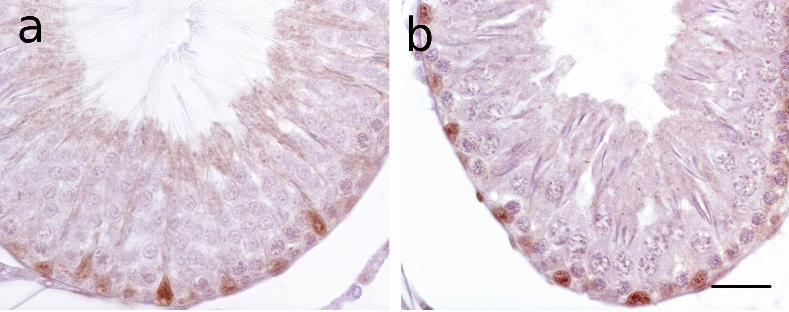
Figure 1. Comparative immunostaining patterns of apoptotic germ cells with the rabbit
polyclonal cleaved caspase-3 antibody in paraffin testicular sections of p-NP-induced and
uninduced rats (a-e), aging human (f) and spermatogenically active catfish (g) testes.
(Sections were counterstained with methyl green containing crystal violet). (a) A stage XIII
tubule of an induced rat with several caspase-3 labeled germ cells showing sequential
alterations in nuclear morphology associated with progressive cytoplasmic immunostaining,
including lateral compression of the nucleus (arrowhead). Inset in (a) is a high magnification
sagittal view of such germ cells which shows the nucleus to be fragmented into at least two
large pale pieces, as seen in this tubule from an uninduced rat. (b) Another aspect (lateral
view) of these large nuclear fragments showing cleaved caspase-3 strongly immunolocalized
to two sharply circumscribed dense chromatin masses fragments, as seen in a tubule from an
induced rat. In other labeled germ cells, (c) the nuclear fragments were much reduced in size
(arrows), as seen in this strongly immunostained spermatogonium in an induced rat. Inset in
(c) is a high magnification of a slightly earlier stage showing a blue stained region (*) in the
cytoplasm associated with three small pale nuclear fragments (arrows), as seen in this
immunostained spermatogonium from an uninduced rat. (d) With diminishing
immunoreactivity, as seen in this tubule from an uninduced rat, only the blue-purplish stained
regions remain readily visible, with these having the same color as the condensed chromatin
in spermatid nuclei (arrow). (e) The blue-purplish-stained regions also eventually become
smaller (arrow) until they are no longer observed in the still moderately immunostained
cytoplasm (arrowhead), as can be seen in this tubule from an induced rat. (f) In the aging
human testis, the entire germ cell, with its large ratio of cytoplasmic volume:nuclear volume,
can be intensely immunostained, or only the cytoplasm with the nucleus staining blue similar
in color to that of the spermatozoa head. Inset in (f) shows a germ cell with cleaved caspase-3
strongly immunolocalised to the large intact nucleus. (g) In the catfish, immunostaining
varied from being strictly cytoplasmic (inset), to nuclear (arrow), and to cytoplasmic at which
point the nuclei have an intense blue color similar to that of condensed chromatin in the
spermatozoa heads. Psc, primary spermatocytes; Ssc, secondary spermatocytes; sz,
spermatozoa. Bar: a = 20 µm; b, c, d, f, g = 10 µm.
Figure 2. Comparative TUNEL-staining patterns of apoptotic germ cells in testicular sections
of the rat (a,b), aging human (c) and spermatogenically active catfish (d) testes. (Sections
were counterstained with methyl green containing crystal violet). Testicular sections from
induced and uninduced rats gave similar results. (a) In the rat testis, non-descript-looking
germ cells are TUNEL-labeled (arrow). Spermatogonia with several blue-purplish colored
regions are TUNEL-negative (*). (b) A stage XIV rat seminiferous tubule showing an out-of-
position TUNEL-labeled primary spermatocyte located among secondary spermatocytes. (c)
The nuclei of TUNEL-labeled germ cells in the human testis are faintly discernible. Inset in
(c) shows the rare occurrence of a TUNEL-labeled human germ cell with two large blue-
purplish colored nuclear fragments. (d) In the catfish testis, primary and secondary
spermatocytes are often strongly TUNEL-labeled. Note that the shrunken germ cells, whose
nuclear compartments stain intense blue, are TUNEL-negative. Psc, primary spermatocytes;
Ssc, secondary spermatocytes; st, spermatids; sz, spermatozoa. Bar = 10 µm.
Figure 3. Immunostaining of rat testicular sections with the mouse anti-human GATA-4
monoclonal antibody. Testicular sections from induced and uninduced rats gave similar
results. (a) A stage III/IV tubule showing distinct labeling of the oblong shaped basally

18
located Sertoli nuclei. Some of the Sertoli nuclei have their long axes perpendicular to the
basement membrane. (b) A stage XII tubule showing oblong Sertoli nuclei with their long
axes lying horizontally along the basement membrane. Bar = 30 µm.
Figure 4. Stages of the rat seminiferous epithelial cycle (Part I), based on the immunostaining
patterns with the goat anti-mouse GATA-4 polyclonal antibody. Testicular sections from
induced and uninduced rats gave similar results. (a) Stage I. Antibody labels the Golgi-phase
of acrosome development. Note that the blue stained nuclei of the elongating spermatids are
generally located adluminally to the round spermatids with their small developing acrosomes.
(b) Stage III. Increased immunoreactivity indicates the enlarged acrosomal region. Note that
the blue-stained nuclei of the elongating nuclei have started to penetrate the seminiferous
epithelium such that they reach the primary spermatocytes. (c) Stage V. Immunostaining
shows the spreading of the acrosome over the round spermatid nucleus. Many of the
elongated spermatid heads are deeply embedded in the epithelium. (d) Stage VI.
Immunostaining labels the spreading acroplaxome across the spermatid nuclear surface, with
the acrosome at the apex of the spermatid nucleus most intensely immunostained. Note that
the nuclei of elongated spermatids have retracted towards the tubule lumen. (e) Stage VIII.
The moderately immunostained acroplaxome covers about a third of the round spermatid
nuclear surface. The blue-purple-stained nascent residual bodies are layered between the
mature spermatid nuclei and the round spermatids. (f) Stage IX. A recently spermiated tubule,
with a few remaining spermatids among the residual bodies near the tubule lumen. Labeling
of the acroplaxome is somewhat reduced. Note the reorientation of the spermatid nuclei with
their apices towards the basement membrane. This stage is one of the few stages in which the
Sertoli nuclei are faintly immunolabeled (arrowheads). Bar = 30 µm.
Figure 5. Stages of the rat seminiferous epithelial cycle (Part II), based on the
immunostaining patterns with the goat anti-mouse GATA-4 polyclonal antibody. Testicular
sections from induced and uninduced rats gave similar results. (a) Late stage IX. Less
prominently immunostained acrosomes clearly identify round spermatids as the only
postmeiotic germ cells present in the epithelium at this stage of the cycle. Note that the
residual bodies are absent. (b) Stage X. The spermatid nuclei have started to elongate.
Immunostaining shows the acrosomic head cap to extend from the apex towards the caudal
region of the V-shaped spermatid head. Some of these early elongating spermatids have
started to embed themselves deep into the epithelium, but they are not bundled yet. (c) Stage
XII. Moderate immunostaining along the length of elongated spermatid nuclei, which
continue to embed themselves deeper into the Sertoli cell body. Early signs of bundling of the
young spermatids can be observed. (d) Stage XIII. The spermatid nuclei are still
immunolabeled. Condensation of the chromatin in the spermatid nuclei has been completed,
as judged by their blue color reaction with the counterstain. The spermatid heads are bundled
together as they penetrate the Sertoli cell bodies through to the level of the pachytene
spermatocytes. (e). Early Stage I. The highly condensed nuclei of the elongated spermatids
are still prominently immunolabeled. The faint specks tightly associated with the nucleus of
the round spermatids are the immunolabeled Golgi vesicles which mark the assembly of the
new acrosome in these newly formed round spermatids. (f) Negative control of a stage VIII
tubule (left) and other stages (right), following the omission of the primary antibody. Bar = 30
µm.