Generation of multipotent cell lines from a distinct population of male germ line stem cells
14
REPRODUCTION RESEARCH Generation of multipotent cell lines from a distinct population of male germ line stem cells Fariborz Izadyar 1 , Francis Pau 1 , Joel Marh 1 , Natalia Slepko 2 , Tracy Wang 1 , Rafael Gonzalez 2 , Thomas Ramos 1 , Kyle Howerton 1 , Chauncey Sayre 1 and Francisco Silva 2 1 PrimeGen Biotech LLC and 2 New Stem Biosciences, 213 Technology Drive, Irvine, California 92618, USA Correspondence should be addressed to F Izadyar; Email: fi[email protected]; www.primegenbiotech.com Abstract Spermatogonial stem cells (SSCs) maintain spermatogenesis by self-renewal and generation of spermatogonia committed to differentiation. Under certain in vitro conditions, SSCs from both neonatal and adult mouse testis can reportedly generate multipotent germ cell (mGC) lines that have characteristics and differentiation potential similar to embryonic stem (ES) cells. However, mGCs generated in different laboratories showed different germ cell characteristics, i.e., some retain their SSC properties and some have lost them completely. This raises an important question: whether mGC lines have been generated from different subpopulations in the mouse testes. To unambiguously identify and track germ line stem cells, we utilized a transgenic mouse model expressing green fluorescence protein under the control of a germ cell-specific Pou5f1 (Oct4) promoter. We found two distinct populations among the germ line stem cells with regard to their expression of transcription factor Pou5f1 and c-Kit receptor. Only the POU5F1C/c-KitC subset of mouse germ line stem cells, when isolated from either neonatal or adult testes and cultured in a complex mixture of growth factors, generates cell lines that express pluripotent ES markers, i.e., Pou5f1, Nanog, Sox2, Rex1, Dppa5, SSEA-1, and alkaline phosphatase, exhibit high telomerase activity, and differentiate into multiple lineages, including beating cardiomyocytes, neural cells, and chondrocytes. These data clearly show the existence of two distinct populations within germ line stem cells: one destined to become SSC and the other with the ability to generate multipotent cell lines with some pluripotent characteristics. These findings raise interesting questions about the relativity of pluripotency and the plasticity of germ line stem cells. Reproduction (2008) 135 771–784 Introduction Application of stem cells for therapeutic purposes has been the focus of stem cell science since the successful derivation of human pluripotent embryonic stem (ES) cells from pre-implantation embryos in 1998 (Thomson et al. 1998). Later on, numerous studies (Thomson & Odorico 2000, Weissman et al. 2001) have explored the potential of different stem cells, including ES, embryonic germ (EG), and adult stem cells for cell replacement therapy. Recently, multipotent germ cell (mGC) lines have been generated from neonatal mouse using culture- induced reprogramming (Kanatsu-Shinohara et al. 2004), indicating the presence of cell populations in the mouse testes with the ability to acquire pluripotency in culture. Multipotent cell lines generated in that study possessed identical ES cell characteristics and were completely devoid of SSC functional properties, i.e., they were unable to repopulate recipient testes following testicular transplantation. Recently, the identity of subpopulation in the mouse testes with the ability to culture-induced reprogramming has been demonstrated (Seandel et al. 2007). Interestingly, the cell lines generated from this cell population acquired ES charac- teristics and yet maintained their SSC functional proper- ties. mGC lines could also be generated from adult mouse testes without reprogramming growth factors (Guan et al. 2006), indicating the presence of a subpopulation of cells with pluripotent characteristics in the adult testes. Altogether, these studies suggest the presence of different subpopulations of mouse testicular stem cells with different abilities. In the present study, to investigate whether the SSCs or different subpopulations of germ line stem cells generate the mGC lines, we took advantage of a transgenic mouse model expressing green fluorescence protein (GFP) driven by a germ line-specific Pou5f1 (Oct4) promoter (Yeom et al. 1996). POU5F1 (previously known as octamer-binding transcription factor 3/4, OCT4) was originally identified as an ES cell and a germ line-specific marker (Okamota et al. 1990, Scholer et al. 1990). The expression of Pou5f1 is regulated by a proximal promoter, a germ-specific distal enhancer, and a retinoic acid- responsive element (Saiti & Lacham-Kaplan 2007). At gastrulation, Pou5f1 expression is down-regulated and thereafter is maintained only in primordial germ cells q 2008 Society for Reproduction and Fertility DOI: 10.1530/REP-07-0479 ISSN 1470–1626 (paper) 1741–7899 (online) Online version via www.reproduction-online.org
-
Upload
independent -
Category
Documents
-
view
2 -
download
0
Transcript of Generation of multipotent cell lines from a distinct population of male germ line stem cells

R
EPRODUCTIONRESEARCHGeneration of multipotent cell lines from a distinct populationof male germ line stem cells
Fariborz Izadyar1, Francis Pau1, Joel Marh1, Natalia Slepko2, Tracy Wang1, Rafael Gonzalez2,Thomas Ramos1, Kyle Howerton1, Chauncey Sayre1 and Francisco Silva2
1PrimeGen Biotech LLC and 2New Stem Biosciences, 213 Technology Drive, Irvine, California 92618, USA
Correspondence should be addressed to F Izadyar; Email: [email protected]; www.primegenbiotech.com
Abstract
Spermatogonial stem cells (SSCs) maintain spermatogenesis by self-renewal and generation of spermatogonia committed to
differentiation. Under certain in vitro conditions, SSCs from both neonatal and adult mouse testis can reportedly generate multipotent
germ cell (mGC) lines that have characteristics and differentiation potential similar to embryonic stem (ES) cells. However, mGCs
generated in different laboratories showed different germ cell characteristics, i.e., some retain their SSC properties and some have lost
them completely. This raises an important question: whether mGC lines have been generated from different subpopulations in the mouse
testes. To unambiguously identify and track germ line stem cells, we utilized a transgenic mouse model expressing green fluorescence
protein under the control of a germ cell-specific Pou5f1 (Oct4) promoter. We found two distinct populations among the germ line stem
cells with regard to their expression of transcription factor Pou5f1 and c-Kit receptor. Only the POU5F1C/c-KitC subset of mouse germ
line stem cells, when isolated from either neonatal or adult testes and cultured in a complex mixture of growth factors, generates cell
lines that express pluripotent ES markers, i.e., Pou5f1, Nanog, Sox2, Rex1, Dppa5, SSEA-1, and alkaline phosphatase, exhibit high
telomerase activity, and differentiate into multiple lineages, including beating cardiomyocytes, neural cells, and chondrocytes. These data
clearly show the existence of two distinct populations within germ line stem cells: one destined to become SSC and the other with the
ability to generate multipotent cell lines with some pluripotent characteristics. These findings raise interesting questions about the
relativity of pluripotency and the plasticity of germ line stem cells.
Reproduction (2008) 135 771–784
Introduction
Application of stem cells for therapeutic purposes hasbeen the focus of stem cell science since the successfulderivation of human pluripotent embryonic stem (ES)cells from pre-implantation embryos in 1998 (Thomsonet al. 1998). Later on, numerous studies (Thomson &Odorico 2000, Weissman et al. 2001) have explored thepotential of different stem cells, including ES, embryonicgerm (EG), and adult stem cells for cell replacementtherapy. Recently, multipotent germ cell (mGC) lines havebeen generated from neonatal mouse using culture-induced reprogramming (Kanatsu-Shinohara et al.2004), indicating the presence of cell populations in themouse testes with the ability to acquire pluripotency inculture. Multipotent cell lines generated in that studypossessed identical ES cell characteristics and werecompletely devoid of SSC functional properties, i.e.,they were unable to repopulate recipient testes followingtesticular transplantation. Recently, the identity ofsubpopulation in the mouse testes with the ability toculture-induced reprogramming has been demonstrated(Seandel et al. 2007). Interestingly, the cell lines
q 2008 Society for Reproduction and Fertility
ISSN 1470–1626 (paper) 1741–7899 (online)
generated from this cell population acquired ES charac-teristics and yet maintained their SSC functional proper-ties. mGC lines could also be generated from adult mousetestes without reprogramming growth factors (Guan et al.2006), indicating the presence of a subpopulation of cellswith pluripotent characteristics in the adult testes.Altogether, these studies suggest the presence of differentsubpopulations of mouse testicular stem cells withdifferent abilities.
In the present study, to investigate whether the SSCs ordifferent subpopulations of germ line stem cells generatethe mGC lines, we took advantage of a transgenic mousemodel expressing green fluorescence protein (GFP)driven by a germ line-specific Pou5f1 (Oct4) promoter(Yeom et al. 1996). POU5F1 (previously known asoctamer-binding transcription factor 3/4, OCT4) wasoriginally identified as an ES cell and a germ line-specificmarker (Okamota et al. 1990, Scholer et al. 1990). Theexpression of Pou5f1 is regulated by a proximal promoter,a germ-specific distal enhancer, and a retinoic acid-responsive element (Saiti & Lacham-Kaplan 2007). Atgastrulation, Pou5f1 expression is down-regulated andthereafter is maintained only in primordial germ cells
DOI: 10.1530/REP-07-0479
Online version via www.reproduction-online.org

772 F Izadyar and others
(Yeom et al. 1996). PGCs, of both males and females,continue to expressPou5f1 as they proliferate and migrateto the genital ridges. In the males, the expression in germcells persists throughout fetal development and ismaintained postnatally in proliferating gonocytes, pros-permatogonia, and undifferentiated spermatogonia,including A single (As), A paired (Apr), and A aligned(Aal) spermatogonia (Pesce et al. 1998, Tadokora et al.2002). As in the mouse only the As spermatogonia areconsidered to be spermatogonial stem cells (SSCs),enriched populations of undifferentiated spermatogoniaincluding SSCs can be isolated by sorting the POU5F1-GFP cells from OG2 transgenic mouse model.
Germ line stem cells could further be subdividedbased on the expression of c-Kit receptor molecule.c-Kit, a tyrosine kinase receptor, and its ligand stem cellfactor (also known as kit ligand or steel factor) are keyregulators of PGC growth and survival (De Miguel et al.2002). c-Kit is expressed in PGCs from their initialsegregation to their arrival at the genital ridge. Inpostnatal mouse testes, it has been shown that c-Kitcan be used as a marker for the differentiation ofundifferentiated and differentiating type A spermato-gonia (Schrans-Stassen et al. 1999). Therefore, theexpression levels of POU5F1 and c-Kit were used inthis study to isolate distinct populations of germ line stemcells. We then analyzed the molecular and phenotypiccharacteristics of these cells extensively before and afterculture and compared their ability to generate multi-potent cell lines under a defined culture condition with amixture of growth factors. In addition, the functionalityof these subpopulations and their descendant mGC linesto repopulate recipient testes was evaluated using theSSC transplantation technique.
Results
For enrichment of germ line stem cells, both neonatal andadult testicular tissues were cultured on gelatin-coatedculture dishes for 2 h for differential adhesion to removesomatic cells but not germ cells (Kanatsu-Shinohara et al.2004). After differential adhesion, cell suspensionscontaining GFP-positive cells (4–10% in the neonates,0.01–0.05% in the adults) could be retrieved (Fig. 1a–c).Harvested germ cells were cultured in a stem cell culturemedium with a mixture of growth factors as described.Initial GFP signals disappeared after a few days in culture(Fig. 2a and b). Thereafter, the cells underwent distinctmorphological changes forming chain-like colonies thatcontinued to grow without GFP signal (Fig. 2c–e).Up-regulation of POU5F1 indicated by the occurrence ofGFP-positive cells within colonies appeared after 3–4weeks of culture (Fig. 2f). For expansion, the cells wereco-cultured with mouse embryonic fibroblast (MEF)feeders in the same culture medium supplemented with15% fetal bovine serum (FBS). For derivation of cell linesfrom adult mice, GFP-positive cells harvested after
Reproduction (2008) 135 771–784
differential adhesion were sorted using fluorescence-assisted cell sorting (FACS) and were directly cultured onMEF. Using OG2 or OG2-LacZ mice, 19 cell lines (10neonatal and 9 adult) have been generated. All cell linesexpressed either GFP (14 lines) or GFP-LacZ (5 lines)(Fig. 2g–i). In addition, a mGC line has been generatedfrom neonatal wild-type CD-1 mice, indicating that themethod is not limited to transgenic OG2 mice (data notshown). Selected cell lines have been frozen/thawed andpropagated for more than 40 splittings with an estimatedcell doubling time of 72 h (using both manual cell countand GFP sort; Fig. 3).
Next, we separated c-Kit-positive GFP-positive cellsfromthe c-Kit-negative GFP-positive cells by FACS.Amongthe GFP-positive cells, 60% were c-Kit negative, and c-Kitwas dimly expressed on the positive fraction. A linearcorrelation was observed between the expression level ofPOU5F1 and c-Kit in germ line stem cells, showing thatonly the POU5F1 high germ cells were c-Kit positive.Interestingly, the POU5F1 high germ line stem cells werelarger than POU5F1 low cells based on their forwardscatter distribution (Fig. 1). Only c-Kit-positive populationsgenerated mGC colonies and no cell line could begenerated from the c-Kit-negative pool. Among the growthfactors used in this study, removal of GDNF resulted insmaller colonies indicating the role of GDNF in the self-renewal of the mGCs. By contrast, removal of FGF2resulted in the differentiation of the colonies indicatingpossible role of FGF2 in the maintenance of the mGCs intheir undifferentiated stage. Removal of leukemia inhibi-tory factor (LIF) or EG factor (EGF) did not affect theexpansion nor the differentiation of the mGCs (Supple-mentary Fig. 1, which can be viewed online at www.re-production-online.org/supplemental).
Marker and imprinting profiles
The majority of cells in the mGC colonies expressedPOU5F1, NANOG, SSEA-1, and VASA (Fig. 4a–d). Theyalso expressed pluripotent genes Sox2, Dppa5, Rex1,eRas, and Crypto along with germ line-specific markers,including Stella, Dazl, Vasa, and cRet (Fig. 4q). Inaddition, the expression of POU5F1, NANOG, andSOX2 was confirmed by Western blot analysis (Fig. 4p).Among the three mGSC lines tested in this study, onlyone line showed Nanog expression and this is the linethat has been used for Western blot analysis. The mousecell line at passage 20 showed high telomerase activity(similar to ES cells) and normal karyotype (40, XY) (Fig. 5).
We also analyzed the global gene expression andimprinting patterns of the mGCs before and after cultureand compared that with ES cells. Interestingly, ourculture condition did not change the imprinting patternof the mGCs in all the DMR sites tested. In contrast to themouse ES cells that showed only a partial androgeneticimprinting, the mGCs clearly exhibited a 100%androgenetic imprinting pattern (Fig. 6). Somewhat
www.reproduction-online.org

Figure 1 Enrichment of GFPC subpopulationsfrom testicular stem cells isolated from thetransgenic OG2 mouse using flow cytometry.POU5F1C cells as indicated by GFP expressionwere found as a distinct cell population in bothneonatal (b) and adult (c) OG2 mouse comparedwith the wild-type (a). Among the POU5F1-positive cells, two clear subpopulations consist-ing of c-Kit positive (R5) and c-Kit negative (R2)were found (d and e). Correlation betweenexpression of GFP and c-Kit is shown in (f–h).Note that the c-Kit dim cells (blue) have also thehighest level of GFP (dashed outline) expression.(G) GFP expression in a 2D plot showing theexpression of c-Kit-negative cells (red) and c-Kitdim cells (blue). The c-Kit dim cells tend todisplay overall higher levels of GFP. Scatter plot ofthe GFP/c-Kit show that GFP-positive c-Kit-negative cells are low in both forward (FSC) andside scatter (SSC), indicating that they are small,round, clear cells. GFP-positive c-Kit-positivecells are forward scatter high and side scatterlow/medium, indicating that they are larger cellswith not much complexity.
Generation of multipotent germ line stem cells 773
surprisingly, our microarray analysis showed that theglobal gene expression pattern of the mGCs had 87%similarity before and after culture.
Differentiation potential
When mGCs were aggregated to form embryoid bodies(EBs), gastrulation was observedwithin 9–15 days (Fig. 7a).Cells in the EBs expressed early developmental markersincluding E-cadherin and laminin-1 (markers of polarizedepithelium, Fig. 7b and c); ZIC1, PAX6, and SOX1 (earlyectodermmarkers, Fig. 7d and f); GATA4 and FOXA2 (earlyendoderm markers, Fig. 7e and f); and BRACHURY, BMP4,and COL2A1 (early mesoderm markers, Fig. 7f). In culture,mGC colonies spontaneously differentiated into pheno-types expressing markers of cardiomyocytes (Fig. 7g–j),adipocytes (Fig. 7k), and neural cells (Fig. 7l and m). Someof the cells that spontaneously differentiated to cardio-myocytes exhibited rhythmic contractions for up to 3 days(Fig. 7i, the beating sequence of representative cells areshown in the Supplementary Video, which can be viewedonline at www.reproduction-online.org/supplemental).Using directed differentiation protocols, mGC lines couldbe induced to differentiate into neural cells representingneural progenitors (nestin, NEUROD1), neurons (MAP2,
www.reproduction-online.org
NF-68, GAD67), and glial cells (GFAP, MBP, A2B5, O4,NG2), as shown in Fig. 8a–g and j. They could alsobe induced to form cardiomyocytes (troponin-1, cardiacmyosin, desmin, NKX2.5, GATA4) or chondrocytes(collagen Xa1 and staining by alcian blue) (Fig. 8h–Iand k–l).
In a separate differentiation study with mGCs, wecounted the number of cells (nuclei) with and withoutstaining of neural markers in seven colonies within aculture, and the average percentage was estimated as17.6% for GFAPC cells, 2.5% for Tuj-1C cells, and 2.3%for MAP2C cells. In general, the efficiency of induceddifferentiation by these protocols was much higher in EScells compared with the mGCs.
Testes regeneration
Four weeks after transplantation, testes of the controlanimals as well as those that received POU5F1-positivec-Kit-positive cells showed no spermatogenesis in themajority of the seminiferous tubules. On the contrary,80% of those that received freshly isolated POU5F1-positive c-Kit-negative cells showed some degrees ofspermatogenesis throughout the testes, indicating thepresence of functional SSCs in the cell suspension.
Reproduction (2008) 135 771–784

Figure 2 Morphological changes during the development of multi-potent germ cell lines in culture. A few mouse POU5F1-GFP cells wereobserved in cell preparations before culture. Note the presence of large(arrow) and small (asterisk) GFP-positive cells (a). Shortly after culture,down-regulation of POU5F1 was observed (b). After the attachment ofthe cells in the second week of culture, obvious morphological changesoccurred (c and d). Approximately, 3 weeks after culture, coloniescontaining small round cells were formed (e). Up-regulation ofPOU5F1 was observed about 1 month after culture (f). Images of threeestablished mGC lines derived from neonatal OG2, adult OG2, orneonatal OG2-LacZ are presented in (g–i). Scale bars: 50 mm.
Figure 3 Doubling time of the mGS cells. Multipotent germ cell lines(mGC) were cultured on mouse embryonic fibroblast (MEF) feeders inculture medium supplemented with 15% fetal bovine serum (FBS). Atdifferent time points during culture, the number of GFPC cells wasanalyzed using fluorescence-assisted cell sorting (FACS). The data arerepresentative of three replicates. Note that the size of the GC coloniesincreased during culture (a–d). The analysis of GFPC cell count duringculture is shown in (e) with a cell doubling time estimated at 3 days(72 h). Scale bars: 60 mm.
774 F Izadyar and others
Similarly, our short-term transplantation experimentusing the cell trace marker carboxyfluorescein diacetatesuccinimidyl ester (CSFE) showed that only the c-Kit-negative subpopulation of germ line stem cells colo-nized the recipient testes (Fig. 9). No spermatogenesiswas found in the majority of seminiferous tubules of therecipient mice testes transplanted with the mGCs,indicating that these cells do not have SSC properties(Table 1).
Teratomas and chimera formation
For teratoma formation, we injected equal numbers ofmouse ES cells (as positive control) or POU5F1-GFP/LacZmGS cells into the skin, muscle, and testis of differentgroups of nude mice (one million cells/site). All recipientmice (6/6) receiving ES cells developed teratomas in allthree tissue types. By contrast, none of the mice (0/20)receiving mGSCs gave rise to teratomas (Fig. 9a–f;Supplementary Fig. 2, which can be viewed online atwww.reproduction-online.org/supplemental). Six weeksafter transplantation, POU5F1-GFP/LacZ cells were foundin skin, muscle, and testicular tissues (Fig. 9g–l). These datashow that mGCs, unlike ES cells, are non-tumorigenic. Wetested chimera formation by injecting cultured POU5F1-GFP/LacZ cells into eight-cell embryos and blastocysts ofCD-1 mice, an approach considered by many to be thehighest standard for proving the pluripotency of cells. Asshown in Fig. 10a–d, POU5F1-GFP/LacZ cells wereincorporated into the inner cell mass of the mouse
Reproduction (2008) 135 771–784
blastocysts. The embryos were transferred to the uterus ofpseudopregnant mice (a total of 45 fetuses from 119transferred embryos). At 12.5 dpc, staining of wholeembryos for LacZ (b-galactosidase activity) showeddistinctive patterns in the eye, brain, and limbs (Fig. 10e).The intensity of LacZ staining was much higher in chimericembryos receiving mouse ES cells than those injected withmGC lines. The distribution of chimeric cells is alsodemonstrated in histological sections of the brain, heart,gonadal ridge, and liver (Fig. 10l–o). The intensity and thenumber of LacZ-positive cells were much higher inchimeric embryos injected with LacZ-ES cells than thoseinjected with LacZ-GS cells (data not shown). Confir-mation of POU5F1-GFP/LacZ chimeric tissues wassupported by the presence of GFP DNA sequence in theectodermal (brain), mesodermal (heart), endodermal(liver), and testis of the chimeric pups (Fig. 10p), as wellas the presence of LacZ DNA (Fig. 10q) in all four tissues.These combined results clearly demonstrate that culturedmGCs are non-teratogenic stem cells with some pluripo-tent characteristics.
www.reproduction-online.org

Figure 4 Phenotypic and molecular charac-terization of multipotent germ cells. Immuno-localization of pluripotent markers are shown for(a–d) POU5F1, (e–h) NANOG, and (m–o) SSEA-1.(i–l) The germ cell marker, VASA, in mGC colonies.Scale bars: (a–h) 25 mm, (i–o) 20 mm; expression ofpluripotent and germ-specific markers determinedby RT-PCR is shown in (q). The Western blotanalysis of protein contents of POU5F1, NANOG,and SOX2 in mGC cells before and afterimmunoprecipitation (IP) is presented in (p). Notein the RT-PCR panel (q), among three mGC linestested only line 3 shows Nanog expression.
Generation of multipotent germ line stem cells 775
Discussion
In this study, using a transgenic mouse model, wespecifically isolated germ line stem cells from bothneonatal and adult testes based on their POU5F1expression. Germ line stem cells were further separatedinto two populations according to their expression levelof c-Kit. Our FACS analysis showed two distinct popu-lations regarding the expression levels of POU5F1 andc-Kit in germ line stem cells. Interestingly, only thePOU5F1/GFPC cells that possess c-Kit receptor moleculeresponded to culture and generated mGC lines and onlythe c-Kit-negative subpopulations repopulated the testisafter SSC transplantation. This indicates the existence oftwo distinct subsets of cells withingerm line stem cells, i.e.,a c-Kit-positive pool containing larger cells with higherPOU5F1 expression with the ability to become multi-potent germ line stem cell and a smaller population ofgerm line stem cells that have lower POU5F1 and no c-Kitexpression with SSC properties. Apparently, at least in themouse, some germ stem cells in the testis are either at
www.reproduction-online.org
different developmental stages or possess various signalingmolecules that allow them to respond to growth factors atdifferent speed. A recent study in the mouse showed that aG-protein (GP-125)-coupled receptor might play animportant role as a signaling molecule in this process(Seandelet al. 2007). The existence of stem cells at differentdevelopmental stages has been proposed in other neonataland adult tissues (Ratajczak et al. 2008).
We have derived multipotent cell lines from postnatalmouse testicular stem cells with some but not allpluripotent characteristics. Thesecell lines are distinctivelydifferent from the mGC lines obtained by the otherlaboratories (Kanatsu-Shinohara et al. 2004, Seandelet al. 2007), most notably, with regard to the extent ofpluripotentiality and teratoma formation. Based onmicroarray analysis, our mGC lines express pluripotentgenes, i.e.,Nanog andCrypto respectively 1000 and 5000times less than ES cells. Similarly, our cell lines expressoncogenes, i.e., p53, Eras, Bak, Int2, and c-myc, severalfolds lower than the ES cells (Supplementary Fig. 3, whichcan be viewed online at www.reproduction-online.org/
Reproduction (2008) 135 771–784

Figure 5 Telomerase activity and karyotype analysis. (a) Telomeraseactivity in adult adipose stem cells (ADSC), mouse ES cells, freshlyisolated germ line stem cells after c-Kit sorting, and multipotent germline stem cells at passage 10 (neonatal OG2). The telomerase activity inthe germ line stem cells is comparable with mouse ES cells and higherthan the ADSC cells. (b) Karyotype of the same neonatal OG2 cell line.The picture is representative of 80 metaphase spreads analyzed. Notethat after 15 passages the cells exhibit normal karyotype.
Figure 6 The imprinting analysis of multipotent germ cells before (NGC)and after culture (GC) and mouse ES cells with differentially methylatedregions such as Meg3, Peg10, Pou5f1, Igfr2, and Rasgrf1. Note, except aslight difference in the Meg3 locus, that the mGC showed strongsimilarity to NGC in their imprinting profile.
776 F Izadyar and others
supplemental). Comparisons of the present findings withpublished reports suggest that mGC lines generated byother groups and our cell lines might differ in terms ofdevelopmental stages, imprinting profiles, and differen-tiation potential. Indeed, germ line-derived cells in thepresent report have properties of ES cells such as markerexpression, broad differentiation potential, and limitedchimera formation. However, these cells also seem to haveretained germ cell-specific imprinting patterns and non-tumorigenic characteristics.
Several lines of evidence support the notion that ourcell lines retain their germ cell properties more than theyresemble the reported properties of ES cells. First, these
Reproduction (2008) 135 771–784
cells doubled their cell numbers in about 72 h(determined by both GFP sorting and manual counting).This cell doubling time, similar to that of germ line stemcells, is three times longer than that of the ES cells.Secondly, they seem to have molecular characteristicsdifferent from those in ES cells or other mGC lines. Ourresults on global gene expression analysis show that ourcell lines have 65% similarity to ES cells and 87% to germline stem cells. Among the genes tested, our cell linesshowed significantly higher expression level of germ line-specific genes (Vasa, Plzf, Gfra1, Dazl) and lowerexpression level of pluripotent genes (Pou5f1, Nanog,Dppa5, Sox2, Crypto). Thirdly, our cell lines are moredependent on GDNF for their self-renewal than LIF orFGF2. GDNF has been shown to be the key regulator ofthe self-renewal of male germ line stem cells (Kubota et al.2004, Ryu et al. 2005, Oatley et al. 2006), while LIF andFGF2 play crucial role in the self-renewal of the ES cells(Cartwright et al. 2005, Levenstein et al. 2005). Fourthly,the expression level of SSEA-1 in our cell lines was lowerthat found either in mouse ES cells (Supplementary Fig. 4,which can be viewed online at www.reproduction-online.org/supplemental) or other mGC line as reported
www.reproduction-online.org

Figure 7 Spontaneous differentiation of multipotentgerm cells. Gastrulation of embryoid body (EB; a)and the expression of markers indicative of polarizedepithelium (E-cadherin and laminin-1; b, c) and earlydevelopment of the three germ layers, i.e., ectoderm(ZIC1, PAX6, SOX1), endoderm (GATA4, FOXA2),and mesoderm (BRACHURY, BMP4, and COL2A1)are shown (d–f) during culture reprogramming mGCsalso differentiated spontaneously into cardiomyo-cytes (g–j), adipocytes (k), and neural cells (l and m).A video showing the rhythmic contraction of cellsand cell aggregates derived from mouse GCs ispresented in the Supplementary Video. Scale bars:(a and i) 50 mm, (c and e) 30 mm, (b and d) 25 mm,(g, h and l) 45 mm, (k) 12 mm.
Generation of multipotent germ line stem cells 777
by Kanatsu-Shinohara et al. (2004). It has been shown thatSSEA-1 may be involved in tumor invasion and metastasisin certain animal model systems (Kajiwara et al. 2005),suggesting that higher expression may reflect higherpotential for tumorigenesis. Finally, our multipotent GCsexhibited an androgenic imprinting pattern that isdifferent from mouse ES cells or other mGC lines reportedby other laboratories (Kanatsu-Shinohara et al. 2004,Seandel et al. 2007).
Despite all of the similarities to their germ lineancestors, our cell lines did not regenerate testesfollowing transplantation. One possibility is that mGC
www.reproduction-online.org
lines might have changed their phenotypic and/ormolecular signature during culture condition. Indeed, aslight difference in gene expression profile betweenthe mGCs and the non-cultured germ line stem cellswas observed. As some of the altered genes were celladhesion molecules, we speculate that at least someof these alterations might be due to the in vitrocondition, i.e., attachment to MEF instead of basementmembrane of testicular epithelium and the absenceof the nursing somatic Sertoli cells. On the contrary,mouse SSCs cultivated for a long period of time arereported to repopulate recipient testes following SSC
Figure 8 Induced differentiation of multipotentgerm cells into lineage-specific phenotypes.Confocal images of the cells that expressed neuralmarkers are shown in (a–g). Expression of theneural gene markers is shown in (j). Confocalimage of mGCs differentiated into cardiomyo-cytes are presented in (i). Expression of the cardiacgene markers is shown in (l). Alcian blue-positivechondrocyte after differentiation of mGCs isshown in (h). Expression of the chondrocyte-specific genes is shown in (k). Scale bars: (a, c andg) 20 mm, (b and h) 50 mm, (d–f and i) 10 mm.
Reproduction (2008) 135 771–784
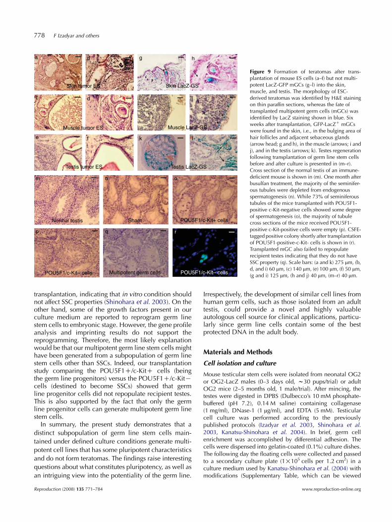
Figure 9 Formation of teratomas after trans-plantation of mouse ES cells (a–f) but not multi-potent LacZ-GFP mGCs (g–l) into the skin,muscle, and testis. The morphology of ESC-derived teratomas was identified by H&E stainingon thin paraffin sections, whereas the fate oftransplanted multipotent germ cells (mGCs) wasidentified by LacZ staining shown in blue. Sixweeks after transplantation, GFP-LacZC mGCswere found in the skin, i.e., in the bulging area ofhair follicles and adjacent sebaceous glands(arrow head; g and h), in the muscle (arrows; i andj), and in the testis (arrows; k). Testes regenerationfollowing transplantation of germ line stem cellsbefore and after culture is presented in (m–r).Cross section of the normal testis of an immune-deficient mouse is shown in (m). One month afterbusulfan treatment, the majority of the seminifer-ous tubules were depleted from endogenousspermatogenesis (n). While 73% of seminiferoustubules of the mice transplanted with POU5F1-positive c-Kit-negative cells showed some degreeof spermatogenesis (o), the majority of tubulecross sections of the mice received POU5F1-positive c-Kit-positive cells were empty (p). CSFE-tagged positive colony shortly after transplantationof POU5F1-positive-c-Kit- cells is shown in (r).Transplanted mGC also failed to repopulaterecipient testes indicating that they do not haveSSC property (q). Scale bars: (a and k) 275 mm, (b,d, and i) 60 mm, (c) 140 mm, (e) 100 mm, (f) 50 mm,(g and i) 125 mm, (h and j) 40 mm, (m–r) 40 mm.
778 F Izadyar and others
transplantation, indicating that in vitro condition shouldnot affect SSC properties (Shinohara et al. 2003). On theother hand, some of the growth factors present in ourculture medium are reported to reprogram germ linestem cells to embryonic stage. However, the gene profileanalysis and imprinting results do not support thereprogramming. Therefore, the most likely explanationwould be that our multipotent germ line stem cells mighthave been generated from a subpopulation of germ linestem cells other than SSCs. Indeed, our transplantationstudy comparing the POU5F1C/c-KitC cells (beingthe germ line progenitors) versus the POU5F1C/c-KitKcells (destined to become SSCs) showed that germline progenitor cells did not repopulate recipient testes.This is also supported by the fact that only the germline progenitor cells can generate multipotent germ linestem cells.
In summary, the present study demonstrates that a
distinct subpopulation of germ line stem cells main-
tained under defined culture conditions generate multi-
potent cell lines that has some pluripotent characteristics
and do not form teratomas. The findings raise interesting
questions about what constitutes pluripotency, as well as
an intriguing view into the potentiality of the germ line.
Reproduction (2008) 135 771–784
Irrespectively, the development of similar cell lines fromhuman germ cells, such as those isolated from an adulttestis, could provide a novel and highly valuableautologous cell source for clinical applications, particu-larly since germ line cells contain some of the bestprotected DNA in the adult body.
Materials and Methods
Cell isolation and culture
Mouse testicular stem cells were isolated from neonatal OG2or OG2-LacZ males (0–3 days old, w30 pups/trial) or adultOG2 mice (2–5 months old, 1 male/trial). After mincing, thetestes were digested in DPBS (Dulbecco’s 10 mM phosphate-buffered (pH 7.2), 0.14 M saline) containing collagenase(1 mg/ml), DNase-1 (1 mg/ml), and EDTA (5 mM). Testicularcell culture was performed according to the previouslypublished protocols (Izadyar et al. 2003, Shinohara et al.2003, Kanatsu-Shinohara et al. 2004). In brief, germ cellenrichment was accomplished by differential adhesion. Thecells were dispensed into gelatin-coated (0.1%) culture dishes.The following day the floating cells were collected and passedto a secondary culture plate (1!105 cells per 1.2 cm2) in aculture medium used by Kanatsu-Shinohara et al. (2004) withmodifications (Supplementary Table, which can be viewed
www.reproduction-online.org

Table 1 Restoration of spermatogenesis following transplantation of subpopulations of germ line stem cells and multipotent germ cell line in recipientmouse testes.
Transplanted cells Recipient animalsTubules with incomplete
spermatogenesisa (meanGS.D.)Tubules with complete
spermatogenesis (meanGS.D.)Empty tubules(meanGS.D.)
POU5F1Cc-KitC 1 17 4 982 11 3 753 12 2 924 15 (14G2.7)* 4 (3G0.9) 97 (90G10.6)
POU5F1Cc-KitK 1 114 6 332 96 8 413b 85 25 84 108 (104G12)† 5 (11G9.4) 34 (29G14.4)
mGC 1 12 11 782 18 13 873 11 8 694 18 (14G3.7)* 9 (10G2.2) 86 (80G8.3)
Sham 1 21 4 1122 15 7 953 18 3 1074 16 (17G2.6)* 6 (5G1.8) 86 (100G11.7)
*, †P!0.01. mGC, multipotent germ cell line.aIncomplete spermatogenesis defined as the tubule cross sections containing spermatogenic cells up to the spermatids. bMouse with the leastnumber of empty tubules and the highest number of tubules with complete spermatogenesis probably due to ineffective busulfan treatment.
Generation of multipotent germ line stem cells 779
online at www.reproduction-online.org/supplemental). After2–4 weeks in culture, GFP-positive colonies were mechani-cally transferred to culture dishes containing mitomycinC-treated murine embryonic fibroblast (MEF) feeder layers(see below). After passage for three to four times, viamechanical transfer, to new MEF cultures, the colonies wereestablished and could be removed from the culture plateenzymatically (collagenase 1 mg/ml, 5–10 min) for furtherexpansion and/or storage in liquid nitrogen. To enhance germline stem cell derivation, spermatogenesis was arrested inadult OG2 mice (4–6 weeks old, nZ4), i.e., testes weresurgically secured to the abdominal wall to becomecryptorchid as described previously (de Rooij et al. 1999).mGC lines were also generated from neonatal and non-cryptorchid adult OG2 mice by sorting the GFP-positive cells.To further study which subpopulation of germ line stem cellsgenerate these cell lines, the GFP-positive cells were sortedbased on their c-Kit expression and were cultured on MEFfeeders as described above. Finally, the effect of the growthfactor removal on self-renewal or differentiation of mGC lineswas investigated.
Preparation of MEF feeders
MEF feeders were made by the standard procedures using 12.5dpc CD-1 mouse embryos. The embryos were evisceratedbefore trypsinization and the dissociated cells plated onto150 mm plates at a plating density of w1.5 embryos per plate.After initial plating, MEFs were split 1:5 and then fro-zen/thawed (passage 1). Thawed MEFs (P1) were passed onlyonce for expansion purposes prior to mitomycin C treatment.MEF feeders were plated at a density of 50!103 to 60!103 percm2. New MEF feeders were used for pluripotent germ cellculture every 7–10 days. All animal experiments wereconducted in accordance with the National Research Council’sGuidelines for the Care and Use of Laboratory Animals.
www.reproduction-online.org
Evaluation of telomerase activity and karyotyping
For the determination of telomerase activity, cell extracts wereisolated from germ cell lines (passage 10 and higher), freshlyisolated POU5F1C/c-KitC sorted, and POU5F1C/c-KitKsorted cells using CHAPS lysis buffer containing 150 U/mlRNase. Cell lysates were centrifuged for 20 min at 12 000 g,4 8C, and the supernatants were stored at K80 8C. Proteinconcentration was measured by Bradford assay using BSA asthe standard. Telomerase activity was measured by PCR-basedassay using TRAPEZE detection kit (Chemicon, Temecula, CA,USA). Two microliters of the cell extract at 750 mg/ml wereadded to a total volume of 50 ml PCR mix containing the TRAPreaction buffer, dNTPs, substrate oligonucleotide, telomeraseprimer, internal standard primer, and Taq polymerase. Twomicroliters of mESC cell extract were added to the reaction mixas positive control, and CHAPS lysis buffer and heat-inactivatedtelomerase were used as negative control for each experi-mental sample. Each sample was incubated at 30 8C for 30 minfor telomerase extension, followed by PCR amplification.Karyotyping was performed at Coriell Cell Repositories,Cytogenetics Laboratory. For karyotyping, proliferating cellswere incubated in culture with 0.1 mg/ml KaryoMAX Colcemid(Invitrogen) for 3–4 h before they were resuspended inhypotonic solution (0.075 M KCl) and incubated at roomtemperature for 10 min. The cells were then resuspended in acold fixative (3:1 methanol:acetic acid) and stored at 4 8C for atleast 30 min. After washing with the fixative, the cells wereapplied to clean glass slides and air dried. Metaphasechromosomes were prepared and karyotypes created usingan Applied Spectral Imaging Band View digital imaging system.
In vitro differentiation
For generating EBs, mGSC colonies were dissociated withcollagenase and plated at a concentration of 1!106/well to2!106/well on 6-well non-adhesive culture plates in the
Reproduction (2008) 135 771–784

Figure 10 Chimeras formation after incorporation of multipotent GCs into blastocysts and host embryos. The incorporation of LacZ-GFPC mGC cellsduring early embryonic development and blastocyst formation is presented in (a–d). The majority of the GFP-LacZ cells injected at eight-cell stagehave been incorporated at day 2 of the embryonic development (arrow head) and some cells have not been incorporated yet (arrows). GFP-positivecells were further found at day 3.5 incorporated in inner cell mass (arrow) of the blastocyst. An example of four chimeric embryos showing differentdegree of chimerism is shown in (e) as whole embryo staining. To visualize the internal organs, sagittal sections of two of the embryos (indicated byasterisks) are also shown (f and g). (h–k) Chimeric pattern in dissected organs and (l–o) chimeric cell population in histological sections of the brain,heart, liver, and gonadal ridge (chimeric LacZ-GFP cells appear in blue). Amplification of the GFP and LacZ DNA in the tissues of the chimeric pupsis shown in (p and q). Scale bars: (a and c) 50 mm, (b and d) 25 mm, (e–g) 1250 mm, (h–k) 625 mm, (l–n) 50 mm, (o) 10 mm.
780 F Izadyar and others
complete medium containing 15% FBS (Hyclone, Logan ,UT,USA). In some experiments, EBs were formed in hanging drops
by aggregating 50!103 to 100!103 cells in 50 ml medium.For differentiation into cells representing the three germ layers,the EBs were cultured for 15 days, and every 3 days 20–30 EBs
Reproduction (2008) 135 771–784
were collected for RT-PCR analysis and 20–30 EBs were usedfor histological examinations. For induced differentiation, the
EBs were cultured in the complete medium for 4 days beforethey were cultured in the serum-free N1 medium for lineageselection, i.e., DMEM/F12 (Invitrogen) supplemented with ITS
www.reproduction-online.org

Generation of multipotent germ line stem cells 781
(insulin, 10 mg/l; transferrin, 5.5 mg/l; selenium, 0.67 mg/l)and fibronectin (50 mg/ml). After 5–7 days, N1-treated cellaggregates were transferred to gelatin-coated culture plates(Ying et al. 2003) in the N2 medium for expansion of neuralprogenitor cells, i.e., N1 medium with ITS, withoutfibronectin, and supplemented with bFGF (10 ng/ml). Fordifferentiation into cardiomyocytes, the EBs were cultured for2 weeks in the presence of different cardiogenic compoundsincluding DMSO (0.06 M), 5 0-aza-2 0-deoxycytidine AZA(5 mM), and Cardiogenol C (25–50 mM; Calbiochem, SanDiego, CA, USA) (Paquin et al. 2002, Choi et al. 2004). Duringthe differentiation process, the morphology of cells wasanalyzed and the samples were taken both for gene expressionanalysis by RT-PCR and immunohistochemical (IHC) staining(see below). Chondrocyte differentiation (Lee et al. 2004) ofmGSCs was induced by adding a chondrogenic inductionmedium (Chondrogenic SingleQuots, Cambrex, Walkersville,MD, USA) supplemented with transforming growth factor-3b(10 ng/ml) and 20% FBS.
Immunocytochemical and IHC staining
Cultured cells were fixed in 4% paraformaldehyde for 10–30 minat room temperature and stored in PBS at 4 8C. For fluorescentimmunocytochemistry, the cells were permeabilized with1!Cytoperm (BD Biosciences, San Jose, CA, USA) or 0.2%Triton X-100 (Sigma) for 15 min and subsequently incubated
Table 2 Antibodies and reagents used for immunohistochemical staining.
Lineage Name of antigen Antibody
Pluripotent POU5F1 Santa CruNANOG Bethyl labSOX2 Stem cellSSEA-1 ChemicoAlkaline phosphatase Abcam, CAlkaline phosphatase Cell Biola
Germ layer markers E-cadherin R&D SystLaminin-1 Santa CruSOX1 Affinity BFOXA2 Chemico
Neural Nestin ChemicoNG2 ChemicoMAP2 ChemicoGFAP ChemicoO4 ChemicoA2B5 ChemicoNeurofilament Chemico
Cardio Troponin-1 ChemicoMyosin heavy chain Abcam
Adipocyte Oil red dye AmericanChondro Alcian blue dye Fluka, StGerm cells VASA AbcamSecond antibodies Alexa 488 Molecula
Alexa 568 MoleculaAlexa 488 IgG1 MoleculaAlexa 488 IgG2b MoleculaAnti-mouse IgG Jackson LAnti-mouse IgM BD PharmAnti-rabbit Ig Jackson L
Nuclear dye DAPI MoleculaNuclear dye TO-PRO-3 MoleculaNuclear dye Hoechst 33342 MoleculaLacZ staining kit b-gal Sigma
www.reproduction-online.org
in 2% (w/v) BSA (Sigma) and 2% (v/v) normal goat serum(GS)/1! Cytoperm–PBS for 30–60 min both at roomtemperature. Primary antibody was either diluted at the optimalconcentration in 2% BSA and 2% GS/1! Cytoperm–PBS andincubated for 3 h at 4 8C or diluted in blocking buffer overnight at4 8C. After two washes, fluorescent secondary antibody wasdiluted accordingly (Supplementary Fig. 2) in 2% BSA and 2%goat serum/1! Cytoperm–PBS and incubated for 1 h at 4 8C inthe dark. The cells were washed twice with PBS, wrapped in foil,and stored at 4 8C until microscopic analysis. Images wererecorded using an Olympus IX71 microscope or Zeiss LSM510confocal microscope equipped with digital image hardwareand software.
For bright-field immunocytochemistry, the cells werewashed once with 1! PBS. Endogenous peroxidase activitywas blocked with 3% (v/v) H2O2 for 15 min followedby permeabilization – blocking with 2% BSA and 2%GS/1! Cytoperm–PBS for 30 min. The primary antibody wasdiluted accordingly (Supplementary Fig. 2) in 2% BSA and 2%GS/1! Cytoperm–PBS and incubated for 3 h at 4 8C. Theremainder of the staining was accomplished using ABCstaining kits (Vector Labs, Burlingame, CA, USA), accordingto the manufacturer’s instructions. Visualization was done withenhanced diaminobenzidine substrate (Sigma) tablet dissolvedin purified water and incubated for 5–10 min. For negativecontrols, the primary antibody was omitted. The primaryantibodies used in this study were obtained from various
source Working dilution
z 1:50oratories 1:50 to 1:100technologies 1:1000
n 1:50ambridge, MA, USA 1:50 to 1:100bs Inc., San Diego, CA, USA According to instructionsems 1:40z 1:100io reagents 1:100n 1:100n 1:200n 1:200n 1:200n 1:1000n 1:100n 1:100n 1:200n 1:200
1:200MasterTech Inc., Lodi, CA, USA Solution supplied by kit
Gallen, Switzerland 1% solution1:100 to 1:200
r Probes, Eugene, OR, USA 1:200r Probes 1:500r Probes (Carlsbad, CA, USA) 1:500r Probes (Carlsbad, CA, USA) 1:500abs, Bar Harbor, ME, USA 1:200ingen, San Diego, CA, USA 1:200
abs 1:200r Probes 1:1000r Probes 1:100r Probes 1:1000
According to instructions
Reproduction (2008) 135 771–784

782 F Izadyar and others
companies. Each antibody was validated and the concen-trations optimized in our laboratory. The source and workingdilutions of these antibodies are presented in Table 2.
Flow cytometry
Specific antibodies, including SSEA-1 and c-Kit, were optimizedin our laboratory for FACS analysis. Cells were sorted on theinFlux Cell Sorter (Cytopeia, Seattle, WA, USA). For c-Kit sorting,freshly isolated testicular cells containing the POU5F1-GFPconstruct were stained with CD117 APC (BD Pharmingen, SanDiego, CA, USA). GFPexcitation was attained with 488 nm solid-state Coherent laser (Coherent, Santa Clara, CA, USA) andemission light collected through a 530-40 dichroic mirror. APCexcitation was attained with 638 nm solid-state Coherent laser(Coherent) and emission light collected through a 710-40dichroic mirror. The cells were stained at a final dilution of1:200 c-Kit APC in the complete culture medium for 45 min onice in the dark. After washing twice with the fresh cold completemedium, the cells were concentrated at 10!106 cells/ml andkept on ice for sorting. To sort for GFP/c-KitC/K cells, first wegated the whole GFP population with the non-stained cells toestablish a baseline for APC channel. A region was placed basedon the baseline to discriminate between negative and positivec-Kit-stained cells. Any cells/events falling into this region wasconsidered to be positively stained for c-Kit and was sorted from
Table 3 Primers for marker determination by RT-PCR and primers used for
LineageNameof gene 5 0-Sequence
(A)Pluripotent Pou5f1 CCCCCTGTCCCCCATTCCTA
Nanog AGAAGGCCTCAGCACCTACDppa5 GAAAGATCCAGAGGTGTTCRex1 GCGTACGCAAATTAAAGTCCAGASox2 CCCCCGGCGGCAATAGCA
Gonadal Dazl GGAGCTATGTTGTACCTCCNeural Nestin GCCCTGACCACTCCAGTT TA
NeuroD1 AAGCCATGAACGCAGAGGAGGACTVimentin CAGGAAGAGCAGAAGAACTGGNF-68 (Nefl) ACCCGACTCAGTTTCACCAGGAD67 CGAGGACTCTGGACAGTAGAGGGFAP ACATCGAGATCGCCACCTACMBP GGAAACCACGCAGGCAAACGAGAG
Cardio Nkx2.5 CTTCAAGCCAGAGGCCTACGGATA4 GACGGGTCACTATCTGTGCAACa-Actin ACTGGGACGACATGGAGAAGANP GAACCAGAGGGGAGAGACAGAG
Chondro Collagen IIa1 GGGAGTAATGCAAGGACCAACollagen Xa1 GATATGGTGCTCCTGGTCGTAggrecan ACAGCTGGGGACATTAGTGGPerlecan TCCACCTGAGTACCCGAAAC
Housekeeping b-Actin ATCTGGCACCACACCTTCTACAATGAHousekeeping Gapdh ACCCAGAAGACTGTGGATGG
Name of gene 5 0-sequence
(B)Meg3 IG GGTTTGGTATATATGGATGTATTGTAARasgrf1 GTGTAGAATATGGGGTTGTTTTATATTIgfr2 TTAGTGGGGTATTTTTATTTGTATGGPeg10 GTAAAGTGATTGGTTTTGTATTTTTAAPou5f1 GGTTTTTTAGAG GATGGTTGAGTG
Reproduction (2008) 135 771–784
the negative c-Kit cells. For some experiments, fresh germ cellcolonies were dissociated and the cells were stained with anti-SSEA-1 antibody followed by goat-anti mouse IGM conjugatedwith PE-Cy7 (BD Biosciences).
Gene expression, imprinting analysis, and GFPamplification
Total RNA was isolated using RNeasy mini kit (Qiagen) and theRNA was used for RT-PCR, quantitative PCR, or microarrayanalysis. For RT-PCR, cDNA was synthesized with the Sensi-script RT kit (Qiagen), and the PCR was performed withHotStarTaq DNA Polymerase (Qiagen). All PCRs began with aninitial incubation at 95 8C for 15 min to activate the enzyme.This was followed by 35 cycles of 95 8C for 15 s, theappropriate annealing temperature for 1 min, and 72 8C for1 min, which was then followed by 1 cycle of 72 8C for 10 minfor final extension. The reactions were carried out using aniCycler Thermal Cycler (Bio-Rad). RT-PCR was carried outusing specific primers including Pou5f1, Nanog, Rex1, Dppa5,Dazl, b-actin (Actb), Nkx2.5, nestin, Mab2, and Gfap (primersequences are presented in Table 3). For internal controls,Gapdh was used as a housekeeping gene for cellular samplesand b-actin or interleukin-2 (IL2) was used in mouse embryos.
Imprinting patterns in mGSCs and mESCs were determined bya PCR-based analysis. PCR amplification of each dimethylated
imprinting study.
3 0-SequenceDNAsize
CGACCATCTGCCGCTTTGAG 577GGCCTGATTGTTCCAGGATT 205TGGTTCACTTCATCCAAG 297CAGATCCTAAACAGCTCGCAG 306TCGGCGCCGGGGAGATACAT 448CCATGTAACTAGATAAGCCAG 203GGAGTCCTGGATTTCCTTCC 199AGCTGTCCATGGTACCGTAA 579GGTAAAGAAGGCTGGTTGTGG 750CTCATCCTTGGCTTCCTCAG 216GATCTTGAGCCCCAGTTTTCT G 182ACATCACATCCTTGTGCTCC 219AAAAGAGGCGGATCAAGTGGGG 284TCATAGACCTGCTCCTGCG 305AGACATCGCACTGACTGAGAAC 475GCAAAGCGTAGCCCTCATAG 282CCCTCAGCTTGCTTTTTAGGAG 406ACCATCATCACCAGGCTTTC 178CACCTCTTTTTCCCACTCCA 96GTGGAATGCAGAGGTGGTTT 189CTGAAGTGACCAGGCTCCTC 130
GCTGCG CGTCATACTCCTGCTTGCTGATCCACATCTGC 838TTCAGCTCAGGGATGACCTT 125
3 0-sequence
TATAGG ATAAAACACCAAATCTATACCAAAATATACCG ATAATACAACAACAACAATAACAATC
AAATATCCTAAAAATACAAACTACACAAGTG-3 TTAATTACTCTCCTACAACTTTCCAAATT
TCCAACCCTACTAACCCATCACC
www.reproduction-online.org
Generation of multipotent germ line stem cells 783
region (DMR) from bisulfite-treated DNAs was carried out byspecific primers as described (Kanatsu-Shinohara et al. 2004).For analysis of the imprinted genes, we used the UVP imagesoftware to quantify the band intensity. For GFP and LacZamplification, individual tissues from chimeric embryos werecarefully collected by dissection, minced into small pieces, andplaced in DNA extraction buffer (DNeasy kit, Qiagen) for DNAisolation and purification, according to the manufacturer’sprotocol. PCR (1 ml DNA template containing 100 ng DNA) wascarried out as described previously (Izadyar et al. 2000).
Microarray analysis
Total cellular RNA was isolated using RNeasy mini kit (QiagenInc.), according to the manufacturer’s recommendations. Toeliminate DNA contamination, the samples were treated with2.0 U DNase I (Amplification grade, Invitrogen) at 37 8C for15 min, and the enzyme is inactivated by the addition of EDTA(2 mM final) at 65 8C for 10 min. The samples were concen-trated by ethanol precipitation and resuspended in RNase-freewater. Two micrograms of total RNA of each sample were sentto the UCI DNA Microarray Facility, where the samples wereprepared and hybridized to Affymetrix Mouse Genome 430 2.0GeneChips (Affymetrix Inc., Santa Clara, CA, USA) andscanned, according to the manufacturer’s protocols.
SSC transplantation
To test the functionality of the subpopulations of germ line stemcells and our mGCs for regeneration of spermatogenesis, we usedthe SSC transplantation technique. Sixteen 6–8 weeks immune-deficient nude male mice (Harlan, Indianapolis, IN, USA) havebeen treated with busulfan (40 mg/kg) and used as recipients.One month after busulfan treatment, 2!105 cells weretransplanted into the seminiferous tubules via rete testis injectionas described previously (Ogawa et al. 2000). Four mice weretransplanted with freshly isolated POU5F1-positive c-Kit-positivesorted cells, four mice injected with freshly isolated POU5F1-positive c-Kit-negative sorted cells, and four mice transplantedwith mGCs. As the mGCs are cultured on MEFs, they were sortedfor GFP to avoid MEF contamination during transplantation. Theremaining four mice served as sham control and not injected.One month after transplantation, the animals were killed and thetestes harvested and used for histological evaluations. To evaluatethe efficiency of transplantation, 100–150 tubule cross sections ofeach animal was examined and the number of tubules withdifferent stages of spermatogenesis was counted. Statisticalanalysis was carried out using ANOVA and P!0.05 wasconsidered significant. To better identify the transplanted cellsin the recipient testes, a fluorescent cell trace marker, CSFE,(Invitrogen) was used. CSFE is colorless and non-fluorescent untilthe acetate groups are cleaved by intracellular esterases to yieldhighly fluorescent product. This fluorescent product is wellretained and can be fixed with an aldehyde fixative; however, itdiminishes following multiple cell divisions and can be usedsuccessfully only in short-term studies. Two mice weretransplanted with POU5F1-positive c-Kit-negative cells and twoother mice received POU5F1-positive c-Kit-positive cells. Tendays after transplantation, the mice were killed and the number of
www.reproduction-online.org
CSFE-positive colonies was determined. Considering 72 h foreach cell doubling of germ line stem cells (Fig. 2), at this time aftertransplantation, SSCs could have gone through two to three celldoublings. Therefore, colonies of four to eight cells could havebeen formed.
Tests for teratoma and chimera formation
To test the ability of the mGCs to form teratomas or chimeras,OG2 mice (Jackson Laboratories, Bar Harbor, ME, USA) werebred with Rosa26 mice (Jackson Laboratories) and a new strain(OG2-R26) was generated. These mice have both GFP andLacZ constructs in their germ cells. Culture was performed asdescribed and the new POU5F1-GFP/LacZ germ cell lineswere produced for testing teratoma and chimera formation.Mouse POU5F1-GFP/LacZ mGSCs were examined for theirability to form teratomas in vivo by s.c., i.m., or injection intothe seminiferous tubules of nude mice (Harlan). As positivecontrols for teratoma formation, the ES cells were injected intosome mice. For s.c., i.m., or testicular injections, w1!106
cells were injected. Mice were killed 6 weeks later and thetissues harvested for morphological and histological analysis.
The ability of mouse POU5F1-GFP/LacZ GSCs to formchimeric cell populations was determined after injection intohost blastocysts, or by their aggregation with morula stageembryos or eight-cell stage embryos (Bradley 1987). Blastocystinjections of 15–20 cells were administered using 3.5-dayblastocysts collected from CD-1 mice following the procedureas described (Chatot et al. 1990). After injection, the blastocystswere transferred (7–8 blastocysts in each horn of the uterus) to2.5-day pseudopregnant CD-1 females, previously mated withvasectomized males. Incorporation of LacZ cells was examinedin different areas of the chimeric 12.5 dpc embryos by theb-galactosidase staining kit (Sigma). In addition, LacZ and GFPPCRs were performed in DNAs isolated from the brain, heart,liver, and gonadal ridges of the chimeric embryos formed fromPOU5F1-GFP/LacZ cells.
Acknowledgements
We would like to thank Dr John Sundsmo for critical reading ofthe manuscript and his valuable comments. We also thankJason Pacchiarotti, Michael Pascual, Carl Javier, Chad Maki,Susanne Csontos, Jadelind Wong, Julio Espinosa, Jessie Kinjo,Sandra Anorve, Jane Pham, and Vanessa Vargas for theirexcellent technical assistance. The authors declare that there isno conflict of interest that would prejudice the impartiality ofthis scientific work.
References
Bradley A 1987 Production and analysis of chimeric mice. In Teratocarci-nomas and Embryonic Stem Cells: A Practical Approach, pp 113–151.Ed. EJ Robertson. Oxford: JRI Press.
Cartwright P, McLean C, Sheppard A, Rivett D, Jones K & Dalton S 2005LIF/STAT3 controls ES cell self-renewal and pluripotency by a Myc-dependent mechanism. Development 132 885–896.
Chatot CL, Lewis JL, Torres I & Ziomek CA 1990 Development of 1-cellembryos from different strains of mice in CZB medium. Biology ofReproduction 42 432–440.
Reproduction (2008) 135 771–784

784 F Izadyar and others
Choi SC, Yoon J, Shim WJ, Ro YM & Lim DS 2004 5-azacytidine inducescardiac differentiation of P19 embryonic stem cells. Experimental andMolecular Medicine 36 515–523.
Guan K, Nayernia K, Maier LS,Wagner S, Dressel R, Lee JH, Nolte J, Wolf F,Li M, Engel W et al. 2006 Pluripotency of spermatogonial stem cells fromadult mouse testis. Nature 440 1199–1203.
Izadyar F, Van Tol HT, Hage WG & Bevers MM 2000 Preimplantationbovine embryos express mRNA of growth hormone receptor and respondto growth hormone addition during in vitro development. MolecularReproduction and Development 57 247–255.
Izadyar F, Den Ouden K, Creemers LB, Posthuma G, Parvinen M &de Rooij DG 2003 Proliferation and differentiation of bovine type Aspermatogonia during long-term culture. Biology of Reproduction 68272–281.
Kajiwara H, Yasuda M, Kumaki N, Shibayama T & Osamura Y 2005Expression of carbohydrate antigens (SSEA-1, sialyl-Lewis X, DU-PAN-2and CA19-9) and E-selectin in urothelial carcinoma of the renal pelvis,ureter, and urinary bladder. Tokai Journal of Experimental and ClinicalMedicine 30 177–182.
Kanatsu-Shinohara M, Inoue K, Lee J, Yoshimoto M, Ogonuki N, Miki H,Baba S, Kato T, Kazuki Y, Toyokuni S et al. 2004 Generation ofpluripotent stem cells from neonatal mouse testis. Cell 119 1001–1012.
Kubota H, Avarbock MR & Brinster RL 2004 Growth factors essential forself-renewal and expansion of mouse spermatogonial stem cells. PNAS101 16489–16494.
Lee OK, Kuo TK, ChenW-M, Lee K-D, Hsieh S-L & Chen T-H 2004 Isolationof multipotent mesenchymal stem cells from umbilical cord blood. Blood103 1669–1675.
Levenstein ME, Ludwig TE, Xu R-H, Llanas RA, VanDenHeuvel-Kramer K, Manning D & Thomson JA 2005 Basic fibroblast growthfactor support of human embryonic stem cell self-renewal. StemCells 24 568–574.
De Miguel MP, Cheng L, Holland EC, Federspiel MJ & Donovan PJ 2002Dissection of the c-Kit signaling pathway in mouse primordial germ cellsby retroviral mediated gene transfer. PNAS 99 10458–10463.
Oatley JM, Avarbock MR, Telaranta AI, Fearon DT & Brinster RL 2006Identifying genes important for spermatogonial stem cell self-renewaland survival. PNAS 103 9524–9529.
Ogawa T, Dobrinski I, Avarbock MR & Brinster RI 2000 Transplantation ofmale germ line stem cells restores fertility in infertile mice. NatureMedicine 6 29–34.
Okamota K, Okazawa H, Okuda A, Sakai M, Muramatsu M & Hamada H1990 A novel octamer binding transcription factor is differentiallyexpressed in mouse embryonic stem cells. Cell 60 461–472.
Paquin J, Danalache BA, Jankowski M, McCann SM & Gutkowska J 2002Oxytocin induces differentiation of P19 embryonic stem cells tocardiomyocytes. PNAS 99 9550–9555.
Pesce M, Wang X, Wolgemuth DJ & Scholer H 1998 Differential expressionof the oct-4 transcription factor during mouse germ cell differentiation.Mechanisms of Development 71 89–98.
Reproduction (2008) 135 771–784
Ratajczak MZ, Zuba-Surma EK, Wysoczynski M, Wan W, Ratajczak J,Wojakowski W & Kucia MA 2008 Hunt for pluripotent stem cell –regenerative medicine search for almighty cell. Journal of Autoimmunity30 151–162.
de Rooij DG, Okabe M & Nishimune Y 1999 Arrest of spermatogonialdifferentiation in jsd/jsd, S117H/S117H, and cryptorchid mice. Biologyof Reproduction 61 842–847.
Ryu B-Y, Kubota H, Avarbock MR & Brinster RL 2005 Conservation ofspermatogonial stem cell self-renewal signaling between mouse and rat.PNAS 102 14302–14307.
Saiti D & Lacham-Kaplan O 2007 Mouse germ cell development in vivoand in vitro. Biomarker Insights 2 241–252.
Scholer HR, Ruppert S, Suzuki N, Chowdhury K & Gruss P 1990 New typeof POU domain in germ line-specific protein Oct-4. Nature 344435–439.
Schrans-Stassen BH, Van de Kant HJ, de Rooij DG & Van Pelt AM 1999Differential expression of c-kit in mouse undifferentiated and differentiat-ing type A spermatogonia. Endocrinology 140 5894–5900.
Seandel M, James D, Shmelkov SV, Falciatori I, Kim J, Chavala S, Scherr DS,Zhang F, Torres R, Gale NW et al. 2007 Generation of functionalmultipotent adult stem cells from GPR125C germline progenitors.Nature 449 346–350.
Shinohara MK, Ogonuki N, Inoue K, Miki H, Ogura A, Toyokuni S &Shinohara T 2003 Long-term proliferation in culture and germlinetransmission of mouse male germline stem cells. Biology of Reproduc-tion 69 612–616.
Tadokoro Y, Yomogida K, Ohta H, Tohda A & Nishimune Y 2002Homeostatic regulation of germinal stem cell proliferation byGDNF/FSH pathway. Mechanisms of Development 113 29–39.
Thomson JA & Odorico JS 2000 Human embryonic stem cell andembryonic germ cell lines. Trends in Biotechnology 18 53–57.
Thomson JA, Itskovitz-Eldor J, Shapiro SS, Waknitz MA, Swiergel JJ,Marshall VS & Jones JM 1998 Embryonic stem cell lines derived fromhuman blastocysts. Science 282 1145–1147.
Weissman IL, Anderson DJ & Gage F 2001 Stem and progenitor cells:origins, phenotypes, lineage commitments, and transdifferentiations.Annual Review of Cell and Developmental Biology 17 387–403.
Yeom YI, Fuhramann G & Ovitt CE 1996 Germline regulatory element ofOct-4 specific for the totipotent cycle of embryonal cells. Development122 881–894.
Ying QL, Stavridis M, Griffiths D, Li M & Smith A 2003 Conversion ofembryonic stem cells into neuroectodermal precursors in adherentmonoculture. Nature Biotechnology 21 183–186.
Received 23 October 2007
First decision 2 January 2008
Revised manuscript received 5 March 2008
Accepted 25 March 2008
www.reproduction-online.org