Male infertility due to germ cell apoptosis in mice lacking the thiamin carrier, Tht1. A new insight...
11
Male infertility due to germ cell apoptosis in mice lacking the thiamin carrier, Tht1. A new insight into the critical role of thiamin in spermatogenesis Kimihiko Oishi, a Marco Barchi, b Audrey C. Au, a Bruce D. Gelb, a and George A. Diaz a, * a Departments of Human Genetics and Pediatrics, Mount Sinai School of Medicine, New York, NY, USA b Cell Biology Program, Memorial Sloan-Kettering Cancer Center, New York, NY, USA Received for publication 14 June 2003, revised 16 October 2003, accepted 20 October 2003 Abstract A mouse model of thiamin-responsive megaloblastic anemia (diabetes mellitus, deafness, megaloblastic anemia) lacking functional Slc19a2 has been generated and unexpectedly found to have a male-specific sterility phenotype. We describe here the characterization of the testis-specific effects of absence of the high-affinity thiamin transporter, Tht1. Null males were found to have hypoplastic testes secondary to germ cell depletion. Morphologic and expression analysis revealed that under conditions of standard thiamin intake, tissues affected in the syndrome (pancreatic h-cell, hematopoietic cells, auditory nerve) maintained normal function but pachytene stage spermatocytes underwent apoptosis. Under conditions of thiamin challenge, the apoptotic cell loss extended to earlier stages of germ cells but spared Sertoli cells and Leydig cells. Injection of high-dose thiamin was effective in reversing the spermatogenic failure, suggesting that the absence of the thiamin carrier could be overcome by diffusion-mediated transport at supranormal thiamin concentrations. These observations demonstrated that male germ cells, particularly those with high thiamin transporter expression beyond the blood –testis barrier, were more susceptible to apoptosis triggered by intracellular thiamin deficiency than any other tissue type. The findings described here highlight an unexpected and critical role for thiamin transport and metabolism in spermatogenesis. D 2003 Elsevier Inc. All rights reserved. Keywords: Spermatogenesis; Male infertility; Germ cells; Apoptosis; Thiamin; Thiamin-responsive megaloblastic anemia syndrome; Slc19a2 Introduction Spermatogenesis is a highly complex process involving several tightly regulated molecular interactions. The ab- sence or dysregulation of genes involved in this process results in male infertility with arrest of spermatogenesis at wide variety of stages (Escalier, 2001). An additional important aspect for sperm production in mammals is the nutritional environment (Wong et al., 2000). Deficien- cies in several vitamins have been found to impact on spermatogenesis through different mechanisms (Kodent- sova et al., 1994). Vitamin A deficiency results in male infertility due to the degeneration of most germ cells (Kim and Wang, 1993). Supplementation with retinoic acid (RA) reverses the pathology (van Pelt and de Rooij, 1991). In mice, loss of the RA receptor (RARs) or retinoid receptor (RXRs) disrupts spermatogenesis (Kast- ner et al., 1996; Lufkin et al., 1993), implicating this signaling pathway in the regulation of germ call matura- tion. Interestingly, RXRh is expressed principally in Sertoli rather than germ cells, illustrating that spermato- genic failure from nutritional deficiency may be mediated by supporting cells. a-Tocopherol (vitamin E) and ascor- bic acid (vitamin C) also have critical roles in spermato- genesis, apparently as antioxidants that counteract oxidative damage occurring during sperm development (Ciereszko and Dabrowski, 1995; Kodentsova et al., 1994; Vezina et al., 1996) rather than through direct effects on differentiation or apoptosis. Thiamin (vitamin B 1 ) is an essential coenzyme for several enzymes including transketolase, pyruvate dehydrogenase (PDH), a-ketoglutarate dehydrogenase, and branched-chain ketoacid dehydrogenase complexes. Dietary deficiency of 0012-1606/$ - see front matter D 2003 Elsevier Inc. All rights reserved. doi:10.1016/j.ydbio.2003.10.026 * Corresponding author. Departments of Human Genetics and Pediatrics, Mount Sinai School of Medicine, One Gustave Levy Place, PO Box 1498, New York, NY 10029. Fax: +1-212-849-2508. E-mail address: [email protected] (G.A. Diaz). www.elsevier.com/locate/ydbio Developmental Biology 266 (2004) 299 – 309
-
Upload
independent -
Category
Documents
-
view
2 -
download
0
Transcript of Male infertility due to germ cell apoptosis in mice lacking the thiamin carrier, Tht1. A new insight...

www.elsevier.com/locate/ydbio
Developmental Biology 266 (2004) 299–309
Male infertility due to germ cell apoptosis in mice lacking
the thiamin carrier, Tht1. A new insight into the critical role of
thiamin in spermatogenesis
Kimihiko Oishi,a Marco Barchi,b Audrey C. Au,a Bruce D. Gelb,a and George A. Diaza,*
aDepartments of Human Genetics and Pediatrics, Mount Sinai School of Medicine, New York, NY, USAbCell Biology Program, Memorial Sloan-Kettering Cancer Center, New York, NY, USA
Received for publication 14 June 2003, revised 16 October 2003, accepted 20 October 2003
Abstract
A mouse model of thiamin-responsive megaloblastic anemia (diabetes mellitus, deafness, megaloblastic anemia) lacking functional
Slc19a2 has been generated and unexpectedly found to have a male-specific sterility phenotype. We describe here the characterization of the
testis-specific effects of absence of the high-affinity thiamin transporter, Tht1. Null males were found to have hypoplastic testes secondary to
germ cell depletion. Morphologic and expression analysis revealed that under conditions of standard thiamin intake, tissues affected in the
syndrome (pancreatic h-cell, hematopoietic cells, auditory nerve) maintained normal function but pachytene stage spermatocytes underwent
apoptosis. Under conditions of thiamin challenge, the apoptotic cell loss extended to earlier stages of germ cells but spared Sertoli cells and
Leydig cells. Injection of high-dose thiamin was effective in reversing the spermatogenic failure, suggesting that the absence of the thiamin
carrier could be overcome by diffusion-mediated transport at supranormal thiamin concentrations. These observations demonstrated that male
germ cells, particularly those with high thiamin transporter expression beyond the blood–testis barrier, were more susceptible to apoptosis
triggered by intracellular thiamin deficiency than any other tissue type. The findings described here highlight an unexpected and critical role
for thiamin transport and metabolism in spermatogenesis.
D 2003 Elsevier Inc. All rights reserved.
Keywords: Spermatogenesis; Male infertility; Germ cells; Apoptosis; Thiamin; Thiamin-responsive megaloblastic anemia syndrome; Slc19a2
Introduction
Spermatogenesis is a highly complex process involving
several tightly regulated molecular interactions. The ab-
sence or dysregulation of genes involved in this process
results in male infertility with arrest of spermatogenesis at
wide variety of stages (Escalier, 2001). An additional
important aspect for sperm production in mammals is
the nutritional environment (Wong et al., 2000). Deficien-
cies in several vitamins have been found to impact on
spermatogenesis through different mechanisms (Kodent-
sova et al., 1994). Vitamin A deficiency results in male
infertility due to the degeneration of most germ cells (Kim
and Wang, 1993). Supplementation with retinoic acid
0012-1606/$ - see front matter D 2003 Elsevier Inc. All rights reserved.
doi:10.1016/j.ydbio.2003.10.026
* Corresponding author. Departments of Human Genetics and
Pediatrics, Mount Sinai School of Medicine, One Gustave Levy Place,
PO Box 1498, New York, NY 10029. Fax: +1-212-849-2508.
E-mail address: [email protected] (G.A. Diaz).
(RA) reverses the pathology (van Pelt and de Rooij,
1991). In mice, loss of the RA receptor (RARs) or
retinoid receptor (RXRs) disrupts spermatogenesis (Kast-
ner et al., 1996; Lufkin et al., 1993), implicating this
signaling pathway in the regulation of germ call matura-
tion. Interestingly, RXRh is expressed principally in
Sertoli rather than germ cells, illustrating that spermato-
genic failure from nutritional deficiency may be mediated
by supporting cells. a-Tocopherol (vitamin E) and ascor-
bic acid (vitamin C) also have critical roles in spermato-
genesis, apparently as antioxidants that counteract
oxidative damage occurring during sperm development
(Ciereszko and Dabrowski, 1995; Kodentsova et al.,
1994; Vezina et al., 1996) rather than through direct
effects on differentiation or apoptosis.
Thiamin (vitamin B1) is an essential coenzyme for several
enzymes including transketolase, pyruvate dehydrogenase
(PDH), a-ketoglutarate dehydrogenase, and branched-chain
ketoacid dehydrogenase complexes. Dietary deficiency of
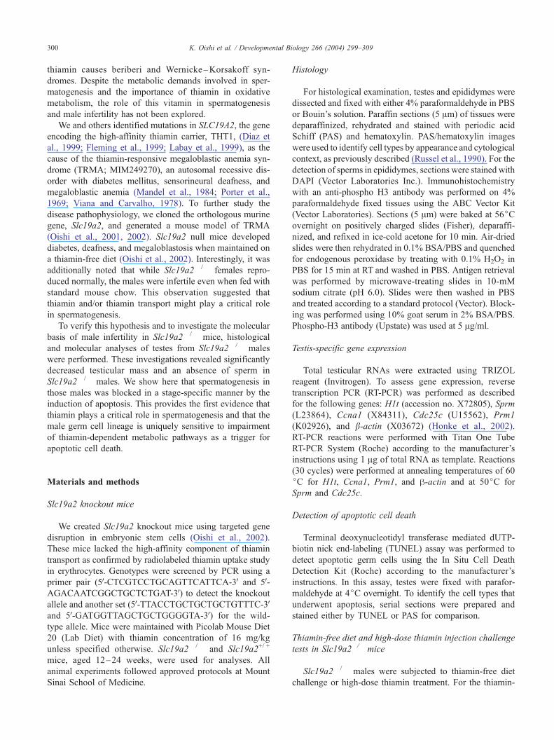
K. Oishi et al. / Developmental B300
thiamin causes beriberi and Wernicke–Korsakoff syn-
dromes. Despite the metabolic demands involved in sper-
matogenesis and the importance of thiamin in oxidative
metabolism, the role of this vitamin in spermatogenesis
and male infertility has not been explored.
We and others identified mutations in SLC19A2, the gene
encoding the high-affinity thiamin carrier, THT1, (Diaz et
al., 1999; Fleming et al., 1999; Labay et al., 1999), as the
cause of the thiamin-responsive megaloblastic anemia syn-
drome (TRMA; MIM249270), an autosomal recessive dis-
order with diabetes mellitus, sensorineural deafness, and
megaloblastic anemia (Mandel et al., 1984; Porter et al.,
1969; Viana and Carvalho, 1978). To further study the
disease pathophysiology, we cloned the orthologous murine
gene, Slc19a2, and generated a mouse model of TRMA
(Oishi et al., 2001, 2002). Slc19a2 null mice developed
diabetes, deafness, and megaloblastosis when maintained on
a thiamin-free diet (Oishi et al., 2002). Interestingly, it was
additionally noted that while Slc19a2� /� females repro-
duced normally, the males were infertile even when fed with
standard mouse chow. This observation suggested that
thiamin and/or thiamin transport might play a critical role
in spermatogenesis.
To verify this hypothesis and to investigate the molecular
basis of male infertility in Slc19a2� /� mice, histological
and molecular analyses of testes from Slc19a2� /� males
were performed. These investigations revealed significantly
decreased testicular mass and an absence of sperm in
Slc19a2� /� males. We show here that spermatogenesis in
those males was blocked in a stage-specific manner by the
induction of apoptosis. This provides the first evidence that
thiamin plays a critical role in spermatogenesis and that the
male germ cell lineage is uniquely sensitive to impairment
of thiamin-dependent metabolic pathways as a trigger for
apoptotic cell death.
Materials and methods
Slc19a2 knockout mice
We created Slc19a2 knockout mice using targeted gene
disruption in embryonic stem cells (Oishi et al., 2002).
These mice lacked the high-affinity component of thiamin
transport as confirmed by radiolabeled thiamin uptake study
in erythrocytes. Genotypes were screened by PCR using a
primer pair (5V-CTCGTCCTGCAGTTCATTCA-3V and 5V-AGACAATCGGCTGCTCTGAT-3V) to detect the knockout
allele and another set (5V-TTACCTGCTGCTGCTGTTTC-3Vand 5V-GATGGTTAGCTGCTGGGGTA-3V) for the wild-
type allele. Mice were maintained with Picolab Mouse Diet
20 (Lab Diet) with thiamin concentration of 16 mg/kg
unless specified otherwise. Slc19a2� / � and Slc19a2+/ +
mice, aged 12–24 weeks, were used for analyses. All
animal experiments followed approved protocols at Mount
Sinai School of Medicine.
Histology
For histological examination, testes and epididymes were
dissected and fixed with either 4% paraformaldehyde in PBS
or Bouin’s solution. Paraffin sections (5 Am) of tissues were
deparaffinized, rehydrated and stained with periodic acid
Schiff (PAS) and hematoxylin. PAS/hematoxylin images
were used to identify cell types by appearance and cytological
context, as previously described (Russel et al., 1990). For the
detection of sperms in epididymes, sections were stained with
DAPI (Vector Laboratories Inc.). Immunohistochemistry
with an anti-phospho H3 antibody was performed on 4%
paraformaldehyde fixed tissues using the ABC Vector Kit
(Vector Laboratories). Sections (5 Am) were baked at 56jCovernight on positively charged slides (Fisher), deparaffi-
nized, and refixed in ice-cold acetone for 10 min. Air-dried
slides were then rehydrated in 0.1% BSA/PBS and quenched
for endogenous peroxidase by treating with 0.1% H2O2 in
PBS for 15 min at RT and washed in PBS. Antigen retrieval
was performed by microwave-treating slides in 10-mM
sodium citrate (pH 6.0). Slides were then washed in PBS
and treated according to a standard protocol (Vector). Block-
ing was performed using 10% goat serum in 2% BSA/PBS.
Phospho-H3 antibody (Upstate) was used at 5 Ag/ml.
Testis-specific gene expression
Total testicular RNAs were extracted using TRIZOL
reagent (Invitrogen). To assess gene expression, reverse
transcription PCR (RT-PCR) was performed as described
for the following genes: H1t (accession no. X72805), Sprm
(L23864), Ccna1 (X84311), Cdc25c (U15562), Prm1
(K02926), and b-actin (X03672) (Honke et al., 2002).
RT-PCR reactions were performed with Titan One Tube
RT-PCR System (Roche) according to the manufacturer’s
instructions using 1 Ag of total RNA as template. Reactions
(30 cycles) were performed at annealing temperatures of 60
jC for H1t, Ccna1, Prm1, and h-actin and at 50jC for
Sprm and Cdc25c.
Detection of apoptotic cell death
Terminal deoxynucleotidyl transferase mediated dUTP-
biotin nick end-labeling (TUNEL) assay was performed to
detect apoptotic germ cells using the In Situ Cell Death
Detection Kit (Roche) according to the manufacturer’s
instructions. In this assay, testes were fixed with parafor-
maldehyde at 4jC overnight. To identify the cell types that
underwent apoptosis, serial sections were prepared and
stained either by TUNEL or PAS for comparison.
Thiamin-free diet and high-dose thiamin injection challenge
tests in Slc19a2�/� mice
Slc19a2� /� males were subjected to thiamin-free diet
challenge or high-dose thiamin treatment. For the thiamin-
iology 266 (2004) 299–309

Fig. 1. Weights of Slc19a2+/ + and Slc19a2� /� testes.
K. Oishi et al. / Developmental Biology 266 (2004) 299–309 301
free diet challenge, mice were maintained for 16 days on the
Thiamin Deficient Diet (0 mg/kg of thiamin; ICN Biomed-
icals, Inc.). For the high-dose thiamin rescue study, daily
intraperitoneal injections of thiamin–HCl (1 mg/day) were
administered for 10 and 20 days while the mice were fed
standard chow. After completion of these challenges, testes
were dissected and analyzed histologically.
In situ hybridization
Paraffin sections (5 Am) of adult wild-type testis were
used for in situ hybridization. Mouse Slc19a2 cDNA (nt
240–739 in exon 2) was PCR amplified using a primer pair
(5V-TTACCTGCTGCTGCTGTTTC-3V and 5V-GATGGT-TAGCTGCTGGGGTA-3V) and then cloned into pCRII-
TOPO vector (Invitrogen). Digoxigenin (DIG)-labeled
probes were synthesized using DIG RNA Labeling Kit
(SP6/T7) (Roche) according to the manufacturer’s instruc-
tion. Hybridization was performed as described previously
by (Braissant and Wahli, 1998). DIG signal was detected by
DIG Nucleic Acid Detection Kit (Roche). To identify the
cell types that showed Slc19a2 expression, serial sections
were prepared and were subjected to either in situ hybrid-
ization or PAS staining for comparison.
Statistics
Statistical significance was calculated using t test (sig-
nificant threshold: P < 0.05). Values are given as mean FSEM.
Results
Analysis of Slc19a2�/� mice
Using targeted gene disruption through homologous
recombination in embryonic stem cells, we generated the
Slc19a2� /� mice, as previously described (Oishi et al.,
2002). These mice showed diabetes mellitus, sensorineural
deafness, and megaloblastosis in their bone marrow when
fed on thiamin-deficient diet. With standard chow, they did
not present any TRMA symptoms and appeared to develop
normally. While expanding the colony, it became apparent
that Slc19a2� /� females reproduced normally but males
were infertile. This infertility was present in males that were
fed standard chow and observed over a period of 6 months.
The sexual behavior of the Slc19a2� /� males was normal
as assessed by the observation of normal mating behavior
and copulatory plugs.
Despite normal sexual behavior, testes from Slc19a2� /�
males were smaller than their wild-type littermates, suggest-
ing an intrinsic defect in spermatogenesis. The average testis
weight of the Slc19a2� /� males (36.3 F 1.5 mg; n= 16)
was approximately half that of wild type males at 12 weeks
of age (72.5 F 4.0 mg, n= 10; P < 0.00001) (Fig. 1). To
assess the basis of the infertility, seminal fluid was
expressed from epididymes. In contrast to the wild-type
males, no sperm was observed in seminal fluid obtained
from Slc19a2� /� males (data not shown). Aspermia was
confirmed by histological test of epididymes. As seen in
Fig. 2, nuclear DAPI staining reveals abundant sperm heads
in the epididymal lumen of Slc19a2+/ + males, but luminal
staining is absent from the Slc19a2� /� mice.
Histology of Slc19a2�/� testis
PAS-stained sections were used to characterize the pro-
gression of spermatogenesis in wild type and Slc19a2� /�
testes. As shown in low- and high-power light micrographs
of PAS-stained tissues, analysis of the Slc19a2� /� testes
revealed a complete disruption of spermatogenesis (Fig. 2).
Leydig cells and Sertoli cells in seminiferous tubules
appeared normal in structure and number but there was a
marked decrease in the cellularity of germ cells, particularly
at the adluminal side of the tubules. No mature sperm was
seen in tubules at any of the developmental stages. Specif-
ically, cells at metaphase I (MI), metaphase II (MII), or
spermatids were never observed in the Slc19a2� /� testes.
The number of middle pachytene spermatocytes was dra-
matically decreased and diplotene spermatocytes were rarely
seen, whereas the numbers of spermatogonia and early
primary spermatocytes were normal. Thus, from a histolog-
ical perspective, an arrest in spermatogenesis appeared to
have occurred at the pachytene stage before the first meiotic
division.
Testis stage-specific gene expression
To verify the stage of spermatogenesis arrest, we per-
formed gene expression analysis using RT-PCR (Fig. 3). In
this assay, we employed H1t, Sprm1, Ccna1, Cdc25c, and
Prm1, which encode histone H1t, POU-domain transcrip-
tion factor, cyclin A1, M-phase inducer phosphatase 3, and
protamine 1, respectively (Andersen et al., 1993; Kleene et
al., 1984; Kremer and Kistler, 1991; Ravnik and Wolge-
muth, 1999; Wu and Wolgemuth, 1995). The window of

Fig. 2. Abnormal spermatogenesis in Slc19a2� /� mice. (A, B) Cross-sections of DAPI-stained epididymes from 12-week-old Slc19a2� /� and Slc19a2+/ +
mice revealed aspermia in Slc19a2� / � males (original magnification 600� ). (C, D) PAS-stained testes exhibited arrest of spermatogenesis with profound loss
of germ cells in the seminiferous tubules compared to controls (magnification 200� ). (E, F) Post-meiotic haploid cells were never observed in the sections
from Slc19a2� /� testes (magnification 600� ).
K. Oishi et al. / Developmental Biology 266 (2004) 299–309302
expression for each of these genes during spermatogenesis
has been well documented in previous studies. H1t is
expressed in mid- to late-pachytene spermatocytes. Sprm1
is transcribed transiently just before the first meiotic divi-
sion from late pachytene to diplotene spermatocytes. Ccna1
message is observed just before or during the first meiotic
division. Cdc25c transcript, which is first expressed in late
pachytene diplotene stages, is highest in round spermatids.
Prm1 is a haploid-specific gene whose expression is high in
round spermatids. In the Slc19a2� /� testes, the expression
levels of early stage-specific genes, H1t and Sprm1, were
the same as observed in the wild type and Slc19a2+/� testes,
while the expression of Ccna1 and Cdc25c, which are
expressed from the pachytene stage onwards, was detected
at lower levels. Expression of the haploid-specific gene
Prm1 was not detectable in Slc19a2� /� testes. The results
of the gene expression analysis were thus consistent with an
arrest in spermatogenesis occurring during the late pachy-
tene or diplotene stages.
Phospho-histone-H3 staining
To further characterize the stage of spermatogenesis
arrest and to examine the proliferative activity of mitotic
cells, we performed immunostaining with anti-phospho-H3
antibody. Phosphorylation of histone H3 on Serine 10
occurs in mitotic cells during chromosome condensation
(Hendzel et al., 1997), in meiosis at the late pachytene-
diplotene stage, when synapsed homologous chromosomes
are separating, and at the MI stage (Cobb et al., 1999). Serial
sections from wild type and Slc19a2� /� testes were stained
with PAS and anti-phospho-H3 antibody and compared. In
wild type mice, H3 phosphorylation was specifically ob-
served in meiotic cells in stage XI diplotene spermatocytes

Fig. 4. Immunostaining with anti-phospho-H3 antibody. Immunostaining
against phospho-H3 was performed in testis sections from Slc19a2+/ + and
Slc19a2� /� mice (magnification 400� ). (A) Section of stage XI tubule
from Slc19a2+/ + testis showed diplotene (Di) spermatocytes with the
characteristic ‘‘speckled’’ staining at the centromeric heterochromatin
located near the nuclear envelope. (B) Testis section from Slc19a2� / �
mouse showed normal proliferating spermatogonia with H3 phosphoryla-
tion, which were observed in the basal area of the seminiferous tubule, but
no proliferating diplotene-stage cells were present.
Fig. 3. Stage-specific gene expression in Slc19a2� / � testes. RT-PCR
analyses of stage-specific genes. For genes expressed in early devel-
opmental stages, H1t and Sprm1, no difference was detected between
Slc19a2+/ +, Slc19a2+/�, and Slc19a2� / � testes, while Ccna1 and Cdc25c
expression, which are expressed from the pachytene stage onwards, was
reduced in Slc19a2� / �. Expression of the haploid-specific gene Prm1 was
not detectable in Slc19a2� /� testes. Experiments were repeated a minimum
of three times.
K. Oishi et al. / Developmental Biology 266 (2004) 299–309 303
(Fig. 4A) and in spermatogonia before stage XI (data not
shown). In Slc19a2� /� testis, many H3 positive spermato-
gonia were observed (Fig. 4B), while rare H3-positive
‘‘diplotene-like’’ and no MI cells were found, demonstrating
that the arrest in spermatogenesis occurred during the
pachytene stage in Slc19a2� /� testis.
Mechanism of spermatogenic block
Since there were decreased numbers of germ cells in the
testes of Slc19a2� /� males, we considered the possibility
that loss of these cells might involve apoptosis. To test this
hypothesis, we performed TUNEL assays on sections of
Slc19a2� /� testes. In agreement with our hypothesis, there
were many TUNEL positive cells in the Slc19a2� /� testes,
while only a few positive spermatogonia were seen in the
wild type control (Figs. 5A–D). In the Slc19a2� /� testes,
apoptotic cells were observed only in the tubules, not in the
interstitial area. Morphologically, the affected cells
appeared to be germ cells. The distribution of apoptotic
cells was not random, with clusters of positive cells
observed in a subset of approximately 10–20% of the
seminiferous tubules in the sections. To verify the identity
of the cell types that were undergoing apoptosis, we
examined serial sections stained with TUNEL and PAS
(Figs. 5E, F). This analysis revealed that spermatogonia
and early meiotic cells at the base of the tubules (prelepto-
tene to zygotene stages) were not TUNEL positive, where-
as early- and mid-pachytene spermatocytes were dying by
apoptosis. These results suggested that the apoptosis ob-
served in the Slc19a2� / � testes occurred in a stage-
specific fashion.

Fig. 5. Loss of high-affinity thiamin transport induces apoptotic cell death in spermatocytes. In situ TUNEL staining of Slc19a2+/ + and Slc19a2� /� testes.
TUNEL-positive cells are stained in green. (A, B) Occasional TUNEL-positive spermatogonia were seen in the wild type testes, but abundant positive cells
were observed in the Slc19a2� / � testes (magnification 200� ). (C, D) Apoptotic cells were observed only in the tubules of Slc19a2� /� testes towards the
adluminal surface, not in the interstitial area (magnification 600� ). (E, F) Serial sections from Slc19a2� /� testis with TUNEL and PAS staining showed that
the distribution of apoptotic cells was restricted to pachytene cells (magnification E: 400� , F: 600� ). E inset highlights the area shown as PAS stained serial
section in F.
K. Oishi et al. / Developmental Biology 266 (2004) 299–309304
Effects of thiamin deficiency and high-dose thiamin
injection
We challenged Slc19a2� /� mice with thiamin-free diet
for 16 days and assessed the effect on the germ cell popu-
lation. At the end of the challenge period, plasma thiamin
concentration was significantly decreased and the TRMA
phenotype appeared, as previously described (Oishi et al.,
2002). The histology of testes from wild type animals main-
tained on thiamin-deficient diet revealed no apparent effects
on the cellularity of seminiferous tubules (data not shown).
In contrast, testes from thiamin-depleted Slc19a2� /� males
demonstrated massive destruction of germ cells (Fig. 6B).
In addition to the loss of late spermatocytes, the spermato-
gonia and early spermatocytes that were unaffected under
normal dietary conditions were almost completely absent
under thiamin-deficient condition. Interestingly, Leydig cells
and Sertoli cells in null animals were resistant to thiamin
deficiency and the integrity of the seminiferous tubule
basement membranes appeared to be maintained.
These results suggested that the male germ cell lineage
was particularly dependent on thiamin status for survival,
but that cells beyond the BTB were particularly sensitive. To
test the hypothesis that higher plasma thiamin levels might
increase intracellular levels by diffusion-mediated transport
and rescue spermatogenesis, we administered daily intraper-

Fig. 6. Effects of thiamin deficiency and high-dose thiamin injection on spermatogenesis in Slc19a2� /� mice. (A–D) PAS-stained sections from Slc19a2� / �
testes at 200� original magnification. (A) Testis section from mouse maintained on standard chow. (B) Testis section from mouse challenged with thiamin-free
diet for 16 days. Thiamin depletion causes massive loss of germ cells in Slc19a2� /�, including spermatogonia and early spermatocytes. (C) The block in
spermatogenesis observed under standard diet is reversed with the administration of intraperitoneal (i.p.) high-dose thiamin (1 mg/day). After 10 days of i.p.
thiamin, the tubule histology normalized significantly, with increased number of germ cells in seminiferous tubules and differentiation of primary
spermatocytes into secondary spermatocytes and round spermatids. (D) After 20 days of i.p. thiamin, spermatogenesis has recovered completely, with the
appearance of all developmental cell types. (E) DAPI staining of Slc19a2� /� epididymis sections confirmed the presence of mature sperm in the lumen
(magnification 600� ). (F) Anti-phospho-H3 immunostaining in sections from mice treated with thiamin for ten days showed many diplotene (arrowheads) and
MI (arrows) cells (magnification 400� ). (G) RT-PCR analysis revealed equivalent Prm1 expression in testes from wild type and Slc19a2� /� mice treated for
ten days, whereas untreated Slc19a2� /� males showed no expression.
K. Oishi et al. / Developmental Biology 266 (2004) 299–309 305
itoneal injection of thiamin–HCl (1 mg/day) to 12-week old
Slc19a2� /� males. After 10 days of treatment, significant
normalization of tubule histology was appreciated with
Fig. 7. Localization of mRNA for Slc19a2 in of adult wild type testes by in situ h
cells from the mid-pachytene stage to the round spermatid stage, with the highest e
Serial sections of stage VIII– IX tubule with in situ hybridization and PAS showed
sense probe exhibited no signal.
increased numbers of germ cells observed in seminiferous
tubules (Figs. 6C, F). In addition to increased cellularity,
differentiation of primary spermatocytes into secondary
ybridization. (A, C) High levels of signal were observed in spermatogenic
xpression in late pachytene spermatocytes in stage VIII –XI tubules. (C, D)
the highest signal in late pachytene spermatocytes. (B) Hybridization with

K. Oishi et al. / Developmental Biology 266 (2004) 299–309306
spermatocytes and round spermatids (stages VII–VIII) and
staining of acrosomes by PAS was observed, but mature
sperm was not seen in any tubules. After 20 days of
treatment, the testicular mass of treated Slc19a2� /� mice
was significantly greater than that of untreated mice and
equivalent to wild type (81.7 F 7.2 mg; n= 6; P < 0.002).
Although histologic remnants of the preexisting pathology
were observed in occasional tubules, the block in spermato-
genesis was overcome, with all developmental cell types
present (Fig. 6D). Mature sperm production was confirmed
both in epididymis and testis (Fig. 6E). As expected from
the histological observation, Prm1 expression in testes from
mice treated with thiamin for 10 days was identical to wild
type controls (Fig. 6G). The reestablishment of spermato-
genesis was accompanied by the reduction of apoptosis to
levels equivalent to those observed in control testes as
assessed by TUNEL staining (data not shown).
To investigate whether fertility of the thiamin-treated
Slc19a2� /� males was restored, we mated two Slc19a2� /�
males after 20 days of high-dose thiamin injection with
non-thiamin-treated Slc19a2� /� females. Over a period of
3 months of observation, females experienced multiple
pregnancies and deliveries from each mated male, confirm-
ing that sperm from the treated males was completely
functional.
Slc19a2 gene expression in testis
To detect and localize the Slc19a2 mRNA message in
mouse testis, we performed in situ hybridization using adult
wild type testis sections. Overall, high expression levels
were detected in a subpopulation of cells in the seminiferous
tubules, while the level of mRNA expression in interstitial
cells was low (Fig. 7). PAS staining of serial sections
confirmed that the expression of Slc19a2 is developmentally
regulated during spermatogenesis. The upregulation of
Slc19a2 gene was initiated at the mid-pachytene stage and
persisted to the round spermatids stage, with the highest
expression observed in late pachytene spermatocytes in
stage VIII–XI tubules. The signal was low in Sertoli cells
as well as spermatogonia, preloptotene, leptotene, zygotene,
pachytene spermatocytes present in stage I–IV tubules, and
in elongated spermatids.
Discussion
In this report, we describe male-specific infertility in
mice lacking the high-affinity thiamin transporter, Tht1, due
to the induction of apoptosis in pachytene-stage spermato-
genic precursors. In contrast to the germ cells, the support-
ing Sertoli and Leydig cells appeared to be resistant to
thiamin deficiency. The dependence of spermatogenic pre-
cursors on Tht1 for survival is striking in light of the fact
that the tissues classically affected in TRMA showed no
phenotype under these dietary thiamin conditions. Plasma
thiamin concentration in animals fed standard mouse chow
reaches > 600 nM (Oishi et al., 2002) and is adequate for
intracellular thiamin in most cell types through non-satura-
ble diffusion. We hypothesize that the sensitivity of sper-
matogenic cells under conditions of normal thiamin intake
derives from several interacting factors including the met-
abolic demands of spermatogenic precursors, the presence
of the blood–testis barrier (BTB), and the relative balance
of pro- and anti-apoptotic factors in male germ cells.
There are several developmental changes associated with
spermatogenesis that are potentially relevant to the Tht1
requirement observed in germ cells. Spermatogonia close to
the seminiferous tubule basement membrane divide and
migrate towards the tubule lumen along the surface of
supporting Sertoli cells as they differentiate. Upon traversing
the BTB at the preleptotene to leptotene stage, spermatocytes
are completely dependent on cell–cell communication with
Sertoli cells for nutritional support and molecular signaling.
Germ cells beyond the BTB no longer have free access to
glucose and the transition to this environment is accompa-
nied by changes in germ cell carbohydrate metabolism
(Bajpai et al., 1998). Unlike spermatogonia and spermatids,
which utilize glucose or glucose/fructose as a principal
source of energy, spermatocytes preferentially utilize lac-
tate/pyruvate (Jutte et al., 1981; Mita and Hall, 1982;
Nakamura et al., 1984a,b). The change in energy substrates
coincides with the transcriptional inactivation of the X–Y
chromosome pair and the expression of autosomal isoforms
of proteins involved in energy metabolism, including phos-
phoglucokinase, cytochrome c, lactate dehydrogenase sub-
unit C, and the E1a subunit of pyruvate dehydrogenase
(PDH) (Goldberg et al., 1977; Iannello and Dahl, 1992;
McCarrey et al., 1992; Wheat et al., 1977). Changes related
to the expression pattern of PDH are of particular signifi-
cance, as this enzyme requires thiamin pyrophosphate as a
cofactor.
The upregulation of the testis-specific autosomal gene
encoding E1a2, Pdha-2, is initiated at the leptotene stage
and high expression levels are observed at the pachytene
stage (Iannello and Dahl, 1992). The switch in expression
pattern occurs in the context of a general increase in PDH
protein levels at the pachytene stage, corresponding to the
point at which apoptosis is initiated in Slc19a2� /� germ
cells. Interestingly, the upregulation of Slc19a2 in late
pachytene spermatocytes mirrors the expression pattern of
Pdha-2. E1a binds to thiamin pyrophosphate at two cata-
lytic sites in the holoenzyme (Ciszak et al., 2003). The
requirement for high-affinity thiamin transport might there-
fore reflect increased cellular requirement reflecting the
increased abundance of the apoenzyme or decreased affinity
of the Pdha-2-encoded subunit for thiamin pyrophosphate
secondary to variations in the coding sequence. This latter
possibility is not supported by studies of kinetic parameters
for thiamin pyrophosphate interactions with the two PDH
isoforms (Patel, personal communication). The change in
energy substrate makes the proper function of PDH critical

K. Oishi et al. / Developmental Biology 266 (2004) 299–309 307
for the production of acetyl-CoA and efficient energy
production by the TCA cycle. Lack of adequate intracellular
thiamin secondary to loss of Slc19a2 would thus be
expected to impair PDH activity and cellular energy metab-
olism, conceivably triggering spermatogenesis arrest at this
stage. Interestingly, antisense inhibition of the E1a subunit
in the anther tapetum of the sugar beet caused male-specific
sterility (Yui et al., 2003), highlighting the role of PDH in
male germ cell development in highly divergent species.
In addition to its effect on the energy substrates avail-
able to developing germ cells, the BTB is likely to have an
effect on the availability of thiamin. Little is known about
thiamin exchange across the BTB but a significant barrier
to diffusion has been demonstrated in the case of metho-
trexate, a small molecule using a transporter homologous to
Tht1 (Goldman and Matherly, 1985; Riccardi et al., 1982).
The induction of Slc19a2 expression in spermatogenic cells
that have crossed the BTB raises the possibility that
expression of the transporter is required to compensate
for low thiamin concentration in the intraluminal fluid.
Thus, it seems reasonable to speculate that inefficient
thiamin diffusion across the BTB may contribute to the
spermatogenic failure of Slc19a2� /� mice. Interestingly,
the central nervous system is not affected to the same
extent as spermatogenic cells, despite a high metabolic
requirement and evidence that thiamin transport across
the blood–brain barrier (BBB) is also carrier-mediated
(Greenwood et al., 1982; Spector, 1976). This difference
may reflect the involvement of alternative thiamin trans-
porters in the BBB or a different intrinsic susceptibility to
thiamin deficiency-induced apoptosis.
We propose a model of male infertility in the Slc19a2� /�
mice that centers on the relationship between the thiamin
metabolic demand of cells and the role of the BTB in
excluding thiamin from germ cells in the adluminal com-
partment of the tubule. In the Slc19a2� /� testis, germ cells
up to leptotene-stage spermatocytes have access to thiamin
available by interstitial-capillary diffusion, although they
have low expression of high-affinity thiamin transporter and
can maintain adequate energy homeostasis at plasma thia-
min concentrations maintained with standard mouse chow.
More differentiated germ cells beyond the BTB without
Tht1 are dependent on the thiamin diffused through BTB
and Sertoli cells and are thus susceptible to decreased
adluminal thiamin concentration, a situation compounded
by the reliance of pachytene spermatocytes on thiamin-
dependent pyruvate dehydrogenase for ATP synthesis. In-
jection of high-dose thiamin serves to increase the adluminal
thiamin concentration by passive diffusion, allowing re-
sumption of spermatogenesis.
The precise trigger for apoptosis in the susceptible germ
cells is not yet known. In contrast to the spermatogenic
failure observed with vitamin A deficiency, the loss of high-
affinity thiamin transport is unlikely to perturb differentia-
tion signals but rather to expose the susceptibility of germ
cells to mitochondrial insult. While we cannot rule out a
mechanism whereby Sertoli cells initiate apoptosis in germ
cells in response to metabolic stress, current models suggest
that maintenance of germ cell population size through
apoptosis is operative at the level of spermatogonia (Rodri-
guez et al., 1997), a population relatively resistant to
apoptosis under standard thiamin intake. In addition to
mitochondrial dysfunction resulting from the block in oxi-
dative phosphorylation, impairment of transketolase func-
tion may lead to dysfunction of the pentose phosphate
pathway resulting in oxidative stress (Calingasan et al.,
1999; Langlais et al., 1997) and impaired DNA synthesis
secondary to inefficient ribose production (Boros et al.,
1998). Both of these processes are candidates in the path-
way leading to spermatogenic failure in Slc19a2� /� mice.
Although our studies focused on mutant animals, extended
(30 days) dietary thiamin deficiency has been reported to
lead to aspermia and decreased numbers of spermatocytes
and spermatogonia in wild-type rats (Onodera et al., 1980),
suggesting that this mechanism is not restricted to organisms
lacking high-affinity thiamin transport.
One of the most intriguing questions arising from this
study is whether humans with TRMA or dietary thiamin
deficiency conditions have impaired spermatogenesis. Up to
now, male infertility has not been reported in TRMA. In an
adolescent male with TRMA, testicular size and develop-
ment was age-appropriate (Shalata, personal communica-
tion), but the significance of this observation is uncertain as
patients are maintained on high-dose thiamin from diagno-
sis, which is generally in early childhood. Nonetheless, mice
have a higher daily thiamin requirement than humans due to
a higher energetic demand increase per unit weight, raising
the possibility that murine germ cells may be intrinsically
more sensitive to thiamin. To our knowledge, the effect of
thiamin deficiency on spermatogenesis in humans has not
been reported. Should human germ cells prove as sensitive
as murine germ cells, it may be reasonable to assess the
contribution of subclinical dietary thiamin deficiency to
male infertility.
Acknowledgments
We thank Cheryl C. Tan for assistance with the mouse
experiments. This work was supported by a grant from the
March of Dimes to B.D.G. (FY00-283). M.B. was
supported by the Lalor Foundation Fellowship 2003.
References
Andersen, B., Pearse II, R.V., Schlegel, P.N., Cichon, Z., Schonemann,
M.D., Bardin, C.W., Rosenfeld, M.G., 1993. Sperm 1: a POU-domain
gene transiently expressed immediately before meiosis I in the male
germ cell. Proc. Natl. Acad. Sci. U. S. A. 90, 11084–11088.
Bajpai, M., Gupta, G., Setty, B.S., 1998. Changes in carbohydrate metab-
olism of testicular germ cells during meiosis in the rat. Eur. J. Endo-
crinol. 138, 322–327.
K. Oishi et al. / Developmental Biology 266 (2004) 299–309308
Boros, L.G., Lee, P.W., Brandes, J.L., Cascante, M., Muscarella, P.,
Schirmer, W.J., Melvin, W.S., Ellison, E.C., 1998. Nonoxidative pen-
tose phosphate pathways and their direct role in ribose synthesis in
tumors: is cancer a disease of cellular glucose metabolism? Med. Hy-
potheses 50, 55–59.
Braissant, O., Wahli, W., 1998. A simplified in situ hybridization protocol
using non-radioactively laveled probes to detect abundant and rare
mRNAs on tissue sections. Biochemica 1, 10–16.
Calingasan, N.Y., Chun, W.J., Park, L.C., Uchida, K., Gibson, G.E., 1999.
Oxidative stress is associated with region-specific neuronal death during
thiamine deficiency. J. Neuropathol. Exp. Neurol. 58, 946–958.
Ciereszko, A., Dabrowski, K., 1995. Sperm quality and ascorbic acid con-
centration in rainbow trout semen are affected by dietary vitamin C: an
across-season study. Biol. Reprod. 52, 982–988.
Ciszak, E.M., Korotchkina, L.G., Dominiak, P.M., Sidhu, S., Patel, M.S.,
2003. Structural basis for flip-flop action of thiamin pyrophosphate-
dependent enzymes revealed by human pyruvate dehydrogenase. J. Biol.
Chem. 278, 21240–21246.
Cobb, J., Miyaike, M., Kikuchi, A., Handel, M.A., 1999. Meiotic events at
the centromeric heterochromatin: histone H3 phosphorylation, topoiso-
merase II alpha localization and chromosome condensation. Chromo-
soma 108, 412–425.
Diaz, G.A., Banikazemi, M., Oishi, K., Desnick, R.J., Gelb, B.D., 1999.
Mutations in a new gene encoding a thiamine transporter cause thi-
amine-responsive megaloblastic anaemia syndrome. Nat. Genet. 22,
309–312.
Escalier, D., 2001. Impact of genetic engineering on the understanding of
spermatogenesis. Hum. Reprod. Updat. 7, 191–210.
Fleming, J.C., Tartaglini, E., Steinkamp, M.P., Schorderet, D.F., Cohen, N.,
Neufeld, E.J., 1999. The gene mutated in thiamine-responsive anaemia
with diabetes and deafness (TRMA) encodes a functional thiamine
transporter. Nat. Genet. 22, 305–308.
Goldberg, E., Sberna, D., Wheat, T.E., Urbanski, G.J., Margoliash, E.,
1977. Cytochrome c: immunofluorescent localization of the testis-spe-
cific form. Science 196, 1010–1012.
Goldman, I.D., Matherly, L.H., 1985. The cellular pharmacology of metho-
trexate. Pharmacol. Ther. 28, 77–102.
Greenwood, J., Love, E.R., Pratt, O.E., 1982. Kinetics of thiamine transport
across the blood–brain barrier in the rat. J. Physiol. 327, 95–103.
Hendzel, M.J., Wei, Y., Mancini, M.A., Van Hooser, A., Ranalli, T., Brink-
ley, B.R., Bazett-Jones, D.P., Allis, C.D., 1997. Mitosis-specific phos-
phorylation of histone H3 initiates primarily within pericentromeric
heterochromatin during G2 and spreads in an ordered fashion coincident
with mitotic chromosome condensation. Chromosoma 106, 348–360.
Honke, K., Hirahara, Y., Dupree, J., Suzuki, K., Popko, B., Fukushima, K.,
Fukushima, J., Nagasawa, T., Yoshida, N., Wada, Y., Taniguchi, N.,
2002. Paranodal junction formation and spermatogenesis require sulfo-
glycolipids. Proc. Natl. Acad. Sci. U. S. A. 99, 4227–4232.
Iannello, R.C., Dahl, H.H., 1992. Transcriptional expression of a testis-
specific variant of the mouse pyruvate dehydrogenase E1 alpha subunit.
Biol. Reprod. 47, 48–58.
Jutte, N.H., Grootegoed, J.A., Rommerts, F.F., van der Molen, H.J., 1981.
Exogenous lactate is essential for metabolic activities in isolated rat
spermatocytes and spermatids. J. Reprod. Fertil. 62, 399–405.
Kastner, P., Mark, M., Leid, M., Gansmuller, A., Chin, W., Grondona, J.M.,
Decimo, D., Krezel, W., Dierich, A., Chambon, P., 1996. Abnormal
spermatogenesis in RXR beta mutant mice. Genes Dev. 10, 80–92.
Kim, K.H., Wang, Z.Q., 1993. Action of vitamin A on the testis: role of the
Sertoli cell. In: Russel, L.D., Grisworld, M.D. (Eds.), The Sertoli Cell.
Cache River Press, Clearwater, FL, pp. 517–535.
Kleene, K.C., Distel, R.J., Hecht, N.B., 1984. Translational regulation and
deadenylation of a protamine mRNA during spermiogenesis in the
mouse. Dev. Biol. 105, 71–79.
Kodentsova, V.M., Vrzesinskaya, O.A., Spirichev, V.B., 1994. Male fertil-
ity: a possible role of vitamins. Ukr. Biokhim. Z. 66, 17–22.
Kremer, E.J., Kistler, W.S., 1991. Localization of mRNA for testis-specific
histone H1t by in situ hybridization. Exp. Cell Res. 197, 330–332.
Labay, V., Raz, T., Baron, D., Mandel, H., Williams, H., Barrett, T., Szar-
gel, R., McDonald, L., Shalata, A., Nosaka, K., Gregory, S., Cohen, N.,
1999. Mutations in SLC19A2 cause thiamine-responsive megaloblastic
anaemia associated with diabetes mellitus and deafness. Nat. Genet. 22,
300–304.
Langlais, P.J., Anderson, G., Guo, S.X., Bondy, S.C., 1997. Increased
cerebral free radical production during thiamine deficiency. Metab.
Brain Dis. 12, 137–143.
Lufkin, T., Lohnes, D., Mark, M., Dierich, A., Gorry, P., Gaub, M.P.,
LeMeur, M., Chambon, P., 1993. High postnatal lethality and testis
degeneration in retinoic acid receptor alpha mutant mice. Proc. Natl.
Acad. Sci. U. S. A. 90, 7225–7229.
Mandel, H., Berant, M., Hazani, A., Naveh, Y., 1984. Thiamine-dependent
beriberi in the ‘‘thiamine-responsive anemia syndrome’’. N. Engl. J.
Med. 311, 836–838.
McCarrey, J.R., Berg, W.M., Paragioudakis, S.J., Zhang, P.L., Dilworth,
D.D., Arnold, B.L., Rossi, J.J., 1992. Differential transcription of Pgk
genes during spermatogenesis in the mouse. Dev. Biol. 154, 160–168.
Mita, M., Hall, P.F., 1982. Metabolism of round spermatids from rats:
lactate as the preferred substrate. Biol. Reprod. 26, 445–455.
Nakamura, M., Okinaga, S., Arai, K., 1984a. Metabolism of pachytene
primary spermatocytes from rat testes: pyruvate maintenance of adeno-
sine triphosphate level. Biol. Reprod. 30, 1187–1197.
Nakamura, M., Okinaga, S., Arai, K., 1984b. Metabolism of round sper-
matids: evidence that lactate is preferred substrate. Am. J. Physiol. 247,
E234–E242.
Oishi, K., Hirai, T., Gelb, B.D., Diaz, G.A., 2001. Slc19a2: cloning and
characterization of the murine thiamin transporter cDNA and genomic
sequence, the orthologue of the human TRMA gene. Mol. Genet.
Metab. 73, 149–159.
Oishi, K., Hofmann, S., Diaz, G.A., Brown, T., Manwani, D., Ng, L., Young,
R., Vlassara, H., Ioannou, Y.A., Forrest, D., Gelb, B.D., 2002. Targeted
disruption of Slc19a2, the gene encoding the high-affinity thiamin trans-
porter Thtr-1, causes diabetes mellitus, sensorineural deafness and mega-
loblastosis in mice. Hum. Mol. Genet. 11, 2951–2960.
Onodera, K., Kisara, K., Okabe, H., Ogura, Y., 1980. Studies on the effects
of thiamine deficiency on rat testes. Nippon Yakurigaku Zasshi 76,
143–152.
Porter, F.S., Rogers, L.E., Sidbury Jr., J.B., 1969. Thiamine-responsive
megaloblastic anemia. J. Pediatr. 74, 494–504.
Ravnik, S.E., Wolgemuth, D.J., 1999. Regulation of meiosis during mam-
malian spermatogenesis: the A-type cyclins and their associated cyclin-
dependent kinases are differentially expressed in the germ-cell lineage.
Dev. Biol. 207, 408–418.
Riccardi, R., Vigersky, R.A., Barnes, S., Bleyer, W.A., Poplack, D.G.,
1982. Methotrexate levels in the interstitial space and seminiferous
tubule of rat testis. Cancer Res. 42, 1617–1619.
Rodriguez, I., Ody, C., Araki, K., Garcia, I., Vassalli, P., 1997. An early and
massive wave of germinal cell apoptosis is required for the development
of functional spermatogenesis. EMBO J. 16, 2262–2270.
Russel, L., Ettlin, R., Hikim, A., Clegg, E., 1990. Histological and Histo-
pathological Evaluation of the Testis. Cache River Press, Clearwater, FL.
Spector, R., 1976. Thiamine transport in the central nervous system. Am. J.
Physiol. 230, 1101–1107.
van Pelt, A.M., de Rooij, D.G., 1991. Retinoic acid is able to reinitiate
spermatogenesis in vitamin A-deficient rats and high replicate doses
support the full development of spermatogenic cells. Endocrinology
128, 697–704.
Vezina, D., Mauffette, F., Roberts, K.D., Bleau, G., 1996. Selenium-vita-
min E supplementation in infertile men. Effects on semen parameters
and micronutrient levels and distribution. Biol. Trace Elem. Res. 53,
65–83.
Viana, M.B., Carvalho, R.I., 1978. Thiamine-responsive megaloblastic
anemia, sensorineural deafness, and diabetes mellitus: a new syn-
drome? J. Pediatr. 93, 235–238.
Wheat, T.E., Hintz, M., Goldberg, E., Margoliash, E., 1977. Analyses of
stage-specific multiple forms of lactate dehydrogenase and of cyto-

K. Oishi et al. / Developmental Biology 266 (2004) 299–309 309
chrome c during spermatogenesis in the mouse. Differentiation 9,
37–41.
Wong, W.Y., Thomas, C.M., Merkus, J.M., Zielhuis, G.A., Steegers-The-
unissen, R.P., 2000. Male factor subfertility: possible causes and the
impact of nutritional factors. Fertil. Steril. 73, 435–442.
Wu, S., Wolgemuth, D.J., 1995. The distinct and developmentally regulated
patterns of expression of members of the mouse Cdc25 gene family
suggest differential functions during gametogenesis. Dev. Biol. 170,
195–206.
Yui, R., Iketani, S., Mikami, T., Kubo, T., 2003. Antisense inhibition of
mitochondrial pyruvate dehydrogenase E1alpha subunit in another ta-
petum causes male sterility. Plant J. 34, 57–66.