Immunohistochemical Studies of Amyloid P Component Distribution in Normal Human Skin
10
British Journal of Dermatology (1983) 108, 267-275. Immunohistochemical studies of amyloid P component and fibronectin in erythropoietic protoporphyria S.M.BREATHNACH*t, B.BHOGAL*, F.C.DE BEER^., S.M.MHLROSE§, M.M.BLACK* AND M.B.PEPYSi: * Institute of Dertnaiology, St John's Hospital for Diseases of the Skin, Lisle Street, Leicester Square, London WC2H 7BJ, t Immunological Medicine Unit, Department of Medicine, and § Department of Histopathology, Royal Postgraduate Medical School, Hammersmith Hospital, Du Cane Road, London W12 OHS Accepted for publication 9 August 1982 SUMMARY Immunofluorescence staining of exposed skin from patients with erythropoietic protoporphyria (EPP) with antibodies to serum amyloid P component (SAP) and to fibronectin produced striking fluorescence of abnormal vascular structures in the upper dermis. An appearance of linear fluorescence along the dermo-epidermal junction with anti-SAP was the result of confluent staining of papillary oxytalan fibres. Amyloid P component (AP) was localized in uitrastructural immunoperoxidase studies to the peripheral (abluminal) regions of thickened dermal vessel walls, the site of maximum concentration of an amorphous matrix containing microfibrillar structures; antibodies to SAP did not bind to leaflets of the reduplicated vascular basal lamina. The characteristic thickening and reduplication of blood vessel walls seen with the electron microscope in EPP therefore involves increased local deposition of both AP and fibronectin. Examination of skin biopsies from light-exposed areas in erythropoietic protoporphyria (EPP) reveals the characteristic deposition of periodic-acid Schiff (PAS)-positive amorphous hyaline material in and around the walls of upper dermal blood vessels (Peterka, Fusaro & Goltz, 1965; Ryan, 1966; Rimington et al., 1967; Ryan & Madill, 1968; Anton-Lamprecht & Meyer, 1970; Epstein, Tuffanelli & Epstein, 1973). Uitrastructural studies of EPP have shown that the hyaline thickening of vascular walls is the result of multiple concentric reduplication of the vascular basal lamina and the presence of afinelyfibrillarmaterial permeating and surrounding the vessel wail (Ryan & Madill, 1968; Anton-Lamprecht & Meyer, 1970; Epstein et al., 1973; t Present address: Dermatology Branch, National Cancer Institute, Bethesda, Maryland 20205, U.S.A. Correspondence: Dr M.B.Pepys. 0007-0963/83/0300-0267S02.00 (; 1983 British Association of Dermatologists 267
-
Upload
independent -
Category
Documents
-
view
2 -
download
0
Transcript of Immunohistochemical Studies of Amyloid P Component Distribution in Normal Human Skin

British Journal of Dermatology (1983) 108, 267-275.
Immunohistochemical studies of amyloidP component and fibronectin inerythropoietic protoporphyria
S.M.BREATHNACH*t, B.BHOGAL*, F.C.DE BEER^., S.M.MHLROSE§,M.M.BLACK* AND M.B.PEPYSi:
* Institute of Dertnaiology, St John's Hospital for Diseases of the Skin, Lisle Street, Leicester Square, LondonWC2H 7BJ, t Immunological Medicine Unit, Department of Medicine, and § Department of Histopathology, Royal
Postgraduate Medical School, Hammersmith Hospital, Du Cane Road, London W12 OHS
Accepted for publication 9 August 1982
SUMMARY
Immunofluorescence staining of exposed skin from patients with erythropoietic protoporphyria(EPP) with antibodies to serum amyloid P component (SAP) and to fibronectin producedstriking fluorescence of abnormal vascular structures in the upper dermis. An appearance oflinear fluorescence along the dermo-epidermal junction with anti-SAP was the result ofconfluent staining of papillary oxytalan fibres. Amyloid P component (AP) was localized inuitrastructural immunoperoxidase studies to the peripheral (abluminal) regions of thickeneddermal vessel walls, the site of maximum concentration of an amorphous matrix containingmicrofibrillar structures; antibodies to SAP did not bind to leaflets of the reduplicated vascularbasal lamina. The characteristic thickening and reduplication of blood vessel walls seen with theelectron microscope in EPP therefore involves increased local deposition of both AP andfibronectin.
Examination of skin biopsies from light-exposed areas in erythropoietic protoporphyria (EPP)reveals the characteristic deposition of periodic-acid Schiff (PAS)-positive amorphous hyalinematerial in and around the walls of upper dermal blood vessels (Peterka, Fusaro & Goltz, 1965;Ryan, 1966; Rimington et al., 1967; Ryan & Madill, 1968; Anton-Lamprecht & Meyer, 1970;Epstein, Tuffanelli & Epstein, 1973). Uitrastructural studies of EPP have shown that thehyaline thickening of vascular walls is the result of multiple concentric reduplication of thevascular basal lamina and the presence of a finely fibrillar material permeating and surroundingthe vessel wail (Ryan & Madill, 1968; Anton-Lamprecht & Meyer, 1970; Epstein et al., 1973;
t Present address: Dermatology Branch, National Cancer Institute, Bethesda, Maryland 20205, U.S.A.
Correspondence: Dr M.B.Pepys.0007-0963/83/0300-0267S02.00 (; 1983 British Association of Dermatologists
267
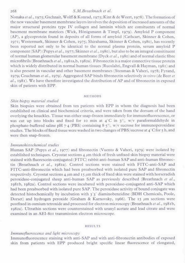
268 S.M.Breathnach et al.Nonakaefa/., i975;Gschnait, Woltf & Konrad, 1975;Kim&deWeert, i978).Theformationofthe new vascular basement membrane layers involves the deposition of increased amounts of themajor structural proteins type IV collagen and laminin which are consituents of normalbasement membrane matrices (Wick, Honigsmann & Timpl, 1979). Amyloid P component(AP), a glycoprotein found in deposits of all forms of amyloid (Cathcart, Skinner & Cohen,1971; Westermark, Skinner & Cohen, 1975; Shirahama, Skinner & Cohen, 1980J, has recentlybeen reported not only to be identical to the normal plasma protein, serum amyloid Pcomponent (SAP) (Pepys et al., 1977; Skinner et ai, 1980), but also to be an integral consitituentof normal human glomerular basement membrane (Dycktv al., 1980) and of normal elastic fibremicrofibrils (Breathnach et al., 1981 a,b, 1982a). Fibronectin is a major connective tissue proteinwhich is widely distributed in normal human tissues (Ruoslahti, Engvall & Hayman, 1981) andis also present in vascular and other basement membranes (Stenman & Vaheri, 1978; Fyrand,1979; Couchman et al., 1979). Aggregated SAP binds fibronectin selectively in vitro (de Beer etal., 1981). We have therefore investigated the distribution of AP and of fibronectin in exposedskin ofpatients with EPP.
METHODS
Skin biopsy material studiedSkin biopsies were obtained from ten patients with EPP in whom the diagnosis had beenestablished on clinical and biochemical criteria, and were taken from the dorsum of the handoverlying the knuckles. Tissue was either snap-frozen immediately for immunotluorescence, orwas cut up into blocks and fixed for 10 min at 4 C in 3",, w/v paraformaldehyde inphosphate-buffered saline pH 74 (PBS) containing 8-5",, w/v sucrose for immunoperoxidasestudies. The blocks of fixed tissue were washed in two changes of PBS/sucrose at 4 C for 5 h, andwere then snap-frozen.
Immunohistochemical studiesHuman SAP (Pepys et al., 1977) and fibronectin (Vuento & Vaheri, 1979) were isolated byestablished techniques. Cryostat sections 4 fim thick of fresh unfixed skin biopsy material werestained with fluorescein-conjugated (FITC) rabbit anti-human SAP and anti-human fibronec-tin (Breathnach et al., 1981a). Control sections were stained with FITC-anti-SAP andFITC-anti-fibronectin which had been preabsorbed with isolated pure SAP and fibronectinrespectively. Cryostat sections 4/im and 15 /im thick of fixed skin were stained with horseradishperoxidase-conjugated sheep anti-human SAP as previously described (Breathnach et al.,1981b, 1982a). Control sections were incubated with peroxidase-conjugated anti-SAP whichhad been preabsorbed with isolated pure SAP. The peroxidase activity of bound conjugate wasdetected histochemically by incubation with 33 ' diaminobenzidine (BDH Chemicals, Poole,Dorset) and hydrogen peroxide (Graham & Karnovsky, 1966). The 15 ;(m sections werepostfixed in osmium tetroxide and processed for electron microscopy (Breathnachera/., 1981b,1982a). Ultrathin sections were counterstained with uranyl acetate and lead citrate and wereexamined in an AEI-801 transmission electron microscope.
RESULTS
Immunofiuorescence and light microscopyImmunofluorescence staining with anti-SAP and with anti-fibronectin antibodies of exposedskin from patients with EPP produced bright specific linear fluorescence of elongated.

Amyloid P component and fibronectin in porphyria 269
FIGURE I. Immunottuorebcence staining of EPP with anti-SAP. There is bright specific fluorescence ofgreatly thickened blood vessel walls in the papillary dermis. An appearance of linear BMZ fluorescence isthe result of confluent staining of oxytalan fibres. E = epidermis. ( x 252J.
FlGtJRE 2. Immunofluorescence staining of EPP with anti-SAP. Under higher power, the linear BMZband is seen as a lower ramifying plexus of oxytalan fibres which connects with a fainter upper line offluorescence (arrowedj. BV = bloud vessel. (

270 S.M.Breathnach et al.
FIGURE 3. Immunofluorescence staining of EPP with ami-fibronectin. There is specific fluorescence ofthickened blood vessel walls in the papillary dermis and in a linear band-like pattern along the BMZ.E = epidermis. ( x 249).
" ' - . . ^ ' - ' ^ ' • ' * % •
FIGURE 4. Immunoperoxidase staining of EPP with HRP-anti-SAP, Specific staining of thickenedvascular walls and oxytalan fibres in the papillary dermis (a) is inhibited by preabsorption of theHRP-anti-SAP with isolated pure SAP (b). Staining is most intense at the peripheral (ablumina!) bordersof the vascular structures. E = epidermis. ( x 313).
Amyloid P component and fibronectin in porphyria 271
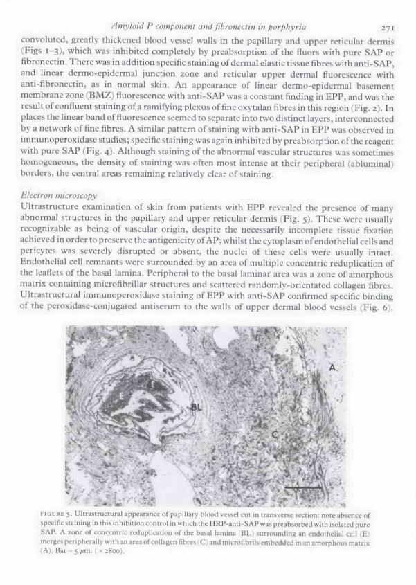
convoluted, greatly thickened blood vessel walls in the papillary and upper reticular dermis(Figs 1-3), which was inhibited completely by preabsorption of tbe fluors with pure SAP orfibronectin. There was in addition specific staining of dermal clastic tissue fibres witb anti-SAP,and linear dermo-epidermal junction zone and reticular upper dermal fluorescence withanti-fibroncctin, as in normal skin. An appearance of linear dermo-cpiderma! basementmembrane zone (BMZ) fluorescence with anti-SAP was a constant finding in EPP, and was theresult of confluent staining of a ramifying plexus of fine oxytalan fibres in this region (Fig. 2). Inplaces the linear band of fluorescence seemed to separate into two distinct layers, interconnectedby a network of fine fibres. A similar pattern of staining with anti-SAP in HPP was observed inimmunoperoxidase studies; specific staining was again inhibited by preabsorption of the reagentwith pure SAP (Fig. 4). Although staining of the abnormal vascular structures was sometimeshomogeneous, the density of staining was often most intense at their peripheral (abluminal)borders, the central areas remaining relatively clear of staining.
Electron microscopyUltrastructure examination of skin from patients with EPP revealed the presence of manyabnormal structures in the papillary and upper reticular dermis (Fig. 5). These were usuallyrecognizable as being of vascular origin, despite the necessarily incomplete tissue fixationachieved in order to preserve the antigenicity of AP; whilst the cytoplasm of endothelial cells andpericytes was severely disrupted or absent, the nuclei of these cells were usually intact.Endothelial cell remnants were surrounded by an area of multiple concentric reduplication ofthe leaflets of the basal lamina. Peripheral to tbe basal laminar area was a zone of amorphousmatrix containing microfibrillar structures and scattered randomly-orientated collagen fibres.Uitrastructural immunoperoxidase staining of HPP with anti-SAP confirmed specific bindingof the peroxidase-conjugated antiserum to the walls of upper dermal blood vessels (Fig. 6).
FIGURE 5. Uitrastructural appearance of papiilary tiliu.d vcs-,tl LU[ in truiisvcrsL' S;JCIKIII; iitjte;ibsi,-nce ofspecific siaining in tliis inhibition tonirul jn which tlii; HRP-anti-SAP wa^ preabsorbed with isolaied pureSAP. A zone of concentric reduplication of the basal lamina > Bl.t surniunding an t-nJiithelial cell (E)merges peripherally with an area of collagen fibres (C; and microtibrils embedded m an amorphous matrix(A). Bar = 5 ;/m. ( x 2800).

272 S.M.Breathnach et al.
BL * ' *^ . -? i
. :
•if jT
V
FIGURE 6. Immunoperoxidase staining of EPP with HRP-anti-SAP. Transverse section of a papillaryblood vessel showing cunceniric reduplication of the basal lamina (BL) surrounding an cndotheiial cell(E). Specific staining is confined to the periphery of the vessel wall. Leafieis of the basal lamina andadjacent collagen fibres (C) are not sttiined. Bar = 5 /im. ( x S250).
Staining was confined to the peripheral (abluminal) areas ofthe blood vessel walls correspond-ing to the regionof greatest concentration of the amorphous matrix containing microfibrils andcollagen fibres. Leafiets of the reduplicated basal lamina and collagen fibres outside the confinesofthe vessel walls were not stained. Specific staining with anti-SAP was observed in a linearpattern around certain blood vessels in the deep reticular dermis at the site ofthe external elasticlamina, and was also noted in association with elastic fibre microfibrils throughout the dermis asin normal skin.
DISCUSSION
Sunlight or long wave ultraviolet light (UVA) irradiation of the skin of patients with EPP(Gschnait et at., 1975) and of mice with griseofulvin-induced protoporphyria (Konrad et at.,1975) results in extensive damage to dermal vascular endothelial cells, leakage of intravascularcontents, and accumulation of amorphous foamy material in the subendothelial area. The exactmechanism of the vascular damage is uncertain, although interactions between protopor-phyrins, UVA, lysosomes (Allison, Magnus & Young, 1966) and/or complement components(Lim & Gigli, 1981) have been postulated. Repeated UVA exposure is followed by concentricreduplication of the vascular basal lamina and tbe appearance of a finely fibrillar substancepermeating and surrounding thickened vessel walls (Ryan & Madili, 1968; Anton-Lamprecht &
Amyloid P component and fibronectin in porphyria 273Meyer, 1970; Epstein et al., 1973; Nonaka et at.., 1975; Gschnait et at.., I975i Kint & de Weert,1978; Honigsmann et al., 1976). The present study has shown that this characteristic thickeningand reduplication of blood vessel walls in EPP involves local deposition of AP and fibronectin, inaddition to that of type IV collagen and laminin as reported elsewhere (Wick ei at., 1979).
Immunofluorescence staining with anti-SAP of exposed skin in EPP revealed confluentfiuorescence of a ramifying plexus of oxytalan fibres along the dermo-epidermal junction.Similar continuous dermo-epidermal BMZ fluorescence with anti-SAP is present in exposedlesiunal skin of patients with discoid lupus erythematosus (LE) (Breathnach e? a/., 1982b) but isnot found in normal skin (Breathnach et al.., 1981a). An increase in the number of oxytalan fibresmay therefore contribute to the thickening of the PAS-positive Mght-microscopic basementmembrane seen in EPP and LE, and which ultrastructurally is composed ofthe sub-basal laminafibrous zone (Briggaman & Wheeler, 1975). A faint linear band of fiuorescence above andconnecting with the plexus of oxytalan fibres may have represented the termination of thesefibres along the basal lamina, into which they are inserted (Kobayasi, 1968; Briggaman &Wheeler, 1975; Cotta-Pereira, Guerra Rodrigo & Bittencourt-Sampaio, 1976).
Fibronectin is a normal constituent of all vascular basement membranes fStenman & Vaheri,1978; Fyrand, 1979; Couchman et at.., 1979; Ruoslahti ei al., 1981) and might accordingly beexpected to be found in increased quantities in reduplicated vascular basement membranes inEPP. AP, however, is not normally present in basement membranes of cutaneous blood vessels(Breathnach ei al., 1981b, 1982a). AP was localized in immuno-electron microscopic studies tothe peripheral (abluminal) regions of thickened vascular walls in EPP, and was not detected inassociation with leaflets of the reduplicated basal lamina. The area of staining with anti-SAPcorresponded to the region of maximum concentration of an amorphous matrix containingrandomly orientated collagen fibres and microfibrillar material. Collagen fibres do not bindantibodies to SAP (Breathnach ei at., I98tb, 1982a). The presence of microfibrillar materialpermeating the walls of dermal blood vessels is a characteristic feature of the histology of EPP(Ryan & Madill, 1968; Anton-Lamprecht & Meyer, 1970; Epstein et al.y 1973; Nonaka et at.,1975; Gschnait et at., 1975; Kint & de Weert, 1978). The specific staining of thickened vascularwalls with anti-SAP observed in EPP may have been associated with this microfibrillar material,as AP is specifically associated witb microfibrils in normal human elastic tissue (Breathnach etat., 1981b, 1982a).
At present it is not clear whether the AP and fibronectin in the abnormal vessel walls arcderived from SAP and plasma fibronectin which have leaked from the intravascularcompartment, or whether they have been synthesized locally by fibroblasts and or vascularendothelial cells (Spark ei at., 1978; Yamada & Weston, 1974; Jaffe & Mosher, 1978) in responseto repeated episodes of protoporphyrin-mediated photochemical injury.
ACKNOWLEDGMENTS
We are grateful to Professor LA. Magnus for permission to study patients under his care. Wethank Dr G. Rook for preparing the peroxidase conjugate, and Mr W. Hinkes for printing thelight photomicrographs. This work was supported in part by MRC Programme Grant 979/51 toMBP.
REFERENCES
ALLISON, A . C , MAGNUS, I, A, & YOUNG, M.R, (1966) Rule of lysosomes and of cell mcmbrnnes in photosensitisation.Nature, 209, 874.

274 S.M.Breathnach et al.ANTON-LAMPRECHT, I. & MEYER, B . (1970) Zur ukrastruktur der haut bei protoporphyrinamie. Dennarohgica, 141,
76.
BREATHNACH, S.M., BHOGAL, B. , DYCK, R.F., DE BEER, F.C., BLACK, M . M . & PEI-YS, M . B . (1981a) Immunohistochemi-
cai demonstration of amyloid P component in skin of nnrmal subjects and patients with cutaneous amyloidosis.
Briiish Journal of Dermatology, 105, 115.
BREATHNACH, S.M., MELROSE, S.M., BHOGAL, B., DE BEER, F.C., DYCK, R.F., TENNENT, G . , BLACK, M.M. & PEPYS,
M.B. (1981b) Amyloid P component is located on elastic fibre microfibrils in normal human tissue. Nature, 293^652.
BREATHNACH, S.M., MELROSE, S.M., BHOGAL, B. , DE BEER, F . C , BLACK, M M . & PEPYS, M.B. (1982a) Immunohisto-chemical studies of amyloid P component distribution in normal human skin. Journal of Investigative Dermatology,in press.
BREATHNACH, S.M., MELROSE, S.M., BHOGAL, B. , DE BEER. F . Q , BLACK, M.M. & PEPYS, M.B. (1982b) Immunohisto-
chemical studies of amyloid P component in disorders of cutaneous elastic tissue. Brimh Journal of Dermatoloev107, 433-
BRIGGAMAN, R . A . & WHEELER, C.E., JR (1975) The epidtrrmal-dermal junction. Journal of Investigative Dermatology,65i 71-
CATHCART, E.S., SKINNER, M . & COHEN, A . S . (19711 Immunogenicity of amyloid, hmnunologv, 20, 945.COTTA-PERHIRA, G . , GUERRA RODRIGO, F . & BITTENCOLRT-SAMPAIO, S. (1976) Oxytalan, eiaunin, and elastic fibers in
the human skin. Journal of Investigaiive Dermatology, 66, 143.COUCHMAN, H.R., GIBSON, W.T., THOM, D . , WEAVER, A.C., REES, D . A . & PARISH, W . E . (1979) Fibronectin
distribution in epithelial and associated tissues of the rat. Archives of Dermaiotogical Research, 266, 295DYCK, R.F., LOCKWOOD, C M . , KEBSHAW, M . , MCHUGH, N . , DUANCE, V .C , BALTZ, M . L . & PEPYS, M.B. (1980)
Amyloid P-component is a constituent of normal human glomerular basement membrane. Journal of ExperimentalMedicine, 152, 1162.
D E B E E R , F . C , B A L T Z , M . L . , H O L F O R D , S . , F E I N S T F : I N , A . & P I ; P Y S , M . B . ( 1981) Fibronectin and C4-binding protein areselectively bound by aggregated amyloid P component. Journal of Experimental Medicine, 154, 1134
EPSTEIN, J.H., TUFFANELLI, D . L . & EPSTEIN, W . L . (1973) Cutaneous changes in the porphyrias. A microscopic study.
Archives of Dermarology, 107, 689.
FYRAND, O . (1979) Studies on fibronectin in the skin. I. Indirect immunofluorescence studies in normal human skin.British Journal of Dermatology, lo i , 263.
GRAHAM, R . C & KARNOVSKY, M.J. (1966) The early stages of absorption of injected horseradish peroxidase in theproximal tubule of mouse kidney: ultrastructural cytochemistry by a new technique. Journal of Hisioclwmimv andCytochemistry, 14, 291.
GscHNAlT, F., WOLFF, K . & KONRAD, K . (1975^ Erythropoietic protoporphyria—submicroscopic events during theacute photosensitivity flare. British Journal of Dcrnuuohgv, 92, 545.
HONIGSMANN, H . , GSCHNAIT, F . , KONRAD, K . , STINGL, G . & WOLFF, K . (1976) Mouse model for protoporphyria-like
skin lesions. Journal of Investigative Dermatology, 66, 188.
jAFFE, E.A. & MOSHKR, D.F. (1978) Synthesis of fibronectin by cultured human endothclial cells. Ammis of the New
York Academy of Sciences, 312, 122.
K I N T , A. & DE WEERT, J. (1978) Vergleichende morphologische untersuchung von porphyria cutanea tarda underythropoetischcr protoporphyric. llauiarzt, 29, 198.
KOBAYASI, T . (1968) Electron microscopy uf the elastic fibers and the dermal membrane on normal human skin. Acta
dcrmatn-venereologica (Stockholm), 48, 303.
KONRAD, D . , HONIGSMANN, H . , GSCHNAIT, F . & WOLFF, K . (1975) Mouse model for protoporphyria. II. Cellular andsubcellular events in the photosensitivity flare of the skin. Journal of Investigative Dermatology, 65, 300.
LiM, H.W. & GiGLl, I. (1981) Role of complement in porphyrin-induced photosensitivity. Journal of InvestigativeDermatology, 76, 4.
NoKAKA, S., NisHiMOTO, K., HiROWATARi, T., HoNDA, T. & NoGiTA, M. (I975J Electron microscopic study of
porphyrias. Journal of Dermatology (Tokyo), 2, 87.
PEPYS, M.B., DASH, A.C, MUNN, E.A., FEINSTEIN, A., SKINNER, M . , COHEN, A.S., GEWURZ, H . , OSMAND, A . P . &
PAINTER, R . H . [1977) Isolation of amyloid P component (protein AP) from normal serum as a calcium-dependent
binding protein. Lancet, i, 1029.
PETERKA, E.S., FUSARO, R.M. & GOLTZ, R . W . (1965J Erythropoietic protoporphyria. II. Histological and histochemicalstudies of cutaneous lesions. Archives of Dermaiohgy, 92, 357.
RIMINGTON, C , MAGNUS, I.A., RYAN, E . A . & CRIPPS, D . J . (1967) Porphyria and photosensitivity. Quarter I v Journal ofMedicine, 36, 29.
Amyloid P component and fibronectin in porphyria 275
RrosLAHTl, E., ENGVALL, E . & HAYMAN, E.G. (1981) Fibronectin: current concepts of its structure and functions.Cotluf;c'i Research, i , 95.
RYAN, E , A . (1966) Histochcmistry nf the skin in erythropoietic prDtoporphyria. Ihitnhjounuit of Dfrmatnlofiy, 78,501.RYAN, E.A. & MADILL, G . T . (1968) Electron microscopy of the skin in erythropoietic protoporphyria. Britiih Journal of
Dermatology, 8o, 561.SHIRAHAMA, T , , SKINNER, M . & COHEN, A.S, (1980) IdeniiRcation and localisation of amyloid by tmmunocytochemis-
try with antisera against amyloid-related proteins, i'cderatwn Proceedings, 39, 102a.SKINNER, M . , PEPYS, M.B,, COHEN, A.S., HELLER, L . M . & LIAN, J . B . (1980) Studies on amyloid protein AP. In;
Antyloid and Amyloidosis. {"Ed. by G.CJ.GIenner, P.Pinho e Costa and A,Falcao de Freitasj, p. 384. Excerpta.Medica, Amsrcrdam,
SPARK, B.C., SHIBAHAMA, T . , SKINNER, M . & COHEN, A , S , (1978) The identification of amyloid P-component (proteinAP) in normal ctiliured human (ibroblasts. Laboratory bwestigation., 38, 556.
STENMAN, S . & VAHEHI, A. (197X) Distribution of a major connective tissue protein, fibroncciin, in normal humantissues. Journal of Experimental Medidnt\ 147, 1054.
VUENTO, M . & VAHEKI, A. (1979) Purification of fibronectin from human plasma by affinity chromatography undernon-denaturing conditions, liiochcinicdt Journal, 183, 331.
WESTERMAKK, P . . SKINNER, M . & {!:OHEN, A.S. (1975) The P-component of human islets of Langerhans. ScandinavianJournal of Immunology, 4, 95.
WtCK, G,, HONIGSMANN, H , & liMfL, R, !I979; Immunofluorescence demonstration of lype IV collagen and anoncollagenous glycoprotein in thickened vascular basal membranes in proioporphyria. Journal of InvestigativeDermalology, 73, 335.
YAMADA, K . M . & WESTON, J.A. (1974) Isolation of a major cell surface glycoprotein from fibroblasts. Proceedings of theNational Academy of Sciences of ihe U.S.A., 71, 3492.


![Menschenhaut [Human skin]](https://static.fdokumen.com/doc/165x107/6326d24f24adacd7250b1364/menschenhaut-human-skin.jpg)