NPM/ALK binds and phosphorylates the RNA/DNA-binding protein PSF in anaplastic large-cell lymphoma
36
doi:10.1182/blood-2006-01-028647 Prepublished online May 30, 2007; Perrotti and Carlo Gambacorti-Passerini Puttini, Rocco G Piazza, Holger Ruchatz, Antonello Villa, Arianna Donella-Deana, Oriano Marin, Danilo Philip W Tucker, Carmen J Tartari, Ching-Jung Huang, Emanuela Colombo, Karen Pulford, Miriam Annamaria Galietta, Rosalind H Gunby, Sara Redaelli, Paola Stano, Cristiana Carniti, Angela Bachi, anaplastic large cell lymphoma NPM/ALK binds and phosphorylates the RNA/DNA binding protein PSF in (4217 articles) Neoplasia Articles on similar topics can be found in the following Blood collections http://bloodjournal.hematologylibrary.org/site/misc/rights.xhtml#repub_requests Information about reproducing this article in parts or in its entirety may be found online at: http://bloodjournal.hematologylibrary.org/site/misc/rights.xhtml#reprints Information about ordering reprints may be found online at: http://bloodjournal.hematologylibrary.org/site/subscriptions/index.xhtml Information about subscriptions and ASH membership may be found online at: digital object identifier (DOIs) and date of initial publication. the indexed by PubMed from initial publication. Citations to Advance online articles must include final publication). Advance online articles are citable and establish publication priority; they are appeared in the paper journal (edited, typeset versions may be posted when available prior to Advance online articles have been peer reviewed and accepted for publication but have not yet Copyright 2011 by The American Society of Hematology; all rights reserved. 20036. the American Society of Hematology, 2021 L St, NW, Suite 900, Washington DC Blood (print ISSN 0006-4971, online ISSN 1528-0020), is published weekly by For personal use only. by guest on May 31, 2013. bloodjournal.hematologylibrary.org From
-
Upload
independent -
Category
Documents
-
view
1 -
download
0
Transcript of NPM/ALK binds and phosphorylates the RNA/DNA-binding protein PSF in anaplastic large-cell lymphoma
doi:10.1182/blood-2006-01-028647Prepublished online May 30, 2007;
Perrotti and Carlo Gambacorti-PasseriniPuttini, Rocco G Piazza, Holger Ruchatz, Antonello Villa, Arianna Donella-Deana, Oriano Marin, DaniloPhilip W Tucker, Carmen J Tartari, Ching-Jung Huang, Emanuela Colombo, Karen Pulford, Miriam Annamaria Galietta, Rosalind H Gunby, Sara Redaelli, Paola Stano, Cristiana Carniti, Angela Bachi, anaplastic large cell lymphomaNPM/ALK binds and phosphorylates the RNA/DNA binding protein PSF in
(4217 articles)Neoplasia �Articles on similar topics can be found in the following Blood collections
http://bloodjournal.hematologylibrary.org/site/misc/rights.xhtml#repub_requestsInformation about reproducing this article in parts or in its entirety may be found online at:
http://bloodjournal.hematologylibrary.org/site/misc/rights.xhtml#reprintsInformation about ordering reprints may be found online at:
http://bloodjournal.hematologylibrary.org/site/subscriptions/index.xhtmlInformation about subscriptions and ASH membership may be found online at:
digital object identifier (DOIs) and date of initial publication. theindexed by PubMed from initial publication. Citations to Advance online articles must include
final publication). Advance online articles are citable and establish publication priority; they areappeared in the paper journal (edited, typeset versions may be posted when available prior to Advance online articles have been peer reviewed and accepted for publication but have not yet
Copyright 2011 by The American Society of Hematology; all rights reserved.20036.the American Society of Hematology, 2021 L St, NW, Suite 900, Washington DC Blood (print ISSN 0006-4971, online ISSN 1528-0020), is published weekly by
For personal use only. by guest on May 31, 2013. bloodjournal.hematologylibrary.orgFrom
1
NPM/ALK Binds and Phosphorylates the RNA/DNA Binding Protein PSF in Anaplastic
Large Cell Lymphoma
Running Title: PSF is a novel ligand and substrate of NPM/ALK
Annamaria Galietta1,2,9, Rosalind H. Gunby1, Sara Redaelli1, Paola Stano2, Cristiana Carniti2,
Angela Bachi3, Philip W. Tucker4, Carmen J. Tartari1,2, Ching-Jung Huang4, Emanuela Colombo5,
Karen Pulford6, Miriam Puttini1, Rocco G. Piazza1, Holger Ruchatz2, Antonello Villa7, Arianna
Donella-Deana8 Oriano Marin8, Danilo Perrotti9, Carlo Gambacorti-Passerini2,10
1Department of Clinical Medicine, University of Milano-Bicocca, Monza, Italy
2Department of Experimental Oncology, The National Cancer Institute of Milan, Italy
3DIBIT, San Raffaele Scientific Institute, Milan, Italy
4Molecular Genetics and Microbiology, University of Texas at Austin, Austin, Texas 78705, USA
5Department of Experimental Oncology, The European Institute of Oncology , Milan, Italy
6Nuffield Department of Clinical Laboratory Sciences, John Radcliffe Hospital, Oxford, UK
7Microscopy and Image Analysis Consortium, Department of Neuroscience, University of Milano-
Bicocca, Monza, Italy
8Department of Biological Chemistry, Institute of Neuroscience of the Italian National Research
Council (C.N.R.), Section of Padua, University of Padua, Padua, Italy
9Dept. Microbiology, Immunology, Virology, and Medical Genetics, Human Cancer Genetics
Program and The Ohio State University Comprehensive Cancer Center, Columbus OH, 43210 USA
10Department of Oncology, McGill University, Montreal, Canada
Blood First Edition Paper, prepublished online May 30, 2007; DOI 10.1182/blood-2006-01-028647
Copyright © 2007 American Society of Hematology
For personal use only. by guest on May 31, 2013. bloodjournal.hematologylibrary.orgFrom

2
Correspondence should be addressed to Annamaria Galietta, Department of Clinical Medicine,
University of Milano-Bicocca, Via Cadore 48, Monza 20052, Italy (Tel: +39 0264488059, Fax: +39
0264488363). Email: [email protected]
For personal use only. by guest on May 31, 2013. bloodjournal.hematologylibrary.orgFrom

3
Abstract
The oncogenic fusion tyrosine kinase, NPM/ALK, induces cellular transformation in anaplastic
large cell lymphomas (ALCL) carrying the t(2;5) chromosomal translocation. Protein-protein
interactions involving NPM/ALK are important for the activation of downstream signaling
pathways. This study was aimed at identifying novel NPM/ALK binding proteins that might
contribute to its oncogenic transformation. Using a proteomic approach, several RNA/DNA binding
proteins were found to co-immunoprecipitate with NPM/ALK, including the multi-functional
polypyrimidine tract binding protein-associated splicing factor (PSF). The interaction between
NPM/ALK and PSF was dependent on an active ALK kinase domain and PSF was found to be
tyrosine phosphorylated in NPM/ALK expressing cell lines and in primary ALK+ ALCL samples.
Furthermore, PSF was shown to be a direct substrate of purified ALK kinase domain in vitro and
PSF Tyr293 was identified as the site of phosphorylation. Y293F PSF was not phosphorylated by
NPM/ALK and was not delocalized in NPM/ALK+ cells. The expression of ALK fusion proteins
induced delocalization of PSF from the nucleus to the cytoplasm and forced overexpression of PSF
inhibited proliferation and induced apoptosis in cells expressing NPM/ALK. PSF phosphorylation
also increased its binding to RNA and decreased the PSF-mediated suppression of GAGE6
expression. These results identify PSF as a novel NPM/ALK binding protein and substrate, and
suggest that PSF function may be perturbed in NPM/ALK transformed cells.
For personal use only. by guest on May 31, 2013. bloodjournal.hematologylibrary.orgFrom

4
INTRODUCTION
Anaplastic large cell lymphoma (ALCL) comprises a group of CD30/Ki-1+ T-cell or null-cell
lymphoid neoplasms.1 A subset of ALCL can be characterized by the expression of fusion proteins
involving the Anaplastic Lymphoma Kinase (ALK).2 ALK is a receptor tyrosine kinase normally
expressed in specific tissues of the central nervous system during embryogenesis.3,4 Due to
chromosomal translocations involving the ALK gene at 2p23, ALK is also aberrantly expressed in
lymphoid tissues. To date, 11 ALK fusion proteins have been detected in ALCL, the most common
of which is nucleophosmin(NPM)/ALK, occurring in 70% of ALK-positive ALCL cases.5
NPM/ALK, the product of the t(2;5)(p23;q35) translocation, encodes a chimeric 80 kDa protein
consisting of the N-terminal portion (amino acids 1-117) of NPM fused to the cytoplasmic portion
of ALK (amino acids 1058-1620).6,7 NPM is a ubiquitously expressed protein normally localized in
the nucleus that has been implicated in nuclear/cytoplasmic trafficking, the cell cycle, centrosome
duplication and maintenance of genomic stability.8-13 The N-terminal portion of NPM contains a
homodimerization domain, which is responsible for the formation of NPM/ALK oligomers capable
of transphosphorylation and activation of the ALK kinase domain.14 NPM/ALK has a nuclear and
cytoplasmic localization, whereas other ALK chimeric proteins (e.g. ATIC/ALK and CLTC/ALK)
possess diffuse or granular cytoplasmic immunostaining patterns.15,16 The constitutive activation of
ALK is sufficient to induce cellular transformation in vitro14,17,18 and lymphoid/myeloid neoplasms
in xenograft or transgenic mice models.19-22
NPM/ALK exerts its transforming potential via its ability to interact and activate several anti-
apoptotic and mitogenic signaling transducers (i.e. PI-3K, JAK, STAT and PLC-γ).17,23-26 The
interaction of NPM/ALK with these signaling molecules is either direct or mediated by adapter
proteins containing SH2 or phosphotyrosine binding domains.17,23 NPM/ALK has been shown to
interact with the adapter proteins IRS1, SHC, GRB2 and CRKL14,17,18,23 and to co-
immunoprecipitate with PI-3K, STAT3, STAT5, JAK3, JAK2, NIPA (Nuclear Interacting Partner
of ALK) and p130CAS.27-29 The significance of these interactions in NPM/ALK-mediated
For personal use only. by guest on May 31, 2013. bloodjournal.hematologylibrary.orgFrom

5
oncogenesis has been partially investigated. For example, activation of the PI-3K pathway is
required for the growth of BaF3-NPM/ALK transformed cells in mice, while STAT3 is essential for
NPM/ALK induced lymphomagenesis in a transgenic mouse model.30,31 In contrast, mutation of
docking sites within NPM/ALK for SHC and IRS1 did not affect the transforming ability of
NPM/ALK in vitro, while mutation of the PLC-γ docking site only impaired mitogenic, but not anti-
apoptotic signaling.17 Since multiple pathways are targeted by NPM/ALK it is possible that
functional redundancy exists between pathways. Furthermore, the oncogenicity of NPM/ALK is
likely to be the result of a complex interplay between these signaling pathways and possibly other,
as yet unidentified, downstream effectors. We report the identification of novel ligands of
NPM/ALK, including several multi-functional RNA/DNA binding proteins such as the
polypyrimidine tract binding protein-associated splicing factor (PSF), the nuclear RNA-binding
protein, 54 kDa (p54nrb), translocated in liposarcomas (FUS/TLS), and EWS (expressed in Ewing’s
sarcoma).
METHODS
Antibodies
The polyclonal anti-PSF and the monoclonal anti-ALK1 antibodies have been previously
described.32,33 The anti-ALK11 polyclonal antibody was kindly supplied by Dr. S.W. Morris (St.
Jude Research Hospital, Memphis, TN, USA). The polyclonal anti-β-actin (Cell Signaling
Technology, Inc. Danvers, MA USA), the polyclonal anti-Lamin B1 (Abcam, Cambrige, UK), the
polyclonal anti-SHC (Upsate Biotech Lake Placid, NY), the monoclonal anti-hemagglutinin (HA)
(clone HA-11; Covance, Berkeley Antibody Company, CA, USA), the monoclonal anti-Histone H1
(Upstate, NY, USA), the monoclonal F2 anti-PARP (Santa Cruz Biotechnology, Inc., Santa Cruz,
CA, USA), the monoclonal anti-PSF Sigma-Aldrich (St. Louis, MO, USA) and the anti-
phosphotyrosine (4G10; Upstate Biotechnology, Lake Placid, NY, USA) antibodies were used as
For personal use only. by guest on May 31, 2013. bloodjournal.hematologylibrary.orgFrom

6
suggested by the manufacturer. Normal rabbit IgG1 was obtained from Amersham (Arlington
Heights, IL, USA). HRP-conjugated anti-mouse or anti-rabbit secondary antibodies were obtained
from Bio-Rad (Hercules, CA, USA). The Alexa Fluor 488 goat anti-mouse IgG was obtained from
Molecular Probes (Eugene, OR, USA) and the Cy5 Conjugated Affinity goat anti-rabbit IgG was
from Rockland (Gilbertsville, PA, USA).
Plasmids
Full-length PSF cDNA cloned into the bacterial expression vector pET 15b (Novagen, Madison,
WI, USA) has been previously described.34 To clone pCR3.1 containing full length HA-tagged PSF
(pCR3.1-HA-PSF), the ORF of PSF was cloned into pCR3.1 (Invitrogen, San Diego, CA, USA)
with BamHI and XhoI, then an HA-tag was created at the 3’ end of the ORF of PSF. Site-directed
mutagenesis of Tyr293 to Phe (Y293F) in HA-PSF was performed using the QuikChange® II XL
Site-Directed Mutagenesis Kit (Stratagene, La Jolla, CA, USA). pcDNA3.0 (Invitrogen) containing
wild type NPM/ALK (pcDNA3-NA) or a kinase-dead NPM/ALK possessing the K210R mutation
(pcDNA3-NA-K210R) were kindly provided by S. W. Morris (St. Jude Research Hospital,
Memphis, TN, USA). pSG5 (Stratagene) containing GCN4/ALK was created from a pSG5 vector
already containing GCN4/RAR. RAR was removed by digestion with EcoRV (blunt) and BglII and
replaced by ALK cytoplasmic domain ORF. To clone GCN4/ALK into the MSCV-IRES-GFP
retroviral expression system (BD Biosciences, NY, USA), GCN4/ALK was excised from pSG5
with BglII and MSCV-IRES-GFP was digested with Xho1. GCN4/ALK and MSCV-IRES-GFP
were blunt-ended with EcoR1 and ligated. For the retroviral construct MigR1-HA-PSF, HA-tagged
PSF was excised from pCR3.1-HA-PSF with BamHI and XhoI, and subcloned into the BglII/XhoI
sites of the bicistronic GFP-containing MigR1 vector.
Cell lines
The murine IL-3-dependent pro-B BaF3 cell line, the SUDHL-1 (t(2:5)-positive) human ALCL-
derived cell line, K562 (Ph+ BCR/ABL-positive) human CML-derived cell line, human embryonic
For personal use only. by guest on May 31, 2013. bloodjournal.hematologylibrary.orgFrom
7
kidney (HEK) 293T cells and the Jurkat T cell line were obtained from DSMZ (Braunschweig,
Germany). The human ALCL (t(2:5)-positive) JB6 cell line was kindly provided by Dr. Turturro
(LSUHSC/Feist-Weiller Cancer Center Shreveport, LA). BaF3 cells were stably transfected with 10
µg pcDNA3-NA (BaF3-NA), pcDNA3-NA-K210R (BaF3-KD) or pcDNA3-Bcr/Abl (BaF3-BA) by
electroporation. Clonal cell lines were obtained by limiting dilution. A stable BaF3 cell line
expressing GCN4/ALK was generated using MSCV-GCN4/ALK. BaF3 cells (1x10⁶) were infected
with supernatant from Phoenix packaging cells transfected with retroviral construct MSCV-
GCN4/ALK. GCN4/ALK expressing cells were selected by culturing in the absence of IL-3. All
cells were maintained in a humidified atmosphere at 37ºC and 5% CO2 .
Expression and purification of recombinant proteins
N-terminally 6xHis-tagged PSF protein was produced using BL21 competent cells (Stratagene) and
pET15b-PSF. Cells were harvested and lysed by sonication in 50 mM NaH2PO4, 300 mM NaCl, pH
8.0. Lysates were clarified by centrifugation at 15000xg for 30 min at 4°C. PSF was purified using
NI-NTA agarose resin (QIAGEN, Hilden, Germany) in batch mode following instructions. 6xHis-
tagged ALK protein containing residues Leu1073 – Ala1459 (ALK_HUMAN, Genbank Accession
Code: Q9UM73), which includes the predicted kinase domain, was produced using a Baculovirus
expression system and purified as described elsewhere.35 GST-tagged ALK (Leu1073 – Ala1459) was
expressed using the Bac-to-Bac Baculorvirus expression system (Invitrogen) and purified using
GSTrap columns (GE Healthcare, NY, USA).
Immunoprecipitation and Western blotting
Cells (20 x 106/sample) were washed in ice-cold PBS and lysed on ice in lysis buffer (50 mM Tris
HCl, pH 7.4; 1% Triton X-100; 5 mM EDTA; 150 mM NaCl, 1 mM Na3VO4; 1 mM NaF; 1 mM
PMSF, protease inhibitor cocktail). Cell lysates were clarified by centrifugation at 12,000xg for 20
min at 4°C and protein was quantified using the Bradford assay (BioRad Laboratories, Hercules,
CA, USA). Frozen lymph nodes were obtained from two ALCL patients and normal spleen tissue
For personal use only. by guest on May 31, 2013. bloodjournal.hematologylibrary.orgFrom
8
from a healthy donor, after informed consent (kindly provided by Dr. P. Collini, Pathology Dept.
NCI Milan, Italy). Tissues were homogenized and lysed as described previously.36 Proteins were
immunoprecipitated by incubating 100 µg (cell lines), or 1.5 mg (patient tissue), of total protein
overnight with anti-PSF and anti-ALK1 at 4°C followed by a 1 h incubation with 30 µl of protein
A-Sepharose (GE Healthcare) at 4°C. Immunocomplexes were washed 5 times with ice-cold lysis
buffer, denatured and subjected to SDS-PAGE. Western blots were performed with anti-PSF, anti-
ALK1 and anti-pTyr primary antibodies.
Tryptic digestion and mass spectrometric peptide sequencing
Anti-ALK1 immunocomplexes obtained by immunoprecipitation of 400x106 cells were resolved by
SDS-PAGE and detected by silver staining. Bands of interest were excised from silver stained gels
reduced, alkylated and digested overnight with bovine trypsin as described.37 One µl of supernatant
containing the generated tryptic peptides was loaded onto the MALDI target using the dried droplet
technique and α-cyano-4-hydroxycinnamic acid (HCCA) acid as matrix. MALDI-TOF (matrix
assisted laser desorption ionization-time of flight) mass measurements were performed on a
Voyager-DE STR TOF mass spectrometer (Applied Biosystems, Framingham, MA, USA) operated
in the delayed extraction and reflector mode. Spectra, internally calibrated, were processed via the
Data Explorer software (Applied Biosystems). Proteins were unambiguously identified by searching
a comprehensive non-redundant protein database using the program ProFound.38
Kinase assays
Full length purified 6xHis-tagged PSF was phosphorylated in 50 µl of reaction buffer containing 25
mM Hepes pH 7.5, 10 mM MgCl2, 10 mM MnCl2, 1 µCi (4500 Ci/mmol) [γ32P]ATP, 20 µM ATP,
2 mM DTT, purified 6xHIS-ALK (0.4 µg) and purified 6xHis-PSF (0.25 µg). After incubation at
30°C for 30 min, reactions were terminated by the addition of 15 µl of 5X Laemmli buffer. Samples
were denatured, subjected to SDS-PAGE and visualized by autoradiography. Synthetic PSF
peptides were synthesized as previously described39 (Tyr293, Tyr251, Tyr470, Tyr488-490,
Tyr597-602, Tyr698) or purchased from Sigma (Tyr527 and Tyr624). PSF peptides (400 µM) were
For personal use only. by guest on May 31, 2013. bloodjournal.hematologylibrary.orgFrom
9
phosphorylated in the presence of 10 units of GST-ALK as described elsewhere.39 One unit was
defined as the amount of GST-ALK transferring 1 pmol phosphate per min to the random polymer
polyGlu4Tyr (0.1 mg/ml) under standard conditions.
Nuclear and cytoplasmic extracts
Cells (3 x 10⁷) were washed in ice-cold PBS, resuspended in a buffer containing 20 mM HEPES pH
7, 2 mM MgCl2, 10 mM KCl, 0.5% NP40 and protease inhibitors, and homogenized using a
Wheaton A Dounce homogenizer (20 strokes). Lysates were clarified by centrifugation at 1500xg
for 5 min. The resulting supernatant was subsequently centrifuged at 15,000xg for 10 min. The
soluble fraction, representing the cytoplasmic extract, was immediately transferred to a pre-chilled
tube and stored on ice. The insoluble fraction, which contains nuclei, was washed twice with ice-
cold buffer and then resuspended in buffer supplemented with 0.5 M NaCl and rocked gently for 30
min at 4°C to extract nuclear proteins. The extracted material was then centrifuged at 15,000xg for
10 min, and the soluble nuclear extract fraction was transferred to a pre-chilled tube and placed on
ice. Extracts were stored at -80°C until use.40
Immunofluorescence
293T cells were seeded on glass coverslips and transiently transfected with 5 µg of pCR3.1-HA-
PSF alone or together with 5 µg of pcDNA3-NA, pcDNA3-NA-K210R or pG5-GCN4/ALK, using
the calcium phosphate precipitation method. Twenty-four hours after transfection the cells were
fixed with 4% paraformaldehyde and permeabilized with 0.2% Triton X-100 and 2% BSA in PBS at
RT for 10 min. Cells were incubated overnight at 4°C with the primary antibody, washed 3 times
with PBS 0.2% Triton X-100 and incubated for 1 h at room temperature with the relevant secondary
antibodies. The nuclear staining was performed by incubating with 50 µg/ml propidium iodide for
30 min at 37°C. The coverslips were then washed and mounted. Fluorescence was detected using
the Nikon Eclipse Є 600 confocal microscope.
Proliferation and apoptosis assays
For personal use only. by guest on May 31, 2013. bloodjournal.hematologylibrary.orgFrom

10
Parental BaF3, BaF3-NA, BaF3-BA and BaF3-KD cells (5x10⁶) were transiently transfected by
electroporation (260V and 1050µF) with 10 µg of pCR3.1 or pCR3.1-HA-PSF. Cells were then
resuspended in medium with (BaF3-Par, BaF3-KD) or without IL-3 (BaF3-NA, BaF3-BA) and cell
proliferation was measured at 24, 48 and 72 h after transfection by 3H-thymidine incorporation as
described previously.25 Apoptosis was assessed by detecting PARP cleavage by immunoblotting
with the anti-PARP monoclonal antibody or by annexin V-fluorescein isothiocyanate (FITC)
binding using the Apoptosis Detection Kit (Bender MedSystems Diagnostic, Vienna, Austria).
Samples were analyzed using a FACScalibur and Cell-quest software.
Colony formation assay
The amphotropic-packaging cell line Phoenix A (G. P. Nolan, Stanford University School of
Medicine) was transiently transfected41 with MigR1-HA-PSF and the infectious supernatant was
used to infect JB6, SUDHL-1 and Jurkat cell lines. After infection, cells were sorted for green
fluorescent protein (GFP) expression using a FACSCalibur. GFP+ cells (103) were plated in 0.9%
Metho-Cult M3234 semisolid medium (Stem Cell Technologies, Vancouver, BC) and colonies were
scored 9 -12 days later.42
RESULTS
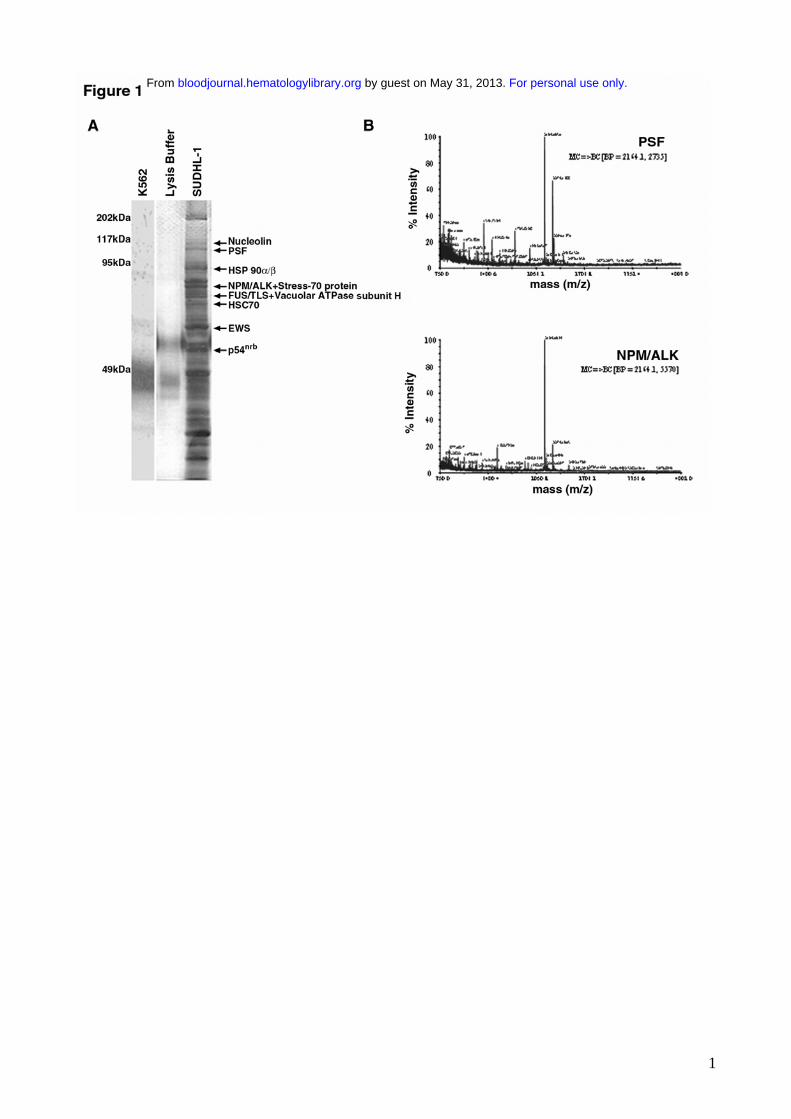
Identification of proteins co-immunoprecipitating with NPM/ALK.
NPM/ALK was immunoprecipitated from SUDHL-1 cells and associating proteins were visualized
by SDS-PAGE and silver staining. Several proteins with diverse molecular weights (MW) co-
immunoprecipitated with NPM/ALK (Figure 1A), but were not present in anti-ALK1
immunoprecipitates from NPM/ALK-negative K562 cells or lysis buffer alone. Co-
immunoprecipitating proteins with a MW greater than 50 kDa were analyzed by MALDI-TOF and
ten were identified. These included the heat shock proteins HSP 90α/β43,44, previously identified as
NPM/ALK interacting proteins45 and novel ligands such as HSC 7046, V-ATPase subunit-H47,
Stress-70 protein48, nucleolin49, PSF50, FUS/TLS51, nonO/p54nrb52 and EWS53 (summarized in table
For personal use only. by guest on May 31, 2013. bloodjournal.hematologylibrary.orgFrom

11
1). NPM/ALK itself was also positively identified demonstrating the reliability of the mass
spectrometry sequencing. MS/MS profiles identifying the PSF and NPM/ALK proteins are shown
in figure 1B.
The binding of NPM/ALK to PSF was further investigated by reciprocal immunoprecipitation and
Western blotting using NPM/ALK-positive cells (SUDHL-1 and NPM/ALK-transfected BaF3 cells
(BaF3-NA)) and NPM/ALK-negative cells (K562 and parental BaF3 cells (BaF3-Par)). PSF was
detected in anti-ALK1 immunoprecipitates derived from t(2;5) SUDHL-1 and BaF3-NA cells
(Figures 2A and 2B). Accordingly, NPM/ALK was detected in anti-PSF immunoprecipitates from
the same cells. Co-immunoprecipitation of PSF and NPM/ALK was also observed in primary
lymph node tissue obtained from two patients with NPM/ALK+ ALCL, but not in spleen tissue from
a healthy donor (Figure 2C). Furthermore, PSF was unable to interact with the kinase-dead
NPM/ALK mutant expressed in BaF3 cells (BaF3-KD) (Figure 2B). Thus, PSF is a bona fide
NPM/ALK ligand and this interaction is strictly dependent on NPM/ALK kinase activity.
To determine whether PSF can interact with ALK fusions other than NPM/ALK we assessed its
ability to co-immunoprecipitate with the GCN4/ALK fusion protein. GCN4/ALK contains the
coiled-coil homodimerisation domain of the yeast GCN4 protein fused to the complete cytoplasmic
domain of ALK.54 PSF was able to interact with GCN4/ALK (Figure 2D), indicating that the NPM
moiety is dispensable for the PSF-ALK interaction, and suggests that the PSF-ALK interaction is
mediated through the ALK kinase domain.
PSF is a direct target of NPM/ALK tyrosine kinase activity.
Since NPM/ALK has a constitutive tyrosine kinase activity and PSF binds to NPM/ALK in cells,
we examined whether PSF could be a substrate of NPM/ALK. Initially, the tyrosine
phosphorylation status of PSF in cell lines expressing or not expressing NPM/ALK was examined.
A 100 kDa tyrosine phosphorylated protein corresponding to PSF was detected in anti-ALK and
anti-PSF immunoprecipitates from SUDHL-1 and BaF3-NA cells (Figure 3A). In contrast, no such
For personal use only. by guest on May 31, 2013. bloodjournal.hematologylibrary.orgFrom
12
tyrosine phosphorylated band was found in anti-ALK and anti-PSF immunoprecipitates from K562,
BaF3-KD and BaF3-Par cells. The fact that PSF was not phosphorylated in K562 cells, which
express the constitutively active tyrosine kinase Bcr/Abl, suggests that PSF tyrosine
phosphorylation is specifically dependent on ALK activity. PSF was also found to be tyrosine
phosphorylated in anti-ALK1 immunoprecipitates from lysates of one ALCL lymph node patient
tissue but not from normal spleen (Figure 3B). Due to insufficient material, it was not possible to
assess PSF tyrosine phosphorylation in ALCL lymph nodes from patient 2.
To determine if PSF could be directly phosphorylated by ALK, an in vitro kinase assay was
performed using purified recombinant 6xHis-tagged ALK kinase domain and full-length 6xHis-
tagged PSF. Purified ALK kinase domain possesses autophosphorylation activity and is able to
phosphorylate PSF (Figure 3C), but not the unrelated GST and BSA proteins (data not shown). In
an attempt to map the PSF tyrosine residue/s that is/are phosphorylated by ALK, we synthesised 8
synthetic PSF peptides containing the potential ALK tyrosine phosphorylation site. Peptides were
chosen on the basis of the ALK substrate consensus sequence39,55 and phosphorylation site
prediction programs (e.g. NetPhos 2.0 Server)56. The ability of purified ALK kinase domain to
phosphorylate these peptides was determined by an in vitro kinase assay. Only the peptide
containing Tyr293 of PSF (PGEKTYTQRCRLFVGNLPADIT) was phosphorylated by ALK
(Figure 3D). To confirm that PSF Tyr293 is indeed a bona fide ALK phosphorylation site, we
mutagenized Tyr293 to Phe and transiently co-expressed the PSF Y293F mutant and NPM/ALK in
293T cells. We observed that mutant PSF was no longer tyrosine phosphorylated (Figure 3E, top
panel, lanes 2,4), unlike control wild type PSF (Figure 3E, top panel, lanes 3,5). Mutant PSF did not
co-immunoprecipitate with NPM/ALK (Figure 3E, lower panel, lane 4). These data suggest that
PSF is a substrate of NPM/ALK and that tyrosine phosphorylation occurs at Tyr293 of PSF and is
critical for the physical association of NPM/ALK and PSF.
ALK fusion proteins alter PSF subcellular localization
For personal use only. by guest on May 31, 2013. bloodjournal.hematologylibrary.orgFrom
13
PSF normally displays a nuclear localization pattern, whereas NPM/ALK is localized in both the
cytoplasm and nucleus.14,57,58 Since NPM/ALK binds PSF, we investigated whether this association
affects the subcellular localization of PSF. Immunofluorescence and confocal microscopy
experiments were performed in 293T cells transiently transfected with HA-PSF alone or co-
transfected with, HA-PSF and NPM/ALK, NPM/ALK KD or GCN4/ALK (Figure 4). Since no anti-
PSF antibody suitable for immunofluorescence was available, ectopically expressed HA-PSF was
evaluated. In 293T cells, HA-PSF showed a characteristic nuclear staining pattern when expressed
alone (Figure 4A). However, expression of HA-PSF together with active ALK fusion proteins,
NPM/ALK or GCN4/ALK, resulted in HA-PSF localization in both the nucleus and cytoplasm
(Figure 4C and 4D). In contrast, kinase dead NPM/ALK did not affect the nuclear localisation of
HA-PSF (Figure 4B), consistent with its inability to co-immunoprecipitate with NPM/ALK (Figure
2B). To confirm the delocalization of PSF in NPM/ALK-expressing cells, we prepared nuclear and
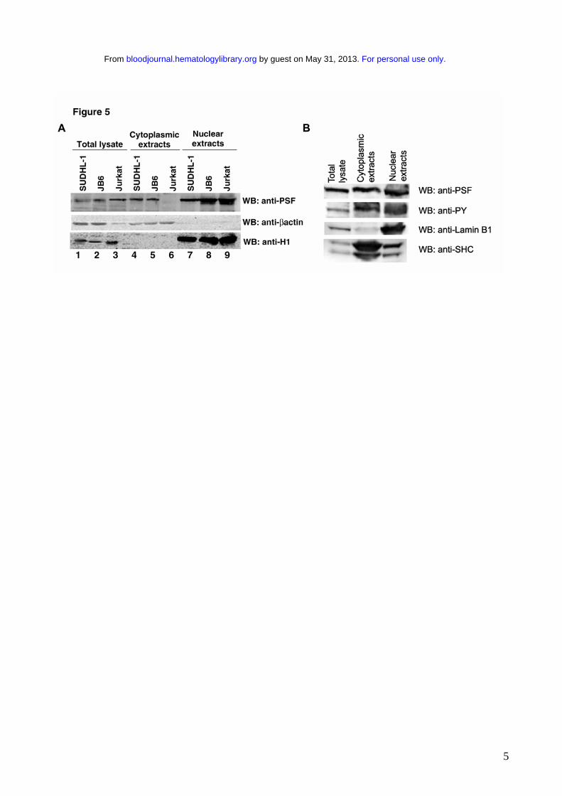
cytoplasmic protein extracts from JB6, SUDHL-1, and Jurkat cells, and detected PSF in these
extracts by Western blotting. In the negative control Jurkat cells PSF was only detected in the
nuclear extract (Figure 5A, lanes 6 and 9), whereas in SUDHL-1 and JB6 cell lines PSF was
detected in both the nuclear and cytoplasmic extracts (Figure 5A, lanes 4, 5, 7, 8). Both nuclear and
cytoplasmic fractions appear to contain phosphorylated PSF (Figure 5B). Interestingly, co-
expression of the mutant Y293F PSF with active NPM/ALK resulted in the localization of mutant
PSF only in the nucleus (Figure 4E). Therefore, these results demonstrate that ALK fusion proteins
can induce the relocalization of PSF from the nucleus to the cytoplasm and indicate that Y293
phosphorylation is needed for PSF delocalization.
Forced PSF expression induces growth arrest, apoptosis and reduces the clonogenic potential
of NPM/ALK positive cells.
For personal use only. by guest on May 31, 2013. bloodjournal.hematologylibrary.orgFrom

14
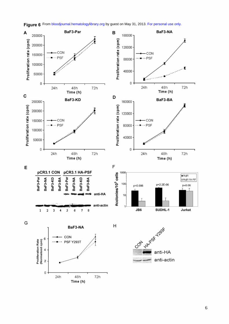
We investigated the effect of overexpression of PSF on cells expressing and not expressing
NPM/ALK. Since NPM/ALK expression is associated with phosphorylation and delocalization of
PSF into the cytoplasm, it is plausible that PSF might contribute to the oncogenic activity of
NPM/ALK. We transiently overexpressed HA-tagged PSF in BaF3-NA cells and observed a
marked inhibition of proliferation in these cells compared with cells transduced with empty vector
alone (Figure 6B). In contrast, PSF overexpression did not affect the growth of BaF3-Par cells,
BaF3-KD cells or BaF3 cells stably transformed with BCR/ABL (BaF3-BA) (Figure 6A, C, D). The
overexpression of HA-PSF was confirmed by Western blotting (Figure 6E). Unlike wild type PSF,
the mutant Y293F PSF had no effect on the proliferation of BaF3-NA cells (Figure 6G, H). We
subsequently evaluated the effect of ectopic PSF expression on the clonogenicity of NPM/ALK-
positive cell lines. A retroviral vector carrying HA-tagged PSF (MigR1-HA-PSF) was used to
transduce the NPM/ALK positive human cell lines, JB6 and SUDHL-1, and the NPM-ALK
negative Jurkat cell line. The effect of PSF overexpression on clonogenic potential was assessed in
transduced cells using a methylcellulose colony formation assay. Overexpression of HA-PSF
induced a marked decrease in the number of colonies derived from the NPM/ALK positive human
cell lines, while having no effect on the NPM/ALK negative Jurkat cells (Figure 6F). Transduction
with MigR1 alone in JB6, SUDHL-1 and Jurkat cells had no effect on colony formation (Figure
6F). These results indicate that PSF overexpression exerts growth arrest specifically in NPM/ALK
expressing cells.
The decrease of BaF3-NA cell proliferation was also accompanied by the induction of apoptosis,
demonstrated by the cleavage of the caspase substrate, PARP59 (~112 kDa), to ~85 and 25 kDa
proteolytic products (Figure 7A). The induction of apoptosis by PSF overexpression in the presence
of NPM/ALK, was also evident when using the Annexin V binding assay (Figure 7F). Apoptosis
was not observed in non-transfected controls or in cells transfected with HA-PSF alone or together
with kinase dead NPM/ALK (Figures 7C-E). The level of HA-PSF and NPM/ALK expression in
For personal use only. by guest on May 31, 2013. bloodjournal.hematologylibrary.orgFrom

15
transfected cells was controlled by Western blotting (Figure 7B). Therefore, forced overexpression
of PSF induces apoptosis in NPM/ALK expressing cells.
PSF phosphorylation alters its RNA binding and transcriptional repression activities. To
further verify if NPM/ALK-mediated phosphorylation of PSF alters its biological functions we
studied two known activities of PSF: its RNA binding ability60,61 and its transcriptional repressor
activity of GAGE662. To study the first activity, the binding of purified PSF to a labeled specific
oligo RNA)60 was evaluated. As shown in Figure S1, the RNA probe specifically recognized PSF as
evidenced by the formation of a band; this binding was increased by the NMP/ALK-mediated
phosphorylation of PSF (lane 2 versus 4). To evaluate the transcriptional activity of PSF on
GAGE6, 293T cells were transfected with PSF and/or NPM/ALK, and the expression of GAGE6
assessed by real time PCR. The results (Figure S2) show that NPM/ALK induced a substantial
increase in GAGE6 expression levels in PSF transfected cells, that were not observed or greatly
reduced when PSFY293F and NPM/ALK were transfected.
DISCUSSION
NPM/ALK induces cellular transformation by constitutive activation of multiple anti-apoptotic and
proliferative signaling pathways, through interactions with components of signaling cascades.5,29
NPM/ALK also associates with the chaperones HSP90 and HSP70 that regulate its degradation,45
and with NPM that localizes NPM/ALK in the nucleus.14 Hence, NPM/ALK ligands play an
important role in mediating not only downstream signaling, but also impact on its subcellular
localization. In this study, we identified novel NPM/ALK ligands, including five nuclear
RNA/DNA binding proteins: PSF, p54nrb, FUS/TLS, EWS and nucleolin. A study by Crockett et al
employing a similar proteomics approach to identify NPM/ALK interacting partners identified a
number of cytoplasmic signaling proteins including adaptor molecules, kinases and phosphatases.63.
The RNA/DNA binding proteins reported here were not identified, most likely due to differences in
For personal use only. by guest on May 31, 2013. bloodjournal.hematologylibrary.orgFrom
16
experimental conditions. However a more recent publication identified an RNA binding protein
associated to NPM/ALK.64. The association of PSF with NPM/ALK was confirmed in ALCL
derived cell lines and patient samples, and required an active ALK kinase domain. PSF also bound
to GCN4/ALK, suggesting that the interaction may be mediated via the ALK portion of the fusion
protein. Therefore, PSF binding may be a common feature of variant ALK fusion proteins.
PSF is a multifunctional nuclear factor that has been implicated in diverse reactions in the nucleus.65
It is normally associated with subnuclear structures known as speckles and with the nuclear
membrane and matrix.57,58,66 PSF consists of an N-terminal proline/glutamine-rich domain that
mediates interactions with proteins and DNA, two RNA-recognition motifs (RRMs) involved in
interactions with proteins and RNA and a C-terminal region containing two nuclear localization
signals (NLS).50 PSF forms multi-protein complexes that are involved in pre-mRNA splicing,50,60,67
gene transcription,32,34,68,69 DNA repair,70 DNA recombination,71,72 and cytoplasmic mRNA
stability.73 PSF is most commonly associated with the closely related p54nrb nuclear factor52 and has
also been reported to interact with EWS and TLS/FUS.74 Since these proteins were also identified
as novel NPM/ALK ligands, it is possible that NPM/ALK interacts with multi-protein complexes
containing PSF and these other RNA/DNA binding proteins.
We also showed that PSF is a substrate of ALK in vitro and is tyrosine phosphorylated in
NPM/ALK expressing cell lines and in primary ALCL lymph node tissue. Mapping of the
phosphorylation sites in PSF identified Tyr293 as the phosphorylated residue. Interestingly, the
Y293F PSF mutant did not co-immunoprecipitate with NPM/ALK, which suggests that PSF
phosphorylation may stabilize the interaction with NPM/ALK, or with another adaptor protein
present in the NPM/ALK complex. The effect of tyrosine phosphorylation on PSF function has not
been described previously, although both PSF and p54nrb have been identified as tyrosine
phosphorylated proteins associating with the nuclear envelope in neuroblastoma cells.75 By contrast,
serine/threonine phosphorylation of PSF, p54nrb, TLS/FUS and the serine-arginine (SR) family of
splicing factors represents a mechanism through which the multiple function(s) of these RNA/DNA
For personal use only. by guest on May 31, 2013. bloodjournal.hematologylibrary.orgFrom
17
binding proteins are regulated.71,74,76-80 For example, PKC-mediated phosphorylation of PSF, p54nrb
and TLS/FUS regulates RNA binding/processing and DNA recombination reactions.71,74,76-78 In
light of the regulation of PSF activity by serine/threonine phosphorylation it is possible that
NPM/ALK may alter PSF function by tyrosine phosphorylation. The experiments performed here
suggest that tyrosine phosphorylation of PSF can increase its RNA binding and inhibit the
transcriptional repressor function of PSF.
PSF function may also be affected by its delocalisation to the cytoplasm in cells expressing active
ALK fusion proteins, including NPM/ALK positive ALCL cell lines. Since kinase dead NPM/ALK
did not cause delocalization of PSF, it is conceivable that delocalization is dependent on tyrosine
phosphorylation, possibly by stabilizing the PSF-NPM/ALK interaction. This hypothesis is
supported by the finding that the mutant Y293F PSF was not phosphorylated, did not associate with
NPM/ALK and was not delocalized in NPM/ALK expressing cells. The finding that both nuclear
and cytoplasmic PSF fractions are tyrosine phosphorylated in NPM/ALK+ cells is compatible with
the subcellular localization of NPM/ALK inside cells.
The delocalization of PSF to the cytoplasm may alter its nuclear-associated functions. The facts that
NPM/ALK-mediated phosphorylation of PSF increases its RNA binding activity (figure S1) and
that the combined transfection of NPM/ALK and PSF leads to an increase in GAGE6 transcription
(figure S2) are compatible with this hypothesis, which however will require further confirmatory
experiments.
The modulation of other nuclear proteins via relocalization to the cytoplasm has been reported. For
example, the oncogenic fusion protein PSF-TFE3, identified in papillary renal cell carcinoma,81
causes endogenous TFE3 and p53 to relocalize into the cytoplasm, thereby stimulating their
degradation and inactivation.82 Another example is offered by the cytoplasmic relocation of NPM
caused by C-terminal mutations, which are currently considered the primary leukemogenic event
that disrupts normal NPM function in a cohort of acute myeloid leukemia patients.83
From these data, it could be hypothesized that NPM/ALK-mediated phosphorylation of PSF may
For personal use only. by guest on May 31, 2013. bloodjournal.hematologylibrary.orgFrom
18
impact on the expression of factors regulating cell growth and apoptosis. Indeed, other ALK-
associated factors such as NIPA protect NPM/ALK-expressing BaF3 cells from apoptosis through a
mechanism requiring NPM/ALK activity and NIPA phosphorylation.27. Interestingly,
overexpression of PSF, but not of Y293F PSF, specifically inhibited proliferation, clonogenic
potential, and induced apoptosis in NPM/ALK-positive cells. The interpretation of these results
requires caution. The data showing that Y293F PSF did not inhibit growth could be interpreted in
two ways: that phosphorylation of overexpressed PSF by NPM/ALK is necessary for such effects,
or that the mutation of residue 293, which is located very close to the RRM1 domain (residues 297-
369), affects other functions of PSF. The mechanisms by which overexpression of PSF leads to
growth inhibition remain to be established and will be the subject of further studies.
In conclusion, we report here that NPM/ALK associates with and phosphorylates several RNA-
DNA binding proteins, in particular the multi-functional nuclear factor PSF. The data also show that
PSF delocalizes to the cytoplasm in cells containing constitutively active forms of ALK through a
mechanism that involves NPM/ALK-dependent phosphorylation of Tyr293 of PSF. Data were also
provided showing that forced PSF expression impairs cell growth, induces apoptosis and decreases
clonogenic potential of NPM/ALK-expressing cells. Therefore, PSF might play a role in
NPM/ALK-mediated lymphomagenesis through mechanisms that require further investigation.
Whether PSF could be a target for other oncogenes also remains to be determined. Such studies
should lead to a greater understanding of the mechanisms of ALK-mediated transformation.
ACKNOWLEDGMENTS
The authors gratefully acknowledge Dr. S.W. Morris (St. Jude Research Hospital, Memphis, TN,
USA) for providing the ALK11 polyclonal antibody, pcDNA3-NA and pcDNA3-NA-K210R
plasmids; Dr. P. Collini, (Pathology Dept. NCI Milan, Italy) for providing ALCL patient samples,
Dr. Turturro (LSUHSC/Feist-Weiller Cancer Center Shreveport, LA) for providing JB6 cell line.
For personal use only. by guest on May 31, 2013. bloodjournal.hematologylibrary.orgFrom
19
The authors are especially grateful to Lorenzo Bertola, Edoardo Marchesi and Elvezia Bonacina
(NCI Milan, Italy) for technical assistance, Barbara Cimbro for the preparation GST-ALK and
Valentina Belloni (UNIMIB, Italy).
Authors Contributions
Annamaria Galietta: designed research, performed research and wrote paper Rosalind H. Gunby: wrote paper Sara Redaelli: performed research Paola Stano: performed research Cristiana Carniti: performed research Angela Bachi: performed research Philip W. Tucker: contributed vital material and analyzed data Carmen J. Tartari: performed research Ching-Jung Huang: contributed vital material Emanuela Colombo: contributed vital material Karen Pulford: contributed vital material
Miriam Puttini: performed research Rocco G. Piazza: analyzed data
Holger Ruchatz: designed research Antonello Villa: contributed vital material Arianna Donella-Deana: performed research Oriano Marin: performed research Danilo Perotti: designed research, analyzed data and wrote paper Carlo Gambacorti-Passerini: designed research, supervised work, analyzed data and wrote paper This work was supported in part by the Italian Association for Cancer Research (AIRC), Min. San.
Ricerca Finalizzata (2003), CNR, MIUR-COFIN and PRIN programs (2004), EU (Prokinase
network, #503467), CIHR, CFI, NCI-C, NIH grants CA92318 (P.W.T) and CA095512 (D.P.) and by
Leukemia Research Fund.
REFERENCES
1. Stein H, Foss HD, Durkop H, et al. CD30(+) anaplastic large cell lymphoma: a review of its histopathologic, genetic, and clinical features. Blood. 2000;96:3681-3695. 2. Morris SW, Xue L, Ma Z, Kinney MC. Alk+ CD30+ lymphomas: a distinct molecular genetic subtype of non-Hodgkin's lymphoma. Br J Haematol. 2001;113:275-295. 3. Morris SW, Naeve C, Mathew P, et al. ALK, the chromosome 2 gene locus altered by the t(2;5) in non-Hodgkin's lymphoma, encodes a novel neural receptor tyrosine kinase that is highly related to leukocyte tyrosine kinase (LTK). Oncogene. 1997;14:2175-2188. 4. Iwahara T, Fujimoto J, Wen D, et al. Molecular characterization of ALK, a receptor tyrosine kinase expressed specifically in the nervous system. Oncogene. 1997;14:439-449.
For personal use only. by guest on May 31, 2013. bloodjournal.hematologylibrary.orgFrom

20
5. Pulford K, Morris SW, Turturro F. Anaplastic lymphoma kinase proteins in growth control and cancer. J Cell Physiol. 2004;199:330-358. 6. Shiota M, Fujimoto J, Semba T, Satoh H, Yamamoto T, Mori S. Hyperphosphorylation of a novel 80 kDa protein-tyrosine kinase similar to Ltk in a human Ki-1 lymphoma cell line, AMS3. Oncogene. 1994;9:1567-1574. 7. Morris SW, Kirstein MN, Valentine MB, et al. Fusion of a kinase gene, ALK, to a nucleolar protein gene, NPM, in non-Hodgkin's lymphoma. Science. 1994;263:1281-1284. 8. Zatsepina OV, Rousselet A, Chan PK, Olson MO, Jordan EG, Bornens M. The nucleolar phosphoprotein B23 redistributes in part to the spindle poles during mitosis. J Cell Sci. 1999;112:455-466. 9. Okuda M, Horn HF, Tarapore P, et al. Nucleophosmin/B23 is a target of CDK2/cyclin E in centrosome duplication. Cell. 2000;103:127-140. 10. Dumbar TS, Gentry GA, Olson MO. Interaction of nucleolar phosphoprotein B23 with nucleic acids. Biochemistry. 1989;28:9495-9501. 11. Borer RA, Lehner CF, Eppenberger HM, Nigg EA. Major nucleolar proteins shuttle between nucleus and cytoplasm. Cell. 1989;56:379-390. 12. Shu X, Fry AM, Tulloch B, et al. RPGR ORF15 isoform co-localizes with RPGRIP1 at centrioles and basal bodies and interacts with nucleophosmin. Hum Mol Genet. 2005;14:1183-1197. Epub 2005 Mar 1116. 13. Grisendi S, Bernardi R, Rossi M, et al. Role of nucleophosmin in embryonic development and tumorigenesis. Nature. 2005;437:147-153. Epub 2005 Jul 2006. 14. Bischof D, Pulford K, Mason DY, Morris SW. Role of the nucleophosmin (NPM) portion of the non-Hodgkin's lymphoma-associated NPM-anaplastic lymphoma kinase fusion protein in oncogenesis. Mol Cell Biol. 1997;17:2312-2325. 15. Ma Z, Cools J, Marynen P, et al. Inv(2)(p23q35) in anaplastic large-cell lymphoma induces constitutive anaplastic lymphoma kinase (ALK) tyrosine kinase activation by fusion to ATIC, an enzyme involved in purine nucleotide biosynthesis. Blood. 2000;95:2144-2149. 16. Touriol C, Greenland C, Lamant L, et al. Further demonstration of the diversity of chromosomal changes involving 2p23 in ALK-positive lymphoma: 2 cases expressing ALK kinase fused to CLTCL (clathrin chain polypeptide-like). Blood. 2000;95:3204-3207. 17. Bai RY, Dieter P, Peschel C, Morris SW, Duyster J. Nucleophosmin-anaplastic lymphoma kinase of large-cell anaplastic lymphoma is a constitutively active tyrosine kinase that utilizes phospholipase C-gamma to mediate its mitogenicity. Mol Cell Biol. 1998;18:6951-6961. 18. Fujimoto J, Shiota M, Iwahara T, et al. Characterization of the transforming activity of p80, a hyperphosphorylated protein in a Ki-1 lymphoma cell line with chromosomal translocation t(2;5). Proc Natl Acad Sci U S A. 1996;93:4181-4186. 19. Chiarle R, Gong JZ, Guasparri I, et al. NPM-ALK transgenic mice spontaneously develop T-cell lymphomas and plasma cell tumors. Blood. 2003;101:1919-1927. Epub 2002 Nov 1917. 20. Miething C, Grundler R, Fend F, et al. The oncogenic fusion protein nucleophosmin-anaplastic lymphoma kinase (NPM-ALK) induces two distinct malignant phenotypes in a murine retroviral transplantation model. Oncogene. 2003;22:4642-4647. 21. Kuefer MU, Look AT, Pulford K, et al. Retrovirus-mediated gene transfer of NPM-ALK causes lymphoid malignancy in mice. Blood. 1997;90:2901-2910. 22. Jager R, Hahne J, Jacob A, et al. Mice transgenic for NPM-ALK develop non-Hodgkin lymphomas. Anticancer Res. 2005;25:3191-3196. 23. Bai RY, Ouyang T, Miething C, Morris SW, Peschel C, Duyster J. Nucleophosmin-anaplastic lymphoma kinase associated with anaplastic large-cell lymphoma activates the phosphatidylinositol 3-kinase/Akt antiapoptotic signaling pathway. Blood. 2000;96:4319-4327. 24. Zamo A, Chiarle R, Piva R, et al. Anaplastic lymphoma kinase (ALK) activates Stat3 and protects hematopoietic cells from cell death. Oncogene. 2002;21:1038-1047.
For personal use only. by guest on May 31, 2013. bloodjournal.hematologylibrary.orgFrom
21
25. Ruchatz H, Coluccia AM, Stano P, Marchesi E, Gambacorti-Passerini C. Constitutive activation of Jak2 contributes to proliferation and resistance to apoptosis in NPM/ALK-transformed cells. Exp Hematol. 2003;31:309-315. 26. Nieborowska-Skorska M, Slupianek A, Xue L, et al. Role of signal transducer and activator of transcription 5 in nucleophosmin/ anaplastic lymphoma kinase-mediated malignant transformation of lymphoid cells. Cancer Res. 2001;61:6517-6523. 27. Ouyang T, Bai RY, Bassermann F, et al. Identification and characterization of a nuclear interacting partner of anaplastic lymphoma kinase (NIPA). J Biol Chem. 2003;278:30028-30036. Epub 32003 May 30014. 28. Ambrogio C, Voena C, Manazza AD, et al. p130Cas mediates the transforming properties of the anaplastic lymphoma kinase. Blood. 2005;106:3907-3916. Epub 2005 Aug 3916. 29. Coluccia AM, Gunby RH, Tartari CJ, Scapozza L, Gambacorti-Passerini C, Passoni L. Anaplastic lymphoma kinase and its signalling molecules as novel targets in lymphoma therapy. Expert Opin Ther Targets. 2005;9:515-532. 30. Slupianek A, Nieborowska-Skorska M, Hoser G, et al. Role of phosphatidylinositol 3-kinase-Akt pathway in nucleophosmin/anaplastic lymphoma kinase-mediated lymphomagenesis. Cancer Res. 2001;61:2194-2199. 31. Chiarle R, Simmons WJ, Cai H, et al. Stat3 is required for ALK-mediated lymphomagenesis and provides a possible therapeutic target. Nat Med. 2005;11:623-629. Epub 2005 May 2015. 32. Mathur M, Tucker PW, Samuels HH. PSF is a novel corepressor that mediates its effect through Sin3A and the DNA binding domain of nuclear hormone receptors. Mol Cell Biol. 2001;21:2298-2311. 33. Pulford K, Lamant L, Morris SW, et al. Detection of anaplastic lymphoma kinase (ALK) and nucleolar protein nucleophosmin (NPM)-ALK proteins in normal and neoplastic cells with the monoclonal antibody ALK1. Blood. 1997;89:1394-1404. 34. Emili A, Shales M, McCracken S, et al. Splicing and transcription-associated proteins PSF and p54nrb/nonO bind to the RNA polymerase II CTD. Rna. 2002;8:1102-1111. 35. Gunby RH, Tartari CJ, Porchia F, Donella-Deana A, Scapozza L, Gambacorti-Passerini C. An enzyme-linked immunosorbent assay to screen for inhibitors of the oncogenic anaplastic lymphoma kinase. Haematologica. 2005;90:988-990. 36. le Coutre P, Mologni L, Cleris L, et al. In vivo eradication of human BCR/ABL-positive leukemia cells with an ABL kinase inhibitor. J Natl Cancer Inst. 1999;91:163-168. 37. Shevchenko A, Wilm M, Vorm O, Mann M. Mass spectrometric sequencing of proteins silver-stained polyacrylamide gels. Anal Chem. 1996;68:850-858. 38. Zhang W, Chait BT. ProFound: an expert system for protein identification using mass spectrometric peptide mapping information. Anal Chem. 2000;72:2482-2489. 39. Donella-Deana A, Marin O, Cesaro L, et al. Unique substrate specificity of anaplastic lymphoma kinase (ALK): development of phosphoacceptor peptides for the assay of ALK activity. Biochemistry. 2005;44:8533-8542. 40. Settleman J, Narasimhan V, Foster LC, Weinberg RA. Molecular cloning of cDNAs encoding the GAP-associated protein p190: implications for a signaling pathway from ras to the nucleus. Cell. 1992;69:539-549. 41. Neviani P, Santhanam R, Trotta R, et al. The tumor suppressor PP2A is functionally inactivated in blast crisis CML through the inhibitory activity of the BCR/ABL-regulated SET protein. Cancer Cell. 2005;8:355-368. 42. Bellon T, Perrotti D, Calabretta B. Granulocytic differentiation of normal hematopoietic precursor cells induced by transcription factor PU.1 correlates with negative regulation of the c-myb promoter. Blood. 1997;90:1828-1839. 43. Hickey E, Brandon SE, Smale G, Lloyd D, Weber LA. Sequence and regulation of a gene encoding a human 89-kilodalton heat shock protein. Mol Cell Biol. 1989;9:2615-2626.
For personal use only. by guest on May 31, 2013. bloodjournal.hematologylibrary.orgFrom

22
44. Rebbe NF, Hickman WS, Ley TJ, Stafford DW, Hickman S. Nucleotide sequence and regulation of a human 90-kDa heat shock protein gene. J Biol Chem. 1989;264:15006-15011. 45. Bonvini P, Gastaldi T, Falini B, Rosolen A. Nucleophosmin-anaplastic lymphoma kinase (NPM-ALK), a novel Hsp90-client tyrosine kinase: down-regulation of NPM-ALK expression and tyrosine phosphorylation in ALK(+) CD30(+) lymphoma cells by the Hsp90 antagonist 17-allylamino,17-demethoxygeldanamycin. Cancer Res. 2002;62:1559-1566. 46. Yamagishi N, Ishihara K, Hatayama T. Hsp105alpha suppresses Hsc70 chaperone activity by inhibiting Hsc70 ATPase activity. J Biol Chem. 2004;279:41727-41733. Epub 42004 Aug 41722. 47. Stevens TH, Forgac M. Structure, function and regulation of the vacuolar (H+)-ATPase. Annu Rev Cell Dev Biol. 1997;13:779-808. 48. Domanico SZ, DeNagel DC, Dahlseid JN, Green JM, Pierce SK. Cloning of the gene encoding peptide-binding protein 74 shows that it is a new member of the heat shock protein 70 family. Mol Cell Biol. 1993;13:3598-3610. 49. Ginisty H, Sicard H, Roger B, Bouvet P. Structure and functions of nucleolin. J Cell Sci. 1999;112:761-772. 50. Patton JG, Porro EB, Galceran J, Tempst P, Nadal-Ginard B. Cloning and characterization of PSF, a novel pre-mRNA splicing factor. Genes Dev. 1993;7:393-406. 51. Rabbitts TH, Forster A, Larson R, Nathan P. Fusion of the dominant negative transcription regulator CHOP with a novel gene FUS by translocation t(12;16) in malignant liposarcoma. Nat Genet. 1993;4:175-180. 52. Dong B, Horowitz DS, Kobayashi R, Krainer AR. Purification and cDNA cloning of HeLa cell p54nrb, a nuclear protein with two RNA recognition motifs and extensive homology to human splicing factor PSF and Drosophila NONA/BJ6. Nucleic Acids Res. 1993;21:4085-4092. 53. Ohno T, Ouchida M, Lee L, Gatalica Z, Rao VN, Reddy ES. The EWS gene, involved in Ewing family of tumors, malignant melanoma of soft parts and desmoplastic small round cell tumors, codes for an RNA binding protein with novel regulatory domains. Oncogene. 1994;9:3087-3097. 54. O'Donoghue SI, Junius FK, King GF. Determination of the structure of symmetric coiled-coil proteins from NMR data: application of the leucine zipper proteins Jun and GCN4. Protein Eng. 1993;6:557-564. 55. Schwartz D, Gygi SP. An iterative statistical approach to the identification of protein phosphorylation motifs from large-scale data sets. Nat Biotechnol. 2005;23:1391-1398. 56. Blom N, Gammeltoft S, Brunak S. Sequence and structure-based prediction of eukaryotic protein phosphorylation sites. J Mol Biol. 1999;294:1351-1362. 57. Meissner M, Dechat T, Gerner C, Grimm R, Foisner R, Sauermann G. Differential nuclear localization and nuclear matrix association of the splicing factors PSF and PTB. J Cell Biochem. 2000;76:559-566. 58. Dye BT, Patton JG. An RNA recognition motif (RRM) is required for the localization of PTB-associated splicing factor (PSF) to subnuclear speckles. Exp Cell Res. 2001;263:131-144. 59. Gu Y, Sarnecki C, Aldape RA, Livingston DJ, Su MS. Cleavage of poly(ADP-ribose) polymerase by interleukin-1 beta converting enzyme and its homologs TX and Nedd-2. J Biol Chem. 1995;270:18715-18718. 60. Gozani O, Patton JG, Reed R. A novel set of spliceosome-associated proteins and the essential splicing factor PSF bind stably to pre-mRNA prior to catalytic step II of the splicing reaction. Embo J. 1994;13:3356-3367. 61. Berglund JA, Fleming ML, Rosbash M. The KH domain of the branchpoint sequence binding protein determines specificity for the pre-mRNA branchpoint sequence. Rna. 1998;4:998-1006. 62. Song X, Sun Y, Garen A. Roles of PSF protein and VL30 RNA in reversible gene regulation. Proc Natl Acad Sci U S A. 2005;102:12189-12193. Epub 12005 Aug 12183.
For personal use only. by guest on May 31, 2013. bloodjournal.hematologylibrary.orgFrom
23
63. Crockett DK, Lin Z, Elenitoba-Johnson KS, Lim MS. Identification of NPM-ALK interacting proteins by tandem mass spectrometry. Oncogene. 2004;23:2617-2629. 64. Fawal M, Armstrong F, Ollier S, et al. A "liaison dangereuse" between AUF1/hnRNPD and the oncogenic tyrosine kinase NPM-ALK. Blood. 2006;108:2780-2788. Epub 2006 Jul 2711. 65. Shav-Tal Y, Zipori D. PSF and p54(nrb)/NonO--multi-functional nuclear proteins. FEBS Lett. 2002;531:109-114. 66. Cronshaw JM, Krutchinsky AN, Zhang W, Chait BT, Matunis MJ. Proteomic analysis of the mammalian nuclear pore complex. J Cell Biol. 2002;158:915-927. Epub 2002 Aug 2026. 67. Peng R, Dye BT, Perez I, Barnard DC, Thompson AB, Patton JG. PSF and p54nrb bind a conserved stem in U5 snRNA. Rna. 2002;8:1334-1347. 68. Urban RJ, Bodenburg Y, Kurosky A, Wood TG, Gasic S. Polypyrimidine tract-binding protein-associated splicing factor is a negative regulator of transcriptional activity of the porcine p450scc insulin-like growth factor response element. Mol Endocrinol. 2000;14:774-782. 69. Urban RJ, Bodenburg Y. PTB-associated splicing factor regulates growth factor-stimulated gene expression in mammalian cells. Am J Physiol Endocrinol Metab. 2002;283:E794-798. 70. Bladen CL, Udayakumar D, Takeda Y, Dynan WS. Identification of the polypyrimidine tract binding protein-associated splicing factor.p54(nrb) complex as a candidate DNA double-strand break rejoining factor. J Biol Chem. 2005;280:5205-5210. Epub 2004 Dec 5207. 71. Akhmedov AT, Lopez BS. Human 100-kDa homologous DNA-pairing protein is the splicing factor PSF and promotes DNA strand invasion. Nucleic Acids Res. 2000;28:3022-3030. 72. Straub T, Grue P, Uhse A, et al. The RNA-splicing factor PSF/p54 controls DNA-topoisomerase I activity by a direct interaction. J Biol Chem. 1998;273:26261-26264. 73. Zolotukhin AS, Michalowski D, Bear J, et al. PSF acts through the human immunodeficiency virus type 1 mRNA instability elements to regulate virus expression. Mol Cell Biol. 2003;23:6618-6630. 74. Deloulme JC, Prichard L, Delattre O, Storm DR. The prooncoprotein EWS binds calmodulin and is phosphorylated by protein kinase C through an IQ domain. J Biol Chem. 1997;272:27369-27377. 75. Otto H, Dreger M, Bengtsson L, Hucho F. Identification of tyrosine-phosphorylated proteins associated with the nuclear envelope. Eur J Biochem. 2001;268:420-428. 76. Rosenberger U, Lehmann I, Weise C, Franke P, Hucho F, Buchner K. Identification of PSF as a protein kinase Calpha-binding protein in the cell nucleus. J Cell Biochem. 2002;86:394-402. 77. Perrotti D, Bonatti S, Trotta R, et al. TLS/FUS, a pro-oncogene involved in multiple chromosomal translocations, is a novel regulator of BCR/ABL-mediated leukemogenesis. Embo J. 1998;17:4442-4455. 78. Perrotti D, Iervolino A, Cesi V, et al. BCR-ABL prevents c-jun-mediated and proteasome-dependent FUS (TLS) proteolysis through a protein kinase CbetaII-dependent pathway. Mol Cell Biol. 2000;20:6159-6169. 79. Yeakley JM, Tronchere H, Olesen J, Dyck JA, Wang HY, Fu XD. Phosphorylation regulates in vivo interaction and molecular targeting of serine/arginine-rich pre-mRNA splicing factors. J Cell Biol. 1999;145:447-455. 80. Stojdl DF, Bell JC. SR protein kinases: the splice of life. Biochem Cell Biol. 1999;77:293-298. 81. Clark J, Lu YJ, Sidhar SK, et al. Fusion of splicing factor genes PSF and NonO (p54nrb) to the TFE3 gene in papillary renal cell carcinoma. Oncogene. 1997;15:2233-2239. 82. Mathur M, Das S, Samuels HH. PSF-TFE3 oncoprotein in papillary renal cell carcinoma inactivates TFE3 and p53 through cytoplasmic sequestration. Oncogene. 2003;22:5031-5044. 83. Falini B, Mecucci C, Tiacci E, et al. Cytoplasmic nucleophosmin in acute myelogenous leukemia with a normal karyotype. N Engl J Med. 2005;352:254-266.
For personal use only. by guest on May 31, 2013. bloodjournal.hematologylibrary.orgFrom
24
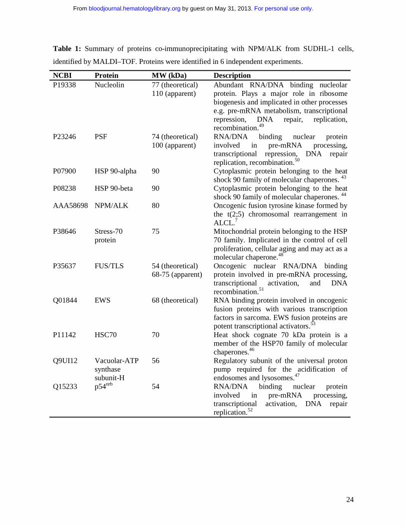
Table 1: Summary of proteins co-immunoprecipitating with NPM/ALK from SUDHL-1 cells,
identified by MALDI–TOF. Proteins were identified in 6 independent experiments.
NCBI Protein MW (kDa) Description P19338
Nucleolin
77 (theoretical) 110 (apparent)
Abundant RNA/DNA binding nucleolar protein. Plays a major role in ribosome biogenesis and implicated in other processes e.g. pre-mRNA metabolism, transcriptional repression, DNA repair, replication, recombination.49
P23246
PSF 74 (theoretical) 100 (apparent)
RNA/DNA binding nuclear protein involved in pre-mRNA processing, transcriptional repression, DNA repair replication, recombination.50
P07900
HSP 90-alpha
90 Cytoplasmic protein belonging to the heat shock 90 family of molecular chaperones. 43
P08238
HSP 90-beta
90 Cytoplasmic protein belonging to the heat shock 90 family of molecular chaperones. 44
AAA58698
NPM/ALK
80 Oncogenic fusion tyrosine kinase formed by the t(2;5) chromosomal rearrangement in ALCL.7
P38646
Stress-70 protein
75 Mitochondrial protein belonging to the HSP 70 family. Implicated in the control of cell proliferation, cellular aging and may act as a molecular chaperone.48
P35637
FUS/TLS 54 (theoretical) 68-75 (apparent)
Oncogenic nuclear RNA/DNA binding protein involved in pre-mRNA processing, transcriptional activation, and DNA recombination.51
Q01844
EWS
68 (theoretical) RNA binding protein involved in oncogenic fusion proteins with various transcription factors in sarcoma. EWS fusion proteins are potent transcriptional activators.53
P11142
HSC70
70 Heat shock cognate 70 kDa protein is a member of the HSP70 family of molecular chaperones.46
Q9UI12
Vacuolar-ATP synthase subunit-H
56 Regulatory subunit of the universal proton pump required for the acidification of endosomes and lysosomes.47
Q15233
p54nrb
54 RNA/DNA binding nuclear protein involved in pre-mRNA processing, transcriptional activation, DNA repair replication.52
For personal use only. by guest on May 31, 2013. bloodjournal.hematologylibrary.orgFrom

25
Figure legends
Figure 1: Identification of proteins co-immunoprecipitating with NPM/ALK by mass
spectrometry. A) Silver stained SDS-PAGE gel of anti-ALK1 immunoprecipitates from
NPM/ALK negative K562 cells (lane 1), lyses buffer alone (lane 2), and NPM/ALK positive
SUDHL-1 cells (lane 3). Bands were excised from the gel, trypsin digested and analysed by
MALDI-TOF. Identified proteins are indicated by arrows. B) MS/MS sequencing of peptides
identifying PSF (genbank: P23246) and NPM/ALK (genbank: AAA58698) proteins.
Figure 2: PSF co-immunoprecipitates with ALK fusion proteins expressed in cell lines and
ALCL tumours. Reciprocal immunoprecipitation (IP) and Western blotting (WB) experiments
were performed using anti-ALK1 and anti-PSF antibodies on lysates derived from: A) NPM/ALK-
positive SUDHL-1 cells and NPM/ALK-negative K562 cells; B) BaF3-NPM/ALK (BaF3-N/A),
BaF3-Parental (BaF3-PAR) and BaF3 cells expressing kinase dead NPM/ALK (BaF3-KD); C)
lymph node tissue taken from two NPM/ALK positive ALCL patients and NPM/ALK negative
spleen tissue; D) BaF3 cells expressing GCN4/ALK (BaF3-GCN4/ALK). As an internal control
SUDHL-1 and K562 cells were subjected to IP with an anti-IgG antibody (A).
Figure 3: PSF is Tyr-phosphorylated in NPM/ALK expressing cells and is a substrate of ALK
tyrosine kinase activity. A) Anti-ALK1 and anti-PSF immunoprecipitates (IP) from SUDHL-1,
K562, BaF3-Par, BaF3-KD and BaF3-NA cells were Western blotted with an anti-phosphotyrosine
(pTyr) antibody. B) Anti-ALK1 immunoprecipitates from lymph nodes derived from patient 1 with
NPM/ALK positive ALCL and NPM/ALK negative spleen tissue were Western blotted using the
anti-pTyr antibody. C) A radioactive in vitro kinase assay was performed with purified recombinant
6xHisALK kinase domain (rALK) and purified full length 6xHis-PSF. Protein purity was checked
by coomassie staining (i). Kinase assay was performed in the presence of rALK kinase domain
For personal use only. by guest on May 31, 2013. bloodjournal.hematologylibrary.orgFrom
26
alone (0.4 µg), PSF alone (0.25 µg), or rALK and PSF together. Samples were analysed by SDS-
PAGE and visualized by autoradiography (ii). Bands corresponding to PSF and ALK are indicated
by arrows. D) Time courses of the phosphorylation of PSF peptides (400 µM) by GST-ALK. The
peptide sequences are as follows: Tyr251–RGGRQHHPPYHQQHHQGP; Tyr293–
PGEKTYTQRCRLFVGNLPADIT; Tyr470–EKLAQKNPMYQKERETPTR; Tyr488-490–
TFEYEYSQRWKSLDEMEKQQR; Tyr527–EMEDAYHEHQANLLRQDLMRRQ; Tyr597-602–
REESYSRMGYMDPRERDMR; Tyr624–MNMGDPYGSGGQKFPPLGGG; Tyr698–
GRGREEYEGPNKKPRF. E) Immunoprecipitation with anti-HA (lanes 2,3) and anti-ALK1 (lanes
4-5) monoclonal antibodies of lysate derived from 293T cells transiently transfected with wild-type
or mutant Y293F HA-PSF together with NPM/ALK. Immunoprecipitates were resolved by SDS-
PAGE and Western blotting with anti-pTyr and anti-HA antibodies was performed. Lane 1 shows
lysate from non-transfected 293T cells.
Figure 4: PSF is delocalized to the cytoplasm in cells expressing ALK fusion proteins.
Indirect immunofluorescence was performed on 293T cells transiently transfected with HA-PSF
alone, or together with kinase dead NPM/ALK (KD-N/A), NPM/ALK (N/A), GCN4/ALK (G/A) or
with mutant HA-PSF-Y293F together with NPM/ALK (N/A). Fluorescence was detected by
confocal microscopy. Nuclei were detected using propidium iodide (PI) shown in red (column 1).
HA-PSF was detected by FITC staining shown in green (column 2). NPM/ALK was detected by
Cy5 staining shown in blue (column 3). A transmission image in place of Cy5 staining is shown for
cells transfected with HA-PSF alone.
Figure 5: PSF content in cytoplasmic and nuclear extracts derived from human cell lines.
Nuclear and cytoplasmic extracts were prepared from the NPM/ALK negative cell line, Jurkat, and
the NPM/ALK positive cell lines, SUDHL-1 and JB6. A. Total cell lysates (lanes 1-3), cytoplasmic
extracts (lanes 4-6), and nuclear extracts (lanes 7-9) were resolved by SDS-PAGE and PSF was
For personal use only. by guest on May 31, 2013. bloodjournal.hematologylibrary.orgFrom

27
detected by Western blotting (top panel). The purity of the extracts were controlled by Western
blotting with anti-β actin for the cytoplasm (middle panel) and anti-Histone H1 for the nucleus
(lower panel). B. Nuclear and cytoplasmic extracts from SUDHL-1 cells were probed with anti-
phosphotyrosine Ab.
Figure 6: PSF overexpression reduces the growth rate and clonogenic potential of NPM/ALK
expressing cells. BaF3-Par (A), BaF3-NA (B), BaF3-KD (C), and BaF3-BA (D) cells were
transiently transfected with pCR3.1 encoding HA-PSF (PSF) or with empty vector (CON). Cell
proliferation was measured by 3[H]-thymidine incorporation at 24, 48 and 72h after transfection.
Results are shown as the mean ± S.D. and are representative of 3 independent experiments (E)
Expression of HA-PSF was verified by Western blotting using anti-HA antibody at 72h and protein
loading was controlled by anti-actin Western blotting. (F) The NPM/ALK positive cell lines, JB6
and SUDHL-1, and the NPM/ALK negative cell line, Jurkat, were transduced with the MigR1-HA-
PSF construct (gray bars), or the MigR1 retrovirus alone (black bars). Cells were selected for GFP
positivity and clonogenic potential was measured by colony formation in methylcellulose. Results
are expressed as the mean ± s.e.m. of three independent clonogenic assays. Significance was
determined by comparing MigR1 alone versus MigR1-HA-PSF in a 2-tail paired samples t test.
(G) BaF3-NA cells were transiently transfected with pCR3.1 encoding HA-Y293T PSF or with
empty vector (CON). (H) Transfection efficiency was controlled by anti-HA immunoblotting at
48h, in control (CON) and HA-PSF Y293F samples, loaded on the same gel. Total protein loading
was controlled by anti-actin immunoblotting.
Figure 7: PSF overexpression induces apoptosis in NPM/ALK expressing cell lines.
A) BaF3-Par, BaF3-KD and Baf3-NA cells were electroporated with pCR3.1 plasmid alone (lanes
1-3) and with pCR3.1 HA-PSF (lanes 4 – 6). After 30h of culture the cell extracts from 5 x 10⁶ cells
were lysed and subjected to denaturing SDS-PAGE and Western blotting with an anti-PARP
For personal use only. by guest on May 31, 2013. bloodjournal.hematologylibrary.orgFrom
28
antibody. PSF expression and protein loading were controlled by anti-HA and anti-actin Western
blotting. B-F) 293T cells were transiently transfected with pCR3.1 empty vector (CON) or HA-PSF
alone or together with kinase dead or wild type NPM/ALK. The efficiency of transfection was
determined by anti-HA and anti-ALK1 Western blotting at 72h post-transfection (B). Apoptosis
was assessed at the same time point by Annexin V analysis in control cells (C), cells transfected
with HA-PSF alone (D), or together with kinase dead NPM/ALK (E: HA-PSF+NA KD), or wild
type NPM/ALK (F: HA-PSF+NA).
For personal use only. by guest on May 31, 2013. bloodjournal.hematologylibrary.orgFrom
1
For personal use only. by guest on May 31, 2013. bloodjournal.hematologylibrary.orgFrom

2
For personal use only. by guest on May 31, 2013. bloodjournal.hematologylibrary.orgFrom

3
For personal use only. by guest on May 31, 2013. bloodjournal.hematologylibrary.orgFrom

Figure 4
4
For personal use only. by guest on May 31, 2013. bloodjournal.hematologylibrary.orgFrom
5
For personal use only. by guest on May 31, 2013. bloodjournal.hematologylibrary.orgFrom
6
For personal use only. by guest on May 31, 2013. bloodjournal.hematologylibrary.orgFrom

7
For personal use only. by guest on May 31, 2013. bloodjournal.hematologylibrary.orgFrom