EML4-ALK Fusion Gene and Efficacy of an ALK Kinase Inhibitor in Lung Cancer
18
EML4-ALK fusion gene and efficacy of an ALK kinase inhibitor in lung cancer Jussi P. Koivunen 1,2 , Craig Mermel 2,4 , Kreshnik Zejnullahu 1,2 , Carly Murphy 5 , Eugene Lifshits 6 , Alison J. Holmes 1,2 , Hwan Geun Choi 3,8 , Jhingook Kim 9,10 , Derek Chiang 2,4 , Roman Thomas 11 , Jinseon Lee 9,10 , William G. Richards 12 , David J. Sugarbaker 12 , Christopher Ducko 12 , Neal Lindeman 5 , J. Paul Marcoux 1,2,5 , Jeffrey A. Engelman 6 , Nathanael S. Gray 3,8 , Charles Lee 5 , Matthew Meyerson 1,2,3,5 , and Pasi A. Jänne 1,2,7,* 1 Lowe Center for Thoracic Oncology, Dana-Farber Cancer Institute, Boston, MA 2 Department of Medical Oncology, Dana-Farber Cancer Institute, Boston, MA 3 Department of Cancer Biology, Dana-Farber Cancer Institute, Boston, MA 4 The Broad Institute of MIT and Harvard, Cambridge, MA 5 Department of Pathology, Brigham and Women’s Hospital and Harvard Medical School, Boston, MA 6 Massachusetts General Hospital Cancer Center, Boston, MA 7 Department of Medicine, Brigham and Women’s Hospital and Harvard Medical School, Boston, MA 8 Department of Biological Chemistry and Molecular Pharmacology, Harvard Medical School, Boston, MA 9 Department of Thoracic Surgery, Samsung Medical Center, Seoul, Korea 10 Sungkyunkwan University School of Medicine, Seoul, Korea 11 Max-Planck Institute, Cologne, Germany 12 Department of Surgery, Brigham and Women’s Hospital, Boston, MA Abstract Purpose—The EML4-ALK fusion gene has been detected in ~7% of Japanese non-small cell lung cancers (NSCLC). We determined the frequency of EML4-ALK in Caucasian NSCLCs and in NSCLC cell lines. We also determined whether TAE684, a specific ALK kinase inhibitor, would inhibit the growth of EML4-ALK containing cell lines in vitro and in vivo. Experimental Design—We screened 305 primary NSCLCs (both US (n=138) and Korean (n=167) patients) and 83 NSCLC cell lines using RT-PCR and by exon array analyses. We evaluated the efficacy of TAE684 against NSCLC cell lines in vitro and in vivo. Results—We detected 4 different variants, including two novel variants, of EML4-ALK using RT-PCR in 8/305 tumors (3%) and in 3/83 (3.6%) NSCLC cell lines. All EML4-ALK containing tumors and cell lines were adenocarcinomas. EML4-ALK was detected more frequently in NSCLC patients who were never or light (< 10 pack years) cigarette smokers compared to current/former smokers (6% vs. 1%; p=0.049). TAE684 inhibited the growth of 1 of 3 (H3122) EML4-ALK * Address Correspondence to: Pasi A. Jänne, M.D., Ph.D., Lowe Center for Thoracic Oncology, Dana-Farber Cancer Institute, D820A, 44 Binney Street, Boston, MA 02115, Phone: (617) 632-6036, Fax: (617) 582-7683, [email protected]. NIH Public Access Author Manuscript Clin Cancer Res. Author manuscript; available in PMC 2011 January 24. Published in final edited form as: Clin Cancer Res. 2008 July 1; 14(13): 4275–4283. doi:10.1158/1078-0432.CCR-08-0168. NIH-PA Author Manuscript NIH-PA Author Manuscript NIH-PA Author Manuscript
-
Upload
independent -
Category
Documents
-
view
1 -
download
0
Transcript of EML4-ALK Fusion Gene and Efficacy of an ALK Kinase Inhibitor in Lung Cancer

EML4-ALK fusion gene and efficacy of an ALK kinase inhibitor inlung cancer
Jussi P. Koivunen1,2, Craig Mermel2,4, Kreshnik Zejnullahu1,2, Carly Murphy5, EugeneLifshits6, Alison J. Holmes1,2, Hwan Geun Choi3,8, Jhingook Kim9,10, Derek Chiang2,4,Roman Thomas11, Jinseon Lee9,10, William G. Richards12, David J. Sugarbaker12,Christopher Ducko12, Neal Lindeman5, J. Paul Marcoux1,2,5, Jeffrey A. Engelman6,Nathanael S. Gray3,8, Charles Lee5, Matthew Meyerson1,2,3,5, and Pasi A. Jänne1,2,7,*1 Lowe Center for Thoracic Oncology, Dana-Farber Cancer Institute, Boston, MA2 Department of Medical Oncology, Dana-Farber Cancer Institute, Boston, MA3 Department of Cancer Biology, Dana-Farber Cancer Institute, Boston, MA4 The Broad Institute of MIT and Harvard, Cambridge, MA5 Department of Pathology, Brigham and Women’s Hospital and Harvard Medical School, Boston,MA6 Massachusetts General Hospital Cancer Center, Boston, MA7 Department of Medicine, Brigham and Women’s Hospital and Harvard Medical School, Boston,MA8 Department of Biological Chemistry and Molecular Pharmacology, Harvard Medical School,Boston, MA9 Department of Thoracic Surgery, Samsung Medical Center, Seoul, Korea10 Sungkyunkwan University School of Medicine, Seoul, Korea11 Max-Planck Institute, Cologne, Germany12 Department of Surgery, Brigham and Women’s Hospital, Boston, MA
AbstractPurpose—The EML4-ALK fusion gene has been detected in ~7% of Japanese non-small celllung cancers (NSCLC). We determined the frequency of EML4-ALK in Caucasian NSCLCs and inNSCLC cell lines. We also determined whether TAE684, a specific ALK kinase inhibitor, wouldinhibit the growth of EML4-ALK containing cell lines in vitro and in vivo.
Experimental Design—We screened 305 primary NSCLCs (both US (n=138) and Korean(n=167) patients) and 83 NSCLC cell lines using RT-PCR and by exon array analyses. Weevaluated the efficacy of TAE684 against NSCLC cell lines in vitro and in vivo.
Results—We detected 4 different variants, including two novel variants, of EML4-ALK usingRT-PCR in 8/305 tumors (3%) and in 3/83 (3.6%) NSCLC cell lines. All EML4-ALK containingtumors and cell lines were adenocarcinomas. EML4-ALK was detected more frequently in NSCLCpatients who were never or light (< 10 pack years) cigarette smokers compared to current/formersmokers (6% vs. 1%; p=0.049). TAE684 inhibited the growth of 1 of 3 (H3122) EML4-ALK
*Address Correspondence to: Pasi A. Jänne, M.D., Ph.D., Lowe Center for Thoracic Oncology, Dana-Farber Cancer Institute, D820A,44 Binney Street, Boston, MA 02115, Phone: (617) 632-6036, Fax: (617) 582-7683, [email protected].
NIH Public AccessAuthor ManuscriptClin Cancer Res. Author manuscript; available in PMC 2011 January 24.
Published in final edited form as:Clin Cancer Res. 2008 July 1; 14(13): 4275–4283. doi:10.1158/1078-0432.CCR-08-0168.
NIH
-PA Author Manuscript
NIH
-PA Author Manuscript
NIH
-PA Author Manuscript

containing cell lines in vitro and in vivo, inhibited Akt phosphorylation and caused apoptosis. Inanother EML4-ALK cell line, DFCI032, TAE684 was ineffective due to co-activation of EGFRand ERBB2. The combination of TAE684 and CL-387,785 (EGFR/ERBB2 kinase inhibitor),inhibited growth and Akt phosphorylation and led to apoptosis in the DFCI032 cell line.
Conclusions—EML4-ALK is found in the minority of NSCLCs. ALK kinase inhibitors alone orin combination may nevertheless be clinically effective treatments for NSCLC patients whosetumors contain EML4-ALK.
KeywordsCarcinoma, Non-Small-Cell lung; EML4-ALK; ALK; Kinase inhibitor
IntroductionAnaplastic lymphoma kinase (ALK) kinase was originally discovered from chromosomaltranslocations leading to the production of fusion proteins consisting of the C-terminalkinase domain of ALK and the N-terminal portions of different genes (1). Translocations ofALK have been identified in 40–60% of anaplastic lymphomas and in B-cell lymphomas,neuroblastomas, and myofibroblastic tumors (2). Nucleophosmin (NPM) is the mostcommon fusion partner of ALK (80% of translocations) but at least six other fusion partnershave been identified (2). In these fusion proteins, the N-terminal portion is responsible forprotein oligomerization, which leads to constitutive activation of ALK kinase, and results inaberrant activation of downstream signalling targets including Akt, STAT3, andextracellular regulated kinase 1/2 (ERK1/2) (2).
The fusion of the ALK gene with echinoderm microtubule-associated protein-like 4 (EML4)has recently been detected in 6.7% (5/75) of Japanese non-small cell lung cancers (NSCLC)(3). ALK and EML4 are both located in the short arm of chromosome 2 separated by 12megabases and are oriented in opposite 5′ to 3′ directions. Two different variants of EML4-ALK fusion gene have been characterized both involving exons 20-29 of ALK fused to exon1-13 (variant 1) or 1–20 (variant 2) of EML4. Both variants of the EML4-ALK fusion genewere transforming in 3T3 cells and in Ba/F3 models (3).
Inhibitors of ALK kinase have been developed and examined in preclinical models. Proof ofconcept studies using shRNA knockdown of ALK in NPM-ALK containing models led togrowth inhibition and apoptosis and suggested that ALK inhibition may be a potentiallyeffective therapeutic strategy (4). This has lead to development and testing of smallmolecule inhibitors of ALK. Initial studies have been performed using less potent ALKinhibitors such as WHI-P154 (IC50 ~5μM), pyridones (IC50 for staurosporine 0.15–0.78μM)or with HSP90 inhibitors (5). Subsequently, more potent and specific ALK inhibitors suchas diamino or aminopyrimidines have been developed including TAE684 and PF02341066(6–8). Both of these inhibitors have good bioavailability and they inhibit ALK kinaseactivity and growth of NPM-ALK positive lymphoma cells in the low nanomolar range (6–8). PF02341066 is an inhibitor of both MET and ALK presently in phase I clinicaldevelopment. TAE684 is not currently under clinical development. Neither agent haspreviously been examined against EML4-ALK.
In the current study we analyzed the frequency of the EML4-ALK fusion gene in NSCLCcell lines and tumors derived from US and Korean NSCLC patients. In addition weexamined the efficacy of an ALK kinase inhibitor, TAE684, in NSCLC cell lines harboringthe EML4-ALK inversion to determine if this would be a potentially effective therapeuticstrategy for NSCLC patients whose tumors contain the EML4-ALK inversion (6).
Koivunen et al. Page 2
Clin Cancer Res. Author manuscript; available in PMC 2011 January 24.
NIH
-PA Author Manuscript
NIH
-PA Author Manuscript
NIH
-PA Author Manuscript

Material and MethodsCell lines and tumors
NSCLC (n=81) and mesothelioma (n=2) cell lines were purchased from ATCC (Manassas,VA), or were kind gifts from Drs. John D. Minna and Adi F. Gazdar (UT Southwestern,Dallas, TX) (Table S1). DFCI024 and DFCI032 were established at DFCI from pleuraleffusions of treatment naïve female NSCLC patients. The PC9, A549, H3122 and H2228cells were cultured in RPMI-1640 (Sigma Chemical Co., St Louis, MO) supplemented with10% fetal bovine serum, 100 U/ml streptomycin and 1 mM sodium pyruvate. The DFCI032cells were cultured in ACL-4 media (Invitrogen, Rockville, MD) supplemented with 5%fetal bovine serum, 100 U/ml streptomycin and 1 mM sodium pyruvate.
NSCLC tumors (n = 305) were collected from surgical resections from patients with stagesI–IIII NSCLC when sufficient material for RNA extraction was available. The majority ofthe specimens (n = 167) were collected at the Samsung Medical Center, Korea. Frozentumor tissues were collected from 809 out of 2442 patients who underwent curativeresection for non-small cell lung cancer (NSCLC) from Nov. 1995 to Feb. 2007 at SamsungMedical Center. One or two pieces from the periphery of the tumor masses—avoidingnecrotic regions—were immediately frozen at −80°C until retrieved. The medical recordsand also hematoxylin/eosin-stained slides of the specimen were reviewed by a singlepathologist. Only frozen tumor tissues from adenocarcinoma or squamous cell carcinoma(according to the 2004 World Health Organization histopathological criteria) were included.Only frozen tumor tissues with a tumor cell content of more than 70% were used for furtheranalysis. In addition, frozen tumor tissues of the following patients were excluded from thestudy: patients who had received preoperative neoadjuvant treatments, patients with doubleprimary lung cancer, and patients who had undergone incomplete resections or who had notbeen subjected to mediastinal lymph node dissections. The selected frozen tumor tissueswere used for the microdissection. Briefly frozen tissues were lightly stained withhematoxylin/eosin to improve visualization and the necrotic tumor tissues and interveningnormal tissues were removed. Each of the microdissected tumor tissues with a tumor cellcontent of more than 90% was placed in 1 ml Easy Blue reagent of a commercially availableRNA isolation kit (easy-spin™ Total RNA Extraction Kit, iNtRON Biotechnology, Korea),immediately homogenized by vortexing, and the total RNA was extracted. The quantity andquality of RNA were analyzed using a spectrometer (Nanodrop Technologies, Rockland,DE) and Agilent 2100 Bioanalyzer (Agilent RNA 6000 Nano Kit, Agilent TechnologiesInc., Germany), respectively. In the end, 167 frozen tissues with acceptable quality of RNA[RNA Integrity Number (RIN) value over 7.0] were used for the current studies. All patientsprovided written informed consent.
The tumors from Caucasian patients (n = 138) were collected at the Brigham and Women’sHospital, Boston, MA between 1991 and 1997 and have been previously published (9,10).Frozen samples of resected lung tumors were obtained within 30 minutes of resection andsubdivided into 100 mg samples and snap frozen at −80 C. Each specimen was associatedwith an immediately adjacent sample embedded for histology in optimal cutting temperature(OCT) medium and stored at −80 C. Six micron frozen sections of embedded samplesstained with hematoxylin/eosin (H&E) was used to confirm the post operative pathologicdiagnosis and to estimate the cellular composition of adjacent samples. All specimensunderwent pathologic review by 2 pathologists. 109 tumors obtained during the same timeperiod were excluded because they did not meet one or more of the eligibility criteria.Tissue samples were homogenized in Trizol (Life Technologies, Gaithersburg, MD) andRNA was extracted and purified by using the RNeasy column purification kit (Qiagen,Chatsworth, CA). Denaturing formaldehyde gel electrophoresis followed by northern
Koivunen et al. Page 3
Clin Cancer Res. Author manuscript; available in PMC 2011 January 24.
NIH
-PA Author Manuscript
NIH
-PA Author Manuscript
NIH
-PA Author Manuscript

blotting using a beta-actin probe assessed RNA integrity. Samples were excluded if beta-actin was not full-length. All patients provided written informed consent.
Cell line specimens were snap frozen, and stored at −80C. RNA was extracted from tumorsand cell lines using Trizol (Invitrogen, Carlsbad, CA), purified with Rneasy Mini Kit(Qiagen, Valencia, CA) and was used for cDNA synthesis using the QuantiTect reversetranscription kit (Qiagen, Valencia, CA).
Exon Array StudiesTo screen for ALK translocations, we used Affymetrix HuEx-1.0 Exon Array (Affymetrix,Santa Clara, CA) data that was previously generated from these cell lines (R.K. Thomas,C.H. Mermel, D. Chiang, and M. Meyerson, unpublished results). The HuEx-1.0 array wasdesigned to contain probes mapping to every known and predicted exon in the humangenome. We reasoned that translocations in the ALK gene would result in disparate levels ofexpression between exons 5′ and 3′ of the breakpoint, with the expression higher in the 3′end (kinase domain). After performing array normalization and background correction forall probes, we restricted our analysis to the 104 probes uniquely mapping to the ALK gene(Refseq NM_004304). To correct for differences in probe response characteristics across thegene, for every sample we divided each probe intensity value by the average probe intensityacross the other wild type specimens. For each cell line, we computed the location of themost likely breakpoint as the probe which gives the maximum deviation between averageexpression of 5′ and 3′ probe subsets. Significance levels for each inferred breakpoint werecomputed using a simple two-sided t-test.
RT-PCR and GenotypingFor RT-PCR analysis of EML4-ALK, we used primer sequences (primer set 1) as describedin (3). The forward primer is located at exon 13 of EML4 while the reverse primer is locatedat exon 20 of ALK. In order to detect other potential EML4-ALK fusion products, wedesigned a second forward primer from exon 3 of EML4 (5′-taccagtgctgtctcaattgcagg-3′)while using the same reverse primer as the primer set 1. PCR amplification was performedusing JumpStart Taq enzyme (Sigma, St. Louis, MO) under manufacturer’s guidelines. Theresulting PCR products were analyzed using agarose gel electrophoresis. Genotyping forKRAS, EGFR, HER2, BRAF and PIK3CA was performed using either a RT-PCR based or agenomic DNA based SURVEYOR-WAVE mutation analysis (11) followed by sequencingof the positive specimens or by direct sequencing of the PCR products. Primer sequencesand PCR conditions are available upon request.
Fluorescence in situ hybridizationBacterial artificial chromosomes (BAC) RP11-667I6 and RP11-100C1 (Children’s HospitalOakland Research Institute, Oakland, CA) were used as probes for the EML4 and ALKgenes, respectively. BAC DNA was labeled with either spectrum red dUTP or spectrumgreen-11-dUTP by nick translation (Vysis, Des Plain, IL) using manufacturer’srecommended conditions. Slides for metaphase FISH from cell lines were prepared usingstandard cytogenetic methodologies. Paraffin embedded slides were prepared as previouslydescribed in (11). Probes were hybridized and washed according to standard FISHprocedures (12).
Kinase InhibitorsTAE684 was synthesized according to published procedures (13). The structure of TAE684was confirmed using liquid chromatography-electrospray mass spectrometry (LC-MS)and 1H and 13C nuclear magnetic resonance (NMR). The synthesized TAE684 was
Koivunen et al. Page 4
Clin Cancer Res. Author manuscript; available in PMC 2011 January 24.
NIH
-PA Author Manuscript
NIH
-PA Author Manuscript
NIH
-PA Author Manuscript

determined to be 98% pure by 1H NMR and 99% pure by LC-MS monitoring at 210nm and254nm wavelengths (data not shown). CL-387,785 was purchased from Calbiochem(Gibbstown, NJ). Erlotinib was purchased from the Dana Farber Cancer Institute pharmacy.All drugs were dissolved in DMSO, stored at −70C and diluted in fresh media prior to use.
Cell Proliferation and Growth AssaysGrowth inhibition was assessed by MTS assay as described in (11). NSCLC cells wereexposed to drugs alone or in combination for 72 hours. All experimental points were set upin six to twelve wells and repeated at least three times. The data was graphically displayedusing GraphPad Prism version 3.00 for Windows, (GraphPad Software;www.graphpad.com). The curves were fitted using a non-linear regression model with asigmoidal dose response.
Antibodies and Western BlottingCells were lysed in buffer containing proteinase inhibitors, proteins separated by gelelectrophoresis on 5–12% polyacrylamide gels selected depending on the target’s molecularweight, transferred to PVDF membranes and detected by immunoblotting using an enhancedchemiluminescence system (Perkin Elmer, Boston, MA) as previously described (11). Thereceptor tyrosine kinase (RTK) array was purchased from R&D Systems (Minneapolis, MN)and used according to the manufacturer’s recommended conditions. Anti-ALK, anti-phospho-ALK (Tyr-1604), anti-phospho-Akt (Ser-473), anti-Akt, anti-STAT3, anti-phosphoSTAT3 (Tyr705), anti-PTEN, and anti-PARP antibodies were obtained from Cell SignalingTechnology (Danvers, MA). Total ERK1/2 and phospho-ERK1/2 (pT185/pY187) antibodieswere purchased from Biosource International (Camarillo, CA). The anti-α-tubulin antibodywas purchased from Sigma-Aldrich (St. Louis, MO).
Fluorescence-activated cell sorting analysisCells were plated at a density of 0.5 to 2 × 10⁵ cells/plate in 10-cm2 plates. Drugs wereadded to the medium after 24 h, and the cells were incubated for another 72 h, after whichthe cells were analyzed as previously described (14). Percent apoptosis was estimated fromthe sub-G1 cell fraction.
Xenograft studiesNude mice (nu/nu; 6–8 weeks old; Charles River Laboratories) were used for in vivo studiesand were cared for in accordance with the standards of the Institutional Animal Care andUse Committee (IACUC) under a protocol approved by the Animal Care and UseCommittee of the Beth Israel Deaconess Medical Center. Mice were anesthetized using a2% Isoflurane (Baxter) inhalation oxygen mixture. A suspension of 5×10⁶ H3122 lungcancer cells (in 0.2 ml of PBS) were inoculated subcutaneously into the lower-right quadrantof the flank of each mouse. Mice were randomized to 4 treatment groups (n=5 per group)once the mean tumor volume reached 500–600 mm3: vehicle (NMP (10% 1-methyl-2-pyrrolidinone: 90% PEG-300) alone, erlotinib, TAE684 10 mg/kg/day and TAE684 25 mg/kg/day p.o. (6). Erlotinib was administered at 100mg/kg/day p.o. as previously described(11). Tumors were measured twice weekly using calipers, and volume was calculated usingthe formula (length × width2 × 0.52). Mice were monitored daily for body weight andgeneral condition. The experiment was terminated when the mean size of either the treatedor control groups reached 2000 mm3.
Koivunen et al. Page 5
Clin Cancer Res. Author manuscript; available in PMC 2011 January 24.
NIH
-PA Author Manuscript
NIH
-PA Author Manuscript
NIH
-PA Author Manuscript

ResultsIdentification of EML4-ALK fusion genes in NSCLC cell lines
In order to rapidly screen our panel of 83 lung cancer cell lines (Table S1) for potential ALKtranslocations, we used Affymetrix HuEx-1.0 mRNA exon arrays and focused on 104unique probes covering the ALK gene. We identified two cell lines, H3122 and H2228,which had statistically significant (p < .001) breakpoints in the ALK gene (Figs 1A and S1).Although our algorithm did not consider the location or direction of the breakpoint, theinferred ALK breakpoints in both samples were very near the conserved exon 20 breakpointin the ALK gene, and in both samples the expression was higher in the 3′ than the 5′ ends.Using RT-PCR, we were able to confirm the presence of the EML4-ALK fusion geneproduct in both H3122 and H2228 but not in any other of the 81 cell lines. In H3122 wedetected variant 1 of EML4-ALK (Figs. 1B and 1C). In H2228 we detected a novel variant(named variant 3a hereafter) resulting from a fusion of exon 6 (codons 1-222) of EML4 withexon 20 (codons 1058-1621) of ALK (Figs. 1C and S2). A second fusion gene (variant 3b)was also detected from this cell line and contains an additional 33bp fragment derived froman alternatively spliced exon of EML4 (exon 6b; Figs. 1C and S2) and is the predominantform in H2228 (data not shown). This alternatively spliced exon was not detected in any ofthe other fusion variants. As both H3122 and H2228 cell lines were established from femaleNSCLC patients with adenocarcinoma and H2228 is from a never-smoker, we screened forthe presence of EML4-ALK in NSCLC cell lines with these clinical features that we hadestablished at Dana Farber Cancer Institute. We identified 2 cell lines, DFCI024 andDFCI032, both derived from chemotherapy naïve female never-smokers withadenocarcinoma. Both cell lines are wild type for EGFR and KRAS. We detected variant 1 ofthe EML4-ALK fusion gene in the DFCI032 cell line and neither variant in DFCI024 (Fig.1B). Overall, we detected the EML4-ALK fusion gene in 3/83 (3.6%) NSCLC cell lines. Wefurther confirmed the presence of the EML4-ALK inversion using FISH (Fig. S3) in these 3cell lines (Fig. 2A–2C). In addition, we confirmed the presence of the EML4-ALK fusion inthe original tumor specimen that gave rise to the DFCI032 cell line using interphase FISH(Fig 2D).
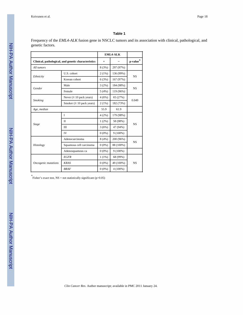
EML4-ALK fusion gene is detected in both Caucasian and Asian NSCLCsWe screened NSCLC (n=305) from patients of U.S. (n=138) and Korean (n=167) origin forthe EML4-ALK fusion gene and detected its presence in 8/305 (3%) NSCLCs (Table 1).Four tumors contained variant 3 (both variants 3a and 3b with 3b being predominant), twocontained variant 1 and two contained a novel variant (named variant 4 here after) (Fig 1Band 1C). In variant 4, exon 15 of EML4 is fused with exon 20 of ALK (EML4 codons 1-569to codons 1078-1621 of ALK; Figs.1C and S2). Six of the EML4-ALK containing tumors(6/167; 3.6%) were from Korean patients while two (2/138 (1.5%)) were detected inNSCLCs from U.S. patients. The frequency of EML4-ALK was higher in females (4%) vs.males (2%). All eight of the EML4-ALK containing tumors were adenocarcinomas.Furthermore, the fusion gene was detected significantly (p=0.049; Table 1) more frequentlyin patients (6%; 4/69) with limited smoking history (≤ 10 pack years) compared to tumorsfrom smokers (1%; 2/184). The tumor from one of the patients had a concurrent EGFRkinase domain mutation (del E746_A750) with the EML4-ALK fusion gene. None of theeight tumors contained a concurrent KRAS or BRAF mutation (data not shown).
Inhibition of ALK kinase activity in EML4-ALK fusion gene in vitro and in vivoIn order to determine whether ALK kinase inhibitors may be therapeutically effective inEML4-ALK containing NSCLCs, we evaluated TAE684, a highly potent ALK kinaseinhibitor (6). We found that TAE684 significantly inhibited the growth of only the H3122cell line while the other two EML4-ALK containing cell lines, H2228 and DFCI032, were as
Koivunen et al. Page 6
Clin Cancer Res. Author manuscript; available in PMC 2011 January 24.
NIH
-PA Author Manuscript
NIH
-PA Author Manuscript
NIH
-PA Author Manuscript

resistant (IC50s 1–10μM) to the inhibitor as those containing an EGFR mutation (PC-9;delE746_A750) or a KRAS mutation (A549; G12S) (Fig. 3A). It should be noted that theIC50 for the H3122 cells is 10nM and that TAE684 exhibits its maximal effects in thisresponsive cell line at 100nM (Fig 3A). At these low concentrations TAE684 is highlyselective for ALK, and therefore the observed response is not likely to be due to off-targeteffects (6). Conversely, the effects on the EGFR mutant PC9 and KRAS mutant A549 cells,which are not ALK dependent for their growth, at concentrations > 1 μM are likely to be dueto off-target effects and thus we used 100 nM TAE684 for subsequent studies. TAE684 (100nM) treatment led to significant apoptosis only in the H3122 cell line as detected byfluorescence activated cell sorting (FACS) (Fig. 3B) or by Western blotting for cleavedPARP (Fig. 3C). No growth arrest or apoptosis was observed in the other cell linesfollowing TAE684 treatment.
In order to determine why the growth of only 1/3 of the EML4-ALK containing cell lineswas inhibited by TAE684 we examined its effects on phosphorylation of ALK anddownstream signalling proteins (Fig. 3D). Following 100 nM TAE684 treatment, completeinhibition of phosphorylated ALK was observed in all three of the EML4-ALK positive celllines (Fig. 3D). However, this was accompanied by substantial inhibition of Akt, and ERK1/2 phosphorylation only in the H3122 cell line consistent with the effects on cell growthand apoptosis in this cell line (Fig 3A–C). In contrast to Akt and ERK 1/2, there was only aminimal decrease in STAT3 phosphorylation following TAE684 treatment in the H3122 cellline (Fig. 3D). In the H2228 cell line there was some but not complete inhibition of Akt andERK1/2 phosphorylation while these were unchanged in the DFCI032 cell line (Fig. 3D).
We also examined the effects of TAE684 treatment on H3122 in vivo using a xenograftmodel. We compared the effects of TAE684 to the epidermal growth factor receptor (EGFR)kinase inhibitor erlotinib which did not inhibit the growth of H3122 cells in vitro (IC50 > 10μM; data not shown). We used erlotinib because we detected EML4-ALK significantly morefrequently in never or former light cigarette smokers with NSCLC (Table 1) and becauseerlotinib is frequently used in clinical trials in this same patient population(11,15). Thus wewished to determine the efficacy of erlotinib in EML4-ALK containing NSCLC. We alsoexplored the dosing of TAE684 by examining 2 different doses in the xenograft studies. Ascan be seen in Figure 4, both doses of TAE684 effectively inhibited the growth of H3122xenografts. Both vehicle and erlotinib treated mice were all sacrificed following 2 weeks oftreatment due to rapid tumor growth. These are consistent with the effects of erlotinib invitro (data not shown). The higher dose of TAE-684 (25 mg/kg/day) more effectivelyinhibited tumor growth than the lower (10 mg/kg/day) dose (Fig. 4). The 25 mg/kg/day dosewas associated with an initial shrinkage of established tumors followed by stabilization. Allmice were sacrificed at day 53 of treatment.
Co-activation of ERBB family members in an EML4-ALK containing NSCLCIn the DFCI032 cell line, which contains the exact same EML4-ALK variant as H3122 (Fig.1B), TAE684 completely inhibited ALK phosphorylation but this was not accompanied byinhibition of growth or changes in phosphorylation of Akt or ERK 1/2 (Fig 3A and D).DFCI032 is not a heterogeneous cell line as by FISH we were able to detect EML4-ALK in100% of the cells (data not shown). We also did not detect a concurrent mutation in theknown oncogenes (EGFR, KRAS, BRAF, HER2 or PIK3CA) commonly mutated in NSCLCin the DFCI032 cell line (data not shown). Recent studies have demonstrated that in somecancers multiple receptor tyrosine kinases (RTKs) can be co-activated (11,16). Inhibition ofonly one of the co-activated kinases is insufficient to result in inhibition of growth or Aktphosphorylation (11,16). In order to determine whether DFCI032 contained other activatedkinases we performed a screen using a phospho-RTK array comprising of 42 receptortyrosine kinases. As can be seen in Figure 5A, the 2 most intense signals detected in this
Koivunen et al. Page 7
Clin Cancer Res. Author manuscript; available in PMC 2011 January 24.
NIH
-PA Author Manuscript
NIH
-PA Author Manuscript
NIH
-PA Author Manuscript

array were for phosphorylated EGFR and ERBB2. Low level activation of other RTKs werealso observed but the signals were substantially weaker than for EGFR and ERBB2 (Fig.5A). ALK is not present on this array (data not shown). We next examined the effects ofCL-387,785 (1 μM), an irreversible EGFR and ERBB2 inhibitor, alone or in combinationwith TAE684 (100 nM) in the DFCI032 cell line. The combination of CL,387,785 andTAE684, but not either agent alone, significantly inhibited the growth of DFCI032 cells andwas associated with significant apoptosis (Fig 5C and D). Furthermore, only thecombination of TAE684 and CL-387,785 was associated inhibition of Akt and ERK 1/2phosphorylation (Fig 5D). We also examined the combination of the EGFR inhibitorgefitinib (1 μM) and TAE684 (100 nM) and observed no effect on growth of DFCI032 cells(data not shown). These findings suggest that inhibition of both EGFR and ERBB2 alongwith ALK is necessary to effectively inhibit growth and induce apoptosis in the DFCI032cell line.
DiscussionThe use of molecular targeted therapy in genetically defined subsets of cancer patients isemerging as an effective therapeutic strategy for many cancers (17–19). In lung cancer forexample, 10–30% of NSCLCs contain activating mutations in the EGFR kinase domain and60–80% of the patients with EGFR mutations obtain dramatic radiographic responsesfollowing treatment with the EGFR kinase inhibitors gefitinib or erlotinib (11,19). Similarly,EGFR mutant NSCLC cell lines are exquisitely sensitive to gefitinib in vitro compared withEGFR wild type cell lines and only EGFR mutant NSCLC cell lines undergo apoptosisfollowing gefitinib treatment (14,20,21). Thus it remains critical to identify subsets of lungcancer patients and to develop effective therapeutic strategies for such patients. As lungcancer is a very common cancer, the identification of even small subsets of lung cancerpatients harbouring specific genetic alterations will translate into a large cohort of patients.
In the present study we characterized the frequency of the EML4-ALK inversion in NSCLCcell lines and primary tumors from NSCLC patients of different ethnic backgrounds. Wedetected the EML4-ALK fusion gene in 3% of NSCLC specimens, numerically morefrequently from Korean than US NSCLC patients, adenocarcinomas and in patients withlimited cigarette smoke exposure. Our study is the first example addressing the frequency ofEML4-ALK in Caucasian NSCLC patients. Despite the low frequency of EML4-ALK inNSCLC, this represents more patients (~5000 annually in U.S.) than those diagnosed withanaplastic large cell lymphoma (ALCL) where ALK translocations have previously beendetected (22,23). We detected EML4-ALK significantly more frequently (Table 1) fromNSCLC patients who were either never or former light (≤ 10 pack years) cigarette smokers.This same clinical feature has also been shown to predict for presence of EGFR mutations(24). Thus it is possible that NSCLC arising in never/former light smokers are geneticallyand biologically different from those arising in smokers and more likely to contain activatedoncogenes. We detected 4 different variants of EML4-ALK (Fig. 1) containing virtuallyidentical portions of ALK, comprising the entire kinase domain, with varying portions ofEML4. These variants are similar to previously described translocations of ALK with othergenes; all of which contain the cytoplasmic portion and entire tyrosine kinase domain ofALK (22). We also developed a FISH assay which can be used to detect the EML4-ALKinversion from routine paraffin embedded lung cancer clinical specimens. This will facilitatethe identification of appropriate NSCLC patients for clinical studies of ALK kinaseinhibitors.
One of the three cell lines with the EML4-ALK translocation (H3122) was also found to beexquisitely sensitive to TAE684 in vitro and in vivo (Fig. 3A and 4). In addition, TAE684treatment was associated with significant apoptosis and downregulation of Akt and ERK 1/2
Koivunen et al. Page 8
Clin Cancer Res. Author manuscript; available in PMC 2011 January 24.
NIH
-PA Author Manuscript
NIH
-PA Author Manuscript
NIH
-PA Author Manuscript

signaling (Fig 3B.–D.). These findings suggest the phenomenon of oncogene addictionwhere ALK kinase solely controls the critical survival signalling pathways in this cell line(25). ALK inhibition leads to inhibition of all of these signalling pathways and subsequentlyto apoptosis. This is analogous to gefitinib treatment of EGFR mutant NSCLC and suggeststhat ALK kinase inhibitors alone may be effective therapies at least for some patients whosetumors contain EML4-ALK (14,26). Interestingly TAE-684 led to only a minimal decrease inSTAT3 phosphorylation in H3122 despite causing apoptosis in this cell line (Fig. 3D).STAT3 has been shown to be critical to NPM-ALK mediated lyphomagenesis and inhibitionof STAT3 alone using a dominant negative STAT3 is sufficient to induce apoptotis (27,28).There may be signalling differences between NPM-ALK and EML4-ALK containing tumorsor this may reflect differences between NSCLC and ALCL. Further studies will be arenecessary to determine the significance of STAT3 signalling in EML4-ALK containingNSCLC.
Only 1 of 3 NSCLC cell lines (H3122) with EML4-ALK was sensitive to the ALK kinaseinhibitor alone. The 2 other EML4-ALK containing cell lines were either resistant toTAE684 alone (H2228) or required concomitant inhibition of EGFR and ERBB2(DFCI032). These findings are quite different from EGFR mutant NSCLC cell lines wherethe majority are exquisitely sensitive to gefitinib or erlotinib in vitro and the growthinhibition is accompanied by apoptosis and significant inhibition of EGFR, ERK 1/2 andAkt phosphorylation (14,29,30). These differences may be clinically significant andhighlight the possibility that ALK inhibitors alone may only be effective in a subset ofNSCLC patients with the EML4-ALK inversion. Our studies of DFCI032 suggest that itcontains co-activation of both EGFR and ERBB2 as concomitant inhibition of ALK, EGFRand ERBB2 is required to significantly effect growth, induce apoptosis and inhibit Akt andERK 1/2 phosphorylation (Figure 5). Thus our findings provide one potential mechanism,activation of other receptor tyrosine kinases, by which resistance could emerge in NSCLCpatients being treated with ALK inhibitors. In addition, these data suggest that in someEML4-ALK containing NSCLC, a combination therapeutic strategy may be necessary. Ourfindings are analogous to those found in subsets of glioblastoma multiforme and in METamplified gefitinib resistant lung cancers where multiple kinases are co-activated andinhibition of one kinase alone is not sufficient to effect growth or lead to down regulation ofAkt (11,16). In H2228 we did not detect co-activation of another kinase (data not shown).The lack of efficacy of TAE684 in our study in H2228 is similar with recent studies using anALK specific siRNA which also did not inhibit the growth of H2228 cells (31). It willcontinue to be important to study H2228 and other EML4-ALK containing tumors in order todetermine whether an ALK inhibitor alone or in combination with other kinase inhibitorswill be necessary for growth inhibition and apoptosis.
Supplementary MaterialRefer to Web version on PubMed Central for supplementary material.
AcknowledgmentsThis study is supported by grants from the National Institutes of Health 1RO1CA114465-01 (P.A.J.), AmericanCancer Society RSG-06-102-01-CCE (P.A.J. and J.A.E.), Finnish Medical Foundation (J.P.K), Finnish CulturalFoundation (J.P.K), and Academy of Finland (J.P.K.) and the Hazel and Samuel Bellin research fund (P.A.J.).P.A.J. and M.M. are part of a pending patent application on EGFR mutations.
References1. Morris SW, Kirstein MN, Valentine MB, et al. Fusion of a kinase gene, ALK, to a nucleolar protein
gene, NPM, in non-Hodgkin’s lymphoma. Science 1994;263:1281–4. [PubMed: 8122112]
Koivunen et al. Page 9
Clin Cancer Res. Author manuscript; available in PMC 2011 January 24.
NIH
-PA Author Manuscript
NIH
-PA Author Manuscript
NIH
-PA Author Manuscript

2. Amin HM, Lai R. Pathobiology of ALK+ anaplastic large-cell lymphoma. Blood 2007;110:2259–67. [PubMed: 17519389]
3. Soda M, Choi YL, Enomoto M, et al. Identification of the transforming EML4-ALK fusion gene innon-small-cell lung cancer. Nature 2007;448:561–6. [PubMed: 17625570]
4. Piva R, Chiarle R, Manazza AD, et al. Ablation of oncogenic ALK is a viable therapeutic approachfor anaplastic large-cell lymphomas. Blood 2006;107:689–97. [PubMed: 16189272]
5. Li R, Morris SW. Development of anaplastic lymphoma kinase (ALK) small-molecule inhibitors forcancer therapy. Med Res Rev. 2007
6. Galkin AV, Melnick JS, Kim S, et al. Identification of NVP-TAE684, a potent, selective, andefficacious inhibitor of NPM-ALK. Proc Natl Acad Sci U S A 2007;104:270–5. [PubMed:17185414]
7. Zou HY, Li Q, Lee JH, et al. An orally available small-molecule inhibitor of c-Met, PF-2341066,exhibits cytoreductive antitumor efficacy through antiproliferative and antiangiogenic mechanisms.Cancer Res 2007;67:4408–17. [PubMed: 17483355]
8. Christensen JG, Zou HY, Arango ME, et al. Cytoreductive antitumor activity of PF-2341066, anovel inhibitor of anaplastic lymphoma kinase and c-Met, in experimental models of anaplasticlarge-cell lymphoma. Mol Cancer Ther 2007;6:3314–22. [PubMed: 18089725]
9. Bhattacharjee A, Richards WG, Staunton J, et al. Classification of human lung carcinomas bymRNA expression profiling reveals distinct adenocarcinoma subclasses. Proc Natl Acad Sci U S A2001;98:13790–5. [PubMed: 11707567]
10. Hayes DN, Monti S, Parmigiani G, et al. Gene expression profiling reveals reproducible humanlung adenocarcinoma subtypes in multiple independent patient cohorts. J Clin Oncol2006;24:5079–90. [PubMed: 17075127]
11. Engelman JA, Zejnullahu K, Gale CM, et al. PF00299804, an irreversible pan-ERBB inhibitor, iseffective in lung cancer models with EGFR and ERBB2 mutations that are resistant to gefitinib.Cancer Res 2007;67:11924–32. [PubMed: 18089823]
12. Lee C, Critcher R, Zhang JG, Mills W, Farr CJ. Distribution of gamma satellite DNA on thehuman X and Y chromosomes suggests that it is not required for mitotic centromere function.Chromosoma 2000;109:381–9. [PubMed: 11072793]
13. Garcia-Echeverria, C.; Kanazawa, T.; Kawahara, E., et al. Preparation of 2,4-pyrimidinediaminesuseful in the treatment of neoplastic diseases, inflammatory and immune system disorders. UnitedStates patent. WO2005016894. 2005.
14. Tracy S, Mukohara T, Hansen M, et al. Gefitinib induces apoptosis in the EGFRL858R non-small-cell lung cancer cell line H3255. Cancer Res 2004;64:7241–4. [PubMed: 15492241]
15. Tsao MS, Sakurada A, Cutz JC, et al. Erlotinib in lung cancer - molecular and clinical predictors ofoutcome. N Engl J Med 2005;353:133–44. [PubMed: 16014883]
16. Stommel JM, Kimmelman AC, Ying H, et al. Coactivation of receptor tyrosine kinases affects theresponse of tumor cells to targeted therapies. Science 2007;318:287–90. [PubMed: 17872411]
17. Druker BJ, Talpaz M, Resta DJ, et al. Efficacy and safety of a specific inhibitor of the BCR-ABLtyrosine kinase in chronic myeloid leukemia. N Engl J Med 2001;344:1031–7. [PubMed:11287972]
18. Demetri GD, von Mehren M, Blanke CD, et al. Efficacy and safety of imatinib mesylate inadvanced gastrointestinal stromal tumors. N Engl J Med 2002;347:472–80. [PubMed: 12181401]
19. Inoue A, Suzuki T, Fukuhara T, et al. Prospective phase II study of gefitinib for chemotherapy-naive patients with advanced non-small-cell lung cancer with epidermal growth factor receptorgene mutations. J Clin Oncol 2006;24:3340–6. [PubMed: 16785471]
20. Paez JG, Janne PA, Lee JC, et al. EGFR mutations in lung cancer: correlation with clinicalresponse to gefitinib therapy. Science 2004;304:1497–500. [PubMed: 15118125]
21. Mukohara T, Civiello G, Johnson BE, Janne PA. Therapeutic targeting of multiple signalingpathways in malignant pleural mesothelioma. Oncology 2005;68:500–10. [PubMed: 16020981]
22. Pulford K, Morris SW, Turturro F. Anaplastic lymphoma kinase proteins in growth control andcancer. J Cell Physiol 2004;199:330–58. [PubMed: 15095281]
23. Jemal A, Siegel R, Ward E, et al. Cancer statistics, 2007. CA Cancer J Clin 2007;57:43–66.[PubMed: 17237035]
Koivunen et al. Page 10
Clin Cancer Res. Author manuscript; available in PMC 2011 January 24.
NIH
-PA Author Manuscript
NIH
-PA Author Manuscript
NIH
-PA Author Manuscript

24. Pham D, Kris MG, Riely GJ, et al. Use of cigarette-smoking history to estimate the likelihood ofmutations in epidermal growth factor receptor gene exons 19 and 21 in lung adenocarcinomas. JClin Oncol 2006;24:1700–4. [PubMed: 16505411]
25. Weinstein IB. Cancer. Addiction to oncogenes--the Achilles heal of cancer. Science 2002;297:63–4. [PubMed: 12098689]
26. Sordella R, Bell DW, Haber DA, Settleman J. Gefitinib-sensitizing EGFR mutations in lung canceractivate anti-apoptotic pathways. Science 2004;305:1163–7. [PubMed: 15284455]
27. Chiarle R, Simmons WJ, Cai H, et al. Stat3 is required for ALK-mediated lymphomagenesis andprovides a possible therapeutic target. Nat Med 2005;11:623–9. [PubMed: 15895073]
28. Amin HM, McDonnell TJ, Ma Y, et al. Selective inhibition of STAT3 induces apoptosis and G(1)cell cycle arrest in ALK-positive anaplastic large cell lymphoma. Oncogene 2004;23:5426–34.[PubMed: 15184887]
29. Mukohara T, Engelman JA, Hanna NH, et al. Differential effects of gefitinib and cetuximab onnon-small-cell lung cancers bearing epidermal growth factor receptor mutations. J Natl Cancer Inst2005;97:1185–94. [PubMed: 16106023]
30. Amann J, Kalyankrishna S, Massion PP, et al. Aberrant epidermal growth factor receptor signalingand enhanced sensitivity to EGFR inhibitors in lung cancer. Cancer Res 2005;65:226–35.[PubMed: 15665299]
31. Rikova K, Guo A, Zeng Q, et al. Global survey of phosphotyrosine signaling identifies oncogenickinases in lung cancer. Cell 2007;131:1190–203. [PubMed: 18083107]
Koivunen et al. Page 11
Clin Cancer Res. Author manuscript; available in PMC 2011 January 24.
NIH
-PA Author Manuscript
NIH
-PA Author Manuscript
NIH
-PA Author Manuscript

Figure 1.EML4-ALK in NSCLC cell lines and tumors. A. Detection of ALK fusion genes in lungcancer cell lines using exon arrays. In the screen of 83 lung cancer cell lines (81/83NSCLCs), exon arrays showed that H3122 and H2228 cell lines had significantly highersignal (log2 difference) for ALK probes #80–140 corresponding to exons 20–29 of ALKcompared with other 81 cell lines. Probes were assigned into three categories based on theirlabeling intensity; non-responsive probes (purple), low-intensity probes (blue), high-intensity probes (black). Only high-intensity probes were used in breakpoint detection. B.RT-PCR detection of EML4-ALK fusion in NSCLC cell lines and tumors. Primer set 2amplifies EML4-ALK fusion genes from H3122, H2228, and DFCI032 cell lines but notfrom A549 line. Primer sets 1 and 2 also detected EML4-ALK fusion from 8 primaryNSCLCs. H3122; positive control, A549; negative control. C. Schematic representation ofthe four different EML4-ALK variants in NSCLC.
Koivunen et al. Page 12
Clin Cancer Res. Author manuscript; available in PMC 2011 January 24.
NIH
-PA Author Manuscript
NIH
-PA Author Manuscript
NIH
-PA Author Manuscript

Figure 2.Detection of EML4-ALK using FISH. A. PC-9 (EGFR del E746_A750) cell line; signals forALK (red dot) and EML4 (green dot) are seen separately. B. H2228 cell line, the fusionsignal of EML4-ALK (arrow) is detected in a small extra-chromosomal fragment (yellow).C. DFCI032 cell line; the EML4-ALK fusion signal in yellow (arrow) is heterozygous.Similar findings were observed for H3122 (data not shown). D. Interphase FISH for EML4-ALK from the formalin fixed paraffin embedded tumor specimen obtained from the pleura ofthe patient whose pleural effusion was used to establish the DFCI032 cell line in C. Thetumor is heterozygous for the EML4-ALK fusion signal (yellow dot; arrow).
Koivunen et al. Page 13
Clin Cancer Res. Author manuscript; available in PMC 2011 January 24.
NIH
-PA Author Manuscript
NIH
-PA Author Manuscript
NIH
-PA Author Manuscript

Figure 3.Effect of TAE684 on growth and signaling in EML4-ALK containing NSCLC cell lines. A.NSCLC cells were treated with TAE-684 at the indicated concentrations, and viable cellswere measured after 72 hours of treatment. The percentage of viable cells is shown relativeto untreated controls. A549 (KRAS G12S); PC9 (EGFR delE746_A750); H2228 (EML4-
Koivunen et al. Page 14
Clin Cancer Res. Author manuscript; available in PMC 2011 January 24.
NIH
-PA Author Manuscript
NIH
-PA Author Manuscript
NIH
-PA Author Manuscript

ALK variant 3); H3122 (EML4-ALK variant 1); DFCI032 (EML4-ALK variant 1). B. FACSanalysis of sub G1 fraction without treatment (left bar) and after treatment with 0.1μMTAE684 for 72h (right bar). Significant apoptosis following TAE-684 treatment is onlyobserved in the H3122 cell line. C. Western analysis of PARP following treatment with0.1μM TAE684 for 72h. The 89 kDa cleaved PARP products is observed only in the H3122cell line consistent with the effects of TAE-684 on cell growth in A. D. Western analysisfollowing TAE684 treatment in wild type and EML4-ALK positive NSCLC cell lines. Totaland phosphorylated ALK are only detected in EML4-ALK positive cell lines (H3122,H2228, DFCI032) but not in wild type control (PC-9). In H3122 and DFCI032 cell lines,ALK positive band migrates at ~120 kDa corresponding to predicted molecular weight(117kDa) of the variant 1 (arrow 1) while in H2228, the band migrates at ~90kDa whichalso corresponds to the predicted molecular weight (90/91kDa) of the variant 3 (arrow 3).ALK phosphorylation is completely inhibited following 0.1μM TAE684 treatment (6 hours)in all the cell lines. Phosphorylation of Akt, STAT3, and ERK1/2 decrease in H3122 andH2228 cell lines with TAE684 but remain unchanged in DFCI032 and PC-9 lines. All thecell lines show presence of PTEN. α-tubulin is used as a loading control.
Koivunen et al. Page 15
Clin Cancer Res. Author manuscript; available in PMC 2011 January 24.
NIH
-PA Author Manuscript
NIH
-PA Author Manuscript
NIH
-PA Author Manuscript

Figure 4.TAE-684 effectively inhibits the growth of H3122 in vivo. Xenografts on H3122 in nu/numice were generated as described in Methods. Erlotinib and TAE-684 treatments wereadministered by oral gavage and tumors were measured three times weekly. The control anderlotinib treated mice reached a median tumor size of 2000 mm3 by 15 days of treatment andwere sacrificed. In contrast the median tumor size of mice treated with TAE684 at either 10mg/kg/day or 25 mg/kg/day did not reach 2000 mm3 even after 53 days of treatment.
Koivunen et al. Page 16
Clin Cancer Res. Author manuscript; available in PMC 2011 January 24.
NIH
-PA Author Manuscript
NIH
-PA Author Manuscript
NIH
-PA Author Manuscript

Figure 5.Co-activation of EGFR and ERBB2 in DFCI032 cell line. A. A phospho-receptor tyrosinekinase (RTK) array reveals that the DFCI032 cells contain strong activation of both EGFRand ERBB2. Cells were grown in media and the cell lysates were hybridized to a phospho-RTK array. In the array, each RTK is spotted in duplicate. Hybridization signals at thecorners serve as controls. B. The combination of TAE684 and CL-387,785 effectivelyinhibits growth of DFCI032 cells. DFCI032 cells were treated with either CL-387,785 (1μM) alone, TAE684 (100 nM) alone or the two in combination for 72 hours. Growth wasassayed by MTS (Methods). The combination of TAE684 and CL-387,785 led to significantinhibition of growth compared to untreated (p < 0.001; paired t-test) or treatment with eitheragent alone (p < 0.001; paired t-test for both comparisons, respectively). **; p < 0.001 C.The combination of CL-387,785 and TAE-684 leads to significant apoptosis. Cells weretreated as in B. and apoptosis was estimated from sub-G1 fraction using FACS (Methods).The combination of CL-387,785 and TAE-684 led to significant increase in apoptosiscompared with untreated (p < 0.05; paired t-test) or treatment with either agent alone (p <0.05; paired t-test for both comparisons, respectively). *; p < 0.05. D. Combination ofCL-387,785 and TAE-684 leads to inhibition of Akt and ERK 1/2 phosphorylation. Cellswere treated as in B. for 6 hours or 48 hours. Cells were lysed and the indicated proteinswere detected by immunoblotting. Only the combination of CL-387,785 and TAE-684 leadsto significant downregulation of Akt and ERK 1/2 signaling and to apoptosis as measured byappearance of cleaved (89 kDa) PARP fragment.
Koivunen et al. Page 17
Clin Cancer Res. Author manuscript; available in PMC 2011 January 24.
NIH
-PA Author Manuscript
NIH
-PA Author Manuscript
NIH
-PA Author Manuscript
NIH
-PA Author Manuscript
NIH
-PA Author Manuscript
NIH
-PA Author Manuscript
Koivunen et al. Page 18
Table 1
Frequency of the EML4-ALK fusion gene in NSCLC tumors and its association with clinical, pathological, andgenetic factors.
EML4-ALK
Clinical, pathological, and genetic characteristics + − p-value*
All tumors 8 (3%) 297 (97%)
EthnicityU.S. cohort 2 (1%) 136 (99%)
NSKorean cohort 6 (3%) 167 (97%)
GenderMale 3 (2%) 184 (98%)
NSFemale 5 (4%) 119 (96%)
SmokingNever (≤ 10 pack years) 4 (6%) 65 (27%)
0.049Smoker (≥ 10 pack years) 2 (1%) 182 (73%)
Age, median 55.9 61.9
Stage
I 4 (2%) 179 (98%)
NSII 1 (2%) 58 (98%)
III 3 (6%) 47 (94%)
IV 0 (0%) 9 (100%)
Histology
Adenocarcinoma 8 (4%) 200 (96%)NS
Squamous cell carcinoma 0 (0%) 88 (100%)
Adenosquamous ca 0 (0%) 9 (100%)
Oncogenic mutations
EGFR 1 (1%) 68 (99%)
NSKRAS 0 (0%) 49 (100%)
BRAF 0 (0%) 4 (100%)
*Fisher’s exact test, NS = not statistically significant (p>0.05)
Clin Cancer Res. Author manuscript; available in PMC 2011 January 24.