Purification and cDNA cloning of HeLa cell p54 nrb , a nuclear protein with two RNA recognition...
8
1993 Oxford University Press Nucleic Acids Research, 1993, Vol. 21, No. 17 4085-4092 Purification and cDNA cloning of HeLa cell p54 n , a nuclear protein with two RNA recognition motifs and extensive homology to human splicing factor PSF and Drosophila NONA/BJ6 Benhao Dong, David S.Horowitz, Ryuji Kobayashi and Adrian R.Krainer* Cold Spring Harbor Laboratory, PO Box 100, 1 Bungtown Road, Cold Spring Harbor, NY 11724-2208, USA Received April 27, 1993; Revised and Accepted June 19, 1993 GenBank accession no. L14599 ABSTRACT While searching for a human homolog of the S.cerevlslae splicing factor PRP18, we found a polypeptlde that reacted strongly with antibodies against PRP18. We purified this polypeptlde from HeLa cells using a Western blot assay, and named it p54 nrb (for nuclear RNA-blnding protein, 54 kDa). cDNAs encoding p54 nrb were cloned with probes derived from partial sequence of the purified protein. These cDNAs have identical coding sequences but differ as a result of alternative splicing In the 5' untranslated region. The cDNAs encode a 471 aa polypeptlde that contains two RNA recognition motifs (RRMs). Human p54 nrt> has no homology to yeast PRP18, except for a common epltope, but is Instead 71% identical to human splicing factor PSF within a 320 aa region that Includes both RRMs. In addition, both p54 nrt> and PSF are rich in Pro and Gin residues outside the main homology region. The Drosophila puff-specif Ic protein BJ6, one of three products encoded by the alternatively spliced no-on- translent A gene (nonA), which is required for normal vision and courtship song, Is 42% identical to p54 nrt> in the same 320 aa region. The striking homology between p54 nrt >, PSF, and NONA/BJ6 defines a novel phylogenetlcally conserved protein segment, termed DBHS domain (for Drosophila behavior, human splicing), which may be Involved in_regulatlng diverse pathways at the level of pre-mRNA splicing. INTRODUCTION Pre-mRNA introns are present in a small proportion of S.cerevisiae genes, almost always as single short sequences near the 5' end of the gene. The 5' and 3' splice sites and the branch site are very highly conserved in budding yeast. In metazoans, intronless genes are infrequent, whereas genes with multiple introns are common, and there is enormous variation in intron sizes. The conserved elements are highly degenerate, and complex patterns of alternative splicing are common. In spite of these substantial differences, the metazoan and yeast pre- mRNA splicing pathways appear very similar (reviewed in 1,2). The structures arid functions of the sphceosomal snRNAs are well conserved from yeast to man, and thus, e.g., human U2 snRNA can functionally replace its S.cerevisiae counterpart in vivo (3). There have also been reports of a putative human homolog of the yeast PRP8 protein (4-6), of yeast homologs of the 70K and A polypeptides of metazoan Ul snRNP (7,8) and the Dl core snRNP polypeptide (9), and of a possible yeast homolog of the human U2AF 65 splicing factor (10). Genetic approaches have facilitated the identification of more than 20 PRP genes required for pre-mRNA splicing in S.cerevisiae (reviewed in 11). One of these genes, PRP18, encodes a 28 kDa U5 snRNP-associated protein involved in the second step reaction of pre-mRNA splicing in vivo and in vitro (12-14). In the absence of PRP18 protein, splicing intermediates accumulate, and upon addition of recombinant PRP 18 protein expressed in bacteria, the second step reaction is restored (15). If there is substantial phylogenetic conservation of protein splicing factors, it may be possible to take advantage of the S.cerevisiae prp mutant strains, genes, proteins or antibodies to identify their metazoan homologs. When antibodies against PRP 18 (14) were used to probe HeLa cell extracts on Western blots, a band of apparent molecular weight 60 kDa was consistently observed. The same antibodies also produced a nucleoplasmic speckled pattern upon indirect immunofluorescence staining of HeLa cells, typical of the pattern generated with antibodies against nucleoplasmic snRNP particles and other splicing factors (reviewed in 16). We report the purification of this cross-reactive protein, termed p54 nrb , and the cloning of the corresponding HeLa cell cDNAs. Surprisingly, the protein sequence of p54 mb bears no sequence homology to PRP18. It is, however, highly homologous to the recently identified human splicing factor PSF (17). The pM"* sequence • To whom correspondence should be addressed by guest on May 30, 2016 http://nar.oxfordjournals.org/ Downloaded from
-
Upload
independent -
Category
Documents
-
view
2 -
download
0
Transcript of Purification and cDNA cloning of HeLa cell p54 nrb , a nuclear protein with two RNA recognition...

1993 Oxford University Press Nucleic Acids Research, 1993, Vol. 21, No. 17 4085-4092
Purification and cDNA cloning of HeLa cell p54n , anuclear protein with two RNA recognition motifs andextensive homology to human splicing factor PSF andDrosophila NONA/BJ6
Benhao Dong, David S.Horowitz, Ryuji Kobayashi and Adrian R.Krainer*Cold Spring Harbor Laboratory, PO Box 100, 1 Bungtown Road, Cold Spring Harbor,NY 11724-2208, USA
Received April 27, 1993; Revised and Accepted June 19, 1993 GenBank accession no. L14599
ABSTRACT
While searching for a human homolog of theS.cerevlslae splicing factor PRP18, we found apolypeptlde that reacted strongly with antibodiesagainst PRP18. We purified this polypeptlde from HeLacells using a Western blot assay, and named it p54nrb
(for nuclear RNA-blnding protein, 54 kDa). cDNAsencoding p54nrb were cloned with probes derived frompartial sequence of the purified protein. These cDNAshave identical coding sequences but differ as a resultof alternative splicing In the 5' untranslated region. ThecDNAs encode a 471 aa polypeptlde that contains twoRNA recognition motifs (RRMs). Human p54nrt> has nohomology to yeast PRP18, except for a commonepltope, but is Instead 71% identical to human splicingfactor PSF within a 320 aa region that Includes bothRRMs. In addition, both p54nrt> and PSF are rich in Proand Gin residues outside the main homology region.The Drosophila puff-specif Ic protein BJ6, one of threeproducts encoded by the alternatively spliced no-on-translent A gene (nonA), which is required for normalvision and courtship song, Is 42% identical to p54nrt>
in the same 320 aa region. The striking homologybetween p54nrt>, PSF, and NONA/BJ6 defines a novelphylogenetlcally conserved protein segment, termedDBHS domain (for Drosophila behavior, humansplicing), which may be Involved in_regulatlng diversepathways at the level of pre-mRNA splicing.
INTRODUCTION
Pre-mRNA introns are present in a small proportion ofS.cerevisiae genes, almost always as single short sequences nearthe 5' end of the gene. The 5' and 3' splice sites and the branchsite are very highly conserved in budding yeast. In metazoans,intronless genes are infrequent, whereas genes with multipleintrons are common, and there is enormous variation in intron
sizes. The conserved elements are highly degenerate, andcomplex patterns of alternative splicing are common. In spiteof these substantial differences, the metazoan and yeast pre-mRNA splicing pathways appear very similar (reviewed in 1,2).The structures arid functions of the sphceosomal snRN As are wellconserved from yeast to man, and thus, e.g., human U2 snRNAcan functionally replace its S.cerevisiae counterpart in vivo (3).There have also been reports of a putative human homolog ofthe yeast PRP8 protein (4-6), of yeast homologs of the 70Kand A polypeptides of metazoan Ul snRNP (7,8) and the Dlcore snRNP polypeptide (9), and of a possible yeast homologof the human U2AF65 splicing factor (10).
Genetic approaches have facilitated the identification of morethan 20 PRP genes required for pre-mRNA splicing inS.cerevisiae (reviewed in 11). One of these genes, PRP18,encodes a 28 kDa U5 snRNP-associated protein involved in thesecond step reaction of pre-mRNA splicing in vivo and in vitro(12-14). In the absence of PRP18 protein, splicing intermediatesaccumulate, and upon addition of recombinant PRP 18 proteinexpressed in bacteria, the second step reaction is restored (15).If there is substantial phylogenetic conservation of protein splicingfactors, it may be possible to take advantage of the S.cerevisiaeprp mutant strains, genes, proteins or antibodies to identify theirmetazoan homologs.
When antibodies against PRP 18 (14) were used to probe HeLacell extracts on Western blots, a band of apparent molecularweight 60 kDa was consistently observed. The same antibodiesalso produced a nucleoplasmic speckled pattern upon indirectimmunofluorescence staining of HeLa cells, typical of the patterngenerated with antibodies against nucleoplasmic snRNP particlesand other splicing factors (reviewed in 16). We report thepurification of this cross-reactive protein, termed p54nrb, and thecloning of the corresponding HeLa cell cDNAs. Surprisingly,the protein sequence of p54mb bears no sequence homology toPRP18. It is, however, highly homologous to the recentlyidentified human splicing factor PSF (17). The pM"* sequence
• To whom correspondence should be addressed
by guest on May 30, 2016
http://nar.oxfordjournals.org/D
ownloaded from

4086 Nucleic Acids Research, 1993, Vol. 21, No. 17
contains two RNA recognition motifs (RRMs, reviewed in 18),strongly suggesting that it is an RNA-binding protein.
MATERIALS AND METHODSAntibodiesThe preparation of rabbit antisera against S.cerevisiae PRP18protein expressed in E.coli has been described (14). Anti-PRP18IgGs, which recognize yeast PRP18 by Western blotting, werepurified from rabbit sera by Protein A-Sepharose chroma-tography.
Western blottingProtein samples were separated by SDS-PAGE and electroblottedonto nitrocellulose membranes (Schleicher & Schuell) in a semi-dry apparatus (Owl Scientific). The filters were probed with anti-PRP18 antibody at 1:500 dilution, using the ProtoBlot AP system(Promega) with 5% nonfat milk as the blocking agent.
Purification of p54nrij
HeLa cell nuclear extract was prepared essentially as described(19). The crude nuclear extract was dialyzed against 100 volumesof buffer 1 (20 mM Hepes-KOH, pH 8 + 0.1 M KC1 + 0.2mM EDTA + 20% glycerol + 1 mM DTT + 0.2 mM PMSF)for 12 hours at 4°C. The dialysate was centrifuged at 16,000 xgfor 30 min. The resulting precipitate was resuspended in buffer1 containing 6 M urea and 100 mM NaCl, and loaded at 0.25ml/min onto a Mono Q column (Pharmacia) pre-equilibrated inthe same buffer. The flowthrough fractions were pooled,combined with an equal volume of buffer 1 + 6 M urea to adjustthe salt concentration to 50 mM, then loaded onto a Mono Scolumn (Pharmacia). Proteins bound to the column were elutedby a 50 mM to 1 M salt gradient in buffer 1 -I- 6 M urea, ata flow rate of 0.25 ml/min. p54nrb eluted at around 170 mMNaCl. The Mono S fraction containing p54nrb was purifiedfurther by preparative SDS-PAGE (20). The gel was stainedbriefly in 0.25% Coomassie Blue R250 in deionized water;p54nrt) was excised, crushed in a Dounce homogenizer, andrecovered from the gel as described (21).
Peptide sequencing of p54nrb
12 ng of purified p54nrt) was treated with 250 ng ofAchromobacter lyticus protease I (lysyl-endopeptidase; Wako)in 0.1 % SDS + 0.15 M Tris HC1, pH 9, at 30°C for 18 hours.In some cases the digestion was performed in an excised gel sliceafter staining with Coomassie Blue G (Aldrich), essentially asdescribed (22). The separation and sequence analysis of peptideswere performed at the Cold Spring Harbor Protein ChemistryFacility. The peptides were separated by reverse-phase HPLCin a Hewlett-Packard 1090 system using a DEAE pre-column(Brownlee GAX-013, 3.2x 15 mm) and a C18 column (Vydac,5 /on, 300 A, 2.1X250 mm) (23). The peptides were eluted withan acetonitrile gradient and monitored at 214 nm. Amino acidsequence analysis of selected peaks was performed by automatedEdman degradation using an Applied Biosystems model 470Asequencer with an on-line 120A PTH analyzer.
Oligonucleotide primersOligonucleotides were synthesized by S.Teplin at the Cold SpringHarbor Oligonucleotide Facility. P54BAM contains three extrant and a BamH I recognition site 5' to the degenerate codingsequence derived from peptide 1. P54HIN contains three extra
nt and a Hind m site 5' of the degenerate antisense sequencederived from peptide 3. P54PRMNDE consists of the first 18nt of the p54rab ORF, preceded by an Nde I recognition site andthree extra nt. P54PRMBAM has the sequence complementaryto the last 13 nt of the p54nrt) ORF including the stop codon,plus a BamH I site and three extra nt at the 5' end of the primer.The sequences of these oligonucleotides are as follows (r=a+g;y=t+c; m=a+c; n=t+g; x=a+c+g+t ) :
P54BAM: cttggatccccxmgxttygcxcarccP54HIN: ctcaagcttcnrtcxacytgrtcytgP54OLJ41: gccttccagcgcatggcatattcatactcaaaggagccaggP54PRMNDE: gtccatatgcagagtaataaaactP54PRMBAM: gctggatccttagtatcggcga
PCR isolation of a specific p54nrtl probe8 /tg of HeLa polyadenylated RNA was annealed to 5 pmol ofoligonucleotide P54HIN by heating in 10 y\ of 10 mM Tris, pH7.6 + 150 mM KC1 at 85°C for 2 min, 65°C for 5 min, thengradually cooling the mixture to 40°C. The mixture was thenincubated with 5 units of AMV reverse transcriptase (LifeSciences) at 42°C for 2 hours in 50 /i\ of 50 mM Tris, pH 8.3+ 120 mM KC1 + 8 mM MgCl2 + 1 mM dNTPs + 0.1mg/ml BSA + 1 mM DTT. The mixture was treated with NaOHto degrade the RNA, neutralized, phenol extracted and ethanolprecipitated. The cDNA was resuspended in 100 /il water.
1 /xl of the first strand cDNA was mixed with 100 pmol eachof oligonucleotides P54BAM and P54HIN, in 100 /tl of 20mMTris, pH 8.3 + 0.2 mM dNTPs + 2 mM MgCl2 + 0.1 mg/mlBSA + 5 units of AmpliTaq polymerase (Perkin Elmer Cetus).The mixture was overlaid with 100 /xl of light mineral oil andincubated in a Hybaid OmniGene thermal cycler. Cycling timesfor denaturation, annealing, and extension were, respectively,1.5 min, 5 min and 5 min for the first cycle, and 0.5 min, 4min and 4 min for 40 repetitive cycles. Final extension was for20 min. Denaturation was at 95°C, annealing at 50°C or 55°C,and extension at 72 °C.
A discrete PCR DNA fragment of approximately 118 bp waspurified on a 1.8% agarose gel, digested with BamH I and HindHI, and subcloned into the corresponding sites of pGEMEX(Promega) to generate pGEMEX-subl. Nucleotide sequencingof the insert was performed with a Sequenase II kit (USBQ usingpGEMEX-subl as a double-stranded template. SP6 and T3promoter primers were used to sequence both DNA strands.Unambiguous nucleotide sequence corresponding to the regionbetween peptides 1 and 3 was obtained, part of which was usedto synthesize an antisense 41-mer oligonucleotide, P54OLJ41.
Library screeningAn amplified oligo(dT)-primed HeLa cell cDNA librarysubcloned with EcoR I linkers into XI149 was kindly providedby C.Schneider. Recombinant phage were plated at a density of105 pfu per 15 cm Petri dish in E.coli LE392. Plaque lifts wereprepared and probed on duplicate filters (GeneScreen, NEN) asdescribed (24). The probe was 5' end-labeled oligonucleotideP54OLJ41. Five positive plaques were isolated and purified, andtheir DNA was extracted by standard methods (25).
Nucleotide sequencing of cDNA clonesDNA purified from recombinant phage isolates was digested withEcoR I, and the inserts were subcloned in both orientations intothe EcoR I site of pGEMEX to generate pGEMEX-54/l + ,
by guest on May 30, 2016
http://nar.oxfordjournals.org/D
ownloaded from
Nucleic Acids Research, 1993, Vol. 21, No. 17 4087
pGEMEX-54/1-, pGEMEX-54/2 + ' pGEMEX-54/2-,pGEMEX-54/3 + , pGEMEX-54/3-, pGEMEX-54/4+,pGEMEX-54/4-, pGEMEX-54/5 + , and pGEMEX-54/5- (theminus sign indicates that the sense strand is in the same orientationas that of T7 gene 10). An Erase-A-Base System kit (Promega)was used to generate a nested set of exonuclease HI deletionsin the insert DNA of pGEMEX-54/4+ and pGEMEX-54/4-. Aseries of pGEMEX-based plasmids containing variably shortenedinsert DNA was obtained and prepared as double-stranded DNAtemplates by alkaline lysis and PEG/NaCl precipitation (25). Theoverlapping sequences obtained were combined to give completesequence information for the insert DNA of pGEMEX-54/4.Since restriction analysis of the other four inserts indicated thatthey were all highly related to that of pGEMEX-54/4, they weresequenced from both ends until the sequence overlapped withthat of the pGEMEX-54/4 insert.
Expression of p54nri) in bacteria1 ng of pGEMEX54/4+ was used as template, and mixed with100 pmol each of oligonucleotides P54PRMNDE andP54PRMBAM in a PCR reaction with Vent polymerase (NewEngland Biolabs) to amplify the open reading frame of p54nib.The PCR product was digested with BamH I and Nde I, andinserted into the corresponding sites of the expression vectorpET-9c (Novagene). The resulting plasmid pET-9cp54 wastransformed into E.coli BL21(DE3)-pLysS cells (26).Transformants resistant to both kanamycin and chloramphenicolwere selected. Bacterial cultures grown in M9ZB (26) wereinduced for 3 hr with 0.4 mM IPTG when their O.Deoo reached0.6. IPTG-induced and mock-induced cells were boiled for 5 minin SDS-PAGE sample buffer (20) and analyzed by Westernblotting.
Northern blottingPolyadenylated RNA was obtained from HeLa cells grown insuspension culture using RNAgents and PolyATract kits(Promega). 5 /tg of polyadenylated RNA was fractionated byagarose-formaldehyde gel electrophoresis. After ethidiumbromide staining to visualize the markers, the RNA wastransferred onto a GeneScreen Plus membrane (NEN) in avacuum blotting apparatus (Pharmacia) in 20 X SSC at 50-55cm H2O. The hybridization probe was made by digestingpGEMEX-54/4+ with Sac I, which cuts once in the cDNA atposition +1021 (Fig. 3), and once in the polylinker, downstreamof the insert. This fragment was labeled with a-32P-dATP byrandom priming using a Prime-a-Gene labeling system(Promega). Aqueous hybridization and washing were carried outas described (25).
Southern blotting20 ng aliquots of human sperm DNA were separately digestedwith EcoR I, Hind m, Nco I, and Pst I. The digested DNA wasseparated by agarose gel electrophoresis and transferred onto aGeneScreen Plus membrane (NEN) in a vacuum blottingapparatus (Pharmacia) as described by the manufacturers. Theblot was pre-hybridized with 6x SSC + 0 . 5 % SDS + 5xDenhardt's solution + 100 /ig/ml sonicated denatured salmonsperm DNA at 68°C for 2 hours, then hybridized with the samehybridization probe used for Northern blotting, in 6x SSC +0.5% SDS + and 100 /tg/ml salmon sperm DNA at 68°C for16-18 hours. The membrane was removed from thehybridization solution and washed with two changes of 100 ml
of 2 x SSC at room temperature for 5 min each, then washedtwice with 2 x SSC + 0.1% SDS at 70°C for 30 min each, andfinally washed twice with O.lx SSC at room temperature for30 min each.
RESULTSIdentification and purification of human p54nrt>
The product of the PRP18 gene of 5. cerevisiae is involved inthe second cleavage-ligation reaction of pre-mRNA splicing invivo and in vitro (12-15). PRP18 protein has been expressedin E.coli and the recombinant protein was used to generatepolyclonal antibodies (14). Splicing intermediates accumulate insplicing reactions with extracts immunodepleted of PRP18, andcan be chased to spliced products upon addition of purifiedrecombinant PRP18 (15).
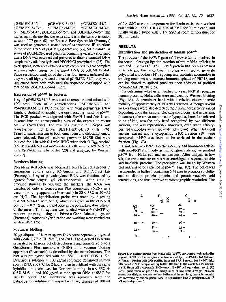
To determine whether antibodies to yeast PRP18 recognizehuman proteins, HeLa cells were analyzed by Western blotting(Fig. 1A). A prominent band with a relative electrophoreticmobility of approximately 60 kDa was detected. Although severalweaker bands were also detected, their immunoreactivity varieddepending upon the sample, blocking conditions, and antiserum.In contrast, the above-mentioned polypeptide, hereafter referredto as p54nrb, was the only band recognized by two differentantisera, and was reproducibly observed, even when affinity-purified antibodies were used (data not shown). When HeLa cellnuclear extract and a cytoplasmic S100 fraction (19) werecompared, pM"* was found to be enriched in the nuclearfraction (Fig. IB).
Using relative electrophoretic mobility and immunoreactivitywith anti-PRP18 antibody as fractionation criteria, we purifiedp54nrt> from HeLa cell nuclear extracts. After dialysis in lowsalt, the crude nuclear extract was centrifuged to separate solubleand insoluble proteins. The precipitate was found by Westernblot analysis to be enriched in p54nrt) (Fig. 1C). The pellet wasresuspended in buffer 1 containing 6 M urea to promote solubilityand to disrupt protein-protein and protein—nucleic acidinteractions, and thus improve chromatographic resolution. The
* § 3 aKDo9 7 -6 9 - __4 6 -
3 0 - -
2 2 -
14 -
kDo
9 7 -6 9 -4 6 -
3 0 -
2 2 -1 4 -
kOo
9 7 -69 -4 6 -
3 0 -
22 -14 -
Figure 1. A nuclear protein from HeLa cells (pM""*) cross-reacts with antibodiesto yeast PRP18. Protein samples were fractionated by SDS-PAGE, and analyzedby Western blotting with IgGs purified from anti-PRP18 serum. (A) 4 x 10* HeLacells boiled in SDS sample loading buffer. (B) lane 1: HeLa cell nuclear extract;lane 2: HeLa cell cytoplasmic S100 extract ( 6 x 10⁵ cell equivalents each). (C)Partial purification of 054°* by precipitation at low ionic strength. Nuclearextract was dialyzed against low salt buffer and the resulting insoluble materialwas recovered by centrifiigation. Lane 1: supernatant; lane 2: precipitate (2 X10^cell equivalents each).
by guest on May 30, 2016
http://nar.oxfordjournals.org/D
ownloaded from

4088 Nucleic Acids Research, 1993, Vol. 21, No. 17
kDo9 7 -6 6 -
4 5 -
31 -
2 2 -
a
I1 2 3 4 5
Figure 2. Purification of HeLa pM"1*. HeLa cell nuclear extract was fractionatedand pM™* followed by Western blot analysis. Sequential peak fractions analyzedby SDS-PAGE and Coomassie Blue R250 staining are shown. Lane 1: proteinsize markers; lane 2: low salt precipitate (2X10⁶ cell equivalents); lane 3:flowthrough fraction from Mono Q column (1.3 x 10⁷ cell equivalents); lane 4:Mono S peak gradient fraction (2xlO7 cell equivalents); lane 5: gel-purifiedP541* (3.5 xl<? cell equivalents).
denatured material was purified by two ion exchangechromatography steps in the presence of 6 M urea (Fig. 2).p54nrb was recovered in the Mono Q flowthrough (lane 3), andis one of the prominent polypeptides in this fraction. The MonoQ fraction was loaded onto a Mono S column and p54nrb elutedat approximately 170 mM salt (lane 4). Finally, p54nrt) wasseparated from minor contaminants in the Mono S fraction bypreparative SDS-PAGE flane 5).
Cloning and analysis of p54nrb cDNAsPurified HeLa p54nrt) was found to have a blocked N-terminus(data not shown). To obtain internal amino acid sequence thepurified protein was digested with lysyl-endopeptidase and dieresulting peptide mixture was fractionated by reverse-phaseHPLC. Sequencing of selected peak fractions yielded thefollowing peptides (bracketed Ks indicate the expected Lysresidues preceding uie cleavage site; X denotes ambiguoussequence):
peptide 1: (K)XXEQPPRFAQPGSFEYEYpeptide 2: (K)ALIEMEKpeptide 3: (K)QQQDQVDRNI
Protein database searches using the BLAST program (27)showed that these three short peptides were highly homologousto a common region in two previously cloned cDNAs. One ofthese, HUM241D5, was reported as a partial cDNA encodinga human myoblast cell-surface antigen (28). The other, knownas BJ6 or NONA, is a Drosophila puff-specific protein, whichcorresponds to one of three proteins encoded by the alternativelyspliced transcripts of the no-on-transient A gene (29,30). Fromthe alignment of the three pSA™* peptides with these twoproteins, the expected order of the peptides in the human p54nrt)
sequence is 1—2—3 (see below).To clone human cDNAs encoding p54nrt) we designed two
degenerate oligonucleotide pools: P54BAM (sense) and P54HIN(antisense), corresponding to peptides 1 and 3, respectively (seeMaterials and Methods). First, P54HIN was used as the primerto syndiesize pM"* first strand cDNA from HeLa poly-adenylated RNA by reverse transcription. Both oligonucleotideswere men used in a polymerase chain reaction to amplify thefirst strand cDNA. Analysis of subcloned PCR products yielded
• !••t t ccQCTCTTTTCTCOOOACeOQAQAQQCCOTOTAOCQTCOCCaTTACTCCOAOaAOATACCAQTCOOTAaAQaAQAAOTCOAOOTTAQAOaOAACIOOOAQQCACTTTOCTaTCTOCAATCOAAOTTaAQQQTQCAAAA ATO CAO AOT AAT AAA
ACT TTT AAC TTO OAQ AAO CAA AAC CAT ACT CCA AQA AAQ CAT CAT CAA CAT0
CAC CAC CAQ CAO CH H 0 0
CCA ATA CCT OCA A
ATT OAC CTQ AAQ A
,0 CAC CAC CAQ CAQ CAA CAO CAO CAO CCO CCA CCA CCOH H O O O O O O P P P P
T QQO CAA CAO OCC AOC AQC CAA AAT QAA OQC TTO ACTO O O A S t O K E Q
TTT AOA AAA CCA QOA OAO AAO ACC TTC ACC CAA COAI D L K K F H K P O E K T F T O n
AOC COT CTT TTT O T ^ Q p ^ A A ^ C T T C C ^ C C C O A C ^ I T C A C ^ O A O O A A Q A A A T O
,0 AAA TAT QOA AAQ OCA 00C QAA QTC TTC ATT CAT AAQ^C^^^T^^QAi
QAT AAA OBA TTT OQC TTT ATC COC TTQ OAA *CC COA *CC CTA
AAQ CAP CTO
QTO TCC AAC QAA CT
AQT OCAi ^ ^ l A ICTQ QAA QA* TCT QTQ TTT QQC CAQ OTA OAQ
QTA QTC ATT QTQ QAT OAT CQA QOA AQO CCC TCA OQA A** 00C A
QCT CQQ AAA OAC AOA TOC AQ
CTA ACC *CA TTT CCT COT
CAQ TTA QATO L D
OAT QAA QAO OQA CTT
OQC TCC
QAO AAQE K
AAQ CTO
CAQ CAQ CAO0 0 0
OAO ATQ OAO
ATO AOA
OAA QAQE E
ATO COOy R
CAQ OAOQ E
CAQ QAT TTQ0 0 1AAC CAA OAOH O E
COC AOQ COC
OAC CAA QTO0 O V
ATO QAA OCTU E A
ATQ A00 CQC
TOO AAO
AAC ATCft I
CAT OAO
QTO CAA AAA» 0 «
COT OAA OAA
CAA OAA0 E
COA AAO
OAA CTTE L
CAA CTQ0 L
COO COO
OCA
AAOK
CAC
CQO
OAQE
CAQ
OTTV
OCA
ATT
QCTA
OTCV
ATQ
CAO CCTo r
QAQ ATOE y
COT OAOR E
ATQ CTAy L
QAA OAO
COA CAO CAOft 0 0
ATT COO ATOI
OAA OQA TTCE O F
QQT CAQ ATO
AAQ QQA ACC TTC CCTK O T F P
QCT ATO QOA OQT OCT
CAO OAO0 E
QAA ATQE y
AQA OAOR E
ATA AAC
1 1 7
III
1 t t
271
111
I M
• 11
511
S 7 |
il;
• 71
7>l
711
111
• II
111
111
o»s
o n
117
1 ••
CCA OQAr o
ACA ACTT T
OOT QUA
CCT OCC ACT
OAA COC TTTE R F
ACT CCT CCT
ATQ ATQ CCQ
QQT CAO QCTO O A
OCA TTC AAC
OAT QQA
OCT ACAA T
COT OCA
ACT TIO
ATQ QAAU E
OCT CCT
0OOA
QOAQ
OQA
ATTI
OCT
CCA CCA
OCA ATTA t
TTT QCC
Figure 3. Nucleotide sequence and translation of p54nrt> HeLa cDNA clone 4.The nucleotide sequence of the sense strand is shown, with the translated proteinsequence in the one-letter code below the corresponding codons. Nucleotideresidues are numbered on the right, and amino acid residues on the left. Thetwo RRMs, separated by a single Ala residue, are highlighted with graybackground. The RNP-2 and RNP-1 subrootifs of each RRM are highlighted withblack background. Except for the 5' untranslated region, the five cDNA clonesshare the same sequence. The EcoR I linker at the 5' end is shown in lowercase.
unique nucleotide sequence of the region between the degenerateoligonucleotides. A 41-mer antisense oligonucleotide, P54OLJ41,was synthesized according to this unique sequence, and was end-labeled as a probe to screen a HeLa cDNA library inbacteriophage X. Dozens of positive plaques were observedamong 6x 10⁵ plaques screened on duplicate filters, suggestingthat p54nrt) mRNAs are abundant. Five positive clones pickedat random were plaque-purified, and the insert DNAs weresubcloned into plasmid pGEMEX. Restriction analysis indicatedthat the five inserts were highly related (data not shown).
Nucleotide sequencing of the cDNA inserts showed that thefive clones represent three independent cDNAs that share acommon open reading frame (ORF) and a short 3' tail (Fig. 3).The three classes of cDNA clones differ in their 5' untranslatedregion, most likely as a result of alternative splicing (Fig. 4).The significance of the presence of alternative exons within the5' untranslated region, all expressed in the same cell line, isunclear. Additional alternatively spliced p54nA mRNAs mayexist, since we only analyzed five out of dozens of positive clones.In all three cDNA classes, the available sequence upstream ofthe first Met codon contains in-frame nonsense codons.Therefore, this shared Met codon, which is present within anacceptable consensus initiation sequence (31), is presumed to bethe start codon. The three classes of cDNA clones all end at thesame position as the one shown in Fig. 3, 75 nt downstream ofthe common ochre codon. We did not find any polyadenylation
by guest on May 30, 2016
http://nar.oxfordjournals.org/D
ownloaded from

Nucleic Acids Research, 1993, Vol. 21, No. 17 4089
Clon« 1 ggcc9tgtagcgtc?ccgtt*ctccgaTO*9B 32Clon* 2,3,5 gccgtgtagcgtcgccgttaetccgaggaga 31clona 4 9Ctcttttctcqqgacqqq*q>qqccgtgt«qcytcqccqtt«ctccq»gqaq> 54
t«cc*9te«9t<gtigg 48i 2,3,5 taccagtcggtagaggaggoaamgtatttaggQgacmgtgmafcttcfctaotct? 65
Clan* 4 t*ccagtcg9tftgag9aga*gtogaggttaga9ggmmctggymg7aaatttgct 101
Clou 2,3,5 ugagTOttctgcac*t*tttoo*atttat*ttmteeto*tcaeaxtagstex 139Clooa 4 gtutgoaatogmaettsag 127
Clooa 1 ggtguaaaUS 51Clooa 2,3,5 taqyaagaiatTtttntoaagOTtgc««««aTQ 169Clooa 4 gytgcuuAIO 136
Figure 4. Comparison of 5' untranslated regions of five pM01* HeLa cDNAclones. Nucleotide residues are numbered on the right. The sequences of clones2, 3 and 5 are identical. All five cDNA sequences are immediately preceded bythe EcoR I linker GGAATTCC. The simplest interpretation of these structuresis that the 5' end of the pM1"* gene comprises at least four exons, with theinitiation codon (bold uppercase) starting 10 nt into exon 4. Clones 2, 3, and5 skip alternative exon 3 (57 nt), whereas cDNA clone 4 slops alternative exon2 (114 nt), and cDNA clone 1 skips both alternative exons 2 and 3. Presumptivealternative exons 2 and 3 (bold lowercase) appear to be mutually exclusive; theirorder in geoomk DNA cannot be deduced from our data. The first 22 or 23nt of presumptive exon 1 in cDNA clone 4 are missing from the remaining clones,perhaps due to incomplete first strand cDNA synthesis or to nibbling of the DNAin steps prior to ligation of the EcoR I linkers.
S °~ —a. 7 ikDo
9 7 -6 9 -
4 6 -
3 0 -
I 2 3
Figure 5. Recombinant pM"1* reacts with anti-PRP18 antibodies and has thesame electrophoretic mobility as purified HeLa pM"*. Protein samples wereanalyzed by Western blot as in Fig. 1. Lane 1: control gel-purified HeLa pM™*;lane 2: 0.2 jil of induced E.coli cells harboring the p54 expression plasmM;lane 3: 0.2 y\ of mock-induced cells. The fast migrating band seen only withinduced cells (lane 2) is probably a degradation product of *
signals in the 3' untranslated region. It appears that all the cloneswere generated by cleavage at an internal EcoR I site duringconstruction of the original library. Since the stop codon wasthe same for all three cDNA classes, we did not attempt to obtainlonger clones to sequence the complete 3' untranslated region.
The p54nrb ORF encodes a 471 amino acid polypeptide witha calculated molecular weight of 54,267, which is about 10%smaller than the apparent molecular weight of the protein purifiedfrom HeLa cells, as determined by SDS-PAGE (Fig. 2). Thethree peptide sequences derived from purified HeLa pM"* (seeabove) are present in the open reading frame of the cloned cDN A(Fig. 3). The p54rab sequence bears no homology to yeastPRP18, although a few very short peptides might be sharedepitopes responsible for cross-reactivity between p54mb and anti-PRP18 antibodies. For example, the peptides RWKA and RSRLare present in p54mt, compared to RWKA and RTRL in PRP18(14). Analysis of the deduced p54nrt) protein sequence with thePC/Gene program Prosite (32) showed the presence of three
PI4 171PIF 501SJI 501
P9 4 1 1 4PSF 917BJI III
Figure 6. Sequence homology between central domains of human pM™*, humanPSF and Drosophila NONA/BJ6. Identical residues are indicated by Mackbackground; conservative substitutions are highlighted in gray. Amino acid residuesin each protein are numbered on both sides.
overlapping potential bipartite nuclear targeting sequences (33)at positions 324-340, 336-352, and 337-353. The presenceof these sequences is consistent with the subcellular fractionariondata (Fig. IB).
To ensure that we had cloned cDNAs encoding the PRP18cross-reacting protein, and that we had obtained the completep54mb coding sequence, we subcloned the pM™1* ORF into theE.coli expression plasmid vector pET-9c (26). After inducingthe expression of recombinant p54nrb protein with IPTG,bacterial ly sates were analyzed by Western blot with anti-PRP18antibodies (Fig. 5). Recombinant p54nrb had identicalelectrophoretic mobility to p54nrt) purified from HeLa cells, andretained immunoreactivity with anti-PRP18 antibodies.
Sequence homology between p54nrt), PSF, and NONA/BJ6Comparison of the pM™*5 protein sequence with those ofHUM241D5 and Drosophila NONA/BJ6 revealed a high degreeof homology. The HUM241D5 sequence was originally reportedas a cDNA fragment encoding a human myoblast cell surfaceantigen (28). However, recent work showed that HUM241D5represents the C-terminal part of PSF, a recently identified HeLacell splicing factor of 712 aa (17). Evidently, the monoclonalantibody 241D5, which stains myoblast cell surfaces (28),fortuitously cross-reacted with PSF upon screening of a myoblastexpression library (17). The 241D5 monoclonal antibody did notrecognize human pM"* on Western blots (data not shown).
A sequence alignment of human p54mt, human PSF, andDrosophila NONA/BJ6 is shown in Fig. 6. No gaps wereintroduced to generate the alignment, except for an extra Alain NONA/BJ6. Human p54mt and human PSF are 70%identical and 7% similar in an internal region of 320 aa. Thisregion contains two consensus RNA recognition motifs (RRMs),a feature found in many RNA-binding proteins (reviewed in 18).Drosophila NONA/BJ6, a protein of 700 aa (29,30), has a 321aa region with 42% identity and 7% similarity to the same 320aa region of p54"rt), and 44% identity and 9% similarity to thatof PSF (Fig. 6). In addition, p54nrt) and PSF are similar in theirN-terminal regions in that both are rich in Pro and Gin residues(Fig. 3; ref. 17). The N-terminus of NONA/BJ6 is rich in Gly,Asn and Gin residues (29,30). All three proteins are also highlybasic and their SDS-PAGE migration is slower than expectedon die basis of calculated molecular weight. The homologybetween NONA/BJ6 and PSF was not noted previously, althoughthe RRMs in both proteins were reported (17,29,34). We note
by guest on May 30, 2016
http://nar.oxfordjournals.org/D
ownloaded from

4090 Nucleic Acids Research, 1993, Vol. 21, No. 17
that the two RRMs comprise only 153 aa of the 320 aa homologyregion, and that the similarity between otherwise unrelated RRMsranges from 10% to 18% identity (35).
Northern and Southern blot analysis of p54nrb
Since 320 out of 471 aa in pM"*" are in a region homologousto PSF, and in addition both proteins have N-termini with longstretches of Pro and Gin residues, we used a Sac I restrictionfragment corresponding to the unique C-terminus of p54nrb asa probe for Northern and Southern blot analysis, in order to avoidcross-hybridization with PSF. Northern blot analysis (Fig. 7) ofHeLa cell polyadenylated RNA at high stringency showed anabundant mRNA of approximately 2.6 kb and a rare mRNA ofapproximately 1.9 kb. The lengths of the five cDNAs range from1,545 to 1,657 bp (Figs. 3 and 4). Assuming an average poly(A)length of 200 nt, the cDNA clones appear to be missing about800 nt of untranslated sequence, most of which is probably fromthe 3' end, since all the cDNAs were truncated upstream of theirpresumptive polyadenylation sites.
Under stringent Southern blot washing conditions (Fig. 8), theprobe hybridizes to a single Nco I fragment of human genomicDNA, but to two or more fragments when the same DNA isdigested by either EcoR I, Hind m, or Pst I. The probeencompasses a cDNA fragment from the Sac I site at +1021to the 3' end at +1491 (Fig. 3). The cDNAs have an Nco I siteat +677 but lack internal sites for the remaining three enzymes.A single 3 kb Nco I genomic DNA fragment hybridized to theprobe (Fig. 8, lane 3); the second genomic Nco I site must belocated within an intron or downstream of the coding region.Hence, the fact that a single Nco I band was detected suggeststhat p54rab is encoded by a single-copy gene. Since two or morebands hybridize to the same probe when genomic DNAs aredigested by the remaining enzymes, which do not cut within thecDNA portion of die probe, we conclude that one or more intronsinterrupt the p54nri) gene in the 3' terminal region encompassedby the cDNA fragment probe.
DISCUSSION
A polypeptide from HeLa cells reacted very strongly inimmunoblots with antibodies raised against the S.cerevisiaesplicing factor PRP18. In addition, indirect immunofluorescencestaining of HeLa cells with PRP18 antiserum produced anucleoplasmic speckled pattern (D.Spector, unpublished data)typical of that generated by antibodies against snRNP particlesand other splicing factors (16). These observations prompted usto look for a possible human homolog of this factor. Usingreactivity with this antibody by Western blotting as a criterion,we purified p54nrtl from HeLa cell nuclear extracts. Threeclasses of cDNAs coding for p54nrt) were cloned based on partialpeptide sequence of the purified protein. Sequence analysis ofthe cDNAs showed no significant homology between theircommon coding sequence and PRP18. We do not know whyp54mt> reacts with anti-PRP18 antibody, although a few veryshort peptides present in both proteins might be common epitopes.At present, we cannot conclude whether there is a humanhomolog of yeast PRP18. Extensive screenings of human cDNAexpression libraries have been carried out, either using anti-PRP18 antibody, or by attempting to complement the conditionalgrowth phenotype of S. cerevisiae prplSP strains (unpublisheddata). No human homolog of yeast PRP18 has been found so far.
kb
4.40-
^ 3 7 •
1.77.1.52.135-1.28 •
0.78-053
Figure 7. Analysis of p54mb mRNA from HeLa cells. Polyadenylated RNA fromHeLa cells was analyzed by Northern blot with a p54ort)-specirk; cDNA probefragment, as described in Materials and Methods. The relative mobilities of RNAsize markers (BRL) are shown on the left. An abundant transcript of approximately2.6 kb and a minor one of 1.9 kb are observed.
1 IKb
3 4
Figure 8. Analysis of the human pM"* gene. Human genomic DNA wasanalyzed by Southern Wot with a p54nrt)-specific probe. 20 ft% of human genomicDNA was digested with EcoR I (lane 1), Hind m (lane 2), Nco I (lane 3), orPst I (lane 4), fractionated by agarose gel electrophoresis, blotted onto a chargednylon membrane, and probed with a p54mb-speci6c cDNA probe fragment, asdescribed in Materials and Methods. The relative mobilities of BstE D-digestedX DNA size markers are shown on the left.
The translated sequence of p54nrb indicates that it is a basicprotein with a calculated pi of 9.54, consistent widi itschromatographic behavior, and a predicted molecular weight of54,267, assuming no post-translational modifications. Its N-terminus contains runs of His, Pro, and Gin residues. The proteinhas two consecutive RNA recognition motifs, strongly suggestingthat it is an RNA-binding protein. Preliminary UV crosslinkingexperiments have confirmed this prediction (data not shown).Further experiments are necessary to determine any sequencespecificity in the binding of p54nrt) to RNA.
Although the function of p^"*" is presently unknown, it hasstriking homology to the newly discovered HeLa cell splicingfactor PSF (17) in a region that comprises 68% of the p54nrt)
by guest on May 30, 2016
http://nar.oxfordjournals.org/D
ownloaded from

Nucleic Acids Research, 1993, Vol. 21, No. 17 4091
residues. This region includes the two RRMs, as well as othersequences of unknown function. In addition, both PSF and^54°^ are unusually rich in Gin and Pro at their N-termini.These PQ domains are present in several hnRNP and snRNPpolypeptides, and by analogy to similar domains in a numberof transcription factors, they are thought to be involved in protein-protein interactions, and perhaps to have effector functions (17and references therein). We speculate that p54nrt) is also asplicing factor, and that PSF and p54nrt) are different membersof a family of splicing factors with related functions, but whichmay have, for example, different pre-mRNA substratespecificities. Additional experiments will be necessary to testwhether p54nrt) is required for pre-mRNA splicing in vitro andin vivo.
It is intriguing to find that the 320 aa region shared by p54nrt>
and PSF also exists in a Drosophila protein, NONA/BJ6. Thisprotein is ubiquitously expressed in neural and non-neural nucleithroughout Drosophila development (34). It is one of threealternatively spliced isoforms encoded by the gene no-on-transientA, which is required for normal response to visual stimuli inDrosophila (29,30). nonA mutants show impaired phototaxis andoptomotor responses, and their electroretinograms displayaberrant on- and off-transient spikes, which reflect electricalcurrents that are thought to originate in the neurons of the lamina(30,34 and references therein). The nonA gene is also the locusof the dissonance (diss) allele, which causes aberrant malecourtship song (34).
Although the molecular function of NONA/BJ6 is currentlyunknown and null alleles have not been isolated, the strikingsimilarity to a splicing factor suggests that its pleiotropic functionsin the central nervous system somehow involve pre-mRNAsplicing. For example, NONA/BJ6 may affect alternative splicingof pre-mRNAs encoding ion channels or other proteins in sucha way as to affect the expression of one or several isoforms thatfunction in specific neural tissues.
The presence of NONA/BJ6 in transcriptionally active puffson Drosophila polytene chromosomes (36) is consistent with theobservation that splicing can take place on nascent transcripts(37,38). Interestingly, two other Drosophila puff-specific proteinsare known to be splicing factors: B52 (39), which is a closelyrelated variant of SRp55 (40,41), and RBP1 (42). Both proteinsare members of the SR family of nuclear phosphoproteins, whichappear to function in constitutive and alternative splicing(40-43). They are characterized by the presence of one or twoRRMs and a C-terminal Arg- and Ser-rich domain. PSF,NONA/BJ6, and p54nrt) have two RRMs but otherwise do notstructurally resemble SR proteins. The subnuclear localizationof PSF coincides with that of SR proteins and other splicingfactors (16,17), and perhaps with that of p54nrb, if the latter isresponsible for the immunofluorescence pattern obtained withanti-PRP18 antibodies.
The extensive sequence homology in the 320 aa central domainsof p54nrb, PSF, and NONA/BJ6 strongly suggests conservationof function over the period of divergence of human and fruit fly,since the homologous segments of the three proteins align withoutany gaps, except for a single insertion of an Ala residue inNONA/BJ6. We propose to name this phylogenetically conservedregion a DBHS domain (for Drosophila behavior and humansplicing), based on the observed functions of its currentrepresentatives. It will be of interest to determine the functionalconsequences of swapping DBHS domains between p54mb, PSF,
and NONA/BJ6 on splicing, visual acuity, and male courtshipsong. Since p54nrt) and PSF are both expressed in HeLa cells,it is possible that these two proteins are representatives of a largefamily of DBHS domain proteins, which may function as generaland/or regulatory splicing factors. Additional proteins with DBHSdomains may be found, which would further show the range ofsize and sequence variation of DBHS domains and whether twoRRMs are always included within the first half of the domain.
ACKNOWLEDGEMENTS
We thank E.Birney and members of our lab for helpfuldiscussions; J.Abelson for providing reagents prior to publication;F.Walsh for the 241D5 monoclonal antibody; J.Patton forcommunicating results prior to publication; D.Spector forimmunofluorescence data; J.Posfai for PostScript software;A.Mayeda, D.Spector, and D.Helfman for comments on themanuscript. This work was supported by a grant from theNational Institutes of Health. A.R.K. is a Pew Scholar in theBiomedical Sciences.
REFERENCES1. Moore.M.J., Query.C.C. and Sharp.P.A. (1993) In Gesteland.R.F. and
AtkinsJ.F. (eds.), The RNA World. Cold Spring Harbor Laboratory Press,New York, pp. 303-357.
2. Guthrie.C. and Patterson.B. (1988) Anna. Rev. Genet. 22, 387-419.3. Shuster.E.O. and Guthrie.C. (1990) Nature MS, 270-273.4. Pinto.A.L. and SteitzJ.A. (1989) Proc. Nail. Acad Sci. USA 86,
8742-8746.5. Anderson.G.J., Bach.M., LOhrmann.R. and BeggsJ.D. (1989) Nature342,
819-821.6. Garcia-Blanco.M.A., Anderson.G.J., BeggsJ. and Sharp.P.A. (1990) Proc.
Nad. Acad. Sci. USA 87, 3082-2086.7. Smith.V. and Barrell.B.G. (1991) EMBO J. 10, 2627-2634.8. Liao.X.C, TangJ. and Rosbash.M. (1993) Genes Dev. 7, 419-428.9. Rymond.B.C. (1993) Proc. Natl Acad. Sci. USA 90, 848-852.
10. Bimey.E., Kumar.S. and Krainer,A.R. (1992) Nucleic Acids Res. 20, 4663.11. Ruby.S.W. and AbelsonJ. (1991) Trends Genet. 7, 79-85.12. Vijayraghavan.U., Company.M. and AbekonJ. (1989) Genes Dev. 3,
1206-1216.13. Vijayraghavan.U. and AbelsonJ. (1990) Mol. Cell. Biol. 10, 324-33X14. Horowitz.D.S. and AbelsonJ. (1993) MoL Cell Bid 13, 2959-2970.15. Horowitz.D.S. and AbeUonJ. (1993) Genes Dev. 7, 320-329.16. Huang.S. and Spector.D. (1992) Current Biol. 2, 188-190.17. Pattern j .G . , Porro.E.B., GalceranJ., Tempst.P. and Nadal-Ginard.B. (1993)
Genes Dev. 7, 393-406.18. Kenan.D.J., Query.C.C. and Keene^.D. (1991) Trends Biochem. Sci.
16, 214-220.19. DignamJ.D., Lebovhz.R.M. and Roeder.R.G. (1983) Nucleic Acids Res.
11, 1475-1489.20. Laemmli.U.K. (1970) Nature 227, 680-685.21. Hager.D.A. and Burgess.R.R. (1980) Anal. Biochem. 109, 76-86.22. Kawasaki.H., Emori.Y. and Suzuki.K. (1990) Anal Biochem. 191, 332-336.23. Kawasaki.H. and Suzuki.K. (1990) Anal. Biochem. 186, 264-268.24. Ausubel.F.M., Brent.R., Kingston.R.E., Moore.D.D., SeidmanJ.G.,
SmithJ.A. and Struhl.K. (1989) In Current Protocols in Molecular Biology,J. Wiley & Sons, New York.
25. SambrookJ., Fritsch.E.F. and Maniatis.T. (1989) In Molecular Cloning:A Laboratory Manual, Cold Spring Harbor Laboratory Press, Cold SpringHarbor, New York.
26. Studier.F.W., Rosenberg.A.H., DunnJ.J. and DubendorffJ.W. (1990)Methods EnzymoL 185, 60-89.
27. Altschul.S.F., Gish.W., Miller.W., Myers.E.W. and Lipman.D.J. (1990)J. MoL Biol 215, 403-410.
28. Gower.H.J., Moore.S.E., Dickson.G., FJsom.V.L., Nayak.R. andWalsh.F.S. (1989) Development 105, 723-731.
29. Besser.H.V., Schnabd.P., Wieland.C., Fritz.E., Stanewsky.R. andSaumweber.H. (1990) Chromosoma 100, 37-47.
by guest on May 30, 2016
http://nar.oxfordjournals.org/D
ownloaded from

4092 Nucleic Acids Research, 1993, Vol. 21, No. 17
30. Jones.K.R. and Rubin.G.M. (1990) Neuron 4, 711-723.31. Kozak.M. (1989) / . Cell Biol. 108, 229-241.32. Bairoch.A. (1992) Nucleic Adds Res. 20, 2013-2018.33. Dingwall.C. and Laskey.R.A. (1991) Trends Biochem. Set. 16, 478-481.34. ReadaMJCG., JooesJC.R., Kulkami.S.J., BaguHy.S.H. and HalU.C. (1992)
J. Neuroscience 12, 390-407.35. Birney.E., Kumar.S. and Krainer.A.R. Manuscript in preparation.36. Frasch.M. and Saumweber.H. (1989) Oiromosoma 97, 272-281.37. Sass.H. and Pederson.T. (1984) / Mol Biol 180, 9111-926.38. Beyer.A.L. and Osheim.Y.N. (1988) Genes Dev. 2, 754-765.39. Champlin.D.T., Frasch.M. Saumweber.H. and LisJ.T. (1991) Genes Dev.
5, 1611-1621.40. MayedaA, Zahler.A.M., Krainer.A.R. and Roth.M.B. (1992) Proc. Nml.
Acad. Set. 89, 1301-1304.41. Zahler.A.M., Lane.W.S., Stolk.J.A. and Roth.M.B. (1992) Genes Dev. 6,
837-848.42. Kim,Y-J., Zuo.P., ManleyJ.L. and Baker.B.S. (1992) Genes Dev. 6,
25159-2579.43. Fu,X.-D., Mayeda.A., Maniatis.T. and Krainer.A.R. (1992) Proc. Natl.
Acad. Sd. USA 89, 11224-11228.
by guest on May 30, 2016
http://nar.oxfordjournals.org/D
ownloaded from