Novel Interactions of the TRTK12 Peptide with S100 Protein Family Members: Specificity and...
13
Novel Interactions of the TRTK12 Peptide with S100 Protein Family Members: Specificity and Thermodynamic Characterization Lucas N. Wafer, † Franco O. Tzul, † Pranav P. Pandharipande, ‡ and George I. Makhatadze* ,† † Department of Biology and ‡ Department of Chemical and Biological Engineering, Center for Biotechnology and Interdisciplinary Studies, Rensselaer Polytechnic Institute, Troy, New York 12180, United States * S Supporting Information ABSTRACT: The S100 protein family consists of small, dimeric proteins that exert their biological functions in response to changing calcium concentrations. S100B is the best-studied member and has been shown to interact with more than 20 binding partners in a calcium-dependent manner. The TRTK12 peptide, derived from the consensus binding sequence for S100B, has previously been found to interact with S100A1 and has been proposed to be a general binding partner of the S100 family. To test this hypothesis and gain a better understanding of the specificity of binding for the S100 proteins, 16 members of the human S100 family were screened against this peptide and its alanine variants. Novel interactions were found with only two family members, S100P and S100A2, indicating that TRTK12 selectively interacts with a small subset of the S100 proteins. Substantial promiscuity was observed in the binding site of S100B thereby accommodating variations in the peptide sequence, while S100A1, S100A2, and S100P exhibited larger differences in the binding constants for the TRTK12 alanine variants. This suggests that single-point substitutions can be used to selectively modulate the affinity of TRTK12 peptides for individual S100 proteins. This study has important implications for the rational drug design of inhibitors for the S100 proteins, which are involved in a variety of cancers and neurodegenerative diseases. T he S100 protein family consists of approximately 25 members found exclusively in vertebrates, making it the largest subset of the EF-hand, calcium-binding proteins. 1 The name derives from the fact that many members are soluble in 100% ammonium sulfate at neutral pH. 2 These proteins are expressed in a tissue-specific manner and have been implicated in a variety of intracellular and extracellular functions, including cell growth and di fferentiation, cytoskeleton dynamics, regulation of calcium homeostasis, inflammation response, and protein phosphorylation. 1,3−6 In addition, the expression levels of the S100 proteins have been directly correlated to the severity of neurodegenerative disorders, 7−11 inflammatory diseases such as irritable bowel syndrome, 12−15 cardiomyopa- thies, 16,17 and various cancers. 6,18−24 S100 proteins are relatively small (9−13 kDa) and exist almost exclusively as homo- or heterodimers in solution. 25 Each S100 protein monomer consists of four α-helices, with helices I and IV arranged in an antiparallel orientation. 1,26 Calcium binding induces a significant structural rearrangement in the S100 proteins that has been well-documented by X-ray crystallography and solution nuclear magnetic resonance (NMR) spectroscopy. 27−32 During this conformational change, helix III reorients itself (>40°) relative to helices II and IV to expose a hydrophobic binding pocket that allows interactions with target proteins and peptides. 27 Because the S100 proteins appeared to be functionally distinct from related, calcium- binding proteins, such as calmodulin and parvalbumin, early work focused on identifying high-affinity biological targets to improve our understanding of the molecular basis of recognition. 33−38 In 1995, Ivankenov et al. screened a phage display library against S100B and provided the first consensus binding sequence for the S100 proteins [(K/R)-(L/I)-X-W-X-X-I- L]. 35 A search of the available protein sequences showed a highly homologous region in the C-terminus of the actin- binding protein CapZ (T-R-T-K-I-D-W-N-K-I-L-S). 35 This sequence was later termed TRTK12. 35 Subsequent studies have shown that this peptide interacts with the two C-terminal helices of S100B (Figure 1), as well as the hinge region, and was able to compete with binding for other known targets. 35,39−46 Furthermore, it was shown to bind S100A1, leading some to propose that TRTK12 represents a general binding motif for the S100 protein family. 32 To gain a better understanding of the specificity of binding within the S100 protein family, we screened the proposed consensus binding sequence (TRTK12) against representative members of the human S100 protein family, using isothermal titration calorimetry (ITC) and steady-state emission fluo- rescence. Unexpectedly, TRTK12 was found to selectively interact with a small subset of S100 proteins. We confirmed previously reported interactions with S100B and S100A1. In Received: June 18, 2013 Revised: July 30, 2013 Article pubs.acs.org/biochemistry © XXXX American Chemical Society A dx.doi.org/10.1021/bi400788s | Biochemistry XXXX, XXX, XXX−XXX
Transcript of Novel Interactions of the TRTK12 Peptide with S100 Protein Family Members: Specificity and...

Novel Interactions of the TRTK12 Peptide with S100 Protein FamilyMembers: Specificity and Thermodynamic CharacterizationLucas N. Wafer,† Franco O. Tzul,† Pranav P. Pandharipande,‡ and George I. Makhatadze*,†
†Department of Biology and ‡Department of Chemical and Biological Engineering, Center for Biotechnology and InterdisciplinaryStudies, Rensselaer Polytechnic Institute, Troy, New York 12180, United States
*S Supporting Information
ABSTRACT: The S100 protein family consists of small,dimeric proteins that exert their biological functions inresponse to changing calcium concentrations. S100B is thebest-studied member and has been shown to interact withmore than 20 binding partners in a calcium-dependent manner.The TRTK12 peptide, derived from the consensus bindingsequence for S100B, has previously been found to interact withS100A1 and has been proposed to be a general binding partnerof the S100 family. To test this hypothesis and gain a betterunderstanding of the specificity of binding for the S100proteins, 16 members of the human S100 family were screened against this peptide and its alanine variants. Novel interactionswere found with only two family members, S100P and S100A2, indicating that TRTK12 selectively interacts with a small subsetof the S100 proteins. Substantial promiscuity was observed in the binding site of S100B thereby accommodating variations in thepeptide sequence, while S100A1, S100A2, and S100P exhibited larger differences in the binding constants for the TRTK12alanine variants. This suggests that single-point substitutions can be used to selectively modulate the affinity of TRTK12 peptidesfor individual S100 proteins. This study has important implications for the rational drug design of inhibitors for the S100proteins, which are involved in a variety of cancers and neurodegenerative diseases.
The S100 protein family consists of approximately 25members found exclusively in vertebrates, making it the
largest subset of the EF-hand, calcium-binding proteins.1 Thename derives from the fact that many members are soluble in100% ammonium sulfate at neutral pH.2 These proteins areexpressed in a tissue-specific manner and have been implicatedin a variety of intracellular and extracellular functions, includingcell growth and differentiation, cytoskeleton dynamics,regulation of calcium homeostasis, inflammation response,and protein phosphorylation.1,3−6 In addition, the expressionlevels of the S100 proteins have been directly correlated to theseverity of neurodegenerative disorders,7−11 inflammatorydiseases such as irritable bowel syndrome,12−15 cardiomyopa-thies,16,17 and various cancers.6,18−24
S100 proteins are relatively small (9−13 kDa) and existalmost exclusively as homo- or heterodimers in solution.25 EachS100 protein monomer consists of four α-helices, with helices Iand IV arranged in an antiparallel orientation.1,26 Calciumbinding induces a significant structural rearrangement in theS100 proteins that has been well-documented by X-raycrystallography and solution nuclear magnetic resonance(NMR) spectroscopy.27−32 During this conformational change,helix III reorients itself (>40°) relative to helices II and IV toexpose a hydrophobic binding pocket that allows interactionswith target proteins and peptides.27 Because the S100 proteinsappeared to be functionally distinct from related, calcium-binding proteins, such as calmodulin and parvalbumin, earlywork focused on identifying high-affinity biological targets to
improve our understanding of the molecular basis ofrecognition.33−38
In 1995, Ivankenov et al. screened a phage display libraryagainst S100B and provided the first consensus bindingsequence for the S100 proteins [(K/R)-(L/I)-X-W-X-X-I-L].35 A search of the available protein sequences showed ahighly homologous region in the C-terminus of the actin-binding protein CapZ (T-R-T-K-I-D-W-N-K-I-L-S).35 Thissequence was later termed TRTK12.35 Subsequent studieshave shown that this peptide interacts with the two C-terminalhelices of S100B (Figure 1), as well as the hinge region, andwas able to compete with binding for other knowntargets.35,39−46 Furthermore, it was shown to bind S100A1,leading some to propose that TRTK12 represents a generalbinding motif for the S100 protein family.32
To gain a better understanding of the specificity of bindingwithin the S100 protein family, we screened the proposedconsensus binding sequence (TRTK12) against representativemembers of the human S100 protein family, using isothermaltitration calorimetry (ITC) and steady-state emission fluo-rescence. Unexpectedly, TRTK12 was found to selectivelyinteract with a small subset of S100 proteins. We confirmedpreviously reported interactions with S100B and S100A1. In
Received: June 18, 2013Revised: July 30, 2013
Article
pubs.acs.org/biochemistry
© XXXX American Chemical Society A dx.doi.org/10.1021/bi400788s | Biochemistry XXXX, XXX, XXX−XXX
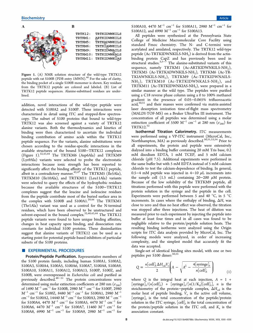
addition, novel interactions of the wild-type peptide weredetected with S100A2 and S100P. These interactions werecharacterized in detail using ITC and stopped-flow spectros-copy. The subset of S100 proteins that bound to wild-typeTRTK12 was also screened against a variety of TRTK12alanine variants. Both the thermodynamics and kinetics ofbinding were then characterized to ascertain the individualbinding contribution of amino acids within the TRTK12peptide sequence. For the variants, alanine substitutions werechosen according to the residue-specific interactions in theavailable structures of the bound S100−TRTK12 complexes(Figure 1).32,43,44 The TRTKM6 (Asp6Ala) and TRTKM9(Lys9Ala) variants were selected to probe the electrostaticinteractions because ionic strength has been reported tosignificantly affect the binding affinity of the TRTK12 peptide,albeit in a contradictory manner.42,47 The TRTKM5 (Ile5Ala),TRTKM10 (Ile10Ala), and TRTKM11 (Leu11Ala) variantswere selected to query the effects of the hydrophobic residuesbecause the available structures of the S100−TRTK12complexes suggest that the leucine and isoleucine residuesfrom the peptide contribute most of the buried surface area inthe complex with S100B and S100A1.29,44 The TRTKM1(Thr1Ala) variant was used as a control for the N-terminalresidues, which have been observed to be unstructured andsolvent-exposed in the bound complex.32,40,43,44 The TRTK12peptide variants were found to have unique binding affinities,changes in heat capacity upon binding, and dissociation rateconstants for individual S100 proteins. These dissimilaritiessuggest that alanine variants of TRTK12 can be used as astarting point for potential peptide-based inhibitors of particularsubsets of the S100 proteins.
■ EXPERIMENTAL PROCEDURESProtein/Peptide Purification. Representative members of
the S100 protein family, including human S100A1, S100A2,S100A3, S100A4, S100A5, S100A6, S100A7, S100A8, S100A9,S100A10, S100A11, S100A12, S100A13, S100P, S100Z, andS100B, were overexpressed in Escherichia coli and purified aspreviously described.48−51 The protein concentrations weredetermined using molar extinction coefficients at 280 nm (ε280)of 1490 M−1 cm−1 for S100B, 2980 M−1 cm−1 for S100P, 2980M−1 cm−1 for S100Z, 8480 M−1 cm−1 for S100A1, 2980 M−1
cm−1 for S100A2, 14440 M−1 cm−1 for S100A3, 2980 M−1 cm−1
for S100A4, 4470 M−1 cm−1 for S100A5, 4470 M−1 cm−1 forS100A6, 4470 M−1 cm−1 for S100A7, 11460 M−1 cm−1 forS100A8, 6990 M−1 cm−1 for S100A9, 2980 M−1 cm−1 for
S100A10, 4470 M−1 cm−1 for S100A11, 2980 M−1 cm−1 forS100A12, and 6990 M−1 cm−1 for S100A13.All peptides were synthesized at the Pennsylvania State
College of Medicine Macromolecular Core Facility usingstandard Fmoc chemistry. The N- and C-termini wereacetylated and amidated, respectively. The TRTK12 wild-typepeptide (Ac-TRTKIDWNKILS-NH2) is derived from the actin-binding protein CapZ and has previously been used instructural studies.3,5−7 The alanine-substituted variants of thissequence, namely TRTKM1 (Ac-ARTKIDWNKILS-NH2),TRTKM5 (Ac-TRTKADWNKILS-NH2), TRTKM6 (Ac-TR-TKIAWNKILS-NH2), TRTKM9 (Ac-TRTKIDWNAILS-NH2), TRTKM10 (Ac-TRTKIDWNKALS-NH2), andTRTKM11 (Ac-TRTKIDWNKIAS-NH2), were prepared in asimilar manner as the wild type. The peptides were purifiedusing a C18 reverse phase column using a 0 to 100% methanolgradient in the presence of 0.05−0.065% trifluoroaceticacid,50,51 and their masses were confirmed via matrix-assistedlaser desorption ionization time-of-flight mass spectrometry(MALDI-TOF-MS) on a Bruker UltraFlex III instrument. Theconcentration of all peptides was determined using a molarextinction coefficient of 5500 M−1 cm−1 at 280 nm (ε280) forTRTK12.52
Isothermal Titration Calorimetry. ITC measurementswere performed using a VP-ITC instrument (MicroCal, Inc.,Northhampton, MA) as previously described.50,51,53,54 Prior toall experiments, the protein and peptide were extensivelydialyzed into a binding buffer containing 20 mM Tris base, 0.2mM disodium EDTA, 1 mM TCEP, and 5 mM calciumchloride (pH 7.5). Additional experiments were performed inthe same buffer but with 5 mM EDTA instead of 5 mM calciumchloride to test the calcium-dependence of binding. In general,0.5−4 mM peptide was injected in 4−10 μL increments intothe sample cell (1.5 mL) containing 20−200 μM protein.Because of the low solubility of the TRTKM9 peptide, alltitrations performed with this peptide were performed with theprotein solution in the syringe and the peptide in the cell.Experiments were performed between 5 and 40 °C, in 5 °Cincrements. In cases where the enthalpy of binding, ΔH, wasclose to zero and thus no heat effect was observed, the titrationwas stopped after three injections. The heat of dilution wasmeasured prior to each experiment by injecting the peptide intobuffer at least four times and in all cases was found to benegligible relative to the protein/peptide solution heats. Theresulting binding isotherms were analyzed using the Originscripts for ITC data analysis provided by MicroCal, Inc. Thefollowing models were analyzed, in order of increasingcomplexity, and the simplest model that accurately fit thedata was accepted.Single-set of identical binding sites model, with one or two
peptides per S100 dimer:50,55
=Δ ° − −
⎛⎝⎜⎜
⎞⎠⎟⎟Q
n H VA A
n[cell]
24[syringe]
[cell]t cal 2 t
t (1)
where Q is the integral heat at each injection, A = 1 +[syringe]t/(n[cell]t) + [syringe]t/[n(1/Kd)[cell]t], n is thestoichiometry of the protein−peptide complex, ΔHcal is themolar heat of peptide binding, Vo is the active cell volume,[syringe]t is the total concentration of the peptide/proteinsolution in the ITC syringe, [cell]t is the total concentration ofthe protein/peptide solution in the ITC cell, and Ka is theassociation constant.
Figure 1. (A) NMR solution structure of the wild-type TRTK12peptide with rat S100B (PDB entry 1MWN).40 For the sake of clarity,the binding pocket of a single S100B monomer is shown. Key residuesfrom the TRTK12 peptide are colored and labeled. (B) List ofTRTK12 peptide sequences. Alanine-substitued residues are under-lined.
Biochemistry Article
dx.doi.org/10.1021/bi400788s | Biochemistry XXXX, XXX, XXX−XXXB

Two-sequential binding sites model, with two peptides perS100 dimer:50,55
= ° Δ
+ Δ + Δ
+ + +
Q V H K
H H K K
K K K
[cell] {[ (1/ )[syringe]
( )(1/ )(1/ )[syringe] ]
/[1 [syringe] (1/ ) 1 (1/ )(1/ )
[syringe] ]}
t cal1 d1 t
cal1 cal2 d1 d2 t2
t d1 d1 d2
t2
(2)
where Q is the integral heat at each injection, ΔHcal1 and ΔHcal2are the molar heats of peptide binding, Vo is the active cellvolume, Kd1 and Kd2 are the dissociation constants for bindingto sites 1 and 2, respectively, and ΔHcal1 and ΔHcal2 are thecalorimetric enthalpies for binding to sites 1 and 2, respectively.Fluorescence Spectroscopy. Steady-state fluorescence
titrations were performed using a Fluoromax-4 spectrofluor-ometer (Horiba Jobin Yvon, Kyoto, Japan) operatingFluorEssence version 3.5. A cuvette with a 3 mm excitationpath length was used throughout all experiments. All TRTK12peptides contain a Trp residue. In the available structures of theS100 proteins in complex with the wild-type peptide (S100Band S100A1), this tryptophan becomes buried in a hydrophobicregion upon binding. Therefore, changes in Trp emissionfluorescence intensity were used to monitor interactionsbetween the TRTK12 peptides and S100 proteins. Theexperiments were performed in binding buffer at 25 ± 0.2 °Cusing a thermostated cell holder. The solution was excited at295 nm and the emission spectra were monitored from 305 to420 nm to monitor changes in λmax; a wavelength of 350 nmwas used to specifically monitor changes in intensity. The initialconcentration of the TRTK12 peptides in the fluorescence cellwas 2−8 μM. Small aliquots of the concentrated protein wereadded until signal saturation was reached or until thefluorescence signal deviated from unity due to inner filtereffects. The experimental emission spectra were corrected bysubtracting the emission control spectra of protein added tobuffer in the absence of peptide. The fluorescence intensity wasalso corrected for fluctuations in lamp intensity by dividing thefluorescence signal by the lamp intensity. The data were fit to asingle-site binding model using the Nonlinear RegressionAnalysis Program (NLREG)56 and the following equation:
A A n2[cell]
( [cell] [titrant] )max
total
2total total
(3)
where Q equals the fluorescence intensity at each titration step,Qmax equals the maximal intensity for the fully saturatedpeptide, [cell]total equals the molar concentration of the peptideat each step, A = Kd + [cell]total + n[titrant]total, n equals thenumber of binding sites, and [titrant]total equals the molarconcentration of the protein at each step.Anisotropy titrations were performed in the same manner as
described above, but using vertical (V) or horizontal (H)excitation and emission polarizations (IVV, IVH, IHH, and IHV).The G factor was calculated as
=GII
HV
HH (4)
where IHV and IHH represent the corrected intensities for thehorizontally polarized light of the peptide in solution. Theanisotropy was calculated as
⟨ ⟩ =−+
rI GI
I GI2VV VH
VV VH (5)
where ⟨r⟩ represents the anisotropy at a given titration step andIVV and IVH represent the corrected intensities of the verticallypolarized light.The relative exposure of the tryptophan residue in the
peptide−protein complex was assayed using dynamic quench-ing with acrylamide. Titrations were performed using 4−8 μMsolutions of the peptide in the presence or absence of saturatingamounts of the S100 proteins and 5 mM CaCl2 or 5 mMEDTA. In cases where the affinity for the complex was low, theprotein concentration was limited by the inner filter effect.Titrations were performed until the final concentration ofacrylamide was ∼0.6 M, and the data were then fit to themodified Stern−Volmer equation:54,57
= +II
K(1 [Q])eV
oSV
[Q]
(6)
where Io is the initial fluorescence intensity, I is the intensity ata given titration step, KSV is the Stern−Volmer constant, [Q] isthe acrylamide concentration at a given titration step, and V[Q]is the constant describing the static component of thequenching reaction. The intensity was corrected for dilution,and the average values are reported.
Structure-Based Calculations of ΔCp. Using the availablethree-dimensional structures of S100A1, S100A2, S100B, andS100P in the calcium-bound state and/or in complex with apeptide target, ΔASAtot was calculated as previouslydescribed.58 In cases where no structure was available,homology models of the peptide-bound state were generatedusing Modeler59 and PDB entries 1MQN,40 1MQ1,43 3IQQ,44
and 2KBM.32 Changes in the accessible surface area uponbinding, ΔASAtotal, were calculated as previously de-scribed.50,51,54 Changes in ΔASA were further subdivided intofour categories (aliphatic surface area, aromatic surface area,peptide backbone surface area, and polar surface area) andconverted into ΔCp using the following empirical relation-ship:50,51,58,60,61
Δ = Δ + Δ − Δ
− Δ
C 2.14 ASA 1.55 ASA 1.81 ASA
0.88 ASA
p alp arm bb
pol (7)
where ΔASAalp, ΔASAarm, and ΔASApol values are the changesin ASA for aliphatic, aromatic, and polar amino acids,respectively, and ΔASAbb values are the changes in ASA forthe polypeptide backbone.
Kinetic Stopped-Flow koff Measurements. Measure-ments of protein−peptide dissociation rate constants (koff)were performed using standard stopped-flow methods. Datawere collected using a JASCO J-815 spectropolarimeterequipped with an SFM 300 mixing module (BioLogic ScienceInstruments), containing an HDS mixer and a FC-15observation cuvette. The koff rate constants were evaluated ata minimum of three temperatures between 5 and 35 °C. Thetemperature of the sample chamber was regulated using acirculating water bath. A mercury lamp source was used forpeptide tryptophan excitation at 295 nm. The emissionfluorescence was collected using an N-WG 320 cutoff filter(BioLogic Science Instruments). Slit widths were adjusted from3 to 5 nm to eliminate or minimize photobleaching. Voltagesapplied to the photomultiplier tube were set accordingly andranged from 700 to 850 V. Kinetic traces were collected under
Biochemistry Article
dx.doi.org/10.1021/bi400788s | Biochemistry XXXX, XXX, XXX−XXXC

an optimal set of push volumes and flow rates for the SFM 300instrument and kept constant for all experiments. Becausepeptide binding is calcium-dependent, peptide dissociation wasinitiated by calcium chelation. For this, preformed protein−peptide complexes (6 or 12 μM peptide) in the binding bufferdescribed above were mixed in a 1:1 mixing ratio with 40 mMEDTA. Special care was taken to control for inner filter effectsby limiting the protein:peptide ratio.Using the Bio-Kine32 software package (BioLogic Science
Instruments), rate constants, koff, were obtained by fitting theexponential change of the emission fluorescence intensity fromEDTA-induced peptide dissociation to the following equation:
= + + ±I t y A( ) XT e k T0
app(8)
where I(t) is the fluorescence intensity as a function of time, y0is the initial fluorescence intensity, A is the amplitude of thechange between the initial and final fluorescence intensity, kappis the observed kinetic rate constant associated with thefluorescence intensity relaxation, and XT is the sloping baselinecorrection used in the Bio-Kine32 software, which accounts forthe photobleaching effect. Errors were calculated from thestandard deviation of five independent traces. A dead time of4.0 ms was electronically estimated with BioLogic’s Bio-Kine32software and experimentally confirmed using the dead-timeassessment procedure of Peterman et al.62 Because theexperimentally determined rate constants were sufficientlyslow, no dead-time data correction was performed.
■ RESULTSThe TRTK12 peptide has been shown to bind human S100Band human S100A1 in a calcium-dependent man-ner29,32,35,40−44,46,47 and has been proposed to be a generalS100 protein-binding motif.32 To probe the specificity ofTRTK12 for these proteins, 16 representative members of thehuman S100 protein family were screened for binding usingisothermal titration calorimetry (ITC) and emission fluores-cence. Unexpectedly, novel interactions were detected withonly two S100 proteins: S100A2 and S100P. To gain a betterunderstanding of these interactions, the binding of the wild-type peptide and its alanine variants was further characterizedusing several biophysical methods.Interactions of TRTK12 Peptides with S100B. Figure 2A
shows the binding isotherm obtained from ITC experiments at25 °C in which the wild-type TRTK12 peptide was titrated intoS100B in the presence of 5 mM CaCl2. As reported previously,
4
two peptides interact with the S100B dimer through identicalbinding sites (eq 1; n = 2). The dissociation constant, Kd,obtained from the fit, 1.7 ± 0.1 μM, is in good agreement withpreviously reported values.44,47,50 The binding model isconsistent with structures derived from X-ray crystallographyand solution NMR involving complexes of TRTK12 and S100Bfrom several orthologs (Homo sapiens, Rattus norveggicus, andBos taurus).5,17,18 ITC experiments were performed over thetemperature range of 5−40 °C. In addition, the thermody-namics of binding were measured for six alanine variants ofTRTK12 (see Experimental Procedures for abbreviated namesand sequences). All peptides, with the exception of TRTKM11,bind with a stoichiometry similar to that of the wild type, i.e.,two peptides per S100B dimer (eq 1; n = 2). TRTKM11appears to interact with a different stoichiometry of one peptideper S100B dimer (eq 1; n = 1). This stoichiometry haspreviously been observed for several peptides binding to the
S100 proteins.50,63−69 The dissociation constants of wild typeand alanine peptides are listed in Table 1.Figure 3A shows the temperature dependence of the
enthalpy of binding, ΔHcal, of the wild-type peptide andalanine variants to S100B. The slope of the temperaturedependence corresponds to the heat capacity change uponbinding, ΔCp, and these values are summarized in Table 1. Ithas previously been shown that ΔCp can be predicted usingempirical changes in the accessible surface area upon binding,provided that accurate structural models are available.2,4,14,21
The experimentally determined heat capacity change for theS100B−TRTK12 interaction (−1.2 ± 0.2 kJ mol−1 K−1) is inexcellent agreement with the structure-based calculated values(Table 1). In addition, the values obtained for all alaninevariants but TRTKM11 are within error of the wild-type ΔCpvalue, suggesting they interact with S100B through a similarstructural mode. This is also expected on the basis of thestructure-based calculations of ΔCp using homology models ofthe alanine variants (Table 1). Again, the only notableexception is the TRTKM11 variant, which interacts with theS100B dimer with a novel stoichiometry of one peptide per
Figure 2. Examples of ITC experiments showing the binding ofTRTK12 to S100B, S100A1, S100A2, and S100P in the presence ofcalcium. The top plots in each panel represent the raw heat effects as afunction of time and the bottom plots represent the cumulative heateffects (■) as a function of the molar ratio of peptide to protein andthe fits to the experimental data using eq 1 () for binding ofTRTK12 to S100B at 25 °C (A), binding of TRTK12 to S100A1 at 25°C (B), binding of TRTK12 to S100A2 at 25 °C (C), and binding ofTRTK12 to S100P at 25 °C (D).
Biochemistry Article
dx.doi.org/10.1021/bi400788s | Biochemistry XXXX, XXX, XXX−XXXD

S100B dimer (eq 1; n = 1) and therefore is inaccuratelyrepresented by the homology models.Steady-state emission fluorescence and fluorescence aniso-
tropy were used to monitor protein−peptide binding anddetermine the dissociation constants as methods complemen-tary to ITC. The TRTK12 peptides contain a tryptophan atposition 7 (W7), which is buried in a hydrophobic bindingpocket in the available structures.29,32,43,44 This burial would beexpected to lead to changes in the fluorescence intensity of W7upon binding because of changes in the polarity of the chemical
environment. The S100 proteins, with the exception of S100A1,do not contain a tryptophan and therefore contribute minimallyto the overall signal intensity. The Kd obtained at 25 °C usingfluorescence anisotropy for binding of the wild-type peptide toS100B (3 ± 1 μM) is in good agreement with that obtainedfrom ITC (1.7 ± 0.1 μM). A similar correspondence in thebinding affinities is observed for all alanine variants (Table 1).Changes in fluorescence intensity using stopped-flow experi-
ments were also used to monitor the koff rates for peptidedissociation (see Experimental Procedures). In these experi-
Table 1. Summary of the Dissociation Constants, Changes in Heat Capacity upon Binding, and Kinetic Rate Constants of theTRTK12 Wild-Type Peptide and Alanine Variants Binding to S100B at 25 °C
peptide Kda (μM) ΔCp,exp
b (kJ mol−1 K−1) ΔCp,calcc (kJ mol−1 K−1) koff (s
−1)d kon (×107 M−1 s−1)e
TRTK12 1.7 ± 0.1 (3 ± 1) −1.2 ± 0.2 −1.4 ± 0.1 53 ± 4 3.1 ± 0.3TRTKM1 2.9 ± 1.0 (0.2 ± 0.1) −1.2 ± 0.1 −1.3 ± 0.1 34 ± 2 1.2 ± 0.1TRTKM5 1.2 ± 0.4 (0.6 ± 0.6) −1.0 ± 0.1 −1.2 ± 0.1 66 ± 7 5.5 ± 1.9TRTKM6 1.3 ± 0.6 (5 ± 1) −1.3 ± 0.1 −1.5 ± 0.1 31 ± 1 6.6 ± 1.0TRTKM9 1.7 ± 0.1 (2 ± 1) −1.5 ± 0.1 −1.5 ± 0.1 53 ± 8 3.1 ± 0.5TRTKM10 3.4 ± 1.0 (1 ± 0.1) −1.0 ± 0.1 −1.1 ± 0.1 36 ± 8 1.1 ± 0.4TRTKM11 3.8 ± 1.0 (10 ± 3) −1.6 ± 0.2 −1.3 ± 0.1 naf naf
aDissociation constants obtained using ITC. Values in parentheses were obtained using emission fluorescence assays. bExperimentally determinedchanges in heat capacity upon binding (Figure 3). cStructure-based calculations (see eq 7) using the average value from homology models based onthe human S100B dimer binding to two peptides using PDB entries 1MQ1,43 1MWN,40 and 3IQQ.44 dkoff values obtained using fluorescencestopped-flow spectroscopy (eq 8). ekon values were calculated using koff rate constants and dissociation constants obtained from ITC. fNot available.
Figure 3. Temperature dependence of the enthalpies of binding, ΔHcal. (A) Binding of TRTK12WT (black circles), TRTK12M1 (red circles),TRTKM5 (green circles), TRTKM6 (yellow circles), TRTKM9 (blue circles), TRTKM10 (pink circles), and TRTKM11 (cyan circles) peptides tothe S100B protein in the presence of 5 mM calcium. Solid lines represent linear fits of the ΔHcal temperature dependencies for each peptide−S100Binteraction. The slopes of these lines represent the changes in heat capacity, which are summarized in Table 1. (B) Temperature dependence of theenthalpies, ΔHcal, of the TRTK12WT peptide binding to S100B (circles), S100A1 (triangles), S100A2 (squares), and S100P (diamonds) in thepresence of 5 mM calcium chloride.
Table 2. Summary of the Dissociation Constants, Changes in Heat Capacity upon Binding, and Kinetic Rate Constants of theTRTK12 Wild-Type Peptide and Alanine Variants Binding to S100A1 at 25 °C
peptide Kda (μM) ΔCp,exp
b (kJ mol−1 K−1) ΔCp,calcc (kJ mol−1 K−1) koff (s
−1)d kon (×107 M−1 s−1)e
TRTK12 3.4 ± 1.0 (1 ± 0.5) −1.6 ± 0.1 −1.0 ± 0.1 (−1.6 ± 0.1) 44 ± 8 1.2 ± 0.3TRTKM1 1.0 ± 0.2 (0.5 ± 0.1) −2.0 ± 0.1 −0.7 ± 0.1 (−1.4 ± 0.1) 6 ± 2f 0.6 ± 0.2f
TRTKM5 11.7 ± 1.0 (6 ± 2) −1.7 ± 0.2 −0.7 ± 0.1 (−1.5 ± 0.1) 77 ± 22 0.7 ± 0.2TRTKM6 5.3 ± 3.0 (7 ± 3) −2.2 ± 0.1 −0.9 ± 0.1 (−1.7 ± 0.1) 87 ± 13 1.6 ± 1.0TRTKM9 3.1 ± 1.0 (0.6 ± 0.1) −1.5 ± 0.1 −1.4 ± 0.1 (−1.7 ± 0.1) 97 ± 21g 2.7 ± 0.8g
TRTKM10 9.9 ± 1.0 (27 ± 15) −1.7 ± 0.1 −0.7 ± 0.1 (−1.3 ± 0.1) 63 ± 6g 1.7 ± 0.4g
TRTKM11 4.5 ± 1.9 (2 ± 1) −1.5 ± 0.1 −1.3 ± 0.1 (−1.5 ± 0.1) 71 ± 12 1.6 ± 0.7aDissociation constants obtained using ITC. Values in parentheses were obtained using emission fluorescence assays. bExperimentally determinedchanges in heat capacity upon binding (Figure 3). cStructure-based calculations (see eq 7) from homology models based on the human S100A1dimer binding to two peptides using PDB entry 2KBM32 or 3IQQ.44 dkoff values obtained using fluorescence stopped-flow spectroscopy (eq 8). ekonvalues calculated using koff rate constants and dissociation constants obtained from ITC. fThe koff value was measured at 5 °C. The kon value wascalculated for the corresponding temperature. gThe koff value was measured at 15 °C. The kon value was calculated for the correspondingtemperature.
Biochemistry Article
dx.doi.org/10.1021/bi400788s | Biochemistry XXXX, XXX, XXX−XXXE

ments, the addition of EDTA to a preformed complex of anS100 protein and the TRTK12 peptides leads to calciumchelation and peptide dissociation. This dissociation ismanifested as a change in fluorescence intensity. The emissionfluorescence relaxation for peptide dissociation fit well to asingle exponential as described by eq 8. The apparent koff rateobtained for the dissociation of the wild-type TRTK12 peptidefrom S100B at 35 °C (86 ± 8 s−1) is in good agreement withthe previously obtained value from line-shape analysis ofchemical shift changes monitored by NMR.50 The kinetic rateconstants for the interactions of S100B with all of the TRTK12peptides are summarized in Table 1. There is only a 2-folddifference in the apparent koff rates between the fastest andslowest peptides, consistent with the narrow range of Kd valuesobserved for the alanine variants in ITC experiments.Interactions of TRTK12 Peptides with S100A1. Figure
2B shows the binding isotherm obtained from ITC experimentsat 25 °C in which the wild-type TRTK12 peptide was titratedinto S100A1 in the presence of 5 mM CaCl2. As with S100B,the stoichiometry of the interaction is two TRTK12 peptidesper S100A1 dimer (eq 1; n = 2). The dissociation constantobtained from the fit was found to be 3.4 ± 1.0 μM (Table 2),which is weaker than that of the S100B-TRTK12 interaction(Table 1), consistent with a previous report of S100A1 bindingthis peptide with a 2-fold reduced affinity.7 For the TRTK12alanine variants, the interactions between S100A1 and a givenTRTK12 peptide are also characterized by weaker bindingaffinities, when compared to those of S100B and the samepeptide. This trend is apparent in the dissociation constantsmeasured by both ITC and emission fluorescence (Table 2). Inparticular, the TRTKM5 peptide binds to S100A1 with a 10-fold weaker affinity than to S100B. Overall, there is also a largervariation within the binding affinities of the TRTK12 peptidesfor S100A1, as compared to those for S100B.Figure 3B shows the comparison of the enthalpies of binding
of the TRTK12 wild-type peptide to different S100 proteins,including S100A1 and S100B. The absolute values of theenthalpies for S100A1 are significantly lower than those forS100B. The temperature dependence of the enthalpies, whichrepresent ΔCp, is also somewhat different for these proteins(Table 2). For the binding of the alanine variants to S100A1,the ΔCp values also differ from the binding of the wild type. Inparticular, binding of the TRTKM6 peptide shows the largestΔCp value (Table 2). Structure-based calculations of thechanges in heat capacity are listed in Table 2. A comparisonshows that, in general, the calculated values are close to theexperimentally determined ΔCp values for the binding of theTRTK12 peptides to S100A1. However, the best agreementappears to be with the homology models derived from X-raystructures of the S100B−TRTK12 complex from the Webergroup,44 as opposed to those derived from rat S100A1.32
Table 2 summarizes the dissociation constants and kineticrate constants for the peptide binding to S100A1. As withS100B, all peptides fit well to a single exponential, consistentwith the binding model of two peptides per S100B dimer (eq 1;n = 2). The apparent koff rates are faster than those observed forS100B, suggesting a compensatory effect in the off rate toaccount for the weaker binding affinities. For both proteins, thekinetics and dissociation constants for all peptides differ by ≤1order of magnitude.Interactions of TRTK12 Peptides with S100A2. The
binding of TRTK12 to human S100A2 is a novel interaction,which was identified by us using tryptophan fluorescence and
was further confirmed using ITC. Figure 2C shows the bindingisotherm obtained from ITC experiments at 25 °C in which thewild-type TRTK12 peptide was titrated into S100A2 in thepresence of 5 mM CaCl2. Although the binding model for thewild-type peptide is consistent with both S100B and S100A1(eq 1; n = 2), the dissociation constant is >1 order ofmagnitude weaker (48 ± 3 μM). Similarly, the dissociationconstants for the binding of the TRTK12 alanine variants toS100A2 are significantly weaker than those for both S100B andS100A1 (Table 3). Notably, the TRTKM5 and TRTKM11
peptides bind to S100A2 with a much weaker affinity than thewild type, 3- and 10-fold, respectively. Furthermore, theTRTKM10 peptide, which substitutes a C-terminal isoleucinefor an alanine, exhibits no detectable binding by ITC oremission fluorescence (data not shown). As with S100A1, theTRTKM1 peptide appears to bind more tightly to S100A2 thandoes the wild type. In addition, the experimentally determinedΔCp values of the alanine variants are generally smaller thanthose of either S100B or S100A1 (Table 3). This possiblyindicates that a smaller amount of hydrophobic surface area isburied in the bound state of the S100A2−TRTK12WTcomplex, although a compensatory effect in the hydrophilicsurface area cannot be ruled out until a structure of the boundcomplex becomes available. There are large deviations in theheat capacity upon binding of the TRTK12 alanine variants toS100A2, relative to both the wild type and the calculated valuesbased on a variety of structural models (Table 3). This suggestsseveral of the alanine variants may adopt different conforma-tions in the bound complex relative to the wild-type peptide.We were unable to measure the dissociation rate constants
for any TRTK12 peptide from S100A2 because there was nodetectable change in the emission fluorescence intensity uponthe addition of EDTA. Dynamic quenching assays withacrylamide confirmed that W7 of the TRTK12 peptides isburied in the bound complex with S100A2 to a similar extent aswith other proteins, such as S100B (Table 4). Furthermore,there are no detectable heats of binding for the TRTK12peptide and S100A2 in the absence of calcium, which suggests
Table 3. Summary of the Dissociation Constants andChanges in the Heat Capacity upon Binding of the TRTK12Wild-Type Peptide and Alanine Variants Binding to S100A2at 25 °C
peptide Kda (μM)
ΔCp,expb
(kJ mol−1 K−1)ΔCp,calc
c
(kJ mol−1 K−1)
TRTK12 48 ± 3 (43 ± 9) −1.5 ± 0.1 −1.5 ± 0.1TRTKM1 15.2 ± 1.0
(16 ± 1)−0.8 ± 0.1 −1.3 ± 0.1
TRTKM5 131.0 ± 6.1(N/A)c
−0.9 ± 0.1 −1.3 ± 0.1
TRTKM6 21.5 ± 4.0(23 ± 3)
−0.7 ± 0.1 −1.5 ± 0.1
TRTKM9 45.0 ± 2.0(17 ± 4)
−0.4 ± 0.1 −1.5 ± 0.1
TRTKM10d − − −TRTKM11 504 ± 9.7
(N/A)c−1.6 ± 0.1 −1.5 ± 0.1
aDissociation constants obtained using ITC. Values in parentheseswere obtained using emission fluorescence assays. bExperimentallydetermined changes in heat capacity upon binding (Figure 3).cStructure-based calculations (see eq 7) from homology models basedon the human S100A2 dimer binding to two peptides using PDB entry3IQQ.44 dNo detectable binding by ITC or emission fluorescence.
Biochemistry Article
dx.doi.org/10.1021/bi400788s | Biochemistry XXXX, XXX, XXX−XXXF

that there is no calcium-independent binding. On the basis ofthese findings, we conclude that the off rates themselves are toofast to be detected by the stopped-flow instrument (instrumentdead time of 4 ms). This places the lower bound of koff at 250s−1 and results in kon rates that are equal to or less than those ofS100B.Interactions of TRTK12 Peptides with S100P. The
binding of wild-type TRTK12 with S100P is the only othernovel interaction identified for this peptide and the humanS100 proteins. Figure 2D shows a typical titration of theTRTK12 peptide into S100P. The dissociation constant (5.1 ±1.0 μM) is weaker than that of S100B and S100A1, but thebinding is still significantly tighter than that of S100A2 (Table5). As with S100A2, the TRTKM5 and TRTKM10 peptidesexhibit substantially reduced affinities, while the TRTKM11peptide binds with a similar stoichiometry to S100B [onepeptide per S100P dimer (eq 1; n = 1)]. This stoichiometry haspreviously been observed for several peptides binding to theS100 proteins,63 most notably to S100B,50,68 S100A4,65−67,69
and S100A10.65−67 The results obtained for the TRTKM11peptide suggest significant plasticity in peptide binding andshow an interesting case in which a single-amino acidsubstitution can switch between different binding modes. Thedifference in the affinities does not correlate with thedifferences in the enthalpies of binding; i.e., there is noenthalpy−entropy compensation (data not shown). In addition,there were no observable changes in the emission fluorescenceintensity of the tryptophan upon peptide binding or
dissociation to S100P for three of the alanine variants:TRTKM5, TRTKM6, and TRTKM9. This suggests that theremay be an alternate mode of binding for these peptides inwhich W7 is solvent-exposed in the bound complex. Dynamicquenching with acrylamide was used to probe the relativeexposure of W7. Judging by the similarity between the Stern−Volmer constants of the S100P−TRTK12 peptide complexesand the free TRTK12 peptides (Table 4), we conclude that W7is indeed exposed in the TRTKM5, TRTKM6, and TRTKM9complexes with S100P.The absolute enthalpies of binding of the TRTK12 wild-type
peptide to S100P are significantly lower than those of the otherS100 proteins, including S100A1 (Figure 3B). However, thechanges in heat capacity upon binding, ΔCp, are very similar tothose of S100B (Table 5). As with S100B, the ΔCp values forthe binding of the alanine variants to S100P are generallywithin error of those of the wild type. The only notableexception is TRTKM11, which binds with a differentstoichiometry (see above). This suggests that the TRTKM5,TRTKM6, and TRTKM9 variants bind in a manner thatcompensates for the exposure of W7, possibly by burying otherhydrophobic residues in the bound complex.Because there are no changes in the emission fluorescence
intensity for TRTKM5, TRTKM6, and TRKTM10 peptides,the kinetic rate constants of binding to S100P could only bemeasured for the remaining TRTK12 peptides (Table 5).Although it is a limited data set, the measured koff rates are thefastest observed among the other studied S100 proteins.
■ DISCUSSION
Promiscuity of the S100B-Binding Site. Since itsdiscovery in 1965, S100B has become the best-studied memberof the S100 protein family.70 To date, more than 20 bindingpartners have been identified, including p53, RAGE, tau, NDR,microtubules, intermediate filaments, GFAP, and CapZ(TRTK12).10,40−42,44,45,50,71−74 Despite the promising workof Ivanenkov et al. to elucidate a target binding motif,35,46
subsequent studies have revealed a large degree of promiscuitywithin the S100B-binding pocket and significant variabilitywithin the consensus binding sequence.50,71,75 In particular,recent work by Weber et al. suggests that no particular residueor position from the original phage display motif, (K/R)-(L/I)-X-W-X-I-L, is a prerequisite for binding.71 Indeed, the onlycharacteristics shared by all available binding partners are the
Table 4. Stern−Volmer (KSV) Constants of the TRTK12Peptides in the Free and Bound States for VariousComplexes with S100B, S100A2, and S100P
peptide onlyKSV (M−1)
S100B/peptide KSV
(M−1)
S100A2/peptide KSV
(M−1)
S100P/peptide KSV
(M−1)
TRTK12 14.0 ± 0.2 2.1 ± 0.1a 4.9 ± 0.5a 8.5 ± 0.2TRTKM1 14.9 ± 0.2 2.4 ± 0.1 4.3 ± 0.2 ndb
TRTKM5 11.9 ± 1.0 2.8 ± 0.1 8.9 ± 0.2 11.2 ± 0.2TRTKM6 11.8 ± 0.2 3.7 ± 0.1a 4.7 ± 0.1a 11.3 ± 0.3TRTKM9 15.4 ± 0.2 7.2 ± 0.2 6.1 ± 1.1 13.6 ± 0.4TRTKM10 13.6 ± 0.3 7.5 ± 0.1 ndb ndb
TRTKM11 13.8 ± 0.1 7.2 ± 0.1 8.7 ± 0.2 ndb
aExperiments performed in the presence of 150 mM NaCl. bNotdetermined.
Table 5. Summary of the Dissociation Constants, Changes in Heat Capacity upon Binding, and Kinetic Rate Constants of theTRTK12 Wild-Type Peptide and Alanine Variants Binding to S100P at 25 °C
peptide Kda (μM) ΔCp,exp
b (kJ mol−1 K−1) ΔCp,calcc (kJ mol−1 K−1) koff (s
−1)d kon (×107 M−1 s−1)e
TRTK12 5.1 ± 0.3 (11 ± 1) −1.1 ± 0.1 −1.7 ± 0.1 286 ± 43f 5.6 ± 0.9f
TRTKM1 1.0 ± 0.5 (7 ± 3) −0.9 ± 0.1 −1.4 ± 0.1 135 ± 18 6.8 ± 1.9TRTKM5 56.2 ± 4.0 (100 ± 23) −1.2 ± 0.1 −1.5 ± 0.1 − −TRTKM6 7.4 ± 1.0 (16 ± 4) −0.9 ± 0.1 −1.6 ± 0.1 − −TRTKM9 7.1 ± 0.5 (30 ± 10) −1.0 ± 0.1 −1.7 ± 0.1 − −TRTKM10 43.1 ± 3.9 (not determined) −1.3 ± 0.1 −1.3 ± 0.1 32 ± 11g 0.07 ± 0.03g
TRTKM11 31.7 ± 0.5 (not determined) −1.8 ± 0.1 −1.5 ± 0.1 151 ± 25h 0.6 ± 0.1h
aDissociation constants obtained using ITC. Values in parentheses were obtained using emission fluorescence assays. bExperimentally determinedchanges in heat capacity upon binding (Figure 3). cStructure-based calculations (see eq 7) using the average value from homology models based onthe human S100P dimer binding to two peptides using PDB entry 3IQQ.44 dkoff value obtained using fluorescence stopped-flow spectroscopy (eq 8).ekon values were calculated using the koff rate constants and the dissociation constants obtained from ITC. fThe koff value is within error of the deadtime but is consistent with the temperature-dependent slope of the rate constant. gThe koff value was measured at 5 °C. The kon value was calculatedfor the corresponding temperature. hThe koff value was measured at 20 °C. The kon value was calculated for the corresponding temperature.
Biochemistry Article
dx.doi.org/10.1021/bi400788s | Biochemistry XXXX, XXX, XXX−XXXG

presence of several hydrophobic residues and at least oneintervening positive residue. This suggests that the bindingaffinity of TRTK12 for S100B is probably dominated bynonspecific, hydrophobic interactions and modulated by onlyelectrostatic interactions. These minimal requirements forbinding S100B may explain why it is able to interact withdifferent targets through novel binding modes, with variationsin stoichiometry, affinity, and cooperativity.50,71,76
The assessment described above is consistent with the resultsof this study, in which no single-alanine mutation of theTRTK12 peptide significantly altered the binding affinity orkinetics [observed changes in Kd and koff are <2-fold (see Table1)]. This includes the charge-neutralizing variants, TRTKM6and TRTKM9, which contain D6A and K9A substitutions,respectively. These results are consistent with the originalphage display assay, which found no amino acid preference forposition 6 within the TRTK12 sequence and no preference fora positively charged residue flanking the C-terminal side of thetryptophan (i.e., residue 9).35 Furthermore, the presence ofphysiological salt concentration had no observable effect on thedissociation constant of the wild-type peptide and caused only aslight decrease in the apparent koff (data not shown). Becausethe TRTK12 alanine variants appear to only slightly modulatethe peptide binding affinity, our findings suggest that thetryptophan (W7) acts as a bulky hydrophobic anchor for theTRTK12 peptide binding to S100B, consistent with previousproposals.43,71,77
The W7 of TRTK12 may also play an important role in thekinetics of peptide binding. It has previously been proposedthat S100B interacts with several of its binding partners througha “fly-casting mechanism”, in which the peptide remainsunstructured in a “low-affinity” encounter complex andproceeds to adopt a folded conformation in the final “high-affinity” complex.50,78,79 This model is increasingly beingexamined as a rational explanation for the promiscuity ofS100B because so many of its binding partners appear to beunstructured in the unbound state.50,77,78 Furthermore,computational models suggest that several binding partners ofS100B form initial contacts via a large, nonpolar residue, suchas a tryptophan or phenylalanine.77,78 The underlying principleis that intrinsically disordered proteins or peptides (IDP), suchas TRTK12, have a larger “capture radius” than peptides with apreformed structure80 and therefore increase the bimolecularassociation rates.81 A recent review by Kiefhaber et al.quantitatively defined the fly-casting mechanism as abimolecular association involving an IDP with an apparent
kon rate of ≥1 × 10⁷ M−1 s−1.82 This mechanism is consistentwith the association rate constants observed for the TRTK12peptides binding to S100B (Table 1), and the observed changesin the conformational ensemble of the peptide between the freeand bound states.40,41,44,50
TRTK12 Selectively Binds a Subset of S100 Proteins.Previously, it has been suggested that TRTK12 binds to several,distantly related members of the S100 family and thereforerepresents a general S100-binding motif.32 In contrast, theresults of this study indicate that TRTK12 selectively bindsS100B and only three other S100 proteins: S100A1, S100P, andS100A2. The available structures of the TRTK12 peptidebound to S100B and S100A1,29,32,43,44 as well as the sequencealignment of the S100 proteins, suggest a rational basis for thespecificity of binding observed in this study. A phylogeneticanalysis of the S100 family indicates that two of these proteins,S100A1 and S100P, are the members most closely related toS100B (Figure 5), consistent with the similar affinities andkinetics observed for wild-type TRTK12 binding (Tables 1, 2,and 5). Furthermore, S100A1 exhibits an insensitivity to alaninemutagenesis similar to that of S100B (Table 2) and alsoappears to interact with an equally large and diverse set ofbinding partners.75 These similarities are most likely due to notonly the global homology between S100A1 and S100B but alsothe sequence conservation within the hinge and C-terminalhelix regions (Figure 4). Previous bioinformatics studies haverevealed that the hinge and C-terminal helix regions of the S100proteins contain the largest degree of sequence variability, and anumber of crystallographic and solution structures havedemonstrated these regions are crucial for S100 targetrecognition.6,30,32,42,50,83−86 The composite binding sites forS100B−TRTK12 and S100A1−TRTK12 complexes differsignificantly only in terms of their charge distributions. Lys58of S100A1 has no homologous residue in S100B, and Asp86 ofS100B is replaced with an asparagine in the S100A1 sequence.These substitutions may be sufficient to explain the 2-foldweaker binding affinity of the wild-type peptide for S100A1 inthe presence of a physiological ionic strength (data not shown).A hydrophobic pocket for W7 of TRTK12 in the structure of
the S100B−peptide complex is formed by several residues,including I36, L44, V52, and V56. The corresponding positionsin the sequence of S100A1 are L36, L44, A52, and V56,respectively (Figure 4). Many of the residues that form thebinding pocket in the S100B−TRTK12 and S100A1−TRTK12complexes are not conserved in other S100 proteins. Thissequence difference likely leads to the increased sensitivity of
Figure 4. Sequence comparison of human S100 proteins. Sequence alignment of human S100 family members generated using ClustalOmega.106
For the sake of clarity, residue numbering in the text is always written in reference to the sequence of S100B.
Biochemistry Article
dx.doi.org/10.1021/bi400788s | Biochemistry XXXX, XXX, XXX−XXXH

the TRTK12 alanine variants for S100A2 and S100P and theinability of most S100 proteins to bind the TRTK12 peptide. Inthe absence of structural information for TRTK12 complexeswith S100P and S100A2, it is difficult to identify the “hot spots”that define the binding specificity. Therefore, one can provideonly a possible rationale based on the sequence alignment. Forexample, within the hinge region of S100P, several substitutionschange both the net polarity and charge, relative to those ofS100B. These include S41P, E45Q, E46S, and Q50Ksubstitutions. Several residues within helices III and IV havealso been replaced, including V56L, M79A, and V80Isubstitutions that modify the shape of the hydrophobic pocketand are likely responsible for the reduced affinity of theTRTKM5, TRTKM10, and TRTKM11 variants for S100P.This may also explain why the TRTKM5, TRTKM6, andTRTKM9 alanine variants are still able to bind S100P, despitethe apparent exposure of W7 to solvent (Table 4).Interestingly, S100A10 and S100Z, which are closely relatedto S100B, S100A1, and S100P, do not appear to interact withthe TRTK12WT peptide (Table 1). This may be the result ofnonconserved substitutions within the hinge region, includingH42E, E45S, and I47Q substitutions in S100Z and I36M, I47Q,and V56I substitutions in S100A10. In addition, S100A10 andS100Z both have an unusually long, unstructured regionfollowing helix IV that consists of four Lys residues, which mayelectrostatically repulse the TRTK12 peptide and/or partiallyocclude the binding site.S100A2 is the only member of the S100A2/S100A3/
S100A4/S100A5/S100A6 phylogenetic branch that is able tobind TRTK12 (Figure 5). Furthermore, it appears to be the
S100 protein most sensitive to the TRTK12 motif, asdemonstrated by the ability of the TRKTM11 variant, whichsubstitutes an alanine for isoleucine, to completely abolishbinding. Similarly, the TRTKM5 and TRTKM11 variants,which contain I5A and L11A substitutions, respectively, bindS100A2 with significantly weaker affinities (Table 2). This islikely the result of key substitutions within the hinge (S41P,E45G, I47K, K48V, and Q50E) and C-terminal helices (V52L,V56L, and A83M)of S100A2, relative to those of S100B. Asequence comparison demonstrates that the other members ofthis phylogenetic branch contain significant variations in thenumber and type of charged residues in their hinge region,relative to those of S100B (Figure 4). In particular, they lack anumber of conserved glutamic acid and aspartic acid residues,
which may contribute to the inability of the positively chargedTRTK12 peptide to bind to these proteins. Furthermore,S100A3 is believed to have a calcium dissociation constant of≥30 mM, meaning that, within this study, the majority of thepopulation would have remained in the apo state, with itshydrophobic binding site secluded.87 S100A3 has beenproposed to bind zinc with high affinity, which induces aconformational change similar to that observed for other S100proteins in the presence of calcium, but the effects of Zn2+-bound S100A3 were not probed in this study. S100A4 is similarto S100Z in that it has a large, positively charged unstructuredregion following the C-terminal helix. This region is thought toplay an important role in the tetramerization of S100A4 bybinding to the canonical binding site of an adjacent dimer,88 butit may also fold back onto the protein surface and interfere withthe binding sites within an individual dimer. S100A5 andS100A6 both contain a truncated hinge region, as demonstratedby their three- and two-residue gaps, respectively, relative to theother S100 proteins (Figure 4). In addition, the residues in thishinge region are not conserved, relative to those of S100B. Aglycine occupies the position of Glu45 in both S100A5 andS100A6, and neither of the sequences contains His42, Phe43,or Ile47. The increased level of sequence divergence within theS100A2 subgroup, especially in terms of polar and chargedresidues, results in a binding pocket that is incompatible withthe TRTK12 peptide.
Selective Inhibitions of S100 Proteins. Differentialexpression of individual S100 proteins has been implicated ina wide variety of aberrant cellular processes, making themprime candidates for therapeutic targeting.1,4,5,23,75 However,each individual S100 protein has been associated with a varietyof diverse, tissue-specific, and sometimes antagonistic functions.For example, upregulation of S100B has been observed inAlzheimer’s disease,7,8,10 Parkinson’s disease,11,89,90 Down’ssyndrome,9 epilepsy,91 melanoma,23,92,93 and glioblastomas.94
In contrast, downregulation of S100A1 is associated with anincreased likelihood of cardiovascular disease and cardiomyo-pathies.16,17 Upregulation of S100A2 has been shown tosuppress tumor growth in certain epithelial tissues95 but topromote metastasis within the lungs and cervix.96,97 Theinteraction of S100 proteins with their cellular targets is furthercomplicated by the ability of these proteins to localize andassociate with other family members, such as in the S100B/S100A1 heterodimer,98 and the ability of multiple S100proteins to interact with the same binding partner, such asp53.22,71,99 Therefore, there is a need to selectively inhibitindividual S100 proteins, without detrimentally affecting thebiological pathways of closely related homologues.The results of this study indicate that single-point variants of
the TRTK12 peptide are sufficient to selectively target S100B,or any subset of TRTK12 binding-competent S100 proteinsthat include S100B. For example, TRTKM1 exhibits 2−4-foldenhanced affinity for S100A1, S100A2, and S100P relative tothe wild-type peptide, while binding to S100B remainsunchanged. Thus, this peptide can putatively act as amicromolar inhibitor for binding of cellular targets for allfour S100 proteins. Similarly, the wild-type or TRTKM9peptides can be used to selectively inhibit S100B, S100A1, andS100P and not S100A2, because S100A2 binds these peptides 1order of magnitude more weakly (Table 3). Likewise, theTRTKM11 peptide can be used to preferentially bind S100Band S100A1, as opposed to S100A2 or S100P. Finally, theTRTKM5 peptide can be used to selectively inhibit only S100B
Figure 5. Phylogenetic tree based on the sequence alignment. For thesake of clarity, proteins that were found to interact with TRTK12(S100B, S100A1, S100P, and S100A2) are underlined.
Biochemistry Article
dx.doi.org/10.1021/bi400788s | Biochemistry XXXX, XXX, XXX−XXXI

as the dissociation constants for S100A1, S100P, and S100A2for this peptide are 1−2 orders of magnitude weaker than thatfor S100B.These in vitro results suggest a potential strategy for targeting
S100 proteins with individual, peptide-based inhibitors.However, ideal drug candidates usually require nanomolarefficacy in vivo,100 as opposed to the micromolar dissociationconstants observed in this study. Furthermore, peptides havetraditionally been avoided as therapeutic candidates because oftheir low bioavailability, decreased membrane permeability, andhigh likelihood of degradation and clearance by the cellularmachinery.101 Nonetheless, several groups have recentlypublished work demonstrating that these drawbacks can beovercome by stabilizing peptides using “hydrocarbon sta-ples”.102 In several cases, these peptides exhibit enhancedbinding affinity, as well as increased cell permeability andresistance to degradation.101−105 In particular, they have shownpromise as cancer therapeutics by binding to previously“undruggable” targets, such as members of the pro-survivorBL-2 family or HDMX/MDMX apoptotic proteins.103,105 TheS100 proteins derived from the A1/A11/B/P/Z and A2/A3/A4/A5/A6 subgroups have been proposed to directly andindirectly regulate these pathways through interactions with thep53, HDM2, and HDM4 proteins.50,71,76 In particular, S100B iscurrently the best biological prognosticator for malignantmelanoma, with serum concentrations of the protein inverselyproportional to long-term survival rates.93 This suggests that amodified TRTK12 peptide may represent a promising startingpoint for an in vivo inhibitor of S100B and/or related S100proteins. In addition, the small size of the TRTK12 peptide andlow sensitivity of its binding to ionic strength are advantageousin comparison to other binding partners of S100B, such as theC-terminus of p53. The ability to individually inhibithomologous proteins, with similar sequences and tertiarystructures, represents an important step toward rational drugdesign.
■ ASSOCIATED CONTENT*S Supporting InformationThree additional figures providing details on stopped-flow,fluorescence anisotropy, and fluorescence quenching. Thismaterial is available free of charge via the Internet at http://pubs.acs.org.
■ AUTHOR INFORMATIONCorresponding Author*Center for Biotechnology and Interdisciplinary Studies,Rensselaer Polytechnic Institute, 110 Eighth St., Troy, NY12180. E-mail: [email protected]. Telephone: (518) 276-4417.Fax: (518) 276-2955.FundingThis work was supported by the National Institute of GeneralMedical Sciences Grant RO1-GM054537.NotesThe authors declare no competing financial interest.
■ ACKNOWLEDGMENTSInstrumentation at the Core Facilities at the Center ofBiotechnology and Interdisciplinary Studies at RensselaerPolytechnic Institute was used for some of the experimentsreported in this paper. We thank Dr. Werner Streicher for hisguidance and insights.
■ ABBREVIATIONS
ITC, isothermal titration calorimetry; EF-hand, calcium-bindingdomain consisting of a helix−loop−helix structure; Fmoc,fluorenylmethyloxycarbonyl chloride; TCEP, tris(2-carboxyethyl)phosphine; PDB, Protein Data Bank.
■ REFERENCES(1) Schafer, B. W., and Heizmann, C. W. (1996) The S100 family ofEF-hand calcium-binding proteins: Functions and pathology. TrendsBiochem. Sci. 21, 134−140.(2) Deloulme, J. C., Mbele, G. O., and Baudier, J. (2002) S100proteins. From purification to functions. Methods Mol. Biol. 172, 185−198.(3) Donato, R. (2001) S100: A multigenic family of calcium-modulated proteins of the EF-hand type with intracellular andextracellular functional roles. Int. J. Biochem. Cell Biol. 33, 637−668.(4) Donato, R. (1999) Functional roles of S100 proteins, calcium-binding proteins of the EF-hand type. Biochim. Biophys. Acta 1450,191−231.(5) Donato, R. (2003) Intracellular and extracellular roles of S100proteins. Microsc. Res. Tech. 60, 540−551.(6) Marenholz, I., Heizmann, C. W., and Fritz, G. (2004) S100proteins in mouse and man: From evolution to function and pathology(including an update of the nomenclature). Biochem. Biophys. Res.Commun. 322, 1111−1122.(7) Chaves, M. L., Camozzato, A. L., Ferreira, E. D., Piazenski, I.,Kochhann, R., Dall’igna, O., Mazzini, G. S., Souza, D. O., and Portela,L. V. (2010) Serum levels of S100B and NSE proteins in Alzheimer’sdisease patients. J. Neuroinflammation 7, 6−7.(8) Anderson, P. J., Watts, H. R., Jen, S., Gentleman, S. M.,Moncaster, J. A., Walsh, D. T., and Jen, L. S. (2009) Differential effectsof interleukin-1β and S100B on amyloid precursor protein in ratretinal neurons. Clin. Ophthalmol. 3, 235−242.(9) Esposito, G., Imitola, J., Lu, J., De Filippis, D., Scuderi, C.,Ganesh, V. S., Folkerth, R., Hecht, J., Shin, S., Iuvone, T., Chesnut, J.,Steardo, L., and Sheen, V. (2008) Genomic and functional profiling ofhuman Down syndrome neural progenitors implicates S100B andaquaporin 4 in cell injury. Hum. Mol. Genet. 17, 440−457.(10) Leclerc, E., Sturchler, E., and Vetter, S. W. (2010) The S100B/RAGE Axis in Alzheimer’s Disease. Cardiovasc. Psychiatry Neurol. 2010,539−581.(11) Liu, J., Wang, H., Zhang, L., Xu, Y., Deng, W., Zhu, H., and Qin,C. (2011) S100B transgenic mice develop features of Parkinson’sdisease. Arch. Med. Res. 42, 1−7.(12) Goyette, J., and Geczy, C. L. (2011) Inflammation-associatedS100 proteins: New mechanisms that regulate function. Amino Acids41, 821−842.(13) Foell, D., Wittkowski, H., Vogl, T., and Roth, J. (2007) S100proteins expressed in phagocytes: A novel group of damage-associatedmolecular pattern molecules. J. Leukocyte Biol. 81, 28−37.(14) Foell, D., Wittkowski, H., Ren, Z., Turton, J., Pang, G., Daebritz,J., Ehrchen, J., Heidemann, J., Borody, T., Roth, J., and Clancy, R.(2008) Phagocyte-specific S100 proteins are released from affectedmucosa and promote immune responses during inflammatory boweldisease. J. Pathol. 216, 183−192.(15) Kaiser, T., Langhorst, J., Wittkowski, H., Becker, K., Friedrich, A.W., Rueffer, A., Dobos, G. J., Roth, J., and Foell, D. (2007) FaecalS100A12 as a non-invasive marker distinguishing inflammatory boweldisease from irritable bowel syndrome. Gut 56, 1706−1713.(16) Remppis, A., Greten, T., Schafer, B. W., Hunziker, P., Erne, P.,Katus, H. A., and Heizmann, C. W. (1996) Altered expression of theCa2+-binding protein S100A1 in human cardiomyopathy. Biochim.Biophys. Acta 1313, 253−257.(17) Rohde, D., Ritterhoff, J., Voelkers, M., Katus, H. A., Parker, T.G., and Most, P. (2010) S100A1: A multifaceted therapeutic target incardiovascular disease. Journal of Cardiovascular Translational Research3, 525−537.
Biochemistry Article
dx.doi.org/10.1021/bi400788s | Biochemistry XXXX, XXX, XXX−XXXJ

(18) Tarabykina, S., Scott, D. J., Herzyk, P., Hill, T. J., Tame, J. R.,Kriajevska, M., Lafitte, D., Derrick, P. J., Dodson, G. G., Maitland, N.J., Lukanidin, E. M., and Bronstein, I. B. (2001) The dimerizationinterface of the metastasis-associated protein S100A4 (Mts1): In vivoand in vitro studies. J. Biol. Chem. 276, 24212−24222.(19) Gibadulinova, A., Tothova, V., Pastorek, J., and Pastorekova, S.(2010) Transcriptional regulation and functional implication of S100Pin cancer. Amino Acids 41, 885−892.(20) Takenaga, K., Nakamura, Y., Endo, H., and Sakiyama, S. (1994)Involvement of S100-related calcium-binding protein pEL98 (or mts1)in cell motility and tumor cell invasion. Jpn. J. Cancer Res. 85, 831−839.(21) Nakazato, Y., Ishizeki, J., Takahashi, K., and Yamaguchi, H.(1982) Immunohistochemical localization of S-100 protein in granularcell myoblastoma. Cancer 49, 1624−1628.(22) Mueller, A., Schafer, B. W., Ferrari, S., Weibel, M., Makek, M.,Hochli, M., and Heizmann, C. W. (2005) The calcium-binding proteinS100A2 interacts with p53 and modulates its transcriptional activity. J.Biol. Chem. 280, 29186−29193.(23) Salama, I., Malone, P. S., Mihaimeed, F., and Jones, J. L. (2008)A review of the S100 proteins in cancer. European Journal of SurgicalOncology 34, 357−364.(24) Botelho, H. M., Fritz, G., and Gomes, C. M. (2012) Analysis ofs100 oligomers and amyloids. Methods Mol. Biol. 849, 373−386.(25) Streicher, W. W., Lopez, M. M., and Makhatadze, G. I. (2010)Modulation of quaternary structure of S100 proteins by calcium ions.Biophys. Chem. 151, 181−186.(26) Lee, Y. C., Volk, D. E., Thiviyanathan, V., Kleerekoper, Q.,Gribenko, A. V., Zhang, S., Gorenstein, D. G., Makhatadze, G. I., andLuxon, B. A. (2004) NMR structure of the Apo-S100P protein. J.Biomol. NMR 29, 399−402.(27) Dempsey, A. C., Walsh, M. P., and Shaw, G. S. (2003)Unmasking the annexin I interaction from the structure of apo-S100A11. Structure 11, 887−897.(28) Wright, N. T., Inman, K. G., Levine, J. A., Cannon, B. R.,Varney, K. M., and Weber, D. J. (2008) Refinement of the solutionstructure and dynamic properties of Ca2+-bound rat S100B. J. Biomol.NMR 42, 279−286.(29) Wright, N. T., Varney, K. M., Ellis, K. C., Markowitz, J., Gitti, R.K., Zimmer, D. B., and Weber, D. J. (2005) The three-dimensionalsolution structure of Ca2+-bound S100A1 as determined by NMRspectroscopy. J. Mol. Biol. 353, 410−426.(30) Lee, Y. T., Dimitrova, Y. N., Schneider, G., Ridenour, W. B.,Bhattacharya, S., Soss, S. E., Caprioli, R. M., Filipek, A., and Chazin, W.J. (2008) Structure of the S100A6 complex with a fragment from theC-terminal domain of Siah-1 interacting protein: A novel mode forS100 protein target recognition. Biochemistry 47, 10921−10932.(31) Bertini, I., Das Gupta, S., Hu, X., Karavelas, T., Luchinat, C.,Parigi, G., and Yuan, J. (2009) Solution structure and dynamics ofS100A5 in the apo and Ca2+-bound states. JBIC, J. Biol. Inorg. Chem.14, 1097−1107.(32) Wright, N. T., Cannon, B. R., Wilder, P. T., Morgan, M. T.,Varney, K. M., Zimmer, D. B., and Weber, D. J. (2009) Solutionstructure of S100A1 bound to the CapZ peptide (TRTK12). J. Mol.Biol. 386, 1265−1277.(33) Zimmer, D. B., and Van Eldik, L. J. (1989) Analysis of thecalcium-modulated proteins, S100 and calmodulin, and their targetproteins during C6 glioma cell differentiation. J. Cell Biol. 108, 141−151.(34) Selinfreund, R. H., Barger, S. W., Pledger, W. J., and Van Eldik,L. J. (1991) Neurotrophic protein S100β stimulates glial cellproliferation. Proc. Natl. Acad. Sci. U.S.A. 88, 3554−3558.(35) Ivanenkov, V. V., Jamieson, G. A., Jr., Gruenstein, E., andDimlich, R. V. (1995) Characterization of S-100b binding epitopes.Identification of a novel target, the actin capping protein, CapZ. J. Biol.Chem. 270, 14651−14658.(36) Henzl, M. T., Davis, M. E., and Tan, A. (2008) Leucine 85 is animportant determinant of divalent ion affinity in rat β-parvalbumin(oncomodulin). Biochemistry 47, 13635−13646.
(37) Henzl, M. T., and Tanner, J. J. (2007) Solution structure ofCa2+-free rat β-parvalbumin (oncomodulin). Protein Sci. 16, 1914−1926.(38) Henzl, M. T., and Tanner, J. J. (2008) Solution structure ofCa2+-free rat α-parvalbumin. Protein Sci. 17, 431−438.(39) Rezvanpour, A., Phillips, J. M., and Shaw, G. S. (2009) Design ofhigh-affinity S100-target hybrid proteins. Protein Sci. 18, 2528−2536.(40) Inman, K. G., Yang, R., Rustandi, R. R., Miller, K. E., Baldisseri,D. M., and Weber, D. J. (2002) Solution NMR structure of S100Bbound to the high-affinity target peptide TRTK-12. J. Mol. Biol. 324,1003−1014.(41) McClintock, K. A., and Shaw, G. S. (2002) Assignment of 1H,
13C and 15N resonances of human Ca2+-S100B in complex with theTRTK-12 peptide. J. Biomol. NMR 23, 255−256.(42) McClintock, K. A., Van Eldik, L. J., and Shaw, G. S. (2002) TheC-terminus and linker region of S100B exert dual control on protein-protein interactions with TRTK-12. Biochemistry 41, 5421−5428.(43) McClintock, K. A., and Shaw, G. S. (2003) A novel S100 targetconformation is revealed by the solution structure of the Ca2+-S100B-TRTK-12 complex. J. Biol. Chem. 278, 6251−6257.(44) Charpentier, T. H., Thompson, L. E., Liriano, M. A., Varney, K.M., Wilder, P. T., Pozharski, E., Toth, E. A., and Weber, D. J. (2010)The effects of CapZ peptide (TRTK-12) binding to S100B-Ca2+ asexamined by NMR and X-ray crystallography. J. Mol. Biol. 396, 1227−1243.(45) Garbuglia, M., Verzini, M., Rustandi, R. R., Osterloh, D., Weber,D. J., Gerke, V., and Donato, R. (1999) Role of the C-terminalextension in the interaction of S100A1 with GFAP, tubulin, theS100A1- and S100B-inhibitory peptide, TRTK-12, and a peptidederived from p53, and the S100A1 inhibitory effect on GFAPpolymerization. Biochem. Biophys. Res. Commun. 254, 36−41.(46) Ivanenkov, V. V., Dimlich, R. V., and Jamieson, G. A., Jr. (1996)Interaction of S100a0 protein with the actin capping protein, CapZ:Characterization of a putative S100a0 binding site in CapZ α-subunit.Biochem. Biophys. Res. Commun. 221, 46−50.(47) Barber, K. R., McClintock, K. A., Jamieson, G. A., Jr., Dimlich, R.V., and Shaw, G. S. (1999) Specificity and Zn2+ enhancement of theS100B binding epitope TRTK-12. J. Biol. Chem. 274, 1502−1508.(48) Gribenko, A., Lopez, M. M., Richardson, J. M., III, andMakhatadze, G. I. (1998) Cloning, overexpression, purification, andspectroscopic characterization of human S100P. Protein Sci. 7, 211−215.(49) Gribenko, A. V., Hopper, J. E., and Makhatadze, G. I. (2001)Molecular characterization and tissue distribution of a novel memberof the S100 family of EF-hand proteins. Biochemistry 40, 15538−15548.(50) Wafer, L. N., Streicher, W. W., McCallum, S. A., andMakhatadze, G. I. (2012) Thermodynamic and kinetic analysis ofpeptides derived from CapZ, NDR, p53, HDM2, and HDM4 bindingto human S100B. Biochemistry 51, 7189−7201.(51) Streicher, W. W., Lopez, M. M., and Makhatadze, G. I. (2009)Annexin I and annexin II N-terminal peptides binding to S100 proteinfamily members: Specificity and thermodynamic characterization.Biochemistry 48, 2788−2798.(52) Wilkins, M. R., Gasteiger, E., Bairoch, A., Sanchez, J. C.,Williams, K. L., Appel, R. D., and Hochstrasser, D. F. (1999) Proteinidentification and analysis tools in the ExPASy server. Methods Mol.Biol. 112, 531−552.(53) Lopez, M. M., and Makhatadze, G. I. (2002) Isothermal titrationcalorimetry. Methods Mol. Biol. 173, 121−126.(54) Gribenko, A. V., Guzman-Casado, M., Lopez, M. M., andMakhatadze, G. I. (2002) Conformational and thermodynamicproperties of peptide binding to the human S100P protein. ProteinSci. 11, 1367−1375.(55) Wiseman, T., Williston, S., Brandts, J. F., and Lin, L.-N. (1989)Rapid measurement of binding constants and heats of binding using anew titration calorimeter. Anal. Biochem. 179, 131−137.(56) Sherrod, P. (2005) Nonlinear Regression Analysis Program,NLREG, version 6.3, www.nlreg.com, Nashville, TN.
Biochemistry Article
dx.doi.org/10.1021/bi400788s | Biochemistry XXXX, XXX, XXX−XXXK

(57) Eftink, M. R., and Ghiron, C. A. (1976) Exposure oftryptophanyl residues in proteins. Quantitative determination byfluorescence quenching studies. Biochemistry 15, 672−680.(58) Makhatadze, G. I., and Privalov, P. L. (1995) Energetics ofprotein structure. Adv. Protein Chem. 47, 307−425.(59) Eswar, N., Webb, B., Marti-Renom, M. A., Madhusudhan, M. S.,Eramian, D., Shen, M. Y., Pieper, U., and Sali, A. (2007) Comparativeprotein structure modeling using MODELLER. Current Protocols inProtein Science, Chapter 2, Unit 2, 9, Wiley, New York.(60) Wafer, L. N., Streicher, W. W., and Makhatadze, G. I. (2010)Thermodynamics of the Trp-cage miniprotein unfolding in urea.Proteins 78, 1376−1381.(61) Brokx, R. D., Lopez, M. M., Vogel, H. J., and Makhatadze, G. I.(2001) Energetics of target peptide binding by calmodulin revealsdifferent modes of binding. J. Biol. Chem. 276, 14083−14091.(62) Peterman, B. F. (1979) Measurement of the dead time of afluorescence stopped-flow instrument. Anal. Biochem. 93, 442−444.(63) Rezvanpour, A., and Shaw, G. S. (2009) Unique S100 targetprotein interactions. Gen. Physiol. Biophys. 28 (Special No Focus),F39−F46.(64) Elliott, P. R., Irvine, A. F., Jung, H. S., Tozawa, K., Pastok, M.W., Picone, R., Badyal, S. K., Basran, J., Rudland, P. S., Barraclough, R.,Lian, L. Y., Bagshaw, C. R., Kriajevska, M., and Barsukov, I. L. (2012)Asymmetric mode of Ca2+-S100A4 interaction with nonmuscle myosinIIA generates nanomolar affinity required for filament remodeling.Structure 20, 654−666.(65) Ozorowski, G., Milton, S., and Luecke, H. (2013) Structure of aC-terminal AHNAK peptide in a 1:2:2 complex with S100A10 and anacetylated N-terminal peptide of annexin A2. Acta Crystallogr. D69,92−104.(66) Dempsey, B. R., Rezvanpour, A., Lee, T. W., Barber, K. R.,Junop, M. S., and Shaw, G. S. (2012) Structure of an asymmetricternary protein complex provides insight for membrane interaction.Structure 20, 1737−1745.(67) Rezvanpour, A., Santamaria-Kisiel, L., and Shaw, G. S. (2011)The S100A10-annexin A2 complex provides a novel asymmetricplatform for membrane repair. J. Biol. Chem. 286, 40174−40183.(68) van Dieck, J., Brandt, T., Teufel, D. P., Veprintsev, D. B.,Joerger, A. C., and Fersht, A. R. (2010) Molecular basis of S100proteins interacting with the p53 homologs p63 and p73. Oncogene 29,2024−2035.(69) Kiss, B., Duelli, A., Radnai, L., Kekesi, K. A., Katona, G., andNyitray, L. (2012) Crystal structure of the S100A4-nonmuscle myosinIIA tail fragment complex reveals an asymmetric target bindingmechanism. Proc. Natl. Acad. Sci. U.S.A. 109, 6048−6053.(70) Moore, B. (1965) A soluble protein characteristic of the nervoussystem. Biochem. Biophys. Res. Commun. 19, 739−744.(71) Wilder, P. T., Lin, J., Bair, C. L., Charpentier, T. H., Yang, D.,Liriano, M., Varney, K. M., Lee, A., Oppenheim, A. B., Adhya, S.,Carrier, F., and Weber, D. J. (2006) Recognition of the tumorsuppressor protein p53 and other protein targets by the calcium-binding protein S100B. Biochim. Biophys. Acta 1763, 1284−1297.(72) Yu, W. H., and Fraser, P. E. (2001) S100β interaction with tau ispromoted by zinc and inhibited by hyperphosphorylation inAlzheimer’s disease. J. Neurosci. 21, 2240−2246.(73) Sorci, G., Agneletti, A. L., Bianchi, R., and Donato, R. (1998)Association of S100B with intermediate filaments and microtubules inglial cells. Biochim. Biophys. Acta 1448, 277−289.(74) Garbuglia, M., Verzini, M., Sorci, G., Bianchi, R., Giambanco, I.,Agneletti, A. L., and Donato, R. (1999) The calcium-modulatedproteins, S100A1 and S100B, as potential regulators of the dynamics oftype III intermediate filaments. Braz. J. Med. Biol. Res. 32, 1177−1185.(75) Santamaria-Kisiel, L., Rintala-Dempsey, A. C., and Shaw, G. S.(2006) Calcium-dependent and -independent interactions of the S100protein family. Biochem. J. 396, 201−214.(76) van Dieck, J., Lum, J. K., Teufel, D. P., and Fersht, A. R. (2010)S100 proteins interact with the N-terminal domain of MDM2. FEBSLett. 584, 3269−3274.
(77) Staneva, I., Huang, Y., Liu, Z., and Wallin, S. (2012) Binding oftwo intrinsically disordered peptides to a multi-specific protein: Acombined Monte Carlo and molecular dynamics study. PLoS Comput.Biol. 8, e1002682.(78) Chen, J. (2009) Intrinsically disordered p53 extreme C-terminusbinds to S100B(ββ) through “fly-casting”. J. Am. Chem. Soc. 131,2088−2089.(79) Trizac, E., Levy, Y., and Wolynes, P. G. (2010) Capillaritytheory for the fly-casting mechanism. Proc. Natl. Acad. Sci. U.S.A. 107,2746−2750.(80) Gsponer, J., and Babu, M. M. (2009) The rules of disorder orwhy disorder rules. Prog. Biophys. Mol. Biol. 99, 94−103.(81) Shoemaker, B. A., Portman, J. J., and Wolynes, P. G. (2000)Speeding molecular recognition by using the folding funnel: The fly-casting mechanism. Proc. Natl. Acad. Sci. U.S.A. 97, 8868−8873.(82) Kiefhaber, T., Bachmann, A., and Jensen, K. S. (2011) Dynamicsand mechanisms of coupled protein folding and binding reactions.Curr. Opin. Struct. Biol. 22, 21−29.(83) Zimmer, D. B., Eubanks, J. O., Ramakrishnan, D., andCriscitiello, M. F. (2013) Evolution of the S100 family of calciumsensor proteins. Cell Calcium 53, 170−179.(84) Rety, S., Osterloh, D., Arie, J. P., Tabaries, S., Seeman, J., Russo-Marie, F., Gerke, V., and Lewit-Bentley, A. (2000) Structural basis ofthe Ca2+-dependent association between S100C (S100A11) and itstarget, the N-terminal part of annexin I. Structure 8, 175−184.(85) Bhattacharya, S., Large, E., Heizmann, C. W., Hemmings, B.,and Chazin, W. J. (2003) Structure of the Ca2+/S100B/NDR kinasepeptide complex: Insights into S100 target specificity and activation ofthe kinase. Biochemistry 42, 14416−14426.(86) Rustandi, R. R., Baldisseri, D. M., Drohat, A. C., and Weber, D.J. (1999) Structural changes in the C-terminus of Ca2+-bound ratS100B(ββ) upon binding to a peptide derived from the C-terminalregulatory domain of p53. Protein Sci. 8, 1743−1751.(87) Fohr, U. G., Heizmann, C. W., Engelkamp, D., Schafer, B. W.,and Cox, J. A. (1995) Purification and cation binding properties of therecombinant human S100 calcium-binding protein A3, an EF-handmotif protein with high affinity for zinc. J. Biol. Chem. 270, 21056−21061.(88) Gingras, A. R., Basran, J., Prescott, A., Kriajevska, M., Bagshaw,C. R., and Barsukov, I. L. (2008) Crystal structure of the Ca2+-formand Ca2+-binding kinetics of metastasis-associated protein, S100A4.FEBS Lett. 582, 1651−1656.(89) Sathe, K., Maetzler, W., Lang, J. D., Mounsey, R. B.,Fleckenstein, C., Martin, H. L., Schulte, C., Mustafa, S., Synofzik,M., Vukovic, Z., Itohara, S., Berg, D., and Teismann, P. (2012) S100Bis increased in Parkinson’s disease and ablation protects againstMPTP-induced toxicity through the RAGE and TNF-α pathway. Brain135, 3336−3347.(90) Schaf, D. V., Tort, A. B., Fricke, D., Schestatsky, P., Portela, L.V., Souza, D. O., and Rieder, C. R. (2005) S100B and NSE serumlevels in patients with Parkinson’s disease. Parkinsonism & RelatedDisorders 11, 39−43.(91) Griffin, W. S., Yeralan, O., Sheng, J. G., Boop, F. A., Mrak, R. E.,Rovnaghi, C. R., Burnett, B. A., Feoktistova, A., and Van Eldik, L. J.(1995) Overexpression of the neurotrophic cytokine S100β in humantemporal lobe epilepsy. J. Neurochem. 65, 228−233.(92) Nonaka, D., Chiriboga, L., and Rubin, B. P. (2008) Differentialexpression of S100 protein subtypes in malignant melanoma, andbenign and malignant peripheral nerve sheath tumors. J. CutaneousPathol. 35, 1014−1019.(93) Tarhini, A. A., Stuckert, J., Lee, S., Sander, C., and Kirkwood, J.M. (2009) Prognostic significance of serum S100B protein in high-risksurgically resected melanoma patients participating in Intergroup TrialECOG 1694. J. Clin. Oncol. 27, 38−44.(94) Camby, I., Nagy, N., Lopes, M. B., Schafer, B. W., Maurage, C.A., Ruchoux, M. M., Murmann, P., Pochet, R., Heizmann, C. W.,Brotchi, J., Salmon, I., Kiss, R., and Decaestecker, C. (1999)Supratentorial pilocytic astrocytomas, astrocytomas, anaplastic astro-
Biochemistry Article
dx.doi.org/10.1021/bi400788s | Biochemistry XXXX, XXX, XXX−XXXL

cytomas and glioblastomas are characterized by a differentialexpression of S100 proteins. Brain Pathol. 9, 1−19.(95) Nagy, N., Brenner, C., Markadieu, N., Chaboteaux, C., Camby,I., Schafer, B. W., Pochet, R., Heizmann, C. W., Salmon, I., Kiss, R.,and Decaestecker, C. (2001) S100A2, a putative tumor suppressorgene, regulates in vitro squamous cell carcinoma migration. Lab. Invest.81, 599−612.(96) Bulk, E., Sargin, B., Krug, U., Hascher, A., Jun, Y., Knop, M.,Kerkhoff, C., Gerke, V., Liersch, R., Mesters, R. M., Hotfilder, M.,Marra, A., Koschmieder, S., Dugas, M., Berdel, W. E., Serve, H., andMuller-Tidow, C. (2009) S100A2 induces metastasis in non-small celllung cancer. Clin. Cancer Res. 15, 22−29.(97) Suzuki, F., Oridate, N., Homma, A., Nakamaru, Y., Nagahashi,T., Yagi, K., Yamaguchi, S., Furuta, Y., and Fukuda, S. (2005) S100A2expression as a predictive marker for late cervical metastasis in stage Iand II invasive squamous cell carcinoma of the oral cavity. Oncol. Rep.14, 1493−1498.(98) Deloulme, J. C., Assard, N., Mbele, G. O., Mangin, C., Kuwano,R., and Baudier, J. (2000) S100A6 and S100A11 are specific targets ofthe calcium- and zinc-binding S100B protein in vivo. J. Biol. Chem. 275,35302−35310.(99) Fernandez-Fernandez, M. R., Rutherford, T. J., and Fersht, A. R.(2008) Members of the S100 family bind p53 in two distinct ways.Protein Sci. 17, 1663−1670.(100) Hilton, A., Taylor, P., and Walkinshaw, M. D. (2004)Biological Ligands. In Encyclopedia of Supramolecular Chemistry(Atwood, J. L., and Steed, J. W., Eds.) Marcel Dekker, New York.(101) Verdine, G. L., and Hilinski, G. J. (2012) Stapled peptides forintracellular drug targets. Methods Enzymol. 503, 3−33.(102) Kim, Y. W., Kutchukian, P. S., and Verdine, G. L. (2010)Introduction of all-hydrocarbon i,i+3 staples into α-helices via ring-closing olefin metathesis. Org. Lett. 12, 3046−3049.(103) Bernal, F., Wade, M., Godes, M., Davis, T. N., Whitehead, D.G., Kung, A. L., Wahl, G. M., and Walensky, L. D. (2010) A stapledp53 helix overcomes HDMX-mediated suppression of p53. Cancer Cell18, 411−422.(104) Estieu-Gionnet, K., and Guichard, G. (2011) Stabilized helicalpeptides: Overview of the technologies and therapeutic promises.Expert Opin. Drug Discovery 6, 937−963.(105) LaBelle, J. L., Katz, S. G., Bird, G. H., Gavathiotis, E., Stewart,M. L., Lawrence, C., Fisher, J. K., Godes, M., Pitter, K., Kung, A. L.,and Walensky, L. D. (2012) A stapled BIM peptide overcomesapoptotic resistance in hematologic cancers. J. Clin. Invest. 122, 2018−2031.(106) Sievers, F., Wilm, A., Dineen, D., Gibson, T. J., Karplus, K., Li,W., Lopez, R., McWilliam, H., Remmert, M., Soding, J., Thompson, J.D., and Higgins, D. G. (2011) Fast, scalable generation of high-qualityprotein multiple sequence alignments using Clustal Omega. Mol. Syst.Biol. 7, 539.
Biochemistry Article
dx.doi.org/10.1021/bi400788s | Biochemistry XXXX, XXX, XXX−XXXM