IEI: A treatment model 1 Idiopathic Environmental Intolerance
Upload
independentCategory
view
4download
0
ORIGINAL PAPER
Noncirrhotic portal fibrosis/idiopathic portal hypertension:APASL recommendations for diagnosis and treatment
Shiv Kumar Sarin Æ Ashish Kumar Æ Yogesh Kumar Chawla Æ Sanjay Saran Baijal ÆRadha Krishna Dhiman Æ Wasim Jafri Æ Laurentius A Lesmana ÆDebendranath Guha Mazumder Æ Masao Omata Æ Huma Qureshi ÆRizvi Moattar Raza Æ Peush Sahni Æ Puja Sakhuja Æ Mohammad Salih ÆAmal Santra Æ Barjesh Chander Sharma Æ Praveen Sharma Æ Gamal Shiha ÆJose Sollano Æ Members of the APASL Working Party on Portal Hypertension
Received: 1 June 2007 / Accepted: 6 June 2007 / Published online: 11 September 2007
� Asian Pacific Association for the Study of the Liver 2007
Abstract The Asian Pacific Association for the Study of
the Liver (APASL) Working Party on Portal Hypertension
has developed consensus guidelines on the disease profile,
diagnosis, and management of noncirrhotic portal fibrosis
and idiopathic portal hypertension. The consensus state-
ments, prepared and deliberated at length by the experts in
this field, were presented at the annual meeting of the
APASL at Kyoto in March 2007. This article includes the
statements approved by the APASL along with brief
backgrounds of various aspects of the disease.
Keywords Noncirrhotic portal fibrosis � Noncirrhotic
portal hypertension � Cirrhosis � Portal hypertension �Consensus � Recommendations
Introduction
The Asian Pacific Association for the Study of the Liver
(APASL) set up a Working Party on Portal Hypertension in
2002 with a mandate to develop a consensus on the various
clinical aspects of portal hypertension. The Asian region is
fully aware and acknowledges the significant contributions
made by the four Baveno consensus conferences on portal
hypertension, the most recent being in 2005 [1]. However,
the etiology, profile, and management options of diseases
causing portal hypertension are quite different in the Asian
region. We, therefore, present in this review consensus
guidelines on noncirrhotic portal fibrosis (NCPF) and idi-
opathic portal hypertension (IPH). Extrahepatic portal vein
obstruction (EHPVO) has been recently covered [2].
Experts predominantly from the Asia-Pacific region were
requested to identify the different aspects of NCPF/IPH and
prepare the consensus statements. We standardized the
process for the development of these consensus statements
with the following steps: review of published literature, a
survey of the current approaches for diagnosis and man-
agement in Asia, discussion on contentious issues and
deliberations to prepare the consensus statement by a core
group of experts. A 2-day meeting was held on January 12
and 13, 2007, at New Delhi, to discuss and finalize the
consensus statements. Only those statements that were
unanimously approved by the experts are presented here.
The working party adopted the Oxford System [3] for
developing an evidence-based approach. The group assessed
the level of existing evidence and accordingly ranked the
recommendations (i.e., level of evidence from 1 [highest] to
5 [lowest]; grade of recommendation from A [strongest] to D
[weakest]). A brief background note has been added to help
us understand the genesis of the consensus statements.
Definition
NCPF/IPH is one of the important disease entities com-
prising noncirrhotic portal hypertension, a group of dis-
eases that are characterized by an increase in portal
pressure, due to intrahepatic or prehepatic lesions, in the
absence of cirrhosis of the liver. Till now there is no uni-
versally accepted definition of this entity and even the
nomenclature is confusing. In the Indian subcontinent, it is
S. K. Sarin (&) � A. Kumar � Y. K. Chawla �S. S. Baijal � R. K. Dhiman � W. Jafri � L. A. Lesmana �D. Guha Mazumder � M. Omata � H. Qureshi �R. M. Raza � P. Sahni � P. Sakhuja � M. Salih �A. Santra � B. C. Sharma � P. Sharma � G. Shiha �J. Sollano
Department of Gastroenterology, G B Pant Hospital, University
of Delhi, Room 201, Academic Block, New Delhi 110 002, India
e-mail: [email protected]
123
Hep Intl (2007) 1:398–413
DOI 10.1007/s12072-007-9010-9

known as noncirrhotic portal fibrosis, while in Japan and
other Asian countries, it is referred to as idiopathic portal
hypertension. In other parts of the world it is variously
named, as hepatoportal sclerosis [4, 5], noncirrhotic intra-
hepatic portal hypertension [6], and idiopathic noncirrhotic
intrahepatic portal hypertension [7].
While there are subtle differences between NCPF and
IPH as mentioned below, for the purposes of this review,
both are considered together.
Consensus statement
(1) NCPF/IPH is a disease of uncertain etiology
characterized by periportal fibrosis and involvement of
small and medium branches of the portal vein, resulting
in the development of portal hypertension. The liver
functions and structure primarily remain normal. (5, D)
Epidemiology of NCPF/IPH
NCPF has been reported from all over the world; however,
the condition is more common in the developing [8–10]
than in the developed countries [4–7]. The reasons for the
marked regional differences in prevalence are not clear, but
differences in socioeconomic status, living conditions,
average lifespan, and ethnic background may be respon-
sible. NCPF and IPH have been commonly seen in people,
who are socioeconomically disadvantaged [8–10].
Epidemiology of IPH
The incidence of IPH has declined in Japan [11]. Two large
series of patients with portal hypertension in Japan before
1975 [12, 13] had shown that about one-third of all patients
with portal hypertension could be classified as IPH. Ima-
naga et al. [12] analyzed 205 patients of portal hyperten-
sion, surgically treated before 1961 at Nagoya University
Hospital and found 70 (31.1%) cases of IPH, while in an-
other series at Kyushu University Hospital [13], there were
225 (29.2%) cases of IPH among 771 cases of portal
hypertension in the period 1955–1975. The incidence of
IPH appears to have declined in Japan since 1970 [11]. A
government-aided survey was conducted throughout the
country by sending out questionnaires to all hospitals, with
more than 200 beds. It was estimated that about 80% of
patients with IPH were compiled with the committee. It
was then calculated that there were 1,376 patients with IPH
living in Japan in 1985 (incidence rate of 0.75 per 100,000
population, with an average duration of morbidity of
12.5 years) [14]. This survey continued and disclosed that
the yearly number of new cases ranged from 8 to 20,
averaging 11 per year up to 1994 [15]. These figures were
much lower compared with the past.
Epidemiology of NCPF
Contrary to Japan, in India, there are no national registry
data of patients with NCPF and there has been no nationwide
study to determine the trend in the incidence of NCPF. Thus,
the Indian data are based on studies performed in the 1970s
and 1980s, the data presented by the National Collaborative
Study on Noncirrhotic Portal Hypertension [9], and the data
collected through personal communication with investiga-
tors having interest in this disease.
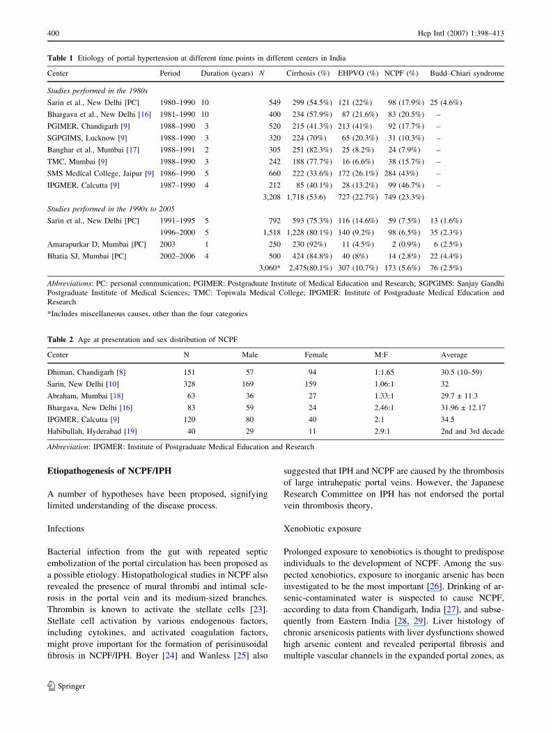
Studies conducted in the 1980s or earlier found an inci-
dence of approximately 23.3% (range 7.9–46.7%; Table 1)
[9, 16, 17]. The majority of the studies found the peak age of
incidence to be the third and fourth decades of life, and this is
one to two decades younger than in patients with IPH from
Japan [8, 16, 18, 19]. Several investigators from India have
found a male dominance or no sex predilection (Table 2) in
contrast to a female dominance (M:F = 1:3) in Japan [20]. A
study from a large tertiary care center at New Delhi has
shown that 98% of patients with NCPF were from the low-
socioeconomic strata of society [10].
The incidence of NCPF has probably declined in India
after 1990. There is also a concomitant decrease in the
incidence of EHPVO (Table 1). It has been speculated that
umbilical sepsis and/or repeated diarrheal episodes during
infancy or in early childhood may be responsible for both
diseases. Over the years, there has been an improvement in
the living standards. The changed scenario of seroepi-
demiology of hepatitis A virus infection also confirms the
improvement in sanitation and hygiene [21, 22]. Prenatal
practices have also changed in India, resulting in a reduc-
tion in the frequency of umbilical sepsis. Only a large
multicenter study could substantiate the decline in the
incidence of NCPF and verify the above-mentioned
observations.
Consensus statements
(2.1) NCPF/IPH is an important cause of portal
hypertension. (4)
(2.2.1) Incidence of IPH has declined in Japan after
the 1970s. (2b)
(2.2.2) There is some indication that the incidence of
NCPF is on the decline in India after 1990. (5, D)
(2.2.3) The current incidence of these diseases in
Asia-Pacific needs to be studied. (5, D)
(2.3.1) The patients of NCPF are generally young
adults in the third and fourth decade of life; IPH
generally presents in the fourth and fifth decade of
life. (3b)
(2.3.2) In NCPF, there is no sex predilection. IPH is
more common in females. (3b)
(2.4) NCPF/IPH is linked to low socioeconomic status.
(4)
Hep Intl (2007) 1:398–413 399
123
Etiopathogenesis of NCPF/IPH
A number of hypotheses have been proposed, signifying
limited understanding of the disease process.
Infections
Bacterial infection from the gut with repeated septic
embolization of the portal circulation has been proposed as
a possible etiology. Histopathological studies in NCPF also
revealed the presence of mural thrombi and intimal scle-
rosis in the portal vein and its medium-sized branches.
Thrombin is known to activate the stellate cells [23].
Stellate cell activation by various endogenous factors,
including cytokines, and activated coagulation factors,
might prove important for the formation of perisinusoidal
fibrosis in NCPF/IPH. Boyer [24] and Wanless [25] also
suggested that IPH and NCPF are caused by the thrombosis
of large intrahepatic portal veins. However, the Japanese
Research Committee on IPH has not endorsed the portal
vein thrombosis theory.
Xenobiotic exposure
Prolonged exposure to xenobiotics is thought to predispose
individuals to the development of NCPF. Among the sus-
pected xenobiotics, exposure to inorganic arsenic has been
investigated to be the most important [26]. Drinking of ar-
senic-contaminated water is suspected to cause NCPF,
according to data from Chandigarh, India [27], and subse-
quently from Eastern India [28, 29]. Liver histology of
chronic arsenicosis patients with liver dysfunctions showed
high arsenic content and revealed periportal fibrosis and
multiple vascular channels in the expanded portal zones, as
Table 1 Etiology of portal hypertension at different time points in different centers in India
Center Period Duration (years) N Cirrhosis (%) EHPVO (%) NCPF (%) Budd–Chiari syndrome
Studies performed in the 1980s
Sarin et al., New Delhi [PC] 1980–1990 10 549 299 (54.5%) 121 (22%) 98 (17.9%) 25 (4.6%)
Bhargava et al., New Delhi [16] 1981–1990 10 400 234 (57.9%) 87 (21.6%) 83 (20.5%) –
PGIMER, Chandigarh [9] 1988–1990 3 520 215 (41.3%) 213 (41%) 92 (17.7%) –
SGPGIMS, Lucknow [9] 1988–1990 3 320 224 (70%) 65 (20.3%) 31 (10.3%) –
Banghar et al., Mumbai [17] 1988–1991 2 305 251 (82.3%) 25 (8.2%) 24 (7.9%) –
TMC, Mumbai [9] 1988–1990 3 242 188 (77.7%) 16 (6.6%) 38 (15.7%) –
SMS Medical College, Jaipur [9] 1986–1990 5 660 222 (33.6%) 172 (26.1%) 284 (43%) –
IPGMER, Calcutta [9] 1987–1990 4 212 85 (40.1%) 28 (13.2%) 99 (46.7%) –
3,208 1,718 (53.6) 727 (22.7%) 749 (23.3%)
Studies performed in the 1990s to 2005
Sarin et al., New Delhi [PC] 1991–1995 5 792 593 (75.3%) 116 (14.6%) 59 (7.5%) 13 (1.6%)
1996–2000 5 1,518 1,228 (80.1%) 140 (9.2%) 98 (6.5%) 35 (2.3%)
Amarapurkar D, Mumbai [PC] 2003 1 250 230 (92%) 11 (4.5%) 2 (0.9%) 6 (2.5%)
Bhatia SJ, Mumbai [PC] 2002–2006 4 500 424 (84.8%) 40 (8%) 14 (2.8%) 22 (4.4%)
3,060* 2,475(80.1%) 307 (10.7%) 173 (5.6%) 76 (2.5%)
Abbreviations: PC: personal communication; PGIMER: Postgraduate Institute of Medical Education and Research; SGPGIMS: Sanjay Gandhi
Postgraduate Institute of Medical Sciences; TMC: Topiwala Medical College; IPGMER: Institute of Postgraduate Medical Education and
Research
*Includes miscellaneous causes, other than the four categories
Table 2 Age at presentation and sex distribution of NCPF
Center N Male Female M:F Average
Dhiman, Chandigarh [8] 151 57 94 1:1.65 30.5 (10–59)
Sarin, New Delhi [10] 328 169 159 1.06:1 32
Abraham, Mumbai [18] 63 36 27 1.33:1 29.7 ± 11.3
Bhargava, New Delhi [16] 83 59 24 2.46:1 31.96 ± 12.17
IPGMER, Calcutta [9] 120 80 40 2:1 34.5
Habibullah, Hyderabad [19] 40 29 11 2.9:1 2nd and 3rd decade
Abbreviation: IPGMER: Institute of Postgraduate Medical Education and Research
400 Hep Intl (2007) 1:398–413
123

seen in NCPF [29]. Similar observations have been made by
other workers in the past [30, 31]. The mechanisms related to
arsenic-induced NCPF are poorly known. Immunologic
mechanisms might play a role in the endothelial cell damage
from arsenic [32]; development of arsenic-induced hepatic
fibrosis was found to be related to high hepatic oxidative
stress and IL-6 and TNF-alpha levels [33–36]. However,
other workers did not find high arsenic content in the liver
tissue of patients with NCPF [9].
Immunologic abnormalities
There is some evidence to support immunologic abnormal-
ities in NCPF [37]. Nayyar et al. found that the population of
total peripheral T lymphocytes (T1) and suppressor/cyto-
toxic phenotype (T8) was significantly decreased in patients
with NCPF compared with controls. While the subpopula-
tions of helper/inducer lymphocytes (T4) and total B lym-
phocytes were comparable in size, the ratio of T4 to T8
lymphocytes was significantly increased in patients with
NCPF in comparison with controls [38, 39]. There is evi-
dence of increased vascular cell adhesion molecule-1
(VCAM-1) [40] and increased soluble TNF-receptor I and II
without any significant increase of TNF in the blood in IPH
[41, 42]. As TNF is involved in the induction and mainte-
nance of fibrotic reaction [42], fibrosis around the portal vein
as observed in patients with IPH could be explained. Fur-
thermore, TNF causes upregulation of VCAM-1, which is
also observed in patients with IPH. Heightened Th1 response
has been noted in NCPF/IPH; however, cellular infiltration is
not so remarkable in these patients [41, 42]. The role of NK
cells, the TH1/TH2 responses and reciprocity, various
cytokines involved, and the role of B-cells need to be studied
further. It remains to be established, whether these immu-
nological anomalies are a result or the cause of NCPF/IPH.
Consensus statements
(3.1) NCPF/IPH is a heterogeneous group of diseases,
which could be a result of the varied degree of portal
venous injury.
(3.2) Injury predominantly manifests in the presinu-
soidal region.
(3.3) The factors/agents that have been reported to be
associated with NCPF/IPH include umbilical/portal
pyemia, diarrheal diseases, or bacterial infections in
infancy; autoimmune disorders; prothrombotic
states; chronic exposure to arsenic, vinyl chloride
monomers, or copper sulfate (vineyard sprayers);
prolonged treatment with methotrexate; hypervita-
minosis A; and renal allograft recipients under
treatment of 6-mercaptopurine, azathioprine, and
corticosteroids. However, the exact etiology in the
majority of cases remains unknown. (4)
Animal models of NCPF/IPH
The etiopathogenesis of NCPF/IPH is still obscure, as pa-
tients generally present with late bleeding from varices.
Animal models help us in exploring the pathophysiology of
NCPF/IPH. The most common models are given below.
Prolonged sensitization with albumin
Prolonged sensitization of rabbits with bovine serum albu-
min [43] was seen to result in splenomegaly. Later, the
experiment was conducted on dogs as rabbits do not tolerate
more than three intraportal injections. The histological
changes that occurred in these animals were characterized
by early portal inflammation immediately followed by portal
fibrosis, aberrant vasculature, and disappearance of portal
venules and were very similar to those in human IPH.
Prolonged sensitization with nonpathogenic
Escherichia coli
In rabbits, killed nonpathogenic E. coli were administered
intraportally. The animals that received an intraportal
mixture of killed E. coli and rabbit antiserum (aggregated
E. coli) developed histologic changes in the liver and portal
hypertension [44]. However, these investigators had used
repeated cannulation of the portal vein, which may itself
cause damage to the portal vein intima, portal pyemia, and
an altered hemodynamic and histological picture in the
animal. Accordingly, alternative routes of introducing
E. coli into the portal circulation have been proposed [44].
Schistosomiasis japonica models
Schistosomiasis japonica and IPH share some histological
features of liver injury. Shekhar et al. [45] produced a
rabbit model by infecting with 250 Schistosoma cercariae
percutaneously and subcutaneously. The angioarchitecture
of chronic schistosomiasis japonica is characterized by
narrowing, obstruction, and obtuse angles of bifurcation of
the peripheral portal veins, and this disease is quite similar
to IPH in both histology and angioarchitecture, strongly
suggesting that portal change is the primary lesion of the
hepatic disorder in IPH. However, splenomegaly invariably
noted in IPH is not necessarily observed in chronic schis-
tosomiasis japonica, suggesting that portal system
involvement may be more extensive in IPH [46].
Chronic arsenic ingestion
Chronic arsenic toxicity is a form of hepatic fibrosis that
causes portal hypertension, but does not progress to cirrhosis.
Hep Intl (2007) 1:398–413 401
123

Hepatotoxic effects of arsenic in humans have been reported
earlier [31, 47–49]. Injury to the intrahepatic portal vein and
even development of cirrhosis has been alleged to occur with
the prolonged use of Fowler’s solution containing sodium
arsenite [49]. Sarin et al. in 1999 [50] produced a repro-
ducible and homogenous murine model of hepatic fibro-
genesis with increased hydroxyproline and collagen levels,
without significant hepatocellular necrosis and inflammation
through chronic arsenic feeding. Development of portal
hypertension was not observed. Guha Mazumder and Santra
[28, 29] demonstrated hepatic fibrosis due to arsenic toxicity
in mice receiving arsenic (3.2 mg/l) in drinking water for at
least 15 months. Arsenic feeding for 6 months showed a
significant decrease in hepatic glutathione and the enzymes
glucose-6-phosphate dehydrogenase and glutathione per-
oxidase in association with a significant increase of lipid
peroxidation compared with a control group [34]. Increasing
dose and duration of arsenic exposure in mice further showed
progressive increase of oxystress and elevation of cytokines
TNF-alpha and IL-6, which are associated with an increasing
level of collagen in the liver [35].
Repeated immunosensitization by rabbit splenic extract
Repeated intramuscular injection of splenic extract was
shown [51] to produce significant splenomegaly and rise in
portal pressure at 1, 3, and 6 months without hepatic
parenchymal injury, quite akin to NCPF seen in humans.
Repeated low-dose endotoxemia of portal circulation
Portal pyelephlebitis, due to repeated abdominal infections
and thrombosis in the portal circulation could lead to the
obstruction of small and middle branches of the portal vein
and development of NCPF. On the basis of this hypothesis,
repeated low-dose portal endotoxemia was produced by
injecting E. coli (heat-killed) through an indwelling can-
nula (placed in the gastrosplenic vein) [52]. Splenomegaly
and rise in portal pressure was noted at 1 and 3 months,
which persisted up to 6 months. Absence of hepatic
parenchymal injury and persistently elevated portal pres-
sure makes this model ideal for investigating the vascular
reactivity to various agents.
Consensus statements
(4.1) Current animal models of NCPF and IPH do not
accurately mimic human disease. (5)
(4.2) Some of the pathophysiological features of this
disease such as hepatic fibrosis or vascular changes
have been reproduced in different animal models.
(2c)
(4.3) Combination of systemic and direct portal
antigen delivery should be evaluated further. (5, D)
Clinical presentation of NCPF/IPH
NCPF
These patients present with well-tolerated episodes of
gastrointestinal hemorrhage, a longstanding mass in the left
upper quadrant (splenomegaly), anemia, and consequences
of hypersplenism. Development of ascites, jaundice, and
hepatic encephalopathy is uncommon and may be seen
only after an episode of gastrointestinal hemorrhage. Of all
the causes of portal hypertension, a massive and dispro-
portionately large spleen is seen most commonly in NCPF.
Left upper quadrant pain due to perisplenitis and splenic
infarction is not uncommon [53]. Like cirrhosis, NCPF also
may have odd presentations, such as glomerulonephritis
[54, 55], hypoxemia [56], or autonomic dysfunction [57].
Over a 24-year-period (1983–2006), Sarin et al. [10] saw
366 patients of NCPF; the clinical presentation profile of
these patients is shown in Table 3. Qureshi from Pakistan
[58, 59] found 73 cases in 20 years (1981–2001) with
similar clinical features.
IPH
Major presenting symptoms in Japanese patients with IPH
[60] were splenomegaly (88%), hepatomegaly (44%),
gastrointestinal (GI) bleeding (35%), and ascites (12%).
These figures are different from NCPF, where the majority
present with upper GI bleeding, splenomegaly, and anemia.
Hematological studies confirmed more severe anemia and
thrombocytopenia in NCPF than in IPH [60].
Consensus statements
(5.1) Patients of NCPF/IPH usually have a longstand-
ing history of mass in the left hypochondrium
(enlarged spleen). (2b)
(5.2) Most cases of NCPF/IPH present with enlarged
spleen and GI bleeding (hematemesis) and some have
features of anemia. (2b)
Table 3 Clinical presentation of patients of NCPF [10]
Parameter NCPF (n = 366)
Mean age 32 ± 14 years
Sex (M:F) 186:180
Hematemesis/melena 72%
Awareness of lump in left upper quadrant 12%
Ascites (transient) 25%
History of jaundice 18%
Esophageal varices 97%
Gastric varicesd 31%
Portal gastropathy 3%
402 Hep Intl (2007) 1:398–413
123

(5.3) Signs of chronic liver disease like palmar
erythema, spider angioma, testicular atrophy in
abdominal wall veins, and gynecomastia are rare.
(3b)
(5.4) Jaundice, ascites/edema, and signs of liver
failure are uncommon. (3b)
(5.5) Ascites is transient and usually seen soon after a
variceal bleed. (2b)
Natural history of NCPF/IPH
In the clinical courses of patients with IPH, the liver slowly
undergoes atrophy, which is not necessarily progressive,
and the liver functional reserve is well maintained. Al-
though mortality from variceal rupture is generally lower in
NCPF/IPH, because of better liver functions compared
with cirrhosis, the major cause of death is variceal bleed-
ing. Repeated uncontrollable bleeding may induce hepatic
insufficiency. The survival curve for patients with NCPF/
IPH is somewhat between that for those with cirrhosis and
for a healthy population of comparable age [11, 60–62].
Good prognostic features in patients with NCPF, a 2- and
5-year survival of nearly 100% after successful eradication
of esophagogastric varices, have been described [63].
Hillaire et al. reported death of 4 out of 28 patients with
IPH owing to terminal liver failure [7]. According to a
clinical study in Japan, 4 out of the 22 patients with IPH
with portal vein thrombosis (PVT) died and all patients
without PVT were alive during the mean clinical course of
12 years [64]. The causes of death were systemic exhaus-
tion as a result of chronic ascites in three patients and
infection in one patient.
The incidence of PVT is higher in patients with IPH than
in those with cirrhosis, and ascites is not uncommon [7,
64]. Hillaire et al. examined the outcome of patients with
IPH having PVT, and found it in 3 of 4 patients who died
and in only 10 out of 24 patients who survived [7]. They
also mentioned that ascites developed in 14 out of the 28
patients with IPH and in 11 with GI bleeding or severe
concurrent diseases, and that ascites was transient in 10
patients and constant in 4 patients. The other study shows
that patients without PVT had less clinical problems after
long-term follow-up [64]. However, marked hypersplenism
and low serum albumin were significantly frequent in pa-
tients with PVT than in patients without PVT. Moreover,
ascites was present only in patients with PVT (seven of
nine) and four patients with PVT died. Development of
PVT in a patient with IPH may be a significant factor for
poor prognosis, and ascites may indicate a deterioration of
the condition in patients with IPH. Furthermore, PVT and
ascites may be mutually related in this disease.
Consensus statements
(6.1.1) Bleeding rate from gastroesophageal varices is
high in patients with NCPF/IPH (32–95%). (2b)
(6.1.2) Mortality from variceal rupture is generally
lower in NCPF/IPH, because of better liver functions
compared with cirrhosis. (2b)
(6.1.3) Management of gastroesophageal varices to
prevent the rupture should be a priority in the care of
patients with NCPF/IPH. (5, D)
(6.2.1) The incidence of PV thrombosis is more
frequent in patients with IPH than in patients with
liver cirrhosis. The same should also be studied in
NCPF. (3b, D)
(6.2.2) Development of portal vein thrombosis in
patients with IPH may be a significant factor for poor
prognosis. The practical benefits of the management
of portal vein thrombosis to improve the clinical
course of IPH should be elucidated in future studies.
(4, C)
(6.3.1) Ascites is not rare in patients with IPH in spite
of preserved liver functions. It occurs in association
with PVT. (4, C)
(6.3.2) However, clinical ascites is rare in patients
with NCPF and it is transient after a bleed. (4)
(6.3.3) Ascites is considered to be a sign for deteri-
oration of the condition in patients with IPH. (5, D)
(6.4.1) In NCPF/IPH, the liver probably undergoes
atrophy, owing to reduced blood supply to the
periphery. It is not necessarily progressive, and the
liver functional reserve is well maintained. (4, C)
(6.4.2) The survival rate for patients with NCPF/IPH
is much better than that for patients with cirrhosis.
(4, C)
(6.4.3) PV thrombosis and ascites may indicate the
deterioration of the condition in certain cases of IPH,
and thrombosis and ascites may be mutually related
in this disease. (5, D)
Histopathology of NCPF/IPH
The histopathology of NCPF/IPH has been described in
studies from India and Japan, including a few cases in
Western literature. These studies include the description of
autopsy livers and wedge and needle biopsies.
Autopsy liver
Gross examination may reveal a normal, enlarged, or even
shrunken liver. The surface is smooth, wrinkled, or even
show some nodularity resembling cirrhosis. Fibrous thick-
ening of the capsule of the liver with increased vascularity
Hep Intl (2007) 1:398–413 403
123
and some inflammation may be seen. Subcapsular septation
can be noted, while deeper parenchyma shows normal
architecture. Sclerosis of large to small intrahepatic portal
vein branches and approximation of portal tracts to surface
has been documented [65, 66]. This has been confirmed by
histomorphometry by Kage et al. [67]. Histological features
noted in autopsies include increased portal collagenous
connective tissue and sclerosis and obliteration of small
branches of portal veins in most cases. This histological
hallmark of NCPF was termed obliterative portal venopathy
by Nayak and Ramalingaswami [68]. Intimal fibrosis and
elastosis is also common, leading to subendothelial thick-
ening of the wall of large- and medium-sized portal vein
branches even compromising the lumen. Veins may be
thickened to the extent that they resemble an artery. Fur-
thermore, aberrant vasculatures characterized by thin-walled
vessels mainly located adjacent to the portal tracts and at
times in the hepatic lobules have been described. Although
some of them are morphologically very similar to hepatic
vein branches, they are portal in nature [69]. Recanalized
thrombi are sometimes seen. The inconspicuous branches of
the terminal portal vein may be replaced by small vascular
channels. Mild inflammation is seen in a few cases. Incom-
plete portal pseudolobule and scattered regenerative nodules
may be noted in a few cases.
Hepatic vein may show sclerosis or small branches may
show slight dilatation. Collagen deposition in the space of
Disse has been observed by electron microscopy [70]. The
collagen and elastin deposition in IPH may be a result of
increased connective tissue growth factor expression and
decreased MMP-9 expression in portal tracts of IPH as
demonstrated by immunohistochemistry in a study by
Tsuneyama et al. [71].
Needle biopsies
Biopsy material may show only mild and subtle changes
from normal. These changes include inconspicuous portal
tracts and obliterated veins, or fibrous expansion of portal
tracts. Alternatively there may be dilatation of vessels in or
near portal tracts, with vessel-like dilatation of sinusoids.
Distortion of portal–central relation may be noted. Ludwig
et al. [72] studied the changes in 25 liver biopsies. Changes
in the portal tract included capillary dilatation, phlebo-
sclerosis, and fibroelastosis of the stroma. Portal vein
dilatation with herniation into the surrounding hepatic
parenchyma was also noted. Portal vein obliteration and
loss of bile ducts were rare in their study. The acinar
architecture showed capillary and necroinflammatory
bridging between portal tracts and terminal hepatic veins,
isolated megasinusoids in a random distribution, displaced,
and abnormally large hepatic vein branches with or without
phlebosclerosis, and slender, curved fibrous septa.
Wedge biopsies
Wedge biopsies show changes similar to autopsy material,
but changes in medium and large portal vein branches may
not be seen if not sampled. The changes in pre- or intra-
operative biopsy specimens are subtle, and may be missed
by the casual observer because of the absence of significant
fibrosis. Nodular regenerative hyperplasia, focal nodular
hyperplasia, and incomplete septal cirrhosis have all been
described with NCPF [73]. Association of NCPF with
hepatocellular carcinoma has also been described [74, 75].
A deep-core wedge biopsy (not broad-based wedge)
along with a needle biopsy should be taken, as they would
complement each other in the information provided. This
material may be valuable in looking for clues to the etio-
pathogenesis of NCPF.
Early changes and staging
Early hepatic changes in NCPF were observed incidentally
in a patient of cervical cancer [76]. These include lymphoid
cell infiltration of the portal tract and of subendothelial
regions of portal vein branches, and nonspecific lobular
hepatitis.
Nakanuma et al. [77] proposed a staging of IPH with a
combination of hepatic parenchymal atrophy and portal
venous thrombosis. Stage I is nonatrophic liver without
subcapsular parenchymal atrophy, stage II is nonatrophic
liver with subcapsular parenchymal atrophy, stage III is
atrophic liver with subcapsular parenchymal atrophy, and
stage IV is portal venous occlusive thrombosis. IPH livers
can progress from stage I to stage III, while stage IV occurs
relatively late.
Consensus statements
(7.1) Diagnostic criteria of NCPF/IPH on needle liver
biopsy are as follows:
Essential for diagnosis:
Absence of regenerative nodules with features
of possible or definite cirrhosis in an adequate-sized
liver biopsy. (1a, A)
Features usually seen (however, these may not be
seen in all):
Small portal vein obliteration; aberrant vascu-
lature; portal tract fibrosis, rounded or streaky;
absence of significant hepatocellular injury. (2b, B)
Laboratory studies in NCPF/IPH
The laboratory evaluation in NCPF/IPH reveals only mild
and subtle abnormalities predominantly related to hy-
persplenism.
404 Hep Intl (2007) 1:398–413
123
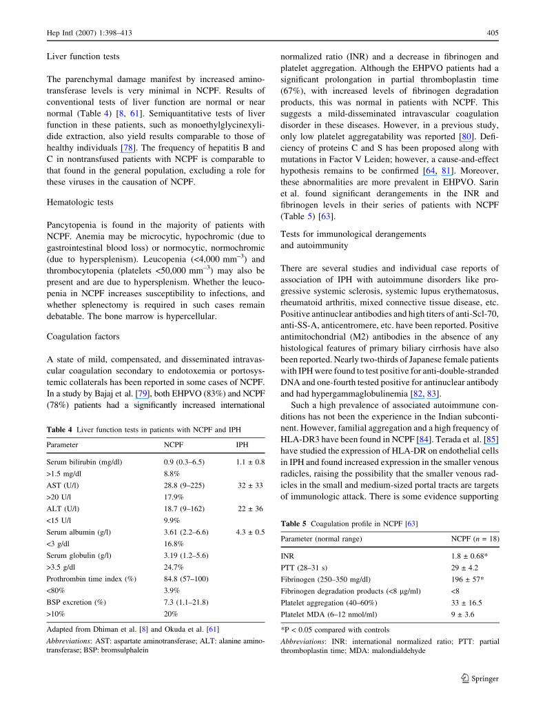
Liver function tests
The parenchymal damage manifest by increased amino-
transferase levels is very minimal in NCPF. Results of
conventional tests of liver function are normal or near
normal (Table 4) [8, 61]. Semiquantitative tests of liver
function in these patients, such as monoethylglycinexyli-
dide extraction, also yield results comparable to those of
healthy individuals [78]. The frequency of hepatitis B and
C in nontransfused patients with NCPF is comparable to
that found in the general population, excluding a role for
these viruses in the causation of NCPF.
Hematologic tests
Pancytopenia is found in the majority of patients with
NCPF. Anemia may be microcytic, hypochromic (due to
gastrointestinal blood loss) or normocytic, normochromic
(due to hypersplenism). Leucopenia (<4,000 mm–3) and
thrombocytopenia (platelets <50,000 mm–3) may also be
present and are due to hypersplenism. Whether the leuco-
penia in NCPF increases susceptibility to infections, and
whether splenectomy is required in such cases remain
debatable. The bone marrow is hypercellular.
Coagulation factors
A state of mild, compensated, and disseminated intravas-
cular coagulation secondary to endotoxemia or portosys-
temic collaterals has been reported in some cases of NCPF.
In a study by Bajaj et al. [79], both EHPVO (83%) and NCPF
(78%) patients had a significantly increased international
normalized ratio (INR) and a decrease in fibrinogen and
platelet aggregation. Although the EHPVO patients had a
significant prolongation in partial thromboplastin time
(67%), with increased levels of fibrinogen degradation
products, this was normal in patients with NCPF. This
suggests a mild-disseminated intravascular coagulation
disorder in these diseases. However, in a previous study,
only low platelet aggregatability was reported [80]. Defi-
ciency of proteins C and S has been proposed along with
mutations in Factor V Leiden; however, a cause-and-effect
hypothesis remains to be confirmed [64, 81]. Moreover,
these abnormalities are more prevalent in EHPVO. Sarin
et al. found significant derangements in the INR and
fibrinogen levels in their series of patients with NCPF
(Table 5) [63].
Tests for immunological derangements
and autoimmunity
There are several studies and individual case reports of
association of IPH with autoimmune disorders like pro-
gressive systemic sclerosis, systemic lupus erythematosus,
rheumatoid arthritis, mixed connective tissue disease, etc.
Positive antinuclear antibodies and high titers of anti-Scl-70,
anti-SS-A, anticentromere, etc. have been reported. Positive
antimitochondrial (M2) antibodies in the absence of any
histological features of primary biliary cirrhosis have also
been reported. Nearly two-thirds of Japanese female patients
with IPH were found to test positive for anti-double-stranded
DNA and one-fourth tested positive for antinuclear antibody
and had hypergammaglobulinemia [82, 83].
Such a high prevalence of associated autoimmune con-
ditions has not been the experience in the Indian subconti-
nent. However, familial aggregation and a high frequency of
HLA-DR3 have been found in NCPF [84]. Terada et al. [85]
have studied the expression of HLA-DR on endothelial cells
in IPH and found increased expression in the smaller venous
radicles, raising the possibility that the smaller venous rad-
icles in the small and medium-sized portal tracts are targets
of immunologic attack. There is some evidence supporting
Table 4 Liver function tests in patients with NCPF and IPH
Parameter NCPF IPH
Serum bilirubin (mg/dl) 0.9 (0.3–6.5) 1.1 ± 0.8
>1.5 mg/dl 8.8%
AST (U/l) 28.8 (9–225) 32 ± 33
>20 U/l 17.9%
ALT (U/l) 18.7 (9–162) 22 ± 36
<15 U/l 9.9%
Serum albumin (g/l) 3.61 (2.2–6.6) 4.3 ± 0.5
<3 g/dl 16.8%
Serum globulin (g/l) 3.19 (1.2–5.6)
>3.5 g/dl 24.7%
Prothrombin time index (%) 84.8 (57–100)
<80% 3.9%
BSP excretion (%) 7.3 (1.1–21.8)
>10% 20%
Adapted from Dhiman et al. [8] and Okuda et al. [61]
Abbreviations: AST: aspartate aminotransferase; ALT: alanine amino-
transferase; BSP: bromsulphalein
Table 5 Coagulation profile in NCPF [63]
Parameter (normal range) NCPF (n = 18)
INR 1.8 ± 0.68*
PTT (28–31 s) 29 ± 4.2
Fibrinogen (250–350 mg/dl) 196 ± 57*
Fibrinogen degradation products (<8 lg/ml) <8
Platelet aggregation (40–60%) 33 ± 16.5
Platelet MDA (6–12 nmol/ml) 9 ± 3.6
*P < 0.05 compared with controls
Abbreviations: INR: international normalized ratio; PTT: partial
thromboplastin time; MDA: malondialdehyde
Hep Intl (2007) 1:398–413 405
123

immunological derangements/deficiency in the causation of
NCPF [38, 86]. In patients with IPH, a poor autologous
mixed lymphocyte reaction has been reported [87, 88]. More
recently, studies have suggested that the imbalance of Th1
and Th2 CD4+ T-cells and TNF may be associated with the
pathogenesis of IPH [89].
Consensus statements
(8.1) Results of liver function tests are normal or near
normal in patients of NCPF/IPH. (2b)
(8.2) Hypersplenism is common but usually asymp-
tomatic. (2b)
(8.3) The frequency and characteristics of the coag-
ulation abnormalities in these patients need to be
investigated further. (5, D)
Features at endoscopy in NCPF/IPH
The risk of bleeding in NCPF/IPH has not been indepen-
dently investigated, but is believed to be similar to the risk
in patients with varices due to other causes. In contrast to
patients with cirrhosis who bleed from varices, patients
with NCPF tolerate variceal bleeding relatively well,
probably because of well-preserved hepatic synthetic
functions. The GI-bleed-related mortality rate varies. In the
West, patients with noncirrhotic etiologies have mortality
ranging from 7 to 31% for a single bleeding episode [90].
The mortality rate is higher in Japan with IPH [62].
Prevalence of varices
Esophagogastric varices have been reported in 85–95% of
patients with NCPF [78]. Gastric varices are more common
in NCPF than in cirrhosis and are reported in up to 44% of
cases. Gastric varices are usually associated with large
esophageal varices [91]. Antral varices are uncommon in
NCPF and may develop after eradication of esophageal
varices in up to 3.8% of patients on follow-up. [92]. Portal
hypertensive gastropathy is relatively less common in
NCPF [57] than in cirrhosis and manifests mainly after
variceal obliteration. Anorectal varices are more common
in NCPF than in cirrhosis. In one study, significantly more
patients with NCPF had anorectal varices than did patients
with cirrhosis (89 vs. 56%) [93].
Bleeding risk from varices in NCPF
Approximately 70% of patients with NCPF present with a
history of major variceal bleeding. NCPF is an important
cause of upper gastrointestinal bleeding, constituting 15%
of the cases of variceal bleed in patients with portal
hypertension [94]. Antral varices rarely bleed, and if they
are not the source of bleeding they can be managed con-
servatively [92].
Consensus statements
(9.1) Esophageal varices are seen in 85–90% of
patients. (2b)
(9.2) These varices are generally large at the time of
diagnosis. (2b)
(9.3) If esophageal varices are small, investigations
for the presence of gastric varices and spontaneous
shunts are required. (5, D)
(9.4) Gastric varices are seen in about 25% of
patients with NCPF/IPH. (2b)
(9.5) Portal hypertensive gastropathy is uncommon in
these patients. (2b)
(9.6.1) Anorectal varices are common in NCPF. (2b)
(9.6.2) Prevalence of anorectal varices in IPH is not
known. (5)
Hemodynamics in NCPF/IPH
The portal vein pressures are elevated markedly in patients
who have NCPF. Two pathoanatomic sites of obstruction
have been identified. A pressure gradient exists between
the spleen and the liver (intrasplenic pressure – intrahepatic
pressure [IHP]) and another exists between the IHP and the
wedged hepatic venous pressure (WHVP) (IHP – WHVP)
[95]. Generally, the WHVP is normal or only slightly
elevated in NCPF. Variceal pressure also has been studied
in these patients and is comparable to that in cirrhotic
portal hypertension [95, 96]. Intravariceal pressure closely
reflects portal pressure in patients who have NCPF and is
the investigation of choice for measurement of portal
pressure. Splenic and portal vein blood flow are known to
be increased markedly in Japanese patients with IPH,
which is suggestive of a hyperdynamic circulatory state.
Consensus statements
(10.1) Hepatic venous pressure gradient (HVPG) is
normal or near normal in NCPF/IPH. (2b)
(10.2) Hemodynamic studies indicate site of resis-
tance as predominantly presinusoidal. (3b)
(10.3) Whether HVPG increases on long-term follow-
up needs to be studied. (5, D)
(10.4) Portal venous blood flow is significantly
increased. (2b)
Diagnosis of NCPF/IPH
The diagnosis of NCPF is comparatively easy in developing
countries, where the disease is common. Clinical presentation
406 Hep Intl (2007) 1:398–413
123
of variceal bleeding, moderate to massive splenomegaly
without features of chronic liver disease, and growth retar-
dation would make one suspect NCPF. In Japan and in the
West, such patients are a decade or so older and often present
with anemia, splenomegaly, and variceal bleeding. NCPF and
IPH are not merely diagnoses of exclusion, and need well-
defined and agreed-upon criteria for diagnosis.
Consensus statements
(11.1) Diagnostic features of NCPF/IPH:
Presence of moderate to massive splenomegaly
Evidence of portal hypertension, varices, and/or
collaterals
Patent spleno-portal axis and hepatic veins on
ultrasound Doppler,
Test results indicating normal or near-normal
liver functions,
Normal or near-normal hepatic venous pressure
gradient, and
Liver histology—no evidence of cirrhosis or
parenchymal injury. (2b)
(11.2) Other features:
Absence of signs of chronic liver disease,
No decompensation after variceal bleed except
occasional transient ascites,
Absence of serum markers of hepatitis B or C
virus infection,
No known etiology of liver disease, and
Imaging with ultrasound or other imaging tech-
niques showing dilated and thickened portal vein
with peripheral pruning and periportal hyperechoic
areas. (2b)
Management of acute bleeding in NCPF/IPH
Variceal bleeding is a common and life-threatening com-
plication of portal hypertension due to NCPF. There is
paucity of data on the management of acute variceal
bleeding in NCPF; however, the principles and modes of
management remain the same as those for patients with
cirrhosis.
General management
Blood transfusion, intravenous fluids, and standard ICU
care are provided [97–99]. Placement of nasogastric tube is
optional, especially if the bleeding has taken place more
than 12 hours ago [100].
Bacterial infections are more common in patients with
cirrhosis having variceal bleeding (35–66%) than in non-
cirrhotic patients (5–7%) [101]. It has been shown that
infected cirrhotic patients have a higher rate of variceal
rebleeding (43%) than noninfected patients (10%) [98,
102]. In patients with cirrhosis and variceal bleeding, the
prophylactic antibiotics reduce variceal rebleeding and
improve survival [103, 104]. In NCPF, however, there is no
study on the use of prophylactic antibiotics.
Balloon tamponade
Balloon tamponade using Sengstaken Blakemore tube en-
ables temporary control of bleeding, by direct compression
of varices at the esophagogastric junction, in 40–90% of
cases [97, 98]. Owing to high rates of complications and
rebleeding, balloon tamponade is not used routinely as the
first-line treatment for control of acute variceal bleeding.
Pharmacological therapy
Vasoactive drugs, such as somatostatin, octreotide, or ter-
lipressin, have been used in the treatment of acute variceal
bleeding while endoscopic therapy is being arranged. The
vasoactive drugs lead to reduction in portal pressure, which
is associated with a better control of variceal bleeding
[105–108]. However, there are no data on the efficacy of
vasoactive drugs in patients with NCPF with acute variceal
bleeding.
Endoscopic therapy
Endoscopic sclerotherapy and band ligation are effective in
80–90% of patients in controlling acute bleeding from
esophageal varices and preventing rebleeding [109, 110].
At present, band ligation is preferred owing to lower
complication rates.
Combined pharmacological and endoscopic therapy
Combination treatment with drugs plus endoscopic therapy
is more effective than endoscopic therapy or drug therapy
alone in controlling acute bleeding (88% vs. 76%) and
preventing rebleeding for 5 days (77% vs. 58%), while
there is no difference in mortality [109, 110]. There is,
however, paucity of data for NCPF.
Failure of endoscopic therapy is defined, as further
variceal bleeding after two endoscopic treatments during a
single hospital admission for acute bleeding. The current
therapies fail to control bleeding or prevent early reblee-
ding in 8–12% of patients [109], who should be treated by
alternative modes of treatment like surgery or transjugular
intrahepatic portosystemic shunt (TIPS).
Consensus statements
(12.1) General measures for the control of acute
bleeding are same as for cirrhosis. (5, D)
Hep Intl (2007) 1:398–413 407
123
(12.2) Endoscopic therapy is effective for the control
of acute variceal bleed in NCPF/IPH. (4, C)
(12.3) Role of vasoactive drugs alone or in combina-
tion with endoscopic therapy needs to be evaluated.
(5, D)
(12.4) If a diagnosis of NCPF/IPH is unlikely, the
condition should be treated as cirrhosis. (5, D)
(12.5) In case of failure of medical management (as in
Baveno IV), decompressive surgery or TIPS is useful
and should be used on the basis of available expertise.
(5, D)
(12.6) Patients with transient ascites should undergo
devascularization procedure. (5, D)
Prevention of variceal bleeding in NCPF/IPH
Primary prophylaxis
The natural history of esophageal varices in NCPF is not
known. Progression of variceal size occurs at a rate of
10–15% per year in patients with cirrhosis, mostly depen-
dent on liver dysfunction. Such a progression of varices in
NCPF is less likely to occur, as the liver function continues
to be normal. Similarly, a decrease in the size of esopha-
geal varices, as seen in patients with cirrhosis with an
improvement in liver functions is unlikely in NCPF, unless
interventions like endoscopic sclerotherapy are applied,
which after variceal obliteration results in the development
of spontaneous splenorenal shunts [111–114].
Endoscopic variceal ligation (EVL) and beta blockers
are the common modes of therapy for the primary pro-
phylaxis of large esophageal varices in cirrhosis [115].
There are no randomized controlled trials on primary
prophylaxis in NCPF. Since patients with NCPF are all in
Child’s A class, the results of EVL and beta blockers could
be extrapolated to NCPF.
In a study by Sarin et al. [116], 8 patients with NCPH
and 18 with Child’s A cirrhosis were given a combination
of EVL and propranolol and compared with 9 and 15 pa-
tients with NCPH and cirrhosis, respectively, given EVL
only. None of the NCPH or cirrhotic patients bled on a
follow-up of an year, indicating that both EVL and drug
therapies are effective in preventing the first bleed. EVL is
the preferred mode of therapy because it is difficult to as-
sess the response to beta blockers in patients with NCPF as
HVPG is near normal. Hence, to assess the efficacy of beta
blocker therapy, measurements of splenic pulp pressure or
direct portal pressure would need to be taken. Moreover,
since the lifespan of patients with NCPF is normal, com-
pliance of drug therapy is not likely to be good.
Shunt surgery for primary prophylaxis is likely to be
indicated if the patient of NCPF has large esophageal
varices with a symptomatic large splenomegaly, a very low
platelet count (<20,000), stays far away from a good
medical center where an upper GI bleed can be tackled, or
has a rare blood group. A study from India on 45 patients
with NCPF [117]—41 of whom were treated with a pro-
phylactic proximal lienorenal shunt, 2 with splenectomy,
and 2 with devascularization—showed no operative mor-
tality. Over a follow-up period of 49 months, three patients
bled and two late deaths unrelated to surgery occurred.
There was delayed morbidity in 47%, including seven
patients, who developed partial splenic embolization; four,
glomerulonephritis; two, pulmonary AV fistulae; and five,
ascites requiring diuretics. It was thus considered safe, but
at the cost of high, delayed morbidity [117]. Patients with
gastric varices of more than 2 cm could be taken up for
surgical shunt or balloon-occluded retrograde transvenous
obliteration (BRTO) if a splenorenal shunt is present,
although studies are lacking.
Secondary prophylaxis
Literature on secondary prophylaxis is scanty. One study
on 72 patients with NCPF with recurrent bleeding [118]
showed that endoscopic sclerotherapy significantly reduced
the bleeding rate over a follow-up period of
21.4 ± 20.4 months; rebleed after obliteration occurred in
seven patients (9.2%). Recurrence occurred in nine
(13.9%) patients. Similarly, Bhargava et al. [119] and
Kochhar et al. [120] have shown sclerotherapy to be
effective in managing patients with NCPF. EVL has been
shown to be better than EST in almost all the studies;
hence, it could be recommended in NCPF [121]. There is
an isolated study using nonselective beta blockers in NCPF
[122]. There is also no information on the role of TIPS in
NCPF except for a case report [123].
Shunt surgery is effective in NCPF. One study involving
30 patients with NCPF reported a significant decrease in
splenic pulp pressure (44.3 ± 13.5 vs. 33.8 ± 7.6 cm of
saline) and splenic size from 9.1 ± 3.3 to 6.8 ± 4.6 cm in
28 patients after successful patent shunt surgery [124].
Shunt occlusion, overt chronic portosystemic encephalop-
athy, and rebleeding after elective shunt surgery were seen
in approximately 10% of patients [125]. A recent trial
comparing shunt surgery with TIPS in patients with cir-
rhosis has shown the two to be equally effective in treating
variceal rebleeding and encephalopathy in Child’s A
patients and ensuring survival [126].
Role of image-guided interventions in preventing
variceal bleeding
Image-guided interventions (IGI) are a relatively recent
means of treating and preventing variceal bleed. These include
408 Hep Intl (2007) 1:398–413
123
partial splenic embolization, BRTO, percutaneous transhe-
patic obliteration (PTO), and TIPS [63, 127]. The concept
behind these modalities is again occlusion of varices and
treatment of splenic lump, in an otherwise well-preserved
patient with a significant lifespan ahead. An approach com-
bining medical, endoscopic, and/or surgical management with
an IGI may be beneficial in certain situations [127].
Transjugular intrahepatic portosystemic shunt
TIPS creation is indicated for uncontrollable variceal
hemorrhage, recurrent variceal hemorrhage despite endo-
scopic therapy, and portal hypertensive gastropathy bleed
[63, 128].
Surgical shunt procedures continue to be a safe, highly
effective, and durable treatment for variceal bleeding in
patients with low operative risk and good liver function
[129, 130]. For patients with NCPH, in particular with
EHPVO, portosystemic shunt surgery represents an effec-
tive therapy that leads to freedom from recurrent bleeding
and repeated endoscopies for many years, and improves
hypersplenism without worsening liver function or
encephalopathy [129].
Primary long-term shunt patency was not as expected.
Wolff et al. [129] found shunt stenosis or occlusion rates to
be about 29% at 6 months, 42% at 1 year, and 51% at
2 years. Balloon angioplasty, new stent placement, or both
can re-establish shunt patency in most patients. In view of
good overall prognosis in patients having NCPF, a routine
follow-up is mandatory; the number of repeat procedures
one might have to undertake in such patients seems to be
higher with the use of presently popular bare stents [129].
PTFE-covered stents (stent-grafts) rather than bare, fenes-
trated stents can help prevent TIPS stenosis and occlusions
in the parenchymal tract as well as the hepatic venous end
[131].
TIPS is still a multistage procedure requiring close
surveillance and frequent maintenance, with interventions
for re-establishing shunt function [132], especially in the
third world countries because of the high cost of a stent-
graft [129]. No published data are available on the rate of
complications of TIPS in patients with NCPF; however,
owing to the good hepatic functions, it might be logical to
conclude that such complications would be uncommon in
NCPF [133]. The role of prophylactic IGI/TIPS in patients
with NCPF has not been evaluated [134].
Balloon-occluded retrograde transvenous obliteration
and percutaneous transhepatic obliteration
These modalities were introduced as less invasive alter-
natives to surgical shunt and TIPS [135–137]. Specific
situations where they may be attempted include poor
hepatic functional reserve, isolated/predominant gastric
cardiac and fundal varices, and gastric fundal varices with
active bleeding (spurting or oozing), signs of recent
bleeding, or in danger of rupturing [136]. The criteria
mentioned above have been derived from studies not
dealing directly with patients with NCPF. This is because
of the scant citation of interventional experiences with
patients with NCPF.
Image-guided interventions: Is there a role?
Image-guided intervention is largely considered a second-
ary tool for managing portal hypertension after conserva-
tive and/or endoscopic management have been explored.
The role of IGI in the treatment of NCPF is still not clear.
We would like to submit that IGI can replace surgical
options in most cases and complement endoscopic man-
agement in managing patients with NCPF.
Consensus statements
Screening
(13.1) All patients of moderate to massive spleno-
megaly with suspected NCPF should have screening
endoscopy. (5, D)
Preprimary and early-primary prophylaxis
(13.2) There is insufficient data to recommend it at
present. (5, D)
Primary prophylaxis
(13.3.1) EVL is recommended for large varices.
There is no consensus on the use of beta blockers in
such patients. (5, D)
(13.3.2) BRTO may be an option in patients with
large gastric varices. (5, D)
(13.3.3) Decompressive shunt surgery is not recom-
mended for primary prophylaxis. (3b, C)
Secondary prophylaxis
(13.4.1) Endoscopic therapy and elective decompres-
sive surgery are effective and safe. (2b)
(13.4.2) There should be head-to-head comparison
between these two modalities. (5, D)
(13.4.3) There is insufficient data on the role of TIPS
in secondary prophylaxis. (5)
(13.4.4) BRTO is effective in patients with NCPF/IPH
with gastric variceal bleed if gastrorenal shunt is
present. (5)
Differences between NCPF and IPH
Although, the terms NCPF and IPH often have been used
interchangeably, there are subtle differences between the
Hep Intl (2007) 1:398–413 409
123

two. NCPF is more common among men. This is in con-
trast to IPH, which is more common among women in
Japan, Europe, and the United States. The mean age of
patients who have NCPF varies from 25 to 35 years, which
is much lower than for patients who have IPH. There are
also distinct differences in the prevalence of autoantibodies
and histopathology between the two diseases [138–140]
Consensus statements
(14.1) While NCPF and IPH represent the same
disease entity, there are some differences. (1a)
(14.2) In NCPF, there is no sex predilection and the
mean age of presentation is about 30 years. This is in
contrast to IPH, which is more common among
middle-aged females in Japan, Europe, and the
United States. (1a)
(14.3) Autoimmune features are common in IPH
while rare in NCPF. (1a)
(14.4) Irregular parenchymal nodules and bile duct
proliferation are more common in NCPF than IPH.
(1a)
(14.5) Wedged hepatic venous pressure is almost
normal in NCPF, while it is moderately raised in IPH.
(1a)
Noncirrhotic portal fibrosis and idiopathic portal
hypertension have withstood scientific indifference, since
their description. It is hoped that the current short review
and guidelines would be seen as an endeavor to enhance
the clinician’s desire and pursuit for this seemingly oriental
fibrosis. These diseases, however, provide unique oppor-
tunities to understand the genesis and pathophysiology of
portal hypertension in the presence of near-normal liver
function.
References
1. de Franchis R. Evolving consensus in portal hypertension. Re-
port of the Baveno IV consensus workshop on methodology of
diagnosis and therapy in portal hypertension. J Hepatol
2005;43:167–76.
2. Sarin SK, Sollano JD, Chawla YK, Amarapurkar D, Hamid S,
Hashizume M et al. Members of the APASL Working Party on
Portal Hypertension. Consensus on extra-hepatic portal vein
obstruction. Liver Int 2006;26:512–9.
3. Centre for Evidence Based Medicine. Department of Primary
Care, Old Road Campus, Headington, Oxford, OX3 7LF, UK.
http://www.cebm.net/index.aspx?o=1025.
4. Mikkelsen WP, Edmondson HA, Peters RL, Redeker AG,
Reynolds TB. Extra- and intra-hepatic portal hypertension
without cirrhosis (hepatoportal sclerosis). Ann Surg
1965;162:602–20.
5. Bioulac-Sage, Le Bail B, Bernard PH, Balabaud C. Hepatoportal
sclerosis. Semin Liver Dis 1995;15:329–39.
6. Kingham JGC, Levison DA, Stansfeld AG, Dawson AM. Non-
cirrhotic intrahepatic portal hypertension: a long term follow-up
study. Q J Med 1981;50:259–68.
7. Hillaire S, Bonte E, Denninger MH, Casadevall N, Cadranel JF,
Lebrec D et al. Idiopathic non-cirrhotic intrahepatic portal
hypertension in the West: a re-evaluation in 28 patients. Gut
2002;51:275–80.
8. Dhiman RK, Chawla Y, Vasishta RK, Kakkar N, Dilawari JB,
Trehan MS et al. Non-cirrhotic portal fibrosis (idiopathic portal
hypertension): experience with 151 patients and a review of the
literature. J Gastroenterol Hepatol 2002;17:6–16.
9. Sarin SK. Non-cirrhotic portal hypertension. In: Advances in
therapeutic hepatology: a world view. Postgraduate Course,
AASLD 1998.
10. Pande C, Kumar A, Sarin SK. Non-cirrhotic portal fibrosis: a
clinical profile of 366 patients [abstract]. Am J Gastroenterol
2006;101:S191.
11. Okuda K. Non-cirrhotic portal hypertension: why is it so com-
mon in India? J Gastroenterol Hepatol 2002;17:1–5.
12. Imanaga H, Yamamoto S, Kuroyanagi Y. Surgical treatment of
portal hypertension according to state of intrahepatic circulation.
Ann Surg 1962;155:43–50.
13. Kobayashi Y, Inokuchi K, Saku M et al. Epidemiology and
clinical features of idiopathic portal hypertension. 1975 Report
of the Ministry of Health and Welfare Research Committee on
Idiopathic Portal Hypertension. Tokyo: Ministry of Health and
Welfare, 1975:10–13.
14. Iwata H, Nishikawa A, Tanaka H et al. National survey on
idiopathic portal hypertension. In: Kameda H, editor. 1985 Re-
port of the Research Committee on Aberrant Portal Hemody-
namics. Tokyo: Ministry of Health and Welfare; 1986. p. 117–
29.
15. Imai F, Kuga K, Komaba M et al. Interim report on IPH survey.
In: Futagawa S, editor. 1992 Report of the Research Committee
on Aberrant Portal Hemodynamics. Tokyo: Ministry of health
and Welfare; 1993. p. 107–10.
16. Bhargava DK, Dasarathy S, Sundaram KR, Ahuja RK. Efficacy
of endoscopic sclerotherapy on long-term management of
esophageal varices: a comparative study of results in patients
with cirrhosis of the liver, non-cirrhotic portal fibrosis (NCPF)
and extra hepatic portal venous obstruction (EHO). J Gastro-
enterol Hepatol 1991;6:471–5.
17. Banghar PK, Abraham P, Mistry FP, Bhatia SJ, Modi A. Profile
of portal hypertension in Bombay. Indian J Gastroenterol
1992;11(Suppl 1):A25.
18. Abraham P, Malkan GH, Bhatia SJ, Parenjape AY, Nagral A,
Mistry FP. Non-cirrhotic portal fibrosis in Bombay. Indian J
Gastroenterol 1995;14(Suppl 1):A97.
19. Habibullah CM, Rao GN, Murthy DK, Padmanabhan CG, Ku-
mar N, Chandra V et al. Non-cirrhotic portal fibrosis in Andhra
Pradesh. (Clinical, radiological, biochemical, hemodynamic,
and histological aspects). J Assoc Physicians India
1978;26:379–82.
20. Okudaira M, Ohbu M, Okuda K. Idiopathic portal hypertension
and its pathology. Semin Liver Dis 2002;22:59–72.
21. Dhawan PS, Shah SS, Alvares JF, Kher A, Shankaran, Kandoth
PW et al. Seroprevalence of hepatitis A virus in Mumbai, and
immunogenicity and safety of hepatitis A vaccine. Indian J
Gastroenterol 1998;17:16–8.
22. Mall ML, Rai RR, Philip M, Naik G, Parekh P, Bhawnani SC
et al. Seroepidemiology of hepatitis A infection in India:
changing pattern. Indian J Gastroenterol 2001;20(4):132–5.
23. Marra F, Valente AJ, Grandaliano G, Abboud HE. Thrombin
stimulates proliferation of fat storing cells and expression of
monocyte chemotactic protein-1. Hepatology 1995;22:780–7.
24. Boyer JL. Non cirrhotic portal hypertension. In: Hoofnagle JH,
Goodman editors. Liver biopsy. Interpretation for the 1990s;
Clinicopathologic correlation in liver disease. Thorofare, NJ:
Slack/AASLD; 1991. p. 428–39.
410 Hep Intl (2007) 1:398–413
123
25. Wanless IR. Non-cirrhotic portal hypertension and nodular
transformation (nodular regenerative hyperplasia). In: Hoofnagle
JH, Goodman editors Liver Biopsy. Interpretation for the 1990s;
Clinicopathologic correlation in liver disease. Thorofare, NJ:
Slack/AASLD; 1991. p. 440–55.
26. Abernathy C, Liu YP, Longfellow D, Aposhian HV, Beck B,
Fowler B et al. Arsenic: health effects, mechanisms of actions
and research issues. Environ Health Perspective 1999;107:593–
7.
27. Dutta DV, Mitra SK, Chhuttani PN, Chakravarti RN. Chronic
oral arsenic intoxication as a possible aetiological factor in
idiopathic portal hypertension (non cirrhotic portal fibrosis) in
India. Gut 1979;20:378–84.
28. Guha Mazumder DN, Das Gupta J, Santra A, Pal A, Ghose A,
Sarkar S. Chronic arsenic toxicity in West Bengal – the worst
calamity in the world. J Indian Med Assoc 1998;96:4–7.
29. Santra A, Das Gupta JD, De B, Roy B, Guha Mazumder DN.
Hepatic manifestations in chronic arsenic toxicity. Indian J
Gastroenterol 1999;18:152–5.
30. Morris JS, Schmid M, Newman S, Scheuer PJ, Sherlock S.
Arsenic and non cirrhotic portal hypertension. Gastroenterology
1974;66:86–94.
31. Huet PM, Guillaume E, Cote J, Legare A, Lavoie P, Viallet A.
Noncirrhotic presinusoidal portal hypertension associated with
chronic arsenical intoxication. Gastroenterology 1975;68:1270–7.
32. Straub AC, Stolz DB, Ross MA, Hernandez-Zavala A, Soucy
NV, Klei LR. Arsenic stimulates sinusoidal endothelial cell
capillarization and vessel remodeling in mouse liver. Hepatol-
ogy 2007;45:205–12.
33. Nevens F, Fevery J, Steenbergen WV, Sciot R, Desmet V, De
Groote J. Arsenic and non-cirrhotic portal hypertension.
J Hepatol 1990;11:80–85.
34. Santra A, Maiti A, Das S, Lahiri S, Charkaborty SK, Mazumder
DN. Hepatic damage caused by arsenic (As) toxicity in exper-
imental animals. Clinical Toxicology 2000;38:395–405.
35. Das S, Santra A, Lahiri S, Guha Mazumder DN. Implications of
oxidative stress and hepatic cytokine (TNF-a and IL-6) response
in the pathogenesis of hepatic collagenesis in chronic arsenic
toxicity. Toxicol Appl Pharmacol 2005;204:18–26.
36. Sakurai T, Kaise T, Matsubara C. Inorganic and methylated
arsenic compounds induced cell death in murine macrophages
via different mechanisms. Chem Res Toxicol 1998;11:273–83.
37. Guha Mazumder DN, Ghose S, Das K, Ghosh A, Ghose KK,
Nag S. Immunological studies in cirrhotic and non-cirrhotic
portal fibrosis. Indian J Med Res 1986;84:59–61.
38. Nayyar AK, Sharma BK, Sarin SK, Malhotra P, Broor SL,
Sachdev G. Characterization of peripheral blood lymphocytes in
patients with non-cirrhotic portal fibrosis: A comparison with
cirrhotic and healthy controls. J Gastroenterol Hepatol
1990;5:554–9.
39. Tokushige K, Yamauchi K, Komatsu T, Takasaki K, Hayashi N.
Predominant T helper 1 cells in patients with idiopathic portal
hypertension. J Gastroenterol Hepatol 2000;15:1312–7.
40. Yamaguchi N, Tokushige K, Haruta I, Yamauchi K, Hayashi N.
Analysis of adhesion molecules in patients with idiopathic portal
hypertension. J Gastroenterol Hepatol 1999;14:364–9.
41. Keane HM, Sheron N, Goka J, Hughes RD, Williams R. Plasma
inhibitory activity against tumor necrosis factor in fulminant
failure. Clin Sci 1996;90:77–80.
42. Miyazaki Y, Araki C, Vesin I, Garcia I, Kapanci Y, Whitsett JA
et al. Expression of a tumor necrosis factor alpha transgene in
murine lung causes lymphocytic and fibrosing alveolitis: A
mouse model of progressive pulmonary fibrosis. J Clin Invest
1996;96:250–9.
43. Albini B, Ito S, Brentjens J, Andres G. Splenomegaly and
immune complex splenitis in rabbits with experimentally
induced chronic serum sickness: immunopathological findings. J
Reticuloendothel Soc 1983;34:485–500.
44. Kono K, Ohnishi K, Omata M, Saito M, Nakayama T, Hatano H
et al. Experimental portal fibrosis produced by intraportal
injection of killed nonpathogenic Escherichia coli in rabbits.
Gastroenterology 1988;94:787–96.
45. Shekhar KC, Pathmanathan R. Hepatosplenic lesions induced by
experimental Schistosoma malayensis in rabbits. Southeast
Asian J Trop Med Public Health 1993;24:333–9.
46. Cheever AW, Duvall RH, Minker RG, Nash TE. Hepatic fibrosis
in rabbits infected with Japanese and Philippine strains of
Schistosoma japonicum. Am J Trop Med Hyg 1980;29:1327–39.
47. Villeneuve JP, Huet PM, Joly JG, Marleau D, Cote J, Legare A
et al. Idiopathic portal hypertension. Am J Med 1976;61:459–64.
48. Narang AP. Arsenicosis in India. J Toxicol Clin Toxicol
1987;25:287–95.
49. Nevens F, Fevery J, Van Steenbergen W, Sciot R, Desmet V, De
Groote J. Arsenic and non-cirrhotic portal hypertension. A re-
port of eight cases. J Hepatol 1990;11:80–5.
50. Sarin SK, Sharma G, Banerjee S, Kathayat R, Malhotra V.
Hepatic fibrogenesis using chronic arsenic ingestion: studies in a
murine model. Indian J Exp Biol 1999;37:147–51.
51. Kathayat R, Pandey GK, Malhotra V, Omanwar S, Sharma BK,
Sarin SK. Rabbit model of non-cirrhotic portal fibrosis with
repeated immunosensitization by rabbit splenic extract. J Gas-
troenterol Hepatol 2002;17:1312–6.
52. Omanwar S, Rizvi MR, Kathayat R, Sharma BK, Pandey GK,
Alam MA et al. A rabbit model of non-cirrhotic portal hyper-
tension by repeated injections of E. coli through indwelling
cannulation of the gastrosplenic vein. Hepatobiliary Pancreat
Dis Int 2004;3:417–22.
53. Vasia M, Curciorello JO, Corron SF, Viola M, Zamboni E,
Castagno M et al. Idiopathic portal hypertension with splenic
infarct. An unreported complication. Acta Gastroenterol Lati-
noam 2001;31:27–30.
54. Soma J, Saieto T, Sato H, Ootaka T, Abe Ke. Membranopro-
liferative glomerulonephritis induced by portosystemic shunt
surgery for non-cirrhotic portal hypertension. Clin Nephrol
1997;48:274–81.
55. Kumar A, Bhuyan UN, Nundy S. Glomerulonephritis compli-
cating non-cirrhotic portal fibrosis. J Gastroenterol Hepatol
1998;13(Suppl 1):271–5.
56. Babbs C, Warnes TW, Haboubi NY. Non-cirrhotic portal
hypertension with hypoxaemia. Gut 1998;29:129–31.
57. Rangari M, Sinha S, Kapoor D, Mohan JC, Sarin SK. Prevalence
of autonomic dysfunction in cirrhotic and noncirrhotic portal
hypertension. Am J Gastroenterol 2002;97:707–13.
58. Qureshi H, Zuberi SJ, Maher M. Idiopathic portal hypertension
an overlooked entity. Hepatol Res 1998; 12:169–76.
59. Qureshi H. Diagnosis and management of portal hypertension
[thesis]. Pakistan: Karachi University; 1996.
60. Okuda K, Nakshima T, Kameda H, Sugirua M, Ohinshi K,
Kobyashi M. Idiopathic noncirrhotic portal hypertension: a na-
tional study, Ministry of Health and Welfare Research Com-
mittee on Idiopathic Portal Hypertension. In: Brunner H, Thaler
H, editors. Hepatology: a festschrift for Hans Popper. New
York: Raven; 1985.
61. Okuda K, Nakashima T, Kameda H et al., for the Japan Ministry
of Heath and Welfare Research Committee on Idiopathic Portal
Hypertension. Idiopathic noncirrhotic portal hypertension: a
national study. In: Brunner H, Thaler H, editors. Hepatology: a
festschrift for Hans Popper. New York: Raven; 1985. p. 95–108.
62. Okuda K, Obata H. Idiopathic portal hypertension (hepatoportal
sclerosis). In: Okuda K, Benhamou JP editors. Portal hyper-
tension: clinical and physiological aspects. Tokyo: Springer-
Verlag; 1991. p. 271–87.
Hep Intl (2007) 1:398–413 411
123
63. Sarin SK, Kapoor D. Non-cirrhotic portal fibrosis: Current
concept and management. J Gastroenterol Hepatol
2002;17:526–34.
64. Ishii M, Katada Y. Idiopathic portal hypertension in a systemic
sclerosis patient heterozygous for factor V Leiden mutation.
Rheumatol Int 2003;23:44–6.
65. Okuda K, Nakashima T, Okudaira M, Kage M, Aida Y, Omata
M et al. Liver pathology of idiopathic portal hypertension.
Comparison with non-cirrhotic portal fibrosis of India. The Ja-
pan idiopathic portal hypertension study. Liver 1982;2:176–92.
66. Okudaira M, Ohbu M, Okuda K. Idiopathic portal hypertension
and its pathology. Semin Liver Dis 2002;22:59–72.
67. Kage M, Arakawa M, Fukuda K, Kojiro M. Pathomorphologic
study on the extrahepatic portal vein in idiopathic portal
hypertension. Liver 1990;10:209–16.
68. Nayak NC, Ramalingaswamy V. Obliterative portal venopathy
of the liver. Arch Pathol 1979;87:359–69.
69. Fukuda K, Kage M, Arakawa M, Nakashima T. Portal vein or
hepatic vein? A curious aberrant vasculature in the liver with
idiopathic portal hypertension. Acta Pathol Jpn 1985;35:885–97.
70. Bredfeldt JE, Enriquez RE, Groszmann RJ. Idiopathic portal
hypertension in a renal transplant recipient. J Clin Gastroenterol
1982;4:157–61.
71. Tsuneyama K, Kouda W, Nakanuma Y. Portal and parenchymal
alterations of the liver in idiopathic portal hypertension: a his-
tological and immunochemical study. Pathol Res Pract
2002;198:597–603.
72. Ludwig J, Hashimoto E, Obata H, Baldus WP. Idiopathic portal
hypertension; a histopathological study of 26 Japanese cases.
Histopathology 1993;22:227–34.
73. Sciot R, Staessen D, Van Damme B, Van Steenbergen W,
Fevery J, De Groote J et al. Incomplete septal cirrhosis: histo-
pathological aspects. Histopathology 1988;13:593–603.
74. Okuda K, Nakashima T, Kojiro M, Kondo Y, Wada K. Hepa-
tocellular carcinoma without cirrhosis in Japanese patients.
Gastroenterology 1989;97:140–6.
75. Hidaka H, Ohbu M, Kokubu S, Shibuya A, Saigenji K, Okayasu
I. Hepatocellular carcinoma associated with idiopathic portal
hypertension: review of large nodules in seven non-cirrhotic
portal hypertensive livers. J Gastroenterol Hepatol 2005;20:
493–4.
76. Nakanuma Y, Kouda W, Nakano T, Uneno K, Tachibana S,
Araki I. A case report of early idiopathic portal hypertension.
Pathol Res Pract 2001;197:759–63.
77. Nakanuma Y, Tsuneyama K, Ohbu M, Katayanagi K. Pathology
and pathogenesis of idiopathic portal hypertension with an
emphasis on the liver. Pathol Res Pract 2001;197:65–76.
78. Sarin SK, Aggarwal SR. Idiopathic portal hypertension. Diges-
tion 1998;59:420–3.
79. Bajaj JS, Bhattacharjee J, Sarin SK. Coagulation profile and
platelet function in patients with extrahepatic portal vein
obstruction and non-cirrhotic portal fibrosis. J Gastroenterol
Hepatol 2001;16:641–6.
80. Prasad CV, Kaur U, Marwaha N, Ghosh K, Chawla YK,
Dilawari JB. Hemostatic alterations in non-cirrhotic portal
fibrosis, extrahepatic portal venous obstruction and Budd–Chiari
syndrome. Indian J Gastroenterol 1990;9:57–60.
81. Matsutani S, Maruyama H, Akiike T, Kobayashi S, Yoshizumi
H, Okugawa H et al. Study of portal vein thrombosis in patients
with idiopathic portal hypertension in Japan. Liver Int
2005;25:978–83.
82. Nakanuma Y, Nonomura A, Hayashi M, Doishita K, Takayanagi
N, Uchida T et al. Pathology of the liver in ‘‘idiopathic portal
hypertension’’ associated with autoimmune disease. The Min-
istry of Health and Welfare Disorders of Portal Circulation
Research Committee. Acta Pathol Jpn 1989;39:586–92.
83. Saito K, Nakanuma Y, Takegoshi K, Ohta G, Obata Y, Okuda K
et al. Non-specific immunological abnormalities and association
of autoimmune diseases in idiopathic portal hypertension. A
study by questionnaire. Hepatogastroenterology 1993;40:163–6.
84. Taneja V, Mehra NK, Sarin SK, Sharma BK, Vaidya MC.
Possible HLA influence in governing susceptibility to non-cir-
rhotic portal fibrosis. Tissue Antigens 1987;30:184–7.
85. Terada T, Nakanuma Y, Obata H. HLA-DR expression on the
microvasculature of portal tracts in idiopathic portal hyperten-
sion. Immunohistochemical characteristics and relation to portal
phlebosclerosis. Arch Pathol Lab Med 1991;115:993–7.
86. Sarin SK, Nayyar AK, Malhotra P, Sharma BK, Kumar R, Broor
SL. Immunological profile of patients with non-cirrhotic portal
fibrosis. Gastroenterol Hepatol 1990;5:425–31.
87. Yamaue H, Tanimura H, Iwahashi M, Tsunoda T, Tani M,
Tamai M et al. Impairment of autologous mixed lymphocyte
reaction in the spleen and peripheral blood lymphocytes of pa-
tients with idiopathic portal hypertension. Gastroenterol Jpn
1990;25:193–8.
88. Tokushige K, Komatsu T, Ohzu K, Yamauchi K, Obata H. A
defective autologous mixed lymphocyte reaction in patients with
idiopathic portal hypertension. J Gastroenterol Hepatol
1992;7:270–3.
89. Tokushige K, Yamauchi K, Komatsu T, Takasaki K, Hayashi N.
Predominant T helper 1 cells in patients with idiopathic portal
hypertension. J Gastroenterol Hepatol 2000;15:1312–7.
90. Walker RM. Treatment of portal hypertension in children. Proc
R Soc Med 1962;55:770–2.
91. Amarapurkar DN, Dhawan PS, Chopra K, Shankaran K, Kalro
RH. Stomach in portal hypertension. J Assoc Physicians India
1993;41:638–40.
92. Shah SR, Desai CS, Mathur SK. Incidence and fate of antral
varices. Eur J Gastroenterol Hepatol 1999;11:1041–3.
93. Chawla Y, Dilawari JB. Anorectal varices—their frequency in
cirrhotic and non-cirrhotic portal hypertension. Gut
1991;32:309–11.
94. Dilawari JB, Kaur U, Narayanan VA, Augustine P, Das J, Ali H,
Bambery P. Pattern of upper gastrointestinal hemorrhage in
northern India: an endoscopic study of 316 patients. J Gastro-
enterol Hepatol 1987;2:443–449.
95. Sarin SK, Sethi KK, Nanda R. Measurement and correlation of
wedged hepatic, intrahepatic, intrasplenic and intravariceal
pressure in patients with cirrhosis of liver and non-cirrhotic
portal fibrosis. Gut 1987;28:260–6.
96. El Atti EA, Nevens F, Bogaerts K, Verbeke G, Fevery J. Vari-
ceal pressure is a strong predictor of variceal hemorrhage in
patients with cirrhosis as well as in patients with non-cirrhotic
portal hypertension. Gut 1999;45:618–21.
97. Gow PJ, Chapman RW. Modern management of esophageal
varices. Postgrad Med J 2001;77:75–81.
98. Zaman A, Chalasani N. Bleeding caused by portal hypertension.
Gastroenterol Clin North Am 2005;34:623–42.
99. Grace ND, Groszmann RJ, Garcia-Tsao G, Burroughs AK,
Pagliaro L, Makuch RW. Portal hypertension and variceal
bleeding: an AASLD single topic symposium. Hepatology
1998;28:868–80.
100. Laine L, Planas R, Nevens F, Banares R, Patch D, Bosch J.
Treatment of the acute bleeding episode. In: de Franchis R
(editor) Portal hypertension IV. Blackwell Publishing, 2006. p.
217–42.
101. Goulis J, Patch D, Burroughs AK. Bacterial infection in the
pathogenesis of variceal hemorrhage. Lancet 1999;353:139–42.
102. Bernard B, Cadranell JF, Valla D, Escolano S, Jarlier V, Opolon
P. Prognostic significance of bacterial infection in bleeding
cirrhotic patients: a prospective study. Gastroenterology
1995;108:1828–34.
412 Hep Intl (2007) 1:398–413
123

103. Bernard B, Nguyen KE, Opolon P, Amiot X, Opolon P, Poynard
T. Antibiotic prophylaxis (ABP) for the prevention of bacterial
infections in cirrhotic patients with gastrointestinal bleeding
(GB): a meta-analysis. Hepatology 1999;29:1655–61.
104. Hou MC, Lin HC, Liu TT, Kuo BI, Lee FY, Chang FY et al.
Antibiotic prophylaxis after endoscopic therapy prevents reb-
leeding in acute variceal hemorrhage: a randomized trial. Hep-
atology 2004;39:746–53.
105. D’Amico G, Pagliaro L, Bosch J. The treatment of portal
hypertension: a meta-analytic review. Hepatology
1995;22(1):332–54.
106. McCormack G, McCormick PA. A practical guide to the man-
agement of esophageal varices. Drug 1999;57:327–35.
107. Garcia-Tsao G. Current management of the complications of
cirrhosis and portal hypertension: variceal hemorrhage, ascites,
and spontaneous bacterial peritonitis. Gastroenterology
2001;120:726–48.
108. Moitinho E, Planas R, Banares R, Albillos A, Ruiz-del-Arbol L,
Galvez C et al. Multicenter randomized controlled trial com-
paring different schedules of somatostatin in the treatment of
acute variceal bleeding. J Hepatol 2001;35:712–8.
109. de Franchis R, Dell’Era A, Iannuzzi F. Diagnosis and treatment
of portal hypertension. Dig Liver Dis 2004;36:787–98.
110. Dagher L, Burroughs A. Variceal bleeding and portal hyper-
tensive gastropathy. Eur J Gastroenterol Hepatol 2001;13:81–8.
111. Sarin SK, Shastri HM, Jain M Jain AK, Issar SK, Murthy NS.
The natural history of portal hypertensive gastropathy. Influence
of variceal eradication. Am J Gastro 2000;95:2888–93.
112. Sarin SK, Govil A, Jain AK, Guptan RC, Issar SK, Jain M.
Prospective trial of endoscopic sclerotherapy versus variceal
band ligation for esophageal varices, influence on gastric varices
and variceal recurrence. J Hepatol 1994;26:826–32.
113. De Franchis R, Primignani M. Natural history of portal hyper-
tension in patients with cirrhosis. Clin Liver Dis 2001;5:645.
114. Chawla YK, Bhushnurmath SR, Dilawari JB. Cruveilhier
Baumgarten syndrome in idiopathic portal hypertension. Am J
Gastro. 1987;82:12.
115. Lay CS, Tsai Y Te, Lee FY, Lai YL, Yu CJ, Chen CB et al.
Endoscopic variceal ligation versus propranolol in prophylaxis
of first variceal bleeding in patients with cirrhosis. Ann Gastro
Hepatol J 2006;21:413–9.
116. Sarin SK, Wadhawan M, Agrawal SR, Tyagi P, Sharma BC.
Endoscopic variceal ligation plus propranolol versus endoscopic
variceal ligation alone in primary prophylaxis of variceal
bleeding. Am J Gastro 2005;100:797–804.
117. Pal S, Radhakrishna P, Sahni P, Pande GK, Nundy S,
Chattopadhyay TK. Prophylactic surgery in non-cirrhotic portal
fibrosis: is it worthwhile? Indian J Gastroenterol 2005;24:239–42.
118. Chawla YK, Dilawari JB, Dhiman RK, Goenka MK, Bhasin
DK, Kochhar R. Sclerotherapy in noncirrhotic portal fibrosis.
Dig Dis Sci 1997;42:1449–53.
119. Bhargav DK. Dasarathy S, Sunderam KR, Ahuja RK. Efficacy
of endoscopic sclerotherapy on long term management of
esophageal varices: a comparative study of results in patients
with cirrhosis beta, NCPF and EHPVO. J Gastroenterol Hepatol
1991;6:471–5.
120. Kochhar R, Goenka MK, Mehta SK. Outcome of infection
sclerotherapy using absolute alcohol in patients with cirrhosis,
NCPF and EHPVO. Gastrointest Endosc 1991;37:460–4.
121. Fakrly S, Omar M, Gannam AI. EST versus EVL in manage-
ment of bleeding esophageal varices: a prospective randomized
study in schiolosomal hepatic fibrosis. Endoscopic Arch Ed
2000;1:39–44.
122. Kire CF. Controlled trial of propranolol to prevent recurrent
variceal bleeding in patients with non cirrhotic portal fibrosis.
BMJ 1989;298:1363–5.
123. Jacobi D, de Muret A, Asbertle B, Perarnan TA. TIPS for
treatment of portal hypertension secondary to noncirrhotic pe-
risinusoidal hepatic fibrosis. Eur J Gastrohepatol 2006;18:
549–51.
124. Sharma BC, Singh RP Chawla YK, Narasimhan KL, Rao KL,
Mitra SK. Effect of shunt surgery on spleen size, portal pressure
and esophageal varices in non cirrhotic portal fibrosis. J Gas-
troenterol Hepatol 1997;12:582–4.
125. Mathur SK, Shah SR, Nagral SS, Soonawala ZF. Transabdom-
inal extensive esophagogastric devascularization with gastro-
esophageal stapling for management of noncirrhotic portal
hypertension: long-term results. World J Surg 1999;23:1168–74.
126. Rosemurgy AS, Serafini FM, Ziverbel BR Black TJ, Kudryk
BT, Nord HJ. TIPS vs small diameter porta caval shunt. Ex-
tended follow up of our expanded randomized prospective trial.
J Gastrointest Surg 2000;4:589–97.
127. Shozo H, Satoshi I, Shinichi M, Tomofumi M, Tetsuya F,
Takeshi Y. Interventional radiologic treatment for idiopathic
portal hypertension. Cardiovasc Intervent Radiol 1999;22:
311–4.
128. Haskal ZJ, Louis M, Cardella JF, Cole PJ, Drooz A, Grassi CJ
et al. Quality improvement guidelines for transjugular intrahe-
patic portosystemic shunts. J Vasc Interv Radiol 2003;14:
S265–S270.
129. Wolff M, Hirner A. Current state of portosystemic shunt
surgery. Langenbeck’s Arch Surg 2003;388:141–9.
130. Rosch J, Keller FS. Transjugular intrahepatic portosystemic
shunt: present status, comparison with endoscopic therapy and
shunt surgery, and future perspectives. World J Surg
2001;25:337–46.
131. Hausegger KA, Karnel F, Georgieva B, Tauss J, Portugaller H,
Deutschmann H et al. Transjugular intrahepatic porto systemic
shunt creation with the Viatorr expanded polytetrafluoroethyl-
ene-covered stent-graft. J Vasc Interv Radiol 2004;15:239–48.
132. Freedman AM, Sanyal AJ, Tisnado J, Cole PE, Sbiffman M,
Luketic VA et al. Complications of transjugular intrahepatic
portosystemic shunt: a comprehensive review. Radiographics
1993;13:1185–210.
133. Barton RE, Rosch J, Saxon RR, Lakin PC, Petersen BD, Keller
FS. TIPS: short and long-term results: a survey of 1750 patients.
Semin Interv Radiol 1995;12:364.
134. Orloff MJ. Prophylactic portasystemic shunt in non-cirrhotic
portal fibrosis: is it worthwhile? Nobody knows. Indian J Gas-
troenterol 2005;24:233–5.
135. Kanagawa H, Mima S, Kouyama H, Gotoh K, Uchida T, Okuda
K. Treatment of gastric fundal varices by balloon-occluded
retrograde transvenous obliteration. J Gastroenterol Hepatol
1996;11:51–8.
136. Hirotaka A, Takehiko A, Hitoshi T, Masatomo M. Efficacy of
balloon-occluded retrograde transvenous obliteration, percuta-
neous transhepatic obliteration and combined techniques for the
management of gastric fundal varices. World J Gastroenterol
2006;12:3866–73.
137. Shimoda R, Horiuchi K, Hagiwara S, Suzuki H, Yamazaki Y,
Kosone T et al. Short-term complications of retrograde trans-
venous obliteration of gastric varices in patients with portal
hypertension: effects of obliteration of major portosystemic
shunts. Abdom Imaging 2005;30:306–13.
138. Sama SK, Bhargawa S, Nath NG, Talwar JR, Nayak NC,
Tandon BN et al. Non-cirrhotic portal fibrosis. Am J Med
1971;51:160–9.
139. Sarin SK. Non-cirrhotic portal fibrosis. Gut 1989;5:336–51.
140. Sarin SK, Kumar A. Non cirrhotic portal hypertension. Clin
Liver Dis 2006;10:627–51.
Hep Intl (2007) 1:398–413 413
123
Copyright © 2022 FDOKUMEN