Ensuring your compliance program is on the mark - IIA Australia
Upload
independentCategory
view
3download
0
Report: Golgi targeting mechanisms for C1GalT1 and C2GnT-M
1
Glycosyltransferase-specific Golgi targeting mechanisms Armen Petrosyan
1,2, Mohamed F. Ali
1,2 & Pi-Wan Cheng
1,2,3+
1VA Nebraska-Western Iowa Health Care System, Department of Research Service, Omaha, NE 68105
and 2Department of Biochemistry and Molecular Biology, College of Medicine and
3Eppley Institute
for Research in Cancer and Allied Diseases, University of Nebraska Medical Center, Omaha, NE
68198 +To whom correspondence should be addressed: Tel: +1402 559-5776; Fax: +1402 559-6650; E-
mail: [email protected]
Keywords: Golgi, glycosyltransferases, golgins, Golgi docking, Glycobiology.
__________________________________________________________________________________
CAPSULE
Background: The Golgi docking mechanisms
for transport vesicles carrying glycosyl-
transferases are largely unknown.
Results: C1GalT1 utilizes GM130-GRASP65
but GM130-Giantin in the absence of
GRASP65 while C2GnT-M employs Giantin
for Golgi targeting.
Conclusion: The Golgi targeting mechanism is
glycosyltransferase-specific.
Significance: Understanding the Golgi
targeting mechanisms of glycosyltransferases
may help uncover altered glycosylation in some
diseases.
SUMMARY
Glycosylation of secreted and membrane-
bound mucins is carried out by glycosyl-
transferases localized to specific Golgi
compartments according to the step in which
each enzyme participates. However, the
Golgi targeting mechanisms of these
enzymes are not clear. Herein, we investigate
the Golgi targeting mechanisms of core 1 β3
galactosyltransferase (C1GalT1) and core 2
N-acetylglucosaminyltransferase 2/M
(C2GnT-2/M), which participate in the early
steps of O-glycosylation. siRNAs, co-
immunoprecipitation and confocal
fluorescence microscopy were employed to
identify the golgins involved in the Golgi
docking of the vesicular complexes (VCs)
that carry these two enzymes. We have
found that these VCs use different golgins
for docking: C2GnT-M-VC utilizes Giantin
while C1GalT1-VC employs GM130-
GRASP65 complex. However, in the absence
of GRASP65, C1GalT1-VC utilizes GM130-
Giantin complex. Also, we have found that
these VCs are 1.1-1.2 μm in diameter and
specific for each enzyme, and independent of
ERGIC, COPI or COPII. These two
fluorescently-tagged enzymes exhibit
different fluorescence recovery times in the
Golgi after photobleaching. In addition,
p115 is not involved in the golgin-dependent
Golgi docking of C2GnT-M-VC or
C1GalT1-VC, and GM130 and Giantin use
p115-independent mechanism to link with
these VCs. Thus, novel enzyme-specific
Golgi targeting mechanisms are employed
by glycosyltransferases, and multiple Golgi
docking strategies are utilized by C1GalT1.
INTRODUCTION
Mucin-type glycans are carbohydrates
linked O-glycosidically via N-acetyl-
galactosamine (GalNAc) to Ser/Thr. They are
found primarily in membrane-bound and
secreted mucins. Membrane-bound mucins
play key roles in cell-cell interactions involved
in cellular immunity and cancer metastasis.
Secreted mucins serve to protect mucus-
secretory epithelium by retention of water, and
entrapment and clearance of inhaled and
ingested pathogens. The functions of mucins
mainly reside in the glycans. Mucin-type
glycans are synthesized in the Golgi apparatus
as catalyzed in a template-independent fashion
by glycosyltransferases localized to the Golgi
compartments according to the biosynthetic
steps in which they participate. Following the
formation of GalNAc-O-Ser/Thr catalyzed by
peptidyl GalNAc transferases (1), core 1
(Galβ1-3GalNAc) is generated by C1GalT1
(2). Then, core 1 is converted to core 2, Galβ1-
3(GlcNAcβ1-6)GalNAc, by core 2 enzymes
which include C2GnT-1/L, C2GnT-2/M, and
C2GnT-3/T (3). These core glycans serve as
the anchors for elaboration of many
biologically important carbohydrate structures.
Therefore, these two enzymes control the
biological functions of mucins. Loss of
C1GalT1 is embryonic lethal (4) and loss of
Report: Golgi targeting mechanisms for C1GalT1 and C2GnT-M
2
C2GnT-M leads to development of colitis and
colon cancer (5, 6).
Golgi glycosyltransferases are synthesized
in the ER, packaged in vesicles, and then
transported to and retained in the Golgi.
Significant progress has been made in
elucidating the molecular determinants of the
glycosyltransferases in their Golgi targeting (7,
8) and recycling processes (9). Golgi matrix
proteins serve as the docking sites for many
Golgi targeting vesicles and are localized
primarily to the cis- and medial-Golgi (10). For
example, Giantin and GM130 have been shown
to be involved in the docking of secreted
proteins, such as vesicular stomatitis virus
protein (VSV) (11). Both golgins are effectors
of Rab proteins (12, 13) and may be
responsible for SNARE-dependent fusion of
transport vesicles to the cis-Golgi (14). Giantin
is a 400 kDa dimeric protein, disulfide bonded
in the small Golgi lumenal domain, with an
extended coiled-coil structure in the cytoplasm
(15). GM130 is a segmented coiled-coil dimer
of which C-terminal region binds to the Golgi
membranes (16) preferentially through
interaction with the Golgi peripheral membrane
protein GRASP65 (17, 18). But, very little is
known about the contribution of the golgins to
the Golgi targeting of glycosyltransferases. In
this study, we examine the Golgi targeting
mechanisms of core 1 and core 2 enzymes. We
focus on the identification of the golgins
(Giantin, GM130, GRASP65, and p115)
involved in the docking of the vesicular
complexes (VCs) that carry C1GalT1 and
C2GnT-2/M, which are localized to the cis-
(19) and cis-medial- (9) Golgi compartments,
respectively. We have found that these
glycosyltransferases utilize distinct ERGIC,
COPI and COPII-independent VCs with
diameter of 1.1-1.2 μm, as well as distinct
Golgi docking sites. Further, C1GalT1-carrying
VCs use multiple docking mechanisms.
EXPERIMENTAL PROCEDURES
Cell culture and antibodies – Panc1-bC2GnT-
M (c-Myc) cells were prepared as previously
described (20). These cells were grown in
MEM/EBSS medium (Thermo Fisher
Scientific, Inc., Waltham, MA) supplemented
with 10% fetal bovine serum and antibiotics
(50 units/ml penicillin and 50 μg/ml
streptomycin) at 37oC under 5% CO2 and water
saturated environment. C-Myc Abs (mouse
monoclonal and rabbit polyclonal) and mouse
monoclonal anti-GRASP65 Ab were obtained
from Santa Cruz Biotechnology (Santa Cruz,
CA). Rabbit polyclonal (anti-Giantin and anti-
p115), rabbit monoclonal anti-GM130 and
mouse monoclonal (anti-Sar1, anti-β-COP,
anti-C1GalT1) Abs were purchased from
Abcam (Cambridge, MA). Mouse monoclonal
anti-β-actin and rabbit polyclonal anti-ERGIC-
53 Abs were purchased from Sigma. The
horseradish peroxidase-conjugated secondary
Abs (donkey anti-rabbit and donkey anti-
mouse) were obtained from Jackson
ImmunoResearch (West Grove, PA).
Plasmids construction and transient
transfection in HEK 293 cells – The wild-type
and mutant hC2GnT-M cDNAs were cloned by
PCR and inserted into the EGFP-N1 eukaryotic
expression vector (Clontech, MountainView,
CA). The coding region of hC2GnT-M gene
(GenBank accession no. NM_004751) was
PCR amplified using the primer set of 5’-
ATctcgagCGCCACCATGGTTCAATGGAA
GAGACTCTGCCAGCTGCATTACTTG-3’
and 5’-ATagatctCCTCCAAGTTCAGTCCCA
TAGATGGCCTTATAAC-3’ and ligated into
XhoI and BamHI sites of the EGFP-N1 vector
to generate hC2GnT-M-pEFGP-N1. The
coding region of hC1GALT1 gene (GenBank
accession no. NM_020156) was cloned by PCR
using the primer pair of 5’-
ATCTCGAGATGGCCTCTAAATCCTGGC
TG-3’ and 5’-GCGGATCCATAGGATTTCC
TAACTTCACTTTTG-3’ and ligated into XhoI
and BamHI (bold) sites of the pDsRed-
Monomer-N1 vector (Clontech), respectively,
to generate the hC1GalT1-pDsRed-Monomer-
N1. The integrity and orientation of the
plasmids were confirmed by sequencing. HEK
293 cells (ATCC) were maintained in DMEM
supplemented with 10% fetal bovine serum and
50 units/ml penicillin and 50 μg/ml
streptomycin. Cells were maintained at 37 °C,
5% CO2. Transfections were carried out using
the Lipofectamine 2000 following the
manufacturer’s protocol. After 2-3 days, the
transfected cells were used for analysis by
confocal immunofluorescence microscopy.
siRNA transfection – Pool of 3 siRNAs
targetting GOLGB1 (Giantin), GOLGA2
(GM130), LMAN1 (ERGIC-53) and scrambled
on-targetplus smartpool siRNA were purchased

Report: Golgi targeting mechanisms for C1GalT1 and C2GnT-M
3
from Dharmacon (Chicago, IL). Pool of 3
siRNAs targetting Sar1a, Sar1b, p115, or
GRASP65 was obtained from Santa Cruz
Biotechnology. Panc1-bC2GnT-M (c-Myc)
cells were transfected with 100-150 nM
siRNAs using Lipofectamine RNAi MAX
reagent (Invitrogen, Carlsbad, CA) and
MEM/EBSS medium without antibiotics
according to the manufacturer’s protocol. After
cultured for 72-96 h, cells were analyzed for
specific proteins by western blotting.
Immunofluorescence analysis – Panc1-
bC2GnT-M (c-Myc) cells grown overnight on
cover slips were fixed in 4% para-
formaldehyde/PBS at RT for 30 min. After
treated with primary Abs (1:100) at 37°C for 1
h, the cells were stained with DyLight 488-
conjugated donkey anti-mouse Ab (green), and
DyLight 594-conjugated donkey anti-rabbit Ab
(red) (1:200) (Santa Cruz Biotechnology, Santa
Cruz, CA) and mounted in ProLong Gold
antifade reagent with and without DAPI
(Invitrogen). hC2GnT-M-pEFGP-N1 and
hC1GalT1-pDsRed-Monomer-N1-transfected
HEK293 cells were cultured in thermo- and
CO2-regulation device and imaged live by
confocal fluorescence microscopy. The GFP
and RFP were excited at 488 and 543 nm,
respectively. One image frame was typically
collected every 10-20 seconds (s). Over a series
of experiments, the scan speed and definition of
pixels varied so that the images were
unsaturated in order to visualize moving
vesicles. For fluorescence recovery after
photobleaching (FRAP), part of the Golgi in
GFP- and RFP-transfected live HEK293 cells
were simultaneously GFP- and RFP-bleached
using 488-nm or 543-nm laser pulse,
respectively. After five iterations, images were
acquired every 8 s using confocal fluorescence
imaging. Fluorescence values in the bleached
and an adjacent non-bleached area were
measured using NIH ImageJ. The fluorescence
recovery was calculated as the ratio of the
bleached to the adjacent areas, normalized to
the pre-bleach and immediate post-bleach
values. Stained or live cells were viewed under
a Zeiss 510 Meta Confocal Laser Scanning
Microscope (63x 1.4 N/A oil for stained and
20x 0.5 N/A air objectives for live). Images
were analyzed using Zeiss 510 software. For
some figures, image analysis was performed
using the Adobe Photoshop and the ImageJ.
Supplemental movies were processed by
Windows Live Movie Maker.
Immunoprecipitation – For identification of
proteins in the complexes pulled down by Co-
IP, confluent Panc1-bC2GnT-M (c-Myc) cells
cultured on a T-75 flask were harvested by
trypsinization, neutralized with soybean trypsin
inhibitor, and then lysed with 1.5 ml of a non-
denaturing lysis buffer, which contained 50
mM Tris (pH 7.4), 150 mM NaCl, 5 mM
EDTA, 0.5% NP-40 (w/w), and 1% (v/v) of
mammalian protease inhibitor cocktail (Sigma).
One ml of cell lysate was pre-cleared with 50
μl of irrelevant antibody (1 mg/ml) of same
species and isotype as those to be employed for
IP for 1 h at 4˚C. It was followed by incubation
with 100 μl of a 50% slurry of protein G plus
Agarose (EMD) at 4˚C for 1 h with gentle
rocking. An aliquot of the supernatant was
incubated with appropriate Abs (1.5 µg Abs to
400 µg protein in 1 ml cell lysate) overnight at
4˚C with gentle rocking. Then, 50 μl of protein
G agarose slurry was added and incubated for 1
h at 4˚C to capture the immunocomplexes,
which were then analyzed by SDS-PAGE.
RESULTS AND DISCUSSION
Different Golgi docking mechanisms for
C2GnT-M and C1GalT1 – We initiated the
study to examine the possible involvement of
Giantin and GM130 in the Golgi targeting of
C1GalT1-VC and C2GnT-M-VC. We found
that knockdown (KD) of Giantin prevented
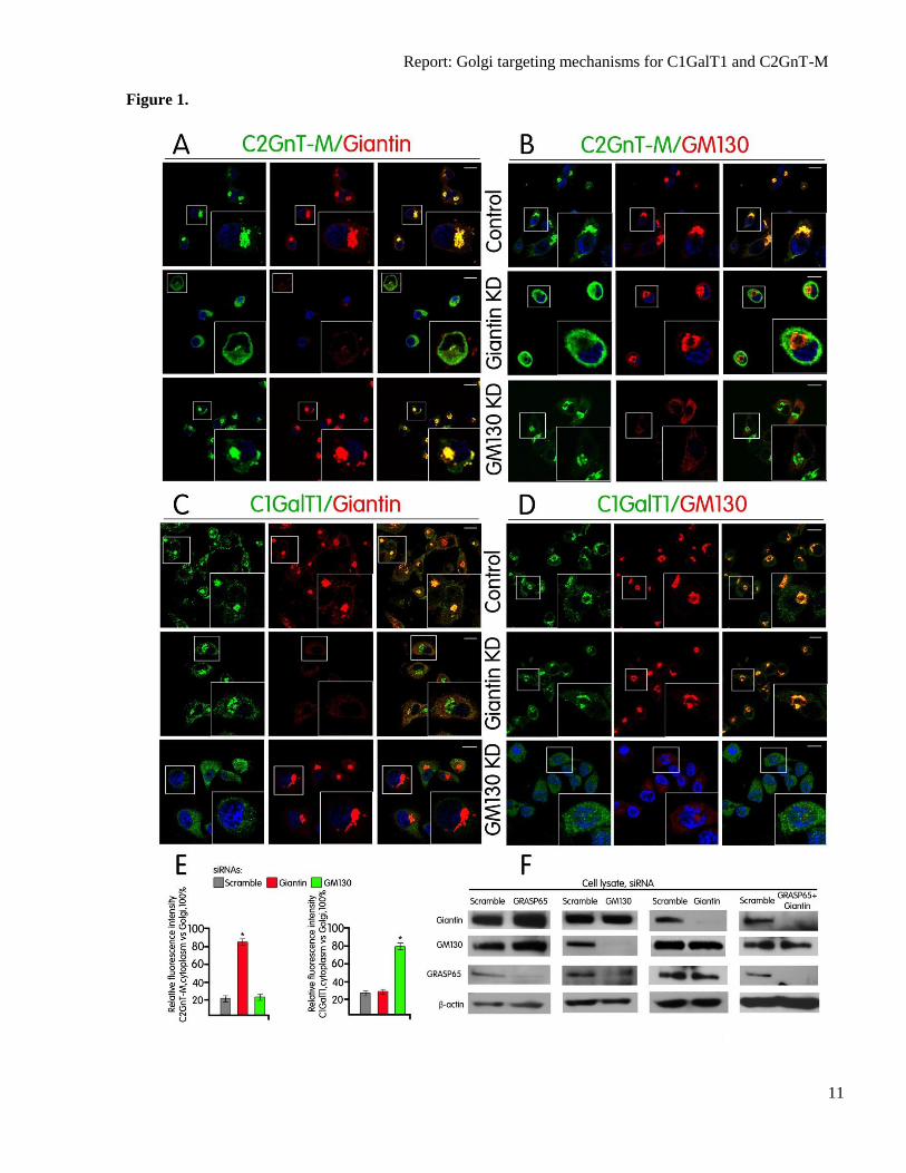
Golgi localization of C2GnT-M (Fig. 1A)
without affecting the Golgi morphology as
shown by immunofluorescence staining of
GM130 (Fig. 1B). Further, KD of GM130 did
not affect Golgi morphology or localization of
C2GnT-M (Fig. 1A & B) (21). Quantification
of the average fluorescence of cytoplasm/Golgi
ratio of C2GnT-M in these cells showed an
increase from 21% in control to 84% in Giantin
KD cells but no change in GM130 KD cells
(Fig. 1E). These data suggest that Giantin and
not GM130 is involved in the Golgi docking of
C2GnT-M-VC.
It is known that GM130 plays an
essential role in mediating fast and complete
incorporation of ER-to-Golgi carriers into the
Golgi stacks (22) independent of Giantin (23).
We found that KD of GM130 prevented Golgi
targeting of C1GalT1 in Panc1-bC2GnT-M
cells (Fig. 1D) without affecting Golgi

Report: Golgi targeting mechanisms for C1GalT1 and C2GnT-M
4
targeting of C2GnT-M (Fig. 1B). KD of
Giantin affected neither Golgi morphology nor
localization of C1GalT1 (Fig. 1B–D).
Quantification of the average fluorescence of
cytoplasm/Golgi ratio of C1GalT1 in these
cells showed a 50% increase in GM130 KD
cells over the control cells but no change in
Giantin KD cells (Fig. 1E). The data suggest
that GM130 and not Giantin is involved in the
docking of C1GalT1-VC.
It has been well established that
GM130 is localized to the Golgi by binding to
a Golgi membrane-associated matrix protein
GRASP65 (17, 18). To confirm that GRASP65
participates in GM130-mediated docking of the
C1GalT1-VC, we examined the Golgi
localization of C1GalT1 after depletion of
GRASP65. To our surprise, C1GalT1 was still
localized to the Golgi after KD of GRASP65
(Fig. 2B). The results suggest that C1GalT1
acquires an alternative Golgi docking
mechanism in the absence of GRASP65. We
found that KD of GM130 led to reduced
GRASP65 (Fig. 1F), which confirmed the
published result (24) that without its binding
partner GM130, GRASP65 was degraded.
Further, KD of GRASP65 resulted in an
increase of not only Giantin and GM130 (Fig.
1F) but also complexes of both GM130-Giantin
and C1GalT1-GM130-Giantin (Fig. 2D and E).
These results led us to hypothesize that these
two Golgi matrix proteins were involved in the
Golgi docking of C1GalT1 when GRASP65
was not available. The hypothesis was proven
by failure of C1GalT1 to target to the Golgi
after KD of both GRASP65 and Giantin (Fig.
2B and C). It is important to point out that prior
to GRASP65 KD, Giantin was not pulled down
by anti-C1GalT1 Ab (Fig. 2D), suggesting that
formation of the C1GalT1-GM130-Giantin
complexes occurred after KD of GRASP65.
Further, GRASP65 was pulled down by anti-
GM130 Ab (Fig. 2E), confirming that GM130
forms complex with GRASP65 in Panc1-
bC2GnT-M cells when GRASP65 is present.
Also, KD of Giantin plus GRASP65 did not
affect the amount of GM130 (Fig. 1F) but
prevented Golgi localization of C1GalT1,
confirming that GM130 alone could not serve
as the Golgi docking site for C1GalT1-VC. We
also noted that the Golgi morphology was not
affected under this condition (supplemental
Fig. S1). Taken together, the results indicate
that GM130-GRASP65 serves as the docking
site for C1GalT1-VC and in the absence of
GRASP65 C1GalT1-VC is targeted to the
Golgi using GM130 and Giantin complex for
docking.
The p115 coiled-coil protein has been
shown to interact with GM130 via its N-
terminal region (16) and bind Giantin via its C-
terminal region (18). The primary role of p115
as a molecule for bridging Giantin and GM130
on Golgi membranes (25), which was not
supported by Sztul and colleagues (11), who
reported that GM130 and Giantin did not
function simultaneously. Nevertheless, the
currently proposed model postulates that these
golgins stimulate p115 binding to Rab1
followed by recruitment of p115 to the Golgi
membrane (11). To further determine whether
p115 is involved in the docking of the Golgi
resident proteins, we performed KD of p115
alone and in combination with GRASP65, and
found that the Golgi localization of both
glycosyltransferases was unaltered (supple-
mental Fig. S2–4). KD of p115 also induced
Golgi morphological changes consistent with
vesiculation at the peri-Golgi area (26) as
demonstrated by immunostaining of Giantin or
GRASP65 (supplemental Fig. S2 and 3).
However, treatment with p115 + GRASP65
siRNAs did not cause an apparent change in
Golgi morphology (supplemental Fig. S4). We
conclude that p115 is not involved in the
golgin-dependent Golgi docking of C2GnT-M-
VC or C1GalT1-VC, and GM130 and Giantin
utilize a p115-independent mechanism to link
with these VCs. Although it is likely that the
GM130-Giantin interaction occurs between 4-6
coiled-coil regions of the Golgi binding site at
the C-terminal region of GM130 and N-
terminal cytoplasmic region of Giantin (27),
details of the interaction of these golgins in the
absence of GRASP65 remain to be elucidated.
Next, we examined the dynamic
intracellular transport of C2GnT-M and
C1GalT1 to the Golgi using time-lapse imaging
in live HEK293 cells which express C2GnT-M-
GFP and C1GalT1-RFP. As shown in Fig. 3A
and supplemental movie S1, many of the
peripheral puncta that contained C2GnT-M-VC
were initially segregated from C1GalT1-VC,
and they remained separated until complete
fusion with the Golgi. To quantify the kinetics
of the formation and fusion of individual VCs,
Report: Golgi targeting mechanisms for C1GalT1 and C2GnT-M
5
we analyzed 90 cells from three independent
experiments and found that C2GnT-M-VC and
C1GalT1-VC were originated in the cytosol
and disappeared at the Golgi surface within 60-
145 s. The shapes of these VCs were ranged
from round to oval with an average diameter of
1.1 ± 0.3 µm for C2GnT-M and 1.2 ± 0.2 µm
for C1GalT1. The sizes of these VCs are quite
different from the 50 to 70 nm sizes reported
for COPI and COPII coated vesicles (28–30). It
is important to note that only 6% of GFP and
RFP-carrying vesicles were totally colocalized
and 7.5% of them were partially colocalized,
while the majority (86.5%) was segregated
(supplemental Fig. S5).
Next, we examined whether C2GnT-
M-VC or C1GalT1-VC used distinct transport
mechanisms by monitoring fluorescence
recovery after photobleaching of a specific
region of the Golgi in cells transiently
expressing C2GnT-M-GFP and C1GalT1-RFP
(Fig. 3B–D). The time-dependent recovery of
the fluorescence in this area represented the
rate of Golgi filling by C2GnT-M-GFP and
C1GalT1-RFP. Fig. 3C and D showed the
distinct kinetics of fluorescence recovery:
C2GnT-M-GFP fluorescence was completely
restored at 168 s, while recovery of C1GalT1-
RFP was completed at 240 s (supplemental
movie S2 and 3).
Golgi targeting of C1GalT1 and C2GnT-M
is independent of ERGIC, COPII, and COPI –
What remains unclear is how are these Golgi
resident enzymes transported to the Golgi. It
has been reported that the ER-to-Golgi
transport of glycosyltransferases requires
ERGIC (31). To examine the involvement of
ERGIC in the Golgi targeting of C1GalT1 and
C2GnT-M, we determined the non-Golgi vs.
Golgi distribution of these two enzymes after
KD of ERGIC-53, a cargo transport receptor
for glycoproteins (32). We found that C1GalT1
and C2GnT-M were localized to the Golgi after
KD of ERGIC-53 (supplemental Fig. S6),
suggesting that VCs carrying these two
enzymes utilized ERGIC-independent process
for Golgi targeting.
COPI and COPII vesicles are engaged in
the ER-to-Golgi transport: COPII in the ER to
ERGIC step while COPI in the ERGIC to cis-
Golgi step (33). However, the ER-to-Golgi
transport does not depend exclusively on COPI
and COPII vesicles because long-range tubular
transport carriers shuttling between ER and
Golgi bi-directionally have been reported.
These non-coated ribosome-free tubules and
vesicles up to 500 nm in length were found in
the pre-Golgi intermediates (34). This transport
mechanism requires intact microtubules and
occurs independent of COPI or COPII coats
(35, 36). To examine the involvement of COPII
in the Golgi targeting of these two enzymes, we
monitored the Golgi localization of both
C1GalT1 and C2GnT-M after KD of Sar1a or
Sar1b, which are GTPases regulating the
assembly and disassembly of COPII coats (28).
The detection of both enzymes in the Golgi
(supplemental Fig. S7) indicates that Golgi
targeting of both Golgi resident proteins is
COPII-independent. Our data fit well with the
observation that non-golgin Golgi proteins are
involved in the docking of COPII (37). The
result is at variance with a previous report (38)
which claims that COPII is involved in the
Golgi targeting of glycosyltransferases. In this
study, the employment of a Sar1 mutant may
have compromised the data interpretation
because this Sar 1 mutant causes alterations of
the Golgi morphology. The COPII-independent
ER-to-Golgi trafficking of C2GnT-M and
C1GalT1 was further supported by unaltered
COPII distribution after KD of Giantin or
GM130 (supplemental Fig. S8).
Previously, we showed that KD of β-
COP, a COPI component (39), did not affect
intra-Golgi localization of C2GnT-M (9), and
herein we show that KD of β-COP also did not
change the Golgi localization of C1GalT1
(supplemental Fig. S9). Further, the Golgi-to-
ER trafficking of the Golgi resident proteins is
a non-muscle myosin IIA-dependent and
COPI-independent process (9), and formation
of COPI vesicles occurs on the Golgi
membrane (40). Therefore, the reported
involvement of β-COP in the ER-to-Golgi
transport (41) may represent its regular
transportation to the Golgi membrane where it
interacts with ADP-ribosylation factor 1 and
other subunits to form COPI vesicles for intra-
Golgi transport (42). However, the possibility
that the COPI subunits are transported to the
Golgi by glycosyltransferase-specific VCs
cannot be ruled out (33).
Our results show that upon depletion of
GM130, β-COP (i.e. COPI vesicles) but not
p115 was present in the Golgi (supplemental
Report: Golgi targeting mechanisms for C1GalT1 and C2GnT-M
6
Fig. S10). Also, KD of either Giantin or
GRASP65 did not affect p115 localization, and
same result was obtained after KD of Sar1a or
Sar1b (supplemental Fig. S10). These data
indicate that p115 is likely not involved in the
ER-to-Golgi trafficking of many Golgi
proteins. The data also suggest that the
transport functions of p115-free COPI are
different from those of p115-bound COPI (43).
In summary, our data show that the
ER-to-Golgi transportation is different for
C2GnT-M and C1GalT1 by not only their VCs
but also their docking sites on the Golgi (Fig.
3E). C1GalT1 is a unique glycosyltransferase
because it is the enzyme that controls the
synthesis of core 1 and core 2-associated
glycans, which constitute the majority of mucin
glycans. It is thus not a surprise that this
enzyme has its own molecular chaperone
Cosmc (44), a unique ER-to-Golgi transport
strategy and multiple Golgi docking
mechanisms. Taken together, our results have
revealed novel Golgi targeting mechanisms for
different glycosyltransferases. It should be
mentioned that C1GalT1 lacks N-glycosylation
site while bC2GnT-M is N-glycosylated (45).
Given that processing of N-glycans starts at the
cis-Golgi (46), it is reasonable to assume that
VCs that carry glycoproteins decorated with N-
glycans use Giantin as a major docking site. On
the other hand, VCs that carry glycoproteins
without N-glycans or proteins without glycans
utilize GM130-dependent docking site(s).
Peptidyl GalNAc T2, which contains N-
glycosylation sites, was reported to target to the
Golgi independent of GM130 (24), which
supports the selectivity of GM130 as a Golgi
docking site for VCs that carry
glycosyltransferases without N-glycans.
Additional evidence that supports the proposed
hypothesis and answers to the question of the
chemical nature and transport mechanisms of
these VCs remain to be discovered.
FUNDING
This work is supported in part by the Office of
Research and Development, Medical Research
Service, Department of Veterans Affairs (VA
1I1BX000985), the National Institutes of
Health (1R21HL097238 and 2RO1HL48282)
and the State of Nebraska (LB506).
The abbreviations used are: KD, knockdown;
VCs, vesicular complexes; C1GalT1, core 1 β3
galactosyltransferase; C2GnT-2/M, core 2 N-
acetylglucosaminyltransferase 2/M; C1GalT1-
VC, C1GalT-1-carrying vesicular complex;
C2GnT-M-VC, C2GnT-M -carrying vesicular
complex; ERGIC, ER-to-Golgi intermediate
compartment; Sar1, Secretion-associated RAS-
related protein 1; COPI/II, coat protein
complex I/II; GRASP65, Golgi ReAssembly
Stacking Protein 65; SNARE, soluble N-
ethylmaleimide sensitive factor attachment
protein receptor.
Report: Golgi targeting mechanisms for C1GalT1 and C2GnT-M
7
REFERENCES
1. Hanisch, F. G. (2001) O-glycosylation of the mucin type. Biol. Chem. 382, 143–149
2. Ju, T., Brewer, K., D'Souza, A., Cummings, R. D., and Canfield, W. M. (2002) Cloning and
expression of human core 1 beta1,3-galactosyltransferase. J. Biol. Chem. 277, 178–186
3. Cheng, P-W., and Radhakrishnan, P. (2011) Mucin glycan branching enzymes: structure, function
and gene regulation. In: Wu A, editor. Molecular immunology of complex carbohydrates-3. Advances
in Experimental Medicine and Biology, vol. 705. pp 465–492. N.Y.: Plenum Press, New York, USA
4. Xia, L., Ju ,T., Westmuckett, A., An, G., Ivanciu, L., McDaniel, J. M., Lupu, F., Cummings, R. D.,
and McEver, R. P. (2004) Defective angiogenesis and fatal embryonic hemorrhage in mice lacking
core 1-derived O-glycans. J. Cell Biol. 164, 451–459
5. Huang, M. C., Chen, H. Y., Huang, H. C., Huang, J., Liang, J. T., Shen, T. L., Lin, N. Y., Ho, C. C.,
Cho, I. M., and Hsu, S. M. (2006) C2GnT-M is downregulated in colorectal cancer and its re-
expression causes growth inhibition of colon cancer cells. Oncogene 25, 3267–3276
6. Stone, E. L., Ismail, M. N., Lee, S. H., Luu, Y., Ramirez, K., Haslam, S. M., Ho, S. B., Dell, A.,
Fukuda, M., and Marth, J. D. (2009) Glycosyltransferase function in core 2-type protein O
glycosylation. Mol. Cell Biol. 29, 3770–3782
7. Colley, K. J. (1997) Golgi localization of glycosyltransferases: more questions than answers.
Glycobiology 7, 1–13
8. Tu, L., and Banfield, D. K. (2010) Localization of Golgi-resident glycosyltransferases. Cell Mol.
Life Sci. 67, 29–41
9. Petrosyan, A., Ali, M. F., Verma, S. K., Cheng, H., and Cheng, P-W. (2012) Non-muscle myosin
IIA transports a Golgi glycosyltransferase to the endoplasmic reticulum by binding to its cytoplasmic
tail. Int. J. Biochem. Cell Biol. 44, 1153–1165
10. Appenzeller-Herzog, C., and Hauri, H. P. (2006) The ER-Golgi intermediate compartment
(ERGIC): in search of its identity and function. J. Cell Sci. 1, 2173–2183
11. Alvarez, C., Garcia-Mata., R, Hauri, H. P., and Sztul, E. (2001) The p115-interactive proteins
GM130 and giantin participate in endoplasmic reticulum-Golgi traffic. J. Biol. Chem. 276, 2693–2700
12. Rosing, M., Ossendorf, E., Rak, A., and Barnekow, A. (2007) Giantin interacts with both the small
GTPase Rab6 and Rab1. Exp Cell Res. 313, 2318–2325
13. Moyer, B. D., Allan, B. B., and Balch, W. E. (2001) Rab1 interaction with a GM130 effector
complex regulates COPII vesicle cis-Golgi tethering. Traffic 2, 268–276
14. Shorter, J., Beard, M. B., Seemann, J., Dirac-Svejstrup, A. B., and Warren, G. (2002) Sequential
tethering of Golgins and catalysis of SNAREpin assembly by the vesicle-tethering protein p115. J.
Cell Biol. 157, 45–62
15. Linstedt, A. D., and Hauri, H. P. (1993) Giantin, a novel conserved Golgi membrane protein
containing a cytoplasmic domain of at least 350 kDa. Mol. Biol. Cell 4, 679–693
16. Nakamura, N., Lowe, M., Levine, T. P., Rabouille, C., and Warren, G. (1997) The vesicle docking
protein p115 binds GM130, a cis-Golgi matrix protein, in a mitotically regulated manner. Cell 89,
445–455
17. Barr, F. A., Puype, M., Vandekerckhove, J., and Warren, G. (1997) GRASP65, a protein involved
in the stacking of Golgi cisternae. Cell 91, 253–262
18. Lesa, G. M., Seemann, J., Shorter, J., Vandekerckhove, J., and Warren, G. (2000) The amino-
terminal domain of the golgi protein giantin interacts directly with the vesicle-tethering protein p115. J
Biol. Chem. 275, 2831–2836
19. Narimatsu, Y., Ikehara, Y., Iwasaki, H., Nonomura, C., Sato, T., Nakanishi, H., and Narimatsu H.
(2008) Immunocytochemical analysis for intracellular dynamics of C1GalT associated with molecular
chaperone, Cosmc. Biochem. Biophys. Res. Commun. 366, 199–205
20. Choi, K. H., Basma, H., Singh, J., and Cheng, P-W. (2005) Activation of CMV promoter-
controlled glycosyltransferase and beta-galactosidase glycogenes by butyrate, tricostatin A, and 5-aza-
2'-deoxycytidine. Glycoconj. J. 22, 63–69
Report: Golgi targeting mechanisms for C1GalT1 and C2GnT-M
8
21. Kodani, A., and Sütterlin, C. (2008) The Golgi protein GM130 regulates centrosome morphology
and function. Mol. Biol. Cell 19, 745–753.
22. Marra, P., Maffucci, T., Daniele, T., Tullio, G. D., Ikehara, Y., Chan, E. K., Luini, A.,
Beznoussenko, G., Mironov, A., and De Matteis, M. A. (2001) The GM130 and GRASP65 Golgi
proteins cycle through and define a subdomain of the intermediate compartment. Nat. Cell. Biol. 3,
1101–1113
23. Weide, T., Bayer, M., Köster, M., Siebrasse, J. P., Peters, R., and Barnekow, A. (2001) The Golgi
matrix protein GM130: a specific interacting partner of the small GTPase rab1b. EMBO Rep. 2, 336–
341
24. Puthenveedu, M. A., Bachert, C., Puri, S., Lanni, F., and Linstedt, A. D. (2006) GM130 and
GRASP65-dependent lateral cisternal fusion allows uniform Golgi-enzyme distribution. Nat. Cell
Biol. 8, 238–248
25. Sönnichsen, B., Lowe, M., Levine, T., Jämsä, E., Dirac-Svejstrup, B., and Warren, G. (1998) A
role for giantin in docking COPI vesicles to Golgi membranes. J. Cell Biol. 140, 1013–1021
26. Puthenveedu, M. A., and Linstedt, A. D. (2004) Gene replacement reveals that p115/SNARE
interactions are essential for Golgi biogenesis. Proc. Natl. Acad. Sci. U S A 101, 1253–1256
27. Nakamura, N. (2010) Emerging new roles of GM130, a cis-Golgi matrix protein, in higher order
cell functions. J. Pharmacol. Sci. 112, 255–264
28. Fromme, J. C., and Schekman, R. (2005) COPII-coated vesicles: flexible enough for large cargo?
Curr. Opin. Cell Biol. 17, 345–352
29. Nickel, W., Brügger, B., and Wieland, F. T. (2002) Vesicular transport: the core machinery of
COPI recruitment and budding. J. Cell Sci. 115, 3235–3240
30. Stephens, D. J., and Pepperkok, R. (2001) Illuminating the secretory pathway: when do we need
vesicles? J. Cell Sci. 114, 1053–1059
31. Marra, P., Salvatore, L., Mironov, A. Jr., Di Campli, A., Di Tullio, G., Trucco, A., Beznoussenko,
G., Mironov, A., and De Matteis, M. A. (2007) The biogenesis of the Golgi ribbon: the roles of
membrane input from the ER and of GM130. Mol. Biol. Cell 18, 1595–1608
32. Hauri, H. P., Kappeler, F., Andersson, H., and Appenzeller, C. (2000) ERGIC-53 and traffic in the
secretory pathway. J. Cell Sci. 113, 587–596
33. Scales, S. J., Pepperkok, R., and Kreis, T. E. (1997) Visualization of ER-to-Golgi transport in
living cells reveals a sequential mode of action for COPII and COPI. Cell 90, 1137–1148
34. Fan, J. Y., Roth, J., and Zuber, C. (2003) Ultrastructural analysis of transitional endoplasmic
reticulum and pre-Golgi intermediates: a highway for cars and trucks. Histochem. Cell Biol. 120, 455–
463
35. Martínez-Alonso, E., Egea, G., Ballesta, J., and Martínez-Menárguez, J. A. (2005) Structure and
dynamics of the Golgi complex at 15 degrees C: low temperature induces the formation of Golgi-
derived tubules. Traffic 6, 32–44
36. Simpson, J. C., Nilsson, T., and Pepperkok, R. (2005) Biogenesis of tubular ER-to-Golgi transport
intermediates. Mol. Biol. Cell 17, 723–737
37. Cao, X., Ballew, N., and Barlowe, C. (1998) Initial docking of ER-derived vesicles requires Uso1p
and Ypt1p but is independent of SNARE proteins. EMBO J. 17, 2156–2165
38. Quintero, C. A., Giraudo, C. G., Villarreal, M., Montich, G., Maccioni, H. J. (2010) Identification
of a site in Sar1 involved in the interaction with the cytoplasmic tail of glycolipid glycosyltransferases.
J. Biol. Chem. 285, 30340–30346
39. Waters, M. G., Serafini, T., and Rothman, J. E. (1991) 'Coatomer': a cytosolic protein complex
containing subunits of non-clathrin-coated Golgi transport vesicles. Nature 349, 248–251
40. Palmer, D. J., Helms, J. B., Beckers, C. J., Orci, L., and Rothman, J. E. (1993) Binding of
coatomer to Golgi membranes requires ADP-ribosylation factor. J. Biol. Chem. 268, 12083–12089
41. Peter, F., Plutner, H., Zhu, H., Kreis, T. E., and Balch, W. E. (1993) Beta-COP is essential for
transport of protein from the endoplasmic reticulum to the Golgi in vitro. J. Cell Biol. 122, 1155–1167
42. Lanoix, J., Ouwendijk, J., Lin, C. C., Stark, A., Love, H. D., Ostermann, J., and Nilsson, T. (1999)
GTP hydrolysis by arf-1 mediates sorting and concentration of Golgi resident enzymes into functional
COP I vesicles. EMBO J. 18, 4935–4948

Report: Golgi targeting mechanisms for C1GalT1 and C2GnT-M
9
43. Malsam, J., Satoh, A., Pelletier, L., and Warren, G. (2005) Golgin tethers define subpopulations of
COPI vesicles. Science 307, 1095–1098
44. Ju, T., and Cummings, R. D. (2002) A unique molecular chaperone Cosmc required for activity of
the mammalian core 1 beta 3-galactosyltransferase. Proc. Natl. Acad. Sci. U S A 99, 16613–16618
45. Singh, J., Khan, G. A., Kinarsky, L., Cheng, H., Wilken, J., Choi, K. H., Bedows, E., Sherman, S.,
and Cheng, P. W. (2004) Identification of disulfide bonds among the nine core 2 N-
acetylglucosaminyltransferase-M cysteines conserved in the mucin beta6-N-
acetylglucosaminyltransferase family. J. Biol. Chem. 279, 38969–38977
46. Beyer, T. A., and Hill, R. L. (1982) The Glycoconjugates (Horowitz, M., ed) Vol. III, Part A, pp.
25–45, Academic Press, New York
Report: Golgi targeting mechanisms for C1GalT1 and C2GnT-M
10
FIGURE LEGENDS
Fig. 1. VCs carrying C2GnT-M utilize Giantin while C1GalT1-VC employs GM130 for Golgi
targeting. (A–D) Confocal immunofluorescence images of Panc1-bC2GnT-M (c-Myc) cells labeled
with green (anti-c-Myc Ab, A and B; anti-C1GalT1 Ab, C and D) and red (anti-Giantin Abs, A and C
and anti-GM130 Ab, B and D) fluorescence after treatment with scramble siRNA or siRNAs specific
for Giantin or GM130. Representative cells showing KD of Giantin are in the second row of A and B
or C and D. Representative cells showing KD of GM130 are in the third row A and B or C and D.
White boxes indicate areas enlarged and shown in the inset. Scale bar, 10 µm. (E) Quantification of
C2GnT-M and C1GalT1 immunofluorescence signals of non-Golgi vs. Golgi (=100%) in cells treated
with scramble siRNA or protein-specific siRNA as shown in A and B, and C and D. The data are
shown as average of three experiments, mean ± s.e.m.. *– P<0.001. (F) Giantin, GM130, and
GRASP65 western blots of the lysates of Panc1-bC2GnT-M (c-Myc) cells treated with scramble
siRNA or siRNA specific for Giantin, GM130, GRASP65, or GRASP65 plus Giantin.
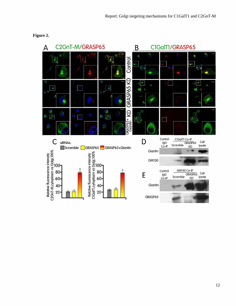
Fig. 2. (A and B) C1GalT1-VC utilizes GM130 and GRASP65 in the presence of GRASP65 but
GM130 and Giantin in the absence of GRASP65 for Golgi targeting. Confocal immunofluorescence
images of Panc1-bC2GnT-M (c-Myc) cells labeled with green (anti-c-Myc Ab, A; anti-C1GalT1 Ab,
B) and red (anti-GRASP65 Ab) fluorescence after treatment with scramble siRNA or siRNA specific
for GRASP65 or GRASP65+Giantin siRNAs. Scale bar, 10 µm. (C) Quantification of C2GnT-M and
C1GalT1 immunofluorescence signals of non-Golgi vs. Golgi (=100%) in cells treated with scramble
siRNA or protein-specific siRNA as shown in A and B. (D) Giantin and GM130 western blots of the
C1GalT1 immunoprecipitates from the lysates of Panc1-bC2GnT-M (c-Myc) cells treated with
scramble or GRASP65-specific siRNA. Knockdown of GRASP65 induced the formation of C1GalT1-
Giantin-GM130 complex. (E) Giantin and GRASP65 western blot of the GM130 immunoprecipitate
from the lysates of Panc1-bC2GnT-M (c-Myc) cells treated with scramble or GRASP65-specific
siRNA. GM130 forms complex with GRASP65, as shown in the control cells, and KD of GRASP65
increased GM130-Giantin complex. Equal amounts of proteins in the lysates were used for Co-IP as
shown by the β-actin blot in Fig. 1F.
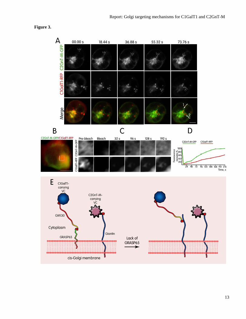
Fig. 3. (A) Direct visualization of C2GnT-M-GFP-VC and C1GalT1-RFP-VC dynamics in live
HEK293 cells. Individual images from the movie of cells expressing C2GnT-M-GFP and C1GalT1-
RFP over time frame 00.00–73.76 s; the positions of anterograde ER-to-Golgi spots are indicated by
arrows. Note that C2GnT-M-GFP-VCs and C1GalT1-RFP-VCs originating from opposite sides of cell
periphery move toward and fuse with Golgi. The dotted lines at 73.76 s indicate the trajectory of
Golgi-targeting C2GnT-M-GFP-VC (1) and C1GalT1-RFP-VC (2). Scale bar, 5 µm. (B-D) Time-
dependent recovery of green (C2GnT-M-GFP) and red (C1GalT1-RFP) fluorescence in the Golgi after
photobleaching. The part of the Golgi (white box in B and highlighted in C) was simultaneously GFP-
and RFP-bleached using the 488-nm and 543-nm laser pulse, respectively. The images of fluorescence
signal were recorded every 8 s to determine recovery of fluorescence intensity. Restoration of C2GnT-
M-GFP fluorescence was completed at 168 s, while recovery of C1GalT1-RFP was substantially
delayed. Representative images of the indicated times are shown on C. (D) Quantification of C2GnT-
M-GFP and C1GalT1-RFP within 216 s after photobleaching. The ratio of fluorescence
(bleached/unbleached) was measured for every 24 s and values were normalized to those of the
prebleached. (E) Proposed model of the interaction of distinct VCs with cis-Golgi membrane before
and after GRASP65 depletion. C2GnT-M-VCs reach the Golgi through association with Giantin,
which is independent of ERGIC-53, COPI, COPII, GM130, or p115, while C1GalT1-VCs preferably
use the GM130–GRASP65 complex, which is also independent of ERGIC-53, COPI, COPII, Giantin,
or p115. In the absence of GRASP65, C1GalT1-VC switch to GM130–Giantin site for Golgi docking.
This new association accompanies elevated amounts of GM130 and Giantin. For simplicity, Giantin,
GM130 and GRASP65 are shown as monomers. Red stars indicate complex formed between golgins
and C2GnT-M-VC or C1GalT1-VCs.
Report: Golgi targeting mechanisms for C1GalT1 and C2GnT-M
11
Figure 1.
Report: Golgi targeting mechanisms for C1GalT1 and C2GnT-M
12
Figure 2.
Report: Golgi targeting mechanisms for C1GalT1 and C2GnT-M
13
Figure 3.
Copyright © 2022 FDOKUMEN