Abrogation of the Fcy Receptor IIA-mediated - NCBI
12
Molecular Biology of the Cell Vol. 7, 1095-1106, July 1996 Abrogation of the Fcy Receptor IIA-mediated Phagocytic Signal by Stem-Loop Syk Antisense Oligonucleotides Mitsuhiro Matsuda,*t Jong-Gu Park,l§ Da-Cheng Wang,* Sharon Hunter,* Paul Chien,* and Alan D. Schreiber*§ *Hematology and Oncology Division, University of Pennsylvania School of Medicine, Philadelphia, Pennsylvania 19104; and IKRIBB, KIST, Yusong, Taejon 305-600, Republic of Korea Submitted November 30, 1995; Accepted May 8, 1996 Monitoring Editor: Keith R. Yamamoto The role of Syk kinase in Fcy receptor (FcyR) IIA-mediated phagocytosis was examined with two forms of antisense oligodeoxynucleotides (ODNs) designed to hybridize to human Syk mRNA. Monocytes were incubated with linear and stem-loop antisense ODNs targeted to Syk mRNA. When complexed with cationic liposomes, stem-loop Syk antisense ODN with phosphorothioate modification exhibited stability in fetal bovine and human serum. The stem-loop Syk antisense ODN at a concentration of 0.2 ,uM inhibited FcyRIIA-mediated phagocytosis by 90% and completely eliminated Syk mRNA and protein in monocytes, whereas scrambled-control ODNs had no effect. The Syk antisense ODNs did not change ,B-actin mRNA levels and FcyRII cell-surface expression. In addition, stem-loop Syk antisense ODN inhibited FcyRI and Fc-yRIIIA-mediated phagocytosis. These data indicate the efficacy of stem-loop Syk antisense ODN for targeting and degrading Syk mRNA and protein and the importance of Syk kinase in Fcy receptor-mediated phagocytosis. Immunoblotting assay demonstrated that FcyRII ty- rosine phosphorylation after FcyRII cross-linking did not change in the absence of Syk protein. These results indicate that Syk kinase is required for Fc-yRIIA-mediated phago- cytic signaling and that FcyRII cross-linking leads to tyrosine phosphorylation of FcyRII independent of Syk kinase. INTRODUCTION Receptors for the Fc portion of immunoglobulin G (IgG)' are expressed on many cells of hematopoietic lineage (Ravetch and Kinet, 1991; van de Winkel and Anderson, 1991; McKenzie and Schreiber, 1994; Indik et al., 1995b). These receptors are important in host defense and ingestion of IgG-coated cells. Three classes of Fcy receptors have been described: Fc-yRI, FcyRII, and FcyRI1, distinguished by IgG subclass affinity, cellular distribution, and reactivity with monoclonal antibodies (Ravetch and Kinet, 1991; t Present address: Third Department of Internal Medicine, Kinki University School of Medicine, 377-2 Ohno-Higashi, Osaka- Sayama, Osaka 589, Japan. § Corresponding authors: 7 Silverstein, University of Pennsylva- nia School of Medicine, 3400 Spruce Street, Philadelphia, PA 19104. McKenzie and Schreiber, 1994; Indik et al., 1995b). The 40-kDa glycoprotein FcyRII has low affinity for mono- meric ligand and interacts only with polymeric IgG (Stuart et al., 1987; Hibbs et al., 1988). FcyRII is ex- pressed on a wide variety of cells and frequently rep- resents the sole Fcy receptor class expressed on hema- topoietic cells (e.g., B lymphocytes, eosinophils, and platelets). Human FcyRII is encoded by three genes: FcyRIIA, IIB, and IIC (Brooks et al., 1989; Cassel et al., 1993). The cytoplasmic domain of FcyRIIA contains two YxxL sequences, similar to those found in other Ig gene family receptors. The YxxL sequences in the cy- Abbreviations used: EA, IgG-sensitized red blood cells; FcyR, Fc-y receptor; IgG, immunoglobulin G; ODN, oligodeoxynucle- otides; PI, phagocytic index; RBCs, red blood cells; RT-PCR, reverse-transcribed polymerase chain reaction. (C 1996 by The American Society for Cell Biology 1095
-
Upload
khangminh22 -
Category
Documents
-
view
1 -
download
0
Transcript of Abrogation of the Fcy Receptor IIA-mediated - NCBI
Molecular Biology of the CellVol. 7, 1095-1106, July 1996
Abrogation of the Fcy Receptor IIA-mediatedPhagocytic Signal by Stem-Loop Syk AntisenseOligonucleotidesMitsuhiro Matsuda,*t Jong-Gu Park,l§ Da-Cheng Wang,* Sharon Hunter,*Paul Chien,* and Alan D. Schreiber*§
*Hematology and Oncology Division, University of Pennsylvania School of Medicine, Philadelphia,Pennsylvania 19104; and IKRIBB, KIST, Yusong, Taejon 305-600, Republic of Korea
Submitted November 30, 1995; Accepted May 8, 1996Monitoring Editor: Keith R. Yamamoto
The role of Syk kinase in Fcy receptor (FcyR) IIA-mediated phagocytosis was examinedwith two forms of antisense oligodeoxynucleotides (ODNs) designed to hybridize tohuman Syk mRNA. Monocytes were incubated with linear and stem-loop antisenseODNs targeted to Syk mRNA. When complexed with cationic liposomes, stem-loop Sykantisense ODN with phosphorothioate modification exhibited stability in fetal bovineand human serum. The stem-loop Syk antisense ODN at a concentration of 0.2 ,uMinhibited FcyRIIA-mediated phagocytosis by 90% and completely eliminated Syk mRNAand protein in monocytes, whereas scrambled-control ODNs had no effect. The Sykantisense ODNs did not change ,B-actin mRNA levels and FcyRII cell-surface expression.In addition, stem-loop Syk antisense ODN inhibited FcyRI and Fc-yRIIIA-mediatedphagocytosis. These data indicate the efficacy of stem-loop Syk antisense ODN fortargeting and degrading Syk mRNA and protein and the importance of Syk kinase in Fcyreceptor-mediated phagocytosis. Immunoblotting assay demonstrated that FcyRII ty-rosine phosphorylation after FcyRII cross-linking did not change in the absence of Sykprotein. These results indicate that Syk kinase is required for Fc-yRIIA-mediated phago-cytic signaling and that FcyRII cross-linking leads to tyrosine phosphorylation of FcyRIIindependent of Syk kinase.
INTRODUCTION
Receptors for the Fc portion of immunoglobulin G(IgG)' are expressed on many cells of hematopoieticlineage (Ravetch and Kinet, 1991; van de Winkel andAnderson, 1991; McKenzie and Schreiber, 1994; Indiket al., 1995b). These receptors are important in hostdefense and ingestion of IgG-coated cells. Threeclasses of Fcy receptors have been described: Fc-yRI,FcyRII, and FcyRI1, distinguished by IgG subclassaffinity, cellular distribution, and reactivity withmonoclonal antibodies (Ravetch and Kinet, 1991;
t Present address: Third Department of Internal Medicine, KinkiUniversity School of Medicine, 377-2 Ohno-Higashi, Osaka-Sayama, Osaka 589, Japan.
§ Corresponding authors: 7 Silverstein, University of Pennsylva-nia School of Medicine, 3400 Spruce Street, Philadelphia, PA19104.
McKenzie and Schreiber, 1994; Indik et al., 1995b). The40-kDa glycoprotein FcyRII has low affinity for mono-meric ligand and interacts only with polymeric IgG(Stuart et al., 1987; Hibbs et al., 1988). FcyRII is ex-pressed on a wide variety of cells and frequently rep-resents the sole Fcy receptor class expressed on hema-topoietic cells (e.g., B lymphocytes, eosinophils, andplatelets). Human FcyRII is encoded by three genes:FcyRIIA, IIB, and IIC (Brooks et al., 1989; Cassel et al.,1993).The cytoplasmic domain of FcyRIIA contains two
YxxL sequences, similar to those found in other Iggene family receptors. The YxxL sequences in the cy-
Abbreviations used: EA, IgG-sensitized red blood cells; FcyR,Fc-y receptor; IgG, immunoglobulin G; ODN, oligodeoxynucle-otides; PI, phagocytic index; RBCs, red blood cells; RT-PCR,reverse-transcribed polymerase chain reaction.
(C 1996 by The American Society for Cell Biology 1095
M. Matsuda et al.
toplasmic domain of FcyRIIA account for its ability tomediate phagocytosis and tyrosine phosphorylation(Indik et al., 1994b; Mitchell et al., 1994). Two otherisoforms of FcyRII, FcyRIIB1 and FcyRIIB2, containonly a single YxxL sequence and do not mediatephagocytosis (Indik et al., 1994b).
Fc,yRII cross-linking results in the tyrosine phos-phorylation of multiple proteins, including the recep-tor itself (Huang et al., 1992; Scholl et al., 1992; Rankinet al., 1993; Ghazizadeh and Fleit, 1994), and inducesrapid hydrolysis of phosphatidylinositol 4,5-diphos-phate by PLC-,yl (Liao et al., 1992). Pretreatment ofcells with tyrosine kinase inhibitors substantially re-duces FcyRII-mediated effector functions, includingcalcium mobilization, tyrosine phosphorylation, andphagocytosis (Liao et al., 1992; Scholl et al., 1992;Rankin et al., 1993; Ghazizadeh and Fleit, 1994; Mitch-ell et al., 1994).The protein tyrosine kinase Syk is expressed in sev-
eral hematopoietic cells, including monocytes / macro-phages and platelets (Taniguchi et al., 1991; Law et al.,1994; Yagi et al., 1994). The induction of Syk phosphor-ylation by cross-linking of FcyRII further suggests thatSyk plays an important role in phagocytic signalingthrough FcyRIIA (Agarwal et al., 1993; Kiener et al.,1993; Greenberg et al., 1994; Hunter et al., 1994; Pan etal., 1994; Ghazizadeh et al., 1995; Indik et al., 1995a). Ina model system Syk kinase enhances the phagocyticsignal induced by FcyRIIA and increases the numberof cells able to mediate phagocytosis (Indik et al.,1995a), but the critical position of Syk kinase in thesignal cascade mediated by FcyRIIA is still unclear.Antisense oligodeoxynucleotides (ODNs) designed
to hybridize to specific mRNA sequences have beenused to inhibit the expression of a number of cellularand viral genes (Chiang et al., 1991; Milligan et al.,1993; Stein and Cheng, 1993; Wagner et al., 1993, 1994).Binding of antisense ODNs to their complementarysequences leads to either degradation of target mRNAby the RNaseH activity or blockage of the ribosomaltranslation of target mRNA (Milligan et al., 1993; Steinand Cheng, 1993; Wagner, 1994). The development ofeffective antisense techniques allows investigators toaddress questions regarding gene function (Chiang etal., 1991; Milligan et al., 1993; Stein and Cheng, 1993;Wagner et al., 1993, 1994). We have designed threelinear ODNs and a stem-loop antisense ODN for hu-man Syk mRNA. The stem-loop antisense ODN wasdesigned to hybridize to three different sites of humanSyk mRNA. These Syk antisense ODNs were used toinvestigate the role of Syk kinase in the Fc-yRIIA-mediated phagocytic signal in cultured monocytes.We report that the stem-loop antisense ODN structuremay provide an improved approach to target mRNAand protein. By using this approach, we demonstratethat the protein tyrosine kinase Syk is required inFcyRIIA-mediated phagocytic signaling and examine
the requirement for Syk kinase in Fcy receptor ty-rosine phosphorylation.
MATERIALS AND METHODS
Construction of Antisense ODNsAntisense and scrambled-control ODNs were modified to be pro-tected from nuclease digestion. One phosphodiester backbone at the5-prime terminus and two at the 3-prime terminus were modifiedwith phosphorothioates. Prediction of secondary structures of SykmRNA (Law et al., 1994) and ODNs was performed with the MacD-NASIS program (Hitachi Software, San Bruno, CA). Three linearSyk antisense ODNs were designed. Target-I linear Syk antisenseODN (5'-CTGTCAGCCATGCCG-3', nucleotides 159-173, accord-ing to the nucleotide numbering of Law et al., 1994) targets the areasurrounding the translation initiation codon of Syk mRNA. Tar-get-II (5'-GCTTCTTGAGGAG-3') and target-III (5'-TGTCTTGTCT-TTGTC-3') linear Syk antisense ODNs are complementary to nucle-otides 451-463 and nucleotides 802-816, respectively. Stem-loopSyk antisense ODN is a 57-mer-containing sequence complemen-tary to these three target sites (Figure 1). The stem-loop Syk anti-sense ODN forms a stem and loop structure by itself (Figure 2). Thestem domain, seven nucleotides in length, is formed by complemen-tary 5- and 3-prime terminal sequences with nucleotide contents ofonly Gs and Cs. The loop domain consists of three antisense se-quences complementary to targets I, II, and III. Three differentantisense sequences were tandemly joined in 5' to 3' order fortargets I, III, and II to contain minimum intramolecular secondarystructure in the loop domain (Figure 2). The sequence of the stem-loop Syk antisense ODN is 5'-GGGGGGGCTGTCAGCCATGCCG-TGTCTTGTCTTTGTCGCTTCTTGAGGAGCCCCCCC-3'. The lin-ear scrambled-control ODN is a 15 mer with the sequence 5'-CCAAGATGATTCCAG-3', and the stem-loop scrambled-controlODN is a 57 mer with 7 Gs and 7 Cs within the stem domain and ascrambled sequence 5'-ATGGAATCATCTTGGGCATTCATTCGT-TCCTCAAAGAAGAATATG-3' within the loop domain. The linearand stem-loop control ODNs were also modified at both the5-prime and 3-prime termini by phosphorothioates. All ODNs weresynthesized by the Nucleic Acid Facility, Department of Chemistry,University of Pennsylvania Cancer Center (Philadelphia, PA).
Stability of Linear and Stem-Loop SykAntisense ODNsOne microgram each of the nonspecific control-phosphodiesterODN and the specific target-I linear and stem-loop Syk antisenseODNs in 50 ,ll of phosphate-buffered saline (PBS) were incubatedwith 2 jig (1 j,l) of Lipofectamine (Life Technologies, Gaithersburg,MD). The ODN-liposome complexes were allowed to form at roomtemperature for 45 min. An equal volume of PBS containing non-heat-inactivated fetal calf serum (FCS; Intergen, Purchase, NY) orhuman serum from a healthy individual was added to each ODN-liposome mixture to a final concentration of 10%, 33% FCS, or 50%human serum and incubated at 37'C for 16 h. ODNs (1 j,g) withoutLipofectamine were also incubated in 100 1.l of PBS with a finalconcentration of 10%, 33% FCS, or 50% human serum. After phenoland chloroform extraction, the ODNs were run on a 12% polyacryl-amide gel, and ODNs were visualized by ethidium bromide stain-ing.
Monocyte Isolation and CulturePeripheral blood mononuclear cells from healthy individuals wereisolated as previously described (Darby et al., 1994). Briefly, theheparinized blood was centrifuged on Ficoll-Hypaque (LymphocyteSeparation Medium; Organon Teknika, Durham, NC), and interfacecells were washed twice in PBS. Mononuclear cells were resus-pended in complete medium containing RPMI-1640 (Life Technol-
Molecular Biology of the Cell1096
Fcy Receptor IIA and Syk Kinase
ogies) with 10% heat-inactivated FCS and 2 mM L-glutamine. Cellswere allowed to adhere at 37°C onto tissue-culture flasks precoatedwith FCS. After 45-90 min, nonadherent cells were removed byextensive washing in Hanks' balanced salt solution. Cells wereharvested by vigorous agitation. Monocytes were routinely >98%viable as judged by trypan blue exclusion. Isolated monocytes weremaintained in RPMI-1640 supplemented with L-glutamine (2 mM)and 10% heat-inactivated FCS at 37°C in 5% CO2.
ODN Treatment of CellsMonocytes (lx 105 or 2.5x 10⁵) were seeded in each well of a24-well plate (Becton Dickinson Labware; Falcon, Lincoln Park, NJ).ODN-liposome complexes were added twice, once on day 0 andonce on day 1. Each time 2 jig of Lipofectamine and 1 jig ofphosphorothioate ODNs (0.5 ,uM for the linear ODNs and 0.1 ,uMfor the stem-loop ODNs) were allowed to form complexes in 100 ,ulRPMI-1640. The ODN-liposome complexes were added to each wellcontaining monocytes in 0.2 ml of the culture medium without FCS.Transfection was performed at 37°C for 4 h. After transfection theculture medium was adjusted to 10% FCS in a final volume of 1 ml.The next day the same volume of ODN-liposome complexes (100,ul) was added again, and the monocytes were incubated at 37°C oneadditional day before assay. Monocytes cultured with the ODNswere >98% viable as judged by trypan blue exclusion.
Preparation of IgG-sensitized Red Blood Cells (EA)Sheep red blood cells (RBCs)/ ml (Ix 109; Rockland, Gilbertsville,PA) were sensitized with an equal volume of the highest subagglu-tinating concentration of rabbit anti-sheep RBC antibody (CappelLaboratories, West Chester, PA) at 37°C for 30 min. The IgG-sensi-tized RBCs were washed twice and resuspended in PBS to a finalconcentration of lx 109 RBCs/ ml, as described previously (Schre-iber et al., 1975).
Phagocytosis of EAMonocytes treated with antisense ODNs were incubated at 37°C for30 min with EA at a ratio of 100:1 (EA to monocytes). Cells wereexposed briefly to hypotonic PBS to remove adherent EA. The cellswere then stained with Wright-Giemsa, and phagocytosed RBCswere microscopically (x 1000) scored. One hundred monocytes werechosen in a random manner, and internalized EA was expressed asa phagocytic index (PI; the number of ingested RBCs per 100 mono-cytes).
Flow Cytometry AnalysisMonocytes were incubated with anti-FcyRI (32.2) (Indik et al.,1994b), anti-FcyRII (IV.3) (Indik et al., 1994b), or anti-FcyRIII (3G8)monoclonal antibody (mAb) (Park et al., 1993a,b) at 4°C for 30 min,washed twice with calcium and magnesium-free PBS containing0.1% bovine serum albumin (BSA) and 0.02% sodium azide, andlabeled with FITC-conjugated goat anti-mouse F(ab')2 IgG (Tago,Burlingame, CA) at 4°C for 30 min. Cells were then washed andfixed with 1% paraformaldehyde. Fluorescence was measured on aFACStar (Becton Dickinson, Mountain View, CA), and mean fluo-rescence intensity data and contour plots were generated by Con-sort 30 software. For all samples 10,000 events were recorded on alogarithmic fluorescence scale.
Reverse-transcribed Polymerase Chain Reaction(RT-PCR)Total RNA was isolated from monocytes (lx 105 or 2.5x 10⁵ cells!ml) treated with scrambled-control or Syk-antisense ODNs. cDNAwas synthesized from total RNA with random hexanucleotide prim-ers (Boehringer Mannheim, Indianapolis, IN). PCR was performed
with synthesized cDNA as templates with two primers: Syk-Hprimer, 5'-GGTGTGTGCCCTCCGGCC-3' corresponding to nucle-otides 122-139 of Syk mRNA (Law et al., 1994), and Syk-M primer,5'-CTGCAGGTTCCATGT-3' (nucleotides 550-564). Human 3-actinprimer (Stratagene, La Jolla, CA) was used as a control. The PCRproducts of Syk were further analyzed by Southern hybridization.
Southern HybridizationRT-PCR products were electrophoresed on a 1.5% agarose gel. DNAwas transferred onto a nylon membrane (New England NuclearResearch Products, Boston, MA) for 5 h in 0.4N NaOH transfersolution. The membrane was hybridized with a biotinylated internalprobe (Syk-pS, 5'-GGGAGTGGTAGTGGCAGAGG-3', nucleotides408-427) in 6x SSPE and 50% formamide. After the membrane waswashed in 0.1 x SSC at 50°C, the hybridized bands were visualizedwith chemiluminescent detection reagent (ProtoGene Nucleic AcidDetection System; Life Technologies).
Antibody Stimulation, Immunoprecipitation,and ImmunoblottingAntibody stimulation, immunoprecipitation, and immunoblottingwere performed as previously described (Darby et al., 1994). Mono-cytes (1 X10⁵ cells/ ml) treated with 0.2 pgM of scrambled-control orSyk-antisense ODN were incubated with 50 ,ug/ml of anti-FcyRIImAb (IV.3) at 0°C for 30 min and centrifuged. The supernatant wasaspirated, and the cells were incubated with 0.5 ml of goat anti-mouse F(ab')2 IgG (50 ,tg/ml) (Organon Teknika) at 37°C for 4 min.After receptor cross-linking the cells were centrifuged, washed inice-cold PBS, and lysed in 0.5 ml of lysis buffer (1% Nonidet P-40,150 mM NaCl, 25 mM Tris-HCl, and 1 mM ethylene glycol-bis[,3-aminoethyl ether]-N,N,N',N'-tetraacetic acid) containing the follow-ing protease inhibitors: 1 mM sodium orthovanadate (Na3VO4), 1mM phenylmethylsulfonyl fluoride, 10 ,Lg/ml of aprotinin, and 50,tg/ml of leupeptin. After a 40-min incubation at 0°C, the lysateswere centrifuged at 4°C. Supernatants were harvested, and celllysates were immunoprecipitated with the saturating amount ofp72sYk antibody (Ab; kindly provided by Andrew C. Chan, HowardHughes Medical Institute, Washington University School of Medi-cine, St. Louis, MO) or anti-FcyRII mAb (IV.3) at 0°C for 2 h.Immune complexes were bound to 35 j,l of washed Staphylococcusaureus cells (Pansorbin; Calbiochem, La Jolla, CA). Pellets werewashed three times in lysis buffer, and adsorbed proteins wereeluted into 20 1.l of reducing-sample buffer. Proteins were resolvedon SDS-PAGE and electrophoretically transferred to nitrocellulose.Immunoblots were probed with murine anti-phosphotyrosine mAb4G10 (Upstate Biotechnology, Lake Placid, NY). After the mem-brane was stripped (55°C for 30 min in 2% SDS plus 0.7% 2-ME),immunoblots were prepared with anti-FcyRII mAb 1I1A5 (kindlyprovided by Jurgen Frey, Bielefeld, Germany) or p72sYk Ab (UpstateBiotechnology). Blots were developed with horseradish peroxidase-conjugated goat anti-mouse IgG (Bio-Rad, Richmond, CA). Boundproteins were visualized with enhanced chemiluminescence (ECL;Amersham, Arlington Heights, IL).
Immunoblotting of p72sYkMonocytes (1 x 10⁵ cells/ ml) treated with scrambled-control orSyk-antisense ODN were washed in ice-cold PBS and lysed asdescribed above. Cell lysates corresponding to 2x 10⁵ cells wereseparated by SDS-PAGE, and immunoblots were performed withp72sYk Ab (Upstate Biotechnology). Blots were developed withhorseradish peroxidase-conjugated goat anti-rabbit IgG (Bio-Rad)and visualized with ECL.
Vol. 7, July 1996 1097
M. Matsuda et al.
RESULTS
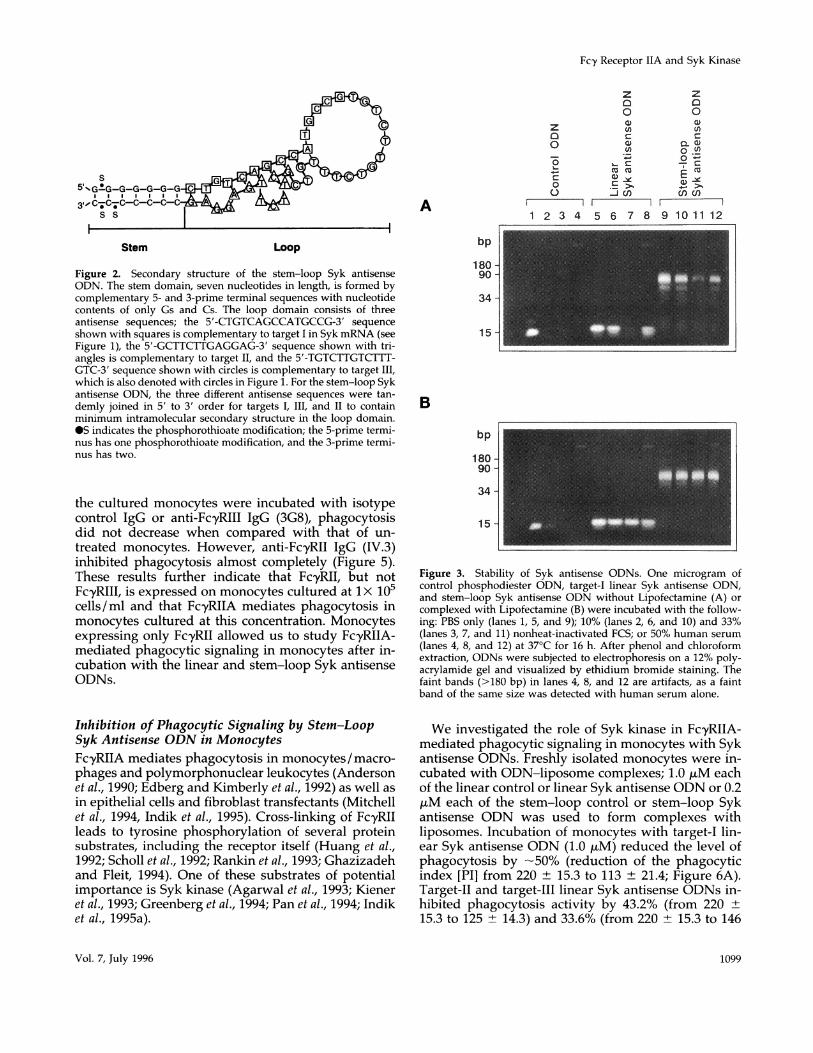
Stability of Syk Antisense ODNsThe efficacy of antisense ODNs for ablating gene ex-pression has not been unequivocal due, in part, to thesensitivity of the ODNs to digestion by nucleases, thedifficulty with delivery into cells, and the inaccessibil-ity of target sequences. To address some of these prob-lems, we devised a novel antisense ODN structurewith additional phosphorothioate modifications atboth the 5-prime and 3-prime termini. The antisenseODN was designed to form a stem and loop structure(Figure 2). The stem domain contains only Gs and Csfor stable base pairing between the 5-prime and3-prime termini and protects the loop domain. Theloop domain consists of three antisense sequencescomplementary to different target sequences of SykmRNA (Figure 1). The loop domain was designed tohave minimal intramolecular secondary structure, al-lowing no more than three contiguous base pairs (bp)to be formed.The stability of the stem-loop Syk antisense ODN
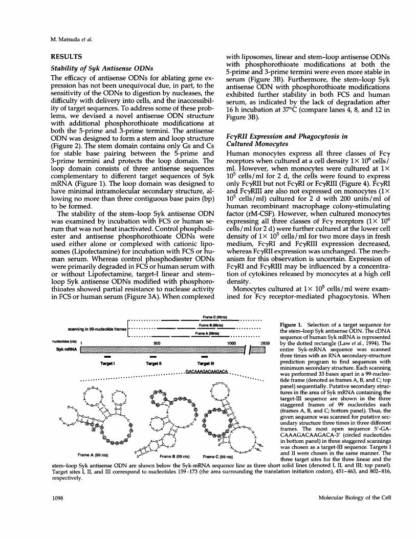
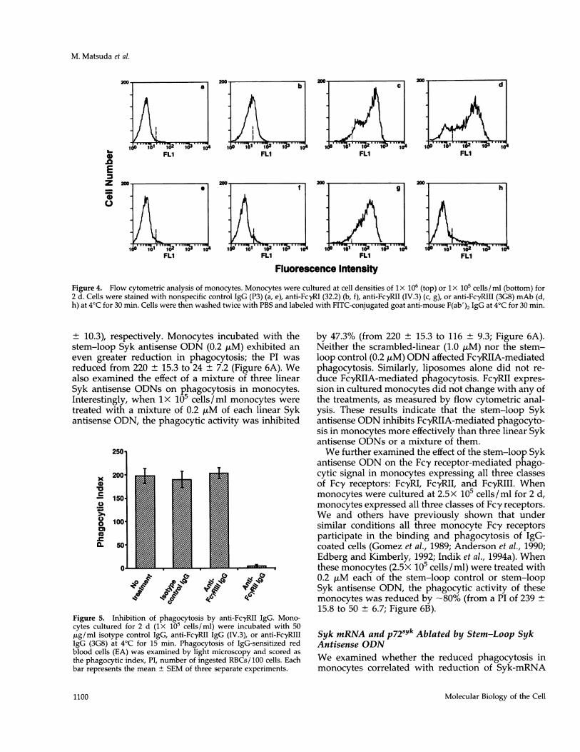
was examined by incubation with FCS or human se-rum that was not heat inactivated. Control phosphodi-ester and antisense phosphorothioate ODNs wereused either alone or complexed with cationic lipo-somes (Lipofectamine) for incubation with FCS or hu-man serum. Whereas control phosphodiester ODNswere primarily degraded in FCS or human serum withor without Lipofectamine, target-I linear and stem-loop Syk antisense ODNs modified with phosphoro-thioates showed partial resistance to nuclease activityin FCS or human serum (Figure 3A). When complexed
with liposomes, linear and stem-loop antisense ODNswith phosphorothioate modifications at both the5-prime and 3-prime termini were even more stable inserum (Figure 3B). Furthermore, the stem-loop Sykantisense ODN with phosphorothioate modificationsexhibited further stability in both FCS and humanserum, as indicated by the lack of degradation after16 h incubation at 37°C (compare lanes 4, 8, and 12 inFigure 3B).
FcyRII Expression and Phagocytosis inCultured MonocytesHuman monocytes express all three classes of Fcyreceptors when cultured at a cell density 1 x 10⁶ cells/ml. However, when monocytes were cultured at 1X10⁵ cells/ml for 2 d, the cells were found to expressonly FcyRII but not FcyRI or FcyRIII (Figure 4). FcyRIand FcyRIII are also not expressed on monocytes (1 x10⁵ cells/ml) cultured for 2 d with 200 units/ml ofhuman recombinant macrophage colony-stimulatingfactor (rM-CSF). However, when cultured monocytesexpressing all three classes of Fcy receptors (ix 106cells/ml for 2 d) were further cultured at the lower celldensity of 1X 10⁵ cells/ml for two more days in freshmedium, FcyRI and FcyRIII expression decreased,whereas FcyRII expression was unchanged. The mech-anism for this observation is uncertain. Expression ofFc'yRI and FcyRIII may be influenced by a concentra-tion of cytokines released by monocytes at a high celldensity.Monocytes cultured at lx 105 cells/ml were exam-
ined for Fcy receptor-mediated phagocytosis. When
Frame C (99nts)
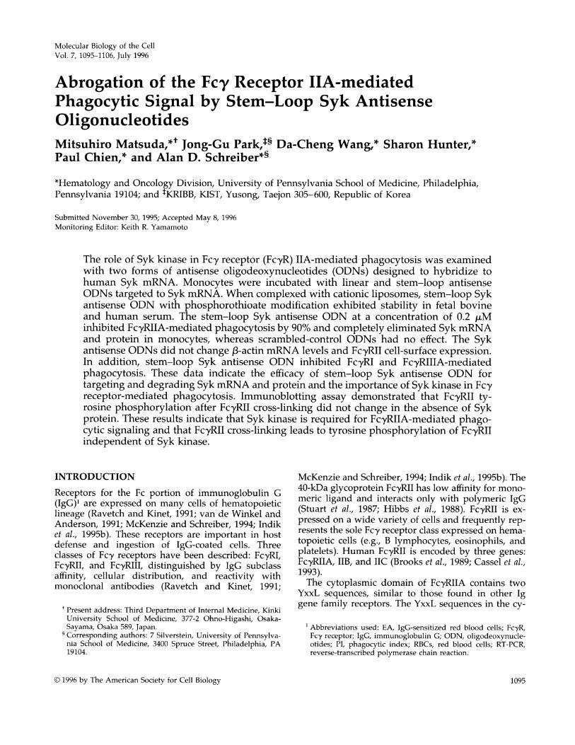
scanNnin 99-nudleotide.fmmes..........Frame B(99nt) ....... Figure 1. Selection of a target sequence forscanr-FranA(WM) the stem-loop Syk antisense ODN. The cDNA........ ....A....s sequence of human Syk mRNA is represented
nudceotides (nt) 1 500 1000 2639 by the dotted rectangle (Law et al., 1994). TheSyk mRNA > entire Syk-mRNA sequence was scanned
_ _ _ three times with an RNA secondary-structureTargt I TarWgt TarStiI prediction program to find sequences with
minimum secondary structure. Each scanning
..................................... was performed 33 bases apart in a 99-nucleo-*C-T-*tide frame (denoted as frames A, B, and C; top
-G-A Ct panel) sequentially. Putafive secondary struc-IfG 'A -A C tures in the area of Syk mRNA containing the
A G T0T AA IGA&J GA-ArAT. target-III sequence are shown in the three5 A.G-C ',C-GaC 'k_C )8 G., staggered frames of 99 nucleotides eachSTG %ATcGacA fae-TvGtAWG G-C t ; sGC C (frames A, B, and C; bottom panel). Thus, the,-TG,GiC TAC-a%C 'c given sequence was scanned for putative sec--G ~~~~~Ia GA-A_Gv-G*#7¢, Si A CG'rT G ' .G ondary structure three times in three differentX ; ciT.._C;<_A-m- Aa frames. The most open sequence 5'-GA->s4,T4>C,- C A CAAAGACAAGACA-3' (circled nucleotides
,G BIA-G\ T G A in bottom panel) in three staggered scanningsA- % C""-~c' 3"C seuneG'%T-A TA T,*.. was chosen as a target-III sequence. Targets I
Frame A (99 nts) 5, Frame -A.(9t and II were chosen in the same manner. The3"' Frame B (99 nta) Frame C (99 nts) three target sites for the three linear and thestem-loop Syk antisense ODN are shown below the Syk-mRNA sequence line as three short solid lines (denoted I, II, and III; top panel).Target sites I, II, and III correspond to nucleotides 159-173 (the area surrounding the translation initiation codon), 451-463, and 802-816,respectively.
Molecular Biology of the Cell1098
Fcy Receptor IIA and Syk Kinase
z
0
0
a)U)
Ca)U1)
._LC
* cnII
za0Q0)
-c~C 0. CE0)-~In en
I I..
1 2 3 4 5 6 7 8 9 10 11 12
Stem Loop
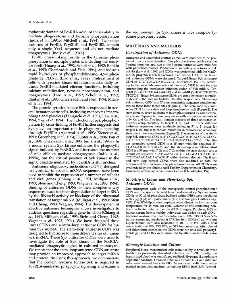
Figure 2. Secondary structure of the stem-loop Syk antisenseODN. The stem domain, seven nucleotides in length, is formed bycomplementary 5- and 3-prime terminal sequences with nucleotidecontents of only Gs and Cs. The loop domain consists of threeantisense sequences; the 5'-CTGTCAGCCATGCCG-3' sequenceshown with squares is complementary to target I in Syk mRNA (seeFigure 1), the 5'-GCTTCTTGAGGAG-3' sequence shown with tri-angles is complementary to target II, and the 5'-TGTCTTGTCTTT-GTC-3' sequence shown with circles is complementary to target III,
which is also denoted with circles in Figure 1. For the stem-loop Sykantisense ODN, the three different antisense sequences were tan-demly joined in 5' to 3' order for targets I, III, and II to containminimum intramolecular secondary structure in the loop domain.OS indicates the phosphorothioate modification; the 5-prime termi-nus has one phosphorothioate modification, and the 3-prime termi-nus has two.
the cultured monocytes were incubated with isotypecontrol IgG or anti-FcyRIII IgG (3G8), phagocytosisdid not decrease when compared with that of un-treated monocytes. However, anti-FcyRII IgG (IV.3)inhibited phagocytosis almost completely (Figure 5).These results further indicate that FcyRII, but notFcyRIII, is expressed on monocytes cultured at 1 x 10⁵cells/ ml and that FcyRIIA mediates phagocytosis inmonocytes cultured at this concentration. Monocytesexpressing only FcyRII allowed us to study FcyRIIA-mediated phagocytic signaling in monocytes after in-cubation with the linear and stem-loop Syk antisenseODNs.
Inhibition of Phagocytic Signaling by Stem-LoopSyk Antisense ODN in MonocytesFcyRIIA mediates phagocytosis in monocytes / macro-phages and polymorphonuclear leukocytes (Andersonet al., 1990; Edberg and Kimberly et al., 1992) as well asin epithelial cells and fibroblast transfectants (Mitchellet al., 1994, Indik et al., 1995). Cross-linking of FcyRIIleads to tyrosine phosphorylation of several proteinsubstrates, including the receptor itself (Huang et al.,1992; Scholl et al., 1992; Rankin et al., 1993; Ghazizadehand Fleit, 1994). One of these substrates of potentialimportance is Syk kinase (Agarwal et al., 1993; Kieneret al., 1993; Greenberg et al., 1994; Pan et al., 1994; Indiket al., 1995a).
bp
180 -
90 -
34 -
15 -
B
bp
180 -
90 -
34 -
15-
Figure 3. Stability of Syk antisense ODNs. One microgram ofcontrol phosphodiester ODN, target-I linear Syk antisense ODN,and stem-loop Syk antisense ODN without Lipofectamine (A) or
complexed with Lipofectamine (B) were incubated with the follow-ing: PBS only (lanes 1, 5, and 9); 10% (lanes 2, 6, and 10) and 33%(lanes 3, 7, and 11) nonheat-inactivated FCS; or 50% human serum
(lanes 4, 8, and 12) at 37°C for 16 h. After phenol and chloroformextraction, ODNs were subjected to electrophoresis on a 12% poly-acrylamide gel and visualized by ethidium bromide staining. Thefaint bands (>180 bp) in lanes 4, 8, and 12 are artifacts, as a faintband of the same size was detected with human serum alone.
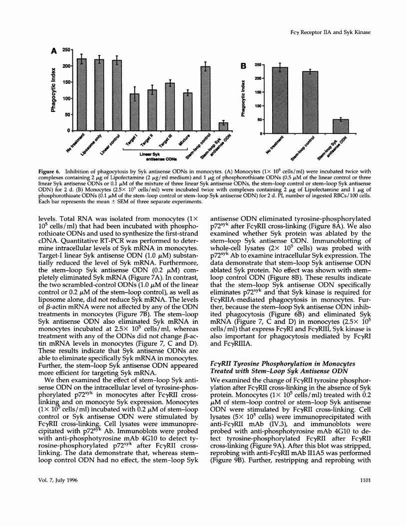
We investigated the role of Syk kinase in FcyRIIA-mediated phagocytic signaling in monocytes with Sykantisense ODNs. Freshly isolated monocytes were in-cubated with ODN-liposome complexes; 1.0 ,tM eachof the linear control or linear Syk antisense ODN or 0.2,uM each of the stem-loop control or stem-loop Sykantisense ODN was used to form complexes withliposomes. Incubation of monocytes with target-I lin-ear Syk antisense ODN (1.0 ,M) reduced the level ofphagocytosis by -50% (reduction of the phagocyticindex [PI] from 220 ± 15.3 to 113 ± 21.4; Figure 6A).Target-II and target-III linear Syk antisense ODNs in-hibited phagocytosis activity by 43.2% (from 220 ±
15.3 to 125 ± 14.3) and 33.6% (from 220 ± 15.3 to 146
Vol. 7, July 1996
zO0
C00
A
1099
M. Matsuda et al.
a
1 1 4
FLI11
i@1 102 1* 3 14IFLI
FLI
lIFL1
d
14T)FLI
h
1 10 1 02 1 1FLI
Fluorescence Intensity
Figure 4. Flow cytometric analysis of monocytes. Monocytes were cultured at cell densities of lx 106 (top) or lx 1i0 cells/ml (bottom) for2 d. Cells were stained with nonspecific control IgG (P3) (a, e), anti-FcyRI (32.2) (b, f), anti-FcyRII (IV.3) (c, g), or anti-FcyRIII (3G8) mAb (d,h) at 4°C for 30 min. Cells were then washed twice with PBS and labeled with FITC-conjugated goat anti-mouse F(ab')2 IgG at 4°C for 30 min.
± 10.3), respectively. Monocytes incubated with thestem-loop Syk antisense ODN (0.2 ,tM) exhibited an
even greater reduction in phagocytosis; the PI was
reduced from 220 ± 15.3 to 24 ± 7.2 (Figure 6A). Wealso examined the effect of a mixture of three linearSyk antisense ODNs on phagocytosis in monocytes.Interestingly, when lx 105 cells/ml monocytes were
treated with a mixture of 0.2 gM of each linear Sykantisense ODN, the phagocytic activity was inhibited
2501
x
0
0
0.
0
200
150
100.
50'
O0
0 0 *-$q li
4, CQIQ~ Cu,CD~~~~~~~~~~'Figure 5. Inhibition of phagocytosis by anti-FcyRII IgG. Mono-cytes cultured for 2 d (lX 105 cells/ml) were incubated with 50Ag/ml isotype control IgG, anti-FcyRII IgG (IV.3), or anti-FcyRIIIIgG (3G8) at 4'C for 15 min. Phagocytosis of IgG-sensitized redblood cells (EA) was examined by light microscopy and scored as
the phagocytic index, PI, number of ingested RBCs/ 100 cells. Eachbar represents the mean ± SEM of three separate experiments.
by 47.3% (from 220 ± 15.3 to 116 ± 9.3; Figure 6A).Neither the scrambled-linear (1.0 ,uM) nor the stem-loop control (0.2 ,uM) ODN affected FcyRIIA-mediatedphagocytosis. Similarly, liposomes alone did not re-
duce Fc-yRIIA-mediated phagocytosis. FcyRII expres-
sion in cultured monocytes did not change with any ofthe treatments, as measured by flow cytometric anal-ysis. These results indicate that the stem-loop Sykantisense ODN inhibits FcyRIIA-mediated phagocyto-sis in monocytes more effectively than three linear Sykantisense ODNs or a mixture of them.We further examined the effect of the stem-loop Syk
antisense ODN on the Fcy receptor-mediated phago-cytic signal in monocytes expressing all three classesof Fcy receptors: Fc-yRI, FcyRII, and FcyRIII. Whenmonocytes were cultured at 2.5x 10⁵ cells/ml for 2 d,monocytes expressed all three classes of Fc-y receptors.We and others have previously shown that undersimilar conditions all three monocyte Fcy receptorsparticipate in the binding and phagocytosis of IgG-coated cells (Gomez et al., 1989; Anderson et al., 1990;Edberg and Kimberly, 1992; Indik et al., 1994a). Whenthese monocytes (2.5x 10⁵ cells/ml) were treated with0.2 ,uM each of the stem-loop control or stem-loopSyk antisense ODN, the phagocytic activity of thesemonocytes was reduced by -80% (from a PI of 23915.8 to 50 6.7; Figure 6B).
Syk mRNA and p72sYk Ablated by Stem-Loop SykAntisense ODNWe examined whether the reduced phagocytosis inmonocytes correlated with reduction of Syk-mRNA
Molecular Biology of the Cell
b
0
Ez
iC.
. 9
0 .1.1 102 i
11
0
1100
Fc,y Receptor IIA and Syk Kinase
B
0
10vc
C
0.
250-
200-
150-
100-
50-
0*
CP 03
10_/ NIP
antinsen ODNs
Figure 6. Inhibition of phagocytosis by Syk antisense ODNs in monocytes. (A) Monocytes (lx 105 cells/ml) were incubated twice withcomplexes containing 2 gg of Lipofectamine (2 jig/ml medium) and 1 ,tg of phosphorothioate ODNs (0.5 ,uM of the linear control or threelinear Syk antisense ODNs or 0.1 ,uM of the mixture of three linear Syk antisense ODNs, the stem-loop control or stem-loop Syk antisenseODN) for 2 d. (B) Monocytes (2.5X 10⁵ cells/ml) were incubated twice with complexes containing 2 ,ug of Lipofectamine and 1 jig ofphosphorothioate ODNs (0.1 ,uM of the stem-loop control or stem-loop Syk antisense ODN) for 2 d. PI, number of ingested RBCs/ 100 cells.Each bar represents the mean ± SEM of three separate experiments.
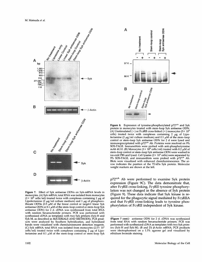
levels. Total RNA was isolated from monocytes (1X10⁵ cells/ml) that had been incubated with phospho-rothioate ODNs and used to synthesize the first-strandcDNA. Quantitative RT-PCR was performed to deter-mine intracellular levels of Syk mRNA in monocytes.Target-I linear Syk antisense ODN (1.0 ,tM) substan-tially reduced the level of Syk mRNA. Furthermore,the stem-loop Syk antisense ODN (0.2 ,uM) com-pletely eliminated Syk mRNA (Figure 7A). In contrast,the two scrambled-control ODNs (1.0 ,uM of the linearcontrol or 0.2 ,uM of the stem-loop control), as well asliposome alone, did not reduce Syk mRNA. The levelsof ,B-actin mRNA were not affected by any of the ODNtreatments in monocytes (Figure 7B). The stem-loopSyk antisense ODN also eliminated Syk mRNA inmonocytes incubated at 2.5x 10⁵ cells/ml, whereastreatment with any of the ODNs did not change n-ac-tin mRNA levels in monocytes (Figure 7, C and D).These results indicate that Syk antisense ODNs areable to eliminate specifically Syk mRNA in monocytes.Further, the stem-loop Syk antisense ODN appearedmore efficient for targeting Syk mRNA.We then examined the effect of stem-loop Syk anti-
sense ODN on the intracellular level of tyrosine-phos-phorylated p72sYk in monocytes after FcyRII cross-
linking and on monocyte Syk expression. Monocytes(ix 10 cells/ ml) incubated with 0.2 p,M of stem-loopcontrol or Syk antisense ODN were stimulated byFcyRII cross-linking. Cell lysates were immunopre-cipitated with p72sYk Ab. Immunoblots were probedwith anti-phosphotyrosine mAb 4G10 to detect ty-rosine-phosphorylated p72sYk after FcyRII cross-linking. The data demonstrate that, whereas stem-loop control ODN had no effect, the stem-loop Syk
antisense ODN eliminated tyrosine-phosphorylatedp72sYk after FcyRII cross-linking (Figure 8A). We alsoexamined whether Syk protein was ablated by thestem-loop Syk antisense ODN. Immunoblotting ofwhole-cell lysates (2X 10⁵ cells) was probed withp72sYk Ab to examine intracellular Syk expression. Thedata demonstrate that stem-loop Syk antisense ODNablated Syk protein. No effect was shown with stem-loop control ODN (Figure 8B). These results indicatethat the stem-loop Syk antisense ODN specificallyeliminates p72sYk and that Syk kinase is required forFc,yRIIA-mediated phagocytosis in monocytes. Fur-ther, because the stem-loop Syk antisense ODN inhib-ited phagocytosis (Figure 6B) and eliminated SykmRNA (Figure 7, C and D) in monocytes (2.5x 10⁵cells/ml) that express FcyRI and FcyRIII, Syk kinase isalso important for phagocytosis mediated by FcyRIand FcyRIIIA.
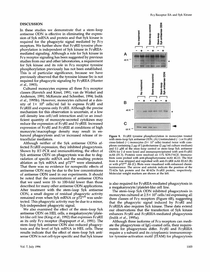
FcyRII Tyrosine Phosphorylation in MonocytesTreated with Stem-Loop Syk Antisense ODNWe examined the change of FcyRII tyrosine phosphor-ylation after FcyRII cross-linking in the absence of Sykprotein. Monocytes (lx 105 cells/ml) treated with 0.2,uM of stem-loop control or stem-loop Syk antisenseODN were stimulated by FcyRII cross-linking. Celllysates (5 x 10⁵ cells) were immunoprecipitated withanti-Fc,yRII mAb (IV.3), and immunoblots wereprobed with anti-phosphotyrosine mAb 4G10 to de-tect tyrosine-phosphorylated FcyRII after FcyRIIcross-linking (Figure 9A). After this blot was stripped,reprobing with anti-FcyRII mAb II1A5 was performed(Figure 9B). Further, restripping and reprobing with
Vol. 7, July 1996
A
x0
£CO0
a.
1101
M. Matsuda et al.
A0a
'to
Co C&
4'0
'?c,w e eA
bp
1,353-1,078-872-603-310
271/281-234 -194=118
97 K -
66 K -- Syk
1 Syk
Figure 7. Effect of Syk antisense ODNs on Syk-mRNA levels in
monocytes. (A) Syk mRNA; total RNA was isolated from monocytes(lx 10 cells/ml) treated twice with complexes containing 2 Mig ofLipofectamine (2 ,Lg/ml culture medium) and 1 ,ug of phosphoro-thioate ODNs (0.5 ,uM of the linear control or target-I linear Sykantisense ODN or 0.1 MuM of the stem-loop control or stem-loop Sykantisense ODN) for 2 d. cDNA was synthesized from total RNAwith random hexanucleotide primers. PCR was performed withsynthesized cDNA as templates with two Syk primers (Syk-H andSyk-M, as described in MATERIALS AND METHODS). PCR prod-ucts were analyzed by Southern hybridization, and hybridizedbands were visualized with chemiluminescent detection reagents.(C) Syk mRNA; total RNA was isolated from monocytes (2.5X 10⁵cells/ml) treated twice with complexes containing 2 Mug of Lipo-fectamine and 0.1 MuM of the stem-loop control or stem-loop Syk
z_E 0
00-0 CL ffi aE o o.r
z CM cn(nm m mI
-. - + - +
oq
Figure 8. Expression of tyrosine-phosphorylated p72sYk and Sykprotein in monocytes treated with stem-loop Syk antisense ODN.(A) Unstimulated (-) or FcyRII cross-linked (+) monocytes (5X 10⁵cells) treated twice with complexes containing 2 Mg of Lipo-fectamine (2 Mug/ml culture medium) and 0.1 MuM of the stem-loopcontrol or stem-loop Syk antisense ODN for 2 d were lysed andimmunoprecipitated with p72sYk Ab. Proteins were resolved on 9%SDS-PAGE. Immunoblots were probed with anti-phosphotyrosinemAb 4GI0. (B) Monocytes (ix 105 cells/ml) treated with 0.2 MM ofstem-loop control or stem-loop Syk antisense ODN were washed inice-cold PBS and lysed. Cell lysates (2x 10⁵ cells) were separated by9% SDS-PAGE, and immunoblots were probed with p72sYk Ab.Blots were visualized with enhanced chemiluminescence. The ar-
row indicates the position of the 72-kDa Syk protein. Molecularweight markers are shown at the left.
p72sYk Ab were performed to examine Syk proteinexpression (Figure 9C). The data demonstrate that,after FcyRII cross-linking, FcyRII tyrosine phosphory-lation was not changed in the absence of Syk protein(Figure 9). These data indicate that Syk kinase is re-
quired for the phagocytic signal mediated by FcyRIIAand that Fc'yRII cross-linking leads to tyrosine phos-phorylation of Fc'yRII independent of Syk kinase.
(Figure 7 cont.) antisense ODN for 2 d. cDNA was synthesizedfrom total RNA with random hexanucleotide primers. PCR was
performed with synthesized cDNA as templates with two Syk prim-ers (Syk-H and Syk-M). (B and D) f3-Actin mRNA; PCR productswere electrophoresed on a 1.5% agarose gel and visualized byethidium bromide staining.
Molecular Biology of the Cell
B
C
D
-*at,, *Now
1-
-
V. 10-00
ILELAL &-l- Ik AlMm Aj, MOSW
-
-0-- li-Actin
1102
--l
'L [.-Actln
Fcy Receptor IIA and Syk Kinase
DISCUSSION
In these studies we demonstrate that a stem-loopantisense ODN is effective in eliminating the expres-sion of Syk mRNA and protein and that Syk kinase isrequired for the phagocytic signal mediated by Fc-yreceptors. We further show that FcyRII tyrosine phos-phorylation is independent of Syk kinase in Fc-yRIIA-mediated signaling. Although a role for Syk kinase inFc,y receptor signaling has been suggested by previousstudies from our and other laboratories, a requirementfor Syk kinase and its role in Fcy receptor tyrosinephosphorylation previously has not been established.This is of particular significance, because we havepreviously observed that the tyrosine kinase Src is notrequired for phagocytic signaling by FcyRIIA (Hunteret al., 1993).Cultured monocytes express all three Fcy receptor
classes (Ravetch and Kinet, 1991; van de Winkel andAnderson, 1991; McKenzie and Schreiber, 1994; Indiket al., 1995b). However, monocytes cultured at a den-sity of lx 105 cells/ ml fail to express FcyRI andFcyRIII and express only FcyRII. Although the precisemechanism for this observation is uncertain, at a lowcell density less cell/cell interaction and/or an insuf-ficient quantity of monocyte-secreted cytokines mayreduce the expression of FcyRI and FcyRIII. Increasedexpression of FcyRI and FcyRIII at conditions of highmonocyte / macrophage density may result in en-hanced phagocytosis and/or increased release of in-tracellular mediators.Although neither of the Syk antisense ODNs af-
fected FcyRII expression, they inhibited phagocytosis.Shown by RT-PCR and immunoblotting, the effect ofSyk antisense ODN on phagocytosis was due to deg-radation of specific mRNA and the resulting proteinablation as Syk mRNA and p72sYk were eliminated.That there was no evidence for nonspecific effects ofantisense ODN may be due to the low concentrationsof antisense ODN used in our experiments. It shouldbe noted that the concentrations of antisense ODNsthat we used were 10- to 100-fold lower than thosedescribed for many other antisense ODN applications.After treatment with the stem-loop Syk antisenseODN, a small degree of residual phagocytic activityremained even when Syk mRNA or p72sY was unde-tected. This phagocytic activity may be due to a minor,Syk-independent phagocytic signal.We also examined the effect of the stem-loop Syk
antisense ODN on HEL cells, a megakaryocyte / plate-let-like cell line (King et al., 1992) that expresses FcyRIIas its only Fcy receptor (Rappaport et al., 1991). Thestem-loop Syk antisense ODN also reduced phagocy-tosis and the level of Syk mRNA in HEL cells. Theseresults indicate that the effect of stem-loop Syk anti:sense ODN is not cell-type specific and that Syk kinase
A46 K -
B
46KK-_
cr66 K -
!8
E 8 IE ~~~III
I I a4_
f.
_--- 1_
r - +
z8
CC nE 8 gX
_-+ _-+ -+
*
-1Figure 9. FcyRII tyrosine phosphorylation in monocytes treatedwith stem-loop Syk antisense ODN. (A) Unstimulated (-) or Fc-yRIIcross-linked (+) monocytes (5X 10⁵ cells) treated twice with com-plexes containing 2 ,ug of Lipofectamine (2 ,ug/ml culture medium)and 0.1 ,uM of the stem-loop control or stem-loop Syk antisenseODN for 2 d were lysed and immunoprecipitated with anti-FcyRIImAb (IV.3). Proteins were resolved on 11% SDS-PAGE. Immuno-blots were probed with anti-phosphotyrosine mAb 4G10. The blotfrom A was stripped and reprobed with anti-Fc-yRII mAb I1A5 (B)or with p72sYk Ab (C). Blots were visualized with enhanced chemi-luminescence. The arrow and asterisk indicate the position of the72-kDa Syk protein and the 40-kDa Fc-yRII protein, respectively.Molecular weight markers are shown at the left.
is also required for Fc'yRIIA-mediated phagocytosis ina megakaryocyte/platelet-like cell line.The stem-loop Syk ODN inhibited phagocytosis in
monocytes cultured at 2.5 x 10⁵ cells/ml expressing allthree classes of Fcy receptors (Figure 6B), suggestingthat the phagocytic signal induced by FcyRI andFcyRIIIA also requires Syk kinase. These data extendour observations that the transfection of Syk kinaseenhances FcyRI and FcyRIIIA-mediated phagocytosis(Indik et al., 1995a).Although three isoforms of Fcy receptors can medi-
ate the phagocytosis of IgG-coated cells, their require-ments for phagocytosis differ. FcyRI and FcyRIIIArequire a y subunit and its cytoplasmic immunorecep-tor tyrosine-activation motif (ITAM) for phagocytosis
Vol. 7, July 1996 1103
M. Matsuda et al.
(Park et al., 1993a,b; Indik et al., 1995a). On the otherhand, FcyRIIA mediates phagocytosis through aunique conserved ITAM sequence present in its owncytoplasmic domain and, thus, does not require the ysubunit (Indik et al., 1994b; Mitchell et al., 1994). Trans-fection of Syk kinase enhances phagocytosis mediatedby FcyRI and FcyRIIIA seven- to tenfold but enhancesphagocytosis by FcyRIIA two- to threefold in COS-1cells (Indik et al., 1995b). In the absence of Syk trans-fection, FcyRIIA-mediated phagocytosis is much moreefficient than phagocytosis by the y subunit of FcyRIand FcyRIIIA (Indik et al., 1995b). The low level ofendogenous Syk kinase in COS-1 cells is likely suffi-cient for FcyRIIA signaling. However, it is uncertain asto precisely how Syk kinase is involved in FcyRIIA-mediated signaling. In this study we demonstrate thatFcyRII tyrosine phosphorylation is independent ofSyk kinase (Figure 9). The data indicate that proteintyrosine kinases other than Syk are involved in ty-rosine phosphorylation of FcyRII. Of interest is thatthe pattern of protein tyrosine phosphorylation afterFc,yRII cross-linking does not seem to change signifi-cantly in the absence of Syk protein. The use of Sykantisense ODNs should allow us in future studies todefine the substrate(s) of Syk kinase. It is possiblethat FcyRIIA-mediated phagocytosis may involvedownstream proteins that are not tyrosine phos-phorylated. Alternatively, anti-phosphotyrosine im-munoblots may not be sufficiently sensitive to dis-criminate decreased protein tyrosine phosphoryla-tion in the absence of Syk.Antisense ODNs offer the potential to reduce or
ablate a specific mRNA. Among the factors that affectthe efficacy of antisense ODNs are the stability of theantisense ODNs, their delivery into the cell cytoplasm,and their accessibility to the target mRNA. We havemodified Syk antisense ODN in three steps to addressthese issues. First, one phosphodiester link at the5-prime and two at the 3-prime termini were modifiedwith phosphorothioates. Second, we have designed astem-loop structure to protect the antisense sequencein the loop domain from nucleases. The stem hascomplementary terminal sequences with only Gs andCs. The loop domain has three antisense sequencestargeting different sites of Syk mRNA. mRNA formssecondary structures by intramolecular hybridization,and mRNA secondary structures may inhibit access ofantisense ODNs to target sequences. To identify se-quences with open structures that may provide betteraccess for antisense ODNs, we scanned the entire Syk-mRNA sequence with an RNA secondary-structureprediction program. Syk mRNA was scanned in threestaggered frames, and the most open sequences withminimum secondary structures were carefully chosen.Our study did not seek to investigate the advantage intargeting open mRNA sequences, but the fact that allthree linear Syk antisense ODNs inhibited phagocyto-
sis suggests that open substrate sequences may bereadily accessible to antisense ODNs. Because stem-loop antisense ODN reduced the phagocytic signalmore dramatically than the mixture of three linearantisense ODNs (Figure 6A), the higher efficacy of thestem-loop Syk antisense ODN may be due to betterstability from nuclease digestion. Third, Syk antisenseODNs were also complexed with cationic liposomes toimprove delivery to the cells. The stability of the stem-loop Syk antisense ODN markedly improved whencomplexed with liposomes, providing an additionalbenefit for the use of ODN-liposome complexes. Ourstem-loop antisense ODN approach has been usedsuccessfully to abrogate the expression of other geneproducts. We have used a stem-loop antisense ODNdirected at the FcyRIIIA y subunit mRNA. With theuse of peripheral blood monocytes and the stem-loopy-chain antisense ODN, the monocyte 'y-chain mes-sage, assessed by RT-PCR, was decreased by >80%.Liposomes can be delivered to the reticuloendothe-lial system, for which monocytes /macrophages area major residential cell population. The complex ofliposome-stem-loop Syk antisense ODN may be ad-vantageous for use as a therapeutic agent(s) for im-munologic disorders requiring down-regulation ofFcy receptor-mediated function in monocytes /macro-phages. Syk kinase is also associated with FcERI andwith the B-cell antigen receptor (Hutchcroft et al.,1992a,b). The stem-loop Syk antisense ODN may alsoprove useful for investigating intracellular signalingevents through these receptors and for developing atherapeutic agent(s) to modulate the signals mediatedby these receptors.
ACKNOWLEDGMENTSWe thank Dr. Zena K. Indik for her critical reading of this manu-script. This work was supported by CorBec Pharmaceuticals andGrants HL-28207 and HL-27068 from the National Institutes ofHealth.
REFERENCESAgarwal, A., Salem, P., and Robbins, K.C. (1993). Involvement ofp72SYk, a protein-tyrosine kinase, in Fcy receptor signaling. J. Biol.Chem. 268, 15900-15905.
Anderson, C.L., Shen, L., Eicher, D.M., Wewers, M.D., and Gill, J.K.(1990). Phagocytosis mediated by three distinct Fcy receptor classeson human leukocytes. J. Exp. Med. 171, 1333-1345.
Brooks, D.G., Qiu, W.Q., Luster, A.D., and Ravetch, J.V. (1989).Structure and expression of human IgG FcRII (CD32). Functionalheterogeneity is encoded by the alternatively spliced products ofmultiple genes. J. Exp. Med. 170, 1369-1385.
Cassel, D.L., Keller, M.A., Surrey, S., Schwartz, E., Schreiber, A.D.,Rappaport, E.F., and McKenzie, S.E. (1993). Differential expressionof FcyRIIA, FcyRIIB, and FcyRIIC in hematopoietic cells: analysis oftranscript. Mol. Immunol. 30, 451-460.
Chiang, M.-Y., Chan, H., Zounes, M.A., Freier, S.M., Lima, W.F., andBennet, C.F. (1991). Antisense oligonucleotides inhibit intercellular
Molecular Biology of the Cell1104
Fc,y Receptor IIA and Syk Kinase
adhesion molecule 1 expression by two distinct mechanisms. J. Biol.Chem. 266, 18162-18171.
Darby, C., Geahlen, R.L., and Schreiber, A.D. (1994). Stimulation ofmacrophage FcyRIIIA activates the receptor-associated protein ty-rosine kinase Syk and induces phosphorylation of multiple proteinsincluding p95Vav and p62/GAP-associated protein. J. Immunol.152, 5429-5437.Edberg, J.C., and Kimberly, R.P. (1992). Receptor-specific probes forthe study of Fcy receptor-specific function. J. Immunol. Methods148, 179-187.
Ghazizadeh, S., Bolen, J.B., and Fleit, H.B. (1995). Tyrosine phos-phorylation and association of Syk with FcyRII in monocytic THP-1cells. Biochem. J. 305, 669-674.
Ghazizadeh, S., and Fleit, H.B. (1994). Tyrosine phosphorylationprovides an obligatory early signal for FcyRII-mediated endocytosisin the monocytic cell line THP-1. J. Immunol. 152, 30-41.
Gomez, F., Chien, P., King, M., McDermott, P., Rossman, M.D., andSchreiber, A.D. (1989). Monocyte Fcy receptor recognition of cell-bound and aggregated IgG. Blood 74, 1058-1065.
Greenberg, S., Chang, P., and Silverstein, S.C. (1994). Tyrosine phos-phorylation of the y subunit of Fcy receptors p72SYk and paxillinduring Fc receptor-mediated phagocytosis in macrophages. J. Biol.Chem. 269, 3897-3902.
Hibbs, M.L., Bonadonna, L., Scott, B.M., McKenzie, I.F., and Hog-arth, P.M. (1988). Molecular cloning of a human immunoglobulin GFc receptor. Proc. Natl. Acad. Sci. USA 85, 2240-2244.
Huang, M.-M., Indik, Z., Brass, L.F., Hoxie, J.A., Schreiber, A.D., andBrugge, J.S. (1992). Activation of FcyRII induces tyrosine phosphor-ylation of multiple proteins including FcyRII. J. Biol. Chem. 267,5467-5473.
Hunter, S., Huang, M.-M., Indik, Z.K., and Schreiber, A.D. (1993).FcyRIIA-mediated phagocytosis and receptor phosphorylation incells deficient in the protein tyrosine kinase Src. Exp. Hematol. 21,1492-1497.
Hunter, S., Kamoun, M., and Schreiber, A.D. (1994). Transfection ofan Fcy receptor cDNA induces T cells to become phagocytic. Proc.Natl. Acad. Sci. USA 91, 10232-10236.
Hutchcroft, J.E., Geahlen, R.L., Deanin, G.G., and Oliver, J.M.(1992a). FcERI-mediated tyrosine phosphorylation and activation ofthe 72-kDa protein tyrosine kinase, PTK72, in RBL-2H3 rat tumormast cells. Proc. Natl. Acad. Sci. USA 89, 9107-9111.Hutchcroft, J.E., Harrison, M.L., and Geahlen, R.L. (1992b). Associ-ation of the 72-kDa protein tyrosine kinase PTK72 with the B cellantigen receptor. J. Biol. Chem. 267, 8613-8619.
Indik, Z.K., Hunter, S., Huang, M.M., Pan, X.Q., Chien, P., Kelly, C.,Levinson, A.I., Kimberly, R.P., and Schreiber, A.D. (1994a). Thehigh-affinity Fcy receptor (CD64) induces phagocytosis in the ab-sence of its cytoplasmic domain: the y subunit of FcyRIIIA impartsphagocytic function to FcyRI. Exp. Hematol. 22, 599-606.
Indik, Z.K., Pan, X.Q., Huang, M.-M., McKenzie, S.E., Levinson, A.I.,and Schreiber, A.D. (1994b). Insertion of cytoplasmic tyrosine se-quences into the nonphagocytic receptor FcyRIIB establishes phago-cytic function. Blood 83, 2072-2080.
Indik, Z.K., Park, J.-G., Pan, X.Q., and Schreiber, A.D. (1995a).Induction of phagocytosis by a protein tyrosine kinase. Blood 85,1175-1180.
Indik, Z.K., Park, J.-G., Hunter, S., and Schreiber, A.D. (1995b). Themolecular dissection of Fcy receptor-mediated phagocytosis. Blood86, 4389-4399.
Kiener, P.A., Rankin, B.M., Burkhardt, A.L., Schieven, G.L., Gilli-land, L.K., Rowley, R.B., Bolen, J.B., and Ledbetter, J.A. (1993).Cross-linking of Fcy receptor I (FcyRI) and receptor II (FcyRII) on
monocytic cells activates a signal transduction pathway common toboth Fc receptors that involves the stimulation of p72 syk proteintyrosine kinase. J. Biol. Chem. 268, 24442-24448.
King, M., Comber, P.G., Chien, P., Ruiz, P., and Schreiber, A.D.(1992). Characterization of Fcy receptors on a human erythroleuke-mia cell line (HEL). Exp. Hematol. 20, 576-581.
Law, C.-L., Sidorenko, S.P., Chandran, K.A., Draves, K.E., Chan,A.C., Weiss, A., Edelhoff, S., Disteche, C.M., and Clark, E.A. (1994).Molecular cloning of human Syk. J. Biol. Chem. 269, 12310-12319.
Liao, F., Shin, H.S., and Rhee, S.G. (1992). Tyrosine phosphorylationof phospholipase C--yl induced by cross-linking of high-affinity orlow-affinity Fc receptor for IgG in U937 cells. Proc. Natl. Acad. Sci.USA 89, 3659-3663.
McKenzie, S.E., and Schreiber, A.D. (1994). Biological advances andclinical applications of Fc receptors for IgG. Leukocytes 1, 45-52.
Milligan, J.F., Matteucci, M.D., and Martin, J.C. (1993). Currentconcepts in antisense drug design. J. Med. Chem. 36, 1923-1937.
Mitchell, M.A., Huang, M.-M., Chien, P., Indik, Z.K., Pan, X.Q., andSchreiber, A.D. (1994). Substitutions and deletions in the cytoplas-mic domain of the phagocytic receptor FcyRIIA: effect on receptortyrosine phosphorylation and phagocytosis. Blood 84, 1753-1759.
Pan, X.Q., Darby, C., Indik, Z.K., and Schreiber, A.D. (1994). Proteintyrosine phosphorylation following activation of monocyte / macro-phage Fcy receptors. J. Immunol. 152, 3231.
Park, J.-G., Isaacs, R.E., Chien, P., and Schreiber, A.D. (1993a). In theabsence of other Fc receptors, FcyRIIIA transmits a phagocyticsignal that requires the cytoplasmic domain of its y subunit. J. Clin.Invest. 92, 1967-1973.
Park, J.-G., Murray, R.K., Chien, P., Darby, C., and Schreiber, A.D.(1993b). Conserved cytoplasmic tyrosine residues of the y subunitare required for a phagocytic signal mediated by Fc-yRIIIA. J. Clin.Invest. 92, 2073-2079.
Rankin, B.M., Yocum, S.A., Mittler, R.S., and Kiener, P.A. (1993).Stimulation of tyrosine phosphorylation and calcium mobilizationby Fc gamma receptor cross-linking: regulation by the phosphoty-rosine phosphatase CD45. J. Immunol. 150, 605-616.
Rappaport, E., Cassel, D.L., McKenzie, S.E., Surrey, S., Schwartz, E.,and Schreiber, A.D. (1991). Expression of FcyRII in hematopoieticcells: analysis of transcripts encoding the soluble and membrane-associated molecular forms. Clin. Res. 39, 151A.
Ravetch, J.V., and Kinet, J.-P. (1991). Fc receptors. Annu. Rev. Im-munol. 9, 457-492.
Scholl, P.R., Ahem, D., and Geha, R.S. (1992). Protein tyrosinephosphorylation induced via the IgG receptors FcyRI and FcyRII inthe human monocytic cell line THP-1. J. Immunol. 149, 1751-1757.
Schreiber, A.D., Parsons, J., McDermott, P., and Cooper, R.A. (1975).Effect of corticosteroids on the human monocyte IgG and comple-ment receptors. J. Clin. Invest. 56, 1189-1197.
Stein, C.A., and Cheng, Y.-C. (1993). Antisense oligonucleotides astherapeutic agents-is the bullet really magical? Science 261, 1004-1012.
Stuart, S.G., Trounstine, M.L., Vaux, D.J., Koch, T., Martens, C.L.,Mellman, I., and Moore, K.W. (1987). Isolation and expression ofcDNA clones encoding a human receptor for IgG (FcyRII). J. Exp.Med. 166, 1668-1684.
Taniguchi, T., Kobayashi, T., Kondo, J., Takahashi, K., Nakamura,H., Suzuki, J., Nagai, K., Yamada, T., Nakamura, S., and Yamamura,H. (1991). Molecular cloning of a porcine gene syk that encodes a72-kDa protein tyrosine kinase showing high susceptibility to pro-teolysis. J. Biol. Chem. 266, 15790-15796.
Vol. 7, July 1996 1105

M. Matsuda et al.
van de Winkel, J.G.J., and Anderson, C.L. (1991). Biology of humanimmunoglobulin G Fc receptors. J. Leukocyte Biol. 49, 511-524.Wagner R.W. (1994). Gene inhibition using antisense oligode-oxynucleotides. Nature 372, 333-335.Wagner, R.W., Matteucci, M.D., Lewis, J.G., Gutierrez, A.J., Moulds,C., and Froehler, B.C. (1993). Antisense gene inhibition by oligonu-
cleotides containing C-5 propyne pyrimidines. Science 260, 1510-1513.
Yagi, S., Suzuki, K., Hasegawa, A., Okumura, K., and Ra, C. (1994).Cloning of the cDNA for the deleted Syk kinase homologous toZAP-70 from human basophilic leukemia cell line (KU812). Bio-chem. Biophys. Res. Commun. 200, 28-34.
Molecular Biology of the Cell1106