New Inhibitors of Laccase and Tyrosinase by ... - MDPI
13
Citation: Chaudhary, D.; Chong, F.; Neupane, T.; Choi, J.; Jee, J.-G. New Inhibitors of Laccase and Tyrosinase by Examination of Cross-Inhibition between Copper-Containing Enzymes. Int. J. Mol. Sci. 2021, 22, 13661. https://doi.org/10.3390/ ijms222413661 Academic Editor: Alexande Baykov Received: 4 November 2021 Accepted: 17 December 2021 Published: 20 December 2021 Publisher’s Note: MDPI stays neutral with regard to jurisdictional claims in published maps and institutional affil- iations. Copyright: © 2021 by the authors. Licensee MDPI, Basel, Switzerland. This article is an open access article distributed under the terms and conditions of the Creative Commons Attribution (CC BY) license (https:// creativecommons.org/licenses/by/ 4.0/). International Journal of Molecular Sciences Article New Inhibitors of Laccase and Tyrosinase by Examination of Cross-Inhibition between Copper-Containing Enzymes Dinesh Chaudhary, Fangchen Chong, Trilok Neupane, Joonhyeok Choi and Jun-Goo Jee * Research Institute of Pharmaceutical Sciences, College of Pharmacy, Kyungpook National University, 80 Daehak-ro, Buk-gu, Daegu 41566, Korea; [email protected] (D.C.); [email protected] (F.C.); [email protected] (T.N.); [email protected] (J.C.) * Correspondence: [email protected]; Tel.: +82-53-950-7568 Abstract: Coppers play crucial roles in the maintenance homeostasis in living species. Approximately 20 enzyme families of eukaryotes and prokaryotes are known to utilize copper atoms for catalytic activities. However, small-molecule inhibitors directly targeting catalytic centers are rare, except for those that act against tyrosinase and dopamine-β-hydroxylase (DBH). This study tested whether known tyrosinase inhibitors can inhibit the copper-containing enzymes, ceruloplasmin, DBH, and laccase. While most small molecules minimally reduced the activities of ceruloplasmin and DBH, aside from known inhibitors, 5 of 28 tested molecules significantly inhibited the function of laccase, with the K i values in the range of 15 to 48 μM. Enzyme inhibitory kinetics classified the molecules as competitive inhibitors, whereas differential scanning fluorimetry and fluorescence quenching supported direct bindings. To the best of our knowledge, this is the first report on organic small- molecule inhibitors for laccase. Comparison of tyrosinase and DBH inhibitors using cheminformatics predicted that the presence of thione moiety would suffice to inhibit tyrosinase. Enzyme assays confirmed this prediction, leading to the discovery of two new dual tyrosinase and DBH inhibitors. Keywords: cheminformatics; ceruloplasmin; dopamine-β-hydroxylase; laccase; tyrosinase 1. Introduction One third of the total enzymes in living organisms include metals to maintain their structures or implement their functions [1,2]. The most common metalloenzymes are zinc- containing proteins, followed by those containing iron. Malfunction of metalloenzymes often leads to diseases, making metalloenzymes a potential target for drug discovery. How- ever, inhibitors of metalloenzymes have been less developed by drug researchers. Thus far, FDA-approved drugs include fewer than 70 metalloenzyme inhibitors [1,3], most of which are inhibitors of four types of proteins: lanosterol 14α-demethylase, carbonic anhydrase, histone deacetylase, and metalloprotease [3]. This limited number may reflect the difficulty caused by the nonspecificity of metal-binding groups (MBGs) in small molecules [4,5]. Copper-containing enzymes account for <5% of all metalloenzymes [3,6–8]. The cellu- lar level of copper is much lower than that of zinc, and copper-containing proteins are much fewer in number than zinc-containing proteins. The small molecules that inhibit copper- containing enzymes are scarce, except for those that inhibit tyrosinase and dopamine-β- hydroxylase (DBH) [9–13]. The FDA has approved hydroquinone as a depigmenting agent through the inhibition of tyrosinase [3]. No other drug targeting copper-containing enzymes has been approved. In addition to tyrosinase and DBH, copper-containing enzymes include ceruloplasmin, lysyl oxidase, peptidylglycine monooxygenase, bilirubin oxidase, galac- tose oxidase, hexose oxidase, ascorbate oxidase, nitrite reductase, nitrous-oxide reductase, quercetin 2,3-dioxygenase, and laccase. Several studies have reported on the selectiveness of metalloenzyme inhibitors across metals, exhibiting off-target effects [2]. However, information on off-target effects in copper- containing enzymes is limited. We previously reported new types of tyrosinase inhibitors by Int. J. Mol. Sci. 2021, 22, 13661. https://doi.org/10.3390/ijms222413661 https://www.mdpi.com/journal/ijms
-
Upload
khangminh22 -
Category
Documents
-
view
2 -
download
0
Transcript of New Inhibitors of Laccase and Tyrosinase by ... - MDPI

�����������������
Citation: Chaudhary, D.; Chong, F.;
Neupane, T.; Choi, J.; Jee, J.-G. New
Inhibitors of Laccase and Tyrosinase
by Examination of Cross-Inhibition
between Copper-Containing
Enzymes. Int. J. Mol. Sci. 2021, 22,
13661. https://doi.org/10.3390/
ijms222413661
Academic Editor: Alexande Baykov
Received: 4 November 2021
Accepted: 17 December 2021
Published: 20 December 2021
Publisher’s Note: MDPI stays neutral
with regard to jurisdictional claims in
published maps and institutional affil-
iations.
Copyright: © 2021 by the authors.
Licensee MDPI, Basel, Switzerland.
This article is an open access article
distributed under the terms and
conditions of the Creative Commons
Attribution (CC BY) license (https://
creativecommons.org/licenses/by/
4.0/).
International Journal of
Molecular Sciences
Article
New Inhibitors of Laccase and Tyrosinase by Examination ofCross-Inhibition between Copper-Containing Enzymes
Dinesh Chaudhary, Fangchen Chong, Trilok Neupane, Joonhyeok Choi and Jun-Goo Jee *
Research Institute of Pharmaceutical Sciences, College of Pharmacy, Kyungpook National University,80 Daehak-ro, Buk-gu, Daegu 41566, Korea; [email protected] (D.C.); [email protected] (F.C.);[email protected] (T.N.); [email protected] (J.C.)* Correspondence: [email protected]; Tel.: +82-53-950-7568
Abstract: Coppers play crucial roles in the maintenance homeostasis in living species. Approximately20 enzyme families of eukaryotes and prokaryotes are known to utilize copper atoms for catalyticactivities. However, small-molecule inhibitors directly targeting catalytic centers are rare, except forthose that act against tyrosinase and dopamine-β-hydroxylase (DBH). This study tested whetherknown tyrosinase inhibitors can inhibit the copper-containing enzymes, ceruloplasmin, DBH, andlaccase. While most small molecules minimally reduced the activities of ceruloplasmin and DBH,aside from known inhibitors, 5 of 28 tested molecules significantly inhibited the function of laccase,with the Ki values in the range of 15 to 48 µM. Enzyme inhibitory kinetics classified the moleculesas competitive inhibitors, whereas differential scanning fluorimetry and fluorescence quenchingsupported direct bindings. To the best of our knowledge, this is the first report on organic small-molecule inhibitors for laccase. Comparison of tyrosinase and DBH inhibitors using cheminformaticspredicted that the presence of thione moiety would suffice to inhibit tyrosinase. Enzyme assaysconfirmed this prediction, leading to the discovery of two new dual tyrosinase and DBH inhibitors.
Keywords: cheminformatics; ceruloplasmin; dopamine-β-hydroxylase; laccase; tyrosinase
1. Introduction
One third of the total enzymes in living organisms include metals to maintain theirstructures or implement their functions [1,2]. The most common metalloenzymes are zinc-containing proteins, followed by those containing iron. Malfunction of metalloenzymesoften leads to diseases, making metalloenzymes a potential target for drug discovery. How-ever, inhibitors of metalloenzymes have been less developed by drug researchers. Thus far,FDA-approved drugs include fewer than 70 metalloenzyme inhibitors [1,3], most of whichare inhibitors of four types of proteins: lanosterol 14α-demethylase, carbonic anhydrase,histone deacetylase, and metalloprotease [3]. This limited number may reflect the difficultycaused by the nonspecificity of metal-binding groups (MBGs) in small molecules [4,5].
Copper-containing enzymes account for <5% of all metalloenzymes [3,6–8]. The cellu-lar level of copper is much lower than that of zinc, and copper-containing proteins are muchfewer in number than zinc-containing proteins. The small molecules that inhibit copper-containing enzymes are scarce, except for those that inhibit tyrosinase and dopamine-β-hydroxylase (DBH) [9–13]. The FDA has approved hydroquinone as a depigmenting agentthrough the inhibition of tyrosinase [3]. No other drug targeting copper-containing enzymeshas been approved. In addition to tyrosinase and DBH, copper-containing enzymes includeceruloplasmin, lysyl oxidase, peptidylglycine monooxygenase, bilirubin oxidase, galac-tose oxidase, hexose oxidase, ascorbate oxidase, nitrite reductase, nitrous-oxide reductase,quercetin 2,3-dioxygenase, and laccase.
Several studies have reported on the selectiveness of metalloenzyme inhibitors acrossmetals, exhibiting off-target effects [2]. However, information on off-target effects in copper-containing enzymes is limited. We previously reported new types of tyrosinase inhibitors by
Int. J. Mol. Sci. 2021, 22, 13661. https://doi.org/10.3390/ijms222413661 https://www.mdpi.com/journal/ijms
Int. J. Mol. Sci. 2021, 22, 13661 2 of 13
computation and experimentation [14–17]. In this study, we extend our knowledge on theselectiveness of copper-containing enzyme inhibitors by determining whether tyrosinaseinhibitors could inhibit DBH and ceruloplasmin in humans and laccase in fungus. Mostmetalloenzyme inhibitors contain a MBG that directly interacts with the catalytic metals.The type of MBG necessary to contact specific catalytic metals relies on the character ofthe metals and the geometry around the metal-containing region. Geometry differs ineach enzyme; however, the atomic properties interacting with metals may persist betweeninhibitors for copper-containing enzymes. Therefore, we hypothesized that the probabilityof finding new inhibitors will increase more by checking cross-inhibition than randomscreening. We selected these copper-containing enzymes considering the commercialavailability of proteins and the feasibility of in-house enzyme assays.
Tyrosinase is a key enzyme that helps in melanin production in living organisms [18,19].It catalyzes the conversion of tyrosine, the substrate for melanin biosynthesis, into dihydrox-yphenylalanine (DOPA) and subsequently into dopaquinone, which is then spontaneouslyconverted into melanin. The protein possesses evolutionarily conserved six histidines andtwo juxtaposed copper ions as metal cofactors. Three histidines form coordinate bondswith a copper ion.
Ceruloplasmin is a ferroxidase enzyme that carries a significant portion of copper inplasma and is mainly present in hepatocytes. It consists of six copper atoms, forming atrinuclear cluster that serves as the binding site for oxygen molecules in the catalytic pro-cess [20,21]. Ceruloplasmin catalyzes oxidation of Fe2+ into Fe3+
, which is associated withtransferrin. Mutation of ceruloplasmin results in high iron contents in the retina, pancreas,liver, and brain, resulting in a condition known as aceruloplasminemia. Ceruloplasminoverexpression is associated with various neoplastic and inflammatory conditions [21–23].
DBH is a monooxygenase that primarily catalyzes the conversion of L-DOPA to nore-pinephrine, which is critical for regulating neurotransmission. DBH is widely expressedin the chromaffin cells of adrenal glands and the neurosecretory vesicles of central andperipheral neurons. Its involvement in many neuronal abnormalities, such as Alzheimer’sdisease, anxiety, and Parkinson’s cocaine resistance has been suggested [24,25]. DBH con-tains two copper-containing domains: CuH and CuM. Three histidines form a coordinatedcluster with a copper atom in CuH, and CuM contains two histidines and one methionine,which coordinate with a copper atom [25,26].
Laccase is an enzyme that catalyzes the oxidization of polyphenol. It is widely dis-tributed in bacteria, fungi, insects, and higher plants [27]. The catalytic center of laccasecontains two evolutionarily conserved clusters that are composed of one (T1 site) andthree (T2 and T3 sites) copper atoms, respectively. Industry has utilized high laccase reac-tivity in various fields, such as wastewater treatment, drug analysis, ethanol production,wine clarification, delignification, and bioremediation, with an emphasis on decolorizingdyes [28,29].
Our experiments quantified the cross-inhibitory activities of known tyrosinase in-hibitors against the ceruloplasmin, DBH, and laccase using biochemical assays and orthog-onal biophysical methods. Prediction through cheminformatics with tyrosinase and DBHinhibitors and experimental tests also resulted in new dual tyrosinase and DBH inhibitors.
2. Results2.1. No New Inhibitor of Ceruloplasmin and Dopamine-β-Hydroxylase Was Identified byEnzyme-Based Assays with Tyrosinase Inhibitors
We selected five copper-containing enzymes from the four types of enzyme families,considering their availabilities. These enzymes are DBH and ceruloplasmin from humans,tyrosinase from mushrooms, and two laccases from fungi. The number of copper atomscontained in tyrosinase, laccase, DBH, and ceruloplasmin are 2, 4 (3 + 1, a cluster withthree coppers and one copper), 2, and 6 (3 + 2 + 1), respectively. The three-dimensional(3D) structures in the four types of enzymes are entirely different from each other, and theircatalytic centers are not overlaid [30] (Figure 1). The two laccases from Trametes versicolor

Int. J. Mol. Sci. 2021, 22, 13661 3 of 13
and Aspergillus oryzae have limited sequence identity, with a value of 26.8% (Figure S1).However, the residues for chelating coppers are well conserved, indicating that the 3Dstructures of the catalytic sites in the laccases are similar [7] (Figure S1).
Figure 1. Structures of copper-containing metalloenzymes in this study. (A) Overlaid laccase struc-tures. Structures of T. versicolor (blue) and A. oryzae (green) laccases are overlaid. The PDB codefor T. versicolor (center) is 1GYC, and the UniProt code for A. oryzae is I8U4Z0. AlphaFold [31]generated the structure for A. oryzae (right). Copper atoms are represented by brown spheres. (B)Catalytic metal-containing regions. The 2D diagram was prepared with Openeye Grapheme Toolkit.Two copper clusters (T1 (left), T2, and T3 (middle) sites) in T. versicolor are drawn with definition(right). (C) Ceruloplasmin, (D) dopamine-β-hydroxylase (DBH), (E) tyrosinase. The used PDB codesfor ceruloplasmin, DBH, and mushroom tyrosinase are 4ENZ, 4ZEL, and 2Y9X, respectively. Thebrown spheres indicate copper atoms. Tropolone complexed in tyrosinase is shown with sticks.
The ChEMBL database for small bioactive molecules contains several hundred andtens of tyrosinase and DBH inhibitors, respectively [32,33]. However, no inhibitor of ceru-loplasmin exists, and only an inorganic compound, sodium azide, is registered as a laccaseinhibitor. For the tyrosinase inhibitors in this study, we first selected 19 molecules based onour previous studies: prothionamide, thioguanine, mercaptopurine, methimazole, thioac-etanilide, thioisonicotinamide, thiouracil, methylthiouracil, propylthiouracil, thiourea,N-methyl thiourea, thiosemicarbazide, ethionamide, pyridine-2-carbothioamide, pyridine-3-carbothioamide, ambazone, thioacetazone, thiobenzamide, and hydroquinone [15–17].These molecules include drugs that have been repurposed by the combined use of compu-tational, biochemical, and biophysical methods. We included captopril and mercaptoimida-zole according to other studies. Three molecules, i.e., phenylthiourea (PTU), kojic acid, andtropolone, were included because they have been widely used as control inhibitors. Two

Int. J. Mol. Sci. 2021, 22, 13661 4 of 13
inorganic molecules, sodium azide and ammonium tetrathiomolybdate (ATMD), were alsoused for comparison. The tyrosinase inhibitors largely comprise molecules that possessa poly hydroxyl moiety and thione [15,16]. Our test molecules cover these two classes(Figures 2 and S2).
Figure 2. The molecules tested in this study were as follows: 1. mercaptopurine, 2. thioguanine,3. methylthiouracil, 4. propylthiouracil, 5. hydroquinone, 6. thioacetanilide, 7. thiobenzamide, 8.ethionamide, 9. thioisonicotinamide, 10. thioacetazone, 11. captopril, 12. kojic acid, 13. thiourea, 14.thiosemicarbazide, 15. N-methylthiourea, 16. methimazole, 17. mercaptoimidazole, 18. dimercapto-propanol, 19. dimercaptosuccinate, 20. pyridine-3-carbothioamide, 21. pyridine-2-carbothioamdie,22. ambazone, 23. prothionamide, 24. phenylthiourea (PTU), 25. thiouracil, 26. tropolone, 27. sodiumazide, and 28. ammonium tetrathiomolybdate (ATMD).
Dimercaptosuccinate and captopril are inhibitors of tyrosinase. Dimercaptosuccinate,which was selected owing to its chemical similarity to dimercaptopropanol, inhibits ty-rosinase activity, with an inhibitory constant (Ki) of 7.6 µM (Figure S3). No studies havereported dimercaptosuccinate as a tyrosinase inhibitor. Day and Cohen showed that cap-topril slightly inhibited the function of tyrosinase [2], which is inconsistent with anotherstudy that reported captopril as an inhibitor [34,35]. Our results confirm that captoprilinhibits tyrosinase activity (Figure S3).
No molecule significantly inhibited ceruloplasmin activity at a concentration of 50 µM,except for sodium azide and ATMD (Figure S4). ATMD and tropolone significantly de-creased DBH activity, whereas sodium azide and dimercaptopropanol showed moderate
Int. J. Mol. Sci. 2021, 22, 13661 5 of 13
inhibition (Figure S5). Our data are consistent with the report that tropolone is an inhibitorof DBH [36].
2.2. Enzyme-Based Assays Identified New Organic Laccase Inhibitors
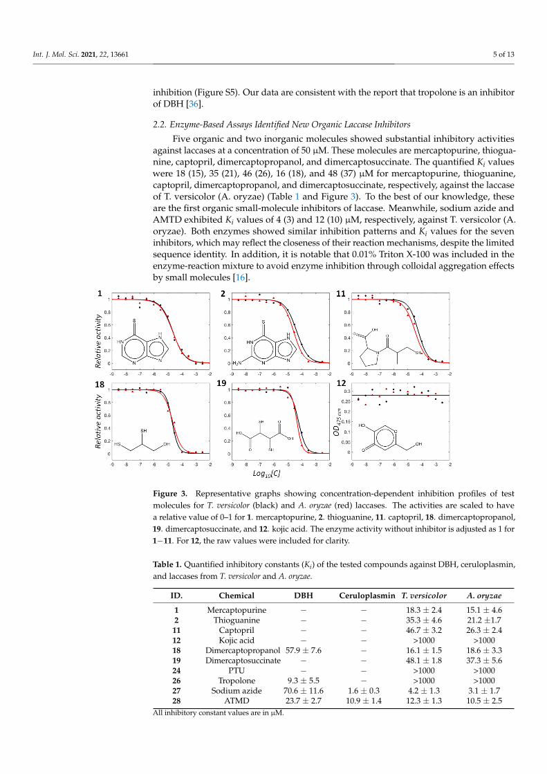
Five organic and two inorganic molecules showed substantial inhibitory activitiesagainst laccases at a concentration of 50 µM. These molecules are mercaptopurine, thiogua-nine, captopril, dimercaptopropanol, and dimercaptosuccinate. The quantified Ki valueswere 18 (15), 35 (21), 46 (26), 16 (18), and 48 (37) µM for mercaptopurine, thioguanine,captopril, dimercaptopropanol, and dimercaptosuccinate, respectively, against the laccaseof T. versicolor (A. oryzae) (Table 1 and Figure 3). To the best of our knowledge, theseare the first organic small-molecule inhibitors of laccase. Meanwhile, sodium azide andAMTD exhibited Ki values of 4 (3) and 12 (10) µM, respectively, against T. versicolor (A.oryzae). Both enzymes showed similar inhibition patterns and Ki values for the seveninhibitors, which may reflect the closeness of their reaction mechanisms, despite the limitedsequence identity. In addition, it is notable that 0.01% Triton X-100 was included in theenzyme-reaction mixture to avoid enzyme inhibition through colloidal aggregation effectsby small molecules [16].
Figure 3. Representative graphs showing concentration-dependent inhibition profiles of testmolecules for T. versicolor (black) and A. oryzae (red) laccases. The activities are scaled to havea relative value of 0–1 for 1. mercaptopurine, 2. thioguanine, 11. captopril, 18. dimercaptopropanol,19. dimercaptosuccinate, and 12. kojic acid. The enzyme activity without inhibitor is adjusted as 1 for1−11. For 12, the raw values were included for clarity.
Table 1. Quantified inhibitory constants (Ki) of the tested compounds against DBH, ceruloplasmin,and laccases from T. versicolor and A. oryzae.
ID. Chemical DBH Ceruloplasmin T. versicolor A. oryzae
1 Mercaptopurine − − 18.3 ± 2.4 15.1 ± 4.62 Thioguanine − − 35.3 ± 4.6 21.2 ±1.711 Captopril − − 46.7 ± 3.2 26.3 ± 2.412 Kojic acid − − >1000 >100018 Dimercaptopropanol 57.9 ± 7.6 − 16.1 ± 1.5 18.6 ± 3.319 Dimercaptosuccinate − − 48.1 ± 1.8 37.3 ± 5.624 PTU − − >1000 >100026 Tropolone 9.3 ± 5.5 − >1000 >100027 Sodium azide 70.6 ± 11.6 1.6 ± 0.3 4.2 ± 1.3 3.1 ± 1.728 ATMD 23.7 ± 2.7 10.9 ± 1.4 12.3 ± 1.3 10.5 ± 2.5
All inhibitory constant values are in µM.

Int. J. Mol. Sci. 2021, 22, 13661 6 of 13
We also evaluated inhibitory enzyme kinetics using various concentrations of sub-strate and inhibitors. The nonlinear fittings of the enzyme kinetics classified all five newinhibitors, i.e., mercaptopurine, thioguanine, captopril, dimercaptopropanol, and dimer-captosuccinate, as competitive mode in the laccases from both sources (Figures 4 and S7,and Table 2), indicating that the inhibitory mechanisms of all inhibitors are similar.
Figure 4. Enzyme inhibitory kinetics with T. versicolor. Left and right panels show Michaelis–Mentenand Lineweaver–Burk plots, respectively, for each inhibitor: 1, 2, 11, 18, and 19. •, �, � and Hrepresent inhibitor concentrations of 10, 20, 30, and 40 µM for 1; 15, 30, 45, and 60 µM for 2; 30, 50, 60,and 80 µM for 11; 10, 20, 30 and 50 µM for 18; and 20, 45, 70, and 90 µM for 19.

Int. J. Mol. Sci. 2021, 22, 13661 7 of 13
Table 2. Parameters of inhibitory kinetics for laccases.
ID Chemical Mechanism * Ki (µM) ** Ki (µM)
1 Mercaptopurine Competitive 2.4 17.22 Thioguanine Competitive 23.4 26.211 Captopril Competitive 5.2 32.418 Dimercaptopropanol Competitive 9.5 18.019 Dimercaptosucciniate Competitive 14.1 13.1
* and ** represent the simulated inhibitory constants by nonlinear fitting, assuming competitive inhibition in T.versicolor and A. oryzae, respectively.
2.3. Differential Scanning Fluorimetry and Fluorescence-Quenching Experiments Support DirectInteractions between Small Molecules and Laccases
Differential scanning fluorimetry (DSF) confirmed direct binding of small moleculeswith laccases. Specific binding of a small molecule can either increase or decrease proteinstability, resulting in a shift in melting temperature (Tm) in DSF. All inhibitors increasedthe Tm values of laccases (Figures 5A and S8). The changes were small but statisticallysignificant for all the inhibitors (p < 0.005). Nevertheless, there was no apparent correlationbetween Tm and Ki values, likely indicating a complicated relationship in binding andinhibition by the small molecules.
Figure 5. (A) Differential scanning fluorimetry (DSF) profiles of inhibitors with laccase fromT. versicolor. DSF profiles for 0 (DMSO), 1 (mercaptopurine), 2 (thioguanine), 11 (captopril),18 (mercaptopropanol), 19 (dimercaptosuccinate), and 27 (sodium azide), respectively. (B) Stern–Volmer constants obtained from the slope of the Stern–Volmer plot in fluorescence-quenchingexperiments are drawn and represented. * and ** represent laccase from T. versicolor andA. oryzae, respectively.
The binding of small molecules can influence the surroundings of fluorophores andtryptophans in a protein, leading to the quenching of fluorescence signals. The alteredpatterns can support the direct interaction between a small molecule and a protein. We

Int. J. Mol. Sci. 2021, 22, 13661 8 of 13
tested whether quenching of fluorescence signals from laccase occurred in a concentration-dependent manner with the inhibitors. The results clearly show that all inhibitors causedquenching (Figures 5B, S9 and S10). The decrements could be classified into two groups byvalues: one of mercaptopurine and thioguanine and the others of captopril, dimercapto-propanol, and mercaptopurine. The inhibitors in each group share chemical similaritiesin the functional moiety. This may support the reliability of the fluorescence-quenchingresults when interpreting the interaction between inhibitors and laccases. We employed twoorthogonal methods, DSF and fluorescence quenching, and both supported direct binding.
2.4. Cheminformatics Analysis Revealed Differences between Tyrosinase and DBH Inhibitors andSuggested New Tyrosinase Inhibitors
One may argue that the test molecules in this study are biased from the viewpoint ofchemical structures, thereby resulting in no finding of new DBH inhibitors. We performeda similarity ensemble approach (SEA) to determine the shared similarity between all tyrosi-nase inhibitors, the test molecules, and DBH inhibitors [37]. SEA reveals the distributionof pairwise Tanimoto coefficients (Tcs) between two small molecules from the respectivegroups. The distribution reflects the shared similarities of the inhibitors in the two groups.
We extracted 579 tyrosinase and 47 DBH organic inhibitors with molecular weightsof <500 Da from the ChEMBL database. The 27,213 (579 × 47) Tcs between tyrosinase andDBH inhibitors share chemical similarities (Figure 6A). Tropolone is a dual inhibitor oftyrosinase and DBH. The pairs that include tropolone or its analogs showed the highest Tcvalues. The analogs are CHEMBL3357560 (Tc = 0.556 against tropolone), CHEMBL48310(0.444), CHEMBL1275999 (0.440), CHEMBL1275969 (0.423), and CHEMBL135189 (0.409).Among them, DBH inhibitors include CHEMBL3357560 and CHEMBL135189. The others,CHEMBL48310, CHEMBL1275999, and CHEMBL1275969, belong to tyrosinase inhibitors.The close similarity to tropolone provoked us to postulate these analogs as dual inhibitorsof tyrosinase and DBH. However, experimental validation was difficult to perform becauseof their commercial unavailability.
Interestingly, the 1222 (26 × 47) Tcs between the 26 tyrosinase inhibitors in this study(omitting two inhibitors, i.e., sodium azide and ATMD, from those in Figure 2) and theDBH inhibitors revealed a similar pattern of distribution. The resembled pattern qualita-tively supports that the 26 test molecules can cover the chemical spaces that tyrosinaseinhibitors map.
The second most highly ranked chemical moiety in pairs was thione. It is an MBGthat is the second most common moiety in tyrosinase inhibitors, present in 202 (35%) of579 ChEMBL-derived inhibitors. Our test molecules also included the 17 thione-containingmolecules (Figure 2). Most DBH inhibitors are thione-containing molecules, accounting for42 of 47 (89%) inhibitors. Of the test molecules, methimazole shared the highest Tc (0.375)with a DBH inhibitor, CHEMBL164791, among the thione-containing pairs (Figure 6B).Despite the apparent similarities with CHEMBL164791, neither methimazole nor otherthione-containing test molecules significantly inhibited DBH at 50 µM. This implies thatthe additional aromatic part in CHEMBL164791 plays a substantial role in inhibition.
We then investigated whether CHEMBL164791 could inhibit tyrosinase activity. BothCHEMBL164791 and its analogous DBH inhibitor, CHEMBL278068 (Tc = 0.353 againstmethimazole), inhibited tyrosinase activity, with Ki values of 15.7 and 12.4 µM, respec-tively (Figure 6C), which suggests that they are dual tyrosinase and DBH inhibitors. Wealso prepared the complex model between tyrosinase and new inhibitor CHEMBL278068by docking simulation with the established protocol [14–17]. The model shows thatCHEMBL278068 shares structural features with those in the complex between PTU and aplant catechol oxidase [38]. Their sulfur positions to the dicopper atoms were almost identi-cal (Figure 6D). These data may suggest the idea that the presence of MBGs is sufficient forinhibition of tyrosinase, at least in thione-containing molecules. However, generalization ofthis idea will require further experimental evidence.

Int. J. Mol. Sci. 2021, 22, 13661 9 of 13
Figure 6. (A) Distribution of Tanimoto coefficients in the chemical pair from tyrosinase and DBHinhibitors. (B) Chemical similarity between methimazole and CHEMBL164791 and CHEMBL278068.(C) Inhibition profiles for tyrosinase by CHEMBL164791 and CHEMBL278068. Activities were scaledwith relative values in the range of 0 to 1. Values in parentheses indicate Ki values. (D) Docked poseof CHEMBL278068 (yellow) to tyrosinase. DOCK3.7 guided the docking of CHEMBL278068 into themushroom tyrosinase coordinate (PDB code: 2Y9X). Brown spheres indicate copper atoms. The PTUstructure (magenta) complexed with catechol oxidase [38] is overlaid for comparison.
3. Discussion
How do the new inhibitors inhibit laccase? The results of fluorescence quenchingand DSF can postulate that the inhibitions occur by direct interactions with laccase. Here,the complex structures of the newly found inhibitors with proteins may provide hints.Two crystal structures possess mercaptopurine as a ligand (PDB ligand name: PM6, https://www.rcsb.org/ligand/PM6 (accessed on 4 November 2021)). Their PDB codes are 3BGDand 3NS1. Thioguanine (ligand name: DX4) is found in four protein structures of 3JQA,4M5M, 4XOY, and 4XP3. There are 14 protein structures containing captopril (ligandname: X8Z). Their PDB codes are 1J37, 2X8Z, 3LUS, 4C1D, 4C1F, 4C1H, 4C2P, 4DPR, 4EXS,4PQA, 5AYA, 5ZIO, 6U10, and 6V61. Mercaptopurine and thioguanine are purine analogsand can inhibit purine-binding proteins. Captopril is a metalloenzyme-specific inhibitor.Therefore, dissection of the complex structures with captopril is informative. There are fourtypes of metalloenzymes complexed with captopril: angiotensin-converting enzyme withmono-Zinc atoms (1J37, 2X8Z, 4C2P), metallo-β-lactamase with di-Zinc (4C1D, 4C1F, 4C1H,4EXS, 5AYA, 5ZIO, 6U10, 6V61), leukotriene A4 hydrolase with mono-Zinc (4DPR), anddesuccinylase with di-Zinc (4PQA). Figure S11 shows 2D diagrams of the intermolecularinteraction between captopril and each type of metalloenzyme. The role of thiol moietyin captopril is to form chelating bonds to catalytic metals. The intermolecular distancebetween sulfur and metal is within 3 Å. It will be reasonable to assume that a similarmechanism exists in the inhibition of laccase by captopril.

Int. J. Mol. Sci. 2021, 22, 13661 10 of 13
Where do the inhibitors bind to the laccase? The substrate binding site will be theprimary candidate. We searched for known laccase structures deposited in the PDBdatabase using the DALI server [39] and extracted 94 coordinates. They include threesubstrates in complex with laccase, which are 2,5-xylidine (PDB ligand: XYD and PDB code:1KYA) [40], 4-methylbenzoic acid (4MA and 2HRG) [41], and sulfoacetate (AS8 and 2XYB).The molecules occupy identical positions on the laccase structures. The closest copper liesat the T1 site. However, the distances between the molecules and the T1 copper are greaterthan 6 Å. The direct chelation found in captopril and metalloenzyme complexes cannot beformed in this condition. We then searched for other potential sites where inhibitors caninteract with copper atoms using FTMap [42] and p2rank [43] methods. However, there isno sufficient space near the copper clusters in the currently available structure (Figure S12).This may suggest the possibility of conformational fluctuation during the catalytic reaction.These complexities make it challenging to prepare an acceptable laccase-inhibitor dock-ing model. Atomistic understanding of the underlying mechanism may necessitate theexperimental 3D complex structure between laccase and inhibitor.
No laccase inhibitor, except sodium azide, has been reported. Our study is one of themost extensive studies on laccase inhibitors. Because the physiological roles of laccaseshave been less studied in pathogens, therapeutic application of laccase inhibitors currentlyremains unclear. New inhibitors may trigger research in this direction.
The cheminformatics-assisted approach to identify novel tyrosinase inhibitors in thisstudy can be a meaningful example for finding new inhibitors. Several small moleculesare known to inhibit various metalloenzymes [2,3]. For instance, captopril, a test moleculein this study, inhibits both zinc-containing angiotensin-converting enzymes and copper-containing tyrosinase. Systematic prediction and comparison across all metalloenzymeinhibitors and their MBGs may be the next step for widening the arsenal space of copper-containing enzyme inhibitors. It is noteworthy that the metal-binding pharmacophorelibrary has shown its usefulness in drug discovery targeting metalloenzymes [1].
4. Materials and Methods
Cheminformatics and docking simulation —RDKit (Open-source cheminformatics;http://www.rdkit.org (accessed on 4 November 2021)) was used to calculate the pairwiseTcs between two molecules by employing ECFP4 as a molecular fingerprint. The data forinhibitors were extracted from ChEMBL25 [32,33]. Docking simulations were tried withDOCK 3.7 [44]. The procedures, in principle, followed what DOCK Blaster had previouslydescribed [45].
Enzyme activity with inhibitors—All chemical reagents were purchased from eitherTokyo Chemical Industry (Tokyo, Japan), ChemBridge (San Diego, CA, USA), or Sigma-Aldrich (St. Louis, MO, USA). The reaction mixture for the tyrosinase assay comprised20 nM tyrosinase and 500 µM L-DOPA in phosphate-buffered saline. The solution for theceruloplasmin activity assay comprised 20 nM ceruloplasmin and 500 µM N,N-dimethyl-p-phenylenediamine (DMPD) in 100 mM of sodium acetate buffer at a pH of 5.5. The DBHactivity assay was conducted in a reaction mixture containing 10 nM DBH, 500 µM DMPD,and 500 µM tyramine. The solution for the laccase activity assay comprised either 150 nM T.versicolor laccase or 350 nM A. oryzae laccase and 500 µM L-DOPA in 100 mM sodium phos-phate buffer with a pH of 5.5. All the solutions included 0.01% Triton X-100 and 5% DMSOin the absence or presence of the inhibitor. After the addition of inhibitors, the solutionswere incubated for 15 min at 30 ◦C. The absorbances of tyrosinase, laccase, ceruloplasmin,and DBH were measured at 475, 475, 550, and 515 nm, respectively, after the additionof substrate to the mixture solution, in a time-dependent manner. After confirming themolecules’ inhibitory activity at a single concentration of 50 µM, concentration-dependentinhibition was observed to calculate the IC50. The Cheng–Prusoff equation with Km wasemployed to convert the IC50 value into the inhibitory constant, Ki, for each inhibitor [46].
Enzyme inhibitory kinetics—The velocities during the generation of the productfor each enzyme were measured by varying the substrate and inhibitor concentrations.

Int. J. Mol. Sci. 2021, 22, 13661 11 of 13
Simultaneous and nonlinear fitting of all profiles in an inhibitor minimized the X2 value ofthe differences of theoretical and experimental values in the Michaelis–Menten equation,expressed with the competitive, uncompetitive, noncompetitive, and mixed models. Thefits resulted in the kinetics parameters Vmax, Km, Kic, and Kiu in each model. The reducedX2 values between the models were compared based on F-statistic to select the mostappropriate model [47]. All fittings and statistical analyses were conducted using MATLABfrom MathWorks (Natick, MA, USA).
Differential scanning fluorimetry—DSF was performed to characterize the directbindings of inhibitors to laccase. SYPROTM orange was added to the enzyme solution inthe presence or absence of 500 µM inhibitor. The fluorescence of the dye was recorded atan excitation wavelength of 492 nm and an emission wavelength of 610 nm by increasingthe temperature from 30◦C to 85◦C. The RT-PCR CFX96 system from BioRad (Hercules,CA, USA) detected the time- and temperature-dependent signals. The mid-point meltingtemperature (Tm) was determined by nonlinearly minimizing the following equation.
I(T) = LL +UL − LL[
1 + exp( Tm−Ta )
] (1)
where LL and UL indicate the top and baseline of the curves, respectively, and a indicatesthe steepness of the slope. Signals in the range of 45 ◦C to 65 ◦C were used for the fitting.
Fluorescence quenching—Fluorescence quenching was performed to evaluate thedirect interaction between the laccases and their inhibitors. The changes in fluorescenceintensity of laccase with excitation (λex) at 280 nm and emission (λem) at 310 nm wererecorded under various inhibitor concentrations. The concentration of laccase was 60 nM,and those for ligands were 0, 0.003, 0.01, 0.03, 0.1, 0.3, 1, 3, 10, 30, 100, and 300 µM. Allspectrophotometric data in this study were obtained using Synergy neo2 from BioTek(Winooski, VT, USA). Changes in the fluorescence intensity by the addition of ligands withthe concentration [L] were interpreted according to the Stern–Volmer equation:
F0F
= 1 + Kqτ0[L] = 1 + Ksv [L] (2)
where F and F0 are the fluorescence intensity of enzymes with and without inhibitors,respectively. Kq is the quencher rate constant, and τ0 is the lifetime of emissive excitedstate of the enzyme without ligands. Ksv is the Stern–Volmer quenching constant. Allexperiments in this study were conducted in triplicate, independently, to generate meanand uncertainty values.
Supplementary Materials: The supplementary materials are available online at https://www.mdpi.com/article/10.3390/ijms222413661/s1.
Author Contributions: J.-G.J. conceived the research project. D.C. and J.-G.J. designed the experi-ments. D.C., F.C., T.N., J.C. and J.-G.J. carried out the experiments and analyzed the data. D.C. andJ.-G.J. wrote the manuscript. D.C., F.C., T.N., J.C. and J.-G.J. agreed with the manuscript. All authorshave read and agreed to the published version of the manuscript.
Funding: This research was supported by the Bio & Medical Technology Development Program of theNational Research Foundation (NRF), funded by the Ministry of Science & ICT (2017M3A9G8083382)and by the 4TH BK21 project (Educational Research Group for Platform development of managementof emerging infectious disease) of the Korean Ministry of Education (5199990614732).
Conflicts of Interest: The authors declare no conflict of interest.

Int. J. Mol. Sci. 2021, 22, 13661 12 of 13
References1. Cohen, S.M. A bioinorganic approach to fragment-based drug discovery targeting metalloenzymes. Acc. Chem. Res. 2017, 50,
2007–2016. [CrossRef] [PubMed]2. Day, J.A.; Cohen, S.M. Investigating the selectivity of metalloenzyme inhibitors. J. Med. Chem. 2013, 56, 7997–8007. [CrossRef]
[PubMed]3. Chen, A.Y.; Adamek, R.N.; Dick, B.L.; Credille, C.V.; Morrison, C.N.; Cohen, S.M. Targeting metalloenzymes for therapeutic
intervention. Chem. Rev. 2019, 119, 1323–1455. [CrossRef] [PubMed]4. Weekley, C.M.; He, C. Developing drugs targeting transition metal homeostasis. Curr. Opin. Chem. Biol. 2017, 37, 26–32. [CrossRef]
[PubMed]5. Riccardi, L.; Genna, V.; De Vivo, M. Metal—ligand interactions in drug design. Nat. Rev. Chem. 2018, 2, 100–112. [CrossRef]6. Caspi, R.; Billington, R.; Keseler, I.M.; Kothari, A.; Krummenacker, M.; Midford, P.E.; Ong, W.K.; Paley, S.; Subhraveti, P.; Karp,
P.D. The MetaCyc database of metabolic pathways and enzymes—A 2019 update. Nucleic Acids Res. 2020, 48, 445–453. [CrossRef]7. Valasatava, Y.; Rosato, A.; Furnham, N.; Thornton, J.M.; Andreini, C. To what extent do structural changes in catalytic metal sites
affect enzyme function? J. Inorg. Biochem. 2018, 179, 40–53. [CrossRef]8. Bowman, S.E.; Bridwell-Rabb, J.; Drennan, C.L. Metalloprotein crystallography: More than a structure. Acc. Chem. Res. 2016, 49,
695–702. [CrossRef]9. Zolghadri, S.; Bahrami, A.; Hassan Khan, M.T.; Munoz-Munoz, J.; Garcia-Molina, F.; Garcia-Canovas, F.; Saboury, A.A. A
comprehensive review on tyrosinase inhibitors. J. Enzyme Inhib. Med. Chem. 2019, 34, 279–309. [CrossRef]10. Pillaiyar, T.; Manickam, M.; Namasivayam, V. Skin whitening agents: Medicinal chemistry perspective of tyrosinase inhibitors.
J. Enzyme Inhib. Med. Chem. 2017, 32, 403–425. [CrossRef]11. Beliaev, A.; Learmonth, D.A.; Soares-da-Silva, P. Synthesis and biological evaluation of novel, peripherally selective chromanyl
imidazolethione-based inhibitors of dopamine β-hydroxylase. J. Med. Chem. 2006, 49, 1191–1197. [CrossRef]12. McCarthy, J.R.; Matthews, D.P.; Broersma, R.J.; McDermott, R.D.; Kastner, P.R.; Hornsperger, J.M.; Demeter, D.A.; Weintraub,
H.J.; Whitten, J.P. 1-(Thienylalkyl) imidazole-2 (3H)-thiones as potent competitive inhibitors of dopamine. beta.-hydroxylase.J. Med. Chem. 1990, 33, 1866–1873. [CrossRef]
13. Kruse, L.I.; Kaiser, C.; DeWolf, W.E., Jr.; Frazee, J.S.; Erickson, R.W.; Ezekiel, M.; Ohlstein, E.H.; Ruffolo, R.R., Jr.; Berkowitz, B.A.Substituted 1-benzylimidazole-2-thiols as potent and orally active inhibitors of dopamine. Beta.-hydroxylase. J. Med. Chem. 1986,29, 887–889. [CrossRef]
14. Choi, J.; Choi, K.-E.; Park, S.J.; Kim, S.Y.; Jee, J.-G. Modeling. Ensemble-based virtual screening led to the discovery of new classesof potent tyrosinase inhibitors. J. Chem. Inf. Model 2016, 56, 354–367. [CrossRef]
15. Choi, J.; Jee, J.-G. Repositioning of thiourea-containing drugs as tyrosinase inhibitors. Int. J. Mol. Sci. 2015, 16, 28534–28548.[CrossRef]
16. Choi, J.; Lee, Y.-M.; Jee, J.-G. Thiopurine drugs repositioned as tyrosinase inhibitors. Int. J. Mol. Sci. 2018, 19, 77. [CrossRef]17. Choi, J.; Park, S.-J.; Jee, J.-G. Analogues of ethionamide, a drug used for multidrug-resistant tuberculosis, exhibit potent inhibition
of tyrosinase. Eur. J Med. Chem. 2015, 106, 157–166. [CrossRef]18. Saghaie, L.; Pourfarzam, M.; Fassihi, A.; Sartippour, B. Synthesis and tyrosinase inhibitory properties of some novel derivatives
of kojic acid. Res. Pharm. Sci. 2013, 8, 233–242.19. Sambasiva Rao, K.; Tripathy, N.; Srinivasa Rao, D.; Prakasham, R. Production, characterization, catalytic and inhibitory activities
of tyrosinase. Res. J. Biotechnol. 2013, 8, 1.20. Bento, I.; Peixoto, C.; Zaitsev, V.N.; Lindley, P.F. Ceruloplasmin revisited: Structural and functional roles of various metal
cation-binding sites. Acta Crystallogr. Sect. D Biol. Crystallogr. 2007, 63, 240–248. [CrossRef]21. Hellman, N.; Gitlin, J. Ceruloplasmin metabolism and function. Annu. Rev. Nutr. 2002, 22, 439–458. [CrossRef] [PubMed]22. Han, I.W.; Jang, J.-Y.; Kwon, W.; Park, T.; Kim, Y.; Lee, K.B.; Kim, S.-W. Ceruloplasmin as a prognostic marker in patients with bile
duct cancer. Oncotarget 2017, 8, 29028. [CrossRef]23. Roberti, M.D.R.F.; Borges Filho, H.M.; Gonçalves, C.H.; Lima, F.L. Aceruloplasminemia: A rare disease-diagnosis and treatment
of two cases. Rev. Bras. Hematol. Hemoter. 2011, 33, 389–392. [CrossRef] [PubMed]24. Rahman, M.K.; Rahman, F.; Rahman, T.; Kato, T. Dopamine-β-hydroxylase (DBH), its cofactors and other biochemical parameters
in the serum of neurological patients in Bangladesh. Int. J. Biomed. Sci. 2009, 5, 395–401. [CrossRef] [PubMed]25. Vendelboe, T.V.; Harris, P.; Zhao, Y.; Walter, T.S.; Harlos, K.; El Omari, K.; Christensen, H.E. The crystal structure of human
dopamine β-hydroxylase at 2.9 Å resolution. Sci. Adv. 2016, 2, e1500980. [CrossRef] [PubMed]26. Tishchenko, K.; Beloglazkina, E.; Mazhuga, A.; Zyk, N. Copper-containing enzymes: Site types and low-molecular-weight model
compounds. Rev. J. Chem. 2016, 6, 49–82. [CrossRef]27. Strong, P.; Claus, H. Laccase: A review of its past and its future in bioremediation. Cri. Rev. Environ. Sci. Technol. 2011, 41, 373–434.
[CrossRef]28. Martínez-Sotres, C.; Rutiaga-Quiñones, J.G.; Herrera-Bucio, R.; Gallo, M.; López-Albarrán, P.; Technology. Molecular docking
insights into the inhibition of laccase activity by medicarpin. Wood Sci. Technol 2015, 49, 857–868. [CrossRef]29. Wang, T.; Xiang, Y.; Liu, X.; Chen, W.; Hu, Y. A novel fluorimetric method for laccase activities measurement using Amplex Red
as substrate. Talanta 2017, 162, 143–150. [CrossRef]

Int. J. Mol. Sci. 2021, 22, 13661 13 of 13
30. Shiro, Y. Structure and function of bacterial nitric oxide reductases: Nitric oxide reductase, anaerobic enzymes. Biochim. Biophys.Acta. Bioenerg. 2012, 1817, 1907–1913. [CrossRef]
31. Jumper, J.; Evans, R.; Pritzel, A.; Green, T.; Figurnov, M.; Ronneberger, O.; Tunyasuvunakool, K.; Bates, R.; Zidek, A.; Potapenko,A.; et al. Highly accurate protein structure prediction with AlphaFold. Nature 2021, 596, 583–589. [CrossRef]
32. Mendez, D.; Gaulton, A.; Bento, A.P.; Chambers, J.; De Veij, M.; Félix, E.; Magariños, M.P.; Mosquera, J.F.; Mutowo, P.; Nowotka,M. ChEMBL: Towards direct deposition of bioassay data. Nucleic Acids Res. 2019, 47, 930–940. [CrossRef]
33. Davies, M.; Nowotka, M.; Papadatos, G.; Dedman, N.; Gaulton, A.; Atkinson, F.; Bellis, L.; Overington, J.P. ChEMBL web services:Streamlining access to drug discovery data and utilities. Nucleic Acids Res. 2015, 43, 612–620. [CrossRef]
34. Kuo, T.; Ho, F. Competitive inhibition of mushroom tyrosinase by captopril. Res. J. Biotechnol. 2013, 8, 26.35. Espín, J.C.; Wichers, H.J. Effect of captopril on mushroom tyrosinase activity in vitro. Biochim. Biophys. Acta 2001, 1544, 289–300.
[CrossRef]36. Meck, C.; D’Erasmo, M.P.; Hirsch, D.R.; Murelli, R.P. The biology and synthesis of α-hydroxytropolones. Med. Chem. Comm. 2014,
5, 842–852. [CrossRef]37. Keiser, M.J.; Roth, B.L.; Armbruster, B.N.; Ernsberger, P.; Irwin, J.J.; Shoichet, B.K. Relating protein pharmacology by ligand
chemistry. Nat. Biotechnol. 2007, 25, 197–206. [CrossRef]38. Klabunde, T.; Eicken, C.; Sacchettini, J.C.; Krebs, B. Crystal structure of a plant catechol oxidase containing a dicopper center.
Nat. Struct. Biol. 1998, 5, 1084–1090. [CrossRef]39. Holm, L.; Rosenstrom, P. Dali server: Conservation mapping in 3D. Nucleic Acids Res. 2010, 38, W545–W549. [CrossRef]40. Bertrand, T.; Jolivalt, C.; Briozzo, P.; Caminade, E.; Joly, N.; Madzak, C.; Mougin, C. Crystal structure of a four-copper laccase
complexed with an arylamine: Insights into substrate recognition and correlation with kinetics. Biochemistry 2002, 41, 7325–7333.[CrossRef]
41. Matera, I.; Gullotto, A.; Tilli, S.; Ferraroni, M.; Scozzafava, A.; Briganti, F. Crystal structure of the blue multicopper oxidase fromthe white-rot fungus Trametes trogii complexed with p-toluate. Inorganica Chim. Acta 2008, 361, 4129–4137. [CrossRef]
42. Kozakov, D.; Grove, L.E.; Hall, D.R.; Bohnuud, T.; Mottarella, S.E.; Luo, L.; Xia, B.; Beglov, D.; Vajda, S. The FTMap family of webservers for determining and characterizing ligand-binding hot spots of proteins. Nat. Protoc. 2015, 10, 733–755. [CrossRef]
43. Krivak, R.; Hoksza, D. P2Rank: Machine learning based tool for rapid and accurate prediction of ligand binding sites from proteinstructure. J. Cheminformatics 2018, 10, 39. [CrossRef]
44. Bender, B.J.; Gahbauer, S.; Luttens, A.; Lyu, J.; Webb, C.M.; Stein, R.M.; Fink, E.A.; Balius, T.E.; Carlsson, J.; Irwin, J.J.; et al. Apractical guide to large-scale docking. Nat. Protoc. 2021, 16, 4799–4832. [CrossRef]
45. Irwin, J.J.; Shoichet, B.K.; Mysinger, M.M.; Huang, N.; Colizzi, F.; Wassam, P.; Cao, Y. Automated docking screens: A feasibilitystudy. J. Med. Chem. 2009, 52, 5712–5720. [CrossRef]
46. Yung-Chi, C.; Prusoff, W.H. Relationship between the inhibition constant (KI) and the concentration of inhibitor which causes 50per cent inhibition (I50) of an enzymatic reaction. Biochem. Pharmacol. 1973, 22, 3099–3108. [CrossRef]
47. Dias, A.A.; Pinto, P.A.; Fraga, I.; Bezerra, R.M. Diagnosis of enzyme inhibition using Excel Solver: A combined dry and wetlaboratory exercise. J. Chem. Educ. 2014, 91, 1017–1021. [CrossRef]