Research on Mushroom as a Potential Source of Nutraceuticals
Upload
independentCategory
view
0download
0
lable at ScienceDirect
European Journal of Medicinal Chemistry 98 (2015) 203e211
Contents lists avai
European Journal of Medicinal Chemistry
journal homepage: http: / /www.elsevier .com/locate/ejmech
Research paper
Kinetic and in silico studies of novel hydroxy-based thymol analoguesas inhibitors of mushroom tyrosinase
Zaman Ashraf a, b, Muhammad Rafiq a, Sung-Yum Seo a, *, Kang Sung Kwon c,Mustafeez Mujtaba Babar d, Najam-us-Sahar Sadaf Zaidi c
a Department of Biology, College of Natural Sciences, Kongju National University, Gongju 314-701, Republic of Koreab Department of Chemistry, Allama Iqbal Open University, Islamabad 44000, Pakistanc Department of Chemistry, Chungnam National University Daejeon, 305-764, Republic of Koread Atta-ur-Rahman School of Applied Biosciences, National University of Sciences and Technology, H-12, Kashmir Highway, Islamabad, 44000, Pakistan
a r t i c l e i n f o
Article history:Received 19 March 2015Received in revised form30 April 2015Accepted 20 May 2015Available online 21 May 2015
Keywords:Thymol analoguesSynthesisAntioxidant activityMushroom tyrosinase inhibitorsKinetic mechanismMolecular docking
* Corresponding author. .E-mail address: [email protected] (S.-Y. Seo).
http://dx.doi.org/10.1016/j.ejmech.2015.05.0310223-5234/© 2015 Published by Elsevier Masson SAS
a b s t r a c t
The present studies reports the synthesis of hydoxylated thymol analogues (4aee) and (6aec) asmushroom tyrosinase inhibitors. The title compounds were obtained in good yield and characterized byFTIR, 1H NMR, 13C NMR, Mass spectral data and X-ray crystallography in case of compound (6a). Theinhibitory effects on mushroom tyrosinase and DPPH were evaluated and it was observed that 2-[5-methyl-2-(propan-2-yl)phenoxy]-2-oxoethyl (2E)-3-(4-hydroxyphenyl)prop-2-enoate (6b) showedtyrosinase inhibitory activity (IC50 15.20 mM) comparable to kojic acid (IC50 16.69 mM) while 2-[5-methyl-2-(propan-2-yl)phenoxy]-2-oxoethyl 3,4-dihydroxybenzoate (4d) exhibited higher antioxidant potential(IC50 11.30 mM) than standard ascorbic acid (IC50 24.20 mM). The docking studies of synthesized thymolanalogues was also performed against tyrosinase protein (PDBID 2ZMX) to compare the binding affinitieswith IC50 values. The predicted binding affinities are in good agreement with the IC50 values as com-pound (6b) showed highest binding affinity �7.1 kcal/mol. The kinetic mechanism analyzed by Line-weavereBurk plots exhibited that compound (4d) and (6b) inhibit the enzyme by two different pathwaysdisplayed mixed-type inhibition. The inhibition constants Ki calculated from Dixon plots for compounds(4d) and (6b) are 34 mM and 25 mM respectively. It was also found from kinetic analysis that derivative(6b) formed reversible enzyme inhibitor complex. It is propose on the basis of our investigation that titlecompound (6b) may serve as lead structure for the design of more potent tyrosinase inhibitors.
© 2015 Published by Elsevier Masson SAS.
1. Introduction
Tyrosinase (EC 1.14.18.1) as a binuclear copper-containing met-alloenzyme catalyzes two distinct reactions of melanin biosyn-thesis. It catalyzes L-tyrosine to L-3,4-dihydroxyphenylalanine (L-DOPA) and further oxidation of (L-DOPA) to dopaquinone whichthen transformed through several reactions into brown to blackmelanin. The distribution patterns of melanin in the surroundingkeratinocytes determine the color of human skin [1,2]. Kadekaroet al. and Petit et al. also reported that there are certain other fac-tors such as UV exposure, a-melanocyte-stimulating hormone,melanocortin 1 receptor and agouti-related protein which involvedin melanin biosynthesis [3,4]. Abnormal melanin production such
.
as observed in melasma, freckles, lentigo senilis, and other forms ofmelanin hyperpigmentation can be a serious aesthetic problemoften causing emotional disturbance [5e7]. Taking into account thekey role of tyrosinase in melanin production, many tyrosinase in-hibitors have found application in cosmetics and pharmaceuticalproducts [8e10]. A large number of tyrosinase inhibitors have beenreported but only a few are used because of their limitations withregards to cytotoxicity, selectivity and stability. Thus, it is in greatneed of developing new tyrosinase inhibitors without causingadverse reactions.
A number of cinnamic acid and benzoic acid analogues pos-sessing hydroxy groups at position 3 and 4 of the phenyl ring havebeen reported as antioxidants and mushroom tyrosinase inhibitors[11,12]. Liu (2003) and Chen (2005) also reported that alkoxybenzoic acids and hydroxy benzoic acids showed potent mushroomtyrosinase inhibitory activity [13,14]. Thymol a naturally occurringmonoterpene phenol which is the main constituent of thyme

Z. Ashraf et al. / European Journal of Medicinal Chemistry 98 (2015) 203e211204
possesses tyrosinase inhibitory activity [15] along with othernumerous pharmacological activities. As phenolic antioxidants,thymol protects food qualities and organisms fromdamage inducedby oxidative stress. Thymol is used as meat preservatives orflavoring agent in the food industry. It exhibited anti-inflammatoryeffect in human neutrophils incubated [16] and also inhibits theformation of lethal products through reactive nitrogen species [17].Thymol displays antimicrobial [18e20] and wound-healing activity[21] and is able to rise the levels of macrophage migration inhibi-tory factor (MIF) in central nervous system [22]. Thymol increasedthe in vitro fibroblast growth [23], effectively inhibited COX-1 [24]and prevented inducible lymphocyte proliferation [25].
Keeping in view the importance of these structural features wehave synthesized the hydroxylated thymol analogues having ben-zoic acid and cinnamic acid moieties in order to discover theirtyrosinase inhibitory potential to offer a source for the develop-ment of new effective tyrosinase inhibitors. The antioxidant activitywas also carried out as most of the clinically used tyrosinase in-hibitors kojic acid, arbutin, kaempferol, hydroquinone, etc. allpossess antioxidant activity. In addition to evaluate the tyrosinaseinhibitory potential and antioxidant activity of synthesized hy-droxylated thymol derivativesmolecular dockingwas carried out topredict the position of the synthesized compounds in the active siteof the 3D structure of tyrosinase (PDB ID 2ZMX). The investigationof the binding interactions during docking analysis between ligand-protein functionalities is important to elucidate the possible mo-lecular mechanism [26,27].
2. Results and discussion
2.1. Chemistry
The title compounds (4aee) and (6aec) were synthesized byfollowing the already reported method [28] with some modifica-tion shown in Schemes 1 and 2. The thymol chlroacetyl derivative(2) was synthesized by esterification of phenolic eOH group ofthymol with chloroacetyl chloride in the presence of (C2H5)3N andanhydrous methylene chloride as solvent. The presence of estercarbonyl stretching at 1723 cm�1 and disappearance of the eOHstretching in FTIR spectra confirmed the formation intermediate(2). The final products (4aee) and (6aec) were prepared by simple
OH
CH3
CH3 CH3
+Cl Cl
O
(1)
(2)
+ OH
O
R
(3a-3e)
(C2H5)
DM
R=3a = 3-OH 3b = 4-OH3c = 2,4-di-OH 3d = 3,4-di-OH 3e = 3,5-di-OH
OO
Cl
CH3
CH3 CH3
(C2H5)
0 t
Scheme 1. Synthesis of thy
nucleophilic substitution at intermediate (2) with hydroxysubstituted benzoic acids (3aee) and substituted and unsubstitutedcinnamic acids (5aec) respectively. All of the synthesized com-pounds have been characterized by FTIR, 1HNMR, 13CNMR andMassspectroscopic data. Compound (6a) yielded single crystals, suitablefor X-ray diffraction studies. The X-ray diffraction analysis alsoconfirmed the formation of the desired product and this is anambiguous evidence of the structure.
2.2. Bioassay for tyrosinase inhibitory activity
Hydroxylated thymol analogues have been designed to evaluatetheir inhibitory effects on mushroom tyrosinase activity. Kojic acida competitive tyrosinase inhibitor was used as standard for com-parison purpose. The hydroxylated tyrosinase inhibitors asdescribed in preceding section are of special interest because oftheir high activity (IC50 < 10 mM). Novel thymol analogues (4aee)and (6aec) have been synthesized by incorporation of hydroxyl-ated benzoic acids and cinnamic acids. The synthesis of mono anddi-hydroxylated derivatives with different position of eOH atphenyl ring was carried out to explore the role of multiple hydroxylgroups in tyrosinase inhibition. It has been exposed from ourbioassay results (Table 1) that the major determining factor ofinhibitory activity is the position and not the number of the hy-droxyl groups. Interestingly, compound (4d) bearing 3,4-dihydroxysubstituted benzoic acid moiety showed higher activity (IC5045.0 mM) than (4c) and (4e) having IC50 56.1 and 220.9 mMrespectively. The derivatives (4c) and (4e) possess 2,4- and 3,5-dihydroxy substituted benzoic acid residues respectively. In caseof compound (4d) two hydroxy groups are present on adjacentpositions of phenyl ring; this impedes the molecule to interact wellwith enzyme. This structural feature can be well correlated with L-DOPA which is used as substrate for tyrosinase enzyme duringbioassay. Thus compound (4d) because of close structural similar-ities with L-DOPA is more active among the dihydroxylated thymolanalogues. Table 1 presented the IC50 values of the synthesizedthymol analogues and it was observed that kojic acid is more activethan all of the synthesized thymol analogues except compound(6b). Compound (6b) showed excellent tyrosinase inhibitory ac-tivity with IC50 15.2 mM and is so inhibitor as kojic acid (IC5016.69 mM). The synthesized thymol derivatives (6a), (6b) and (6c)
OO
Cl
CH3
CH3 CH3
(2)
3N/KI
FR
(4a-4e)
R is same as in (3a-3e)
O
O
CH3
CH3CH3
O
O
3N/CH2Cl2
o -5°C
mol analogues (4aee).

Table 1Tyrosinase inhibitory activity and free radical scavenging activity of the synthesized thymol analogues (4aee) and (6aec).
Codes Tyrosinase inhibition Activity DPPH Activity
% Inhibition (25 mg/mL) IC50 ± SEM mM % Inhibition (500 mg/mL) IC50 ± SEM mM
4a 48 ± 1 79.3 ± 5.3 3 ± 1 a
4b 33 ± 2 91.5 ± 9.4 2 ± 1 a
4c 68 ± 2 56.1 ± 5.9 3 ± 1 a
4d 55 ± 3 45.0 ± 1.5 a 11.30 ± 2.094e 5 ± 2 220.9 ± 11.6 9 ± 2 a
6a 42 ± 3 100.8 ± 14.3 8 ± 3 a
6b 93 ± 4 15.2 ± 0.42 5 ± 2 a
6c 77 ± 2 45.2 ± 4.3 50 ± 6 1344.0 ± 18.76Kojic acid 100 16.69 ± 2.8 a a
Ascorbic acid a a 97 ± 1 24.20 ± 1.93
a Not determined.
Z. Ashraf et al. / European Journal of Medicinal Chemistry 98 (2015) 203e211 205
all possess cinnamic acid moiety but varied in substitution patternon phenyl ring. The derivative (6a) have unsubstituted cinnamicacid residue while compound (6b) and (6c) possess 4-hydroxyl and4-chloro cinnamic acid moiety respectively. Interestingly, presenceof 4-hydroxyl group on cinnamic acid phenyl ring in compound(6b) led to a dramatic increase in tyrosinase inhibitory activity. Thecompound (6a) (IC50 100.8 mM) is 6.6 times less active while (6c)(IC50 45.2 mM) is 2.97 times less active than the most potent de-rivative (6b). Based upon our results we propose that thymolanalogue (6b) may serve as a lead structure for the design of morepotent tyrosinase inhibitors.
2.3. Kinetic mechanism
The inhibitory mechanism of the most active compounds (6b)and (4d) on mushroom tyrosinase for the oxidation of L-DOPA wasstudied. Kinetic studies showed a concentration dependent inhi-bition of mushroom tyrosinase by thymol derivatives (4d) and (6b).Continuous monitoring of the reaction showed a marked decreasein reaction rate in the presence of the inhibitors, which is ulti-mately, indicated the decrease in the final absorbance whencompared with controls containing no inhibitor. The potency ofinhibition exhibited by these compounds varied depending on thepresence of type and position of the functionalities on the aromaticrings [29]. Inhibition kinetics was analyzed by Lineweaver-Burkplot and Dixon plots to determine the type of inhibition and inhi-bition constant (Ki). The results of Figs. 1e2(a) showed that in caseof compounds (4d) and (6b) by increasing the concentration ofsubstrate (L-DOPA) gave family of straight lines, which intersectedwithin the second quadrant. The analysis showed that Vmaxdecreased with changing Km in the presence of increasing
Fig. 1. a) Lineweaver-Burk plots for the inhibition of the diphenolase activity of mushroomconcentrations of L-DOPA. b) Dixon plots for the inhibition of the diphenolase activity of mdifferent concentrations of L-DOPA (0.062, 0.125, 0.25, 0.5 and 1 mM, respectively).
concentrations of compounds. This behavior of compounds (4d)and (6b) indicated that it inhibits tyrosinase by two differentpathways and show mixed type inhibition. On the other hand inDixon plot, slope obtained from the plots for uninhibited enzymeand with different concentrations of inhibitors (4d) and (6b) wasconsistent with the characteristic patterns of mixed-type inhibitionwith Ki value 34 mM and 25 mM as shown in Figs. 1e2(b)respectively.
2.4. Reversible inhibition
The most potent derivative (6b) was selected to ascertain theformation of reversible enzyme-Inhibitor complex between tyros-inase and inhibitor (6b). The plots of initial rate verses enzymeconcentrations (4, 6, 8, 10, 15 and 20 mg/mL) for compound (6b) allconsisted of straight lines passing through the origin. Increasing theinhibitor concentration resulted in a decrease in the slope of thelines, which indicate that the enzyme undergoes a reversible in-hibition [30] (Fig. 3).
2.5. DPPH radical scavenging assay
Thymol derivatives were evaluated for the DPPH free radicalscavenging ability as excessive melanogenesis generates free radi-cals which start unwanted reactions. The thymol derivative (4d)exhibited excellent radical scavenging activity when comparedwith the standard ascorbic acid. The IC50 value of compound (4d) is11.30 mMwhile IC50 value of ascorbic acid is 24.20 mM. The presenceof ortho disubstituted phenyl ring in compound (4d) play signifi-cant role in radical scavenging activity as it showed more potentradical scavenging activity than ascorbic acid. All of the other
tyrosinase by various concentrations of compound (4d) in the presence of differentushroom tyrosinase by various concentrations of compound (4d) in the presence of

Fig. 2. a) Lineweaver-Burk plots for the inhibition of the diphenolase activity of mushroom tyrosinase by various concentrations of compound (6b) in the presence of differentconcentrations of L-DOPA. b) Dixon plots for the inhibition of the diphenolase activity of mushroom tyrosinase by various concentrations of compound (6b) in the presence ofdifferent concentrations of L-DOPA (0.062, 0.125, 0.25, 0.5 and 1 mM, respectively).
Fig. 3. Effects of concentrations of mushroom tyrosinase on its activity for the catalysisof L-DOPA at different concentrations of compound (6b).
Z. Ashraf et al. / European Journal of Medicinal Chemistry 98 (2015) 203e211206
thymol derivatives showed little activity even at high concentration(500 mg/mL) Table 1.
2.6. Docking studies
The in silico docking of the synthesized thymol derivatives wasperformed against crystal structure of tyrosinase protein (PDBID2ZMX) to compare their experimental IC50 with binding affinities.Matoba et al. published recently the crystallographic structure oftyrosinase (PDBID 2ZMX) which enables us to predict the bindingaffinities of thymol derivatives. The compound (6b) showed highestbinding affinity with the target protein having binding energyvalue �7.1 kcal/mol. The experimentally determined tyrosinaseinhibitory activity results are in good agreement with the predictedbinding affinities by in silico docking. The 4ehydroxy group ofcinnamic acid phenyl ring in compound (6b) formed hydrogenbonds with GLU67 having distance 1.99 Å. The p-p stacking alsopresent in the same compound between phenyl ring of thymolmoiety and PHE34 having distance 3.81 Å (Fig. 4).
The phenolic hydroxyls present at positions 3 and 4 in com-pound (4d) formed hydrogen bonds with SER96 having bondlengths 2.36 Å and 2.89 Å respectively (Fig. 5) The hydrogen of 3-hydroxyl in the same compound also interact with VAL95 havingdistance 2.40 Å between two interacting groups. The ester carbonyloxygen in compound (4d) also formed hydrogen bondwith ARG140having bond length 2.40 Å. Table 2 presented the calculated bind-ing energies of the synthesized thymol analogues and it determines
the stability of ligandetarget complex. There is hydrogen bondbetween 4-hydroxy group of derivative (4c) and amino acid THY37of the target protein with bond length 1.84 Å (Fig. 6). The bindingenergy of thymol analogue (4c) is �6.5 kcal/mo. The presence ofphenolic hydroxyl on acid moiety in the synthesized compounds isthe determining factor of binding energies. Another importantstructural feature is the cinnamic acid moiety. It was observed thatwhen phenolic hydroxyl is present on para position of the cinnamicacid residue then ligand has maximum binding affinity with thereceptor protein. The experimentally determined IC50 values alsoconfirmed the docking results as compound (6b) showed themaximum inhibition of the mushroom tyrosinase activity. Thepartition coefficient values (log P) of the synthesized compoundswere calculated by ChemSketch ACD labs 2012 to verify the drug-likeness properties of the compounds. The most potent de-rivatives (6b) and (4d) have partition coefficient values 4.92 ± 0.33and 4.97 ± 0.32 respectively which are not beyond the limits of (logP) values.
3. Conclusion
The hydroxylated thymol analogues (4aee) and (6aec) havingsubstituted benzoic acids and cinnamic acids moieties have beensynthesized and fully characterized. The computational studiesinvestigation revealed that thymol analogue (6b) possessesmaximum binding affinity (�7.1 kcal/mol) with the target protein(PDBID 2ZMX). The wet lab results are in good agreement with thecalculated docking scores. The most potent activity against mush-room tyrosinase was exhibited by the compound (6b) having IC5015.2 mM. The presence of phenolic eOH at para-position on cin-namic acid phenyl ring play very significant role in tyrosinase in-hibition activity. The kinetic analysis of compound (6b) shown thatit is mixed type inhibitor with Ki value 25 mMand formed reversibledrugereceptor complex with mushroom tyrosinase. It wasconcluded from our results that the title compound (6b) may serveas lead structure for the design of more potent tyrosinaseinhibitors.
4. Experimental
All chemicals used for the synthesis of compounds were pur-chased from Sigma Chemical Co. Melting points were determinedusing a Digimelt MPA 160, USA melting point apparatus and arereported uncorrected. The FTIR spectra were recorded with Shi-madzu FTIRe8400S spectrometer (Kyoto, Japan, y, cm�1). Massspectra were performed on an Agilent 6460 Series Triple

Fig. 4. The interactions of compound (6b) with the active site of mushroom tyrosinase (PDBID 2ZMX) generated by using Discovery Studio 4.1.0. The a) shows the two dimensionalinteraction patterns. The legend inset represents the type of interaction between the ligand atoms and the amino acid residues of the protein. The b) shows the three-dimensionaldocking of the compound in the binding pocket. Dashed lines indicate bond distances between interacting functionalities of the ligand and receptor.
Fig. 5. The interactions of compound (4d) with the active site of mushroom tyrosinase (PDBID 2ZMX) generated by using Discovery Studio 4.1.0. The a) shows the two dimensionalinteraction patterns. The legend inset represents the type of interaction between the ligand atoms and the amino acid residues of the protein. The b) shows the three-dimensionaldocking of the compound in the binding pocket. Dashed lines indicate bond distances between interacting functionalities of the ligand and receptor.
Table 2Average binding affinity and Log P values of the synthesized thymol analogues(4aed) and (6aec).
Compound Average binding affinity (kcal/mol) Log pa
4a 5.8 5.17 ± 0.304b 6.4 5.14 ± 0.304c 6.5 5.20 ± 0.334d 6.6 4.97 ± 0.324e 6.5 4.88 ± 0.326a 5.6 5.45 ± 0.306b 7.1 4.92 ± 0.336c 5.7 5.98 ± 0.33
a Log p calculated from ChemSketch ACD labs 2012.
Z. Ashraf et al. / European Journal of Medicinal Chemistry 98 (2015) 203e211 207
Quadrupole instrument (Agilent). The ionization was achieved byelectrospray ionization in the positive ion mode (ESIþ) and nega-tive ion mode (ESI-). The capillary voltage was set to 4.0 kV. Thesource temperature was 120 �C, and the desolvation temperature
was 350 �C. Nitrogen was used as a desolvation gas (flow 600 L/h).Elemental Analysis (C, H) were carried out on a Flash 2000 serieselenatal analyzer with TCD detector system and results are with±0.3%. The 1H NMR and 13C NMR spectra (CDCl3) and (DMSO-d6)were recorded using a Bruker 400 MHz spectrometer. Chemicalshifts (d) are reported in ppm downfield from the internal standardtetramethylsilane (TMS). The purity of the compoundswas checkedby thin layer chromatography (TLC) on silica gel plate using n-hexane and ethyl acetate as mobile phase. The procedure for thesynthesis of the desired compounds is depicted in Schemes 1 and 2.
4.1. Reagents
Mushroom tyrosinase was purchased from Sigma (USA); L-DOPAand thymol were purchased from Sigma (USA). Stock solutions ofthe reducing substrates were prepared in phosphate buffer (20mM,pH 6.8).
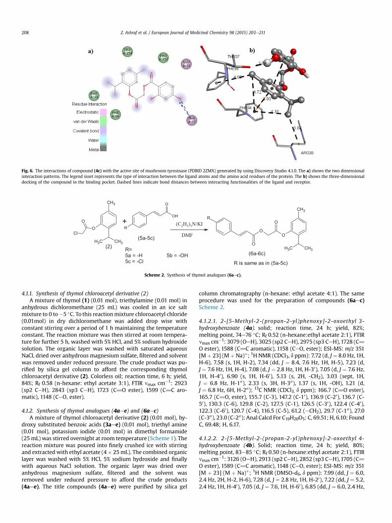
Fig. 6. The interactions of compound (4c) with the active site of mushroom tyrosinase (PDBID 2ZMX) generated by using Discovery Studio 4.1.0. The a) shows the two dimensionalinteraction patterns. The legend inset represents the type of interaction between the ligand atoms and the amino acid residues of the protein. The b) shows the three-dimensionaldocking of the compound in the binding pocket. Dashed lines indicate bond distances between interacting functionalities of the ligand and receptor.
(2)
+O
O
Cl
CH3
CH3 CH3
O
O
CH3
CH3CH3
O
O
R
R
OH
O
(5a-5c)
(C2H5)3N/KI
DMF
(6a-6c)R=5a = -H 5b = -OH5c = -Cl R is same as in (5a-5c)
Scheme 2. Synthesis of thymol analogues (6aec).
Z. Ashraf et al. / European Journal of Medicinal Chemistry 98 (2015) 203e211208
4.1.1. Synthesis of thymol chloroacetyl derivative (2)A mixture of thymol (1) (0.01 mol), triethylamine (0.01 mol) in
anhydrous dichloromethane (25 mL) was cooled in an ice saltmixture to 0 to�5 �C. To this reactionmixture chloroacetyl chloride(0.01mol) in dry dichloromethane was added drop wise withconstant stirring over a period of 1 h maintaining the temperatureconstant. The reaction mixture was then stirred at room tempera-ture for further 5 h, washed with 5% HCl, and 5% sodium hydroxidesolution. The organic layer was washed with saturated aqueousNaCl, dried over anhydrous magnesium sulfate, filtered and solventwas removed under reduced pressure. The crude product was pu-rified by silica gel column to afford the corresponding thymolchloroacetyl derivative (2). Colorless oil; reaction time, 6 h; yield,84%; Rf 0.58 (n-hexane: ethyl acetate 3:1), FTIR nmax cm�1: 2923(sp2 CeH), 2843 (sp3 CeH), 1723 (C]O ester), 1599 (C]C aro-matic), 1148 (CeO, ester).
4.1.2. Synthesis of thymol analogues (4aee) and (6aec)A mixture of thymol chloroacetyl derivative (2) (0.01 mol), hy-
droxy substituted benzoic acids (3aee) (0.01 mol), triethyl amine(0.01 mol), potassium iodide (0.01 mol) in dimethyl formamide(25mL) was stirred overnight at room temperature (Scheme 1). Thereaction mixture was poured into finely crushed ice with stirringand extractedwith ethyl acetate (4� 25mL). The combined organiclayer was washed with 5% HCl, 5% sodium hydroxide and finallywith aqueous NaCl solution. The organic layer was dried overanhydrous magnesium sulfate, filtered and the solvent wasremoved under reduced pressure to afford the crude products(4aee). The title compounds (4aee) were purified by silica gel
column chromatography (n-hexane: ethyl acetate 4:1). The sameprocedure was used for the preparation of compounds (6aec)Scheme 2.
4.1.2.1. 2-[5-Methyl-2-(propan-2-yl)phenoxy]-2-oxoethyl 3-hydroxybenzoate (4a). solid; reaction time, 24 h; yield, 82%;melting point, 74e76 �C; Rf 0.52 (n-hexane:ethyl acetate 2:1), FTIRnmax cm�1: 3079 (OeH), 3025 (sp2 CeH), 2975 (sp3 CeH),1728 (C]O ester), 1588 (C]C aromatic), 1158 (CeO, ester); ESI-MS: m/z 351[Mþ 23] (Mþ Na)þ; 1H NMR (CDCl3, d ppm): 7.72 (d, J¼ 8.0 Hz, 1H,H-6), 7.58 (s, 1H, H-2), 7.34 (dd, J ¼ 8.4, 7.6 Hz, 1H, H-5), 7.23 (d,J ¼ 7.6 Hz, 1H, H-4), 7.08 (d, J ¼ 2.8 Hz, 1H, H-30), 7.05 (d, J ¼ 7.6 Hz,1H, H-40), 6.90 (s, 1H, H-60), 5.13 (s, 2H, -CH2), 3.03 (sept, 1H,J ¼ 6.8 Hz, H-100), 2.33 (s, 3H, H-300), 1.37 (s, 1H, -OH), 1.21 (d,J ¼ 6.8 Hz, 6H, H-200); 13C NMR (CDCl3, d ppm); 166.7 (C]O ester),165.7 (C]O, ester), 155.7 (C-3), 147.2 (C-10), 136.9 (C-20), 136.7 (C-50), 130.3 (C-6), 129.8 (C-2), 127.5 (C-1), 126.5 (C-30), 122.4 (C-40),122.3 (C-60), 120.7 (C-4), 116.5 (C-5), 61.2 (eCH2), 29.7 (C-100), 27.0(C-300), 23.0 (C-200); Anal Calcd For C19H20O5: C, 69.51; H, 6.10; FoundC, 69.48; H, 6.17.
4.1.2.2. 2-[5-Methyl-2-(propan-2-yl)phenoxy]-2-oxoethyl 4-hydroxybenzoate (4b). Solid; reaction time, 24 h; yield, 80%;melting point, 83e85 �C; Rf 0.50 (n-hexane:ethyl acetate 2:1), FTIRnmax cm�1: 3126 (OeH), 2913 (sp2 CeH), 2852 (sp3 CeH),1705 (C]O ester), 1589 (C]C aromatic), 1148 (CeO, ester); ESI-MS: m/z 351[M þ 23] (M þ Na)þ; 1H NMR (DMSO-d6, d ppm): 7.99 (dd, J ¼ 6.0,2.4 Hz, 2H, H-2, H-6), 7.28 (d, J ¼ 2.8 Hz, 1H, H-20), 7.22 (dd, J ¼ 5.2,2.4 Hz, 1H, H-40), 7.05 (d, J ¼ 7.6, 1H, H-60), 6.85 (dd, J ¼ 6.0, 2.4 Hz,

Z. Ashraf et al. / European Journal of Medicinal Chemistry 98 (2015) 203e211 209
2H, H-3, H-5), 5.11 (s, 2H, -CH2), 3.57 (s, 1H, -OH), 3.04 (sept,J ¼ 6.8 Hz, 1H, H-100), 2.32 (s, 3H, -H-300)), 1.21 (d, J ¼ 3.6 Hz, 6H, H-200); 13C NMR (DMSO-d6, d ppm); 167.2 (C]O ester), 165.7 (C]O,ester), 160.5 (C-4), 147.2 (C-10), 136.9 (C-20), 136.7 (C-50), 132.3 (C-3,C-5), 127.5 (C-1), 126.5 (C-30), 122.4 (C-40), 121.2 (C-60), 115.3 (C-2,C-6), 60.9 (eCH2), 29.7 (C-100), 27.0 (C-300), 23.0 (C-200); Anal Calcd ForC19H20O5: C, 69.51; H, 6.10; Found C, 69.57; H, 6.02.
4.1.2.3. 2-[5-Methyl-2-(propan-2-yl)phenoxy]-2-oxoethyl 2,4-dihydroxybenzoate (4c). Solid; reaction time, 24 h; yield, 78%;melting point, 103e105 �C; Rf 0.46 (n-hexane:ethyl acetate 2:1),FTIR nmax cm�1: 3093 (OeH), 2924 (sp2 CeH), 2852 (sp3 CeH),1710(C]O ester), 1602 (C]C aromatic), 1159 (CeO, ester); ESI-MS: m/z367 [M þ 23] (M þ Na)þ; 1H NMR (DMSO-d6, d ppm): 7.82 (d,J ¼ 8.0 Hz, 1H, H-6), 7.23 (d, J ¼ 8.0 Hz, 1H, H-5), 7.08 (d, J ¼ 0.8 Hz,1H, H-60), 6.89 (s, 1H, H-3), 6.38 (d, J ¼ 2.4 Hz, 1H, H-30), 6.36 (dd,J¼ 6.4, 2.4 Hz, 1H, H-40), 5.12 (s, 2H, -CH2), 3.02 (sept, J¼ 7.2 Hz, 1H,H-100), 2.34 (s, 3H, H-300), 1.32 (s, 2H, -OH), 1.22 (d, J ¼ 6.8 Hz, 6H, H-200); 13C NMR (DMSO-d6, d ppm); 169.0 (C]O ester), 166.8 (C]Oester), 163.9 (C-2), 162.7 (C-4), 147.1 (C-10), 136.9 (C-20), 136.8 (C-50),132.1 (C-6), 127.6 (C-40), 126.6 (C-30), 122.3 (C-60), 108.2 (C-3), 104.8(C-5), 103.1 (C-1), 60.9 (eCH2), 29.7 (C-100), 27.0 (C-300), 23.0 (C-200);Anal Calcd For C19H20O6: C, 66.28; H, 5.81; Found C, 66.16; H, 5.73.
4.1.2.4. 2-[5-Methyl-2-(propan-2-yl)phenoxy]-2-oxoethyl 3,4-dihydroxybenzoate (4d). Solid; reaction time, 24 h; yield, 75%;melting point, 159e161 �C; Rf 0.42 (n-hexane:ethyl acetate 2:1),FTIR nmax cm�1: 3126 (OeH), 2965 (sp2 CeH), 2827 (sp3 CeH),1712(C]O ester), 1604 (C]C aromatic), 1128 (CeO, ester); ESI-MS: m/z367 [M þ 23] (M þ Na)þ; 1H NMR (DMSO-d6, d ppm): 7.43 (d,J ¼ 2.4 Hz, 1H, H-2), 7.39 (dd, J ¼ 6.0, 2.4 Hz, 1H, H-6), 7.25 (d,J ¼ 8.0 Hz, 1H, H-5), 7.07 (d, J ¼ 0.8 Hz, 1H, H-50), 6.96 (d, J ¼ 7.6 Hz,1H, H-30), 6.87 (dd, J ¼ 8.4, 0.8 Hz, 1H, H-40), 5.14 (s, 2H, -CH2), 3.36(s, 2H, -OH), 2.98 (sept, J ¼ 6.8 Hz, 1H, H-100), 2.26 (s, 3H, H-300), 1.11(d, J ¼ 6.8 Hz, 6H, H-200); 13C NMR (DMSO-d6, d ppm); 167.6 (C]Oester), 165.7 (C]O ester), 151.5 (C-3), 147.4 (C-4), 145.6 (C-10), 137.2(C-20), 136.7 (C-50), 127.7 (C-6), 126.9 (C-40), 122.8 (C-30), 122.7 (C-60), 119.8 (C-2),116.9 (C-5),115.9 (C-1), 61.4 (eCH2), 26.7 (C-100), 23.3(C-300), 20.7 (C-200); Anal Calcd For C19H20O6: C, 66.28; H, 5.81;Found C, 66.39; H, 5.89.
4.1.2.5. 2-[5-Methyl-2-(propan-2-yl)phenoxy]-2-oxoethyl 3,5-dihydroxybenzoate (4e). Solid; reaction time, 24 h; yield, 82%;melting point, 110e112 �C; Rf 0.46 (n-hexane:ethyl acetate 2:1),FTIR nmax cm�1: 3125 (OeH), 2924 (sp2 CeH), 2852 (sp3 CeH),1709(C]O ester), 1600 (C]C aliphatic) 1603 (C]C aromatic), 1132(CeO, ester); ESI-MS:m/z 367 [Mþ 23] (MþNa)þ; 1H NMR (DMSO-d6, d ppm): 7.26 (d, J¼ 8.0 Hz,1H, H-30), 7.06 (d, J¼ 8.0 Hz,1H, H-40),6.89 (m, 3H, H-2, H-4, H-6), 6.48 (s, 1H, H-60), 5.17 (s, 2H, -CH2), 3.47(s, 2H, -OH), 2.99 (sept, J ¼ 6.8 Hz, 1H, H-100), 2.27 (s, 3H, H-300), 1.10(d, J ¼ 6.8 Hz, 6H, H-200); 13C NMR (DMSO-d6, d ppm); 169.0 (C]Oester), 166.9 (C]O ester), 163.9 (C-3, C-5), 162.8 (C-10), 147.1 (C-20),136.9 (C-50), 132.1 (C-2, C-6), 127.7 (C-40), 126.6 (C-30), 122.2 (C-60),108.2 (C-4), 104.7 (C-1), 60.9 (eCH2), 29.7 (C-100), 27.0 (C-300), 23.0(C-200); Anal Calcd For C19H20O6: C, 66.28; H, 5.81; Found C, 66.12;H, 5.91.
4.1.2.6. 2-[5-Methyl-2-(propan-2-yl)phenoxy]-2-oxoethyl (2E)-3-phenylprop-2-enoate (6a). Solid; reaction time, 24 h; yield, 86%;melting point, 94e96 �C; Rf 0.54 (n-hexane:ethyl acetate 2:1), FTIRnmax cm�1: 3918 (sp2 CeH), 2820 (sp3 CeH), 1723 (C]O ester),1593 (C]C aromatic), 1146 (CeO, ester); ESI-MS: m/z 361 [M þ 23](M þ Na)þ; 1H NMR (DMSO-d6, d ppm): 7.80 (d, J ¼ 16.0 Hz, 1H, H-2), 7.50 (dd, J ¼ 4.8, 2.0 Hz, 2H, H-20, 60), 7.39e7.41 (m, 3H, H-30, H-40, H-50), 7.25 (d, J ¼ 4.8 Hz, 1H, H-300), 7.09 (d, J ¼ 6.4 Hz, 1H, H-400),
6.89 (s, 1H, H-600), 6.56 (d, J ¼ 16.0 Hz, 1H, H-1), 5.02 (s, 2H, -CH2),3.03 (sept, J ¼ 6.8 Hz, 1H, H-1000), 2.34 (s, 3H, H-3000), 1.19 (d,J ¼ 6.8 Hz, 6H, H-2000); 13C NMR (DMSO-d6, d ppm); 166.7 (C]Oester), 166.1 (C]O, ester), 147.3 (C-100), 146.4 (C-2), 136.9 (C-200),134.1 (C-500), 130.6 (C-20,C-60), 128.9 (C-30, C-50), 128.2 (C-40), 127.5(C-10), 126.5 (C-300), 122.4 (C-400), 116.6 (C-600), 60.6 (eCH2), 27.1 (C-1000), 23.0 (C-3000), 20.7 (C-2000); Anal Calcd For C21H22O4: C, 74.56; H,6.51; Found C, 74.45; H, 6.69.
4.1.2.7. 2-[5-Methyl-2-(propan-2-yl)phenoxy]-2-oxoethyl (2E)-3-(4-hydroxyphenyl)prop-2-enoate (6b). Solid; reaction time, 24 h;yield, 80%; melting point, 117e119 �C; Rf 0.48 (n-hexane:ethyl ac-etate 2:1), FTIR nmax cm�1: 3103 (eOH), 2945 (sp2 CeH), 2864 (sp3CeH), 1726 (C]O), 1601 (C]C aromatic), 1122 (CeO, ester); ESI-MS: m/z 377 [M þ 23] (M þ Na)þ; 1H NMR (DMSO-d6, d ppm):7.83 (d, J ¼ 16.0 Hz, 1H, H-2), 7.57 (dd, J ¼ 4.0, 2.0 Hz, 2H, H-20, 60),7.42 (dd, J ¼ 4.0, 2.4 Hz, 2H, H-30, 50), 7.24 (d, J ¼ 6.8 Hz, 1H, H-300),7.07 (d, J ¼ 8.0 Hz, 1H, H-400), 6.90 (s, 1H, H-600), 6.57 (d, J ¼ 16.0 Hz,1H, H-1), 5.03 (s, 2H, -CH2), 3.04 (sept, J¼ 6.8 Hz, 1H, H-1000), 2.34 (s,3H, H-3000), 1.23 (d, J ¼ 6.8 Hz, 6H, H-2000); 13C NMR (DMSO-d6,d ppm); 167.3 (C]O ester), 166.6 (C]O, ester), 158.1 (C-100), 147.2(C-2), 146.3 (C-200), 136.9 (C-500), 130.2 (C-30, C-50), 127.5 (C-40), 126.8(C-10), 126.5 (C-300), 122.4 (C-400), 115.9 (C-20,C-60), 113.7 (C-600), 56.6(eCH2), 29.1 (C-1000), 27.1 (C-3000), 23.0 (C-2000); Anal Calcd ForC21H22O5: C, 71.19; H, 6.21; Found C, 71.28; H, 6.13.
4.1.2.8. 2-[5-Methyl-2-(propan-2-yl)phenoxy]-2-oxoethyl (2E)-3-(4-chlorophenyl)prop-2-enoate (6c). solid; reaction time, 24 h; yield,84%; melting point, 82e84 �C; Rf 0.52 (n-hexane:ethyl acetate 2:1),FTIR nmax cm�1: 3923 (sp2 CeH), 2854 (sp3 CeH), 1728 (C]Oester), 1627 (C]C aromatic), 1162 (CeO, ester); ESI-MS: m/z 395[M þ 23] (M þ Na)þ; 1H NMR (DMSO-d6, d ppm): 7.75 (d,J ¼ 16.0 Hz, 1H, H-2), 7.42 (d, J ¼ 8.8 Hz, 2H, H-20, 60), 7.24 (d,J¼ 8.0 Hz, 1H, H-300), 7.09 (d, J¼ 7.2 Hz, 1H, H-400), 6.89 (s, 1H, H-600),6.83 (d, J¼ 8.8 Hz, 2H, H-30, H-50), 6.38 (d, J¼ 16.0 Hz,1H, H-1), 5.03(s, 2H, -CH2), 3.03 (sept, J¼ 6.8 Hz,1H, H-1000), 2.33 (s, 3H, H-3000), 1.19(d, J ¼ 6.8 Hz, 6H, H-2000); 13C NMR (CDCl3, d ppm); 166.6 (C]Oester), 165.9 (C]O, ester), 147.2 (C-100), 144.9 (C-2), 136.9 (C-200),136.6 (C-500), 132.6 (C-20,C-60), 129.4 (C-30, C-50), 129.2 (C-40), 127.5(C-10), 126.5 (C-300), 122.4 (C-400), 117.2 (C-600), 60.8 (eCH2), 27.0 (C-1000), 23.0 (C-3000), 20.7 (C-2000); Anal Calcd For C21H21O4Cl: C, 67.65;H, 5.64; Found C, 67.51; H, 5.73.
4.2. Mushroom tyrosinase inhibition assay
The mushroom tyrosinase (EC 1.14.18.1) (Sigma Chemical Co.)was used for in vitro bioassays as described previously with somemodifications [31,32]. Briefly, 140 mL of phosphate buffer (20 mM,pH 6.8), 20 mL of mushroom tyrosinase (30 U/mL) and 20 mL of theinhibitor solution were placed in the wells of a 96-well micro plate.After pre-incubation for 10 min at room temperature, 20 mL of L-DOPA (3,4-dihydroxyphenylalanine) (0.85 mM) was added and theplate was further incubated at 25 �C for 20 min. Subsequently theabsorbance of dopachrome was measured at 475 nm using a microplate reader (OPTI Max, Tunable). Kojic acid was used as a referenceinhibitor and for negative tyrosinase inhibitor phosphate bufferwas used instead of the inhibitor solution. The extent of inhibitionby the test compounds was expressed as the percentage of con-centration necessary to achieve 50% inhibition (IC50). Each con-centration was analyzed in three independent experiments. TheIC50 values were determined by the data analysis and graphingsoftware Origin 8.6, 64-bit.

Z. Ashraf et al. / European Journal of Medicinal Chemistry 98 (2015) 203e211210
4.3. Free radical scavenging assay
Radical scavenging activities was determined by modifyingmethod of [33] by 2,2-diphenyl-1- picrylhydrazyl (DPPH) assay. Theassay solution consisted of 100 mL of (150 mM) 2,2-diphenyl-1-picrylhydrazyl (DPPH), 20 mL of increasing concentration of testcompounds and the volume was adjusted to 200 mL in each wellwith DMSO. This reaction mixturewas then incubated for 30min atroom temperature. Ascorbic acid (Vitamin C) was used as a refer-ence inhibitor. The assaymeasurements were carried out by using amicro plate reader (OPTI Max, Tunable) at 517 nm. The reaction rateswere compared and the percent inhibition due to the presence oftested inhibitors was calculated. Each concentration was analyzedin three independent experiments run in triplicate. The concen-tration required for 50% decrease in the absorbance of a controlsolution of DPPH was expressed as IC50. The IC50 values weredetermined by the Data analysis and graphing software Origin 8.6,64-bit.
4.4. Kinetic analysis of the inhibition of tyrosinase
A series of experiments were performed to determine the in-hibition kinetics by following method [34]. Inhibitor (4d) withconcentrations 0, 9, 18, 36.2, 72.5 mM, and (6b)with concentrations0, 7, 14 and 28 mM, respectively were used. Substrate L-DOPA con-centration was among 0.0625e2 mM in all kinetic study. Pre-incubation and measurement time was the same as discussed inmushroom tyrosinase inhibition assay protocol. Formation ofDOPAchrome was continuously monitored at 475 nm for 5 min at a30s interval in the microplate reader after addition of enzyme. Theinhibition type on the enzyme was assayed by LineweavereBurkplots of inverse of velocities (1/V) versus inverse of substrate con-centration 1/[S] mM�1, and the inhibition constant Ki was deter-mined by Dixon plot of 1/V versus inhibitor concentrations. Thereversible kinetics of the enzyme inhibitor complex was alsodetermined for different concentration (0.0, 2.2, 4.4, 8.8 and17.5 mM) of derivatives (6b) versus the enzyme concentration (4, 6,8, 10, 15 and 20 mg/mL).
5. Molecular docking
5.1. Preparation of target protein
Docking analysis of the thymol analogues (4aee) and (6aec)against the tyrosinase enzyme was performed in order to comparethe relative affinity of the chemicals against the enzyme. For thispurpose, AutoDock Vina (1.1.2) was utilized which provides an ac-curate and efficient means to determine the protein-ligand bindingmode predictions [35]. The tertiary structure of the protein (PDB ID2ZMX) was retrieved from RCSB Protein DataBank Site [36,37].Bound ligand and solvent molecules were removed from the pro-tein structure using the Chimera Visualization software (1.8) [38].The protein was saved in the pdb format. AutoDock Tools (1.5.6)was then used to protonate the protein tertiary structure and finallysave it in the dockable file format (pdbqt).
5.2. Ligand preparation
Thymol analogues were drawn in the ChemSketch software(11.02). After the 3-D optimization, the ligand structures weresaved in themol format. For file format conversion, ArgusLab (4.0.1)was used and the ligands were saved in the pdb format [39]. Thesefiles were then retrieved in the AutoDock Tools which was used toobserve the ligand geometry and bond flexibility. The ligands werethen saved in the pdbqt format.
5.3. Grid parameters
For the docking of ligand molecules to the tyrosinase proteinstructure, search space coordinates were provided to the AutoDockVina using AutoDock Tools. The dimensions of the grid box were setin a manner to ensure that the ligand could bind to all the potentialbinding sites of the protein and, hence, provide the best bindingconformation. Default values provided by the program wereretained for the rest of the parameters. The number of grid points inxyz was set to the spacing value equivalent to 1.0 Å and the gridcenter to �5, �15 and 18.
5.4. Docking analysis and visulaization of binding conformations
Docking analysis was carried out using AutoDock Vina whichuses an iterated local search algorithm by employing the searchspace parameters provided by the user. The output was obtained inthe form of binding energies (Kcal/mol) and the best binding con-formations. The binding conformations with the lowest energycoefficients were selected and visualized in Discovery Studio (4.1.0)[40]. Spatial (3D) and linear (2D) interaction maps were studied inorder to determine the amino acids involved in the ligand-proteininteractions.
5.5. Crystallographic data collection for (6a)
Single crystal X-ray diffraction data set for (6a) was collected atroom temperature up to a max 2.04 q of ca. 28.22� on a BrukerSMART CCD area-detector diffractometer using monochromaticMoKa radiation, l ¼ 0.71069 Å. The structure was solved by directmethod [41] and refined by full matrix least squares on F2 withanisotropic displacement parameters for non-H atoms usingSHELXL-2013 [42]. All hydrogen atoms were located from differ-ence Fourier maps and refined as riding on their parent atoms.
5.6. Crystal structure description
The colorless single crystals of compound (6a) suitable for X-raydiffraction were grown from ethyl acetate using the slow evapo-ration technique. Diffraction intensity data were collected withFR590 MACH3 single crystal diffractometer using Moka mono-chromatic radiation (l ¼ 0.71073 Å) at room temperature (296 K).The compound (6a) crystallizes in orthorhombic crystal systemwith Pna21 space group and detailed structure refinement data andcrystallographic data are presented in (Table S1) as supplementarymaterial. The bond length of carbonyl p-bond between C(5)-O(19)is less than carbonyl p-bond between C(2)-O(18) due top-electronsdelocalization in C(5)-O(19) p-bond. As these p-electrons are inconjugation with the p-electrons of cinnamic acid double bondbetween C(6)-C(7). The detailed bond lengths and bond angle ofatomic coordinates for all atoms are tabulated as (Table S2 & S3).The additional data for the molecule (6a) are alternatively availablefrom the Cambridge Crystallographic Data Centre as CCDC 1054911.An ORTEP drawing of the compound (6a) with the atomicnumbering scheme is shown in Figure S1 (Supplementarymaterial).
Acknowledgements
This work (Grants No.C0036335) was supported by Business forCooperative R & D between Industry, Academy, and ResearchInstitute and funded by Korea Small and Medium BusinessAdministration in 2012.

Z. Ashraf et al. / European Journal of Medicinal Chemistry 98 (2015) 203e211 211
Appendix A. Supplementary data
Supplementary data related to this article can be found at http://dx.doi.org/10.1016/j.ejmech.2015.05.031.
References
[1] G. Prota, Melanins and Melanogenesis, Academic, New York, 1992.[2] G. Prota, Fortsch. Chem. Organ Natur. 64 (1995) 93e148.[3] A.L. Kadekaro, H. Kanto, R. Kavanagh, Z.A. Abdel-Malek, Ann. N. Y. Acad. Sci.
994 (2003) 359e365.[4] L. Petit, G.E. Pierard, Int. J. Cosmet. Sci. 25 (2003) (2003) 169e181.[5] K. Urabe, J. Nakayama, Y. Hori, in: J.J. Norlund, R.E. Boissy, V.J. Hearing,
R.A. King, J.P. Ortonne (Eds.), The Pigmentary System: Physiology and Path-ophysiology, Oxford University Press, New York, 1998, pp. 909e911.
[6] C.B. Lynde, J.N. Kraft, C.W. Lynde, Skin. Ther. Lett. 11 (2006) 1e6.[7] M.K. Cullen, in: J.J. Norlund, R.E. Boissy, V.J. Hearing, R.A. King, J.P. Ortonne
(Eds.), The Pigmentary System: Physiology and Pathophysiology, OxfordUniversity Press, New York, 1998, pp. 760e766.
[8] K. Maeda, M. Fukuda, J. Soc. Cosmet. Chem. 42 (1991) 361e368.[9] Y. Mishima, S. Hatta, Y. Ohyama, M. Inazu, Pigment. Cell. Res. 1 (1988)
367e374.[10] C.J. Smith, K.B. O'Hare, J.C. Allen, Pigment. Cell. Res. 1 (1988) 386e389.[11] J.C.J.M.D.S. Menezes, S.P. Kamat, J.A.S. Cavaleiro, A. Gaspar, J. Garrido, F. Borges,
Eur. J. Med. Chem. 46 (2011) 773e777.[12] M. Miliovsky, I. Svinyarov, Y. Mitrev, Y. Evstatieva, D. Nikolova, M. Chochkova,
M.G. Bogdanov, Eur. J. Med. Chem. 66 (2013) 185e192.[13] X.D. Liu, H. Huang, Q.X. Chen, J. Xiamen Uni, Natur. Sci. 42 (2003) 102e106.[14] Q.X. Chen, K.K. Song, L. Qiu, X.D. Liu, H. Huang, H.Y. Guo, Food Chem. 91 (2005)
269e274.[15] H. Satooka, I. Kubo, J. Agric, Food Chem. 59 (2011) 8908e8914.[16] P.C. Braga, M. Dal Sasso, M. Culici, T. Bianchi, L. Bordoni, L. Marabini, Phar-
macology 77 (2006) 130e136.[17] J.M. Prieto, P. Iacopini, P. Cioni, S. Chericoni, Food Chem. 104 (2007) 889e895.[18] G. Haeseler, D. Maue, J. Grosskreutz, J. Bufler, B. Nentwig, S. Piepenbrock,
R. Dengler, M. Leuwer, Eur. J. Anaesthesiol. 19 (2002) 571e579.[19] T.J. Karpanen, T. Worthington, E.R. Hendry, B.R. Conway, P.A. Lambert,
J. Antimicrob, Chemother 62 (2008) 1031e1036.[20] J. Mastelic, I. Jerkovic, I. Blazevic, M. Poljak-Blazi, S. Borovi, I. Ivancic-Bace,
V. Smrecki, N. Zarkovic, K. Brci-Kostic, D. Vikic-Topic, N. Muller, J. Agric. Food
Chem. 56 (2008) 3989e3996.[21] K.R. Riella, R.R. Marinho, J.S. Santos, R.N. Pereira-Filho, J.C. Cardoso,
R.L.C. Albuquerque-Junior, S.M. Thomazzi, J. Ethnopharmacol. 143 (2012)656e663.
[22] M.D. Denkinger, T. Nikolaus, C. Denkinger, A. Lukas, Z. Gerontol. Geriatr. 45(2012) 11e16.
[23] F. Khorshid, S.S. Ali, T. Alsofyani, H. Albar, Int. J. Bot. 6 (2010) 69e80.[24] G. Badr, S. Alwasel, H. Ebaid, M. Mohany, I. Alhazza, Cell. Immunol. 267 (2011)
133e140.[25] Z. Amirghofran, R. Hashemzadeh, K. Javidnia, H. Golmoghaddam,
A. Esmaeilbeig, J. Immunotoxicol. 8 (2011) 265e273.[26] I.A. Hamed, T. Keiichiro, A. Eiichi, K. Hiroto, M. Shinji, H. Hiroyuki, A. Noriyuki,
K. Yutaka, Y. Takehiro, Bioorg. Med. Chem. 15 (2007) 242e256.[27] G.M. Maria, Z. Daniele, V. Luciano, F. Maurizio, F. Marco, P. Sabrina, S. Giuditta,
B. Elena, Bioorg. Med. Chem. 13 (2005) 3797e3800.[28] W. Chu, Z. Tu, E. McElveen, J. Xu, M. Taylor, R.R. Luedtke, R.H. Mach, Bioorg.
Med. Chem. 13 (2005) 77e87.[29] U. Ghani, N. Ullah, Bioorg. Med. Chem. 18 (2010) 4042e4048.[30] M.E. Chiari, D.M.A. Vera, S.M. Palacios, M.C. Carpinella, Bioorg. Med. Chem. 19
(2011) 3474e3482.[31] W.D. Seo, Y.B. Ryu, M.J. Curtis-Long, C.W. Lee, H.W. Ryu, K.C. Jang, K.H. Park,
Eur. J. Med. Chem. 45 (2010) 2010e2017.[32] Z. Ashraf, M. Rafiq, S.Y. Seo, M.M. Babar, N.S.S. Zaidi, J. Enzyme Inhib. Med.
Chem. (2014) 1e10, http://dx.doi.org/10.3109/14756366.2014.979346.[33] K. Sapkota, E. Roh, E. Lee, E.-M. Ha, J.-H. Yang, E.-S. Lee, Y. Kwon, Y. Kim, Y. Na,
Bioorg. Med. Chem. 19 (2011) 2168e2175.[34] Q. Wang, L. Qiu, X.eR. Chen, K.eK. Song, Y. Shi, Q.eX. Chen, Bioorg. Med.
Chem. 15 (2007) 1568e1571.[35] O. Trott, A.J. Olson, J. Comput. Chem. 31 (2010) 455e461.[36] Y. Matoba, K. Takanori, Y. Aiko, Y. Hironari, S. Masanori, J. Biol. Chem. 281
(2006) 8981e8990.[37] H.M. Berman, W. John, F. Zukang, G. Gary, T.N. Bhat, W. Helge, N.S. Ilya,
E.B. Philip, Nucleic Acids Res. 28 (2000) 235e242.[38] E.F. Pettersen, D.G. Thomas, C.H. Conrad, S.C. Gregory, M.G. Daniel, C.M. Elain,
E.Y. Thomas, J. Comput. Chem. 25 (2004) 1605e1612.[39] M.A. Thompson, Molecular docking using ArgusLab, an efficient shape-based
search algorithm and the AScore scoring function, in: ACS Meeting, Phila-delphia, 2004.
[40] D. Studio, Version 2.0, Accelrys Software Inc., San Diego, CA, USA, 2007.[41] G.M. Sheldrick, Acta Crystallogr. A64 (2008) 112e122.[42] G.M. Sheldrick, Acta Crystallogr. C71 (2015) 3e8.
Copyright © 2022 FDOKUMEN